User login
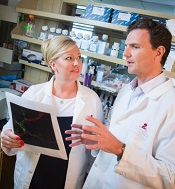
Sequencing aids management of young cancer patients

patient and her father
Photo by Rhoda Baer
Exome and transcriptome sequencing results can inform the management of young patients with relapsed, refractory, and rare malignancies, a new study suggests.
In a consecutive case series, sequencing data revealed potentially actionable findings for 46% of patients.
As a result, 15% of patients changed treatment, and 10% underwent genetic counseling.
Investigators described this research in JAMA.
“We found that, for some children with rare, difficult-to-treat, and aggressive cancers, this technology can dramatically change the course of their treatment,” said study author Rajen Mody, MBBS, of the University of Michigan in Ann Arbor.
Dr Mody and his colleagues evaluated 102 patients with relapsed, refractory, or rare cancers. Their median age was 11.5 (range, 0-22).
The patients underwent integrative clinical exome (tumor and germline DNA) and transcriptome (tumor RNA) sequencing. Ninety-one patients (89%) had adequate tumor tissue to complete sequencing, including 28 patients (31%) with hematologic malignancies and 63 (69%) with solid tumors.
All sequencing results were discussed at a precision medicine tumor board, which included pediatric and adult oncologists, pathologists, genetics specialists, and other professionals. This group discussed the results and assessed the feasibility of pursuing treatment options based on the findings.
Actionable findings
Forty-two patients (46%) had potentially actionable findings, 15 (54%) with hematologic malignancies and 27 (43%) with solid tumors.
Actionable findings included a change in diagnosis (n=2), the presence of a genetic anomaly that could be targeted by an approved or experimental drug (n=31), and the need for genetic counseling for inherited cancer risk that could affect the patient or the whole family (n=9).
“We were excited to see an actionable finding in such a substantial percentage of patients, and we think it could potentially be higher over time,” said study author Arul Chinnaiyan, MD, PhD, also of the University of Michigan.
“These are patients who had exhausted all proven therapeutic options or who had an extremely rare diagnosis. If we can find a clinically actionable event and have a chance to act upon it, we show in this study that it can have a big impact on that patient.”
Actions were taken in 23 of the 42 patients. Fourteen patients (15%) had their treatment changed, and 9 of these patients (10%) had durable partial or complete remissions (CRs) as a result.
Nine patients (10%) underwent genetic counseling because of sequencing results. The researchers noted that 4 of these patients had no notable family history to suggest an inherited risk, and they would not otherwise have been referred for genetic counseling.
Hematologic malignancies
Fifteen patients with hematologic malignancies had potentially actionable findings, and 4 underwent treatment changes as a result. (None of the patients required genetic counseling.)
For a patient with pre-B acute lymphoblastic leukemia (ALL), sequencing revealed a homozygous CDKN2A deletion and an ETV6-ABL1 fusion. So the patient was placed on imatinib and had a sustained CR for 21 months.
A patient with early T-cell precursor ALL had a FLT3-ITD mutation, Chr16p gain, Chr16q loss, and FLT3 overexpression. The patient achieved a CR after transplant, was placed on the FLT3 inhibitor sorafenib, and remained in CR for 15 months.
Another patient with pre-B ALL had a FLT3 nonframeshift deletion and BLK and FLT3 overexpression. The patient was in CR for 9 months after a transplant and received sorafenib for 6 months.
A patient with biphenotypic leukemia had mutations in NRAS and PHF6; SPI1, ASXL1, and CBLC frameshift insertions; a JAK3-activating mutation; and JAK3 overexpression. The patient received the JAK3 inhibitor tofacitinib but could not tolerate the full dose and died of progressive disease.
Cost and turn-around time
The cost for sequencing was approximately $6000 per patient and was covered under the research protocol.
It took the researchers about 7 to 8 weeks to report the sequencing results back to treating physicians and families.
“These are early days, and the full promise of precision medicine is yet to be fully realized,” Dr Mody said. “We need better targeted therapies designed for children, and turnaround time for sequencing needs to be less than 2 weeks for it to be a regular part of a patient’s treatment plan.” ![]()
Retinoids, FAK inhibitors may aid TKIs in treating ALL subtype
and Charles Mullighan
Photo courtesy of St. Jude
Children’s Research Hospital
and Peter Barta
Retinoids and FAK inhibitors may override resistance to tyrosine kinase inhibitors (TKIs) in IKZF1-mutated, Philadelphia chromosome-positive acute lymphoblastic leukemia (Ph+ ALL), according to preclinical research published in Cancer Cell.
Experiments showed that, in Ph+ ALL, IKZF1 mutations prompt changes that reduce responsiveness to TKIs. But combining a TKI with a retinoid or FAK inhibitor can overcome this problem.
“The research shows why, in this era of targeted therapies, Ph+ ALL patients who also have IKZF1 mutations fare so poorly,” said study author Charles Mullighan, MD, MBBS, of St. Jude Children’s Research Hospital in Memphis, Tennessee. “The insight also led us to a promising new treatment strategy.”
To conduct this research, Dr Mullighan and his colleagues began with mouse models of IKZF1-mutated Ph+ ALL, with and without mutations in the ARF gene. ARF encodes a tumor suppressor protein and is altered in about half of Ph+ ALL cases.
With these models, the researchers showed that the addition of IKZF1 mutations, particularly in combination with ARF mutations, was a central event in driving ALL.
In pre-B cells with BCR-ABL1, IKZF1 mutations induced a stem cell-like phenotype, increased stromal bone marrow adhesion, and reduced responsiveness to the TKI dasatinib.
When the researchers investigated the increased adhesion of mutated cells, they found overexpression of FAK and other molecules implicated in leukemic and stem cell adherence. This led the researchers to speculate that FAK inhibitors might prove useful against IKZF1-mutated Ph+ ALL.
But the team also found, through a screen of 483 compounds, that retinoids can reverse the effects of IKZF1 mutations. The antineoplastic agent bexarotene and 4 nuclear hormone receptor effectors—carbacyclin, all-trans retinoic acid (ATRA), 9-cis RA, and 13-cis RA—proved particularly effective.
The drugs worked, in part, by inducing expression of wild-type IKZF1. But they also worked in other ways to reverse the stem-cell phenotype, halt cell proliferation, and promote differentiation of altered cells.
The researchers then tested bexarotene and dasatinib, alone and in combination, in mice transplanted with ARF-/- BCR-ABL1 pre-B cells, with or without IK6 expression. Bexarotene alone produced “significant benefit without detectable toxicity.”
Dasatinib alone increased survival, but dasatinib and bexarotene in combination resulted in a greater survival advantage. The combination nearly doubled the survival time of mice with IK6 tumors, when compared to dasatinib alone.
The researchers also established xenografts of Ph+ ALL that recapitulate a range of IKZF1 genotypes. They administered dasatinib plus bexarotene, ATRA, or the FAK inhibitors PF-562271, NVPTAE226, or PF-573228 ex vivo and observed “significant potentiation of cell killing.”
The team is currently investigating how to incorporate retinoids or FAK inhibitors into the existing treatment of IKZF1-mutated Ph+ ALL. ![]()
New hope for treating fatal subtype of ALL

Photo by Aaron Logan
Preclinical research has revealed potential therapeutic options for TCF3-HLF-positive acute lymphoblastic leukemia (ALL).
Investigators discovered a range of mutations in this subtype of ALL and identified features that appear to contribute to treatment resistance.
However, the team also found that TCF3-HLF-positive ALL is sensitive to treatment with glucocorticoids, anthracyclines, and certain agents in clinical development.
The BCL2-specific inhibitor venetoclax (ABT-199) proved particularly active against the disease.
Jean-Pierre Bourquin, MD, PhD, of the University Children’s Hospital Zurich in Switzerland, and his colleagues reported these findings in Nature Genetics.
The investigators sequenced samples from patients with TCF3-HLF-positive ALL and found a range of mutations. Most samples (67%) had deletions in PAX5, and most of the samples without PAX5 deletions had deletions in VPREB1.
The team also found recurrent mutations of TCF3, a new fusion gene (KHDRBS1-LCK), and activating mutations in RAS signaling pathway genes (NRAS, KRAS, and PTPN11), among other mutations.
After additional investigation, Dr Bourquin and his colleagues hypothesized that the initiating TCF3-HLF fusion in this disease occurs in a B-cell progenitor, and the specific lineage context is constrained further in a restricted developmental stage by additional mutations.
The investigators also tested various treatments in mouse models of TCF3-HLF-positive ALL. They observed resistance to dasatinib, nucleotide analogs, mitotic spindle inhibitors, and polo-like and aurora kinase inhibitors.
On the other hand, the disease was sensitive to glucocorticoids, mTOR inhibitors, anthracyclines, bortezomib, the HSP90 inhibitor AUY922, and panobinostat.
The team also found evidence suggesting that BCL2 might promote leukemic cell survival and constitute a druggable target in TCF3-HLF-positive ALL. So they tested the BCL2 inhibitor venetoclax in the mice.
A 2-week course of daily venetoclax significantly delayed leukemia progression, and xenografts from relapsed patients and those with minimal residual disease remained sensitive to venetoclax. The drug also exhibited synergistic effects when combined with vincristine or dexamethasone.
“Further studies are now needed to test how the results of our study might be used for therapeutic possibilities,” Dr Bourquin concluded. ![]()
JAK2 inhibitor could treat B-ALL
Photo courtesy of the
Dana-Farber Cancer Institute
A type II JAK2 inhibitor has shown activity against B-cell acute lymphoblastic leukemia (B-ALL) in preclinical experiments.
The inhibitor, known as CHZ868, works by binding JAK2 into a tightly clenched position, which prevents the protein from functioning.
Researchers tested CHZ868 in samples from patients with CRLF2-rearranged B-ALL, in mice with the disease, and in mice implanted with human B-ALL tissue.
“In each case, we saw good activity: leukemia cells died, JAK2 signaling was suspended, and survival rates increased,” said David Weinstock, MD, of Dana-Farber Cancer Institute in Boston, Massachusetts.
“When we combined CHZ868 with the steroid dexamethasone, the killing of leukemia cells was much more extensive, and the animals lived longer than they did with CHZ868 alone.”
Dr Weinstock and his colleagues reported these results in Cancer Cell. Some of the researchers involved in this work are employees of, or have received research funding from, Novartis.
The team found that CHZ868 inhibited JAK2 signaling in B-ALL, both in vitro and in vivo. CHZ868 could overcome persistent JAK2 signaling where type I JAK2 inhibitors (BSK805 and BVB808) could not.
However, the researchers also identified a mutation—JAK2 L884P—that conferred resistance to CHZ868 and another type II JAK2 inhibitor, BBT594.
Nevertheless, CHZ868 suppressed the growth of CRLF2-rearranged human B-ALL cells and improved survival in mice with human or murine B-ALL.
CHZ868 worked synergistically with dexamethasone to induce apoptosis in JAK2-dependent B-ALL. The combination also improved survival in mice with B-ALL, when compared to either dexamethasone or CHZ868 alone.
The researchers noted that, when given at 30 mg/kg/day, CHZ868 was tolerated in NSG mice for up to 25 days and in immunocompetent mice for up to 44 days. And the drug had “essentially no effects” on peripheral blood counts.
This result and the tolerability of dexamethasone make CHZ868 and dexamethasone a “particularly attractive” combination that should be investigated in clinical trials, the team said.
They also speculated that CHZ868 or other type II JAK2 inhibitors could prove effective against malignancies other than B-ALL.
“JAK2 abnormalities are found in some cases of triple-negative breast cancer and Hodgkin lymphoma,” Dr Weinstock noted. “The success of CHZ868 in B-ALL suggests that it, or a compound that works by a similar mechanism, may also be effective in these cancers.” ![]()
Drug worth pursuing as T-ALL therapy, researchers say

Photo courtesy of
Children’s Cancer Institute
A drug that previously fell short of expectations holds promise for treating T-cell acute lymphoblastic leukemia (T-ALL), according to researchers.
The drug, PR-104, was originally designed to target hypoxic cells in solid tumors, but it showed less activity than expected in clinical trials, and its development was suspended.
Now, preclinical research has shown that PR-104 can be activated by AKR1C3, an enzyme that is overexpressed in T-ALL.
The researchers described this work in Blood.
“We were so encouraged by our first results with PR-104 that we undertook additional studies which showed the drug to be preferentially active against T-ALL . . . ,” said study author Richard B. Lock, PhD, of the Children’s Cancer Institute in Sydney, New South Wales, Australia.
“We believe that PR-104 might be an effective drug for patients who have initially benefited from conventional treatment for T-ALL but who have subsequently relapsed.”
Developing PR-104: A rocky road
PR-104 is a phosphate ester of the nitrogen mustard prodrug PR-104A. It was invented by William R. Wilson, PhD, of the University of Auckland (UoA) in New Zealand, and licensed to a UoA start-up company called Proacta Inc.
In a phase 1 study of patients with solid tumor malignancies, PR-104 failed to produce responses. The drug did elicit responses in a phase 1/2 trial of patients with advanced ALL or acute myeloid leukemia, but results fell short of expectations, and Proacta suspended development of PR-104.
Another drug Proacta was developing, PR-610, also failed to meet expectations. Because of these setbacks, the company closed its doors.
“As a fragile start-up, [Proacta] could not survive two serial ‘failures’ in phase 1/2,” Dr Wilson said. “Arguably . . . , the failure was more to do with the attempt to develop these compounds without biomarker support . . . than lack of potential of the compounds. Interestingly, PR-610 has subsequently been licensed by UoA to Threshold Pharmaceuticals, who are continuing its development (with biomarker support) as TH-4000.”
“We have a more challenging problem with PR-104 because the original patents have lapsed thanks to the decision of the UoA to not maintain the national phase filings after Proacta pulled the plug. [However,] as a result of [Dr Lock’s] work, it is now clear that PR-104 has exciting potential in leukemias with high activity of
AKR1C3.”
Results in T-ALL
Dr Lock and his colleagues tested PR-104 in a panel of 7 patient-derived pediatric ALL xenografts. Two weekly doses of PR-104 at 200 mg/kg significantly delayed progression in both T-ALL (n=4) and B-cell-precursor (BCP) ALL (n=3) xenografts.
The delay ranged from 10.3 days to 59.2 days and was significantly longer for the T-ALL xenografts (P=0.03).
PR-104 produced objective responses in all 4 T-ALL xenografts, including 2 complete responses. The drug also produced complete responses in 2 of the 3 BCP-ALL xenografts, but the third exhibited progressive disease.
Additional experiments showed that AKR1C3 expression was significantly higher in T-ALL than BCP-ALL, and AKR1C3 was “a major determinant” of sensitivity to PR-104, both in vitro and in vivo.
The researchers confirmed this by overexpressing AKR1C3 in a resistant BCP-ALL xenograft. Once AKR1C3 was overexpressed, the team observed “dramatic sensitization” to PR-104.
The path ahead
Now, Dr Lock and his colleagues are trying to determine why T-ALL cells express high levels of AKR1C3.
“If we can work out what activates this enzyme in T cells, we might find a way of activating it in B cells, making the B-cell disease sensitive to the drug as well,” Dr Lock said. “Obviously, it would be ideal if we could extend this drug’s reach to include all acute lymphoblastic leukemia patients.”
“In the meantime, we can envisage using PR-104 to target highly aggressive T-ALLs that express high levels of AKR1C3. We are in the process of working with our clinician colleagues in Australia and the US to organize a clinical trial of PR-104 in T-ALL.”
Dr Wilson noted that finding a path forward for PR-104 will be challenging due to the lack of patent support.
“[But] there are two reasons that make me think it is worth trying to do so,” he said. “One is the proximate concern that there are kids with high-AKR1C3 leukemias (adults too) who could benefit from this opportunity. The other is that this problem links to a looming paradigm shift in drug development. As we dissect cancer based on molecular analysis . . . , the commercial model will have to change.”
“There will still be ‘blockbuster’ drugs from time to time that address very high numbers of cancers . . . , but my expectation is that most cancer control in the future will depend on understanding the peculiarities of individual tumors and matching these with drugs that exploit these features. PR-104 is currently stuck in the past but could be a poster child for that future.” ![]()
Inactivating an enzyme can eradicate T-ALL

Preclinical research suggests that inactivating a single enzyme could eradicate or prevent T-cell acute lymphoblastic leukemia (T-ALL).
The researchers knew that T-ALL onset is linked to microRNAs, and most are generated with the help of the enzyme Dicer1.
Now, the team has found evidence to suggest that Dicer1 is crucial for the development of T-ALL, and inhibiting Dicer1 can actually prevent the disease altogether.
They reported these findings in Blood.
The researchers used mice that were genetically modified to develop T-ALL and in which Dicer1 could be abrogated. The team “switched off” Dicer1 in the mice at different stages of T-ALL development to see what role the enzyme plays in disease evolution.
Switching Dicer1 off at an early stage completely prevented T-ALL. In mice where Dicer1 was completely abrogated, T-ALL cells were entirely eliminated, allowing all the mice to survive.
The researchers were able to confirm this effect by monitoring the few residual leukemic cells taken from these animals.
“You can actually see the cancer cells dying off after Dicer1 has been abrogated,” said study author Freddy Radtke, PhD, of Ecole Polytechnique Fédérale de Lausanne in Lausanne, Switzerland.
He and his colleagues found that the key to this cell death is Dicer1’s role in producing microRNAs. The team discovered that a previously unrecognized microRNA, miR-21, was deregulated in both mouse and human T-ALL.
In the context of T-ALL, miR-21 inhibits the tumor suppressor gene Pdcd4. Without Dicer1, there is no miR-21 to do this, which allows Pdcd4 to fight the disease.
This study is the first to conclusively demonstrate that Dicer1 plays a role in T-ALL, the researchers said. The work paves the way for a new set of treatment for this malignancy and possibly others.
However, the team also noted that it can be challenging to target cells of interest when dealing with molecules that are so fundamental to the cell’s life.
“We can’t just go shutting down Dicer1 across the board,” Dr Radtke explained. “Otherwise, we’ll end up killing healthy cells as well.”
His lab is now focused on tackling this obstacle. ![]()
EHA: Inotuzumab rallies against refractory/relapsed ALL
VIENNA – The investigational agent inotuzumab ozagamicin more than doubled complete remission rates compared with standard therapy in relapsed or refractory acute lymphoblastic leukemia, preliminary results from the INO-VATE study show.
The co-primary endpoint of complete remission or CR with incomplete hematologic recovery (CRi) by independent review was achieved by 80.7% of patients treated with inotuzumab and 33.3% treated with standard of care (SOC) (P < .0001).
Significantly more CR/CRi responders treated with inotuzumab were minimal residual disease (MRD)-negative by multicolor flow cytometry (78.4% vs. 28.1%; P < .0001), Dr. Daniel DeAngelo reported in a late-breaking abstract (LBA2073) at the annual congress of the European Hematology Association.

“The fact that the response rate was astronomically high with a high MRD-negative status really allows this or this should be an opportunity for patients with relapsed/refractory disease,” he said in an interview.
Inotuzumab ozagamicin is an investigational anti-CD22 antibody conjugated to calicheamicin, an antitumor antibiotic. CD22 is expressed on the surface of about 90% of B-cell ALL cells.
Previous phase II studies reported strong initial antitumor activity and safety with inotuzumab in relapsed or refractory ALL, Dr. DeAngelo, of the Dana Farber Cancer Institute in Boston, said.
The ongoing phase III trial randomized 326 patients with relapsed/refractory CD22-positive ALL due for salvage 1 or 2 therapy to inotuzumab or SOC: either the FLAG regimen (fludarabine (Fludara)/cytarabine (Ara-C)/granulocyte colony-stimulating factor), Ara-C plus mitoxantrone (Novantrone), or high-dose Ara-C. The starting dose for inotuzumab was 1.8 mg/m2/cycle and was reduced to 1.5 mg/m2/cycle once CR/CRi was achieved. Patients were stratified by duration of first remission, salvage 1 or 2, and age.
The first 218 randomized patients were included in the intention-to-treat CR/CRi analysis, which was modified after excluding 13 patients from the SOC arm who refused to start treatment.
The patients’ median age was 47 years (ranging up to 79 years), two-thirds were salvage 1, and more than half had a remission duration of less than 12 months, an adverse prognostic feature.
Data for the co-primary endpoint of overall survival in all 326 patients are still blinded and not expected to mature until 2016, Dr. DeAngelo said.
CR/CRi analyses significantly favored inotuzumab in all stratification factors and baseline factors including peripheral blasts and CD22 expression. Cytogenetics are still being evaluated, but 11 of 14 (79%) patients with Philadelphia-positive karyotype achieved a CR or CRi, he said.
Median duration of remission among responders was 4.6 months in the inotuzumab arm and 3.1 months in the SOC arm (hazard ratio, 0.55; P = .016).
Safety assessed in 259 patients who received at least one dose of study drug showed similar incidence of grade 3 or higher adverse events in the inotuzumab and SOC arms (91% vs. 95%). There were 2 fatal events in the SOC arm and 4 in the inotuzumab arm: 2 veno-occlusive disease (VOD)/sinusoidal obstruction syndrome (SOS), both after poststudy transplant, 1 intestinal ischemia/septic shock, and 1 acute respiratory distress syndrome as a terminal event of pneumonia. In multivariate analysis, dual alkylator conditioning was the only significant covariate of VOD/SOS (P = .039), Dr. DeAngelo said.
An audience member chided the author for the short duration of remission, but session co-moderator Dr. Anthony Moorman, of Newcastle University, Newcastle upon Tyne, England, said it is not that concerning because of the aggressive nature of ALL.
“For all patients that have relapsed or refractory adult ALL, their responses are incredibly low. So any kind of complete remission is a major achievement in this patient population, especially if they are refractory or relapse after tyrosine kinase inhibitors or Philadelphia-positive,” he said in an interview.
“When you have an active agent that works with relapsed refractory disease, in this case leukemia, the goal is to move it up front,” Dr. DeAngelo told this publication.
Indeed, updated results presented at the meeting from M.D. Anderson Cancer Center of frontline inotuzumab added to low-intensity chemotherapy (Mini-hyper CVD) in elderly ALL patients were “provocative,” he added. CR rates reached 97% in the study, according to the abstract (S114).
VIENNA – The investigational agent inotuzumab ozagamicin more than doubled complete remission rates compared with standard therapy in relapsed or refractory acute lymphoblastic leukemia, preliminary results from the INO-VATE study show.
The co-primary endpoint of complete remission or CR with incomplete hematologic recovery (CRi) by independent review was achieved by 80.7% of patients treated with inotuzumab and 33.3% treated with standard of care (SOC) (P < .0001).
Significantly more CR/CRi responders treated with inotuzumab were minimal residual disease (MRD)-negative by multicolor flow cytometry (78.4% vs. 28.1%; P < .0001), Dr. Daniel DeAngelo reported in a late-breaking abstract (LBA2073) at the annual congress of the European Hematology Association.
“The fact that the response rate was astronomically high with a high MRD-negative status really allows this or this should be an opportunity for patients with relapsed/refractory disease,” he said in an interview.
Inotuzumab ozagamicin is an investigational anti-CD22 antibody conjugated to calicheamicin, an antitumor antibiotic. CD22 is expressed on the surface of about 90% of B-cell ALL cells.
Previous phase II studies reported strong initial antitumor activity and safety with inotuzumab in relapsed or refractory ALL, Dr. DeAngelo, of the Dana Farber Cancer Institute in Boston, said.
The ongoing phase III trial randomized 326 patients with relapsed/refractory CD22-positive ALL due for salvage 1 or 2 therapy to inotuzumab or SOC: either the FLAG regimen (fludarabine (Fludara)/cytarabine (Ara-C)/granulocyte colony-stimulating factor), Ara-C plus mitoxantrone (Novantrone), or high-dose Ara-C. The starting dose for inotuzumab was 1.8 mg/m2/cycle and was reduced to 1.5 mg/m2/cycle once CR/CRi was achieved. Patients were stratified by duration of first remission, salvage 1 or 2, and age.
The first 218 randomized patients were included in the intention-to-treat CR/CRi analysis, which was modified after excluding 13 patients from the SOC arm who refused to start treatment.
The patients’ median age was 47 years (ranging up to 79 years), two-thirds were salvage 1, and more than half had a remission duration of less than 12 months, an adverse prognostic feature.
Data for the co-primary endpoint of overall survival in all 326 patients are still blinded and not expected to mature until 2016, Dr. DeAngelo said.
CR/CRi analyses significantly favored inotuzumab in all stratification factors and baseline factors including peripheral blasts and CD22 expression. Cytogenetics are still being evaluated, but 11 of 14 (79%) patients with Philadelphia-positive karyotype achieved a CR or CRi, he said.
Median duration of remission among responders was 4.6 months in the inotuzumab arm and 3.1 months in the SOC arm (hazard ratio, 0.55; P = .016).
Safety assessed in 259 patients who received at least one dose of study drug showed similar incidence of grade 3 or higher adverse events in the inotuzumab and SOC arms (91% vs. 95%). There were 2 fatal events in the SOC arm and 4 in the inotuzumab arm: 2 veno-occlusive disease (VOD)/sinusoidal obstruction syndrome (SOS), both after poststudy transplant, 1 intestinal ischemia/septic shock, and 1 acute respiratory distress syndrome as a terminal event of pneumonia. In multivariate analysis, dual alkylator conditioning was the only significant covariate of VOD/SOS (P = .039), Dr. DeAngelo said.
An audience member chided the author for the short duration of remission, but session co-moderator Dr. Anthony Moorman, of Newcastle University, Newcastle upon Tyne, England, said it is not that concerning because of the aggressive nature of ALL.
“For all patients that have relapsed or refractory adult ALL, their responses are incredibly low. So any kind of complete remission is a major achievement in this patient population, especially if they are refractory or relapse after tyrosine kinase inhibitors or Philadelphia-positive,” he said in an interview.
“When you have an active agent that works with relapsed refractory disease, in this case leukemia, the goal is to move it up front,” Dr. DeAngelo told this publication.
Indeed, updated results presented at the meeting from M.D. Anderson Cancer Center of frontline inotuzumab added to low-intensity chemotherapy (Mini-hyper CVD) in elderly ALL patients were “provocative,” he added. CR rates reached 97% in the study, according to the abstract (S114).
AT THE EHA CONGRESS
Key clinical point: Inotuzumab ozagamicin shows promise as a new treatment option for relapsed or refractory acute lymphoblastic leukemia.
Major finding: The rate of complete remission or CR with incomplete hematologic recovery was 80.7% with inotuzumab vs. 28.1% with standard of care (P < .0001).
Data source: Randomized, phase III study in the first 218 of 326 patients.
Disclosures: Pfizer sponsored the study and funded editorial assistance supplied by Complete Heathcare Communications. Dr. De Angelo reported research support from Sigma Tau and consulting for Novartis, Sigma Tau, Bristol-Myers Squibb, Amgen, and Pfizer.
Study reveals potential target for T-ALL

Photo by Rhoda Baer
Targeting CXCL12/CXCR4 signaling could be a “powerful” approach to treating T-cell acute lymphoblastic leukemia (T-ALL), according to researchers.
Results of their preclinical experiments indicate that deleting CXCL12 from vascular endothelial cells can stall T-ALL progression.
And inhibiting or deleting CXCR4 can halt tumor growth and induce remission in mice with T-ALL.
The researchers reported these findings in Cancer Cell.
“Our experiments showed that blocking CXCR4 decimated leukemia cells,” said study author Susan Schwab, PhD, of the New York University School of Medicine in New York, New York.
This suggests CXCR4 antagonists could treat T-ALL, she added, noting that such drugs are already in preliminary testing for myeloid leukemia and have proven well-tolerated thus far.
Dr Schwab and her colleagues conducted this research to build upon previous work, which showed that leukemia-initiating cells concentrate in the bone marrow near CXCL12-producing blood vessels.
This finding inspired the researchers to investigate the expression and function of CXCR4 because it binds to CXCL12, as well as the role CXCR4-CXCL12 molecular signaling plays in disease growth.
With this study, the team found that T-ALL cells are in direct, stable contact with CXCL12-producing bone marrow stroma.
They showed that deleting CXCL12 in vascular endothelial cells suppressed T-ALL. But they did not observe the same benefit when they deleted CXCL12 from perivascular cells.
The researchers also found that T-ALL cells express high surface levels of CXCR4, and deleting CXCR4 can produce sustained remission in mice with T-ALL.
Similarly, a small-molecule inhibitor of CXCR4, AMD3465, exhibited antileukemic activity in murine T-ALL and human xenografts.
Experiments with CXCR4-deficient T-ALL cells showed that CXCR4 expression and signaling influences leukemic cell localization and survival. And further investigation revealed that the loss of CXCR4 expression is associated with decreased levels of MYC.
The researchers said additional work is needed to determine exactly how CXCR4 is able to promote and sustain T-ALL. ![]()
Photo by Rhoda Baer
Targeting CXCL12/CXCR4 signaling could be a “powerful” approach to treating T-cell acute lymphoblastic leukemia (T-ALL), according to researchers.
Results of their preclinical experiments indicate that deleting CXCL12 from vascular endothelial cells can stall T-ALL progression.
And inhibiting or deleting CXCR4 can halt tumor growth and induce remission in mice with T-ALL.
The researchers reported these findings in Cancer Cell.
“Our experiments showed that blocking CXCR4 decimated leukemia cells,” said study author Susan Schwab, PhD, of the New York University School of Medicine in New York, New York.
This suggests CXCR4 antagonists could treat T-ALL, she added, noting that such drugs are already in preliminary testing for myeloid leukemia and have proven well-tolerated thus far.
Dr Schwab and her colleagues conducted this research to build upon previous work, which showed that leukemia-initiating cells concentrate in the bone marrow near CXCL12-producing blood vessels.
This finding inspired the researchers to investigate the expression and function of CXCR4 because it binds to CXCL12, as well as the role CXCR4-CXCL12 molecular signaling plays in disease growth.
With this study, the team found that T-ALL cells are in direct, stable contact with CXCL12-producing bone marrow stroma.
They showed that deleting CXCL12 in vascular endothelial cells suppressed T-ALL. But they did not observe the same benefit when they deleted CXCL12 from perivascular cells.
The researchers also found that T-ALL cells express high surface levels of CXCR4, and deleting CXCR4 can produce sustained remission in mice with T-ALL.
Similarly, a small-molecule inhibitor of CXCR4, AMD3465, exhibited antileukemic activity in murine T-ALL and human xenografts.
Experiments with CXCR4-deficient T-ALL cells showed that CXCR4 expression and signaling influences leukemic cell localization and survival. And further investigation revealed that the loss of CXCR4 expression is associated with decreased levels of MYC.
The researchers said additional work is needed to determine exactly how CXCR4 is able to promote and sustain T-ALL.
CAR produces high CR rate in adults with rel/ref ALL

the 2015 ASCO Annual Meeting
© ASCO/Max Gersh
CHICAGO—A CD19-targeted chimeric antigen receptor (CAR) T-cell therapy can provide durable complete responses (CRs) or a bridge to allogeneic transplant in adults with relapsed or refractory acute lymphoblastic leukemia (ALL), updated results of a phase 1 study suggest.
The therapy, JCAR015, produced a CR rate of 87%, and 33% of these patients went on to transplant.
The median duration of response or relapse-free survival was 5.3 months. The median overall survival was 8.5 months.
Nearly a quarter of patients developed severe cytokine release syndrome (CRS), and nearly 30% experienced neurological toxicities. But researchers said these effects were largely treatable and reversible.
This study was temporarily placed on clinical hold last year, after 2 patients died from complications related to CRS. But the hold was soon lifted and enrollment and dosing criteria were changed in an attempt to prevent severe CRS.
Jae H. Park, MD, of Memorial Sloan Kettering Cancer Center in New York, presented updated results of this trial (NCT01044069) at the 2015 ASCO Annual Meeting (abstract 7010*). The study is sponsored by Memorial Sloan Kettering, but funding has also been provided by Juno Therapeutics, the company developing JCAR015.
Results from this trial have previously been reported in Science Translational Medicine (Davila et al 2014; Brentjens et al 2013), at AACR 2014, and at ASH 2014.
At ASCO, Dr Park presented results in 39 patients with relapsed/refractory, CD19+ ALL. All of them were evaluable for toxicity assessment, and 38 were evaluable for response with at least 1 month of follow-up.
There were 29 males, and the patients’ median age was 45 (range, 22-74). Thirty-three percent had Ph+ ALL, and 11% had the T315I mutation.
Forty-nine percent of patients had received 2 prior therapies, 23% had received 3, and 28% had received 4 or more. Thirty-six percent of patients had a prior allogeneic hematopoietic stem cell transplant (HSCT).
For this study, patients first underwent leukapheresis. While their T cells were being manufactured, they were allowed to receive salvage chemotherapy. Patients underwent repeat bone marrow biopsy to assess their disease status immediately prior to T-cell infusion.
Fifty-four percent of patients (n=21) had morphologic disease (>5% blasts in the bone marrow, median 52%) immediately prior to JCAR015 infusion, and the remaining patients (n=18) had minimal residual disease (MRD).
Two days after conditioning with cyclophosphamide, patients received an infusion of 1-3 x 10⁶ CAR T cells/kg. At day 28, the researchers assessed patients’ disease with a repeat bone marrow biopsy.
Treatment results
The median follow-up was 5.6 months (1 to >38 months). Six patients had more than a year of follow-up.
The CR rate was 87% (33/38), and 81% of evaluable patients (26/32) were MRD-negative. The median time to CR was 23 days, and the median duration of response or relapse-free survival was 5.3 months.
“We examined the CR rates by different subgroup,” Dr Park noted. “We looked at whether patients had a pre-T-cell disease burden: morphologic disease vs minimal residual disease, whether they had an allogeneic bone marrow transplant prior to CAR T-cell infusion, their Ph+ status, age at infusion, and prior lines of therapy. And there was no [significant] difference between these groups for CRs and MRD-negative CR rate.”
At the time of presentation, 14 patients were disease-free, 10 of whom had not gone on to HSCT. In all, 11 patients went on to allogeneic HSCT.
Fourteen patients relapsed during follow-up, 3 after HSCT. Two of these patients had CD19-negative bone marrow blasts.
The median overall survival was 8.5 months in all patients and 10.8 months in patients who were MRD-negative. The median overall survival was 9.9 months in patients who underwent allogeneic HSCT and 8.5 months in patients who did not.
Dr Park said key adverse events were CRS—clinically manifested by fever, hypotension, and respiratory insufficiency—and neurological changes such as delirium, global encephalopathy, aphasia, and seizures.
Twenty-three percent of patients (n=9) developed severe CRS, 28% (n=11) had grade 3/4 neurotoxicity, and 8% (n=3) had grade 5 toxicity. The patients with grade 5 toxicities died of ventricular arrhythmia, sepsis, and an unknown cause (although this patient suffered a seizure).
The severity of CRS correlated with disease burden, and CRS was managed with an IL-6R inhibitor (n=4), a steroid (n=2), or both (n=9). Neurological symptoms were reversible and could occur independently of CRS, Dr Park said.
*Information in the abstract differs from that presented at the meeting.
Breastfeeding may lower risk of ALL

Photo by Petr Kratochvil
Breastfeeding a child may reduce his risk of developing acute lymphoblastic leukemia (ALL) but perhaps not acute myeloid leukemia (AML), according to a review published in JAMA Pediatrics.
Researchers found that breastfeeding a child for 6 months or longer was associated with a 19% lower risk of childhood leukemia, compared with no
breastfeeding or breastfeeding for a shorter period of time.
And children who were breastfed for any amount of time had an 11% lower risk of childhood leukemia than children who were never breastfed.
However, when the researchers analyzed studies of ALL and AML separately, they found that breastfeeding was not associated with a significantly lower risk of AML.
Efrat L. Amitay, PhD, and Lital Keinan-Boker, MD, PhD, of the University of Haifa in Israel, conducted this research.
In a review of 18 studies, the pair found that breastfeeding a child for 6 months or longer was associated with a significantly lower risk of childhood leukemia, compared with no breastfeeding or breastfeeding for a shorter period of time (odds ratio [OR]=0.81).
And a separate analysis of 15 studies showed that children who were breastfed for any amount of time had a lower risk of childhood leukemia than children who were never breastfed (OR=0.89).
The researchers also conducted meta-analyses of AML studies and ALL studies separately—11 ALL and 6 AML studies. And they found a significant inverse association between breastfeeding for 6 months or more and ALL risk (OR=0.82) but no significant association for AML risk (OR=0.74).
The researchers said several biological mechanisms may explain the association between breastfeeding and a reduced risk of childhood leukemia. However, more high-quality studies are needed to clarify those mechanisms.