User login
Standard BMI inadequate for ALL patients

Photo by Bill Branson
New research suggests that body mass index (BMI) is an inadequate method for estimating changes in body fat and obesity in children with acute lymphoblastic leukemia (ALL).
Investigators found a discrepancy between BMI and body composition in this population, and the cause of this appeared to be increases in body fat with simultaneous loss of lean muscle mass during treatment.
The team reported these findings in Leukemia & Lymphoma.
With previous work, the investigators found that obese children diagnosed with high-risk ALL had a 50% greater risk of their disease recurring compared with children who were not obese.
“In my lab, we’ve seen a direct interaction between fat cells and leukemia cells that may help explain this increased risk of disease relapse,” said study author Steven Mittelman, MD, PhD, of Children’s Hospital Los Angeles in California.
“It appears that the fat cells ‘protect’ leukemia cells, making them less susceptible to chemotherapy and making an accurate measure of body fat essential.”
To determine if BMI accurately reflects body fat in ALL, the investigators analyzed 50 patients. They were predominantly Hispanic, between the ages of 10 to 21, and had newly diagnosed high-risk B-precursor ALL or T-cell ALL.
The team measured the percentage of total body fat and lean muscle mass at the time of diagnosis, at the end of induction, and at the end of delayed intensification. They also calculated BMI Z-score—a measure of how a given child’s BMI deviates from a population of children of the same age and sex—at these time points.
The investigators said sarcopenic obesity—gain in body fat percentage with loss of lean muscle mass—was “surprisingly common” during ALL treatment.
And sarcopenic obesity resulted in poor correlation between changes in BMI Z-score and body fat percentage overall (r=-0.05), within the time points (r=0.02), and within patients (r=-0.09, all not significant). BMI Z-score and body fat percentage changed in opposite directions in more than 50% of interval assessments.
“We found that change in BMI did not reflect changes in body fat or obesity,” said Etan Orgel, MD, of Children’s Hospital Los Angeles.
“In some patients, reaching a ‘healthy’ BMI was due solely to loss of muscle even while body fat continued to rise. Based on these results, we believe that evaluation of obesity in patients with leukemia should include direct measures of body composition.” ![]()
CAR T-cells induce remissions, avoid GVHD in relapsed B-cell malignancies
In patients with B-cell malignancies who did not obtain remissions after allogeneic hematopoietic stem-cell transplantation (alloHSCT), infusion of anti-CD19 chimeric antigen receptor (CAR19) T-cells induced remissions without causing graft-versus-host disease (GVHD).
Of 20 patients treated, 6 obtained complete remission and 2 obtained partial remissions, while none of the patients experienced new-onset acute GVHD.
“The increased antimalignancy potency of CAR19 T-cells allowed small doses of T-cells to eradicate malignancy without causing GVHD; therefore, this work demonstrates a solution to the central problem of alloHSCT, the separation of [graft-versus-malignancy] from GVHD,” wrote Dr. Jennifer Brudno of the National Cancer Institute, Bethesda, Md., and colleagues (J Clin Onc. 2016 Jan. 25. doi: 10.1200/JCO.2015.64.5929).
In general, CAR19 T-cells did not persist longer than 4 weeks, the median time it takes for GVHD to develop after donor lymphocyte infusion, which may be a factor in avoiding GVHD. Smaller doses may be another contributing factor: CAR19 T-cell doses administered in the trial ranged from 106/kg to 107/kg, which is 10-fold smaller than typical donor lymphocyte infusions.
In contrast to previous CAR T-cell studies, patients did not receive chemotherapy before CAR19 T-cell infusion, so endogenous T-cells and natural killer cells were not depleted. Evidence indicates that lymphocyte depletion enhances antitumor activity of adoptively transferred T-cells, but the current results indicate that prior lymphocyte depletion is not an absolute requirement.
The CAR19 T-cell infusion was especially effective in acute lymphoblastic leukemia (ALL), with four of the five ALL patients obtaining complete remission. Patients with chronic lymphocyte leukemia and lymphoma also obtained remissions.
Toxicities observed were consistent with previous CAR T-cell studies and included fever, tachycardia, and hypotension, indicative of cytokine release syndrome.
Peak levels of CAR19 T-cells were significantly higher in patients with complete or partial responses, and patients who did not obtain a complete or partial response were more likely to have undetectable or very low levels. Increasing peak blood levels of CAR19 T-cells in vivo is an important goal for future research, according to investigators. Since endogenous CD19+ cells may promote proliferation of CAR19 T- cells, CD19+ cellular vaccines might enhance CAR19 T-cell proliferation in patients with low levels, they suggested.
Administration of programmed cell death protein-1 (PD-1) antagonists may also offer improvements. High levels of PD-1 expression on CAR19 T-cells were observed at the time of peak blood CAR19 T-cell levels.
Dr. Brudno reported having no disclosures. Dr. Goy and Dr. Rosenberg reported ties to industry, and Dr. Kochenderfer reported patents pending on CAR T-cell therapy.
FROM JOURNAL OF CLINICAL ONCOLOGY
Key clinical point: In patients who relapsed after allogeneic hematopoietic stem-cell transplantation, anti-CD19 chimeric antigen receptor (CAR) T-cell therapy induced remissions without causing graft-versus-host disease (GVHD).
Major finding: Of 20 patients treated, 6 obtained complete remission and 2 obtained partial remissions; none experienced new-onset acute GVHD.
Data source: Patients with B-cell malignancies who had progressed after allogeneic hematopoietic stem-cell transplantation.
Disclosures: Dr. Brudno reported having no disclosures. Dr. Goy and Dr. Rosenberg reported ties to industry, and Dr. Kochenderfer reported patents pending on CAR T-cell therapy.
Drug approved to treat ALL in EU

The European Commission has granted marketing authorization for pegaspargase (Oncaspar) to be used as part of combination antineoplastic therapy for pediatric and adult patients with acute lymphoblastic leukemia (ALL).
The approval means the drug can be marketed for this indication in the 28 member countries of the European Union (EU), as well as Iceland, Liechtenstein, and Norway.
Pegaspargase was already approved for use in Argentina, Belarus, Germany, Kazakhstan, Poland, Russia, Ukraine, and the US.
“Oncaspar has been used as an integral component of the treatment regimen for pediatric and adult patients with ALL for many years, in Europe and worldwide,” said Martin Schrappe, of Schleswig-Holstein University Hospital in Kiel, Germany.
“Today’s marketing authorization will ensure that more patients across the EU will benefit from access to Oncaspar as part of a standard of care regimen.”
The drug is being developed by Baxalta Incorporated.
First-line ALL
Researchers have evaluated the safety and effectiveness of pegaspargase in a study of 118 pediatric patients (ages 1 to 9) with newly diagnosed ALL. The patients were randomized 1:1 to pegaspargase or native E coli L-asparaginase, both as part of combination therapy.
Asparagine depletion (magnitude and duration) was similar between the 2 treatment arms. Event-free survival rates were also similar (about 80% in both arms), but the study was not designed to evaluate differences in event-free survival.
Grade 3/4 adverse events occurring in the pegaspargase and native E coli L-asparaginase arms, respectively, were abnormal liver tests (5% and 8%), elevated transaminases (3% and 7%), hyperbilirubinemia (2% and 2%), hyperglycemia (5% and 3%), central nervous system thrombosis (3% and 3%), coagulopathy (2% and 5%), pancreatitis (2% and 2%), and clinical allergic reactions to asparaginase (2% and 0%).
Previously treated ALL
Researchers have evaluated the effectiveness of pegaspargase in 4 open-label studies of patients with a history of prior clinical allergic reaction to asparaginase. The studies enrolled a total of 42 patients with multiply relapsed acute leukemia (39 with ALL).
Patients received pegaspargase as a single agent or as part of multi-agent chemotherapy. The re-induction response rate was 50%—36% complete responses and 14% partial responses. Three responses occurred in patients who received single-agent pegaspargase.
Adverse event information on pegaspargase in relapsed ALL has been compiled from 5 clinical trials. The studies enrolled a total of 174 patients with relapsed ALL who received pegaspargase as a single agent or as part of combination therapy.
Sixty-two of the patients had prior hypersensitivity reactions to asparaginase, and 112 did not. Allergic reactions to pegaspargase occurred in 32% of previously hypersensitive patients and 10% of non-hypersensitive patients.
The most common adverse events observed in patients who received pegaspargase were clinical allergic reactions, elevated transaminases, hyperbilirubinemia, and coagulopathies.
The most common serious adverse events due to pegaspargase were thrombosis (4%), hyperglycemia requiring insulin therapy (3%), and pancreatitis (1%).
For more details on these trials and pegaspargase in general, see the product information. ![]()
Cancer survival tied to reduction in treatment
Photo by Bill Branson
Results of a large study suggest that long-term survivors of childhood cancer are living longer, partly due to a reduction in the use of certain treatments.
The 15-year death rate among the more than 34,000 childhood cancer survivors studied decreased steadily from 1970 onward.
And this decline coincided with changes in pediatric cancer therapy, including reductions in the use and dose of radiation therapy and anthracyclines.
These therapies are known to put cancer survivors at increased risk for developing second malignancies, heart failure, and other serious health problems.
“This study is the first to show that younger survivors from more recent treatment eras are less likely to die from the late effects of cancer treatment and more likely to enjoy longer lives,” said study author Greg Armstrong, MD, of St. Jude Children's Research Hospital in Memphis, Tennessee.
He and his colleagues reported these results in NEJM.
The study included 34,033 subjects who had been diagnosed with cancer and received treatment between 1970 and 1999 when they were age 20 or younger. All patients lived at least 5 years after their cancers were discovered and were considered long-term survivors.
Changes in mortality
At a median follow-up of 21 years (range, 5 to 38), there were 3958 deaths. Forty-one percent of deaths (n=1618) were considered health-related. This included 746 deaths from subsequent neoplasms, 241 from cardiac causes, 137 from pulmonary causes, and 494 from other causes.
The 15-year death rate (death from any cause) fell from 12.4% in the early 1970s to 6% in the 1990s (P<0.001). During the same period, the rate of death from health-related causes fell from 3.5% to 2.1% (P<0.001).
The researchers said there were significant reductions across treatment eras in the rates of death from any health-related cause among patients with acute lymphoblastic leukemia (ALL), Hodgkin lymphoma (HL), Wilms’ tumor, and astrocytoma, but not among patients with other cancers.
The rate of health-related death among ALL patients fell from 3.2% in the early 1970s to 2.1% in the 1990s (P<0.001). The rate fell from 5.3% to 2.6% (P=0.006) for HL patients, from 2.6% to 0.4% (P=0.005) for Wilms’ tumor patients, and from 4.7% to 1.8% (P=0.02) for astrocytoma patients.
The researchers said these reductions in mortality were attributable to decreases in the rates of death from subsequent neoplasm (P<0.001), cardiac causes (P<0.001), and pulmonary causes (P=0.04).
Treatment changes
The overall use of anthracyclines fell from 73% in the 1970s to 42% in the 1990s. And the use of any radiation decreased from 77% to 41%.
The use of cranial radiotherapy for ALL fell from 85% to 19%. The use of abdominal radiotherapy for Wilms’ tumor decreased from 78% to 43%. And the use of chest radiotherapy for HL fell from 87% to 61%.
The researchers noted that temporal reductions in 15-year rates of death from health-related causes followed temporal reductions in therapeutic exposure for patients with ALL, HL, Wilms’ tumor, and astrocytoma.
However, when the team adjusted their analysis for therapy (eg, anthracycline dose), the effect of the treatment era on the relative rate of death from health-related causes was attenuated in ALL (unadjusted relative rate=0.88, adjusted relative rate=1.02) and Wilms’ tumor (0.68 and 0.80, respectively) but not in HL (0.79 in both models) and astrocytoma (0.81 and 0.82, respectively).
Still, the researchers said the results of this study suggest the strategy of lowering therapeutic exposure has contributed to the decline in late mortality among 5-year survivors of childhood cancer. ![]()
Death from late effects of childhood cancer on decline
The rate of death from treatment-related late effects such as subsequent cancers and cardiopulmonary conditions has decreased among childhood cancer survivors, according to researchers.
At 15 years post diagnosis, the cumulative incidence of death from any cause for survivors diagnosed in the 1970s was 10.7%, 7.9% for those diagnosed in the 1980s, and 5.8% for those diagnosed in the 1990s (P less than .001). The cumulative incidence of death due to health-related causes, which include late effects of cancer therapy, were 3.1%, 2.4%, and 1.9%, respectively (P less than .001).
Results indicate that “the strategy of reducing treatment exposures in order to decrease the frequency of late effects is translating into a significant reduction in observed late mortality and an extension of the life span of children and adolescents who are successfully treated for cancer,” wrote Dr. Gregory Armstrong of St. Jude Children’s Research Hospital, Memphis, Tenn., and colleagues (N Engl J Med. 2016 Jan 13. doi: 10.1056/NEJMoa1510795).
A multivariate model showed that more recent treatment eras were associated with a reduced rate of death. The adjusted relative rate per every 5 years for death due to subsequent neoplasms was 0.83 (95% CI, 0.78-0.88), for cardiac causes, 0.77 (0.68-0.86), and for pulmonary causes, 0.77 (0.66-0.89).
Reductions across treatment eras in the rate of death from health-related causes were observed among survivors of acute lymphoblastic leukemia (3.2% in the early 1970s to 2.1% in the 1990s, P less than .001), Hodgkin lymphoma (5.3% to 2.6%, P = .006), Wilms tumor (2.6% to 0.4%, P = .005), and astrocytoma (4.7% to 1.8%, P = .02).
Temporal reductions in exposure to radiotherapy and anthracyclines occurred in treatment for acute lymphoblastic leukemia, Hodgkin lymphoma, Wilms tumor, and astrocytoma. Health-related mortality reductions were observed concurrently with reduced therapeutic exposures for acute lymphoblastic leukemia and Wilms tumor. For Hodgkin lymphoma and astrocytoma, other factors such as improved screening for late effects of cancer treatment appear to account for reductions in late health-related mortality.
For certain cancers, primarily neuroblastoma, late mortality has increased in recent decades. Increased therapeutic intensity has improved 5-year survival but increased the risk of late effects.
The reduced rate of death from recurrence or progression of primary cancers is the main driver to reductions in all-cause mortality, consistent with results from previous studies.
The retrospective Childhood Cancer Survivor Study evaluated 34,033 patients diagnosed at 31 hospitals in the United States and Canada from 1970 through 1999. In total, 3,958 deaths occurred, 2,002 due to primary cancer and 1,618 due to health-related causes: 746 subsequent neoplasms, 241 cardiac causes, 137 pulmonary causes, and 494 from other causes.
Key clinical point: Five-year survivors of childhood cancers have increased lifespans due in part to reduced rates of treatment-related late effects.
Major finding: At 15 years post diagnosis, the cumulative incidence of death from any cause for survivors diagnosed in the 1970s was 10.7%; in the 1980s, 7.9%; and in the 1990s, 5.8% (P less than .001). The cumulative incidence of death due to health-related causes, which include late effects of cancer therapy, were 3.1%, 2.4%, and 1.9%, respectively (P less than .001).
Data source: The retrospective Childhood Cancer Survivor Study, evaluating 34,033 patients diagnosed at 31 hospitals in the United States and Canada from 1970 through 1999; 3,958 deaths occurred.
Disclosures: Dr. Armstrong and coauthors reported having no disclosures.

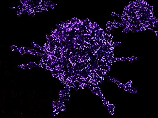
Increasing eligibility for engineered T-cell therapy

Image courtesy of NIAID
A new study suggests that having a certain type of cancer or receiving certain chemotherapeutic agents
can affect T-cell function and make patients ineligible for engineered T-cell therapy.
However, researchers found that proper timing of T-cell collection can increase the number of patients eiligible for the therapy.
And the team developed a culture technique that can boost T cells’ fitness for expansion, which can increase eligibility as well.
Nathan Singh, MD, of the University of Pennsylvania in Philadelphia, and his colleagues described this work in Science Translational Medicine.
The researchers set out to determine why some patients’ T cells fail to multiply in culture. The team studied T cells from children with acute lymphoblastic leukemia (ALL) or non-Hodgkin lymphoma (NHL) who were undergoing chemotherapy. (NHL subtypes included Burkitt lymphoma, diffuse large B-cell lymphoma, primary mediastinal large B-cell lymphoma, primary lymphoma of bone, and follicular lymphoma.)
The researchers found that T cells from patients with ALL expanded better in culture than those from patients with NHL.
The team said a threshold of greater than 5-fold expansion during test expansion was associated with a high likelihood of successful clinical expansion.
Nearly 80% of patients with ALL met this threshold at diagnosis, but the rate declined over the course of therapy, falling to about 40% during maintenance therapy.
About 25% of NHL patients met the threshold at diagnosis, but few samples demonstrated any expansion after therapy began (12.5% of samples at all remaining time points tested).
The researchers said the difference in the proportion of ALL and NHL samples that met the expansion threshold was significant at all time points tested.
Analysis revealed that ALL patients had higher numbers of naïve T cells and stem central memory T cells, T cell subtypes known to be highly potent and proliferative with an enhanced capacity for self-renewal.
The researchers also found that certain chemotherapy drugs—namely, cyclophosphamide and cytarabine—selectively depleted early lineage T cells.
Fortunately, the team discovered that poor expansion can be rescued by exposing T cells to signaling molecules that stimulate T-cell activity. Culture with IL-7 and IL-15 boosted the expansion capacity of T cells from patients with NHL and those with ALL.
The researchers therefore concluded that using this culture technique or collecting T cells prior to chemotherapy can increase the number of patients eligible for engineered T-cell therapy. ![]()
High-risk B-ALL subgroup has ‘outstanding outcomes’
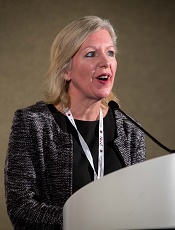
Photo courtesy of ASH
ORLANDO, FL—A subgroup of young patients with high-risk B-cell acute lymphoblastic leukemia (B-ALL) can have “outstanding outcomes” with contemporary therapy, according to researchers.
Results of a large study suggested that patients ages 1 to 30 who have high-risk B-ALL according to National Cancer Institute (NCI) classification can have high rates of event-free survival (EFS) and overall survival (OS) if they have favorable cytogenetic features, have no evidence of CNS disease, and have rapid minimal residual disease (MRD) responses.
The research suggested these patients will not benefit from further chemotherapy intensification.
Elizabeth Raetz, MD, of the University of Utah in Salt Lake City, presented these results at the 2015 ASH Annual Meeting (abstract 807).
She and her colleagues analyzed patients enrolled on the Children’s Oncology Group (COG) AALL03B1 classification study at the time of B-ALL diagnosis. From December 2003 to September 2011, there were 11,144 eligible patients enrolled on this trial.
Eighty-nine percent of these patients were also enrolled on a frontline ALL therapeutic trial, and 96% of these patients were evaluable for post-induction treatment assignment. Sixty-five percent of these patients were treated on a trial for NCI standard-risk B-ALL (COG-AALL0331), and 35% were treated on a trial for high-risk B-ALL (COG-AALL0232).
At the end of induction therapy, patients were classified into low-risk (29%), standard-risk (33%), high-risk (34%), and very-high-risk (4%) groups for further treatment allocation. The variables used for risk classification were age, initial white blood cell count, extramedullary disease status, blast cytogenetics, and early treatment response based on bone marrow morphology and day 29 MRD.
Patients with very-high-risk features (BCR-ABL1, hypodiploidy, induction failure, or poor response at day 43) did not continue on AALL0232/AALL0331 post-induction but did have outcome data captured for analysis.
Response and survival
Rapid early response was defined as M1 (<5% blasts) bone marrow by day 15 plus flow cytometry-based MRD <0.1% on day 29 of induction. Patients with either M2/M3 (≥5% blasts) day 15 marrow or MRD ≥0.1% at day 29 were deemed slow early responders.
Eighty-four percent of patients had a rapid early response to induction, and 16% had a slow early response.
For rapid early responders, the 5-year EFS was 89.3%, and the 5-year OS was 95.2%. For slow early responders, the EFS and OS rates were 67.9% and 84.3%, respectively (P<0.0001 for both EFS and OS comparisons).
Survival according to cytogenetics
Having favorable cytogenetic abnormalities (triple trisomies of chromosomes 4, 10, and 17 or ETV6-RUNX1 fusion) was associated with significantly better EFS and OS than having unfavorable cytogenetics (hypodiploidy [DNA index <0.81 or chromosomes < 44], MLL rearrangements, BCR-ABL1, or iAMP21).
And Dr Raetz pointed out that the 5-year OS exceeded 98% for patients with either standard- or high-risk disease who had favorable cytogenetics.
For patients who were ETV6-RUNX1-positive, the EFS was 93.2% and the OS was 98.3%. For patients who were ETV6-RUNX1 negative, the rates were 83.5% and 92%, respectively (P<0.0001).
For patients with triple trisomy, EFS was 94.7% and OS was 98.7%. For those without triple trisomy, the rates were 83.6% and 92.2%, respectively (P<0.0001).
For patients with MLL rearrangement, the EFS was 73.9% and the OS was 83.1%. For patients without MLL rearrangement, the rates were 85.9% and 93.6%, respectively (P<0.0001).
For patients who were positive for iAMP21, the EFS was 69.5% and the OS was 90.1%. For iAMP21-negative patients, the rates were 86.1% and 93.4%, respectively (P<0.0001 for PFS comparison and P=0.0026 for OS comparison).
Survival according to risk group and MRD
The researchers also assessed EFS and OS among patients with favorable cytogenetics according to NCI risk group and MRD at days 8 and 29.
“One thing to point out is that, regardless of having favorable cytogenetics, those individuals who had end-induction MRD values of greater than 0.01% had inferior outcomes, so that was still a prognostic marker,” Dr Raetz said.
“And one thing that we were pleasantly surprised to see was that, among the NCI high-risk patients, those who had very rapid MRD responses—so less than 1% at day 8 in the blood and less than 0.01% in the marrow on day 29—had a 94.9% 5-year event-free survival and 98.1% overall survival.”
The researchers also divided this group according to age—patients younger than 10 and those 10 years or older. There was no significant difference in EFS or OS between the age groups (P=0.126 and P=0.411).
Standard-risk group
Among patients with <1% MRD on day 8 and <0.01% MRD on day 29, the EFS was 95.7% and the OS was 99.1%.
Among patients with ≥1% MRD on day 8 and <0.01% MRD on day 29, the EFS was 91.7% and the OS was 99.4%.
Among patients with any MRD on day 8 and ≥0.01% MRD on day 29, the EFS was 88.1% and the OS was 96.8%.
High-risk group
Among patients with <1% MRD on day 8 and <0.01% MRD on day 29, the EFS was 94.9% and the OS was 98.1%.
Among patients with ≥1% MRD on day 8 and <0.01% MRD on day 29, the EFS was 93.6% and the OS was 95.5%.
Among patients with any MRD on day 8 and ≥0.01% MRD on day 29, the EFS was 75.4% and the OS was 90.4%.
In closing, Dr Raetz said this study showed that real‐time classification incorporating clinical features, blast cytogenetics, and early response was feasible in a large group of patients enrolled on COG ALL trials and identified patients with varying outcomes for risk‐based treatment allocation.
She noted that early response by marrow morphology was not prognostic when MRD response was used and is therefore no longer used in COG studies.
And although favorable cytogenetic features were not prognostic in NCI high-risk B‐ALL patients in prior COG studies, the current study indicates that these patients can have “excellent outcomes” if they have no evidence of CNS leukemia and are rapid MRD responders. So these patients will not benefit from further chemotherapy intensification. ![]()
CAR T cells persist for 3 years in young ALL patients

Photo courtesy of Penn Medicine
ORLANDO, FL—CTL019, a CD19 chimeric antigen receptor (CAR) T-cell therapy, can persist for 3 years or longer in children and young adults with relapsed/refractory acute lymphoblastic leukemia (ALL), according to the latest results of a pilot study.
This suggests CTL019 can offer long-term disease control without subsequent therapy, such as stem cell transplant, said study author Stephan Grupp, MD, PhD, of the University of Pennsylvania in Philadelphia.
Dr Grupp presented these results at the 2015 ASH Annual Meeting (abstract 681*).
Previously, Dr Grupp and his colleagues reported that CTL019 led to durable antitumor activity, including sustained complete responses (CRs) in adults and children with ALL (ASH 2012, NEJM 2013, ASH 2013, NEJM 2014, ASH 2014).
At this year’s ASH meeting, he reported on outcomes and longer follow-up of the first 59 patients with relapsed/refractory ALL treated in a pilot trial. The patients had a median age of 11 years.
The median follow-up was 12 months. Fifty-five patients (93%) achieved a CR at 1 month. Six patients went on to receive a transplant, and 1 patient went on to receive donor lymphocyte infusion.
The relapse-free survival was 76% at 6 months and 55% at 12 months. Overall survival was 79% at 12 months.
“There were no relapses past 1 year,” Dr Grupp said, noting that 18 patients beyond 1 year are in remission, and 13 patients have not received further therapy.
“We are able to get patients into remission,” he said, adding that response is similar at high and low disease burden.
“We see massive proliferation of CAR T cells. Prolonged CTL019 persistence is detected by flow cytometry. About 70% of patients maintain CAR T cells.”
Resistance was seen in 7% of patients due to failure of T cells to proliferate. One-third of recurrences occurred in those with CD19-positive relapse and two-thirds with CD19-negative relapse.
Dr Grupp noted that CTL019 has an impact on central nervous system (CNS) disease.
“We are able to control CNS disease,” he said. “We found no CNS relapses in patients who were treated, and 98% of them still have CTL019 in the cerebral spinal fluid.”
B-cell aplasia persists beyond 3.5 years in all responding patients. This is managed by immunoglobulin replacement.
“Patients in remission and with B-cell aplasia at 1 year are still alive at 2 to 3 years,” Dr Grupp said. “We do not know how long this will last and if B cells will recover.”
Severe cytokine release syndrome (CRS), observed in 88% of patients, is the principal toxicity. This is controlled with anti-IL6 therapy. Patients who are less likely to have CRS are those with a lower disease burden.
“IL-6 is a major player but does not predict CRS; it only correlates strongly with it,” Dr Grupp said.
Some toxicity is also associated with macrophage activation syndrome and neurotoxicity.
CTL019 was invented at The University of Pennsylvania but has been licensed to Novartis. Most of the researchers involved in this study reported research funding and/or consultancy payments from Novartis, and 1 researcher is employed by the company. ![]()
*Data in the abstract differ from the presentation.
Regimen with intensified PEG-ASP feasible in young adults with ALL
Annual Meeting
Photo courtesy of ASH
ORLANDO, FL—Results of a DFCI ALL Consortium trial have shown that adults with acute lymphoblastic leukemia (ALL) can be successfully and safely treated with a pediatric regimen using intensified pegylated asparaginase (PEG-ASP).
Investigators recently reported that young adults treated with native E coli asparaginase as part of their regimen had improved 4-year disease-free survival and overall survival (OS) rates.
Now, the team has shown it is possible to use PEG-ASP to improve young adult outcomes as well.
The investigators also described the toxicities with PEG-ASP and compared them to the prior DFCI ALL Consortium trial with native E coli asparaginase.
“[Wendy] Stock, years ago, analyzed young adult patients 16 to 20 based on whether or not they were treated on Children’s Cancer Group trials or CALGB trials,” said Daniel J. DeAngelo, MD, PhD, of the Dana-Farber Cancer Institute in Boston, Massachusetts.
“And what she reported was that there was a dramatic improvement in the disease- and event-free survival . . . . And since that publication and presentation at ASH several years ago, there’ve been a large number of us who’ve tried to adapt pediatric trials or pediatric-inspired trials for the treatment of young adults with acute lymphoblastic leukemia.”
The PEG-ASP DFCI ALL trial is one such effort. Dr DeAngelo discussed the results of this trial at the 2015 ASH Annual Meeting (abstract 80).
Patient population
Investigators enrolled 110 patients on the trial.
Patients had to be between 18 and 50 years of age, with untreated ALL and no history of secondary ALL. Patients with Burkitt’s lymphoma were excluded.
The patients’ median age was 32 (range, 18–50), 62% were male, 80% were white, and 85% were non-Hispanic. Eleven percent had central nervous system (CNS) status 2 or 3 prior to the initiation of chemotherapy.
Most (87%) had a performance status of 0 or 1, 82% had the B-cell and 18% the T-cell phenotype, 19% were Ph-positive, 6% had an MLL translocation (11q23), and 18% had other translocations.
Study design
Induction chemotherapy consisted of doxorubicin, prednisone, vincristine, PEG-ASP, and intrathecal therapy.
The first consolidation consisted of high-dose methotrexate followed by a BFM-like intensification and high-dose cytarabine, etoposide, and dexamethasone.
CNS prophylaxis included intrathecal chemotherapy and cranial irradiation.
The second consolidation consisted of eight 3-week courses of doxorubicin, vincristine, dexamethasone, 6-mercaptopurine, and 30 weeks of PEG-ASP.
The PEG-ASP was initially dosed at the pediatric level of 2500 IU/m2 every 2 weeks.
“But due to some toxicity concerns in this treatment strategy—with specific emphasis on liver function abnormalities with hyperbilirubinemia, elevations of AST/ALT—we decided to amend the protocol and decrease the PEG dose from 2500 to 2000 and increase the interval from every 2 weeks to every 3 weeks,” Dr DeAngelo said.
Therefore, during this 30-week course, patients received 10 doses of PEG ASP as opposed to 15.
“We also went back and swapped out the PEG asparaginase during induction and reinserted native E coli to really ascertain comparative properties,” he said.
Maintenance therapy consisted of 3-week courses of vincristine, dexamethasone, methotrexate, and 6-mercaptopurine for 2 years from achievement of complete remission (CR).
During PEG-ASP therapy, patients received anticoagulation prophylaxis, preferably with low-molecular-weight heparin, as long as patients had a platelet count greater than 30,000/μL.
Results
Of the 110 patients enrolled, 65 received the higher dose of asparaginase, and 45 received the amended lower dose.
Ninety-one patients (89%) achieved a CR, 57 of whom received the higher dose of asparaginase and 34 the lower dose.
There were 2 induction deaths, both in the higher-dose group.
Twenty-one patients went on to transplant in CR1, 15 in the higher-dose group and 6 in the lower-dose group.
Twenty-three patients relapsed, 17 in the higher-dose group and 6 in the lower-dose group. Two of the relapses were CNS only.
Three patients died in remission, 2 in the higher-dose asparaginase group and 1 in the lower. And 3 patients died after stem cell transplant in CR, 2 in the higher-dose group and 1 in the lower.
At a median follow-up of 42.2 months, the 3-year disease-free survival for the entire cohort was 73%, and overall survival was 75%.
Subset analyses
The investigators performed subgroup analyses and came up with some “interesting observations,” Dr DeAngelo said.
Younger patients, ages 18 to 19 and 20 to 29, had a better OS than the older patients. The OS for younger patients is in the 80% to 85% range, “which is significantly better than the other patients in the 30 to 40 or 40 to 50 age groups,” Dr DeAngelo said.
Patients with T-cell ALL had a better OS than those with B-cell ALL and Ph-positive ALL, who had the worst OS of approximately 50%. The vast majority of Ph-negative patients were transplanted, and all received imatinib in addition to chemotherapy.
“[Patients with the T-cell phenotype] seemed to do particularly well on this strategy, which is something we showed in the last study as well, with an overall survival of 80%, compared to 70% for the B-cell Philadelphia-negative [patients],” Dr DeAngelo said.
He and his colleagues also found an association between OS and body mass index (BMI).
Obese or morbidly obese patients with a BMI of 30 or over had an OS of around 40%, while underweight or normal-weight patients had an OS of almost 90%, a “profound overall survival in the less-than-obese patients,” Dr DeAngelo said.
“And I think one of the things that is bringing down the curves is the obese,” he added, “which is a concern as the body mass index of the American population increases.”
Another discovery was that patients with minimal residual disease (MRD) of less than 10-4 had better OS than those with a high MRD level.
After a single dose of PEG ASP during induction on day 4, asparagine was depleted for a median of 3 weeks. With a single dose of E coli asparaginase, on the other hand, asparagine is depleted for about a week.
Asparaginase levels during consolidation were “extraordinarily elevated” with the 2500 dose level of PEG-ASP compared to the lower dose level. Toxicity was much more manageable, however, as the dose was reduced, Dr DeAngelo said. And asparagine was still depleted throughout the 30 weeks, as per protocol.
Toxicity
The higher asparaginase dose group “surprisingly, had a very low rate of clinical pancreatitis,” Dr DeAngelo said.
At the higher asparaginase dose—2500 IU/m2 every 2 weeks—66 patients experienced grade 3-5 adverse events: 30 (46%) febrile neutropenia, 1 (1%) pancreatitis, 19 (29%) AST, 34 (52%) ALT, 24 (36%) bilirubin, 19 (29%) thrombosis, 2 (3%) CNS hemorrhage, 3 (4%) hypersensitivity, and 4 (6%) osteonecrosis.
After the protocol amendment, “we saw a marked reduction in hyperbilirubinemia,” Dr DeAngelo said.
Grade 3–4 hyperbilirubinemia decreased from 36% to 7%, grade 3–4 ALT elevation decreased from 52% to 29%, and the rate of thrombosis decreased from 29% to 16%.
“Whether the latter decrease [thrombosis] was reflective of the decreased dose of asparaginase or the addition of anticoagulation, I can’t determine,” Dr DeAngelo said, “but it seemed to reflect other studies.”
The investigators concluded that a dose-intensified pediatric regimen in adults is feasible, with an acceptable toxicity profile.
The team said this approach may translate to better survival for adults with ALL, with the exception of older adults and patients with a high BMI. PEG-ASP has increased toxicity in these patients.
The investigators recommend addressing the challenges that remain—psychosocial issues, practice patterns, and biology—with a unified approach and more cooperative group trials in young adults. ![]()
Annual Meeting
Photo courtesy of ASH
ORLANDO, FL—Results of a DFCI ALL Consortium trial have shown that adults with acute lymphoblastic leukemia (ALL) can be successfully and safely treated with a pediatric regimen using intensified pegylated asparaginase (PEG-ASP).
Investigators recently reported that young adults treated with native E coli asparaginase as part of their regimen had improved 4-year disease-free survival and overall survival (OS) rates.
Now, the team has shown it is possible to use PEG-ASP to improve young adult outcomes as well.
The investigators also described the toxicities with PEG-ASP and compared them to the prior DFCI ALL Consortium trial with native E coli asparaginase.
“[Wendy] Stock, years ago, analyzed young adult patients 16 to 20 based on whether or not they were treated on Children’s Cancer Group trials or CALGB trials,” said Daniel J. DeAngelo, MD, PhD, of the Dana-Farber Cancer Institute in Boston, Massachusetts.
“And what she reported was that there was a dramatic improvement in the disease- and event-free survival . . . . And since that publication and presentation at ASH several years ago, there’ve been a large number of us who’ve tried to adapt pediatric trials or pediatric-inspired trials for the treatment of young adults with acute lymphoblastic leukemia.”
The PEG-ASP DFCI ALL trial is one such effort. Dr DeAngelo discussed the results of this trial at the 2015 ASH Annual Meeting (abstract 80).
Patient population
Investigators enrolled 110 patients on the trial.
Patients had to be between 18 and 50 years of age, with untreated ALL and no history of secondary ALL. Patients with Burkitt’s lymphoma were excluded.
The patients’ median age was 32 (range, 18–50), 62% were male, 80% were white, and 85% were non-Hispanic. Eleven percent had central nervous system (CNS) status 2 or 3 prior to the initiation of chemotherapy.
Most (87%) had a performance status of 0 or 1, 82% had the B-cell and 18% the T-cell phenotype, 19% were Ph-positive, 6% had an MLL translocation (11q23), and 18% had other translocations.
Study design
Induction chemotherapy consisted of doxorubicin, prednisone, vincristine, PEG-ASP, and intrathecal therapy.
The first consolidation consisted of high-dose methotrexate followed by a BFM-like intensification and high-dose cytarabine, etoposide, and dexamethasone.
CNS prophylaxis included intrathecal chemotherapy and cranial irradiation.
The second consolidation consisted of eight 3-week courses of doxorubicin, vincristine, dexamethasone, 6-mercaptopurine, and 30 weeks of PEG-ASP.
The PEG-ASP was initially dosed at the pediatric level of 2500 IU/m2 every 2 weeks.
“But due to some toxicity concerns in this treatment strategy—with specific emphasis on liver function abnormalities with hyperbilirubinemia, elevations of AST/ALT—we decided to amend the protocol and decrease the PEG dose from 2500 to 2000 and increase the interval from every 2 weeks to every 3 weeks,” Dr DeAngelo said.
Therefore, during this 30-week course, patients received 10 doses of PEG ASP as opposed to 15.
“We also went back and swapped out the PEG asparaginase during induction and reinserted native E coli to really ascertain comparative properties,” he said.
Maintenance therapy consisted of 3-week courses of vincristine, dexamethasone, methotrexate, and 6-mercaptopurine for 2 years from achievement of complete remission (CR).
During PEG-ASP therapy, patients received anticoagulation prophylaxis, preferably with low-molecular-weight heparin, as long as patients had a platelet count greater than 30,000/μL.
Results
Of the 110 patients enrolled, 65 received the higher dose of asparaginase, and 45 received the amended lower dose.
Ninety-one patients (89%) achieved a CR, 57 of whom received the higher dose of asparaginase and 34 the lower dose.
There were 2 induction deaths, both in the higher-dose group.
Twenty-one patients went on to transplant in CR1, 15 in the higher-dose group and 6 in the lower-dose group.
Twenty-three patients relapsed, 17 in the higher-dose group and 6 in the lower-dose group. Two of the relapses were CNS only.
Three patients died in remission, 2 in the higher-dose asparaginase group and 1 in the lower. And 3 patients died after stem cell transplant in CR, 2 in the higher-dose group and 1 in the lower.
At a median follow-up of 42.2 months, the 3-year disease-free survival for the entire cohort was 73%, and overall survival was 75%.
Subset analyses
The investigators performed subgroup analyses and came up with some “interesting observations,” Dr DeAngelo said.
Younger patients, ages 18 to 19 and 20 to 29, had a better OS than the older patients. The OS for younger patients is in the 80% to 85% range, “which is significantly better than the other patients in the 30 to 40 or 40 to 50 age groups,” Dr DeAngelo said.
Patients with T-cell ALL had a better OS than those with B-cell ALL and Ph-positive ALL, who had the worst OS of approximately 50%. The vast majority of Ph-negative patients were transplanted, and all received imatinib in addition to chemotherapy.
“[Patients with the T-cell phenotype] seemed to do particularly well on this strategy, which is something we showed in the last study as well, with an overall survival of 80%, compared to 70% for the B-cell Philadelphia-negative [patients],” Dr DeAngelo said.
He and his colleagues also found an association between OS and body mass index (BMI).
Obese or morbidly obese patients with a BMI of 30 or over had an OS of around 40%, while underweight or normal-weight patients had an OS of almost 90%, a “profound overall survival in the less-than-obese patients,” Dr DeAngelo said.
“And I think one of the things that is bringing down the curves is the obese,” he added, “which is a concern as the body mass index of the American population increases.”
Another discovery was that patients with minimal residual disease (MRD) of less than 10-4 had better OS than those with a high MRD level.
After a single dose of PEG ASP during induction on day 4, asparagine was depleted for a median of 3 weeks. With a single dose of E coli asparaginase, on the other hand, asparagine is depleted for about a week.
Asparaginase levels during consolidation were “extraordinarily elevated” with the 2500 dose level of PEG-ASP compared to the lower dose level. Toxicity was much more manageable, however, as the dose was reduced, Dr DeAngelo said. And asparagine was still depleted throughout the 30 weeks, as per protocol.
Toxicity
The higher asparaginase dose group “surprisingly, had a very low rate of clinical pancreatitis,” Dr DeAngelo said.
At the higher asparaginase dose—2500 IU/m2 every 2 weeks—66 patients experienced grade 3-5 adverse events: 30 (46%) febrile neutropenia, 1 (1%) pancreatitis, 19 (29%) AST, 34 (52%) ALT, 24 (36%) bilirubin, 19 (29%) thrombosis, 2 (3%) CNS hemorrhage, 3 (4%) hypersensitivity, and 4 (6%) osteonecrosis.
After the protocol amendment, “we saw a marked reduction in hyperbilirubinemia,” Dr DeAngelo said.
Grade 3–4 hyperbilirubinemia decreased from 36% to 7%, grade 3–4 ALT elevation decreased from 52% to 29%, and the rate of thrombosis decreased from 29% to 16%.
“Whether the latter decrease [thrombosis] was reflective of the decreased dose of asparaginase or the addition of anticoagulation, I can’t determine,” Dr DeAngelo said, “but it seemed to reflect other studies.”
The investigators concluded that a dose-intensified pediatric regimen in adults is feasible, with an acceptable toxicity profile.
The team said this approach may translate to better survival for adults with ALL, with the exception of older adults and patients with a high BMI. PEG-ASP has increased toxicity in these patients.
The investigators recommend addressing the challenges that remain—psychosocial issues, practice patterns, and biology—with a unified approach and more cooperative group trials in young adults.
Group recommends adding rituximab to ALL therapy
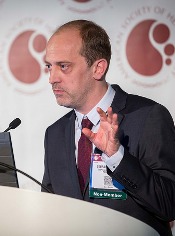
Photo courtesy of ASH
ORLANDO, FL—Investigators from the Group for Research on Adult Lymphoblastic Leukemia (GRAALL) recommend integrating rituximab into the treatment of adult patients with acute lymphoblastic leukemia (ALL) based on results of the GRAALL-R 2005 study.
Patients who received rituximab as part of their therapy had a median event-free survival (EFS) at 2 years of 65%, compared to 52% for patients who did not receive rituximab. After censoring for stem cell transplant in first complete remission, the benefit was even greater.
Sébastien Maury, MD, PhD, of Hȏpital Hénri Mondor in Creteil, France, presented the results during the plenary session of the 2015 ASH Annual Meeting as abstract 1.
Dr Maury said GRAALL-R 2005 is the first phase 3, randomized study to evaluate the role of rituximab in the treatment of B-cell precursor (BCP) ALL.
Only one previous study, he said, suggested a potential benefit of adding rituximab compared to historic controls of chemotherapy alone.
He explained that, because the CD20 antigen is expressed at diagnosis in 30% to 40% of patients with BCP-ALL, investigators undertook to evaluate whether adding the anti-CD20 monoclonal antibody rituximab to the ALL treatment regimen could be beneficial for newly diagnosed Ph-negative BCP-ALL patients.
Study design & population
Investigators randomized 105 patients to receive the pediatric-inspired GRAALL protocol plus rituximab and 104 patients to the same regimen without rituximab.
Patients had to have 20% or more CD20-positive leukemic blasts.
Patients in the rituximab arm received 375 mg/m2 during induction on days 1 and 7, during salvage reinduction (if needed) on days 1 and 7, during consolidation blocks (6 infusions), during late intensification on days 1 and 7, and during the first year of maintenance (6 infusions), for a total of 16 to 18 infusions.
“In this trial, allogeneic transplantation was offered in first remission to high-risk patients who were those patients with at least one of these baseline or response-related criteria,” Dr Maury said.
Investigators defined high-risk at baseline as having a white blood cell count of 30 x 109/L or higher, CNS involvement, CD10-negative disease, or unfavorable cytogenetics.
And response-related criteria for high-risk disease included poor peripheral blast clearance after the 1-week steroid pre-phase, poor bone marrow blast clearance after the first week of chemotherapy, or no hematologic complete response after the first induction course.
Patient characteristics were well balanced between the arms, with a median age for the entire group of 40.2 years. Rituximab-treated patients had 61% CD20-positive blasts, and the no-rituximab arm had 69%.
More patients in the rituximab arm had a better ECOG performance status, although the difference was not significant. Thirteen percent were assessed as being grade 2 or higher in the rituximab arm, compared with 18% in the no-rituximab arm (P=0.06).
“The proportion of high-risk patients was comparable in both arms,” Dr Maury said, “representing around two-thirds of the study population.”
In the rituximab arm, 70% were considered high-risk, compared with 64% in the no-rituximab arm (P=0.46).
“However, despite this,” he said, “a significantly higher proportion of patients received allo transplant at first remission in the rituximab arm, 34% versus 20%. And since this was not explained by a different proportion of high-risk patients, this was probably due to differences in donor availability.”
Dr Maury noted that compliance to treatment was “quite good.”
Efficacy
The median follow-up was 30 months, and the primary endpoint was EFS.
The EFS rate for rituximab-treated patients at 2 years was 65%, compared with 52% for the non-rituximab patients (hazard ratio=0.66, P=0.038).
EFS was also significantly better with rituximab when patients were censored at allogeneic transplant, with a hazard ratio of 0.59 and a significance of 0.021.
However, there were no significant differences in early complete response rates, minimal residual disease (MRD) after induction, and MRD after consolidation.
“[O]nly 40% of patients could be centrally analyzed [for MRD],” Dr Maury explained, “which may be the reason why we could not detect any impact of rituximab on MRD.”
The cumulative incidence of relapse at 2 years was 18% in the rituximab arm and 32% in the no-rituximab arm (hazard ratio=0.52, P=0.017). And after censoring for stem cell transplant in first complete remission, the hazard ratio was 0.49 in favor of rituximab (P=0.018).
Overall survival (OS) was not significantly different between the arms. Rituximab-treated patients had an OS rate of 71%, compared with 64% in the no-rituximab arm (P=0.095).
“However, this difference became significant when censoring patients at time of allo-transplant,” Dr Maury said.
There was a 12% cumulative incidence of death in first complete remission at 2 years in each arm.
Investigators performed multivariate analysis and found that treatment with rituximab (P=0.020), age (P=0.022), white blood cell count of 30 x 109/L or higher (P=0.005), and CNS involvement all significantly impacted EFS.
When they introduced stem cell transplant in first remission as a covariable, the same factors remained significant. Allogeneic stem cell transplant in first remission did not make a significant difference on EFS (P=0.62).
Safety
One hundred twenty-four patients reported 246 severe adverse events, the most frequent of which was infection—71 in the rituximab arm and 55 in the no-rituximab arm, a difference that was not significant (P=0.16).
Severe allergic events were significantly different between the arms, with 2 severe allergic events reported in the rituximab arm and 14 in the no-rituximab arm (P=0.002). Of these 16 events, all but one were due to asparaginase.
“We believe that this may reflect the protective effect of rituximab that might inhibit B-cell protection of antibodies against asparaginase,” Dr Maury said, although the investigators did not actually measure the antibodies.
Severe lab abnormalities, neurologic and pulmonary events, coagulopathy, cardiologic and gastrointestinal events were not significantly different between the arms.
Dr Maury emphasized that the addition of rituximab to standard intensive chemotherapy is well tolerated, significantly improves EFS, and prolongs OS in patients not receiving allogeneic transplant in first remission.
While the optimal dose schedule of rituximab still remains to be determined, the GRAALL investigators believe that “the addition of rituximab should be the new standard of care for these patients,” Dr Maury declared.