User login
MDedge conference coverage features onsite reporting of the latest study results and expert perspectives from leading researchers.
Full 2-year follow-up vindicates EVOLUT Low-Risk TAVR data
After taking it on the chin for previously reporting Bayesian estimates, actual 2-year data from the EVOLUT Low Risk trial confirm that transcatheter aortic valve replacement (TAVR) is noninferior to surgery for the primary endpoint of death or disabling stroke.
Among 1,414 as-treated patients, Kaplan-Meier rates for all-cause death or disabling stroke at 24 months were 4.3% with TAVR and 6.3% with surgery (P = .084).
There was also no difference in the individual components of all-cause death (3.5% vs. 4.4%; log-rank P = .366) and disabling stroke (1.5% vs. 2.7%; log-rank P = .119).
Recent low-risk TAVR studies have raised questions about whether there’s a possible catch-up for surgery between 12 and 24 months, given the early mortality benefit from the less-invasive transcatheter procedure, prompting a landmark analysis, John K. Forrest, MD, said during the virtual presentation at the 2021 Congress of European Association of Percutaneous Cardiovascular Interventions, EuroPCR 2021.
“Between 1 and 2 years, there was no convergence of the Kaplan-Meier curves for death or disabling stroke,” with an incidence of 1.9% for the TAVR group and 2.1% for the surgery group (log-rank P = .742), said Dr. Forrest, of Yale University, New Haven, Conn. “The lines were almost superimposed upon each other.”
Session moderator Bernard Prendergast, MD, observed that the Bayesian analysis, which was reported in 2019 and used 12-month follow-up to predict 2-year outcomes, generated questions and criticism over whether this was an appropriate method, compared with traditional Kaplan-Meier analysis. “Indeed, some people accused the investigators of gaming it with this form of statistical analysis.”
To act as a “fact checker,” Dr. Prendergast called in Christopher Cook, MRC, from the PCR Clinical Research Group and Imperial College London. The key methodologic question, Dr. Cook said, is whether Bayesian methods accurately predict actual clinical outcomes in this randomized clinical trial. “The simple answer to this for me, is yes.”
He pointed out that the Kaplan-Meier data for the primary outcome at 2 years were, in fact, numerically better than Bayesian estimates of 5.3% in the TAVR group and 6.7% in the surgery group.
“This validates the use of the original Bayesian methods to estimate patient outcomes in low-risk TAVI patients and, indeed, it may act as an example of where Bayesian methods can be safely applied in order to fast track potentially transformative procedures and technologies to our patients,” Dr. Cook said.
The rate of disabling stroke with TAVR was 1.5% in the new analysis, up from 1.1% in the Bayesian analysis, and 2.7% with surgery, down from 3.5% in the Bayesian analysis.
All-cause mortality, also noted earlier, was 3.5% with TAVR and 4.4% with surgery, whereas the Bayesian estimate was 4.5% for each group.
Dr. Prendergast of St. Thomas’ Hospital, London, said the actual 2-year data are reassuring regarding the statistical tools used and supplement those recently reported from low-risk patients in PARTNER 3.
But, he asked, “what does this mean for practice, what does it mean for guidelines, and how long do we need to wait until we are comfortable and reassured that we can apply TAVI in younger and low-risk patients with a durable outcome?”
Dr. Forrest said that clinicians can be reassured that these patients “are doing very well” but that long-term follow-up is critical.
“We need to be realistic here. We’re really going to be interested in 5- and 10-year outcomes and potentially even thereafter,” he said. “What happens to these valves when they eventually fail? Are superior hemodynamics going to give us longer valve durability in some way or are there going to be other unforeseen things that come up 10 years out? We don’t know those answers.”
TAVR with a supra-annular, self-expanding valve (CoreValve , Evolut R, or Evolut PRO) had superior hemodynamics in the new 2-year analysis with lower aortic valve gradients (9.0 vs. 11.7 mm Hg) and larger valve areas (2.2 vs. 2.0 cm2).
Prosthesis-patient mismatch also favored TAVR, with moderate or severe mismatch occurring in 7.2% and 2.1%, respectively, compared with 19.1% and 4.9%, respectively, with surgery. “We know that this has an impact on long-term outcomes, so it’s important to note that significant difference here,” Dr. Forrest said.
The chink in TAVR’s armor remains paravalvular leak, particularly mild leak, which was significantly higher at 26.6%, compared with only 2.6% with surgery. Moderate to severe leaks were seen in 1.7% and 0.4%, respectively, reflecting the improvement in TAVR with new iterations, he said.
Surgery was also superior to TAVR with regard to the need for permanent pacemaker implantation (7.9% vs. 21.1%). This compares with Bayesian estimates of 6.7% and 23.0%, respectively.
Rates of myocardial infarction remained constant in the two analyses for the TAVR (2.2%) and surgery (1.6%) groups, whereas heart failure hospitalizations improved slightly, from 5.4% versus 7.9%, respectively, in the Bayesian analysis to 5.3% versus 7.1%, respectively, in the new analysis.
Fellow discussant Marie-Claude Morice, MD, Institute Hospitalier Jacques Cartier, Massy, France, highlighted several meta-analyses in different risk patients showing “a lot of good news,” including decreased stroke and mortality rates and the combined outcome clearly favoring TAVR.
“The remaining question is the longevity of the valve, but with 5 years’ follow-up we have for comparison [in high-risk patients], it is the same,” she said. “What this illustrates is that the tidal wave of TAVR is continuing, and we can look optimistically to the future. Is it the nonsymptomatic patients?”
Medtronic funded the study. Dr. Forrest reported grant support from, serving on the advisory board, and proctoring for Edwards Lifesciences and Medtronic. Dr. Prendergast has received grants from Edwards Lifesciences; and speaker/consultancy fees from Abbott, Anteris, and Edwards.
A version of this article first appeared on Medscape.com.
After taking it on the chin for previously reporting Bayesian estimates, actual 2-year data from the EVOLUT Low Risk trial confirm that transcatheter aortic valve replacement (TAVR) is noninferior to surgery for the primary endpoint of death or disabling stroke.
Among 1,414 as-treated patients, Kaplan-Meier rates for all-cause death or disabling stroke at 24 months were 4.3% with TAVR and 6.3% with surgery (P = .084).
There was also no difference in the individual components of all-cause death (3.5% vs. 4.4%; log-rank P = .366) and disabling stroke (1.5% vs. 2.7%; log-rank P = .119).
Recent low-risk TAVR studies have raised questions about whether there’s a possible catch-up for surgery between 12 and 24 months, given the early mortality benefit from the less-invasive transcatheter procedure, prompting a landmark analysis, John K. Forrest, MD, said during the virtual presentation at the 2021 Congress of European Association of Percutaneous Cardiovascular Interventions, EuroPCR 2021.
“Between 1 and 2 years, there was no convergence of the Kaplan-Meier curves for death or disabling stroke,” with an incidence of 1.9% for the TAVR group and 2.1% for the surgery group (log-rank P = .742), said Dr. Forrest, of Yale University, New Haven, Conn. “The lines were almost superimposed upon each other.”
Session moderator Bernard Prendergast, MD, observed that the Bayesian analysis, which was reported in 2019 and used 12-month follow-up to predict 2-year outcomes, generated questions and criticism over whether this was an appropriate method, compared with traditional Kaplan-Meier analysis. “Indeed, some people accused the investigators of gaming it with this form of statistical analysis.”
To act as a “fact checker,” Dr. Prendergast called in Christopher Cook, MRC, from the PCR Clinical Research Group and Imperial College London. The key methodologic question, Dr. Cook said, is whether Bayesian methods accurately predict actual clinical outcomes in this randomized clinical trial. “The simple answer to this for me, is yes.”
He pointed out that the Kaplan-Meier data for the primary outcome at 2 years were, in fact, numerically better than Bayesian estimates of 5.3% in the TAVR group and 6.7% in the surgery group.
“This validates the use of the original Bayesian methods to estimate patient outcomes in low-risk TAVI patients and, indeed, it may act as an example of where Bayesian methods can be safely applied in order to fast track potentially transformative procedures and technologies to our patients,” Dr. Cook said.
The rate of disabling stroke with TAVR was 1.5% in the new analysis, up from 1.1% in the Bayesian analysis, and 2.7% with surgery, down from 3.5% in the Bayesian analysis.
All-cause mortality, also noted earlier, was 3.5% with TAVR and 4.4% with surgery, whereas the Bayesian estimate was 4.5% for each group.
Dr. Prendergast of St. Thomas’ Hospital, London, said the actual 2-year data are reassuring regarding the statistical tools used and supplement those recently reported from low-risk patients in PARTNER 3.
But, he asked, “what does this mean for practice, what does it mean for guidelines, and how long do we need to wait until we are comfortable and reassured that we can apply TAVI in younger and low-risk patients with a durable outcome?”
Dr. Forrest said that clinicians can be reassured that these patients “are doing very well” but that long-term follow-up is critical.
“We need to be realistic here. We’re really going to be interested in 5- and 10-year outcomes and potentially even thereafter,” he said. “What happens to these valves when they eventually fail? Are superior hemodynamics going to give us longer valve durability in some way or are there going to be other unforeseen things that come up 10 years out? We don’t know those answers.”
TAVR with a supra-annular, self-expanding valve (CoreValve , Evolut R, or Evolut PRO) had superior hemodynamics in the new 2-year analysis with lower aortic valve gradients (9.0 vs. 11.7 mm Hg) and larger valve areas (2.2 vs. 2.0 cm2).
Prosthesis-patient mismatch also favored TAVR, with moderate or severe mismatch occurring in 7.2% and 2.1%, respectively, compared with 19.1% and 4.9%, respectively, with surgery. “We know that this has an impact on long-term outcomes, so it’s important to note that significant difference here,” Dr. Forrest said.
The chink in TAVR’s armor remains paravalvular leak, particularly mild leak, which was significantly higher at 26.6%, compared with only 2.6% with surgery. Moderate to severe leaks were seen in 1.7% and 0.4%, respectively, reflecting the improvement in TAVR with new iterations, he said.
Surgery was also superior to TAVR with regard to the need for permanent pacemaker implantation (7.9% vs. 21.1%). This compares with Bayesian estimates of 6.7% and 23.0%, respectively.
Rates of myocardial infarction remained constant in the two analyses for the TAVR (2.2%) and surgery (1.6%) groups, whereas heart failure hospitalizations improved slightly, from 5.4% versus 7.9%, respectively, in the Bayesian analysis to 5.3% versus 7.1%, respectively, in the new analysis.
Fellow discussant Marie-Claude Morice, MD, Institute Hospitalier Jacques Cartier, Massy, France, highlighted several meta-analyses in different risk patients showing “a lot of good news,” including decreased stroke and mortality rates and the combined outcome clearly favoring TAVR.
“The remaining question is the longevity of the valve, but with 5 years’ follow-up we have for comparison [in high-risk patients], it is the same,” she said. “What this illustrates is that the tidal wave of TAVR is continuing, and we can look optimistically to the future. Is it the nonsymptomatic patients?”
Medtronic funded the study. Dr. Forrest reported grant support from, serving on the advisory board, and proctoring for Edwards Lifesciences and Medtronic. Dr. Prendergast has received grants from Edwards Lifesciences; and speaker/consultancy fees from Abbott, Anteris, and Edwards.
A version of this article first appeared on Medscape.com.
After taking it on the chin for previously reporting Bayesian estimates, actual 2-year data from the EVOLUT Low Risk trial confirm that transcatheter aortic valve replacement (TAVR) is noninferior to surgery for the primary endpoint of death or disabling stroke.
Among 1,414 as-treated patients, Kaplan-Meier rates for all-cause death or disabling stroke at 24 months were 4.3% with TAVR and 6.3% with surgery (P = .084).
There was also no difference in the individual components of all-cause death (3.5% vs. 4.4%; log-rank P = .366) and disabling stroke (1.5% vs. 2.7%; log-rank P = .119).
Recent low-risk TAVR studies have raised questions about whether there’s a possible catch-up for surgery between 12 and 24 months, given the early mortality benefit from the less-invasive transcatheter procedure, prompting a landmark analysis, John K. Forrest, MD, said during the virtual presentation at the 2021 Congress of European Association of Percutaneous Cardiovascular Interventions, EuroPCR 2021.
“Between 1 and 2 years, there was no convergence of the Kaplan-Meier curves for death or disabling stroke,” with an incidence of 1.9% for the TAVR group and 2.1% for the surgery group (log-rank P = .742), said Dr. Forrest, of Yale University, New Haven, Conn. “The lines were almost superimposed upon each other.”
Session moderator Bernard Prendergast, MD, observed that the Bayesian analysis, which was reported in 2019 and used 12-month follow-up to predict 2-year outcomes, generated questions and criticism over whether this was an appropriate method, compared with traditional Kaplan-Meier analysis. “Indeed, some people accused the investigators of gaming it with this form of statistical analysis.”
To act as a “fact checker,” Dr. Prendergast called in Christopher Cook, MRC, from the PCR Clinical Research Group and Imperial College London. The key methodologic question, Dr. Cook said, is whether Bayesian methods accurately predict actual clinical outcomes in this randomized clinical trial. “The simple answer to this for me, is yes.”
He pointed out that the Kaplan-Meier data for the primary outcome at 2 years were, in fact, numerically better than Bayesian estimates of 5.3% in the TAVR group and 6.7% in the surgery group.
“This validates the use of the original Bayesian methods to estimate patient outcomes in low-risk TAVI patients and, indeed, it may act as an example of where Bayesian methods can be safely applied in order to fast track potentially transformative procedures and technologies to our patients,” Dr. Cook said.
The rate of disabling stroke with TAVR was 1.5% in the new analysis, up from 1.1% in the Bayesian analysis, and 2.7% with surgery, down from 3.5% in the Bayesian analysis.
All-cause mortality, also noted earlier, was 3.5% with TAVR and 4.4% with surgery, whereas the Bayesian estimate was 4.5% for each group.
Dr. Prendergast of St. Thomas’ Hospital, London, said the actual 2-year data are reassuring regarding the statistical tools used and supplement those recently reported from low-risk patients in PARTNER 3.
But, he asked, “what does this mean for practice, what does it mean for guidelines, and how long do we need to wait until we are comfortable and reassured that we can apply TAVI in younger and low-risk patients with a durable outcome?”
Dr. Forrest said that clinicians can be reassured that these patients “are doing very well” but that long-term follow-up is critical.
“We need to be realistic here. We’re really going to be interested in 5- and 10-year outcomes and potentially even thereafter,” he said. “What happens to these valves when they eventually fail? Are superior hemodynamics going to give us longer valve durability in some way or are there going to be other unforeseen things that come up 10 years out? We don’t know those answers.”
TAVR with a supra-annular, self-expanding valve (CoreValve , Evolut R, or Evolut PRO) had superior hemodynamics in the new 2-year analysis with lower aortic valve gradients (9.0 vs. 11.7 mm Hg) and larger valve areas (2.2 vs. 2.0 cm2).
Prosthesis-patient mismatch also favored TAVR, with moderate or severe mismatch occurring in 7.2% and 2.1%, respectively, compared with 19.1% and 4.9%, respectively, with surgery. “We know that this has an impact on long-term outcomes, so it’s important to note that significant difference here,” Dr. Forrest said.
The chink in TAVR’s armor remains paravalvular leak, particularly mild leak, which was significantly higher at 26.6%, compared with only 2.6% with surgery. Moderate to severe leaks were seen in 1.7% and 0.4%, respectively, reflecting the improvement in TAVR with new iterations, he said.
Surgery was also superior to TAVR with regard to the need for permanent pacemaker implantation (7.9% vs. 21.1%). This compares with Bayesian estimates of 6.7% and 23.0%, respectively.
Rates of myocardial infarction remained constant in the two analyses for the TAVR (2.2%) and surgery (1.6%) groups, whereas heart failure hospitalizations improved slightly, from 5.4% versus 7.9%, respectively, in the Bayesian analysis to 5.3% versus 7.1%, respectively, in the new analysis.
Fellow discussant Marie-Claude Morice, MD, Institute Hospitalier Jacques Cartier, Massy, France, highlighted several meta-analyses in different risk patients showing “a lot of good news,” including decreased stroke and mortality rates and the combined outcome clearly favoring TAVR.
“The remaining question is the longevity of the valve, but with 5 years’ follow-up we have for comparison [in high-risk patients], it is the same,” she said. “What this illustrates is that the tidal wave of TAVR is continuing, and we can look optimistically to the future. Is it the nonsymptomatic patients?”
Medtronic funded the study. Dr. Forrest reported grant support from, serving on the advisory board, and proctoring for Edwards Lifesciences and Medtronic. Dr. Prendergast has received grants from Edwards Lifesciences; and speaker/consultancy fees from Abbott, Anteris, and Edwards.
A version of this article first appeared on Medscape.com.
In Zambia, PCR tracks pertussis
In the periurban slum of Lusaka, Zambia, asymptomatic pertussis infections were common among both mothers and infants, a surprising finding since asymptomatic infections are assumed to be rare in infants. The findings suggested that pertussis should be considered in cases of chronic cough, and that current standards of treating pertussis infections in low-resource settings may need to be reexamined.
The results come from testing of 1,320 infant-mother pairs who were first enrolled at a public health clinic, then followed over at least four visits. The researchers tracked pertussis infection using quantitative PCR (qPCR) on nasopharyngeal swabs. Over the course of the study, 8.9% tested positive, although only one infant developed clinical pertussis during the study.
The study was presented by Christian Gunning, a postdoctoral researcher at the University of Georgia, at the annual meeting of the European Society for Paediatric Infectious Diseases, held virtually this year. The group also included researchers at Boston University and the University of Zambia, where PCR tests were conducted.
“That was amazing,” said session moderator Vana Spoulou, MD, PhD, professor of pediatric infectious diseases at National and Kapodistrian University of Athens, who is associated with Aghia Sofia Children’s Hospital of Athens. She noted that the study found that many physicians misdiagnosed coughs, believing them to be caused by another agent. “It was very interesting that there was so much pertussis spreading around in that community, and that nobody knew that it was around,” said Dr. Spoulou.
It’s important that physicians provide appropriate treatment, since ampicillin, which is typically prescribed for childhood upper respiratory illnesses, is believed to be ineffective against pertussis, while macrolides are effective and can prevent transmission.
Dr. Spoulou also noted that Zambia uses a whole cell vaccine, which is contraindicated in pregnant women because of potential side effects. “The good thing, despite that there was [a lot of] infection, there were no deaths, which means that maybe because the mother was infected, maybe some antibodies of the mother had passed to the child and could help the child to develop milder symptoms. So these are the pros and cons of natural infection,” said Dr. Spoulou.
The study took place in 2015, and participants were seen at the Chawama Public Health Clinic from about age 1 week to 4 months (with a target of seven clinic visits). Researchers recorded respiratory symptoms and antibiotics use at each visit, and collected a nasopharyngeal swab that was tested retrospectively using qPCR for Bordetella pertussis.
Real-time PCR analysis of the samples yields the CT value, which represents the number of amplification cycles that the PCR test must complete before Bordetella pertussis is detectable. The fewer the cycles (and the lower the CT value), the more infectious particles must have been present in the sample. For pertussis testing, a value below 35 is considered a clinically positive result. Tests that come back with higher CT values are increasingly likely to be false positives.
The researchers plotted a value called evidence for infection (EFI), which combined a range of CT values with the number of positive tests over the seven clinic visits to group patients into none, weak, or strong EFI. Among infants with no symptoms, 77% were in the no EFI category, 16% were in the weak category, and 7% were in the strong EFI group. Of infants with minimal respiratory symptoms, 18% were in the strong group, and 20% with moderate to severe symptoms were in the strong EFI group. Among mothers, 13% with no symptoms were in the strong group. 19% in the minimal symptom group were categorized as strong EFI, as were 11% in the moderate to severe symptom group.
The study used a full range of CT, not just positive test results (for pertussis, CT ≤ 35). Beyond contributing to composite measures such as EFI, CT values can serve as leading indicators of infectious disease outbreaks in a population, according to Dr. Gunning. That’s because weaker qPCR signals (CT > 35) can provide additional information within a large sample population. Higher CT values are successively more prone to false positives, but that’s less important for disease surveillance where sensitivity is of the highest importance. The false positive “noise” tends to cancel out over time. “It may be the case that you don’t make that call (correctly) 100% of the time for 100% of the people, but if you get it right in 80 out of 100 people, that’s sufficient to say we see this pathogen circulating in the population,” said Dr. Gunning.
The study was funded by the National Institute of Allergy and Infectious Diseases. Dr. Gunning and Dr. Spoulou have no relevant financial disclosures.
In the periurban slum of Lusaka, Zambia, asymptomatic pertussis infections were common among both mothers and infants, a surprising finding since asymptomatic infections are assumed to be rare in infants. The findings suggested that pertussis should be considered in cases of chronic cough, and that current standards of treating pertussis infections in low-resource settings may need to be reexamined.
The results come from testing of 1,320 infant-mother pairs who were first enrolled at a public health clinic, then followed over at least four visits. The researchers tracked pertussis infection using quantitative PCR (qPCR) on nasopharyngeal swabs. Over the course of the study, 8.9% tested positive, although only one infant developed clinical pertussis during the study.
The study was presented by Christian Gunning, a postdoctoral researcher at the University of Georgia, at the annual meeting of the European Society for Paediatric Infectious Diseases, held virtually this year. The group also included researchers at Boston University and the University of Zambia, where PCR tests were conducted.
“That was amazing,” said session moderator Vana Spoulou, MD, PhD, professor of pediatric infectious diseases at National and Kapodistrian University of Athens, who is associated with Aghia Sofia Children’s Hospital of Athens. She noted that the study found that many physicians misdiagnosed coughs, believing them to be caused by another agent. “It was very interesting that there was so much pertussis spreading around in that community, and that nobody knew that it was around,” said Dr. Spoulou.
It’s important that physicians provide appropriate treatment, since ampicillin, which is typically prescribed for childhood upper respiratory illnesses, is believed to be ineffective against pertussis, while macrolides are effective and can prevent transmission.
Dr. Spoulou also noted that Zambia uses a whole cell vaccine, which is contraindicated in pregnant women because of potential side effects. “The good thing, despite that there was [a lot of] infection, there were no deaths, which means that maybe because the mother was infected, maybe some antibodies of the mother had passed to the child and could help the child to develop milder symptoms. So these are the pros and cons of natural infection,” said Dr. Spoulou.
The study took place in 2015, and participants were seen at the Chawama Public Health Clinic from about age 1 week to 4 months (with a target of seven clinic visits). Researchers recorded respiratory symptoms and antibiotics use at each visit, and collected a nasopharyngeal swab that was tested retrospectively using qPCR for Bordetella pertussis.
Real-time PCR analysis of the samples yields the CT value, which represents the number of amplification cycles that the PCR test must complete before Bordetella pertussis is detectable. The fewer the cycles (and the lower the CT value), the more infectious particles must have been present in the sample. For pertussis testing, a value below 35 is considered a clinically positive result. Tests that come back with higher CT values are increasingly likely to be false positives.
The researchers plotted a value called evidence for infection (EFI), which combined a range of CT values with the number of positive tests over the seven clinic visits to group patients into none, weak, or strong EFI. Among infants with no symptoms, 77% were in the no EFI category, 16% were in the weak category, and 7% were in the strong EFI group. Of infants with minimal respiratory symptoms, 18% were in the strong group, and 20% with moderate to severe symptoms were in the strong EFI group. Among mothers, 13% with no symptoms were in the strong group. 19% in the minimal symptom group were categorized as strong EFI, as were 11% in the moderate to severe symptom group.
The study used a full range of CT, not just positive test results (for pertussis, CT ≤ 35). Beyond contributing to composite measures such as EFI, CT values can serve as leading indicators of infectious disease outbreaks in a population, according to Dr. Gunning. That’s because weaker qPCR signals (CT > 35) can provide additional information within a large sample population. Higher CT values are successively more prone to false positives, but that’s less important for disease surveillance where sensitivity is of the highest importance. The false positive “noise” tends to cancel out over time. “It may be the case that you don’t make that call (correctly) 100% of the time for 100% of the people, but if you get it right in 80 out of 100 people, that’s sufficient to say we see this pathogen circulating in the population,” said Dr. Gunning.
The study was funded by the National Institute of Allergy and Infectious Diseases. Dr. Gunning and Dr. Spoulou have no relevant financial disclosures.
In the periurban slum of Lusaka, Zambia, asymptomatic pertussis infections were common among both mothers and infants, a surprising finding since asymptomatic infections are assumed to be rare in infants. The findings suggested that pertussis should be considered in cases of chronic cough, and that current standards of treating pertussis infections in low-resource settings may need to be reexamined.
The results come from testing of 1,320 infant-mother pairs who were first enrolled at a public health clinic, then followed over at least four visits. The researchers tracked pertussis infection using quantitative PCR (qPCR) on nasopharyngeal swabs. Over the course of the study, 8.9% tested positive, although only one infant developed clinical pertussis during the study.
The study was presented by Christian Gunning, a postdoctoral researcher at the University of Georgia, at the annual meeting of the European Society for Paediatric Infectious Diseases, held virtually this year. The group also included researchers at Boston University and the University of Zambia, where PCR tests were conducted.
“That was amazing,” said session moderator Vana Spoulou, MD, PhD, professor of pediatric infectious diseases at National and Kapodistrian University of Athens, who is associated with Aghia Sofia Children’s Hospital of Athens. She noted that the study found that many physicians misdiagnosed coughs, believing them to be caused by another agent. “It was very interesting that there was so much pertussis spreading around in that community, and that nobody knew that it was around,” said Dr. Spoulou.
It’s important that physicians provide appropriate treatment, since ampicillin, which is typically prescribed for childhood upper respiratory illnesses, is believed to be ineffective against pertussis, while macrolides are effective and can prevent transmission.
Dr. Spoulou also noted that Zambia uses a whole cell vaccine, which is contraindicated in pregnant women because of potential side effects. “The good thing, despite that there was [a lot of] infection, there were no deaths, which means that maybe because the mother was infected, maybe some antibodies of the mother had passed to the child and could help the child to develop milder symptoms. So these are the pros and cons of natural infection,” said Dr. Spoulou.
The study took place in 2015, and participants were seen at the Chawama Public Health Clinic from about age 1 week to 4 months (with a target of seven clinic visits). Researchers recorded respiratory symptoms and antibiotics use at each visit, and collected a nasopharyngeal swab that was tested retrospectively using qPCR for Bordetella pertussis.
Real-time PCR analysis of the samples yields the CT value, which represents the number of amplification cycles that the PCR test must complete before Bordetella pertussis is detectable. The fewer the cycles (and the lower the CT value), the more infectious particles must have been present in the sample. For pertussis testing, a value below 35 is considered a clinically positive result. Tests that come back with higher CT values are increasingly likely to be false positives.
The researchers plotted a value called evidence for infection (EFI), which combined a range of CT values with the number of positive tests over the seven clinic visits to group patients into none, weak, or strong EFI. Among infants with no symptoms, 77% were in the no EFI category, 16% were in the weak category, and 7% were in the strong EFI group. Of infants with minimal respiratory symptoms, 18% were in the strong group, and 20% with moderate to severe symptoms were in the strong EFI group. Among mothers, 13% with no symptoms were in the strong group. 19% in the minimal symptom group were categorized as strong EFI, as were 11% in the moderate to severe symptom group.
The study used a full range of CT, not just positive test results (for pertussis, CT ≤ 35). Beyond contributing to composite measures such as EFI, CT values can serve as leading indicators of infectious disease outbreaks in a population, according to Dr. Gunning. That’s because weaker qPCR signals (CT > 35) can provide additional information within a large sample population. Higher CT values are successively more prone to false positives, but that’s less important for disease surveillance where sensitivity is of the highest importance. The false positive “noise” tends to cancel out over time. “It may be the case that you don’t make that call (correctly) 100% of the time for 100% of the people, but if you get it right in 80 out of 100 people, that’s sufficient to say we see this pathogen circulating in the population,” said Dr. Gunning.
The study was funded by the National Institute of Allergy and Infectious Diseases. Dr. Gunning and Dr. Spoulou have no relevant financial disclosures.
FROM ESPID 2021
Semaglutide boosts weight loss following endoscopic gastroplasty
Combining minimally invasive endoscopic sleeve gastroplasty with a weekly injection of the glucagonlike peptide–1 agonist semaglutide (Ozempic, Novo Nordisk) leads to significantly greater weight loss than ESG alone in patients with diabetes and excess weight who are not candidates for bariatric surgery, new research shows.
During minimally invasive ESG, a flexible endoscope equipped with an endoscopic suturing device is inserted down the esophagus and into the stomach. The endoscopist then applies the sutures to the upper portion of the stomach, minimizing its size to restrict the amount of food a patient can ingest.
“Our stomachs can stretch back a bit, but we can use the suturing device again,” explained the lead investigator of the research Anna Carolina Hoff, MD, founder and clinical director of Angioskope Brazil in São José dos Campos.
“It’s important that patients with diabetes lose as much weight as possible because, if they lose about 10% of their total body weight, they have a great improvement in their glycemic levels, and some patients can even stop taking their [antidiabetic] medications,” Dr. Hoff said in an interview.
“And we found that by adding the GLP-1 agonist [semaglutide], we could increase weight loss from, on average, about 16%-18% of total body weight with ESG alone to up to 27%, so it’s a great metabolic combination,” she noted.
Dr. Hoff presented the findings at the annual Digestive Disease Week® (DDW).
Asked to comment, Scott Kahan, MD, MPH, director, National Center for Weight and Wellness, George Washington University, Washington, cautioned that it’s still early days for minimally invasive ESG.
“It is reasonable to assume that the long-term outcomes [with ESG] won’t be as good or durable over time as with bariatric surgery, but ... we will have to see.”
However, “we know that, typically, combinations of therapeutic options work better than a one-off option, so I think the real benefit of this study – outside the specific procedure and this specific medication – is that it is a very valuable proof-of-principle study showing that combinations do work better,” Dr. Kahan said in an interview.
Minimally invasive endoscopic sleeve gastroplasty
ESG is a surrogate for laparoscopic sleeve gastrectomy that can offer the benefits of such a procedure to those who don’t qualify for, or don’t wish to pursue, bariatric surgery. It can be performed at an earlier stage of disease, in those with a body mass index of 30 mg/kg2, whereas generally people are not offered bariatric procedures unless they have a BMI of at least 35 with comorbidities or a BMI of at least 40 if they do not have comorbidities.
Subcutaneous semaglutide is already approved for the treatment of type 2 diabetes in adults at doses of up to 1 mg/week; higher doses are needed for weight loss. Novo Nordisk has been investigating higher doses for weight loss in the STEP trial program, which is now complete, and the company has submitted the data to the Food and Drug Administration and European Medicines Agency for an additional indication of adults with obesity (BMI ≥30) or who are overweight (BMI ≥27) and who have at least one weight-related comorbidity, as an adjunct to a reduced-calorie diet and increased physical activity, with a decision expected soon.
Novo Nordisk has also developed an oral form of semaglutide, which has been approved as a once-daily agent for type 2 diabetes (Rybelsus) in doses of 7 mg and 14 mg to improve glycemic control along with diet and exercise. It is the first GLP-1 agonist available in tablet form.
Patients lost fat mass as well as excess weight
The Brazilian study involved 58 patients with obesity or overweight who also had diabetes and were undergoing minimally invasive ESG; they were further randomized to receive semaglutide or placebo.
The GLP-1 agonist (or sham placebo) was initiated 1 month after participants had undergone the procedure and patients were monitored each month for weight loss and type of fat loss achieved with the combination versus ESG alone. The initial dose of semaglutide used was 0.25 mg subcutaneous a week but could be titrated up to a maximum dose of 1.5 mg.
At the end of 11 months of active treatment versus placebo (12 months after ESG), patients who received additional semaglutide lost 86.3% of their excess body weight – the amount of weight patients needed to lose to reach normal BMI – compared with only 60.4% for ESG controls.
Specifically, the mean percentage total body weight loss at the end of 12 months was 25.2% for those in the combination group, compared with 18.6% for those treated with ESG alone (P < .001).
More importantly, patients in the combination group lost 12.6% of their body fat mass, compared with 9% for ESG controls, while mean A1c levels fell more in those treated with additional semaglutide compared with controls (P = .0394).
Indeed, five patients in the combination group reverted to a nondiabetic state and were able to discontinue antidiabetic medications altogether, Dr. Hoff noted.
“Our main goal is not just to lose weight but to lose body mass fat, which is very different from just losing weight,” she explained.
If patients lose weight but still maintain a high percentage of body fat mass, they have what she refers to as “sarcopenic obesity” because in this state patients have lost a lot of muscle mass but still have high levels of metabolically active visceral fat. Among many other inflammatory complexes, metabolically active visceral fat contains a large number of inflammasomes, and it is the latter that have been associated with obesity-related cancers.
“Obesity is a progressive disease, so what we are trying to do here is buy time for patients so they do not progress to [bariatric] surgery, and this approach gives patients a chance to act earlier before obesity takes over and more metabolic consequences occur,” Dr. Hoff emphasized.
So, when combined with semaglutide, “we now have a minimally invasive procedure that can be just as successful [as surgery] and which can be made available to even more people looking to lose a significant amount of weight,” she concluded.
Dr. Hoff and Dr. Kahan have reported no relevant financial relationships.
A version of this article first appeared on Medscape.com.
Combining minimally invasive endoscopic sleeve gastroplasty with a weekly injection of the glucagonlike peptide–1 agonist semaglutide (Ozempic, Novo Nordisk) leads to significantly greater weight loss than ESG alone in patients with diabetes and excess weight who are not candidates for bariatric surgery, new research shows.
During minimally invasive ESG, a flexible endoscope equipped with an endoscopic suturing device is inserted down the esophagus and into the stomach. The endoscopist then applies the sutures to the upper portion of the stomach, minimizing its size to restrict the amount of food a patient can ingest.
“Our stomachs can stretch back a bit, but we can use the suturing device again,” explained the lead investigator of the research Anna Carolina Hoff, MD, founder and clinical director of Angioskope Brazil in São José dos Campos.
“It’s important that patients with diabetes lose as much weight as possible because, if they lose about 10% of their total body weight, they have a great improvement in their glycemic levels, and some patients can even stop taking their [antidiabetic] medications,” Dr. Hoff said in an interview.
“And we found that by adding the GLP-1 agonist [semaglutide], we could increase weight loss from, on average, about 16%-18% of total body weight with ESG alone to up to 27%, so it’s a great metabolic combination,” she noted.
Dr. Hoff presented the findings at the annual Digestive Disease Week® (DDW).
Asked to comment, Scott Kahan, MD, MPH, director, National Center for Weight and Wellness, George Washington University, Washington, cautioned that it’s still early days for minimally invasive ESG.
“It is reasonable to assume that the long-term outcomes [with ESG] won’t be as good or durable over time as with bariatric surgery, but ... we will have to see.”
However, “we know that, typically, combinations of therapeutic options work better than a one-off option, so I think the real benefit of this study – outside the specific procedure and this specific medication – is that it is a very valuable proof-of-principle study showing that combinations do work better,” Dr. Kahan said in an interview.
Minimally invasive endoscopic sleeve gastroplasty
ESG is a surrogate for laparoscopic sleeve gastrectomy that can offer the benefits of such a procedure to those who don’t qualify for, or don’t wish to pursue, bariatric surgery. It can be performed at an earlier stage of disease, in those with a body mass index of 30 mg/kg2, whereas generally people are not offered bariatric procedures unless they have a BMI of at least 35 with comorbidities or a BMI of at least 40 if they do not have comorbidities.
Subcutaneous semaglutide is already approved for the treatment of type 2 diabetes in adults at doses of up to 1 mg/week; higher doses are needed for weight loss. Novo Nordisk has been investigating higher doses for weight loss in the STEP trial program, which is now complete, and the company has submitted the data to the Food and Drug Administration and European Medicines Agency for an additional indication of adults with obesity (BMI ≥30) or who are overweight (BMI ≥27) and who have at least one weight-related comorbidity, as an adjunct to a reduced-calorie diet and increased physical activity, with a decision expected soon.
Novo Nordisk has also developed an oral form of semaglutide, which has been approved as a once-daily agent for type 2 diabetes (Rybelsus) in doses of 7 mg and 14 mg to improve glycemic control along with diet and exercise. It is the first GLP-1 agonist available in tablet form.
Patients lost fat mass as well as excess weight
The Brazilian study involved 58 patients with obesity or overweight who also had diabetes and were undergoing minimally invasive ESG; they were further randomized to receive semaglutide or placebo.
The GLP-1 agonist (or sham placebo) was initiated 1 month after participants had undergone the procedure and patients were monitored each month for weight loss and type of fat loss achieved with the combination versus ESG alone. The initial dose of semaglutide used was 0.25 mg subcutaneous a week but could be titrated up to a maximum dose of 1.5 mg.
At the end of 11 months of active treatment versus placebo (12 months after ESG), patients who received additional semaglutide lost 86.3% of their excess body weight – the amount of weight patients needed to lose to reach normal BMI – compared with only 60.4% for ESG controls.
Specifically, the mean percentage total body weight loss at the end of 12 months was 25.2% for those in the combination group, compared with 18.6% for those treated with ESG alone (P < .001).
More importantly, patients in the combination group lost 12.6% of their body fat mass, compared with 9% for ESG controls, while mean A1c levels fell more in those treated with additional semaglutide compared with controls (P = .0394).
Indeed, five patients in the combination group reverted to a nondiabetic state and were able to discontinue antidiabetic medications altogether, Dr. Hoff noted.
“Our main goal is not just to lose weight but to lose body mass fat, which is very different from just losing weight,” she explained.
If patients lose weight but still maintain a high percentage of body fat mass, they have what she refers to as “sarcopenic obesity” because in this state patients have lost a lot of muscle mass but still have high levels of metabolically active visceral fat. Among many other inflammatory complexes, metabolically active visceral fat contains a large number of inflammasomes, and it is the latter that have been associated with obesity-related cancers.
“Obesity is a progressive disease, so what we are trying to do here is buy time for patients so they do not progress to [bariatric] surgery, and this approach gives patients a chance to act earlier before obesity takes over and more metabolic consequences occur,” Dr. Hoff emphasized.
So, when combined with semaglutide, “we now have a minimally invasive procedure that can be just as successful [as surgery] and which can be made available to even more people looking to lose a significant amount of weight,” she concluded.
Dr. Hoff and Dr. Kahan have reported no relevant financial relationships.
A version of this article first appeared on Medscape.com.
Combining minimally invasive endoscopic sleeve gastroplasty with a weekly injection of the glucagonlike peptide–1 agonist semaglutide (Ozempic, Novo Nordisk) leads to significantly greater weight loss than ESG alone in patients with diabetes and excess weight who are not candidates for bariatric surgery, new research shows.
During minimally invasive ESG, a flexible endoscope equipped with an endoscopic suturing device is inserted down the esophagus and into the stomach. The endoscopist then applies the sutures to the upper portion of the stomach, minimizing its size to restrict the amount of food a patient can ingest.
“Our stomachs can stretch back a bit, but we can use the suturing device again,” explained the lead investigator of the research Anna Carolina Hoff, MD, founder and clinical director of Angioskope Brazil in São José dos Campos.
“It’s important that patients with diabetes lose as much weight as possible because, if they lose about 10% of their total body weight, they have a great improvement in their glycemic levels, and some patients can even stop taking their [antidiabetic] medications,” Dr. Hoff said in an interview.
“And we found that by adding the GLP-1 agonist [semaglutide], we could increase weight loss from, on average, about 16%-18% of total body weight with ESG alone to up to 27%, so it’s a great metabolic combination,” she noted.
Dr. Hoff presented the findings at the annual Digestive Disease Week® (DDW).
Asked to comment, Scott Kahan, MD, MPH, director, National Center for Weight and Wellness, George Washington University, Washington, cautioned that it’s still early days for minimally invasive ESG.
“It is reasonable to assume that the long-term outcomes [with ESG] won’t be as good or durable over time as with bariatric surgery, but ... we will have to see.”
However, “we know that, typically, combinations of therapeutic options work better than a one-off option, so I think the real benefit of this study – outside the specific procedure and this specific medication – is that it is a very valuable proof-of-principle study showing that combinations do work better,” Dr. Kahan said in an interview.
Minimally invasive endoscopic sleeve gastroplasty
ESG is a surrogate for laparoscopic sleeve gastrectomy that can offer the benefits of such a procedure to those who don’t qualify for, or don’t wish to pursue, bariatric surgery. It can be performed at an earlier stage of disease, in those with a body mass index of 30 mg/kg2, whereas generally people are not offered bariatric procedures unless they have a BMI of at least 35 with comorbidities or a BMI of at least 40 if they do not have comorbidities.
Subcutaneous semaglutide is already approved for the treatment of type 2 diabetes in adults at doses of up to 1 mg/week; higher doses are needed for weight loss. Novo Nordisk has been investigating higher doses for weight loss in the STEP trial program, which is now complete, and the company has submitted the data to the Food and Drug Administration and European Medicines Agency for an additional indication of adults with obesity (BMI ≥30) or who are overweight (BMI ≥27) and who have at least one weight-related comorbidity, as an adjunct to a reduced-calorie diet and increased physical activity, with a decision expected soon.
Novo Nordisk has also developed an oral form of semaglutide, which has been approved as a once-daily agent for type 2 diabetes (Rybelsus) in doses of 7 mg and 14 mg to improve glycemic control along with diet and exercise. It is the first GLP-1 agonist available in tablet form.
Patients lost fat mass as well as excess weight
The Brazilian study involved 58 patients with obesity or overweight who also had diabetes and were undergoing minimally invasive ESG; they were further randomized to receive semaglutide or placebo.
The GLP-1 agonist (or sham placebo) was initiated 1 month after participants had undergone the procedure and patients were monitored each month for weight loss and type of fat loss achieved with the combination versus ESG alone. The initial dose of semaglutide used was 0.25 mg subcutaneous a week but could be titrated up to a maximum dose of 1.5 mg.
At the end of 11 months of active treatment versus placebo (12 months after ESG), patients who received additional semaglutide lost 86.3% of their excess body weight – the amount of weight patients needed to lose to reach normal BMI – compared with only 60.4% for ESG controls.
Specifically, the mean percentage total body weight loss at the end of 12 months was 25.2% for those in the combination group, compared with 18.6% for those treated with ESG alone (P < .001).
More importantly, patients in the combination group lost 12.6% of their body fat mass, compared with 9% for ESG controls, while mean A1c levels fell more in those treated with additional semaglutide compared with controls (P = .0394).
Indeed, five patients in the combination group reverted to a nondiabetic state and were able to discontinue antidiabetic medications altogether, Dr. Hoff noted.
“Our main goal is not just to lose weight but to lose body mass fat, which is very different from just losing weight,” she explained.
If patients lose weight but still maintain a high percentage of body fat mass, they have what she refers to as “sarcopenic obesity” because in this state patients have lost a lot of muscle mass but still have high levels of metabolically active visceral fat. Among many other inflammatory complexes, metabolically active visceral fat contains a large number of inflammasomes, and it is the latter that have been associated with obesity-related cancers.
“Obesity is a progressive disease, so what we are trying to do here is buy time for patients so they do not progress to [bariatric] surgery, and this approach gives patients a chance to act earlier before obesity takes over and more metabolic consequences occur,” Dr. Hoff emphasized.
So, when combined with semaglutide, “we now have a minimally invasive procedure that can be just as successful [as surgery] and which can be made available to even more people looking to lose a significant amount of weight,” she concluded.
Dr. Hoff and Dr. Kahan have reported no relevant financial relationships.
A version of this article first appeared on Medscape.com.
Sickle cell disease: Epidemiological change in bacterial infections
Among children with sickle cell disease who have not undergone hematopoietic stem cell transplant, Salmonella is now the leading cause of invasive bacterial infection (IBI), according to a new retrospective study (BACT-SPRING) conducted in Europe. Streptococcus pneumoniae was the second most common source of infection, marking a shift from years past, when S. pneumoniae was the most common source. The epidemiology of IBI in Europe has been altered by adoption of prophylaxis and the introduction of the pneumococcal conjugated vaccine (PCV13) in 2009.
Previous studies of IBI have been single center with small sample sizes, and few have been conducted since 2016, said Jean Gaschignard, MD, PhD, during his presentation of the study at the annual meeting of the European Society for Paediatric Infectious Diseases, held virtually this year.
Dr. Gaschignard is head of pediatrics at Groupe Hospitalier Nord Essonne in Longjumeau, France.
The study produced some unexpected results. “We were surprised,” said Dr. Gaschignard, by results indicating that not all children aged under 10 years were undergoing prophylaxis. Instead, the figures were closer to 80% or 90%. Among children over 10, the rate of prophylaxis varies between countries. “Our study is a clue to discuss again the indications for the age limit for prophylaxis against pneumococcus,” said Dr. Gauschignard, during the question-and-answer session following his talk.

The data give clinicians an updated picture of the epidemiology in this population following introduction of the PCV13 vaccine. “It was very important to have new data on microbiology after this implementation,” said Marie Rohr, MD, who is a fellow in pediatric infectious diseases at the University Hospitals of Geneva. Dr. Rohr moderated the session where the study was presented.
Dr. Rohr noted the shift from the dominant cause of IBI after the introduction of the PCV10/13 vaccine, from S. pneumoniae to Salmonella. The researchers also found a preponderance of bacteremia and osteoarticular infections. “The mortality and morbidity are still considerable despite infection preventive measures,” said Dr. Rohr.
The results should also prompt a second look at prevention strategies. “Even if the antibiotic prophylaxis is prescribed for a large [proportion of children with sickle cell disease] under 10 years old, the median age of invasive bacterial infection is 7 years old. This calls into question systematic antibiotic prophylaxis and case-control studies are needed to evaluate this and possibly modify antibiotic prophylaxis recommendations in the future,” said Dr. Rohr.
The BACT-SPRING study was conducted between Jan. 1, 2014, and Dec. 31, 2019, using online data. It included 217 IBI episodes from 26 centers in five European countries. Just over half were from France, while about a quarter occurred in Spain. Other countries included Belgium, Portugal, and Great Britain. Participants were younger than 18 and had an IBI confirmed by bacterial culture or PCR from normally sterile fluid.
Thirty-eight episodes occurred in children who had undergone hematopoietic stem cell transplantation (HSCT), and 179 in children who had not undergone HSCT. The presentation focused exclusively on the latter group.
Among episodes in children without HSCT, the mean age was 7. Forty-eight patients had a history of acute chest syndrome, 47 had a history of ICU admission, 29 had a history of IBI, and 27 had a history of acute splenic sequestration. Thirteen underwent a splenectomy. Almost half of children had none of these characteristics, while about one-fourth had two or more.
In the HSCT group, 141 children were on prophylaxis at the time of the infection; 74 were on hydroxyurea, and 36 were currently or previously on a transfusion program. Sixty-eight cases were primary bacteremia and 55 were osteoarticular. Other syndromes included pneumonia empyema (n = 18), and meningitis (n = 17), among others. In 44 cases, the isolated bacteria was Salmonella, followed by S. pneumoniae in 32 cases. Escherichia coli accounted for 22. Haemophilus influenza was identified in six episodes, and group A Streptococcus in three.
The study is the first large European epidemiologic study investigating IBI in children with sickle cell disease, and one of its strengths was the strict inclusion criteria. However, it was limited by its retrospective nature.
Dr. Gaschignard and Dr. Rohr have no relevant financial disclosures.
Among children with sickle cell disease who have not undergone hematopoietic stem cell transplant, Salmonella is now the leading cause of invasive bacterial infection (IBI), according to a new retrospective study (BACT-SPRING) conducted in Europe. Streptococcus pneumoniae was the second most common source of infection, marking a shift from years past, when S. pneumoniae was the most common source. The epidemiology of IBI in Europe has been altered by adoption of prophylaxis and the introduction of the pneumococcal conjugated vaccine (PCV13) in 2009.
Previous studies of IBI have been single center with small sample sizes, and few have been conducted since 2016, said Jean Gaschignard, MD, PhD, during his presentation of the study at the annual meeting of the European Society for Paediatric Infectious Diseases, held virtually this year.
Dr. Gaschignard is head of pediatrics at Groupe Hospitalier Nord Essonne in Longjumeau, France.
The study produced some unexpected results. “We were surprised,” said Dr. Gaschignard, by results indicating that not all children aged under 10 years were undergoing prophylaxis. Instead, the figures were closer to 80% or 90%. Among children over 10, the rate of prophylaxis varies between countries. “Our study is a clue to discuss again the indications for the age limit for prophylaxis against pneumococcus,” said Dr. Gauschignard, during the question-and-answer session following his talk.
The data give clinicians an updated picture of the epidemiology in this population following introduction of the PCV13 vaccine. “It was very important to have new data on microbiology after this implementation,” said Marie Rohr, MD, who is a fellow in pediatric infectious diseases at the University Hospitals of Geneva. Dr. Rohr moderated the session where the study was presented.
Dr. Rohr noted the shift from the dominant cause of IBI after the introduction of the PCV10/13 vaccine, from S. pneumoniae to Salmonella. The researchers also found a preponderance of bacteremia and osteoarticular infections. “The mortality and morbidity are still considerable despite infection preventive measures,” said Dr. Rohr.
The results should also prompt a second look at prevention strategies. “Even if the antibiotic prophylaxis is prescribed for a large [proportion of children with sickle cell disease] under 10 years old, the median age of invasive bacterial infection is 7 years old. This calls into question systematic antibiotic prophylaxis and case-control studies are needed to evaluate this and possibly modify antibiotic prophylaxis recommendations in the future,” said Dr. Rohr.
The BACT-SPRING study was conducted between Jan. 1, 2014, and Dec. 31, 2019, using online data. It included 217 IBI episodes from 26 centers in five European countries. Just over half were from France, while about a quarter occurred in Spain. Other countries included Belgium, Portugal, and Great Britain. Participants were younger than 18 and had an IBI confirmed by bacterial culture or PCR from normally sterile fluid.
Thirty-eight episodes occurred in children who had undergone hematopoietic stem cell transplantation (HSCT), and 179 in children who had not undergone HSCT. The presentation focused exclusively on the latter group.
Among episodes in children without HSCT, the mean age was 7. Forty-eight patients had a history of acute chest syndrome, 47 had a history of ICU admission, 29 had a history of IBI, and 27 had a history of acute splenic sequestration. Thirteen underwent a splenectomy. Almost half of children had none of these characteristics, while about one-fourth had two or more.
In the HSCT group, 141 children were on prophylaxis at the time of the infection; 74 were on hydroxyurea, and 36 were currently or previously on a transfusion program. Sixty-eight cases were primary bacteremia and 55 were osteoarticular. Other syndromes included pneumonia empyema (n = 18), and meningitis (n = 17), among others. In 44 cases, the isolated bacteria was Salmonella, followed by S. pneumoniae in 32 cases. Escherichia coli accounted for 22. Haemophilus influenza was identified in six episodes, and group A Streptococcus in three.
The study is the first large European epidemiologic study investigating IBI in children with sickle cell disease, and one of its strengths was the strict inclusion criteria. However, it was limited by its retrospective nature.
Dr. Gaschignard and Dr. Rohr have no relevant financial disclosures.
Among children with sickle cell disease who have not undergone hematopoietic stem cell transplant, Salmonella is now the leading cause of invasive bacterial infection (IBI), according to a new retrospective study (BACT-SPRING) conducted in Europe. Streptococcus pneumoniae was the second most common source of infection, marking a shift from years past, when S. pneumoniae was the most common source. The epidemiology of IBI in Europe has been altered by adoption of prophylaxis and the introduction of the pneumococcal conjugated vaccine (PCV13) in 2009.
Previous studies of IBI have been single center with small sample sizes, and few have been conducted since 2016, said Jean Gaschignard, MD, PhD, during his presentation of the study at the annual meeting of the European Society for Paediatric Infectious Diseases, held virtually this year.
Dr. Gaschignard is head of pediatrics at Groupe Hospitalier Nord Essonne in Longjumeau, France.
The study produced some unexpected results. “We were surprised,” said Dr. Gaschignard, by results indicating that not all children aged under 10 years were undergoing prophylaxis. Instead, the figures were closer to 80% or 90%. Among children over 10, the rate of prophylaxis varies between countries. “Our study is a clue to discuss again the indications for the age limit for prophylaxis against pneumococcus,” said Dr. Gauschignard, during the question-and-answer session following his talk.
The data give clinicians an updated picture of the epidemiology in this population following introduction of the PCV13 vaccine. “It was very important to have new data on microbiology after this implementation,” said Marie Rohr, MD, who is a fellow in pediatric infectious diseases at the University Hospitals of Geneva. Dr. Rohr moderated the session where the study was presented.
Dr. Rohr noted the shift from the dominant cause of IBI after the introduction of the PCV10/13 vaccine, from S. pneumoniae to Salmonella. The researchers also found a preponderance of bacteremia and osteoarticular infections. “The mortality and morbidity are still considerable despite infection preventive measures,” said Dr. Rohr.
The results should also prompt a second look at prevention strategies. “Even if the antibiotic prophylaxis is prescribed for a large [proportion of children with sickle cell disease] under 10 years old, the median age of invasive bacterial infection is 7 years old. This calls into question systematic antibiotic prophylaxis and case-control studies are needed to evaluate this and possibly modify antibiotic prophylaxis recommendations in the future,” said Dr. Rohr.
The BACT-SPRING study was conducted between Jan. 1, 2014, and Dec. 31, 2019, using online data. It included 217 IBI episodes from 26 centers in five European countries. Just over half were from France, while about a quarter occurred in Spain. Other countries included Belgium, Portugal, and Great Britain. Participants were younger than 18 and had an IBI confirmed by bacterial culture or PCR from normally sterile fluid.
Thirty-eight episodes occurred in children who had undergone hematopoietic stem cell transplantation (HSCT), and 179 in children who had not undergone HSCT. The presentation focused exclusively on the latter group.
Among episodes in children without HSCT, the mean age was 7. Forty-eight patients had a history of acute chest syndrome, 47 had a history of ICU admission, 29 had a history of IBI, and 27 had a history of acute splenic sequestration. Thirteen underwent a splenectomy. Almost half of children had none of these characteristics, while about one-fourth had two or more.
In the HSCT group, 141 children were on prophylaxis at the time of the infection; 74 were on hydroxyurea, and 36 were currently or previously on a transfusion program. Sixty-eight cases were primary bacteremia and 55 were osteoarticular. Other syndromes included pneumonia empyema (n = 18), and meningitis (n = 17), among others. In 44 cases, the isolated bacteria was Salmonella, followed by S. pneumoniae in 32 cases. Escherichia coli accounted for 22. Haemophilus influenza was identified in six episodes, and group A Streptococcus in three.
The study is the first large European epidemiologic study investigating IBI in children with sickle cell disease, and one of its strengths was the strict inclusion criteria. However, it was limited by its retrospective nature.
Dr. Gaschignard and Dr. Rohr have no relevant financial disclosures.
FROM ESPID 2021
Antiviral may improve hearing loss in congenital CMV
Infants with isolated sensorineural hearing loss as a result of congenital cytomegalovirus (cCMV) infection may benefit from treatment with valganciclovir, according to results from the CONCERT nonrandomized trial.
Subjects were found through the Newborn Hearing Screening program, using dried blood spot screening to confirm cCMV Infection. As a result of 6 weeks of therapy, more patients in the treatment group had improvements in hearing at age 20 months, and fewer had deterioration compared with untreated controls.
There is a general consensus that symptomatic cCMV should be treated with valganciclovir for 6 weeks or 6 months, but treatment of patients with only hearing loss is still under debate. The average age of participants was 8 weeks.
The study was presented by Pui Khi Chung, MD, a clinical microbiologist at the Leiden University Medical Center, the Netherlands, at the annual meeting of the European Society for Paediatric Infectious Diseases, held virtually this year.
Out of 1,377 NHS-referred infants, 59 were diagnosed with cCMV (4.3%), and 35 were included in the study. Twenty-five patients received 6 weeks of valganciclovir, while 10 patients received placebo. The control group was expanded to 12 when two additional subjects were identified retrospectively and were successfully followed up at 20 months. Subjects in the treatment group were an average of 8 weeks old when treatment began. Both groups had similar neurodevelopmental outcomes at 20 months, as measured by the Bayley Scales of Infant and Toddler Development (BSID-III) and the Child Development Inventory (CDI). There were no serious adverse events associated with treatment.
To measure efficacy, the researchers used a random intercept, random slope model that accounted for repeated measurements. The differences in slopes for analyses of the best ear were significantly different between the treatment and control groups (estimated difference in slopes, –0.93; P = .0071). Further analyses of total hearing found that improvement was more common in the treatment group, and deterioration/no change was more common in the nontreatment group (P = .044). In another analysis that excluded the most profoundly impaired ears (> 70 db hearing loss), none in the control group experienced improvement and almost half deteriorated. In the treatment group, most were unchanged and a small number improved, with almost none deteriorating (P = .006).
Asked whether the treatment has any effect on the most profoundly impaired ears, Dr. Chung said she had not yet completed that analysis, but the hypothesis is that the treatment is unlikely to lead to any improvement. “When you take out the severely impaired ears, you can see a greater [treatment] effect, so it does suggest that it doesn’t do anything for those ears,” Dr. Chung said during the Q&A session following her talk.
She was also asked why the treatment period was 6 weeks, rather than 6 months – a period of treatment that has shown a better effect on long-term hearing and developmental outcomes than 6 weeks of treatment in symptomatic patients. Dr. Chung replied that she wasn’t involved in the study design, but said that at her center, the 6-month regimen is not standard.
There were two key weaknesses in the study. One was the small sample size, and the other was its nonrandomized nature, which could have led to bias in the treated versus untreated group. “Although we don’t see any baseline differences between the groups, we have to be wary in analyses. Unfortunately, an RCT proved impossible in our setting. The CONCERT Trial started as randomized but this was amended to nonrandomized, as both parents and pediatricians had a clear preference for treatment,” said Dr. Chung.
The study could provide useful information about the timing of oral antiviral medication, according to Vana Spoulou, MD, who moderated the session where the research was presented. “The earliest you can give it is best, but sometimes it’s not easy to get them diagnosed immediately after birth. What they showed us is that even giving it so late, there was some improvement,” Dr. Spoulou said in an interview.
Dr. Spoulou isn’t ready to change practice based on the results, because she noted that some other studies have shown no benefit of treatment at 3 months. “But this was a hint that maybe even in these later diagnosed cases there could be some benefit,” she said.
Dr. Chung and Dr. Spoulou have no relevant financial disclosures.
Infants with isolated sensorineural hearing loss as a result of congenital cytomegalovirus (cCMV) infection may benefit from treatment with valganciclovir, according to results from the CONCERT nonrandomized trial.
Subjects were found through the Newborn Hearing Screening program, using dried blood spot screening to confirm cCMV Infection. As a result of 6 weeks of therapy, more patients in the treatment group had improvements in hearing at age 20 months, and fewer had deterioration compared with untreated controls.
There is a general consensus that symptomatic cCMV should be treated with valganciclovir for 6 weeks or 6 months, but treatment of patients with only hearing loss is still under debate. The average age of participants was 8 weeks.
The study was presented by Pui Khi Chung, MD, a clinical microbiologist at the Leiden University Medical Center, the Netherlands, at the annual meeting of the European Society for Paediatric Infectious Diseases, held virtually this year.
Out of 1,377 NHS-referred infants, 59 were diagnosed with cCMV (4.3%), and 35 were included in the study. Twenty-five patients received 6 weeks of valganciclovir, while 10 patients received placebo. The control group was expanded to 12 when two additional subjects were identified retrospectively and were successfully followed up at 20 months. Subjects in the treatment group were an average of 8 weeks old when treatment began. Both groups had similar neurodevelopmental outcomes at 20 months, as measured by the Bayley Scales of Infant and Toddler Development (BSID-III) and the Child Development Inventory (CDI). There were no serious adverse events associated with treatment.
To measure efficacy, the researchers used a random intercept, random slope model that accounted for repeated measurements. The differences in slopes for analyses of the best ear were significantly different between the treatment and control groups (estimated difference in slopes, –0.93; P = .0071). Further analyses of total hearing found that improvement was more common in the treatment group, and deterioration/no change was more common in the nontreatment group (P = .044). In another analysis that excluded the most profoundly impaired ears (> 70 db hearing loss), none in the control group experienced improvement and almost half deteriorated. In the treatment group, most were unchanged and a small number improved, with almost none deteriorating (P = .006).
Asked whether the treatment has any effect on the most profoundly impaired ears, Dr. Chung said she had not yet completed that analysis, but the hypothesis is that the treatment is unlikely to lead to any improvement. “When you take out the severely impaired ears, you can see a greater [treatment] effect, so it does suggest that it doesn’t do anything for those ears,” Dr. Chung said during the Q&A session following her talk.
She was also asked why the treatment period was 6 weeks, rather than 6 months – a period of treatment that has shown a better effect on long-term hearing and developmental outcomes than 6 weeks of treatment in symptomatic patients. Dr. Chung replied that she wasn’t involved in the study design, but said that at her center, the 6-month regimen is not standard.
There were two key weaknesses in the study. One was the small sample size, and the other was its nonrandomized nature, which could have led to bias in the treated versus untreated group. “Although we don’t see any baseline differences between the groups, we have to be wary in analyses. Unfortunately, an RCT proved impossible in our setting. The CONCERT Trial started as randomized but this was amended to nonrandomized, as both parents and pediatricians had a clear preference for treatment,” said Dr. Chung.
The study could provide useful information about the timing of oral antiviral medication, according to Vana Spoulou, MD, who moderated the session where the research was presented. “The earliest you can give it is best, but sometimes it’s not easy to get them diagnosed immediately after birth. What they showed us is that even giving it so late, there was some improvement,” Dr. Spoulou said in an interview.
Dr. Spoulou isn’t ready to change practice based on the results, because she noted that some other studies have shown no benefit of treatment at 3 months. “But this was a hint that maybe even in these later diagnosed cases there could be some benefit,” she said.
Dr. Chung and Dr. Spoulou have no relevant financial disclosures.
Infants with isolated sensorineural hearing loss as a result of congenital cytomegalovirus (cCMV) infection may benefit from treatment with valganciclovir, according to results from the CONCERT nonrandomized trial.
Subjects were found through the Newborn Hearing Screening program, using dried blood spot screening to confirm cCMV Infection. As a result of 6 weeks of therapy, more patients in the treatment group had improvements in hearing at age 20 months, and fewer had deterioration compared with untreated controls.
There is a general consensus that symptomatic cCMV should be treated with valganciclovir for 6 weeks or 6 months, but treatment of patients with only hearing loss is still under debate. The average age of participants was 8 weeks.
The study was presented by Pui Khi Chung, MD, a clinical microbiologist at the Leiden University Medical Center, the Netherlands, at the annual meeting of the European Society for Paediatric Infectious Diseases, held virtually this year.
Out of 1,377 NHS-referred infants, 59 were diagnosed with cCMV (4.3%), and 35 were included in the study. Twenty-five patients received 6 weeks of valganciclovir, while 10 patients received placebo. The control group was expanded to 12 when two additional subjects were identified retrospectively and were successfully followed up at 20 months. Subjects in the treatment group were an average of 8 weeks old when treatment began. Both groups had similar neurodevelopmental outcomes at 20 months, as measured by the Bayley Scales of Infant and Toddler Development (BSID-III) and the Child Development Inventory (CDI). There were no serious adverse events associated with treatment.
To measure efficacy, the researchers used a random intercept, random slope model that accounted for repeated measurements. The differences in slopes for analyses of the best ear were significantly different between the treatment and control groups (estimated difference in slopes, –0.93; P = .0071). Further analyses of total hearing found that improvement was more common in the treatment group, and deterioration/no change was more common in the nontreatment group (P = .044). In another analysis that excluded the most profoundly impaired ears (> 70 db hearing loss), none in the control group experienced improvement and almost half deteriorated. In the treatment group, most were unchanged and a small number improved, with almost none deteriorating (P = .006).
Asked whether the treatment has any effect on the most profoundly impaired ears, Dr. Chung said she had not yet completed that analysis, but the hypothesis is that the treatment is unlikely to lead to any improvement. “When you take out the severely impaired ears, you can see a greater [treatment] effect, so it does suggest that it doesn’t do anything for those ears,” Dr. Chung said during the Q&A session following her talk.
She was also asked why the treatment period was 6 weeks, rather than 6 months – a period of treatment that has shown a better effect on long-term hearing and developmental outcomes than 6 weeks of treatment in symptomatic patients. Dr. Chung replied that she wasn’t involved in the study design, but said that at her center, the 6-month regimen is not standard.
There were two key weaknesses in the study. One was the small sample size, and the other was its nonrandomized nature, which could have led to bias in the treated versus untreated group. “Although we don’t see any baseline differences between the groups, we have to be wary in analyses. Unfortunately, an RCT proved impossible in our setting. The CONCERT Trial started as randomized but this was amended to nonrandomized, as both parents and pediatricians had a clear preference for treatment,” said Dr. Chung.
The study could provide useful information about the timing of oral antiviral medication, according to Vana Spoulou, MD, who moderated the session where the research was presented. “The earliest you can give it is best, but sometimes it’s not easy to get them diagnosed immediately after birth. What they showed us is that even giving it so late, there was some improvement,” Dr. Spoulou said in an interview.
Dr. Spoulou isn’t ready to change practice based on the results, because she noted that some other studies have shown no benefit of treatment at 3 months. “But this was a hint that maybe even in these later diagnosed cases there could be some benefit,” she said.
Dr. Chung and Dr. Spoulou have no relevant financial disclosures.
FROM ESPID 2021
Venetoclax shows activity against T-ALL in children
Data from a small retrospective study suggest that venetoclax-based regimens may have activity against relapsed or refractory T-lineage acute lymphoblastic leukemia (T-ALL) in children and young adults.
Among seven patients with T-ALL treated with venetoclax (Venclexta) in combination with chemotherapy, four had complete remissions and one had a CR with incomplete recovery of blood counts (CRi), and all four patients had undetectable minimal residual disease (MRD), reported pediatric hematology/oncology fellow Amber Gibson, MD, and colleagues from the University of Texas MD Anderson Cancer Center Children’s Cancer Hospital in Houston.
“This single-institution retrospective review found that venetoclax was safe and well tolerated in combination chemotherapy regimens, thrombocytopenia and neutropenia were the most common toxicities identified, [and] venetoclax should be considered for patients with refractory T-cell ALL and investigated as up-front therapy for this patient population,” they wrote in the abstract accompanying a poster presentation at the annual meeting of the American Society of Pediatric Hematology/Oncology.
Children with relapsed T-ALL and T-lymphoblastic lymphoma (T-LL) have a dismal prognosis, with a 3-year event-free survival rate less than 10%, according to the researchers.
To see whether venetoclax, an inhibitor of the antiapoptotic protein B-cell lymphoma-2 (BCL-2), could improve outcomes for children with ALL, the investigators conducted a retrospective chart review of the safety and efficacy of venetoclax in young patients with relapsed/refractory ALL/LL who received the drug at their center.
They identified 10 patients aged 6-21 years (median, 18), 5 of whom had T-ALL (1 with early T-cell precursor ALL), 2 with T-LL, and 3 with B-lineage ALL (B-ALL).
The median number of prior lines of therapy was 3.5. Three of the 10 patients had received hematopoietic stem cell transplants, and the 3 patients with B-ALL had all received prior CD19-directed chimeric antigen receptor T-cell (CAR T) therapy. One of these patients received a dual CD19/CD22 CAR T product, one received CD19-directed blinotumumab.
There were no new safety signals with venetoclax, no treatment-related deaths, and no deaths within 30 days of starting venetoclax.
All 10 patients had grade 4 thrombocytopenias, 6 had grade 4 neutropenia, 3 had grade 4 febrile neutropenia, 2 had grade 4 anemia, and 1 each had grade 4 sepsis, pneumonia, or coagulopathy.
As noted, there were three CRs and one CRi, all in patients with T-ALL. All four of these patients were MRD negative by flow cytometry at a median of 22 days. The median duration of response was 17.4 months (range, 2-18 months).
At the most recent follow-up five patients were still alive, three without disease, one was still undergoing treatment, and one was alive following an allogeneic HSCT.
Early studies
Shilpa Shahani, MD, a pediatric oncologist and assistant clinical professor of pediatrics at City of Hope in Duarte, Calif., who was not involved in the study, said that there are early studies exploring the use of venetoclax in infants with ALL.
“Venetoclax is a BCL-2 inhibitor that is pretty well tolerated, but you can also have cytopenias with it,” she said.
She noted that it is not typically used in the frontline setting in pediatric populations, but may be considered for patients with difficult-to-treat disease or for whom the relatively good toxicity profile might be appropriate.
The MD Anderson investigators did not report a funding source. The authors and Dr. Shahani reported no relevant conflicts of interest.
Data from a small retrospective study suggest that venetoclax-based regimens may have activity against relapsed or refractory T-lineage acute lymphoblastic leukemia (T-ALL) in children and young adults.
Among seven patients with T-ALL treated with venetoclax (Venclexta) in combination with chemotherapy, four had complete remissions and one had a CR with incomplete recovery of blood counts (CRi), and all four patients had undetectable minimal residual disease (MRD), reported pediatric hematology/oncology fellow Amber Gibson, MD, and colleagues from the University of Texas MD Anderson Cancer Center Children’s Cancer Hospital in Houston.
“This single-institution retrospective review found that venetoclax was safe and well tolerated in combination chemotherapy regimens, thrombocytopenia and neutropenia were the most common toxicities identified, [and] venetoclax should be considered for patients with refractory T-cell ALL and investigated as up-front therapy for this patient population,” they wrote in the abstract accompanying a poster presentation at the annual meeting of the American Society of Pediatric Hematology/Oncology.
Children with relapsed T-ALL and T-lymphoblastic lymphoma (T-LL) have a dismal prognosis, with a 3-year event-free survival rate less than 10%, according to the researchers.
To see whether venetoclax, an inhibitor of the antiapoptotic protein B-cell lymphoma-2 (BCL-2), could improve outcomes for children with ALL, the investigators conducted a retrospective chart review of the safety and efficacy of venetoclax in young patients with relapsed/refractory ALL/LL who received the drug at their center.
They identified 10 patients aged 6-21 years (median, 18), 5 of whom had T-ALL (1 with early T-cell precursor ALL), 2 with T-LL, and 3 with B-lineage ALL (B-ALL).
The median number of prior lines of therapy was 3.5. Three of the 10 patients had received hematopoietic stem cell transplants, and the 3 patients with B-ALL had all received prior CD19-directed chimeric antigen receptor T-cell (CAR T) therapy. One of these patients received a dual CD19/CD22 CAR T product, one received CD19-directed blinotumumab.
There were no new safety signals with venetoclax, no treatment-related deaths, and no deaths within 30 days of starting venetoclax.
All 10 patients had grade 4 thrombocytopenias, 6 had grade 4 neutropenia, 3 had grade 4 febrile neutropenia, 2 had grade 4 anemia, and 1 each had grade 4 sepsis, pneumonia, or coagulopathy.
As noted, there were three CRs and one CRi, all in patients with T-ALL. All four of these patients were MRD negative by flow cytometry at a median of 22 days. The median duration of response was 17.4 months (range, 2-18 months).
At the most recent follow-up five patients were still alive, three without disease, one was still undergoing treatment, and one was alive following an allogeneic HSCT.
Early studies
Shilpa Shahani, MD, a pediatric oncologist and assistant clinical professor of pediatrics at City of Hope in Duarte, Calif., who was not involved in the study, said that there are early studies exploring the use of venetoclax in infants with ALL.
“Venetoclax is a BCL-2 inhibitor that is pretty well tolerated, but you can also have cytopenias with it,” she said.
She noted that it is not typically used in the frontline setting in pediatric populations, but may be considered for patients with difficult-to-treat disease or for whom the relatively good toxicity profile might be appropriate.
The MD Anderson investigators did not report a funding source. The authors and Dr. Shahani reported no relevant conflicts of interest.
Data from a small retrospective study suggest that venetoclax-based regimens may have activity against relapsed or refractory T-lineage acute lymphoblastic leukemia (T-ALL) in children and young adults.
Among seven patients with T-ALL treated with venetoclax (Venclexta) in combination with chemotherapy, four had complete remissions and one had a CR with incomplete recovery of blood counts (CRi), and all four patients had undetectable minimal residual disease (MRD), reported pediatric hematology/oncology fellow Amber Gibson, MD, and colleagues from the University of Texas MD Anderson Cancer Center Children’s Cancer Hospital in Houston.
“This single-institution retrospective review found that venetoclax was safe and well tolerated in combination chemotherapy regimens, thrombocytopenia and neutropenia were the most common toxicities identified, [and] venetoclax should be considered for patients with refractory T-cell ALL and investigated as up-front therapy for this patient population,” they wrote in the abstract accompanying a poster presentation at the annual meeting of the American Society of Pediatric Hematology/Oncology.
Children with relapsed T-ALL and T-lymphoblastic lymphoma (T-LL) have a dismal prognosis, with a 3-year event-free survival rate less than 10%, according to the researchers.
To see whether venetoclax, an inhibitor of the antiapoptotic protein B-cell lymphoma-2 (BCL-2), could improve outcomes for children with ALL, the investigators conducted a retrospective chart review of the safety and efficacy of venetoclax in young patients with relapsed/refractory ALL/LL who received the drug at their center.
They identified 10 patients aged 6-21 years (median, 18), 5 of whom had T-ALL (1 with early T-cell precursor ALL), 2 with T-LL, and 3 with B-lineage ALL (B-ALL).
The median number of prior lines of therapy was 3.5. Three of the 10 patients had received hematopoietic stem cell transplants, and the 3 patients with B-ALL had all received prior CD19-directed chimeric antigen receptor T-cell (CAR T) therapy. One of these patients received a dual CD19/CD22 CAR T product, one received CD19-directed blinotumumab.
There were no new safety signals with venetoclax, no treatment-related deaths, and no deaths within 30 days of starting venetoclax.
All 10 patients had grade 4 thrombocytopenias, 6 had grade 4 neutropenia, 3 had grade 4 febrile neutropenia, 2 had grade 4 anemia, and 1 each had grade 4 sepsis, pneumonia, or coagulopathy.
As noted, there were three CRs and one CRi, all in patients with T-ALL. All four of these patients were MRD negative by flow cytometry at a median of 22 days. The median duration of response was 17.4 months (range, 2-18 months).
At the most recent follow-up five patients were still alive, three without disease, one was still undergoing treatment, and one was alive following an allogeneic HSCT.
Early studies
Shilpa Shahani, MD, a pediatric oncologist and assistant clinical professor of pediatrics at City of Hope in Duarte, Calif., who was not involved in the study, said that there are early studies exploring the use of venetoclax in infants with ALL.
“Venetoclax is a BCL-2 inhibitor that is pretty well tolerated, but you can also have cytopenias with it,” she said.
She noted that it is not typically used in the frontline setting in pediatric populations, but may be considered for patients with difficult-to-treat disease or for whom the relatively good toxicity profile might be appropriate.
The MD Anderson investigators did not report a funding source. The authors and Dr. Shahani reported no relevant conflicts of interest.
FROM ASPHO 2021
Sporebiotics improve functional dyspepsia symptoms
Compared with placebo, sporebiotics significantly reduced postprandial distress, epigastric pain, and several other symptoms of functional dyspepsia, reported lead author Lucas Wauters, MD, PhD, of University Hospitals Leuven (Belgium), and colleagues.
“Acid suppressive or first-line therapy with PPIs [proton pump inhibitors] for functional dyspepsia has limited efficacy and potential long-term side effects,” the investigators reported at the annual Digestive Disease Week® (DDW). “Spore-forming bacteria or sporebiotics may be effective for postprandial distress and epigastric pain or burning symptoms, offering benefits which may differ in relation to PPI intake.”
Sporebiotics improve variety of symptoms
To test this hypothesis, the investigators recruited 68 patients with functional dyspepsia who had similar characteristics at baseline. Half of the participants (n = 34) were taking PPIs.
Patients were randomized in a 1:1 ratio to receive 2.5 x 10⁹ CFU of Bacillus coagulans MY01 and B. subtilis MY02 twice daily for 8 weeks, or matching placebo. Following this period, an additional 8-week open-label regimen was instituted, during which time all patients received sporebiotics. Throughout the study, a daily diary was used to self-report symptoms.
The primary outcome, measured at 8 weeks, was clinical response, defined by a decrease in weekly postprandial distress symptoms greater than 0.7 among patients who had a baseline score greater than 1.0. Secondary outcomes included change in postprandial distress symptoms greater than 0.5 (minimal clinical response), as well as changes in cardinal epigastric pain, cardinal postprandial distress, and other symptoms. At baseline and 8 weeks, patients taking PPIs underwent a 14C-glycocolic acid breath test to detect changes in small intestinal bacterial overgrowth.
At 8 weeks, a clinical response was observed in 48% of patients taking sporebiotics, compared with 20% of those in the placebo group (P = .03). At the same time point, 56% of patients in the treatment group had a minimal clinical response versus 27% in the control group (P = .03).
Spore-forming probiotics were also associated with significantly greater improvements in cardinal postprandial distress, cardinal epigastric pain, postprandial fullness, and upper abdominal pain. A trend toward improvement in upper abdominal bloating was also seen (P = .07).
Among patients taking PPIs, baseline rates of positivity for bile acid breath testing were similar between those in the sporebiotic and placebo group, at 18% and 25%, respectively (P = .29). After 8 weeks, however, patients taking spore-forming probiotics had a significantly lower rate of bile acid breath test positivity (7% vs. 36%; P = .04), suggesting improvements in small intestinal bacterial overgrowth.
In the open-label portion of the trial, patients in the treatment group maintained improvements in postprandial distress. Patients who switched from placebo to sporebiotics had a significant reduction in postprandial distress symptoms.
At 8 weeks, sporebiotics were associated with a trend toward fewer side effects of any kind (16% vs. 33%; P = .09), while rates of GI-specific side effects were comparable between groups, at 3% and 15% for sporebiotics and placebo, respectively (P = .2).“Spore-forming probiotics are effective and safe in patients with functional dyspepsia, decreasing both postprandial distress and epigastric pain symptoms,” the investigators concluded. “In patients [taking PPIs], sporebiotics decrease the percentage of positive bile acid breath tests, suggesting a reduction of small intestinal bacterial overgrowth.”
Results are promising, but big questions remain
Pankaj Jay Pasricha, MBBS, MD, vice chair of medicine innovation and commercialization at Johns Hopkins and director of the Johns Hopkins Center for Neurogastroenterology, Baltimore, called the results “very encouraging.”
“This [study] is the first of its kind for this condition,” Dr. Pasricha said in an interview. “It will be very interesting to see whether others can reproduce these findings, and whether [these improvements] are sustained beyond the first few weeks or months.”
He noted that determining associated mechanisms of action could potentially open up new lines of therapy, and provide greater understanding of pathophysiology, which is currently lacking.
“We don’t fully understand the pathophysiology [of functional dyspepsia],” Dr. Pasricha said. “If you don’t understand the pathophysiology, then it’s difficult to identify the right molecular target to address the root cause. Instead, we use a variety of symptomatic treatments that aren’t actually addressing the root cause, but studies like this may help us gain some insight into the cause of the problem, and if it is in fact a fundamental imbalance in the intestinal microbiota, then this would be a rational approach.”
It’s unclear how sporebiotics may improve functional dyspepsia, Dr. Pasricha noted. He proposed three possible mechanisms: the bacteria could be colonizing the intestine, they could be releasing products as they pass through the intestine that have a therapeutic effect, or they may be altering bile acid metabolism in the colon or having some other effect there.
“It’s speculative on my part to say how it works,” Dr. Pasricha said. “All the dots remain to be connected. But it’s a good start, and an outstanding group of investigators.”Dr. Wauters and colleagues reported no conflicts of interest. Dr. Pasricha disclosed a relationship with Pendulum Therapeutics.
Compared with placebo, sporebiotics significantly reduced postprandial distress, epigastric pain, and several other symptoms of functional dyspepsia, reported lead author Lucas Wauters, MD, PhD, of University Hospitals Leuven (Belgium), and colleagues.
“Acid suppressive or first-line therapy with PPIs [proton pump inhibitors] for functional dyspepsia has limited efficacy and potential long-term side effects,” the investigators reported at the annual Digestive Disease Week® (DDW). “Spore-forming bacteria or sporebiotics may be effective for postprandial distress and epigastric pain or burning symptoms, offering benefits which may differ in relation to PPI intake.”
Sporebiotics improve variety of symptoms
To test this hypothesis, the investigators recruited 68 patients with functional dyspepsia who had similar characteristics at baseline. Half of the participants (n = 34) were taking PPIs.
Patients were randomized in a 1:1 ratio to receive 2.5 x 10⁹ CFU of Bacillus coagulans MY01 and B. subtilis MY02 twice daily for 8 weeks, or matching placebo. Following this period, an additional 8-week open-label regimen was instituted, during which time all patients received sporebiotics. Throughout the study, a daily diary was used to self-report symptoms.
The primary outcome, measured at 8 weeks, was clinical response, defined by a decrease in weekly postprandial distress symptoms greater than 0.7 among patients who had a baseline score greater than 1.0. Secondary outcomes included change in postprandial distress symptoms greater than 0.5 (minimal clinical response), as well as changes in cardinal epigastric pain, cardinal postprandial distress, and other symptoms. At baseline and 8 weeks, patients taking PPIs underwent a 14C-glycocolic acid breath test to detect changes in small intestinal bacterial overgrowth.
At 8 weeks, a clinical response was observed in 48% of patients taking sporebiotics, compared with 20% of those in the placebo group (P = .03). At the same time point, 56% of patients in the treatment group had a minimal clinical response versus 27% in the control group (P = .03).
Spore-forming probiotics were also associated with significantly greater improvements in cardinal postprandial distress, cardinal epigastric pain, postprandial fullness, and upper abdominal pain. A trend toward improvement in upper abdominal bloating was also seen (P = .07).
Among patients taking PPIs, baseline rates of positivity for bile acid breath testing were similar between those in the sporebiotic and placebo group, at 18% and 25%, respectively (P = .29). After 8 weeks, however, patients taking spore-forming probiotics had a significantly lower rate of bile acid breath test positivity (7% vs. 36%; P = .04), suggesting improvements in small intestinal bacterial overgrowth.
In the open-label portion of the trial, patients in the treatment group maintained improvements in postprandial distress. Patients who switched from placebo to sporebiotics had a significant reduction in postprandial distress symptoms.
At 8 weeks, sporebiotics were associated with a trend toward fewer side effects of any kind (16% vs. 33%; P = .09), while rates of GI-specific side effects were comparable between groups, at 3% and 15% for sporebiotics and placebo, respectively (P = .2).“Spore-forming probiotics are effective and safe in patients with functional dyspepsia, decreasing both postprandial distress and epigastric pain symptoms,” the investigators concluded. “In patients [taking PPIs], sporebiotics decrease the percentage of positive bile acid breath tests, suggesting a reduction of small intestinal bacterial overgrowth.”
Results are promising, but big questions remain
Pankaj Jay Pasricha, MBBS, MD, vice chair of medicine innovation and commercialization at Johns Hopkins and director of the Johns Hopkins Center for Neurogastroenterology, Baltimore, called the results “very encouraging.”
“This [study] is the first of its kind for this condition,” Dr. Pasricha said in an interview. “It will be very interesting to see whether others can reproduce these findings, and whether [these improvements] are sustained beyond the first few weeks or months.”
He noted that determining associated mechanisms of action could potentially open up new lines of therapy, and provide greater understanding of pathophysiology, which is currently lacking.
“We don’t fully understand the pathophysiology [of functional dyspepsia],” Dr. Pasricha said. “If you don’t understand the pathophysiology, then it’s difficult to identify the right molecular target to address the root cause. Instead, we use a variety of symptomatic treatments that aren’t actually addressing the root cause, but studies like this may help us gain some insight into the cause of the problem, and if it is in fact a fundamental imbalance in the intestinal microbiota, then this would be a rational approach.”
It’s unclear how sporebiotics may improve functional dyspepsia, Dr. Pasricha noted. He proposed three possible mechanisms: the bacteria could be colonizing the intestine, they could be releasing products as they pass through the intestine that have a therapeutic effect, or they may be altering bile acid metabolism in the colon or having some other effect there.
“It’s speculative on my part to say how it works,” Dr. Pasricha said. “All the dots remain to be connected. But it’s a good start, and an outstanding group of investigators.”Dr. Wauters and colleagues reported no conflicts of interest. Dr. Pasricha disclosed a relationship with Pendulum Therapeutics.
Compared with placebo, sporebiotics significantly reduced postprandial distress, epigastric pain, and several other symptoms of functional dyspepsia, reported lead author Lucas Wauters, MD, PhD, of University Hospitals Leuven (Belgium), and colleagues.
“Acid suppressive or first-line therapy with PPIs [proton pump inhibitors] for functional dyspepsia has limited efficacy and potential long-term side effects,” the investigators reported at the annual Digestive Disease Week® (DDW). “Spore-forming bacteria or sporebiotics may be effective for postprandial distress and epigastric pain or burning symptoms, offering benefits which may differ in relation to PPI intake.”
Sporebiotics improve variety of symptoms
To test this hypothesis, the investigators recruited 68 patients with functional dyspepsia who had similar characteristics at baseline. Half of the participants (n = 34) were taking PPIs.
Patients were randomized in a 1:1 ratio to receive 2.5 x 10⁹ CFU of Bacillus coagulans MY01 and B. subtilis MY02 twice daily for 8 weeks, or matching placebo. Following this period, an additional 8-week open-label regimen was instituted, during which time all patients received sporebiotics. Throughout the study, a daily diary was used to self-report symptoms.
The primary outcome, measured at 8 weeks, was clinical response, defined by a decrease in weekly postprandial distress symptoms greater than 0.7 among patients who had a baseline score greater than 1.0. Secondary outcomes included change in postprandial distress symptoms greater than 0.5 (minimal clinical response), as well as changes in cardinal epigastric pain, cardinal postprandial distress, and other symptoms. At baseline and 8 weeks, patients taking PPIs underwent a 14C-glycocolic acid breath test to detect changes in small intestinal bacterial overgrowth.
At 8 weeks, a clinical response was observed in 48% of patients taking sporebiotics, compared with 20% of those in the placebo group (P = .03). At the same time point, 56% of patients in the treatment group had a minimal clinical response versus 27% in the control group (P = .03).
Spore-forming probiotics were also associated with significantly greater improvements in cardinal postprandial distress, cardinal epigastric pain, postprandial fullness, and upper abdominal pain. A trend toward improvement in upper abdominal bloating was also seen (P = .07).
Among patients taking PPIs, baseline rates of positivity for bile acid breath testing were similar between those in the sporebiotic and placebo group, at 18% and 25%, respectively (P = .29). After 8 weeks, however, patients taking spore-forming probiotics had a significantly lower rate of bile acid breath test positivity (7% vs. 36%; P = .04), suggesting improvements in small intestinal bacterial overgrowth.
In the open-label portion of the trial, patients in the treatment group maintained improvements in postprandial distress. Patients who switched from placebo to sporebiotics had a significant reduction in postprandial distress symptoms.
At 8 weeks, sporebiotics were associated with a trend toward fewer side effects of any kind (16% vs. 33%; P = .09), while rates of GI-specific side effects were comparable between groups, at 3% and 15% for sporebiotics and placebo, respectively (P = .2).“Spore-forming probiotics are effective and safe in patients with functional dyspepsia, decreasing both postprandial distress and epigastric pain symptoms,” the investigators concluded. “In patients [taking PPIs], sporebiotics decrease the percentage of positive bile acid breath tests, suggesting a reduction of small intestinal bacterial overgrowth.”
Results are promising, but big questions remain
Pankaj Jay Pasricha, MBBS, MD, vice chair of medicine innovation and commercialization at Johns Hopkins and director of the Johns Hopkins Center for Neurogastroenterology, Baltimore, called the results “very encouraging.”
“This [study] is the first of its kind for this condition,” Dr. Pasricha said in an interview. “It will be very interesting to see whether others can reproduce these findings, and whether [these improvements] are sustained beyond the first few weeks or months.”
He noted that determining associated mechanisms of action could potentially open up new lines of therapy, and provide greater understanding of pathophysiology, which is currently lacking.
“We don’t fully understand the pathophysiology [of functional dyspepsia],” Dr. Pasricha said. “If you don’t understand the pathophysiology, then it’s difficult to identify the right molecular target to address the root cause. Instead, we use a variety of symptomatic treatments that aren’t actually addressing the root cause, but studies like this may help us gain some insight into the cause of the problem, and if it is in fact a fundamental imbalance in the intestinal microbiota, then this would be a rational approach.”
It’s unclear how sporebiotics may improve functional dyspepsia, Dr. Pasricha noted. He proposed three possible mechanisms: the bacteria could be colonizing the intestine, they could be releasing products as they pass through the intestine that have a therapeutic effect, or they may be altering bile acid metabolism in the colon or having some other effect there.
“It’s speculative on my part to say how it works,” Dr. Pasricha said. “All the dots remain to be connected. But it’s a good start, and an outstanding group of investigators.”Dr. Wauters and colleagues reported no conflicts of interest. Dr. Pasricha disclosed a relationship with Pendulum Therapeutics.
FROM DDW 2021
Some nasogastric intubation procedures lead to less aerosolization than feared
Nasogastric intubation for esophageal manometry or impedance monitoring does not generate significant aerosol particles and is associated with minimal droplet spread, according to a Belgian study presented at the annual Digestive Disease Week® (DDW). These findings suggest that standard personal protective equipment and appropriate patient positioning are likely sufficient to protect health care workers from increased risk of coronavirus transmission during tube placement and removal, reported lead author Wout Verbeure, PhD, of Leuven University Hospital, Belgium, and colleagues.
“Subsequent to the COVID-19 peak, [nasogastric tube insertion and extraction] were scaled back based on the assumption that they generate respiratory aerosol particles and droplet spread,” the investigators reported. “However, there is no scientific evidence for this theory.”
To address this knowledge gap, the investigators conducted an observational trial involving SARS-CoV-2-negative patients and including 21 insertions and removals for high-resolution manometry (HRM), plus 12 insertions and 10 removals for 24-hour multichannel intraluminal impedance-pH monitoring (MII-pH). During the study, a Camfil City M Air Purifier was added to the examination room. This was present during 13 of the 21 HRM insertions and removals, allowing for comparison of aerosol particle measurements before and after introduction of the device.
The mechanics of the study
Aerosol particles (0.3-10 mcm) were measured with a Particle Measuring Systems LASAIR II Particle Counter positioned 1 cm away from the patient’s face. For both procedures, measurements were taken before, during, and up to 5 minutes after each nasogastric tube placement and removal. Additional measurements were taken while the HRM examination was being conducted.
To measure droplet spread, 1% medical fluorescein in saline was applied to each patient’s nasal cavity; droplets were visualized on a white sheet covering the patient and a white apron worn by the health care worker. The patients’ masks were kept below their noses but were covering their mouths.
“During the placement and removal of the catheter, the health care worker was always standing sideways or even behind the patient, and they always stood higher relative to the patient to ensure that when there was aerosol or droplet spread, it was not in their direction,” Dr. Verbeure said during his virtual presentation.
During placement for HRM and removal for MII-pH, aerosol particles (excluding those that were 0.3 mcm), decreased significantly. Otherwise, particle counts remained stable. “This shows that these investigations do not generate additional aerosol [particles], which is good news,” Dr. Verbeure said.
When the air purifier was present, placement and examination for HRM were associated with significant reductions in aerosol particles (excluding those that were 0.3 mcm or 0.5 mcm), whereas removal caused a slight uptick in aerosol particles (excluding those that were 0.3 mcm or 0.5 mcm) that did not decline after 5 minutes. “This was actually a surprise to us,” Dr. Verbeure said. “Because we now had an air purifier present, and we expected an even lower number of particles.”
He suggested that the purifier may have been reducing particle counts during HRM examination, thereby lowering baseline values before removal, making small changes more noticeable; or the purifier may have been causing turbulence that spread particles during removal. Whether either of these hypotheses is true, Dr. Verbeure noted that particle counts were never higher than at the start of the examination. Fluorescein visualization showed “surprisingly little droplet spread,” Dr. Verbeure said, apart from some contamination around the patient’s neck.
“Esophageal investigations do not seem to generate additional [aerosol] particles,” Dr. Verbeure concluded. “So wearing the recommended protective gear and also considering the right positioning of the health care worker relative to the patient is important to keep performing this daily clinical routine.” To avoid droplet spread, health care workers should “be aware of the [patient’s] neck region and the direction of the catheter,” Dr. Verbeure added.
SORTing the results
According to Mahdi Najafi, MD, associate professor in the department of anesthesiology at Tehran University of Medical Sciences, Iran, and adjunct professor at Schulich School of Medicine & Dentistry, Western University, London, Ontario, the findings offer valuable insights. “[This study] is very important for at least two reasons: The extent of using this procedure in patient care, especially in the critical care setting, and the paucity of information for COVID-19 transmission and route of transmission as well,” Dr. Najafi said in an interview.
Yet he cautioned against generalizing the results. “We cannot extend the results to all nasogastric tube intubations,” Dr. Najafi said. “There are reasons for that. The tube for manometry is delicate and flexible, while the nasogastric tube used for drainage and GI pressure release – which is used commonly in intensive care and the operating room – is larger and rather rigid. Moreover, the patient is awake and conscious for manometry while the other procedures are done in sedated or unconscious patients.”
He noted that nasogastric intubation is more challenging in unconscious patients, and often requires a laryngoscope and/or Magill forceps. “The result [of using these instruments] is coughing, which is undoubtedly the most important cause of aerosol generation,” Dr. Najafi said. “It can be regarded as a drawback to this study as well. The authors would be better to report the number and/or severity of the airway reactions during the procedures, which are the main source of droplets and aerosols.”
To reduce risk of coronavirus transmission during nasogastric intubation of unconscious patients, Dr. Najafi recommended the SORT (Sniffing position, nasogastric tube Orientation, contralateral Rotation, and Twisting movement) maneuver, which he introduced in 2016 for use in critical care and operating room settings.
“The employment of anatomical approach and avoiding equipment for intubation were devised to increase the level of safety and decrease hazards and adverse effects,” Dr. Najafi said of the SORT maneuver. “The procedure needs to be done step-by-step and as smooth as possible.”
In a recent study, the SORT maneuver was compared with nasogastric intubation using neck flexion lateral pressure in critically ill patients. The investigators concluded that the SORT maneuver is “a promising method” notable for its simple technique, and suggested that more trials are needed.
The investigators and Dr. Najafi reported no conflicts of interest.
Nasogastric intubation for esophageal manometry or impedance monitoring does not generate significant aerosol particles and is associated with minimal droplet spread, according to a Belgian study presented at the annual Digestive Disease Week® (DDW). These findings suggest that standard personal protective equipment and appropriate patient positioning are likely sufficient to protect health care workers from increased risk of coronavirus transmission during tube placement and removal, reported lead author Wout Verbeure, PhD, of Leuven University Hospital, Belgium, and colleagues.
“Subsequent to the COVID-19 peak, [nasogastric tube insertion and extraction] were scaled back based on the assumption that they generate respiratory aerosol particles and droplet spread,” the investigators reported. “However, there is no scientific evidence for this theory.”
To address this knowledge gap, the investigators conducted an observational trial involving SARS-CoV-2-negative patients and including 21 insertions and removals for high-resolution manometry (HRM), plus 12 insertions and 10 removals for 24-hour multichannel intraluminal impedance-pH monitoring (MII-pH). During the study, a Camfil City M Air Purifier was added to the examination room. This was present during 13 of the 21 HRM insertions and removals, allowing for comparison of aerosol particle measurements before and after introduction of the device.
The mechanics of the study
Aerosol particles (0.3-10 mcm) were measured with a Particle Measuring Systems LASAIR II Particle Counter positioned 1 cm away from the patient’s face. For both procedures, measurements were taken before, during, and up to 5 minutes after each nasogastric tube placement and removal. Additional measurements were taken while the HRM examination was being conducted.
To measure droplet spread, 1% medical fluorescein in saline was applied to each patient’s nasal cavity; droplets were visualized on a white sheet covering the patient and a white apron worn by the health care worker. The patients’ masks were kept below their noses but were covering their mouths.
“During the placement and removal of the catheter, the health care worker was always standing sideways or even behind the patient, and they always stood higher relative to the patient to ensure that when there was aerosol or droplet spread, it was not in their direction,” Dr. Verbeure said during his virtual presentation.
During placement for HRM and removal for MII-pH, aerosol particles (excluding those that were 0.3 mcm), decreased significantly. Otherwise, particle counts remained stable. “This shows that these investigations do not generate additional aerosol [particles], which is good news,” Dr. Verbeure said.
When the air purifier was present, placement and examination for HRM were associated with significant reductions in aerosol particles (excluding those that were 0.3 mcm or 0.5 mcm), whereas removal caused a slight uptick in aerosol particles (excluding those that were 0.3 mcm or 0.5 mcm) that did not decline after 5 minutes. “This was actually a surprise to us,” Dr. Verbeure said. “Because we now had an air purifier present, and we expected an even lower number of particles.”
He suggested that the purifier may have been reducing particle counts during HRM examination, thereby lowering baseline values before removal, making small changes more noticeable; or the purifier may have been causing turbulence that spread particles during removal. Whether either of these hypotheses is true, Dr. Verbeure noted that particle counts were never higher than at the start of the examination. Fluorescein visualization showed “surprisingly little droplet spread,” Dr. Verbeure said, apart from some contamination around the patient’s neck.
“Esophageal investigations do not seem to generate additional [aerosol] particles,” Dr. Verbeure concluded. “So wearing the recommended protective gear and also considering the right positioning of the health care worker relative to the patient is important to keep performing this daily clinical routine.” To avoid droplet spread, health care workers should “be aware of the [patient’s] neck region and the direction of the catheter,” Dr. Verbeure added.
SORTing the results
According to Mahdi Najafi, MD, associate professor in the department of anesthesiology at Tehran University of Medical Sciences, Iran, and adjunct professor at Schulich School of Medicine & Dentistry, Western University, London, Ontario, the findings offer valuable insights. “[This study] is very important for at least two reasons: The extent of using this procedure in patient care, especially in the critical care setting, and the paucity of information for COVID-19 transmission and route of transmission as well,” Dr. Najafi said in an interview.
Yet he cautioned against generalizing the results. “We cannot extend the results to all nasogastric tube intubations,” Dr. Najafi said. “There are reasons for that. The tube for manometry is delicate and flexible, while the nasogastric tube used for drainage and GI pressure release – which is used commonly in intensive care and the operating room – is larger and rather rigid. Moreover, the patient is awake and conscious for manometry while the other procedures are done in sedated or unconscious patients.”
He noted that nasogastric intubation is more challenging in unconscious patients, and often requires a laryngoscope and/or Magill forceps. “The result [of using these instruments] is coughing, which is undoubtedly the most important cause of aerosol generation,” Dr. Najafi said. “It can be regarded as a drawback to this study as well. The authors would be better to report the number and/or severity of the airway reactions during the procedures, which are the main source of droplets and aerosols.”
To reduce risk of coronavirus transmission during nasogastric intubation of unconscious patients, Dr. Najafi recommended the SORT (Sniffing position, nasogastric tube Orientation, contralateral Rotation, and Twisting movement) maneuver, which he introduced in 2016 for use in critical care and operating room settings.
“The employment of anatomical approach and avoiding equipment for intubation were devised to increase the level of safety and decrease hazards and adverse effects,” Dr. Najafi said of the SORT maneuver. “The procedure needs to be done step-by-step and as smooth as possible.”
In a recent study, the SORT maneuver was compared with nasogastric intubation using neck flexion lateral pressure in critically ill patients. The investigators concluded that the SORT maneuver is “a promising method” notable for its simple technique, and suggested that more trials are needed.
The investigators and Dr. Najafi reported no conflicts of interest.
Nasogastric intubation for esophageal manometry or impedance monitoring does not generate significant aerosol particles and is associated with minimal droplet spread, according to a Belgian study presented at the annual Digestive Disease Week® (DDW). These findings suggest that standard personal protective equipment and appropriate patient positioning are likely sufficient to protect health care workers from increased risk of coronavirus transmission during tube placement and removal, reported lead author Wout Verbeure, PhD, of Leuven University Hospital, Belgium, and colleagues.
“Subsequent to the COVID-19 peak, [nasogastric tube insertion and extraction] were scaled back based on the assumption that they generate respiratory aerosol particles and droplet spread,” the investigators reported. “However, there is no scientific evidence for this theory.”
To address this knowledge gap, the investigators conducted an observational trial involving SARS-CoV-2-negative patients and including 21 insertions and removals for high-resolution manometry (HRM), plus 12 insertions and 10 removals for 24-hour multichannel intraluminal impedance-pH monitoring (MII-pH). During the study, a Camfil City M Air Purifier was added to the examination room. This was present during 13 of the 21 HRM insertions and removals, allowing for comparison of aerosol particle measurements before and after introduction of the device.
The mechanics of the study
Aerosol particles (0.3-10 mcm) were measured with a Particle Measuring Systems LASAIR II Particle Counter positioned 1 cm away from the patient’s face. For both procedures, measurements were taken before, during, and up to 5 minutes after each nasogastric tube placement and removal. Additional measurements were taken while the HRM examination was being conducted.
To measure droplet spread, 1% medical fluorescein in saline was applied to each patient’s nasal cavity; droplets were visualized on a white sheet covering the patient and a white apron worn by the health care worker. The patients’ masks were kept below their noses but were covering their mouths.
“During the placement and removal of the catheter, the health care worker was always standing sideways or even behind the patient, and they always stood higher relative to the patient to ensure that when there was aerosol or droplet spread, it was not in their direction,” Dr. Verbeure said during his virtual presentation.
During placement for HRM and removal for MII-pH, aerosol particles (excluding those that were 0.3 mcm), decreased significantly. Otherwise, particle counts remained stable. “This shows that these investigations do not generate additional aerosol [particles], which is good news,” Dr. Verbeure said.
When the air purifier was present, placement and examination for HRM were associated with significant reductions in aerosol particles (excluding those that were 0.3 mcm or 0.5 mcm), whereas removal caused a slight uptick in aerosol particles (excluding those that were 0.3 mcm or 0.5 mcm) that did not decline after 5 minutes. “This was actually a surprise to us,” Dr. Verbeure said. “Because we now had an air purifier present, and we expected an even lower number of particles.”
He suggested that the purifier may have been reducing particle counts during HRM examination, thereby lowering baseline values before removal, making small changes more noticeable; or the purifier may have been causing turbulence that spread particles during removal. Whether either of these hypotheses is true, Dr. Verbeure noted that particle counts were never higher than at the start of the examination. Fluorescein visualization showed “surprisingly little droplet spread,” Dr. Verbeure said, apart from some contamination around the patient’s neck.
“Esophageal investigations do not seem to generate additional [aerosol] particles,” Dr. Verbeure concluded. “So wearing the recommended protective gear and also considering the right positioning of the health care worker relative to the patient is important to keep performing this daily clinical routine.” To avoid droplet spread, health care workers should “be aware of the [patient’s] neck region and the direction of the catheter,” Dr. Verbeure added.
SORTing the results
According to Mahdi Najafi, MD, associate professor in the department of anesthesiology at Tehran University of Medical Sciences, Iran, and adjunct professor at Schulich School of Medicine & Dentistry, Western University, London, Ontario, the findings offer valuable insights. “[This study] is very important for at least two reasons: The extent of using this procedure in patient care, especially in the critical care setting, and the paucity of information for COVID-19 transmission and route of transmission as well,” Dr. Najafi said in an interview.
Yet he cautioned against generalizing the results. “We cannot extend the results to all nasogastric tube intubations,” Dr. Najafi said. “There are reasons for that. The tube for manometry is delicate and flexible, while the nasogastric tube used for drainage and GI pressure release – which is used commonly in intensive care and the operating room – is larger and rather rigid. Moreover, the patient is awake and conscious for manometry while the other procedures are done in sedated or unconscious patients.”
He noted that nasogastric intubation is more challenging in unconscious patients, and often requires a laryngoscope and/or Magill forceps. “The result [of using these instruments] is coughing, which is undoubtedly the most important cause of aerosol generation,” Dr. Najafi said. “It can be regarded as a drawback to this study as well. The authors would be better to report the number and/or severity of the airway reactions during the procedures, which are the main source of droplets and aerosols.”
To reduce risk of coronavirus transmission during nasogastric intubation of unconscious patients, Dr. Najafi recommended the SORT (Sniffing position, nasogastric tube Orientation, contralateral Rotation, and Twisting movement) maneuver, which he introduced in 2016 for use in critical care and operating room settings.
“The employment of anatomical approach and avoiding equipment for intubation were devised to increase the level of safety and decrease hazards and adverse effects,” Dr. Najafi said of the SORT maneuver. “The procedure needs to be done step-by-step and as smooth as possible.”
In a recent study, the SORT maneuver was compared with nasogastric intubation using neck flexion lateral pressure in critically ill patients. The investigators concluded that the SORT maneuver is “a promising method” notable for its simple technique, and suggested that more trials are needed.
The investigators and Dr. Najafi reported no conflicts of interest.
FROM DDW 2021
Garbage out: How much trash does a Mohs surgery practice produce?
left behind after surgical procedures. Their findings: Just two physicians – a surgeon and a surgical fellow – manage to produce nearly a ton of noncontaminated surgical waste annually even though they only see patients twice a week.

“While our emissions as Mohs surgeons are relatively small compared to other types of surgeries, we still emit a notable amount of greenhouse gases compared to nonmedical fields. Mohs surgeons tend to produce the most noncontaminated waste versus other categories, and that’s the category that could be most recyclable,” said Mohs surgeon Simon S. Yoo, MD, of Northwestern University, Chicago, who presented the results at the annual meeting of the American College of Mohs Surgery.
Dr. Yoo, who spoke in an interview, said the coronavirus pandemic spurred the waste analysis. “In the past year, there seemed to be many questions as to the environmental causes and impacts of the pandemic,” he said. “We decided to investigate the environmental impact of Mohs surgery.”
He and surgical fellow Alvin Li, MD, analyzed all waste produced by their clinic over a 3-week period when 106 procedures were performed. They discovered that the surgeries produced 25.8 kg of biohazardous waste (29%), 2.2 kg of packaging waste (3%), 56.4 kg of noncontaminated waste (63%), and 7.5 kg of sharps waste (8%).
“The majority of the waste we produced was noncontaminated and possibly recyclable,” Dr. Yoo said. “However, most of this waste and its packaging did not have clear recycling instructions and presented a significant barrier to recycling by our staff.”
The study authors extrapolated the waste amount to annual totals of 413.5 kg of biohazardous waste, 34.9 kg of packaging waste, 902.3 kg of noncontaminated waste, and 119.9 kg of sharps waste. That adds up to 1,471 kg. The total of noncontaminated waste is the equivalent of nearly 2,000 pounds – a ton.
Dr. Yoo and Dr. Li estimate that the waste produced annual emissions equal to 6.5 metric tons of carbon dioxide equivalent. They estimate that the amount of emissions produced by Mohs surgeons nationally each year is 7,592 metric tons of carbon dioxide equivalent, equal to emissions produced by 19 million miles of passenger automobile travel.
Still, Dr. Yoo said, Mohs surgeries appear to produce fewer emissions than some other operations. “We estimate that an individual Mohs procedure generates around 10 kg of carbon dioxide equivalent whereas a single hysterectomy generates about 380 kg; much of this is due to the use of volatile anesthetics.”
Environmental protection advocate Mary Maloney, MD, professor of medicine and director of dermatologic surgery at the University of Massachusetts, Worcester, urged colleagues to launch a similar waste-weighing project in their own clinics. “I challenge dermatologists to take a bag of your daily plastic waste and weigh it,” she said. “We’ll all be astounded by how much we throw away each day. Until you do that experiment yourself, you’ll have a hard time getting your arms around how much plastic we’re using.”
Dr. Maloney, a member of the American Academy of Dermatology Expert Resource Group for Climate Change and Environmental Issues, urged colleagues to consider strategies to reduce plastic use specifically. “Look at everything you use and see if there’s a nonplastic equivalent,” she said. Even reducing the use of plastic writing pens can make a difference, she said, as can cutting back on syringes and revising procedures so gloves don’t have to be changed as often.
No study funding was reported. Dr. Yoo and Dr. Maloney report no disclosures.
left behind after surgical procedures. Their findings: Just two physicians – a surgeon and a surgical fellow – manage to produce nearly a ton of noncontaminated surgical waste annually even though they only see patients twice a week.
“While our emissions as Mohs surgeons are relatively small compared to other types of surgeries, we still emit a notable amount of greenhouse gases compared to nonmedical fields. Mohs surgeons tend to produce the most noncontaminated waste versus other categories, and that’s the category that could be most recyclable,” said Mohs surgeon Simon S. Yoo, MD, of Northwestern University, Chicago, who presented the results at the annual meeting of the American College of Mohs Surgery.
Dr. Yoo, who spoke in an interview, said the coronavirus pandemic spurred the waste analysis. “In the past year, there seemed to be many questions as to the environmental causes and impacts of the pandemic,” he said. “We decided to investigate the environmental impact of Mohs surgery.”
He and surgical fellow Alvin Li, MD, analyzed all waste produced by their clinic over a 3-week period when 106 procedures were performed. They discovered that the surgeries produced 25.8 kg of biohazardous waste (29%), 2.2 kg of packaging waste (3%), 56.4 kg of noncontaminated waste (63%), and 7.5 kg of sharps waste (8%).
“The majority of the waste we produced was noncontaminated and possibly recyclable,” Dr. Yoo said. “However, most of this waste and its packaging did not have clear recycling instructions and presented a significant barrier to recycling by our staff.”
The study authors extrapolated the waste amount to annual totals of 413.5 kg of biohazardous waste, 34.9 kg of packaging waste, 902.3 kg of noncontaminated waste, and 119.9 kg of sharps waste. That adds up to 1,471 kg. The total of noncontaminated waste is the equivalent of nearly 2,000 pounds – a ton.
Dr. Yoo and Dr. Li estimate that the waste produced annual emissions equal to 6.5 metric tons of carbon dioxide equivalent. They estimate that the amount of emissions produced by Mohs surgeons nationally each year is 7,592 metric tons of carbon dioxide equivalent, equal to emissions produced by 19 million miles of passenger automobile travel.
Still, Dr. Yoo said, Mohs surgeries appear to produce fewer emissions than some other operations. “We estimate that an individual Mohs procedure generates around 10 kg of carbon dioxide equivalent whereas a single hysterectomy generates about 380 kg; much of this is due to the use of volatile anesthetics.”
Environmental protection advocate Mary Maloney, MD, professor of medicine and director of dermatologic surgery at the University of Massachusetts, Worcester, urged colleagues to launch a similar waste-weighing project in their own clinics. “I challenge dermatologists to take a bag of your daily plastic waste and weigh it,” she said. “We’ll all be astounded by how much we throw away each day. Until you do that experiment yourself, you’ll have a hard time getting your arms around how much plastic we’re using.”
Dr. Maloney, a member of the American Academy of Dermatology Expert Resource Group for Climate Change and Environmental Issues, urged colleagues to consider strategies to reduce plastic use specifically. “Look at everything you use and see if there’s a nonplastic equivalent,” she said. Even reducing the use of plastic writing pens can make a difference, she said, as can cutting back on syringes and revising procedures so gloves don’t have to be changed as often.
No study funding was reported. Dr. Yoo and Dr. Maloney report no disclosures.
left behind after surgical procedures. Their findings: Just two physicians – a surgeon and a surgical fellow – manage to produce nearly a ton of noncontaminated surgical waste annually even though they only see patients twice a week.
“While our emissions as Mohs surgeons are relatively small compared to other types of surgeries, we still emit a notable amount of greenhouse gases compared to nonmedical fields. Mohs surgeons tend to produce the most noncontaminated waste versus other categories, and that’s the category that could be most recyclable,” said Mohs surgeon Simon S. Yoo, MD, of Northwestern University, Chicago, who presented the results at the annual meeting of the American College of Mohs Surgery.
Dr. Yoo, who spoke in an interview, said the coronavirus pandemic spurred the waste analysis. “In the past year, there seemed to be many questions as to the environmental causes and impacts of the pandemic,” he said. “We decided to investigate the environmental impact of Mohs surgery.”
He and surgical fellow Alvin Li, MD, analyzed all waste produced by their clinic over a 3-week period when 106 procedures were performed. They discovered that the surgeries produced 25.8 kg of biohazardous waste (29%), 2.2 kg of packaging waste (3%), 56.4 kg of noncontaminated waste (63%), and 7.5 kg of sharps waste (8%).
“The majority of the waste we produced was noncontaminated and possibly recyclable,” Dr. Yoo said. “However, most of this waste and its packaging did not have clear recycling instructions and presented a significant barrier to recycling by our staff.”
The study authors extrapolated the waste amount to annual totals of 413.5 kg of biohazardous waste, 34.9 kg of packaging waste, 902.3 kg of noncontaminated waste, and 119.9 kg of sharps waste. That adds up to 1,471 kg. The total of noncontaminated waste is the equivalent of nearly 2,000 pounds – a ton.
Dr. Yoo and Dr. Li estimate that the waste produced annual emissions equal to 6.5 metric tons of carbon dioxide equivalent. They estimate that the amount of emissions produced by Mohs surgeons nationally each year is 7,592 metric tons of carbon dioxide equivalent, equal to emissions produced by 19 million miles of passenger automobile travel.
Still, Dr. Yoo said, Mohs surgeries appear to produce fewer emissions than some other operations. “We estimate that an individual Mohs procedure generates around 10 kg of carbon dioxide equivalent whereas a single hysterectomy generates about 380 kg; much of this is due to the use of volatile anesthetics.”
Environmental protection advocate Mary Maloney, MD, professor of medicine and director of dermatologic surgery at the University of Massachusetts, Worcester, urged colleagues to launch a similar waste-weighing project in their own clinics. “I challenge dermatologists to take a bag of your daily plastic waste and weigh it,” she said. “We’ll all be astounded by how much we throw away each day. Until you do that experiment yourself, you’ll have a hard time getting your arms around how much plastic we’re using.”
Dr. Maloney, a member of the American Academy of Dermatology Expert Resource Group for Climate Change and Environmental Issues, urged colleagues to consider strategies to reduce plastic use specifically. “Look at everything you use and see if there’s a nonplastic equivalent,” she said. Even reducing the use of plastic writing pens can make a difference, she said, as can cutting back on syringes and revising procedures so gloves don’t have to be changed as often.
No study funding was reported. Dr. Yoo and Dr. Maloney report no disclosures.
FROM THE ACMS ANNUAL MEETING
How early can laser treatment for port wine stains in infants be initiated?
without any complications, results from a single-center study showed.
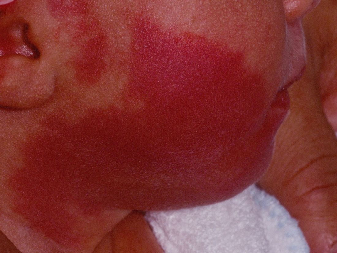
“The current modality of choice for the treatment of port wine birthmarks is pulsed dye laser,” Chelsea Grimes Fidai, MD, said during the annual conference of the American Society for Laser Medicine and Surgery. “When performed by a highly trained expert at efficient frequencies, PDL is a safe, effective treatment that is successful in the majority of patients. We know that earlier treatment yields maximal clearance. However, just how early can you initiate treatment?”
To find out, Dr. Fidai, Roy G. Geronemus, MD, and colleagues at the Laser and Skin Surgery Center of New York, conducted a retrospective chart review of 39 infants with port wine birthmarks who were treated with a 595-nm PDL between 2015 and 2020 at the center. Of the 39 infants, the average age at first treatment was 18 days, with a range from 5 to 29 days. The youngest patient was born prematurely at 35 weeks’ gestation and presented for his first treatment even before his expected due date. Most (74%) had facial lesions with the remaining distributed on the trunk or extremities. The average number of treatments was 15 over the course of 15 months.
The initial settings chosen for facial lesions were a 10-mm spot size, a fluence of 8.0 J/cm2, and a 1.5-millisecond pulse duration. For body lesions, the typical initial settings were a 12-mm spot size, a fluence of 6.7 J/cm2, and 1.5-millisecond pulse duration. Corneal eye shields were placed for all cases with port wine birthmarks approaching the eyelid. “We do recommend a treatment interval of every 2-3 weeks, with longer intervals for patients of darker skin type until the child is 2 years old, at which time the interval is increased to every 3-6 months,” said Dr. Fidai.
Patients in the study experienced the expected short-term side effects of erythema, edema, purpura, and mild transient postinflammatory hyperpigmentation, but there were no cases of atrophy, scarring, infection, or permanent pigmentary change.
“Families seeking early treatment of port wine birthmarks can be reassured that it can be safely initiated within the first few days after birth,” Dr. Fidai concluded. “This procedure can be quickly and confidently performed as an in-office procedure without any complications. The early intervention allows for treatment without general anesthesia and it maximizes the chance of significant clearance as early in life as possible.”
During a question-and-answer session, the abstract section chair, Albert Wolkerstorfer, MD, PhD, expressed concern about the effect of PDL on developing infants. “We do repeated treatments at this young age without any type of anesthesia,” said Dr. Wolkerstorfer, a dermatologist at the Netherlands Institute for Pigment Disorders, department of dermatology, University of Amsterdam.
“Will that influence the development of the child, especially when I hear there might be 15 or 20 treatments done within the first year of life? I think this is a problem where we need to ask the experts in the field of pain management in children, like pediatric anesthesiologists, to find the right way, because I think that the results that you showed are fantastic. I don’t think we can achieve that at a later age, although there’s no direct comparison at this moment.”
Dr. Fidai said that she understood the concern, but pointed to a 2020 article by Dr. Geronemus and colleagues that assessed treatment tolerance and parental perspective of outpatient PDL treatment for port-wine birthmarks without general anesthesia in infants and toddlers. “The kids recover pretty quickly after the treatment,” she said. “There has never been any longstanding issue from the parents’ perspective.”
Dr. Fidai reported having no financial disclosures. Dr. Geronemus disclosed having financial conflicts with numerous device and pharmaceutical companies. Dr. Wolkerstorfer disclosed that he has received consulting fees from Lumenis and InCyte and equipment from Humeca and PerfAction Technologies. He has also received grant funding from Novartis and InCyte and he is a member of InCyte’s advisory board.
without any complications, results from a single-center study showed.
“The current modality of choice for the treatment of port wine birthmarks is pulsed dye laser,” Chelsea Grimes Fidai, MD, said during the annual conference of the American Society for Laser Medicine and Surgery. “When performed by a highly trained expert at efficient frequencies, PDL is a safe, effective treatment that is successful in the majority of patients. We know that earlier treatment yields maximal clearance. However, just how early can you initiate treatment?”
To find out, Dr. Fidai, Roy G. Geronemus, MD, and colleagues at the Laser and Skin Surgery Center of New York, conducted a retrospective chart review of 39 infants with port wine birthmarks who were treated with a 595-nm PDL between 2015 and 2020 at the center. Of the 39 infants, the average age at first treatment was 18 days, with a range from 5 to 29 days. The youngest patient was born prematurely at 35 weeks’ gestation and presented for his first treatment even before his expected due date. Most (74%) had facial lesions with the remaining distributed on the trunk or extremities. The average number of treatments was 15 over the course of 15 months.
The initial settings chosen for facial lesions were a 10-mm spot size, a fluence of 8.0 J/cm2, and a 1.5-millisecond pulse duration. For body lesions, the typical initial settings were a 12-mm spot size, a fluence of 6.7 J/cm2, and 1.5-millisecond pulse duration. Corneal eye shields were placed for all cases with port wine birthmarks approaching the eyelid. “We do recommend a treatment interval of every 2-3 weeks, with longer intervals for patients of darker skin type until the child is 2 years old, at which time the interval is increased to every 3-6 months,” said Dr. Fidai.
Patients in the study experienced the expected short-term side effects of erythema, edema, purpura, and mild transient postinflammatory hyperpigmentation, but there were no cases of atrophy, scarring, infection, or permanent pigmentary change.
“Families seeking early treatment of port wine birthmarks can be reassured that it can be safely initiated within the first few days after birth,” Dr. Fidai concluded. “This procedure can be quickly and confidently performed as an in-office procedure without any complications. The early intervention allows for treatment without general anesthesia and it maximizes the chance of significant clearance as early in life as possible.”
During a question-and-answer session, the abstract section chair, Albert Wolkerstorfer, MD, PhD, expressed concern about the effect of PDL on developing infants. “We do repeated treatments at this young age without any type of anesthesia,” said Dr. Wolkerstorfer, a dermatologist at the Netherlands Institute for Pigment Disorders, department of dermatology, University of Amsterdam.
“Will that influence the development of the child, especially when I hear there might be 15 or 20 treatments done within the first year of life? I think this is a problem where we need to ask the experts in the field of pain management in children, like pediatric anesthesiologists, to find the right way, because I think that the results that you showed are fantastic. I don’t think we can achieve that at a later age, although there’s no direct comparison at this moment.”
Dr. Fidai said that she understood the concern, but pointed to a 2020 article by Dr. Geronemus and colleagues that assessed treatment tolerance and parental perspective of outpatient PDL treatment for port-wine birthmarks without general anesthesia in infants and toddlers. “The kids recover pretty quickly after the treatment,” she said. “There has never been any longstanding issue from the parents’ perspective.”
Dr. Fidai reported having no financial disclosures. Dr. Geronemus disclosed having financial conflicts with numerous device and pharmaceutical companies. Dr. Wolkerstorfer disclosed that he has received consulting fees from Lumenis and InCyte and equipment from Humeca and PerfAction Technologies. He has also received grant funding from Novartis and InCyte and he is a member of InCyte’s advisory board.
without any complications, results from a single-center study showed.
“The current modality of choice for the treatment of port wine birthmarks is pulsed dye laser,” Chelsea Grimes Fidai, MD, said during the annual conference of the American Society for Laser Medicine and Surgery. “When performed by a highly trained expert at efficient frequencies, PDL is a safe, effective treatment that is successful in the majority of patients. We know that earlier treatment yields maximal clearance. However, just how early can you initiate treatment?”
To find out, Dr. Fidai, Roy G. Geronemus, MD, and colleagues at the Laser and Skin Surgery Center of New York, conducted a retrospective chart review of 39 infants with port wine birthmarks who were treated with a 595-nm PDL between 2015 and 2020 at the center. Of the 39 infants, the average age at first treatment was 18 days, with a range from 5 to 29 days. The youngest patient was born prematurely at 35 weeks’ gestation and presented for his first treatment even before his expected due date. Most (74%) had facial lesions with the remaining distributed on the trunk or extremities. The average number of treatments was 15 over the course of 15 months.
The initial settings chosen for facial lesions were a 10-mm spot size, a fluence of 8.0 J/cm2, and a 1.5-millisecond pulse duration. For body lesions, the typical initial settings were a 12-mm spot size, a fluence of 6.7 J/cm2, and 1.5-millisecond pulse duration. Corneal eye shields were placed for all cases with port wine birthmarks approaching the eyelid. “We do recommend a treatment interval of every 2-3 weeks, with longer intervals for patients of darker skin type until the child is 2 years old, at which time the interval is increased to every 3-6 months,” said Dr. Fidai.
Patients in the study experienced the expected short-term side effects of erythema, edema, purpura, and mild transient postinflammatory hyperpigmentation, but there were no cases of atrophy, scarring, infection, or permanent pigmentary change.
“Families seeking early treatment of port wine birthmarks can be reassured that it can be safely initiated within the first few days after birth,” Dr. Fidai concluded. “This procedure can be quickly and confidently performed as an in-office procedure without any complications. The early intervention allows for treatment without general anesthesia and it maximizes the chance of significant clearance as early in life as possible.”
During a question-and-answer session, the abstract section chair, Albert Wolkerstorfer, MD, PhD, expressed concern about the effect of PDL on developing infants. “We do repeated treatments at this young age without any type of anesthesia,” said Dr. Wolkerstorfer, a dermatologist at the Netherlands Institute for Pigment Disorders, department of dermatology, University of Amsterdam.
“Will that influence the development of the child, especially when I hear there might be 15 or 20 treatments done within the first year of life? I think this is a problem where we need to ask the experts in the field of pain management in children, like pediatric anesthesiologists, to find the right way, because I think that the results that you showed are fantastic. I don’t think we can achieve that at a later age, although there’s no direct comparison at this moment.”
Dr. Fidai said that she understood the concern, but pointed to a 2020 article by Dr. Geronemus and colleagues that assessed treatment tolerance and parental perspective of outpatient PDL treatment for port-wine birthmarks without general anesthesia in infants and toddlers. “The kids recover pretty quickly after the treatment,” she said. “There has never been any longstanding issue from the parents’ perspective.”
Dr. Fidai reported having no financial disclosures. Dr. Geronemus disclosed having financial conflicts with numerous device and pharmaceutical companies. Dr. Wolkerstorfer disclosed that he has received consulting fees from Lumenis and InCyte and equipment from Humeca and PerfAction Technologies. He has also received grant funding from Novartis and InCyte and he is a member of InCyte’s advisory board.
FROM ASLMS 2021