User login
Death and dying
Learning about death and dying in residency is perhaps one of the hardest things about being a doctor. ... This list might make it a tiny bit easier.
1. The Hippocratic oath challenges us to be teachers not only to students, but also to our patients – especially to our dying patients. Remember you are teachers.
2. Nurses spend the most time with your patients; utilize them and learn from them. Nurses will make you better teachers and physicians.
3. When a patient is dying, hold that person’s hand if family isn’t around. Touch your patient.
4. Accompany your residents, fellows, or attending physicians to deliver bad news.
5. Ask to debrief after a patient dies.

6. While discussing treatment or prognosis with families, pay attention to the emotional data in the room. Tears should prompt tissues or hand holding. A wrinkled forehead or fearful face warrants a pause in conversation for the family to absorb the content. Tears don’t mean to hurry.
7. Attempt to understand your patient’s expectations. In other words, learn what the family’s goals are for treatment.
8. When a patient dies, reflect upon it in moderation. Don’t just move on to the next task. It’s okay to feel and to grieve … you are human.
9. When families declare DNR [do not resuscitate], don’t ask them repeatedly if they are sure. Also, make certain that they know they can change their minds at any time. Respect a patient’s values and wishes.
10. Take the time to have the difficult conversations early in diagnosis so families know what to expect. This empowers decisions that are proactive as opposed to reactive. Be proactive in your role as a teacher.
11. When discussing options, talk about what you CAN do before you talk about what you CAN’T do.
12. Withdrawing treatment and forgoing treatment are the same thing ethically and legally, but not emotionally.
13. When your patient is actively dying, see it out. Stay with the family, even if it’s time to go home.
14. When the family prays, be present.
15. Palliative care is neither hospice nor social work.
16. Ask your dying patients if they are afraid of anything – if the moment presents itself. Answer questions about death or find someone who can answer their questions.
17. No patient should die in pain. The doctrine of double effect allows a caregiver to provide medication for pain even though it may hasten death.
18. For the dying child, ask the family where they want their child to die. Don’t assume the family wants the death of their child to be at home or in the hospital. Ask.
19. Transitioning from treatment-oriented management to comfort care does not ever mean “there is nothing we can do.”
20. Lastly, and most importantly, I leave you with a question that should be in the back of our minds regarding quality of life and treatment options: Just because we can, should we?
Dr. Morvant is a second-year pediatric resident at Monroe Carell Jr. Children’s Hospital at Vanderbilt, Nashville, Tenn. E-mail her at pdnews@frontlinemedcom.com.
Learning about death and dying in residency is perhaps one of the hardest things about being a doctor. ... This list might make it a tiny bit easier.
1. The Hippocratic oath challenges us to be teachers not only to students, but also to our patients – especially to our dying patients. Remember you are teachers.
2. Nurses spend the most time with your patients; utilize them and learn from them. Nurses will make you better teachers and physicians.
3. When a patient is dying, hold that person’s hand if family isn’t around. Touch your patient.
4. Accompany your residents, fellows, or attending physicians to deliver bad news.
5. Ask to debrief after a patient dies.
6. While discussing treatment or prognosis with families, pay attention to the emotional data in the room. Tears should prompt tissues or hand holding. A wrinkled forehead or fearful face warrants a pause in conversation for the family to absorb the content. Tears don’t mean to hurry.
7. Attempt to understand your patient’s expectations. In other words, learn what the family’s goals are for treatment.
8. When a patient dies, reflect upon it in moderation. Don’t just move on to the next task. It’s okay to feel and to grieve … you are human.
9. When families declare DNR [do not resuscitate], don’t ask them repeatedly if they are sure. Also, make certain that they know they can change their minds at any time. Respect a patient’s values and wishes.
10. Take the time to have the difficult conversations early in diagnosis so families know what to expect. This empowers decisions that are proactive as opposed to reactive. Be proactive in your role as a teacher.
11. When discussing options, talk about what you CAN do before you talk about what you CAN’T do.
12. Withdrawing treatment and forgoing treatment are the same thing ethically and legally, but not emotionally.
13. When your patient is actively dying, see it out. Stay with the family, even if it’s time to go home.
14. When the family prays, be present.
15. Palliative care is neither hospice nor social work.
16. Ask your dying patients if they are afraid of anything – if the moment presents itself. Answer questions about death or find someone who can answer their questions.
17. No patient should die in pain. The doctrine of double effect allows a caregiver to provide medication for pain even though it may hasten death.
18. For the dying child, ask the family where they want their child to die. Don’t assume the family wants the death of their child to be at home or in the hospital. Ask.
19. Transitioning from treatment-oriented management to comfort care does not ever mean “there is nothing we can do.”
20. Lastly, and most importantly, I leave you with a question that should be in the back of our minds regarding quality of life and treatment options: Just because we can, should we?
Dr. Morvant is a second-year pediatric resident at Monroe Carell Jr. Children’s Hospital at Vanderbilt, Nashville, Tenn. E-mail her at pdnews@frontlinemedcom.com.
Learning about death and dying in residency is perhaps one of the hardest things about being a doctor. ... This list might make it a tiny bit easier.
1. The Hippocratic oath challenges us to be teachers not only to students, but also to our patients – especially to our dying patients. Remember you are teachers.
2. Nurses spend the most time with your patients; utilize them and learn from them. Nurses will make you better teachers and physicians.
3. When a patient is dying, hold that person’s hand if family isn’t around. Touch your patient.
4. Accompany your residents, fellows, or attending physicians to deliver bad news.
5. Ask to debrief after a patient dies.
6. While discussing treatment or prognosis with families, pay attention to the emotional data in the room. Tears should prompt tissues or hand holding. A wrinkled forehead or fearful face warrants a pause in conversation for the family to absorb the content. Tears don’t mean to hurry.
7. Attempt to understand your patient’s expectations. In other words, learn what the family’s goals are for treatment.
8. When a patient dies, reflect upon it in moderation. Don’t just move on to the next task. It’s okay to feel and to grieve … you are human.
9. When families declare DNR [do not resuscitate], don’t ask them repeatedly if they are sure. Also, make certain that they know they can change their minds at any time. Respect a patient’s values and wishes.
10. Take the time to have the difficult conversations early in diagnosis so families know what to expect. This empowers decisions that are proactive as opposed to reactive. Be proactive in your role as a teacher.
11. When discussing options, talk about what you CAN do before you talk about what you CAN’T do.
12. Withdrawing treatment and forgoing treatment are the same thing ethically and legally, but not emotionally.
13. When your patient is actively dying, see it out. Stay with the family, even if it’s time to go home.
14. When the family prays, be present.
15. Palliative care is neither hospice nor social work.
16. Ask your dying patients if they are afraid of anything – if the moment presents itself. Answer questions about death or find someone who can answer their questions.
17. No patient should die in pain. The doctrine of double effect allows a caregiver to provide medication for pain even though it may hasten death.
18. For the dying child, ask the family where they want their child to die. Don’t assume the family wants the death of their child to be at home or in the hospital. Ask.
19. Transitioning from treatment-oriented management to comfort care does not ever mean “there is nothing we can do.”
20. Lastly, and most importantly, I leave you with a question that should be in the back of our minds regarding quality of life and treatment options: Just because we can, should we?
Dr. Morvant is a second-year pediatric resident at Monroe Carell Jr. Children’s Hospital at Vanderbilt, Nashville, Tenn. E-mail her at pdnews@frontlinemedcom.com.
Seeing the ob.gyn. field through a med student’s eyes
Each fall, as part of their residency applications, medical students around the country struggle to describe the reasons they have picked their chosen specialty. We all have a sense of what attracts us to a field, or to anything for that matter, and it is often difficult to put into words. That is why the personal essay, as a part of the application, is so challenging and often so bland.

Occasionally though, we are privileged to read an essay that connects with the deeper motivations that lead us to choose the paths we follow. When we read such an essay, it can serve to refresh and replenish the idealism that we all have and that is sometimes a struggle to maintain amid concerns about ACOs, RVUs, EHRs, ICD-10, and the rest of the alphabet soup that requires attention and effort, but that can distract us from our core mission of caring for patients and block us from participating in the wonder and majesty that such care can bring.
This is such an essay, and to read it is like drinking a refreshing glass of water on a hot day.
BY ALIZA MACHEFSKY
In Judaism, a baby is named within the context of a prayer for the mother and child’s health: “May He who blessed our fathers ... bless the woman who has given birth (mother’s name) together with the daughter who was born to her in an auspicious time, her name shall be called in Israel: (child’s name) ...” This prayer simultaneously acknowledges the risks and the possibility of labor – on one side the birth of a new life and continued health of the mother; on the other side the possibility of death and sadness.
Family planning and safe delivery impact not only the individual woman and her family but also society as a whole. The United Nations prioritized this concern in their fifth Millennium Development Goal of improving maternal health and decreasing maternal mortality by increasing access to reproductive health services, family planning, and skilled antenatal care in the developing world.

The obstetrician delivering a baby practices medicine at the edge of opposites: the promise of new life and the possibility of impending death; the end of intrauterine existence for the fetus and the beginning of life outside the womb; the end of singlehood and the beginning of parenthood. All physicians are privy to vulnerable aspects of their patients’ lives, all will see them naked and listen to their fears, but it is rare to share in the joy that comes from shepherding a successful pregnancy and guiding the simultaneously joyous and terrifying event of birth. The assisting physician operates at the place where scientific knowledge and medical skills meet the mystery of human existence.
What draws me to the field of obstetrics and gynecology is the unique combination of being the physician who can provide counseling and primary care throughout a woman’s reproductive and postmenopausal life, while at the same time provide surgical and interventional procedures at critical times in a woman’s life. Ob.gyns. straddle the best possibilities medicine has to offer.
What I find so appealing about ob.gyn. – what had me scuttling off to the labor and delivery floor during slow afternoons on internal medicine, or requesting opportunities to have more exposure to the ob.gyn. subspecialties – is how unique, cutting edge, and imperative each aspect of ob.gyn. and its subspecialties are.
On the labor and delivery floor, I felt privileged to be a part of the birth and safe delivery of a new life. While working with the reproductive endocrinology and infertility team, I saw physicians help infertile patients have a chance of beginning a family. On urogynecology, I witnessed a skilled surgeon perform a precise procedure that enabled a marathoner to race again without fear of urinary incontinence. Rotating through gynecology-oncology, I saw the combination of cutting edge robotic surgery and compassionate care drastically improve the lives of oncology patients. While watching anatomy scans in the maternal-fetal medicine clinic, I was in awe of the beating chambers of each baby’s heart. And at the vaginitis clinic I learned how careful questioning, complete exams, and meticulous study of specimens could reveal vulvar and vaginal pathology that is often overlooked, but when discovered can have a huge impact on quality of life.
I want to be an ob.gyn. who has the capacity to council and teach women about their own health care and to provide comfort and calm during the excitement, anxiety, and pain of birth. But I also want to have the knowledge and skills to make difficult decisions, the capacity to recognize what needs to be done in times of crisis, and the surgical skills to intervene in the right way. I look forward to being a part of a specialty that is filled with compassionate learning, teamwork, and empathy worthy of the women who seek our care.
Ms. Machefsky is a fourth-year medical student at Drexel University in Philadelphia and a Gold Humanism Honor Society member. She recently welcomed the birth of her first child. Dr. Skolnick is a professor of family and community medicine at Temple University in Philadelphia and associate director of the Family Medicine Residency Program at Abington Jefferson Health.
Each fall, as part of their residency applications, medical students around the country struggle to describe the reasons they have picked their chosen specialty. We all have a sense of what attracts us to a field, or to anything for that matter, and it is often difficult to put into words. That is why the personal essay, as a part of the application, is so challenging and often so bland.
Occasionally though, we are privileged to read an essay that connects with the deeper motivations that lead us to choose the paths we follow. When we read such an essay, it can serve to refresh and replenish the idealism that we all have and that is sometimes a struggle to maintain amid concerns about ACOs, RVUs, EHRs, ICD-10, and the rest of the alphabet soup that requires attention and effort, but that can distract us from our core mission of caring for patients and block us from participating in the wonder and majesty that such care can bring.
This is such an essay, and to read it is like drinking a refreshing glass of water on a hot day.
BY ALIZA MACHEFSKY
In Judaism, a baby is named within the context of a prayer for the mother and child’s health: “May He who blessed our fathers ... bless the woman who has given birth (mother’s name) together with the daughter who was born to her in an auspicious time, her name shall be called in Israel: (child’s name) ...” This prayer simultaneously acknowledges the risks and the possibility of labor – on one side the birth of a new life and continued health of the mother; on the other side the possibility of death and sadness.
Family planning and safe delivery impact not only the individual woman and her family but also society as a whole. The United Nations prioritized this concern in their fifth Millennium Development Goal of improving maternal health and decreasing maternal mortality by increasing access to reproductive health services, family planning, and skilled antenatal care in the developing world.
The obstetrician delivering a baby practices medicine at the edge of opposites: the promise of new life and the possibility of impending death; the end of intrauterine existence for the fetus and the beginning of life outside the womb; the end of singlehood and the beginning of parenthood. All physicians are privy to vulnerable aspects of their patients’ lives, all will see them naked and listen to their fears, but it is rare to share in the joy that comes from shepherding a successful pregnancy and guiding the simultaneously joyous and terrifying event of birth. The assisting physician operates at the place where scientific knowledge and medical skills meet the mystery of human existence.
What draws me to the field of obstetrics and gynecology is the unique combination of being the physician who can provide counseling and primary care throughout a woman’s reproductive and postmenopausal life, while at the same time provide surgical and interventional procedures at critical times in a woman’s life. Ob.gyns. straddle the best possibilities medicine has to offer.
What I find so appealing about ob.gyn. – what had me scuttling off to the labor and delivery floor during slow afternoons on internal medicine, or requesting opportunities to have more exposure to the ob.gyn. subspecialties – is how unique, cutting edge, and imperative each aspect of ob.gyn. and its subspecialties are.
On the labor and delivery floor, I felt privileged to be a part of the birth and safe delivery of a new life. While working with the reproductive endocrinology and infertility team, I saw physicians help infertile patients have a chance of beginning a family. On urogynecology, I witnessed a skilled surgeon perform a precise procedure that enabled a marathoner to race again without fear of urinary incontinence. Rotating through gynecology-oncology, I saw the combination of cutting edge robotic surgery and compassionate care drastically improve the lives of oncology patients. While watching anatomy scans in the maternal-fetal medicine clinic, I was in awe of the beating chambers of each baby’s heart. And at the vaginitis clinic I learned how careful questioning, complete exams, and meticulous study of specimens could reveal vulvar and vaginal pathology that is often overlooked, but when discovered can have a huge impact on quality of life.
I want to be an ob.gyn. who has the capacity to council and teach women about their own health care and to provide comfort and calm during the excitement, anxiety, and pain of birth. But I also want to have the knowledge and skills to make difficult decisions, the capacity to recognize what needs to be done in times of crisis, and the surgical skills to intervene in the right way. I look forward to being a part of a specialty that is filled with compassionate learning, teamwork, and empathy worthy of the women who seek our care.
Ms. Machefsky is a fourth-year medical student at Drexel University in Philadelphia and a Gold Humanism Honor Society member. She recently welcomed the birth of her first child. Dr. Skolnick is a professor of family and community medicine at Temple University in Philadelphia and associate director of the Family Medicine Residency Program at Abington Jefferson Health.
Each fall, as part of their residency applications, medical students around the country struggle to describe the reasons they have picked their chosen specialty. We all have a sense of what attracts us to a field, or to anything for that matter, and it is often difficult to put into words. That is why the personal essay, as a part of the application, is so challenging and often so bland.
Occasionally though, we are privileged to read an essay that connects with the deeper motivations that lead us to choose the paths we follow. When we read such an essay, it can serve to refresh and replenish the idealism that we all have and that is sometimes a struggle to maintain amid concerns about ACOs, RVUs, EHRs, ICD-10, and the rest of the alphabet soup that requires attention and effort, but that can distract us from our core mission of caring for patients and block us from participating in the wonder and majesty that such care can bring.
This is such an essay, and to read it is like drinking a refreshing glass of water on a hot day.
BY ALIZA MACHEFSKY
In Judaism, a baby is named within the context of a prayer for the mother and child’s health: “May He who blessed our fathers ... bless the woman who has given birth (mother’s name) together with the daughter who was born to her in an auspicious time, her name shall be called in Israel: (child’s name) ...” This prayer simultaneously acknowledges the risks and the possibility of labor – on one side the birth of a new life and continued health of the mother; on the other side the possibility of death and sadness.
Family planning and safe delivery impact not only the individual woman and her family but also society as a whole. The United Nations prioritized this concern in their fifth Millennium Development Goal of improving maternal health and decreasing maternal mortality by increasing access to reproductive health services, family planning, and skilled antenatal care in the developing world.
The obstetrician delivering a baby practices medicine at the edge of opposites: the promise of new life and the possibility of impending death; the end of intrauterine existence for the fetus and the beginning of life outside the womb; the end of singlehood and the beginning of parenthood. All physicians are privy to vulnerable aspects of their patients’ lives, all will see them naked and listen to their fears, but it is rare to share in the joy that comes from shepherding a successful pregnancy and guiding the simultaneously joyous and terrifying event of birth. The assisting physician operates at the place where scientific knowledge and medical skills meet the mystery of human existence.
What draws me to the field of obstetrics and gynecology is the unique combination of being the physician who can provide counseling and primary care throughout a woman’s reproductive and postmenopausal life, while at the same time provide surgical and interventional procedures at critical times in a woman’s life. Ob.gyns. straddle the best possibilities medicine has to offer.
What I find so appealing about ob.gyn. – what had me scuttling off to the labor and delivery floor during slow afternoons on internal medicine, or requesting opportunities to have more exposure to the ob.gyn. subspecialties – is how unique, cutting edge, and imperative each aspect of ob.gyn. and its subspecialties are.
On the labor and delivery floor, I felt privileged to be a part of the birth and safe delivery of a new life. While working with the reproductive endocrinology and infertility team, I saw physicians help infertile patients have a chance of beginning a family. On urogynecology, I witnessed a skilled surgeon perform a precise procedure that enabled a marathoner to race again without fear of urinary incontinence. Rotating through gynecology-oncology, I saw the combination of cutting edge robotic surgery and compassionate care drastically improve the lives of oncology patients. While watching anatomy scans in the maternal-fetal medicine clinic, I was in awe of the beating chambers of each baby’s heart. And at the vaginitis clinic I learned how careful questioning, complete exams, and meticulous study of specimens could reveal vulvar and vaginal pathology that is often overlooked, but when discovered can have a huge impact on quality of life.
I want to be an ob.gyn. who has the capacity to council and teach women about their own health care and to provide comfort and calm during the excitement, anxiety, and pain of birth. But I also want to have the knowledge and skills to make difficult decisions, the capacity to recognize what needs to be done in times of crisis, and the surgical skills to intervene in the right way. I look forward to being a part of a specialty that is filled with compassionate learning, teamwork, and empathy worthy of the women who seek our care.
Ms. Machefsky is a fourth-year medical student at Drexel University in Philadelphia and a Gold Humanism Honor Society member. She recently welcomed the birth of her first child. Dr. Skolnick is a professor of family and community medicine at Temple University in Philadelphia and associate director of the Family Medicine Residency Program at Abington Jefferson Health.
Carnosine
A powerful endogenous antioxidant found most abundantly in mammalian tissues, especially brain and skeletal muscle tissue, carnosine is a dipeptide of alanine and histidine.1,2,3,4,5.
Carnosine was first isolated in 1900 by the Russian scientist Gulewitsch as a substance extracted from muscle tissue.6,4. L-carnosine (beta-alanyl-L-histidine) is the synthetic version identical to the natural form alpha-alanyl-L-histidine.7 Carnosine has long been reputed to confer immunomodulating, wound healing, antiglycating, and antineoplastic effects.2 Several reports have shown that carnosine can accelerate the healing of surface skin wounds and burns.4,8
Wound healing
An early study by Nagai et al. in 1986 on carnosine in wound healing showed that rats treated locally with carnosine exhibited greater tensile skin strength at an incision site after hydrocortisone had been administered to hinder healing. The investigators concluded that carnosine bolsters wound healing by stimulating early effusion by histamine and of collagen biosynthesis by beta-alanine. They also found that the compound significantly augmented granulation inhibited by cortisone, mitomycin C, 5-fluorouracil, and bleomycin.9
Studies by Fitzpatrick and Fisher in the early 1980s revealed that carnosine acts as a histidine reserve in relation to histamine production during trauma, suggesting a role for carnosine in wound healing.10,11
In 2012, Ansurudeen et al. examined the effects of carnosine in wound healing in a diabetic mouse model. Carnosine was applied locally and injected daily, yielding significant amelioration in wound healing, with analysis revealing elevated expression of growth factors and cytokines implicated in wound healing. The investigators also observed that carnosine supported cell viability in the presence of high glucose in human dermal fibroblasts and microvascular endothelial cells in vitro.2
Other findings with implications for cutaneous therapy
In 2006, Babizhayev reported that the L-carnosine-related peptidomimetic N-acetylcarnosine (N-acetyl-beta-alanyl-L-histidine) can act as a timed-release (carrier) stable version of L-carnosine in cosmetic preparations, including lubricants.6 Babizhayev et al. have since claimed that they have developed a technology using imidazole-containing dipeptide-based compounds (including L-carnosine and derivatives) that enhances protein hydration in photoaged skin.12,13,14
A double-blind comparative study conducted by Dieamant et al. in 2008 in 124 volunteers with sensitive skin aimed to evaluate the therapeutic potential of the combination of the antioxidant L-carnosine and neuromodulatory Rhodiola rosea. For 28 days, the groups of 62 received twice-daily applications of the 1% combination formulation or placebo. Skin barrier function (reduction of transepidermal water loss) improved in the treatment group, and favorable subjective responses regarding skin dryness were reported. Discomfort after the stinging test was also reduced. In vitro results showed that the release of proopiomelanocortin peptides was spurred by treatment, with the elevated levels of neuropeptides and cytokines produced by keratinocytes exposed to UV radiation returning to normal.15
Two years later, Renner et al. showed that carnosine hindered tumor growth in vivo in an NIH3T3-HER2/neu mouse model. They contended that this naturally occurring dipeptide warrants increased consideration and study for its potential as an anticancer agent.16
In 2012, Federici et al. conducted a randomized, evaluator-blinded, controlled comparative trial over 1month to assess the efficacy of twice-daily topical urea 5% with arginine and carnosine (Ureadin Rx) as compared with twice-daily application of a glycerol-based emollient topical product (Dexeryl) in treating xerosis in 40 type 2 diabetes patients (40-75 years of age). Use of the carnosine-containing formulation yielded significantly greater hydration and an 89% decline in Dryness Areas Severity Index (DASI) scores, compared with baseline. The DASI score after 4 weeks of treatment was much lower in the treatment group than the control group. The Visual Analog Scale (VAS) score was also significantly higher in the Ureadin group than the Dexeryl group. The investigators concluded that the topical application of a urea 5%, arginine, and carnosine cream enhances skin hydration and relieves dryness in type 2 diabetic patients in comparison with a control glycerol-based emollient formulation.17
Antiaging potential
In 1993, Reeve et al. showed that dietary or topically applied carnosine potentiated the contact hypersensitivity reaction in hairless mice and prevented the systemic inhibition of this reaction after dorsal skin exposure to UVB. Carnosine was found to also prevent the systemic suppression provoked by the topical application of a lotion containing cis-urocanic acid.3
Carnosine was a key active ingredient in antiaging products evaluated by Kaczvinsky et al. in 2009 in two double-blind, randomized, controlled, split-face studies. The researchers used the Fast Optical in vivo Topometry of Human Skin (FOITS) technique to measure changes in periorbital wrinkles in the two studies in women between the ages of 30 and 70 years old (study 1, n = 42; study 2, n = 35). They reported that 4 weeks of treatment with the test products, which contained niacinamide, the peptides Pal-KT and Pal-KTTKS, and carnosine, ameliorated periorbital skin, enhancing smoothness and diminishing larger wrinkle depth.18
In 2012, Babizhayev et al. conducted a 4-month randomized, double-blind, controlled study with 42 subjects to evaluate the effects on skin aging of oral nonhydrolyzed carnosine (Can-C Plus formulation). Skin parameters exhibited a consistent and significant improvement during 3 months of supplementation in the treatment group, compared with the placebo group, with overall skin appearance enhanced and fine lines diminished based on visual inspection. There were no reports of adverse effects. The investigators concluded that supplementation with nonhydrolyzed carnosine or carcinine in patented oral formulations has potential as an agent for antiaging purposes.19
Two years later, Emanuele et al. conducted an experimental double-blind irradiation study to compare a complex novel topical product (TPF50) consisting of three active ingredients (traditional physical sunscreens, SPF 50; a liposome-encapsulated DNA repair enzymes complex – photolyase, endonuclease, and 8-oxoguanine glycosylase [OGG1]; and a robust antioxidant complex containing carnosine, arazine, and ergothionine) to available DNA repair and antioxidant and growth factor topical products. They found that the new topical agent was the most effective product in reducing three molecular markers (cyclobutane pyrimidine dimers, protein carbonylation, and 8-oxo-7,8-dihydro-2’-deoxyguanosine) in human skin biopsies. The researchers concluded that the carnosine-containing formulation enhances the genomic and proteomic integrity of skin cells after continual UV exposure, suggesting its potential efficacy in lowering the risk of UV-induced cutaneous aging and nonmelanoma skin cancer.20
Conclusion
Carnosine is an intriguing compound with well-documented antioxidant and wound healing activity. While more research is necessary to determine its wider applications in dermatology, recent work in formulating topical products to impart antiaging effects appears to show promise.
References
1. Nutr. Res. Pract. 2011;5:421-8.
2. Amino Acids 2012;43:127-34.
4. Mol. Aspects Med. 1992;13:379-444.
5. Am. J. Ther. 2012;19:e69-89.
7. J. Cosmet. Dermatol. 2004;3:26-34.
8. Nihon Yakurigaku Zasshi. 1992;100:165-72.
12. Int. J. Cosmet. Sci. 2011;33:1-16.
13. Crit. Rev. Ther. Drug Carrier Syst. 2011;28:203-53.
14. Crit. Rev. Ther. Drug Carrier Syst. 2010;27:85-154.
15. J. Cosmet. Dermatol. 2008;7:112-9.
18. J. Cosmet. Dermatol. 2009;8:228-33.
19. J. Dermatolog. Treat. 2012;23:345-84.
20. J. Drugs Dermatol. 2014;13:309-14.
Dr. Baumann is chief executive officer of the Baumann Cosmetic & Research Institute in the Design District in Miami. She founded the Cosmetic Dermatology Center at the University of Miami in 1997. Dr. Baumann wrote the textbook, “Cosmetic Dermatology: Principles and Practice” (New York: McGraw-Hill, 2002), and a book for consumers, “The Skin Type Solution” (New York: Bantam Dell, 2006). She has contributed to the Cosmeceutical Critique column in Dermatology News since January 2001. Her latest book, “Cosmeceuticals and Cosmetic Ingredients,” was published in November 2014. Dr. Baumann has received funding for clinical grants from Allergan, Aveeno, Avon Products, Evolus, Galderma, GlaxoSmithKline, Kythera Biopharmaceuticals, Mary Kay, Medicis Pharmaceuticals, Neutrogena, Philosophy, Topix Pharmaceuticals, and Unilever.
A powerful endogenous antioxidant found most abundantly in mammalian tissues, especially brain and skeletal muscle tissue, carnosine is a dipeptide of alanine and histidine.1,2,3,4,5.
Carnosine was first isolated in 1900 by the Russian scientist Gulewitsch as a substance extracted from muscle tissue.6,4. L-carnosine (beta-alanyl-L-histidine) is the synthetic version identical to the natural form alpha-alanyl-L-histidine.7 Carnosine has long been reputed to confer immunomodulating, wound healing, antiglycating, and antineoplastic effects.2 Several reports have shown that carnosine can accelerate the healing of surface skin wounds and burns.4,8
Wound healing
An early study by Nagai et al. in 1986 on carnosine in wound healing showed that rats treated locally with carnosine exhibited greater tensile skin strength at an incision site after hydrocortisone had been administered to hinder healing. The investigators concluded that carnosine bolsters wound healing by stimulating early effusion by histamine and of collagen biosynthesis by beta-alanine. They also found that the compound significantly augmented granulation inhibited by cortisone, mitomycin C, 5-fluorouracil, and bleomycin.9
Studies by Fitzpatrick and Fisher in the early 1980s revealed that carnosine acts as a histidine reserve in relation to histamine production during trauma, suggesting a role for carnosine in wound healing.10,11
In 2012, Ansurudeen et al. examined the effects of carnosine in wound healing in a diabetic mouse model. Carnosine was applied locally and injected daily, yielding significant amelioration in wound healing, with analysis revealing elevated expression of growth factors and cytokines implicated in wound healing. The investigators also observed that carnosine supported cell viability in the presence of high glucose in human dermal fibroblasts and microvascular endothelial cells in vitro.2
Other findings with implications for cutaneous therapy
In 2006, Babizhayev reported that the L-carnosine-related peptidomimetic N-acetylcarnosine (N-acetyl-beta-alanyl-L-histidine) can act as a timed-release (carrier) stable version of L-carnosine in cosmetic preparations, including lubricants.6 Babizhayev et al. have since claimed that they have developed a technology using imidazole-containing dipeptide-based compounds (including L-carnosine and derivatives) that enhances protein hydration in photoaged skin.12,13,14
A double-blind comparative study conducted by Dieamant et al. in 2008 in 124 volunteers with sensitive skin aimed to evaluate the therapeutic potential of the combination of the antioxidant L-carnosine and neuromodulatory Rhodiola rosea. For 28 days, the groups of 62 received twice-daily applications of the 1% combination formulation or placebo. Skin barrier function (reduction of transepidermal water loss) improved in the treatment group, and favorable subjective responses regarding skin dryness were reported. Discomfort after the stinging test was also reduced. In vitro results showed that the release of proopiomelanocortin peptides was spurred by treatment, with the elevated levels of neuropeptides and cytokines produced by keratinocytes exposed to UV radiation returning to normal.15
Two years later, Renner et al. showed that carnosine hindered tumor growth in vivo in an NIH3T3-HER2/neu mouse model. They contended that this naturally occurring dipeptide warrants increased consideration and study for its potential as an anticancer agent.16
In 2012, Federici et al. conducted a randomized, evaluator-blinded, controlled comparative trial over 1month to assess the efficacy of twice-daily topical urea 5% with arginine and carnosine (Ureadin Rx) as compared with twice-daily application of a glycerol-based emollient topical product (Dexeryl) in treating xerosis in 40 type 2 diabetes patients (40-75 years of age). Use of the carnosine-containing formulation yielded significantly greater hydration and an 89% decline in Dryness Areas Severity Index (DASI) scores, compared with baseline. The DASI score after 4 weeks of treatment was much lower in the treatment group than the control group. The Visual Analog Scale (VAS) score was also significantly higher in the Ureadin group than the Dexeryl group. The investigators concluded that the topical application of a urea 5%, arginine, and carnosine cream enhances skin hydration and relieves dryness in type 2 diabetic patients in comparison with a control glycerol-based emollient formulation.17
Antiaging potential
In 1993, Reeve et al. showed that dietary or topically applied carnosine potentiated the contact hypersensitivity reaction in hairless mice and prevented the systemic inhibition of this reaction after dorsal skin exposure to UVB. Carnosine was found to also prevent the systemic suppression provoked by the topical application of a lotion containing cis-urocanic acid.3
Carnosine was a key active ingredient in antiaging products evaluated by Kaczvinsky et al. in 2009 in two double-blind, randomized, controlled, split-face studies. The researchers used the Fast Optical in vivo Topometry of Human Skin (FOITS) technique to measure changes in periorbital wrinkles in the two studies in women between the ages of 30 and 70 years old (study 1, n = 42; study 2, n = 35). They reported that 4 weeks of treatment with the test products, which contained niacinamide, the peptides Pal-KT and Pal-KTTKS, and carnosine, ameliorated periorbital skin, enhancing smoothness and diminishing larger wrinkle depth.18
In 2012, Babizhayev et al. conducted a 4-month randomized, double-blind, controlled study with 42 subjects to evaluate the effects on skin aging of oral nonhydrolyzed carnosine (Can-C Plus formulation). Skin parameters exhibited a consistent and significant improvement during 3 months of supplementation in the treatment group, compared with the placebo group, with overall skin appearance enhanced and fine lines diminished based on visual inspection. There were no reports of adverse effects. The investigators concluded that supplementation with nonhydrolyzed carnosine or carcinine in patented oral formulations has potential as an agent for antiaging purposes.19
Two years later, Emanuele et al. conducted an experimental double-blind irradiation study to compare a complex novel topical product (TPF50) consisting of three active ingredients (traditional physical sunscreens, SPF 50; a liposome-encapsulated DNA repair enzymes complex – photolyase, endonuclease, and 8-oxoguanine glycosylase [OGG1]; and a robust antioxidant complex containing carnosine, arazine, and ergothionine) to available DNA repair and antioxidant and growth factor topical products. They found that the new topical agent was the most effective product in reducing three molecular markers (cyclobutane pyrimidine dimers, protein carbonylation, and 8-oxo-7,8-dihydro-2’-deoxyguanosine) in human skin biopsies. The researchers concluded that the carnosine-containing formulation enhances the genomic and proteomic integrity of skin cells after continual UV exposure, suggesting its potential efficacy in lowering the risk of UV-induced cutaneous aging and nonmelanoma skin cancer.20
Conclusion
Carnosine is an intriguing compound with well-documented antioxidant and wound healing activity. While more research is necessary to determine its wider applications in dermatology, recent work in formulating topical products to impart antiaging effects appears to show promise.
References
1. Nutr. Res. Pract. 2011;5:421-8.
2. Amino Acids 2012;43:127-34.
4. Mol. Aspects Med. 1992;13:379-444.
5. Am. J. Ther. 2012;19:e69-89.
7. J. Cosmet. Dermatol. 2004;3:26-34.
8. Nihon Yakurigaku Zasshi. 1992;100:165-72.
12. Int. J. Cosmet. Sci. 2011;33:1-16.
13. Crit. Rev. Ther. Drug Carrier Syst. 2011;28:203-53.
14. Crit. Rev. Ther. Drug Carrier Syst. 2010;27:85-154.
15. J. Cosmet. Dermatol. 2008;7:112-9.
18. J. Cosmet. Dermatol. 2009;8:228-33.
19. J. Dermatolog. Treat. 2012;23:345-84.
20. J. Drugs Dermatol. 2014;13:309-14.
Dr. Baumann is chief executive officer of the Baumann Cosmetic & Research Institute in the Design District in Miami. She founded the Cosmetic Dermatology Center at the University of Miami in 1997. Dr. Baumann wrote the textbook, “Cosmetic Dermatology: Principles and Practice” (New York: McGraw-Hill, 2002), and a book for consumers, “The Skin Type Solution” (New York: Bantam Dell, 2006). She has contributed to the Cosmeceutical Critique column in Dermatology News since January 2001. Her latest book, “Cosmeceuticals and Cosmetic Ingredients,” was published in November 2014. Dr. Baumann has received funding for clinical grants from Allergan, Aveeno, Avon Products, Evolus, Galderma, GlaxoSmithKline, Kythera Biopharmaceuticals, Mary Kay, Medicis Pharmaceuticals, Neutrogena, Philosophy, Topix Pharmaceuticals, and Unilever.
A powerful endogenous antioxidant found most abundantly in mammalian tissues, especially brain and skeletal muscle tissue, carnosine is a dipeptide of alanine and histidine.1,2,3,4,5.
Carnosine was first isolated in 1900 by the Russian scientist Gulewitsch as a substance extracted from muscle tissue.6,4. L-carnosine (beta-alanyl-L-histidine) is the synthetic version identical to the natural form alpha-alanyl-L-histidine.7 Carnosine has long been reputed to confer immunomodulating, wound healing, antiglycating, and antineoplastic effects.2 Several reports have shown that carnosine can accelerate the healing of surface skin wounds and burns.4,8
Wound healing
An early study by Nagai et al. in 1986 on carnosine in wound healing showed that rats treated locally with carnosine exhibited greater tensile skin strength at an incision site after hydrocortisone had been administered to hinder healing. The investigators concluded that carnosine bolsters wound healing by stimulating early effusion by histamine and of collagen biosynthesis by beta-alanine. They also found that the compound significantly augmented granulation inhibited by cortisone, mitomycin C, 5-fluorouracil, and bleomycin.9
Studies by Fitzpatrick and Fisher in the early 1980s revealed that carnosine acts as a histidine reserve in relation to histamine production during trauma, suggesting a role for carnosine in wound healing.10,11
In 2012, Ansurudeen et al. examined the effects of carnosine in wound healing in a diabetic mouse model. Carnosine was applied locally and injected daily, yielding significant amelioration in wound healing, with analysis revealing elevated expression of growth factors and cytokines implicated in wound healing. The investigators also observed that carnosine supported cell viability in the presence of high glucose in human dermal fibroblasts and microvascular endothelial cells in vitro.2
Other findings with implications for cutaneous therapy
In 2006, Babizhayev reported that the L-carnosine-related peptidomimetic N-acetylcarnosine (N-acetyl-beta-alanyl-L-histidine) can act as a timed-release (carrier) stable version of L-carnosine in cosmetic preparations, including lubricants.6 Babizhayev et al. have since claimed that they have developed a technology using imidazole-containing dipeptide-based compounds (including L-carnosine and derivatives) that enhances protein hydration in photoaged skin.12,13,14
A double-blind comparative study conducted by Dieamant et al. in 2008 in 124 volunteers with sensitive skin aimed to evaluate the therapeutic potential of the combination of the antioxidant L-carnosine and neuromodulatory Rhodiola rosea. For 28 days, the groups of 62 received twice-daily applications of the 1% combination formulation or placebo. Skin barrier function (reduction of transepidermal water loss) improved in the treatment group, and favorable subjective responses regarding skin dryness were reported. Discomfort after the stinging test was also reduced. In vitro results showed that the release of proopiomelanocortin peptides was spurred by treatment, with the elevated levels of neuropeptides and cytokines produced by keratinocytes exposed to UV radiation returning to normal.15
Two years later, Renner et al. showed that carnosine hindered tumor growth in vivo in an NIH3T3-HER2/neu mouse model. They contended that this naturally occurring dipeptide warrants increased consideration and study for its potential as an anticancer agent.16
In 2012, Federici et al. conducted a randomized, evaluator-blinded, controlled comparative trial over 1month to assess the efficacy of twice-daily topical urea 5% with arginine and carnosine (Ureadin Rx) as compared with twice-daily application of a glycerol-based emollient topical product (Dexeryl) in treating xerosis in 40 type 2 diabetes patients (40-75 years of age). Use of the carnosine-containing formulation yielded significantly greater hydration and an 89% decline in Dryness Areas Severity Index (DASI) scores, compared with baseline. The DASI score after 4 weeks of treatment was much lower in the treatment group than the control group. The Visual Analog Scale (VAS) score was also significantly higher in the Ureadin group than the Dexeryl group. The investigators concluded that the topical application of a urea 5%, arginine, and carnosine cream enhances skin hydration and relieves dryness in type 2 diabetic patients in comparison with a control glycerol-based emollient formulation.17
Antiaging potential
In 1993, Reeve et al. showed that dietary or topically applied carnosine potentiated the contact hypersensitivity reaction in hairless mice and prevented the systemic inhibition of this reaction after dorsal skin exposure to UVB. Carnosine was found to also prevent the systemic suppression provoked by the topical application of a lotion containing cis-urocanic acid.3
Carnosine was a key active ingredient in antiaging products evaluated by Kaczvinsky et al. in 2009 in two double-blind, randomized, controlled, split-face studies. The researchers used the Fast Optical in vivo Topometry of Human Skin (FOITS) technique to measure changes in periorbital wrinkles in the two studies in women between the ages of 30 and 70 years old (study 1, n = 42; study 2, n = 35). They reported that 4 weeks of treatment with the test products, which contained niacinamide, the peptides Pal-KT and Pal-KTTKS, and carnosine, ameliorated periorbital skin, enhancing smoothness and diminishing larger wrinkle depth.18
In 2012, Babizhayev et al. conducted a 4-month randomized, double-blind, controlled study with 42 subjects to evaluate the effects on skin aging of oral nonhydrolyzed carnosine (Can-C Plus formulation). Skin parameters exhibited a consistent and significant improvement during 3 months of supplementation in the treatment group, compared with the placebo group, with overall skin appearance enhanced and fine lines diminished based on visual inspection. There were no reports of adverse effects. The investigators concluded that supplementation with nonhydrolyzed carnosine or carcinine in patented oral formulations has potential as an agent for antiaging purposes.19
Two years later, Emanuele et al. conducted an experimental double-blind irradiation study to compare a complex novel topical product (TPF50) consisting of three active ingredients (traditional physical sunscreens, SPF 50; a liposome-encapsulated DNA repair enzymes complex – photolyase, endonuclease, and 8-oxoguanine glycosylase [OGG1]; and a robust antioxidant complex containing carnosine, arazine, and ergothionine) to available DNA repair and antioxidant and growth factor topical products. They found that the new topical agent was the most effective product in reducing three molecular markers (cyclobutane pyrimidine dimers, protein carbonylation, and 8-oxo-7,8-dihydro-2’-deoxyguanosine) in human skin biopsies. The researchers concluded that the carnosine-containing formulation enhances the genomic and proteomic integrity of skin cells after continual UV exposure, suggesting its potential efficacy in lowering the risk of UV-induced cutaneous aging and nonmelanoma skin cancer.20
Conclusion
Carnosine is an intriguing compound with well-documented antioxidant and wound healing activity. While more research is necessary to determine its wider applications in dermatology, recent work in formulating topical products to impart antiaging effects appears to show promise.
References
1. Nutr. Res. Pract. 2011;5:421-8.
2. Amino Acids 2012;43:127-34.
4. Mol. Aspects Med. 1992;13:379-444.
5. Am. J. Ther. 2012;19:e69-89.
7. J. Cosmet. Dermatol. 2004;3:26-34.
8. Nihon Yakurigaku Zasshi. 1992;100:165-72.
12. Int. J. Cosmet. Sci. 2011;33:1-16.
13. Crit. Rev. Ther. Drug Carrier Syst. 2011;28:203-53.
14. Crit. Rev. Ther. Drug Carrier Syst. 2010;27:85-154.
15. J. Cosmet. Dermatol. 2008;7:112-9.
18. J. Cosmet. Dermatol. 2009;8:228-33.
19. J. Dermatolog. Treat. 2012;23:345-84.
20. J. Drugs Dermatol. 2014;13:309-14.
Dr. Baumann is chief executive officer of the Baumann Cosmetic & Research Institute in the Design District in Miami. She founded the Cosmetic Dermatology Center at the University of Miami in 1997. Dr. Baumann wrote the textbook, “Cosmetic Dermatology: Principles and Practice” (New York: McGraw-Hill, 2002), and a book for consumers, “The Skin Type Solution” (New York: Bantam Dell, 2006). She has contributed to the Cosmeceutical Critique column in Dermatology News since January 2001. Her latest book, “Cosmeceuticals and Cosmetic Ingredients,” was published in November 2014. Dr. Baumann has received funding for clinical grants from Allergan, Aveeno, Avon Products, Evolus, Galderma, GlaxoSmithKline, Kythera Biopharmaceuticals, Mary Kay, Medicis Pharmaceuticals, Neutrogena, Philosophy, Topix Pharmaceuticals, and Unilever.
So much sugar in long-term care
The prevalence of diabetes increases as patients age and gain weight. More than one-third of nursing home residents have diabetes. The overall treatment goals for elderly patients with diabetes are similar to those for younger patients, but somehow the stakes feel higher. Polypharmacy, decreased activity, shifting dietary patterns, hyperglycemia, fears of hypoglycemia leading to falls, worsening comorbid conditions, and hospitalization pose great challenges to ideal management.
Because of these concerns, caution is raised about the use of insulin or oral agents that cause hypoglycemia in frail older adults in long-term care facilities. But these agents are used, and perhaps we understand little about their comparative risks.
Dr. Francisco J. Pasquel of Emory University, Atlanta, and his colleagues conducted a randomized clinical trial evaluating the comparative safety and effectiveness of basal insulin or an oral antidiabetic drug (OAD) for 26 weeks (BMJ Open Diab Res Care. 2015;3:e000104).
A total of 150 patients, average age 79 years, with a blood glucose level greater than 180 mg/dL or a hemoglobin A1c greater than 7.5%, treated with diet or an oral agent, were randomized to either 0.1 U/kg per day of glargine or continuation of oral agents (metformin, insulin secretagogues, thiazolidinediones, or DPP-4 inhibitors). Glargine was adjusted based on blood sugar readings.
In the OAD group, 16% of patients were treated with metformin plus sulfonylurea, 27% with a sulfonylurea alone, and 8% with sulfonylurea and a DPP-4 inhibitor.
There were 62 hypoglycemic events in the OAD group and 43 in the basal insulin group (P = .4). Overall, daily blood glucose levels did not differ between the groups. Rates of cardiovascular events, renal failure, infection, falls, emergency department visits, hospital admissions, and mortality were similar between the two groups.
Although these data are somewhat reassuring, power may have been an issue, and a larger sample size may have resulted in detection of more hypoglycemic events in the OAD group. On the other hand, the data are balanced and resonate with previous data showing that in older adults with diabetes (about 74 years of age), intensive glycemic control is associated with an increased risk of falls in insulin users but not in those treated with OADs. The goal of the current study was not intensive glycemic control.
So, for patients in a long-term care facility, metformin and the DPP-4 inhibitors will be weight neutral without risk of hypoglycemia. Therefore, these may be the “go-to” drugs if the DPP-4 inhibitors are affordable and you do not have a long way to go for control (DPP-4 inhibitors tend to be relatively mild agents, lowering HbA1c by 0.6%). If a sulfonylurea must be used, glipizide should be chosen, because it has a shorter half-life.
Balance all of this with appropriate HbA1c goals for your patient adjusted for medical comorbidity, goals of care, and life expectancy.
Dr. Ebbert is professor of medicine, a general internist at the Mayo Clinic in Rochester, Minn., and a diplomate of the American Board of Addiction Medicine. The opinions expressed are those of the author and do not necessarily represent the views and opinions of the Mayo Clinic. The opinions expressed in this article should not be used to diagnose or treat any medical condition nor should they be used as a substitute for medical advice from a qualified, board-certified practicing clinician. Dr. Ebbert has no relevant financial disclosures about this article. Follow him on Twitter @jonebbert.
The prevalence of diabetes increases as patients age and gain weight. More than one-third of nursing home residents have diabetes. The overall treatment goals for elderly patients with diabetes are similar to those for younger patients, but somehow the stakes feel higher. Polypharmacy, decreased activity, shifting dietary patterns, hyperglycemia, fears of hypoglycemia leading to falls, worsening comorbid conditions, and hospitalization pose great challenges to ideal management.
Because of these concerns, caution is raised about the use of insulin or oral agents that cause hypoglycemia in frail older adults in long-term care facilities. But these agents are used, and perhaps we understand little about their comparative risks.
Dr. Francisco J. Pasquel of Emory University, Atlanta, and his colleagues conducted a randomized clinical trial evaluating the comparative safety and effectiveness of basal insulin or an oral antidiabetic drug (OAD) for 26 weeks (BMJ Open Diab Res Care. 2015;3:e000104).
A total of 150 patients, average age 79 years, with a blood glucose level greater than 180 mg/dL or a hemoglobin A1c greater than 7.5%, treated with diet or an oral agent, were randomized to either 0.1 U/kg per day of glargine or continuation of oral agents (metformin, insulin secretagogues, thiazolidinediones, or DPP-4 inhibitors). Glargine was adjusted based on blood sugar readings.
In the OAD group, 16% of patients were treated with metformin plus sulfonylurea, 27% with a sulfonylurea alone, and 8% with sulfonylurea and a DPP-4 inhibitor.
There were 62 hypoglycemic events in the OAD group and 43 in the basal insulin group (P = .4). Overall, daily blood glucose levels did not differ between the groups. Rates of cardiovascular events, renal failure, infection, falls, emergency department visits, hospital admissions, and mortality were similar between the two groups.
Although these data are somewhat reassuring, power may have been an issue, and a larger sample size may have resulted in detection of more hypoglycemic events in the OAD group. On the other hand, the data are balanced and resonate with previous data showing that in older adults with diabetes (about 74 years of age), intensive glycemic control is associated with an increased risk of falls in insulin users but not in those treated with OADs. The goal of the current study was not intensive glycemic control.
So, for patients in a long-term care facility, metformin and the DPP-4 inhibitors will be weight neutral without risk of hypoglycemia. Therefore, these may be the “go-to” drugs if the DPP-4 inhibitors are affordable and you do not have a long way to go for control (DPP-4 inhibitors tend to be relatively mild agents, lowering HbA1c by 0.6%). If a sulfonylurea must be used, glipizide should be chosen, because it has a shorter half-life.
Balance all of this with appropriate HbA1c goals for your patient adjusted for medical comorbidity, goals of care, and life expectancy.
Dr. Ebbert is professor of medicine, a general internist at the Mayo Clinic in Rochester, Minn., and a diplomate of the American Board of Addiction Medicine. The opinions expressed are those of the author and do not necessarily represent the views and opinions of the Mayo Clinic. The opinions expressed in this article should not be used to diagnose or treat any medical condition nor should they be used as a substitute for medical advice from a qualified, board-certified practicing clinician. Dr. Ebbert has no relevant financial disclosures about this article. Follow him on Twitter @jonebbert.
The prevalence of diabetes increases as patients age and gain weight. More than one-third of nursing home residents have diabetes. The overall treatment goals for elderly patients with diabetes are similar to those for younger patients, but somehow the stakes feel higher. Polypharmacy, decreased activity, shifting dietary patterns, hyperglycemia, fears of hypoglycemia leading to falls, worsening comorbid conditions, and hospitalization pose great challenges to ideal management.
Because of these concerns, caution is raised about the use of insulin or oral agents that cause hypoglycemia in frail older adults in long-term care facilities. But these agents are used, and perhaps we understand little about their comparative risks.
Dr. Francisco J. Pasquel of Emory University, Atlanta, and his colleagues conducted a randomized clinical trial evaluating the comparative safety and effectiveness of basal insulin or an oral antidiabetic drug (OAD) for 26 weeks (BMJ Open Diab Res Care. 2015;3:e000104).
A total of 150 patients, average age 79 years, with a blood glucose level greater than 180 mg/dL or a hemoglobin A1c greater than 7.5%, treated with diet or an oral agent, were randomized to either 0.1 U/kg per day of glargine or continuation of oral agents (metformin, insulin secretagogues, thiazolidinediones, or DPP-4 inhibitors). Glargine was adjusted based on blood sugar readings.
In the OAD group, 16% of patients were treated with metformin plus sulfonylurea, 27% with a sulfonylurea alone, and 8% with sulfonylurea and a DPP-4 inhibitor.
There were 62 hypoglycemic events in the OAD group and 43 in the basal insulin group (P = .4). Overall, daily blood glucose levels did not differ between the groups. Rates of cardiovascular events, renal failure, infection, falls, emergency department visits, hospital admissions, and mortality were similar between the two groups.
Although these data are somewhat reassuring, power may have been an issue, and a larger sample size may have resulted in detection of more hypoglycemic events in the OAD group. On the other hand, the data are balanced and resonate with previous data showing that in older adults with diabetes (about 74 years of age), intensive glycemic control is associated with an increased risk of falls in insulin users but not in those treated with OADs. The goal of the current study was not intensive glycemic control.
So, for patients in a long-term care facility, metformin and the DPP-4 inhibitors will be weight neutral without risk of hypoglycemia. Therefore, these may be the “go-to” drugs if the DPP-4 inhibitors are affordable and you do not have a long way to go for control (DPP-4 inhibitors tend to be relatively mild agents, lowering HbA1c by 0.6%). If a sulfonylurea must be used, glipizide should be chosen, because it has a shorter half-life.
Balance all of this with appropriate HbA1c goals for your patient adjusted for medical comorbidity, goals of care, and life expectancy.
Dr. Ebbert is professor of medicine, a general internist at the Mayo Clinic in Rochester, Minn., and a diplomate of the American Board of Addiction Medicine. The opinions expressed are those of the author and do not necessarily represent the views and opinions of the Mayo Clinic. The opinions expressed in this article should not be used to diagnose or treat any medical condition nor should they be used as a substitute for medical advice from a qualified, board-certified practicing clinician. Dr. Ebbert has no relevant financial disclosures about this article. Follow him on Twitter @jonebbert.
Patchouli
Pogostemon cablin, known in the West as patchouli or guang huo-xiang in China, is a long-time staple in traditional Chinese medicine for various indications, particularly gastrointestinal and skin disorders1.
Patchouli oil, which contains several mono- and sesquiterpenoids, alkaloids, and flavonoids, is thought to possess significant anti-inflammatory and antioxidant qualities2.In fact, it is reputed to impart antiviral, antioxidant, anti-inflammatory, and analgesic effects, and is also known to protect intestinal barrier function3. Peng et al. have found that patchouli oil exerts significant antibacterial activity against methicillin-resistant Staphylococcus aureus (MRSA)4.
After a comprehensive 2013 review, Chen et al. deemed P. cablin to have potential clinical benefits as an effective adaptogenic herbal treatment3. It is thought to have some antiacne properties as well1. Further, P. cablin is among the Top 10 most-often-used traditional Chinese medicine prescriptions for skin care and appearance1.
In Brazil, China, Indonesia, and Malaysia, P. cablin is cultivated for its essential oil, which plays an important role in the perfume industry. Patchouli essential oil, featured in perfumes, soaps, cosmetics, and as incense, is used by aromatherapists for its calming and reviving effects. The essential oil has also been shown to impart antioxidant activity5.
In 2014, Lin et al. studied the protective effects of P. cablin essential oil against ultraviolet (UV)-induced skin photoaging in mice. The researchers applied patchouli oil for 2 hours before UV exposure to the dorsal depilated skin of mice. They found that patchouli oil doses of 6 mg/mouse and 9 mg/mouse significantly suppressed skin wrinkle formation, mitigated skin elasticity impairment, and augmented collagen content (21.9% and 26.3%, respectively). The same doses also yielded significant reductions in epidermal thickness and malondialdehyde content, and blocked the disruption of collagen and elastic fibers. Patchouli oil treatment also resulted in the up-regulation of the antioxidant enzymes superoxide dismutase, glutathione peroxidase, and catalase. The investigators concluded that patchouli oil, perhaps due to its antioxidant characteristics, and sesquiterpene constituents in particular, was effective in preventing photoaging in mice, and warrants attention as a potential agent to hinder photoaging in humans1.
Feng et al. also investigated the effects of topically applied patchouli alcohol on UV-induced photoaging in mice that year. For 9 weeks, investigators applied patchouli oil solution or a vehicle to the depilated dorsal skin of 6-week-old mice. They found that patchouli oil significantly hastened the recovery of UV-induced skin lesions, which they ascribed to the antioxidant and anti-inflammatory activity of the agent and its down-regulation of the expression of matrix metalloproteinase (MMP)-1 and MMP-32.
Antimicrobial and mosquito repellent activity
In a 2005 study by Trongtokit et al. of the mosquito-repellent activity of 38 essential oils at three concentrations (10%, 50%, or undiluted) against the mosquito Aedes aegypti under laboratory conditions using human volunteers, undiluted P. cablin oil was one of four [along with Cymbopogon nardus (citronella), Syzygium aromaticum (clove), and Zanthoxylum limonella (Thai name: makaen)] undiluted oils to yield an effect, 2 hours of full repellency. The investigators then tested the same concentrations of these oils for repellency against Culex quinquefasciatus (the Southern house mosquito) and Anopheles dirus (the mosquito considered to be a vector of malaria in Asian forested zones. The undiluted oils provided the greatest protection, with clove oil rendering the most durable repellency6.
Photoaging

Wu et al. determined the acaricidal activity of compounds extracted from patchouli oil against the house dust mite (Dermatophagoides farinae) in 2012. They isolated 2-(1,3-dihydroxy-but-2-enylidene)-6-methyl-3-oxo-heptanoic acid (DHEMH), the hydrolysate of pogostone, and 15 other constituents in patchouli oil, ultimately ascertaining that DHEMH and patchouli oil itself were the most toxic substances to D. farinae. The investigators concluded that patchouli oil and DHEMH warrant consideration and more study for their acaricidal potential as environmentally friendly, effective, and simple fumigant alternatives to chemical agents7.
In 2013, Yang et al. used molecular docking technology to evaluate the antibacterial activity of patchouli oil in vitro. They identified 26 compounds in patchouli oil displaying antibacterial activity, with pogostone and (-)-patchouli alcohol exhibiting the strongest activity8. Later that year, Yang et al. used the same technology to establish that Herba pogostemonis oil exhibited potent antibacterial effects, especially the constituents pogostone and (-)-Herba pogostemonis alcohol9. Raharjo and Fatchiyah also used molecular docking tools and Chimera 1.7s viewer software in a virtual screening of compounds from patchouli oil, concluding that alpha-patchouli alcohol is a potential inhibitor of the cyclo-oxygenase (COX)-1 enzyme. This is notable given the pivotal role of COX-1 in the inflammatory response10.
The next year, Peng et al. isolated one of the primary constituents of patchouli oil, pogostone, and assessed its antibacterial activity in vitro and in vivo. They found that pogostone suppressed both gram-negative and gram-positive bacteria in vitro. The researchers noted that pogostone was active against some drug-resistant bacteria (such as MRSA). Via intraperitoneal injection, pogostone displayed antibacterial activity in male and female Kunming mice against Escherichia coli and MRSA. At concentrations of 50 and 100 mg/kg, 90% of the mice infected with E. coli were protected; 60% of the mice at 25 mg/kg were protected. For mice with MRSA, 60% were protected at a dose of 100 mg/kg and 50% at a dose of 50 mg/kg. The investigators concluded that pogostone is a viable antibacterial agent for clinical use4.
Transdermal delivery
A 2008 study by Luo et al. showed that patchouli oil was among three volatile oils that improved the skin penetration of the flavonoids baicalin11. It was less effective than several compounds, including clove oil, camphor, menthol, and oleic acid, as a transdermal enhancer in a subsequent study by Zheng et al.12.
Conclusion
Patchouli oil continues to be used today in traditional Chinese medicine. In the West, the established literature on Pogostemon cablin is thin, but what has emerged recently, particularly studies on the protection against photoaging in mice, supports the continued investigation of this ancient herb to determine its potential role in dermatologic practice. As it is, much more research is necessary.
References
1. J Ethnopharmacol. 2014;154(2):408-18.
2. Eur J Pharm Sci. 2014;63:113-23.
3. Expert Opin Investig Drugs. 2013;22(2):245-57.
4. Chin Med J. (Engl) 2014;127(23):4001-5.
5. J Agric Food Chem. 2007;55(5):1737-42
6. Phytother Res. 2005;19(4):303-9.
7. Chem Pharm Bull (Tokyo). 2012;60(2):178-82.
8. Iran J Pharm Res. 2013 Summer;12(3):307-16.
9. Pak J Pharm Sci. 2013;26(6):1173-9.
10. Bioinformation 2013;9(6):321-4.
11. Zhong Yao Cai. 2008;31(11):1721-4
12. Zhongguo Zhong Yao Za Zhi. 2009;34(20):2599-603.
Dr. Baumann is chief executive officer of the Baumann Cosmetic & Research Institute in the Design District in Miami. She founded the Cosmetic Dermatology Center at the University of Miami in 1997. Dr. Baumann wrote the textbook, “Cosmetic Dermatology: Principles and Practice” (New York: McGraw-Hill, 2002), and a book for consumers, “The Skin Type Solution” (New York: Bantam Dell, 2006). She has contributed to the Cosmeceutical Critique column in Dermatology News since January 2001. Her latest book, “Cosmeceuticals and Cosmetic Ingredients,” was published in November 2014. Dr. Baumann has received funding for clinical grants from Allergan, Aveeno, Avon Products, Evolus, Galderma, GlaxoSmithKline, Kythera Biopharmaceuticals, Mary Kay, Medicis Pharmaceuticals, Neutrogena, Philosophy, Topix Pharmaceuticals, and Unilever.
Pogostemon cablin, known in the West as patchouli or guang huo-xiang in China, is a long-time staple in traditional Chinese medicine for various indications, particularly gastrointestinal and skin disorders1.
Patchouli oil, which contains several mono- and sesquiterpenoids, alkaloids, and flavonoids, is thought to possess significant anti-inflammatory and antioxidant qualities2.In fact, it is reputed to impart antiviral, antioxidant, anti-inflammatory, and analgesic effects, and is also known to protect intestinal barrier function3. Peng et al. have found that patchouli oil exerts significant antibacterial activity against methicillin-resistant Staphylococcus aureus (MRSA)4.
After a comprehensive 2013 review, Chen et al. deemed P. cablin to have potential clinical benefits as an effective adaptogenic herbal treatment3. It is thought to have some antiacne properties as well1. Further, P. cablin is among the Top 10 most-often-used traditional Chinese medicine prescriptions for skin care and appearance1.
In Brazil, China, Indonesia, and Malaysia, P. cablin is cultivated for its essential oil, which plays an important role in the perfume industry. Patchouli essential oil, featured in perfumes, soaps, cosmetics, and as incense, is used by aromatherapists for its calming and reviving effects. The essential oil has also been shown to impart antioxidant activity5.
In 2014, Lin et al. studied the protective effects of P. cablin essential oil against ultraviolet (UV)-induced skin photoaging in mice. The researchers applied patchouli oil for 2 hours before UV exposure to the dorsal depilated skin of mice. They found that patchouli oil doses of 6 mg/mouse and 9 mg/mouse significantly suppressed skin wrinkle formation, mitigated skin elasticity impairment, and augmented collagen content (21.9% and 26.3%, respectively). The same doses also yielded significant reductions in epidermal thickness and malondialdehyde content, and blocked the disruption of collagen and elastic fibers. Patchouli oil treatment also resulted in the up-regulation of the antioxidant enzymes superoxide dismutase, glutathione peroxidase, and catalase. The investigators concluded that patchouli oil, perhaps due to its antioxidant characteristics, and sesquiterpene constituents in particular, was effective in preventing photoaging in mice, and warrants attention as a potential agent to hinder photoaging in humans1.
Feng et al. also investigated the effects of topically applied patchouli alcohol on UV-induced photoaging in mice that year. For 9 weeks, investigators applied patchouli oil solution or a vehicle to the depilated dorsal skin of 6-week-old mice. They found that patchouli oil significantly hastened the recovery of UV-induced skin lesions, which they ascribed to the antioxidant and anti-inflammatory activity of the agent and its down-regulation of the expression of matrix metalloproteinase (MMP)-1 and MMP-32.
Antimicrobial and mosquito repellent activity
In a 2005 study by Trongtokit et al. of the mosquito-repellent activity of 38 essential oils at three concentrations (10%, 50%, or undiluted) against the mosquito Aedes aegypti under laboratory conditions using human volunteers, undiluted P. cablin oil was one of four [along with Cymbopogon nardus (citronella), Syzygium aromaticum (clove), and Zanthoxylum limonella (Thai name: makaen)] undiluted oils to yield an effect, 2 hours of full repellency. The investigators then tested the same concentrations of these oils for repellency against Culex quinquefasciatus (the Southern house mosquito) and Anopheles dirus (the mosquito considered to be a vector of malaria in Asian forested zones. The undiluted oils provided the greatest protection, with clove oil rendering the most durable repellency6.
Photoaging
Wu et al. determined the acaricidal activity of compounds extracted from patchouli oil against the house dust mite (Dermatophagoides farinae) in 2012. They isolated 2-(1,3-dihydroxy-but-2-enylidene)-6-methyl-3-oxo-heptanoic acid (DHEMH), the hydrolysate of pogostone, and 15 other constituents in patchouli oil, ultimately ascertaining that DHEMH and patchouli oil itself were the most toxic substances to D. farinae. The investigators concluded that patchouli oil and DHEMH warrant consideration and more study for their acaricidal potential as environmentally friendly, effective, and simple fumigant alternatives to chemical agents7.
In 2013, Yang et al. used molecular docking technology to evaluate the antibacterial activity of patchouli oil in vitro. They identified 26 compounds in patchouli oil displaying antibacterial activity, with pogostone and (-)-patchouli alcohol exhibiting the strongest activity8. Later that year, Yang et al. used the same technology to establish that Herba pogostemonis oil exhibited potent antibacterial effects, especially the constituents pogostone and (-)-Herba pogostemonis alcohol9. Raharjo and Fatchiyah also used molecular docking tools and Chimera 1.7s viewer software in a virtual screening of compounds from patchouli oil, concluding that alpha-patchouli alcohol is a potential inhibitor of the cyclo-oxygenase (COX)-1 enzyme. This is notable given the pivotal role of COX-1 in the inflammatory response10.
The next year, Peng et al. isolated one of the primary constituents of patchouli oil, pogostone, and assessed its antibacterial activity in vitro and in vivo. They found that pogostone suppressed both gram-negative and gram-positive bacteria in vitro. The researchers noted that pogostone was active against some drug-resistant bacteria (such as MRSA). Via intraperitoneal injection, pogostone displayed antibacterial activity in male and female Kunming mice against Escherichia coli and MRSA. At concentrations of 50 and 100 mg/kg, 90% of the mice infected with E. coli were protected; 60% of the mice at 25 mg/kg were protected. For mice with MRSA, 60% were protected at a dose of 100 mg/kg and 50% at a dose of 50 mg/kg. The investigators concluded that pogostone is a viable antibacterial agent for clinical use4.
Transdermal delivery
A 2008 study by Luo et al. showed that patchouli oil was among three volatile oils that improved the skin penetration of the flavonoids baicalin11. It was less effective than several compounds, including clove oil, camphor, menthol, and oleic acid, as a transdermal enhancer in a subsequent study by Zheng et al.12.
Conclusion
Patchouli oil continues to be used today in traditional Chinese medicine. In the West, the established literature on Pogostemon cablin is thin, but what has emerged recently, particularly studies on the protection against photoaging in mice, supports the continued investigation of this ancient herb to determine its potential role in dermatologic practice. As it is, much more research is necessary.
References
1. J Ethnopharmacol. 2014;154(2):408-18.
2. Eur J Pharm Sci. 2014;63:113-23.
3. Expert Opin Investig Drugs. 2013;22(2):245-57.
4. Chin Med J. (Engl) 2014;127(23):4001-5.
5. J Agric Food Chem. 2007;55(5):1737-42
6. Phytother Res. 2005;19(4):303-9.
7. Chem Pharm Bull (Tokyo). 2012;60(2):178-82.
8. Iran J Pharm Res. 2013 Summer;12(3):307-16.
9. Pak J Pharm Sci. 2013;26(6):1173-9.
10. Bioinformation 2013;9(6):321-4.
11. Zhong Yao Cai. 2008;31(11):1721-4
12. Zhongguo Zhong Yao Za Zhi. 2009;34(20):2599-603.
Dr. Baumann is chief executive officer of the Baumann Cosmetic & Research Institute in the Design District in Miami. She founded the Cosmetic Dermatology Center at the University of Miami in 1997. Dr. Baumann wrote the textbook, “Cosmetic Dermatology: Principles and Practice” (New York: McGraw-Hill, 2002), and a book for consumers, “The Skin Type Solution” (New York: Bantam Dell, 2006). She has contributed to the Cosmeceutical Critique column in Dermatology News since January 2001. Her latest book, “Cosmeceuticals and Cosmetic Ingredients,” was published in November 2014. Dr. Baumann has received funding for clinical grants from Allergan, Aveeno, Avon Products, Evolus, Galderma, GlaxoSmithKline, Kythera Biopharmaceuticals, Mary Kay, Medicis Pharmaceuticals, Neutrogena, Philosophy, Topix Pharmaceuticals, and Unilever.
Pogostemon cablin, known in the West as patchouli or guang huo-xiang in China, is a long-time staple in traditional Chinese medicine for various indications, particularly gastrointestinal and skin disorders1.
Patchouli oil, which contains several mono- and sesquiterpenoids, alkaloids, and flavonoids, is thought to possess significant anti-inflammatory and antioxidant qualities2.In fact, it is reputed to impart antiviral, antioxidant, anti-inflammatory, and analgesic effects, and is also known to protect intestinal barrier function3. Peng et al. have found that patchouli oil exerts significant antibacterial activity against methicillin-resistant Staphylococcus aureus (MRSA)4.
After a comprehensive 2013 review, Chen et al. deemed P. cablin to have potential clinical benefits as an effective adaptogenic herbal treatment3. It is thought to have some antiacne properties as well1. Further, P. cablin is among the Top 10 most-often-used traditional Chinese medicine prescriptions for skin care and appearance1.
In Brazil, China, Indonesia, and Malaysia, P. cablin is cultivated for its essential oil, which plays an important role in the perfume industry. Patchouli essential oil, featured in perfumes, soaps, cosmetics, and as incense, is used by aromatherapists for its calming and reviving effects. The essential oil has also been shown to impart antioxidant activity5.
In 2014, Lin et al. studied the protective effects of P. cablin essential oil against ultraviolet (UV)-induced skin photoaging in mice. The researchers applied patchouli oil for 2 hours before UV exposure to the dorsal depilated skin of mice. They found that patchouli oil doses of 6 mg/mouse and 9 mg/mouse significantly suppressed skin wrinkle formation, mitigated skin elasticity impairment, and augmented collagen content (21.9% and 26.3%, respectively). The same doses also yielded significant reductions in epidermal thickness and malondialdehyde content, and blocked the disruption of collagen and elastic fibers. Patchouli oil treatment also resulted in the up-regulation of the antioxidant enzymes superoxide dismutase, glutathione peroxidase, and catalase. The investigators concluded that patchouli oil, perhaps due to its antioxidant characteristics, and sesquiterpene constituents in particular, was effective in preventing photoaging in mice, and warrants attention as a potential agent to hinder photoaging in humans1.
Feng et al. also investigated the effects of topically applied patchouli alcohol on UV-induced photoaging in mice that year. For 9 weeks, investigators applied patchouli oil solution or a vehicle to the depilated dorsal skin of 6-week-old mice. They found that patchouli oil significantly hastened the recovery of UV-induced skin lesions, which they ascribed to the antioxidant and anti-inflammatory activity of the agent and its down-regulation of the expression of matrix metalloproteinase (MMP)-1 and MMP-32.
Antimicrobial and mosquito repellent activity
In a 2005 study by Trongtokit et al. of the mosquito-repellent activity of 38 essential oils at three concentrations (10%, 50%, or undiluted) against the mosquito Aedes aegypti under laboratory conditions using human volunteers, undiluted P. cablin oil was one of four [along with Cymbopogon nardus (citronella), Syzygium aromaticum (clove), and Zanthoxylum limonella (Thai name: makaen)] undiluted oils to yield an effect, 2 hours of full repellency. The investigators then tested the same concentrations of these oils for repellency against Culex quinquefasciatus (the Southern house mosquito) and Anopheles dirus (the mosquito considered to be a vector of malaria in Asian forested zones. The undiluted oils provided the greatest protection, with clove oil rendering the most durable repellency6.
Photoaging
Wu et al. determined the acaricidal activity of compounds extracted from patchouli oil against the house dust mite (Dermatophagoides farinae) in 2012. They isolated 2-(1,3-dihydroxy-but-2-enylidene)-6-methyl-3-oxo-heptanoic acid (DHEMH), the hydrolysate of pogostone, and 15 other constituents in patchouli oil, ultimately ascertaining that DHEMH and patchouli oil itself were the most toxic substances to D. farinae. The investigators concluded that patchouli oil and DHEMH warrant consideration and more study for their acaricidal potential as environmentally friendly, effective, and simple fumigant alternatives to chemical agents7.
In 2013, Yang et al. used molecular docking technology to evaluate the antibacterial activity of patchouli oil in vitro. They identified 26 compounds in patchouli oil displaying antibacterial activity, with pogostone and (-)-patchouli alcohol exhibiting the strongest activity8. Later that year, Yang et al. used the same technology to establish that Herba pogostemonis oil exhibited potent antibacterial effects, especially the constituents pogostone and (-)-Herba pogostemonis alcohol9. Raharjo and Fatchiyah also used molecular docking tools and Chimera 1.7s viewer software in a virtual screening of compounds from patchouli oil, concluding that alpha-patchouli alcohol is a potential inhibitor of the cyclo-oxygenase (COX)-1 enzyme. This is notable given the pivotal role of COX-1 in the inflammatory response10.
The next year, Peng et al. isolated one of the primary constituents of patchouli oil, pogostone, and assessed its antibacterial activity in vitro and in vivo. They found that pogostone suppressed both gram-negative and gram-positive bacteria in vitro. The researchers noted that pogostone was active against some drug-resistant bacteria (such as MRSA). Via intraperitoneal injection, pogostone displayed antibacterial activity in male and female Kunming mice against Escherichia coli and MRSA. At concentrations of 50 and 100 mg/kg, 90% of the mice infected with E. coli were protected; 60% of the mice at 25 mg/kg were protected. For mice with MRSA, 60% were protected at a dose of 100 mg/kg and 50% at a dose of 50 mg/kg. The investigators concluded that pogostone is a viable antibacterial agent for clinical use4.
Transdermal delivery
A 2008 study by Luo et al. showed that patchouli oil was among three volatile oils that improved the skin penetration of the flavonoids baicalin11. It was less effective than several compounds, including clove oil, camphor, menthol, and oleic acid, as a transdermal enhancer in a subsequent study by Zheng et al.12.
Conclusion
Patchouli oil continues to be used today in traditional Chinese medicine. In the West, the established literature on Pogostemon cablin is thin, but what has emerged recently, particularly studies on the protection against photoaging in mice, supports the continued investigation of this ancient herb to determine its potential role in dermatologic practice. As it is, much more research is necessary.
References
1. J Ethnopharmacol. 2014;154(2):408-18.
2. Eur J Pharm Sci. 2014;63:113-23.
3. Expert Opin Investig Drugs. 2013;22(2):245-57.
4. Chin Med J. (Engl) 2014;127(23):4001-5.
5. J Agric Food Chem. 2007;55(5):1737-42
6. Phytother Res. 2005;19(4):303-9.
7. Chem Pharm Bull (Tokyo). 2012;60(2):178-82.
8. Iran J Pharm Res. 2013 Summer;12(3):307-16.
9. Pak J Pharm Sci. 2013;26(6):1173-9.
10. Bioinformation 2013;9(6):321-4.
11. Zhong Yao Cai. 2008;31(11):1721-4
12. Zhongguo Zhong Yao Za Zhi. 2009;34(20):2599-603.
Dr. Baumann is chief executive officer of the Baumann Cosmetic & Research Institute in the Design District in Miami. She founded the Cosmetic Dermatology Center at the University of Miami in 1997. Dr. Baumann wrote the textbook, “Cosmetic Dermatology: Principles and Practice” (New York: McGraw-Hill, 2002), and a book for consumers, “The Skin Type Solution” (New York: Bantam Dell, 2006). She has contributed to the Cosmeceutical Critique column in Dermatology News since January 2001. Her latest book, “Cosmeceuticals and Cosmetic Ingredients,” was published in November 2014. Dr. Baumann has received funding for clinical grants from Allergan, Aveeno, Avon Products, Evolus, Galderma, GlaxoSmithKline, Kythera Biopharmaceuticals, Mary Kay, Medicis Pharmaceuticals, Neutrogena, Philosophy, Topix Pharmaceuticals, and Unilever.
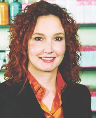
F43.22 Adjustment disorder with anxiety (about ICD-10!)
On Oct. 1, 2015, take a few moments to reflect on 17th century British haberdasher John Graunt. Why? Because Graunt was not merely a purveyor of men’s clothing, but also a demographer and developer of one of the earliest known statistical studies of disease.
Historians consider his 1662 book, “Natural and Political Observations Made Upon the Bills of Mortality,” to be a seminal moment in the genesis of disease classification. Though it was merely a rudimentary beginning, it led others to build upon its framework in categorizing causes of death. But it wasn’t until almost 200 years later that British statistician William Farr proposed a new system to bring medical statistics beyond just mortality. This newer schema, promoted even by Florence Nightingale, would go on to become the impetus for the International Classification of Diseases, Injuries, and Causes of Death, eventually known as the International Classification of Diseases (ICD).
In just a few short weeks, the U.S. health care system will finally embrace the 10th revision of that system, but even this version is far from new. The World Health Organization completed work on ICD-10 in 1992, and it was first adopted by other countries starting with Australia in 1998. In fact, we are among the last developed nations to move into the modern era of disease classification with ICD-10, and this month we felt it would be worthwhile to mark that milestone with a bit of history and a commonsense explanation of what to expect.
Modern terminology
A modern health care system can’t function optimally with a disease classification scheme that is 40 years old; ICD-9 codes, initially conceived in 1975, lack specificity and are replete with antiquated descriptors.
One need not look very far to find examples. The code 200.1, for example, was originally termed “lymphosarcoma,” a deprecated term now replaced with “non-Hodgkin’s lymphoma.” More strikingly, the code 318.0 was initially termed “imbecile.” Today we know this term more appropriately as “moderate intellectual disabilities.” Even if we put political correctness aside, the modern term is clearly a much more accurate and useful descriptor for the diagnosis. But the enhancements in ICD-10 don’t end there.
Increased specificity
Perhaps more important than the descriptors themselves is the logic behind the coding system.
All ICD-10 codes begin with a letter, followed by a series of numbers. These numbers reveal a much greater level of detail than was ever part of ICD-9, such as causal relationship, severity, temporal factors, location, quality, and status. For example, as revised in the ICD-10 system, the ICD-9 code 725.9 (pain in limb) “explodes” into dozens of codes specifying location, all beginning with the prefix M79. These include M79.622 (pain in left upper arm), M79.674 (pain in right toes), M79.652 (pain in left thigh), and so on. Or, in the case of 465.9 (upper respiratory infection), the codes expand to specify the area of infection and causal agent, such as J02.0 (acute streptococcal pharyngitis) or J20.4 (acute bronchitis due to parainfluenza virus).
While this all might seem overwhelming at first, it’s important to note there will be some initial flexibility. The Centers for Medicare & Medicaid Services has stated that for the first year of implementation, it will accept any ICD-10 code from the correct “family,” regardless of specificity (i.e., as long as the letter and first two numbers are correct). In the case of “pain in limb” as above, any 3 digits following M79 with be acceptable. But there is a legitimately good reason to be as specific as possible with the new codes starting now: medical cost management.
Medical cost management is the idea that reimbursement should be related to patient complexity. In other words, caring for sicker patients is more costly and time consuming, so providers should be compensated appropriately. Private insurers have been focusing a lot of attention on this, as their reimbursement rates through Medicare Advantage plans are based on the complexity of the patients they cover. Gradually, this has trickled down to physician payments, as value-based care is growing in importance. Accordingly, we would encourage all physicians to code in a way that accurately reflects the level of morbidity in their patients. After all, if you’re taking care of really complicated patients, you should be getting paid appropriately for it. That’s where ICD-10 can be advantageous, as it allows providers to plainly document the severity of disease in their population through a system that’s easy to capture and collate.
Avoiding insanity
There is a point where the need to be so specific translates into some fairly funny ICD-10 codes. We simply can’t avoid citing a few examples: Z63.1 (problems in relationship with in-laws), W61.52XA (struck by goose, initial encounter), and our personal favorite, V91.07XD (burn due to water skis on fire). While quite funny, this level of specificity also brings up a critical question: With so many codes, how does one find the correct one for the diagnosis when seeing a patient?
Thankfully, 21st century tools make the task of locating the appropriate code much simpler than it would have been when ICD-10 was first conceived. Using plain-text search engines such as those from Intelligent Medical Objects, clinicians can be as specific as possible when searching for diagnoses. There are also apps for mobile devices and Web-based tools like ICD10Data.com that are user friendly and completely free. Most importantly, EHR vendors have been anticipating the transition to ICD-10 and have built many of these tools into their products already.
Closing thoughts
We fully acknowledge that this transition to increased specificity may be anxiety provoking, but we are also forced to accept that it is necessary. While it is debatable whether figures like Graunt and Farr are worthy of accolade or ire (though on Oct. 1 most will probably choose the latter), it’s undeniable that there is a need to gather and tabulate statistics on medical diagnoses. That need was first formally addressed well over 300 years before the dawn of electronic health records and will continue to be addressed for hundreds more. In the meantime, we have a code we’d like to propose for ICD-11: E011.13 – anxiety due to heath information technology. Like ICD-10, this is a concept that’s far from new and certainly here to stay.
Dr. Notte is a family physician and clinical informaticist for Abington (Pa.) Memorial Hospital. He is a partner in EHR Practice Consultants, a firm that aids physicians in adopting electronic health records. Dr. Skolnik is associate director of the family medicine residency program at Abington Memorial Hospital and professor of family and community medicine at Temple University in Philadelphia.
On Oct. 1, 2015, take a few moments to reflect on 17th century British haberdasher John Graunt. Why? Because Graunt was not merely a purveyor of men’s clothing, but also a demographer and developer of one of the earliest known statistical studies of disease.
Historians consider his 1662 book, “Natural and Political Observations Made Upon the Bills of Mortality,” to be a seminal moment in the genesis of disease classification. Though it was merely a rudimentary beginning, it led others to build upon its framework in categorizing causes of death. But it wasn’t until almost 200 years later that British statistician William Farr proposed a new system to bring medical statistics beyond just mortality. This newer schema, promoted even by Florence Nightingale, would go on to become the impetus for the International Classification of Diseases, Injuries, and Causes of Death, eventually known as the International Classification of Diseases (ICD).
In just a few short weeks, the U.S. health care system will finally embrace the 10th revision of that system, but even this version is far from new. The World Health Organization completed work on ICD-10 in 1992, and it was first adopted by other countries starting with Australia in 1998. In fact, we are among the last developed nations to move into the modern era of disease classification with ICD-10, and this month we felt it would be worthwhile to mark that milestone with a bit of history and a commonsense explanation of what to expect.
Modern terminology
A modern health care system can’t function optimally with a disease classification scheme that is 40 years old; ICD-9 codes, initially conceived in 1975, lack specificity and are replete with antiquated descriptors.
One need not look very far to find examples. The code 200.1, for example, was originally termed “lymphosarcoma,” a deprecated term now replaced with “non-Hodgkin’s lymphoma.” More strikingly, the code 318.0 was initially termed “imbecile.” Today we know this term more appropriately as “moderate intellectual disabilities.” Even if we put political correctness aside, the modern term is clearly a much more accurate and useful descriptor for the diagnosis. But the enhancements in ICD-10 don’t end there.
Increased specificity
Perhaps more important than the descriptors themselves is the logic behind the coding system.
All ICD-10 codes begin with a letter, followed by a series of numbers. These numbers reveal a much greater level of detail than was ever part of ICD-9, such as causal relationship, severity, temporal factors, location, quality, and status. For example, as revised in the ICD-10 system, the ICD-9 code 725.9 (pain in limb) “explodes” into dozens of codes specifying location, all beginning with the prefix M79. These include M79.622 (pain in left upper arm), M79.674 (pain in right toes), M79.652 (pain in left thigh), and so on. Or, in the case of 465.9 (upper respiratory infection), the codes expand to specify the area of infection and causal agent, such as J02.0 (acute streptococcal pharyngitis) or J20.4 (acute bronchitis due to parainfluenza virus).
While this all might seem overwhelming at first, it’s important to note there will be some initial flexibility. The Centers for Medicare & Medicaid Services has stated that for the first year of implementation, it will accept any ICD-10 code from the correct “family,” regardless of specificity (i.e., as long as the letter and first two numbers are correct). In the case of “pain in limb” as above, any 3 digits following M79 with be acceptable. But there is a legitimately good reason to be as specific as possible with the new codes starting now: medical cost management.
Medical cost management is the idea that reimbursement should be related to patient complexity. In other words, caring for sicker patients is more costly and time consuming, so providers should be compensated appropriately. Private insurers have been focusing a lot of attention on this, as their reimbursement rates through Medicare Advantage plans are based on the complexity of the patients they cover. Gradually, this has trickled down to physician payments, as value-based care is growing in importance. Accordingly, we would encourage all physicians to code in a way that accurately reflects the level of morbidity in their patients. After all, if you’re taking care of really complicated patients, you should be getting paid appropriately for it. That’s where ICD-10 can be advantageous, as it allows providers to plainly document the severity of disease in their population through a system that’s easy to capture and collate.
Avoiding insanity
There is a point where the need to be so specific translates into some fairly funny ICD-10 codes. We simply can’t avoid citing a few examples: Z63.1 (problems in relationship with in-laws), W61.52XA (struck by goose, initial encounter), and our personal favorite, V91.07XD (burn due to water skis on fire). While quite funny, this level of specificity also brings up a critical question: With so many codes, how does one find the correct one for the diagnosis when seeing a patient?
Thankfully, 21st century tools make the task of locating the appropriate code much simpler than it would have been when ICD-10 was first conceived. Using plain-text search engines such as those from Intelligent Medical Objects, clinicians can be as specific as possible when searching for diagnoses. There are also apps for mobile devices and Web-based tools like ICD10Data.com that are user friendly and completely free. Most importantly, EHR vendors have been anticipating the transition to ICD-10 and have built many of these tools into their products already.
Closing thoughts
We fully acknowledge that this transition to increased specificity may be anxiety provoking, but we are also forced to accept that it is necessary. While it is debatable whether figures like Graunt and Farr are worthy of accolade or ire (though on Oct. 1 most will probably choose the latter), it’s undeniable that there is a need to gather and tabulate statistics on medical diagnoses. That need was first formally addressed well over 300 years before the dawn of electronic health records and will continue to be addressed for hundreds more. In the meantime, we have a code we’d like to propose for ICD-11: E011.13 – anxiety due to heath information technology. Like ICD-10, this is a concept that’s far from new and certainly here to stay.
Dr. Notte is a family physician and clinical informaticist for Abington (Pa.) Memorial Hospital. He is a partner in EHR Practice Consultants, a firm that aids physicians in adopting electronic health records. Dr. Skolnik is associate director of the family medicine residency program at Abington Memorial Hospital and professor of family and community medicine at Temple University in Philadelphia.
On Oct. 1, 2015, take a few moments to reflect on 17th century British haberdasher John Graunt. Why? Because Graunt was not merely a purveyor of men’s clothing, but also a demographer and developer of one of the earliest known statistical studies of disease.
Historians consider his 1662 book, “Natural and Political Observations Made Upon the Bills of Mortality,” to be a seminal moment in the genesis of disease classification. Though it was merely a rudimentary beginning, it led others to build upon its framework in categorizing causes of death. But it wasn’t until almost 200 years later that British statistician William Farr proposed a new system to bring medical statistics beyond just mortality. This newer schema, promoted even by Florence Nightingale, would go on to become the impetus for the International Classification of Diseases, Injuries, and Causes of Death, eventually known as the International Classification of Diseases (ICD).
In just a few short weeks, the U.S. health care system will finally embrace the 10th revision of that system, but even this version is far from new. The World Health Organization completed work on ICD-10 in 1992, and it was first adopted by other countries starting with Australia in 1998. In fact, we are among the last developed nations to move into the modern era of disease classification with ICD-10, and this month we felt it would be worthwhile to mark that milestone with a bit of history and a commonsense explanation of what to expect.
Modern terminology
A modern health care system can’t function optimally with a disease classification scheme that is 40 years old; ICD-9 codes, initially conceived in 1975, lack specificity and are replete with antiquated descriptors.
One need not look very far to find examples. The code 200.1, for example, was originally termed “lymphosarcoma,” a deprecated term now replaced with “non-Hodgkin’s lymphoma.” More strikingly, the code 318.0 was initially termed “imbecile.” Today we know this term more appropriately as “moderate intellectual disabilities.” Even if we put political correctness aside, the modern term is clearly a much more accurate and useful descriptor for the diagnosis. But the enhancements in ICD-10 don’t end there.
Increased specificity
Perhaps more important than the descriptors themselves is the logic behind the coding system.
All ICD-10 codes begin with a letter, followed by a series of numbers. These numbers reveal a much greater level of detail than was ever part of ICD-9, such as causal relationship, severity, temporal factors, location, quality, and status. For example, as revised in the ICD-10 system, the ICD-9 code 725.9 (pain in limb) “explodes” into dozens of codes specifying location, all beginning with the prefix M79. These include M79.622 (pain in left upper arm), M79.674 (pain in right toes), M79.652 (pain in left thigh), and so on. Or, in the case of 465.9 (upper respiratory infection), the codes expand to specify the area of infection and causal agent, such as J02.0 (acute streptococcal pharyngitis) or J20.4 (acute bronchitis due to parainfluenza virus).
While this all might seem overwhelming at first, it’s important to note there will be some initial flexibility. The Centers for Medicare & Medicaid Services has stated that for the first year of implementation, it will accept any ICD-10 code from the correct “family,” regardless of specificity (i.e., as long as the letter and first two numbers are correct). In the case of “pain in limb” as above, any 3 digits following M79 with be acceptable. But there is a legitimately good reason to be as specific as possible with the new codes starting now: medical cost management.
Medical cost management is the idea that reimbursement should be related to patient complexity. In other words, caring for sicker patients is more costly and time consuming, so providers should be compensated appropriately. Private insurers have been focusing a lot of attention on this, as their reimbursement rates through Medicare Advantage plans are based on the complexity of the patients they cover. Gradually, this has trickled down to physician payments, as value-based care is growing in importance. Accordingly, we would encourage all physicians to code in a way that accurately reflects the level of morbidity in their patients. After all, if you’re taking care of really complicated patients, you should be getting paid appropriately for it. That’s where ICD-10 can be advantageous, as it allows providers to plainly document the severity of disease in their population through a system that’s easy to capture and collate.
Avoiding insanity
There is a point where the need to be so specific translates into some fairly funny ICD-10 codes. We simply can’t avoid citing a few examples: Z63.1 (problems in relationship with in-laws), W61.52XA (struck by goose, initial encounter), and our personal favorite, V91.07XD (burn due to water skis on fire). While quite funny, this level of specificity also brings up a critical question: With so many codes, how does one find the correct one for the diagnosis when seeing a patient?
Thankfully, 21st century tools make the task of locating the appropriate code much simpler than it would have been when ICD-10 was first conceived. Using plain-text search engines such as those from Intelligent Medical Objects, clinicians can be as specific as possible when searching for diagnoses. There are also apps for mobile devices and Web-based tools like ICD10Data.com that are user friendly and completely free. Most importantly, EHR vendors have been anticipating the transition to ICD-10 and have built many of these tools into their products already.
Closing thoughts
We fully acknowledge that this transition to increased specificity may be anxiety provoking, but we are also forced to accept that it is necessary. While it is debatable whether figures like Graunt and Farr are worthy of accolade or ire (though on Oct. 1 most will probably choose the latter), it’s undeniable that there is a need to gather and tabulate statistics on medical diagnoses. That need was first formally addressed well over 300 years before the dawn of electronic health records and will continue to be addressed for hundreds more. In the meantime, we have a code we’d like to propose for ICD-11: E011.13 – anxiety due to heath information technology. Like ICD-10, this is a concept that’s far from new and certainly here to stay.
Dr. Notte is a family physician and clinical informaticist for Abington (Pa.) Memorial Hospital. He is a partner in EHR Practice Consultants, a firm that aids physicians in adopting electronic health records. Dr. Skolnik is associate director of the family medicine residency program at Abington Memorial Hospital and professor of family and community medicine at Temple University in Philadelphia.
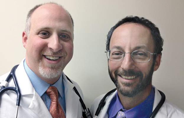
The pros and cons of novel anticoagulants
Novel anticoagulants will likely replace need for vitamin K antagonists
BY MADHUKAR S. PATEL, M.D., AND ELLIOT L. CHAIKOF, M.D.
The discovery of oral anticoagulants began in 1924, when Schofield linked the death of grazing cattle from internal hemorrhage to the consumption of spoiled sweet clover hay.1 It was not until 1941, however, while trying to understand this observation, that Campbell & Link were able to identify the dicoumarol anticoagulant, which formed as a result of the spoiling process.2 Ultimately, after noting that vitamin K led to reversal of the dicoumarol effect, synthesis of the first class of oral anticoagulants, known as vitamin K antagonists (VKAs), began.

Despite the numerous challenges associated with managing patients using this class of anticoagulants, VKAs have become the mainstay of oral anticoagulation therapy for the past 70 years. Over the past 5 years, however, new oral anticoagulants (NOACs) have emerged and are changing clinical practice.
Mechanistically, these medications are targeted therapies and work as either direct thrombin inhibitors (dabigatran etexilate) or direct factor Xa inhibitors (rivaroxaban, apixaban, and edoxaban). Given their favorable pharmacologic design, NOACs have the potential to replace VKAs as they not only have an encouraging safety profile, but also are therapeutically equivalent or even superior to VKAs when used in certain patient populations.
Pharmacologic design
The targeted drug design of NOACs provides many pharmacologic advantages. Compared to VKAs, NOACs have a notably more predictable pharmacologic profile and relatively wide therapeutic window, which allows for fixed dosing, a rapid onset and offset, and fewer drug interactions.3 These characteristics eliminate the need for the routine dose monitoring and serial dose adjustments frequently associated with VKAs.
NOACs less commonly require bridging therapy with parenteral unfractionated heparin or low-molecular-weight heparins (LMWH) while awaiting therapeutic drug levels, as these levels are reached sooner and more predictably than with VKAs.4 As with any medication, however, appropriate consideration should to be given to specific patient populations such as those who are older or have significant comorbidities that may influence drug effect and clearance. Lastly, it should be mentioned that the pharmacologic benefits of NOACs apply not only from a patient perspective, but also from a health care systems standpoint, as their use may provide an opportunity to deliver more cost-effective care.
Specifically, economic models using available clinical trial data for stroke prevention in nonvalvular atrial fibrillation have shown that NOACs (apixaban, dabigatran, and rivaroxaban) are cost-effective alternatives when compared to warfarin.5 Although the results from such economic analyses are limited by the modeling assumptions they rely upon, these findings suggest that at least initially, cost should not be used as a prohibitive reason for adopting these new therapeutics.
Patient selection
The decision to institute oral anticoagulation therapy depends on each patient’s individualized bleeding risk to benefit of ischemia prevention ratio. A major determinant of this ratio is the clinical indication for which anticoagulation is begun. Numerous phase III clinical trials have been conducted comparing the use of NOACs to VKAs or placebos for the management of nonvalvular atrial fibrillation and venous thromboembolism, and as adjunctive therapy for patients with acute coronary syndrome.6
Meta-analyses of randomized trials have shown the most significant benefit to be in patients with nonvalvular atrial fibrillation, where NOACs yield significant reductions in stroke, intracranial hemorrhage, and all-cause mortality compared to warfarin, while displaying variable effects with regard to gastrointestinal bleeding.6,7 In patients with VTE, NOACs have been found to have efficacy similar to that of VKAs with regard to the prevention of VTE or VTE-related death, and have been noted to have a better safety profile.6
Lastly, when studied as an adjunctive agent to dual antiplatelet therapy in patients with acute coronary syndrome, NOACs have been associated with an increased bleeding risk without a significant decrease in thrombosis risk.6 Taken together, these data suggest that the primary indication for instituting NOAC therapy should be considered strongly when deciding upon which class of anticoagulant to use.
Overcoming challenges
Since the introduction of NOACs, there has been concern over the lack of specific antidotes to therapy, especially when administered in patients with impaired clearance, a high likelihood of need for an urgent or emergent procedure, or those presenting with life threatening bleeding complications.
Most recently, however, interim analysis from clinical trial data has shown complete reversal of the direct thrombin inhibitor dabigatran with the humanized monoclonal antibody idarucizumab within minutes of administration in greater than 88% of patients studied.8 Similarly, agents such as a PER977 are currently under phase II clinical trials as they have been shown to form noncovalent hydrogen bonds and charge-charge interactions with oral factor Xa inhibitors as well as oral thrombin inhibitors leading to their reversal.9
Given these promising findings, it likely will not be long until reversal agents for NOACs become clinically available. Until that time, it is encouraging that the bleeding profile of these drugs has been found to be favorable compared to VKAs and their short half-life allows for a relatively expeditious natural reversal of their anticoagulant effect as the drug is eliminated.
Conclusion
Unlike the serendipitous path leading to the discovery of the first class of oral anticoagulants (VKAs), NOACs have been specifically designed to provide targeted anticoagulation and to address the shortcomings of VKAs. To this end, NOACs are becoming increasingly important in the management of patients with specific clinical conditions such as nonvalvular atrial fibrillation and venous thromboembolism, where they have been shown to provide a larger net clinical benefit relative to the available alternatives. Furthermore, with economic analyses providing evidence that NOACs are cost-effective for the healthcare system and clinical trial results suggesting progress in the development of antidotes for reversal, it is likely that with growing experience, these agents will replace VKAs as the mainstay for prophylactic and therapeutic oral anticoagulation in targeted patient populations.
Dr. Patel is a research fellow and Dr. Chaikof is surgeon-in-chief, both at the department of surgery, Beth Israel Deaconess Medical Center, Boston. They reported no conflicts of interest.
References
1. J Am Vet Med Assoc. 1924;64:553-75 (See Br J Haematol 2008 Mar 18;141[6]:757-63).
2. J Biol Chem. 1941;138:21-33 (See Nutr Rev. 1974 Aug;32[8]:244-6).
3. Am Soc Hematol Educ Program. 2013;2013:464-70.
4. Eur Heart J. 2013 Jul;34(27):2094-2106.
5. Stroke. 2013 Jun;44(6):1676-81.
6. Nat Rev Cardiol. 2014 Dec;11(12):693-703.
7. Lancet. 2014 Mar 15;383(9921):955-62.
8. N Engl J Med. 2015;373(6):511-20.
9. N Engl J Med. 2014;371(22):2141-2.
What the doctor didn’t order: unintended consequences and pitfalls of NOACs
BY THOMAS WAKEFIELD, M.D., ANDREA OBI, M.D., AND DAWN COLEMAN, M.D.
Recently, several new oral anticoagulants have gained FDA approval to replace warfarin, capturing the attention of popular media. These include dabigatran, rivaroxaban, apixaban, and edoxaban. Dabigatran targets activated factor II (factor IIa), while rivaroxaban, apixaban, and edoxaban target activated factor X (factor Xa). Easy to take with a once- or twice-daily pill, with no cumbersome monitoring, they represent a seemingly ideal treatment for the chronically anticoagulated patient. All agents are currently FDA approved in the United States for treatment of acute venous thromboembolism (VTE) and atrial fibrillation (AF).

Dabigatran and edoxaban
As with warfarin, dabigatran and edoxaban require the use of a low-molecular-weight heparin (LMWH) or unfractionated heparin “bridge” when therapy is beginning, while rivaroxaban and apixaban are instituted as monotherapy without such a bridge. Dabigatran etexilate (PradaxaR, Boehringer Ingelheim) has the longest half-life of all of the NOACs at 12-17 hours, and this half-life is prolonged with increasing age and decreasing renal function.1 It is the only new agent that can be at least partially reversed with dialysis.2 Edoxaban (SavaysaR, Daiichi Sankyo) carries a boxed warning stating that this agent is less effective in AF patients with a creatinine clearance greater than 95 mL/min, and that kidney function should be assessed prior to starting treatment: Such patients have a greater risk of stroke compared with similar patients treated with warfarin. Edoxaban is the only agent specifically tested at a lower dose in patients at significantly increased risk of bleeding complications (low body weight and/or decreased creatinine clearance).3
Rivaroxaban and apixaban
Rivaroxaban (XareltoR, Bayer and Janssen), and apixaban (EliquisR, Bristol Myers-Squibb), unique among the NOACs, have been tested for extended therapy of acute DVT after treatment of 6-12 months. They were found to result in a significant decrease in recurrent VTE without an increase in major bleeding compared to placebo.4,5 Rivaroxaban has once-daily dosing and apixaban has twice-daily dosing; both are immediate monotherapy, making them quite convenient for patients. Apixaban is the only agent among the NOACs to have a slight decrease in gastrointestinal bleeding compared to warfarin.6
Consequences and pitfalls with NOACs
Problems with these new drugs, which may diminish our current level of enthusiasm for these agents to totally replace warfarin, include the inability to reliably follow their levels and to reverse their anticoagulant effects, the lack of data available on bridging when other procedures need to be performed, their short half-lives, and the lack of data on their anti-inflammatory effects.
With regard to monitoring of anticoagulation, the International Society of Thrombosis and Hemostasis (ISTH) has published a recommendation7 that lists these scenarios:
• When a patient is bleeding.
• Before surgery or an invasive procedure when the patient has taken the drug in the previous 24 hours, or longer if creatinine clearance (CrCl) is less than 50 mL/min.
• Identification of subtherapeutic or supratherapeutic levels in patients taking other drugs that are known to affect pharmacokinetics.
• Identification of subtherapeutic or supratherapeutic levels in patients at body weight extremes.
• Patients with deteriorating renal function.
• During perioperative management.
• During reversal of anticoagulation.
• When there is suspicion of overdose.
• Assessment of compliance in patients suffering thrombotic events while on treatment.
Currently, there exists no commercially available reversal agent for any of the NOACs and existing reversal agents for traditional anticoagulants are of limited, if any, use. Drugs under development include agents for the factor Xa inhibitors and for the thrombin inhibitor. Until the time that specific reversal agents exist, supportive care is the mainstay of therapy. In cases of trauma or severe or life-threatening bleeding, administration of concentrated clotting factors (prothrombin complex concentrate) or dialysis (dabigatran only) may be utilized. However, data from large clinical trials is lacking. A recent study of 90 patients receiving an antibody directed against dabigatran has revealed that the anticoagulant effects of dabigatran were reversed safely within minutes of administration; however, drug levels were not consistently suppressed at 24 hours in 20% of the cohort.8
There are no national guidelines nor large scale studies to guide bridging NOACs for procedures. The relatively short half-life for these agents makes it likely that traditional bridging as is practiced for warfarin is not necessary.9 However, this represents a double edged sword; withholding anticoagulation for two doses (such as if a patient becomes ill or a clinician is overly cautious around the time of a procedure) may leave the patient unprotected.
The final question with the new agents is their anti-inflammatory effects. We know that heparin and LMWH have significant pleiotropic effects that are not necessarily related to their anticoagulant effects. These effects are important to decrease the inflammatory nature of the thrombus and its effect on the vein wall. We do not know if the new oral agents have similar effects, as this has never fully been tested. In view of the fact that two of the agents are being used as monotherapy agents without any heparin/LMWH bridge, the anti-inflammatory properties of these new agents should be defined to make sure that such a bridge is not necessary.
Conclusion
So, in summary, although these agents have much to offer, there are many questions that remain to be addressed and answered before they totally replace traditional approaches to anticoagulation, in the realm of VTE. It must not be overlooked that for all the benefits, they each carry a risk of bleeding as they all target portions of the coagulation mechanism. We believe, that as with any “gift horse,” physicians should perhaps examine the data more closely and proceed with caution.
Dr. Wakefield is director of the Samuel and Jean Frankel Cardiovascular Center, Dr. Obi is a vascular surgery fellow, and Dr. Coleman is program director, section of vascular surgery, at the University of Michigan, Ann Arbor. They reported no conflicts of interest.
References
1. N Engl J Med. 2009;361:2342-52.
2. J Vasc Surg: Venous Lymphat Disord. 2013;1:418-26.
3. N Engl J Med. 2013;369:1406-15.
4. N Engl J Med. 2010;363:2499-2510.
5. N Engl J Med. 2013;368:699-708.
6. Arterioscler Thromb Vasc Biol. 2015;35:1056-65.
7. J Thromb Haemost. 2013;11:756-60.
Novel anticoagulants will likely replace need for vitamin K antagonists
BY MADHUKAR S. PATEL, M.D., AND ELLIOT L. CHAIKOF, M.D.
The discovery of oral anticoagulants began in 1924, when Schofield linked the death of grazing cattle from internal hemorrhage to the consumption of spoiled sweet clover hay.1 It was not until 1941, however, while trying to understand this observation, that Campbell & Link were able to identify the dicoumarol anticoagulant, which formed as a result of the spoiling process.2 Ultimately, after noting that vitamin K led to reversal of the dicoumarol effect, synthesis of the first class of oral anticoagulants, known as vitamin K antagonists (VKAs), began.
Despite the numerous challenges associated with managing patients using this class of anticoagulants, VKAs have become the mainstay of oral anticoagulation therapy for the past 70 years. Over the past 5 years, however, new oral anticoagulants (NOACs) have emerged and are changing clinical practice.
Mechanistically, these medications are targeted therapies and work as either direct thrombin inhibitors (dabigatran etexilate) or direct factor Xa inhibitors (rivaroxaban, apixaban, and edoxaban). Given their favorable pharmacologic design, NOACs have the potential to replace VKAs as they not only have an encouraging safety profile, but also are therapeutically equivalent or even superior to VKAs when used in certain patient populations.
Pharmacologic design
The targeted drug design of NOACs provides many pharmacologic advantages. Compared to VKAs, NOACs have a notably more predictable pharmacologic profile and relatively wide therapeutic window, which allows for fixed dosing, a rapid onset and offset, and fewer drug interactions.3 These characteristics eliminate the need for the routine dose monitoring and serial dose adjustments frequently associated with VKAs.
NOACs less commonly require bridging therapy with parenteral unfractionated heparin or low-molecular-weight heparins (LMWH) while awaiting therapeutic drug levels, as these levels are reached sooner and more predictably than with VKAs.4 As with any medication, however, appropriate consideration should to be given to specific patient populations such as those who are older or have significant comorbidities that may influence drug effect and clearance. Lastly, it should be mentioned that the pharmacologic benefits of NOACs apply not only from a patient perspective, but also from a health care systems standpoint, as their use may provide an opportunity to deliver more cost-effective care.
Specifically, economic models using available clinical trial data for stroke prevention in nonvalvular atrial fibrillation have shown that NOACs (apixaban, dabigatran, and rivaroxaban) are cost-effective alternatives when compared to warfarin.5 Although the results from such economic analyses are limited by the modeling assumptions they rely upon, these findings suggest that at least initially, cost should not be used as a prohibitive reason for adopting these new therapeutics.
Patient selection
The decision to institute oral anticoagulation therapy depends on each patient’s individualized bleeding risk to benefit of ischemia prevention ratio. A major determinant of this ratio is the clinical indication for which anticoagulation is begun. Numerous phase III clinical trials have been conducted comparing the use of NOACs to VKAs or placebos for the management of nonvalvular atrial fibrillation and venous thromboembolism, and as adjunctive therapy for patients with acute coronary syndrome.6
Meta-analyses of randomized trials have shown the most significant benefit to be in patients with nonvalvular atrial fibrillation, where NOACs yield significant reductions in stroke, intracranial hemorrhage, and all-cause mortality compared to warfarin, while displaying variable effects with regard to gastrointestinal bleeding.6,7 In patients with VTE, NOACs have been found to have efficacy similar to that of VKAs with regard to the prevention of VTE or VTE-related death, and have been noted to have a better safety profile.6
Lastly, when studied as an adjunctive agent to dual antiplatelet therapy in patients with acute coronary syndrome, NOACs have been associated with an increased bleeding risk without a significant decrease in thrombosis risk.6 Taken together, these data suggest that the primary indication for instituting NOAC therapy should be considered strongly when deciding upon which class of anticoagulant to use.
Overcoming challenges
Since the introduction of NOACs, there has been concern over the lack of specific antidotes to therapy, especially when administered in patients with impaired clearance, a high likelihood of need for an urgent or emergent procedure, or those presenting with life threatening bleeding complications.
Most recently, however, interim analysis from clinical trial data has shown complete reversal of the direct thrombin inhibitor dabigatran with the humanized monoclonal antibody idarucizumab within minutes of administration in greater than 88% of patients studied.8 Similarly, agents such as a PER977 are currently under phase II clinical trials as they have been shown to form noncovalent hydrogen bonds and charge-charge interactions with oral factor Xa inhibitors as well as oral thrombin inhibitors leading to their reversal.9
Given these promising findings, it likely will not be long until reversal agents for NOACs become clinically available. Until that time, it is encouraging that the bleeding profile of these drugs has been found to be favorable compared to VKAs and their short half-life allows for a relatively expeditious natural reversal of their anticoagulant effect as the drug is eliminated.
Conclusion
Unlike the serendipitous path leading to the discovery of the first class of oral anticoagulants (VKAs), NOACs have been specifically designed to provide targeted anticoagulation and to address the shortcomings of VKAs. To this end, NOACs are becoming increasingly important in the management of patients with specific clinical conditions such as nonvalvular atrial fibrillation and venous thromboembolism, where they have been shown to provide a larger net clinical benefit relative to the available alternatives. Furthermore, with economic analyses providing evidence that NOACs are cost-effective for the healthcare system and clinical trial results suggesting progress in the development of antidotes for reversal, it is likely that with growing experience, these agents will replace VKAs as the mainstay for prophylactic and therapeutic oral anticoagulation in targeted patient populations.
Dr. Patel is a research fellow and Dr. Chaikof is surgeon-in-chief, both at the department of surgery, Beth Israel Deaconess Medical Center, Boston. They reported no conflicts of interest.
References
1. J Am Vet Med Assoc. 1924;64:553-75 (See Br J Haematol 2008 Mar 18;141[6]:757-63).
2. J Biol Chem. 1941;138:21-33 (See Nutr Rev. 1974 Aug;32[8]:244-6).
3. Am Soc Hematol Educ Program. 2013;2013:464-70.
4. Eur Heart J. 2013 Jul;34(27):2094-2106.
5. Stroke. 2013 Jun;44(6):1676-81.
6. Nat Rev Cardiol. 2014 Dec;11(12):693-703.
7. Lancet. 2014 Mar 15;383(9921):955-62.
8. N Engl J Med. 2015;373(6):511-20.
9. N Engl J Med. 2014;371(22):2141-2.
What the doctor didn’t order: unintended consequences and pitfalls of NOACs
BY THOMAS WAKEFIELD, M.D., ANDREA OBI, M.D., AND DAWN COLEMAN, M.D.
Recently, several new oral anticoagulants have gained FDA approval to replace warfarin, capturing the attention of popular media. These include dabigatran, rivaroxaban, apixaban, and edoxaban. Dabigatran targets activated factor II (factor IIa), while rivaroxaban, apixaban, and edoxaban target activated factor X (factor Xa). Easy to take with a once- or twice-daily pill, with no cumbersome monitoring, they represent a seemingly ideal treatment for the chronically anticoagulated patient. All agents are currently FDA approved in the United States for treatment of acute venous thromboembolism (VTE) and atrial fibrillation (AF).
Dabigatran and edoxaban
As with warfarin, dabigatran and edoxaban require the use of a low-molecular-weight heparin (LMWH) or unfractionated heparin “bridge” when therapy is beginning, while rivaroxaban and apixaban are instituted as monotherapy without such a bridge. Dabigatran etexilate (PradaxaR, Boehringer Ingelheim) has the longest half-life of all of the NOACs at 12-17 hours, and this half-life is prolonged with increasing age and decreasing renal function.1 It is the only new agent that can be at least partially reversed with dialysis.2 Edoxaban (SavaysaR, Daiichi Sankyo) carries a boxed warning stating that this agent is less effective in AF patients with a creatinine clearance greater than 95 mL/min, and that kidney function should be assessed prior to starting treatment: Such patients have a greater risk of stroke compared with similar patients treated with warfarin. Edoxaban is the only agent specifically tested at a lower dose in patients at significantly increased risk of bleeding complications (low body weight and/or decreased creatinine clearance).3
Rivaroxaban and apixaban
Rivaroxaban (XareltoR, Bayer and Janssen), and apixaban (EliquisR, Bristol Myers-Squibb), unique among the NOACs, have been tested for extended therapy of acute DVT after treatment of 6-12 months. They were found to result in a significant decrease in recurrent VTE without an increase in major bleeding compared to placebo.4,5 Rivaroxaban has once-daily dosing and apixaban has twice-daily dosing; both are immediate monotherapy, making them quite convenient for patients. Apixaban is the only agent among the NOACs to have a slight decrease in gastrointestinal bleeding compared to warfarin.6
Consequences and pitfalls with NOACs
Problems with these new drugs, which may diminish our current level of enthusiasm for these agents to totally replace warfarin, include the inability to reliably follow their levels and to reverse their anticoagulant effects, the lack of data available on bridging when other procedures need to be performed, their short half-lives, and the lack of data on their anti-inflammatory effects.
With regard to monitoring of anticoagulation, the International Society of Thrombosis and Hemostasis (ISTH) has published a recommendation7 that lists these scenarios:
• When a patient is bleeding.
• Before surgery or an invasive procedure when the patient has taken the drug in the previous 24 hours, or longer if creatinine clearance (CrCl) is less than 50 mL/min.
• Identification of subtherapeutic or supratherapeutic levels in patients taking other drugs that are known to affect pharmacokinetics.
• Identification of subtherapeutic or supratherapeutic levels in patients at body weight extremes.
• Patients with deteriorating renal function.
• During perioperative management.
• During reversal of anticoagulation.
• When there is suspicion of overdose.
• Assessment of compliance in patients suffering thrombotic events while on treatment.
Currently, there exists no commercially available reversal agent for any of the NOACs and existing reversal agents for traditional anticoagulants are of limited, if any, use. Drugs under development include agents for the factor Xa inhibitors and for the thrombin inhibitor. Until the time that specific reversal agents exist, supportive care is the mainstay of therapy. In cases of trauma or severe or life-threatening bleeding, administration of concentrated clotting factors (prothrombin complex concentrate) or dialysis (dabigatran only) may be utilized. However, data from large clinical trials is lacking. A recent study of 90 patients receiving an antibody directed against dabigatran has revealed that the anticoagulant effects of dabigatran were reversed safely within minutes of administration; however, drug levels were not consistently suppressed at 24 hours in 20% of the cohort.8
There are no national guidelines nor large scale studies to guide bridging NOACs for procedures. The relatively short half-life for these agents makes it likely that traditional bridging as is practiced for warfarin is not necessary.9 However, this represents a double edged sword; withholding anticoagulation for two doses (such as if a patient becomes ill or a clinician is overly cautious around the time of a procedure) may leave the patient unprotected.
The final question with the new agents is their anti-inflammatory effects. We know that heparin and LMWH have significant pleiotropic effects that are not necessarily related to their anticoagulant effects. These effects are important to decrease the inflammatory nature of the thrombus and its effect on the vein wall. We do not know if the new oral agents have similar effects, as this has never fully been tested. In view of the fact that two of the agents are being used as monotherapy agents without any heparin/LMWH bridge, the anti-inflammatory properties of these new agents should be defined to make sure that such a bridge is not necessary.
Conclusion
So, in summary, although these agents have much to offer, there are many questions that remain to be addressed and answered before they totally replace traditional approaches to anticoagulation, in the realm of VTE. It must not be overlooked that for all the benefits, they each carry a risk of bleeding as they all target portions of the coagulation mechanism. We believe, that as with any “gift horse,” physicians should perhaps examine the data more closely and proceed with caution.
Dr. Wakefield is director of the Samuel and Jean Frankel Cardiovascular Center, Dr. Obi is a vascular surgery fellow, and Dr. Coleman is program director, section of vascular surgery, at the University of Michigan, Ann Arbor. They reported no conflicts of interest.
References
1. N Engl J Med. 2009;361:2342-52.
2. J Vasc Surg: Venous Lymphat Disord. 2013;1:418-26.
3. N Engl J Med. 2013;369:1406-15.
4. N Engl J Med. 2010;363:2499-2510.
5. N Engl J Med. 2013;368:699-708.
6. Arterioscler Thromb Vasc Biol. 2015;35:1056-65.
7. J Thromb Haemost. 2013;11:756-60.
Novel anticoagulants will likely replace need for vitamin K antagonists
BY MADHUKAR S. PATEL, M.D., AND ELLIOT L. CHAIKOF, M.D.
The discovery of oral anticoagulants began in 1924, when Schofield linked the death of grazing cattle from internal hemorrhage to the consumption of spoiled sweet clover hay.1 It was not until 1941, however, while trying to understand this observation, that Campbell & Link were able to identify the dicoumarol anticoagulant, which formed as a result of the spoiling process.2 Ultimately, after noting that vitamin K led to reversal of the dicoumarol effect, synthesis of the first class of oral anticoagulants, known as vitamin K antagonists (VKAs), began.
Despite the numerous challenges associated with managing patients using this class of anticoagulants, VKAs have become the mainstay of oral anticoagulation therapy for the past 70 years. Over the past 5 years, however, new oral anticoagulants (NOACs) have emerged and are changing clinical practice.
Mechanistically, these medications are targeted therapies and work as either direct thrombin inhibitors (dabigatran etexilate) or direct factor Xa inhibitors (rivaroxaban, apixaban, and edoxaban). Given their favorable pharmacologic design, NOACs have the potential to replace VKAs as they not only have an encouraging safety profile, but also are therapeutically equivalent or even superior to VKAs when used in certain patient populations.
Pharmacologic design
The targeted drug design of NOACs provides many pharmacologic advantages. Compared to VKAs, NOACs have a notably more predictable pharmacologic profile and relatively wide therapeutic window, which allows for fixed dosing, a rapid onset and offset, and fewer drug interactions.3 These characteristics eliminate the need for the routine dose monitoring and serial dose adjustments frequently associated with VKAs.
NOACs less commonly require bridging therapy with parenteral unfractionated heparin or low-molecular-weight heparins (LMWH) while awaiting therapeutic drug levels, as these levels are reached sooner and more predictably than with VKAs.4 As with any medication, however, appropriate consideration should to be given to specific patient populations such as those who are older or have significant comorbidities that may influence drug effect and clearance. Lastly, it should be mentioned that the pharmacologic benefits of NOACs apply not only from a patient perspective, but also from a health care systems standpoint, as their use may provide an opportunity to deliver more cost-effective care.
Specifically, economic models using available clinical trial data for stroke prevention in nonvalvular atrial fibrillation have shown that NOACs (apixaban, dabigatran, and rivaroxaban) are cost-effective alternatives when compared to warfarin.5 Although the results from such economic analyses are limited by the modeling assumptions they rely upon, these findings suggest that at least initially, cost should not be used as a prohibitive reason for adopting these new therapeutics.
Patient selection
The decision to institute oral anticoagulation therapy depends on each patient’s individualized bleeding risk to benefit of ischemia prevention ratio. A major determinant of this ratio is the clinical indication for which anticoagulation is begun. Numerous phase III clinical trials have been conducted comparing the use of NOACs to VKAs or placebos for the management of nonvalvular atrial fibrillation and venous thromboembolism, and as adjunctive therapy for patients with acute coronary syndrome.6
Meta-analyses of randomized trials have shown the most significant benefit to be in patients with nonvalvular atrial fibrillation, where NOACs yield significant reductions in stroke, intracranial hemorrhage, and all-cause mortality compared to warfarin, while displaying variable effects with regard to gastrointestinal bleeding.6,7 In patients with VTE, NOACs have been found to have efficacy similar to that of VKAs with regard to the prevention of VTE or VTE-related death, and have been noted to have a better safety profile.6
Lastly, when studied as an adjunctive agent to dual antiplatelet therapy in patients with acute coronary syndrome, NOACs have been associated with an increased bleeding risk without a significant decrease in thrombosis risk.6 Taken together, these data suggest that the primary indication for instituting NOAC therapy should be considered strongly when deciding upon which class of anticoagulant to use.
Overcoming challenges
Since the introduction of NOACs, there has been concern over the lack of specific antidotes to therapy, especially when administered in patients with impaired clearance, a high likelihood of need for an urgent or emergent procedure, or those presenting with life threatening bleeding complications.
Most recently, however, interim analysis from clinical trial data has shown complete reversal of the direct thrombin inhibitor dabigatran with the humanized monoclonal antibody idarucizumab within minutes of administration in greater than 88% of patients studied.8 Similarly, agents such as a PER977 are currently under phase II clinical trials as they have been shown to form noncovalent hydrogen bonds and charge-charge interactions with oral factor Xa inhibitors as well as oral thrombin inhibitors leading to their reversal.9
Given these promising findings, it likely will not be long until reversal agents for NOACs become clinically available. Until that time, it is encouraging that the bleeding profile of these drugs has been found to be favorable compared to VKAs and their short half-life allows for a relatively expeditious natural reversal of their anticoagulant effect as the drug is eliminated.
Conclusion
Unlike the serendipitous path leading to the discovery of the first class of oral anticoagulants (VKAs), NOACs have been specifically designed to provide targeted anticoagulation and to address the shortcomings of VKAs. To this end, NOACs are becoming increasingly important in the management of patients with specific clinical conditions such as nonvalvular atrial fibrillation and venous thromboembolism, where they have been shown to provide a larger net clinical benefit relative to the available alternatives. Furthermore, with economic analyses providing evidence that NOACs are cost-effective for the healthcare system and clinical trial results suggesting progress in the development of antidotes for reversal, it is likely that with growing experience, these agents will replace VKAs as the mainstay for prophylactic and therapeutic oral anticoagulation in targeted patient populations.
Dr. Patel is a research fellow and Dr. Chaikof is surgeon-in-chief, both at the department of surgery, Beth Israel Deaconess Medical Center, Boston. They reported no conflicts of interest.
References
1. J Am Vet Med Assoc. 1924;64:553-75 (See Br J Haematol 2008 Mar 18;141[6]:757-63).
2. J Biol Chem. 1941;138:21-33 (See Nutr Rev. 1974 Aug;32[8]:244-6).
3. Am Soc Hematol Educ Program. 2013;2013:464-70.
4. Eur Heart J. 2013 Jul;34(27):2094-2106.
5. Stroke. 2013 Jun;44(6):1676-81.
6. Nat Rev Cardiol. 2014 Dec;11(12):693-703.
7. Lancet. 2014 Mar 15;383(9921):955-62.
8. N Engl J Med. 2015;373(6):511-20.
9. N Engl J Med. 2014;371(22):2141-2.
What the doctor didn’t order: unintended consequences and pitfalls of NOACs
BY THOMAS WAKEFIELD, M.D., ANDREA OBI, M.D., AND DAWN COLEMAN, M.D.
Recently, several new oral anticoagulants have gained FDA approval to replace warfarin, capturing the attention of popular media. These include dabigatran, rivaroxaban, apixaban, and edoxaban. Dabigatran targets activated factor II (factor IIa), while rivaroxaban, apixaban, and edoxaban target activated factor X (factor Xa). Easy to take with a once- or twice-daily pill, with no cumbersome monitoring, they represent a seemingly ideal treatment for the chronically anticoagulated patient. All agents are currently FDA approved in the United States for treatment of acute venous thromboembolism (VTE) and atrial fibrillation (AF).
Dabigatran and edoxaban
As with warfarin, dabigatran and edoxaban require the use of a low-molecular-weight heparin (LMWH) or unfractionated heparin “bridge” when therapy is beginning, while rivaroxaban and apixaban are instituted as monotherapy without such a bridge. Dabigatran etexilate (PradaxaR, Boehringer Ingelheim) has the longest half-life of all of the NOACs at 12-17 hours, and this half-life is prolonged with increasing age and decreasing renal function.1 It is the only new agent that can be at least partially reversed with dialysis.2 Edoxaban (SavaysaR, Daiichi Sankyo) carries a boxed warning stating that this agent is less effective in AF patients with a creatinine clearance greater than 95 mL/min, and that kidney function should be assessed prior to starting treatment: Such patients have a greater risk of stroke compared with similar patients treated with warfarin. Edoxaban is the only agent specifically tested at a lower dose in patients at significantly increased risk of bleeding complications (low body weight and/or decreased creatinine clearance).3
Rivaroxaban and apixaban
Rivaroxaban (XareltoR, Bayer and Janssen), and apixaban (EliquisR, Bristol Myers-Squibb), unique among the NOACs, have been tested for extended therapy of acute DVT after treatment of 6-12 months. They were found to result in a significant decrease in recurrent VTE without an increase in major bleeding compared to placebo.4,5 Rivaroxaban has once-daily dosing and apixaban has twice-daily dosing; both are immediate monotherapy, making them quite convenient for patients. Apixaban is the only agent among the NOACs to have a slight decrease in gastrointestinal bleeding compared to warfarin.6
Consequences and pitfalls with NOACs
Problems with these new drugs, which may diminish our current level of enthusiasm for these agents to totally replace warfarin, include the inability to reliably follow their levels and to reverse their anticoagulant effects, the lack of data available on bridging when other procedures need to be performed, their short half-lives, and the lack of data on their anti-inflammatory effects.
With regard to monitoring of anticoagulation, the International Society of Thrombosis and Hemostasis (ISTH) has published a recommendation7 that lists these scenarios:
• When a patient is bleeding.
• Before surgery or an invasive procedure when the patient has taken the drug in the previous 24 hours, or longer if creatinine clearance (CrCl) is less than 50 mL/min.
• Identification of subtherapeutic or supratherapeutic levels in patients taking other drugs that are known to affect pharmacokinetics.
• Identification of subtherapeutic or supratherapeutic levels in patients at body weight extremes.
• Patients with deteriorating renal function.
• During perioperative management.
• During reversal of anticoagulation.
• When there is suspicion of overdose.
• Assessment of compliance in patients suffering thrombotic events while on treatment.
Currently, there exists no commercially available reversal agent for any of the NOACs and existing reversal agents for traditional anticoagulants are of limited, if any, use. Drugs under development include agents for the factor Xa inhibitors and for the thrombin inhibitor. Until the time that specific reversal agents exist, supportive care is the mainstay of therapy. In cases of trauma or severe or life-threatening bleeding, administration of concentrated clotting factors (prothrombin complex concentrate) or dialysis (dabigatran only) may be utilized. However, data from large clinical trials is lacking. A recent study of 90 patients receiving an antibody directed against dabigatran has revealed that the anticoagulant effects of dabigatran were reversed safely within minutes of administration; however, drug levels were not consistently suppressed at 24 hours in 20% of the cohort.8
There are no national guidelines nor large scale studies to guide bridging NOACs for procedures. The relatively short half-life for these agents makes it likely that traditional bridging as is practiced for warfarin is not necessary.9 However, this represents a double edged sword; withholding anticoagulation for two doses (such as if a patient becomes ill or a clinician is overly cautious around the time of a procedure) may leave the patient unprotected.
The final question with the new agents is their anti-inflammatory effects. We know that heparin and LMWH have significant pleiotropic effects that are not necessarily related to their anticoagulant effects. These effects are important to decrease the inflammatory nature of the thrombus and its effect on the vein wall. We do not know if the new oral agents have similar effects, as this has never fully been tested. In view of the fact that two of the agents are being used as monotherapy agents without any heparin/LMWH bridge, the anti-inflammatory properties of these new agents should be defined to make sure that such a bridge is not necessary.
Conclusion
So, in summary, although these agents have much to offer, there are many questions that remain to be addressed and answered before they totally replace traditional approaches to anticoagulation, in the realm of VTE. It must not be overlooked that for all the benefits, they each carry a risk of bleeding as they all target portions of the coagulation mechanism. We believe, that as with any “gift horse,” physicians should perhaps examine the data more closely and proceed with caution.
Dr. Wakefield is director of the Samuel and Jean Frankel Cardiovascular Center, Dr. Obi is a vascular surgery fellow, and Dr. Coleman is program director, section of vascular surgery, at the University of Michigan, Ann Arbor. They reported no conflicts of interest.
References
1. N Engl J Med. 2009;361:2342-52.
2. J Vasc Surg: Venous Lymphat Disord. 2013;1:418-26.
3. N Engl J Med. 2013;369:1406-15.
4. N Engl J Med. 2010;363:2499-2510.
5. N Engl J Med. 2013;368:699-708.
6. Arterioscler Thromb Vasc Biol. 2015;35:1056-65.
7. J Thromb Haemost. 2013;11:756-60.

Brain’s marvels pop up even in life’s simple experiences
This summer my daughter spent a week at Astrocamp. She wasn’t allowed to have her phone, so we went a week wondering what she was up to.
Each night the camp staff would upload 200-300 pictures of that day’s activities, so every morning I’d go to their website and scan through them. I’d see her launching rockets, blowing things up, and doing blacksmithing. (I’m not sure how the last got in there, but she came home with a big piece of metal she calls “the brother poker.”)
It took me maybe 5 minutes to go click through all the shots. A few were of just one person, but most were of a group working on something.
While doing so I became fascinated with the brain’s ability to almost instantaneously sort faces into those that were familiar and those that weren’t, picking my daughter out quickly. We all read about these things in training, and see them in practice all the time, but it’s still a marvel when you realize how fast and precise the system is. Even when she was in the background I quickly identified her (although her habitual hat and jacket helped). After seeing other faces just one or two times I quickly recognized them in later pictures, too.
After she got back, we went on a cruise. I’m not prone to seasickness, and it’s impressive how quickly the vestibular system adjusts to the constant motion. The complex four-way rocking as the ship pushes through water quickly fades into the background. The semicircular canals and their input centers in the brain rapidly adjust to the moving world around you.
And when I return to land … the world keeps moving. For 3-4 weeks after a cruise, I continue to have a constant, mild rocking sensation. In my case, the “mal de débarquement” is more interesting than bothersome. Perhaps even a bit relaxing. My brain and vestibular apparatus, after syncing themselves to the constant motion of the ship, have trouble returning to the everyday stability of land. So my home and office slowly roll and pitch around me, gradually decreasing with each passing day.
Even as a doctor who specializes in the brain, its abilities still strike me as something to be marveled at. We take its 2-3 pounds of highly specialized nerve tissue for granted, not noticing its functioning as it guides our every activity (such as writing and reading this article). Yet, some innocuous events of this past summer again reminded me what an amazing thing it is.
Dr. Block has a solo neurology practice in Scottsdale, Ariz.
This summer my daughter spent a week at Astrocamp. She wasn’t allowed to have her phone, so we went a week wondering what she was up to.
Each night the camp staff would upload 200-300 pictures of that day’s activities, so every morning I’d go to their website and scan through them. I’d see her launching rockets, blowing things up, and doing blacksmithing. (I’m not sure how the last got in there, but she came home with a big piece of metal she calls “the brother poker.”)
It took me maybe 5 minutes to go click through all the shots. A few were of just one person, but most were of a group working on something.
While doing so I became fascinated with the brain’s ability to almost instantaneously sort faces into those that were familiar and those that weren’t, picking my daughter out quickly. We all read about these things in training, and see them in practice all the time, but it’s still a marvel when you realize how fast and precise the system is. Even when she was in the background I quickly identified her (although her habitual hat and jacket helped). After seeing other faces just one or two times I quickly recognized them in later pictures, too.
After she got back, we went on a cruise. I’m not prone to seasickness, and it’s impressive how quickly the vestibular system adjusts to the constant motion. The complex four-way rocking as the ship pushes through water quickly fades into the background. The semicircular canals and their input centers in the brain rapidly adjust to the moving world around you.
And when I return to land … the world keeps moving. For 3-4 weeks after a cruise, I continue to have a constant, mild rocking sensation. In my case, the “mal de débarquement” is more interesting than bothersome. Perhaps even a bit relaxing. My brain and vestibular apparatus, after syncing themselves to the constant motion of the ship, have trouble returning to the everyday stability of land. So my home and office slowly roll and pitch around me, gradually decreasing with each passing day.
Even as a doctor who specializes in the brain, its abilities still strike me as something to be marveled at. We take its 2-3 pounds of highly specialized nerve tissue for granted, not noticing its functioning as it guides our every activity (such as writing and reading this article). Yet, some innocuous events of this past summer again reminded me what an amazing thing it is.
Dr. Block has a solo neurology practice in Scottsdale, Ariz.
This summer my daughter spent a week at Astrocamp. She wasn’t allowed to have her phone, so we went a week wondering what she was up to.
Each night the camp staff would upload 200-300 pictures of that day’s activities, so every morning I’d go to their website and scan through them. I’d see her launching rockets, blowing things up, and doing blacksmithing. (I’m not sure how the last got in there, but she came home with a big piece of metal she calls “the brother poker.”)
It took me maybe 5 minutes to go click through all the shots. A few were of just one person, but most were of a group working on something.
While doing so I became fascinated with the brain’s ability to almost instantaneously sort faces into those that were familiar and those that weren’t, picking my daughter out quickly. We all read about these things in training, and see them in practice all the time, but it’s still a marvel when you realize how fast and precise the system is. Even when she was in the background I quickly identified her (although her habitual hat and jacket helped). After seeing other faces just one or two times I quickly recognized them in later pictures, too.
After she got back, we went on a cruise. I’m not prone to seasickness, and it’s impressive how quickly the vestibular system adjusts to the constant motion. The complex four-way rocking as the ship pushes through water quickly fades into the background. The semicircular canals and their input centers in the brain rapidly adjust to the moving world around you.
And when I return to land … the world keeps moving. For 3-4 weeks after a cruise, I continue to have a constant, mild rocking sensation. In my case, the “mal de débarquement” is more interesting than bothersome. Perhaps even a bit relaxing. My brain and vestibular apparatus, after syncing themselves to the constant motion of the ship, have trouble returning to the everyday stability of land. So my home and office slowly roll and pitch around me, gradually decreasing with each passing day.
Even as a doctor who specializes in the brain, its abilities still strike me as something to be marveled at. We take its 2-3 pounds of highly specialized nerve tissue for granted, not noticing its functioning as it guides our every activity (such as writing and reading this article). Yet, some innocuous events of this past summer again reminded me what an amazing thing it is.
Dr. Block has a solo neurology practice in Scottsdale, Ariz.

Appearances can be deceiving
One of the best parts of my job is meeting people. You can tell a lot by a patient’s name, age, ethnicity, speech, dress, or number and nature of his or her tattoos. But there is a lot more that you won’t know until you have a conversation, and that’s the part that always surprises me. Every interaction offers an opportunity to learn something unexpected about the patient or about oneself.
I recently met a lovely young patient with chronic pain. She had some challenges, including being morbidly obese and on welfare. She had a scar across her left forearm – a deep, well-executed, self-inflicted wound requiring 16 stitches. She’d done it as a teenager and readily admitted that it was a tough time in her life.
But when I got to asking her social history, she lit up with pride. When she was down on her luck some years ago, she decided to learn sign language. She then started a business to incorporate sign language into programs for children with learning disabilities. That left me in awe but also surprised at myself for being so surprised.
And how about a nun who, in addition to having rheumatoid arthritis, also had complex regional pain syndrome after foot surgery. For a year and a half, all she could talk about was how painful her foot was, how miserable she was – so much so that I dreaded each visit, knowing it would make me feel inadequate. I discovered later on that my one-dimensional view of this person as patient was quite limited. “Nun” is not her job description. Her job is with the Social Justice Advocacy arm of her congregation, and most recently her work has focused on interpreting Pope Francis’s encyclical on climate change to make it accessible to congregants. Again, a pleasant surprise.
I was raised Catholic: Heaven and hell, good and evil, Immanuel Kant’s moral imperative. But to be totally postmodern about it, I have a great appreciation for how, unless I walk in another person’s shoes, I will never fully understand them and therefore cannot be the judge of them. In fact, those judgments speak more about me than they do of the patient. What a treat, then, that with each patient interaction I shine a light on my own spirit, and get to know myself a little bit better.
Let me end with a little tribute to Oliver Sacks, who devoted his life to shining a light on the complexities of his patients’ minds: “People will make a life in their own terms, whether they are deaf or colorblind or autistic or whatever. And their world will be quite as rich and interesting and full as our world.”
Dr. Chan practices rheumatology in Pawtucket, R.I.
One of the best parts of my job is meeting people. You can tell a lot by a patient’s name, age, ethnicity, speech, dress, or number and nature of his or her tattoos. But there is a lot more that you won’t know until you have a conversation, and that’s the part that always surprises me. Every interaction offers an opportunity to learn something unexpected about the patient or about oneself.
I recently met a lovely young patient with chronic pain. She had some challenges, including being morbidly obese and on welfare. She had a scar across her left forearm – a deep, well-executed, self-inflicted wound requiring 16 stitches. She’d done it as a teenager and readily admitted that it was a tough time in her life.
But when I got to asking her social history, she lit up with pride. When she was down on her luck some years ago, she decided to learn sign language. She then started a business to incorporate sign language into programs for children with learning disabilities. That left me in awe but also surprised at myself for being so surprised.
And how about a nun who, in addition to having rheumatoid arthritis, also had complex regional pain syndrome after foot surgery. For a year and a half, all she could talk about was how painful her foot was, how miserable she was – so much so that I dreaded each visit, knowing it would make me feel inadequate. I discovered later on that my one-dimensional view of this person as patient was quite limited. “Nun” is not her job description. Her job is with the Social Justice Advocacy arm of her congregation, and most recently her work has focused on interpreting Pope Francis’s encyclical on climate change to make it accessible to congregants. Again, a pleasant surprise.
I was raised Catholic: Heaven and hell, good and evil, Immanuel Kant’s moral imperative. But to be totally postmodern about it, I have a great appreciation for how, unless I walk in another person’s shoes, I will never fully understand them and therefore cannot be the judge of them. In fact, those judgments speak more about me than they do of the patient. What a treat, then, that with each patient interaction I shine a light on my own spirit, and get to know myself a little bit better.
Let me end with a little tribute to Oliver Sacks, who devoted his life to shining a light on the complexities of his patients’ minds: “People will make a life in their own terms, whether they are deaf or colorblind or autistic or whatever. And their world will be quite as rich and interesting and full as our world.”
Dr. Chan practices rheumatology in Pawtucket, R.I.
One of the best parts of my job is meeting people. You can tell a lot by a patient’s name, age, ethnicity, speech, dress, or number and nature of his or her tattoos. But there is a lot more that you won’t know until you have a conversation, and that’s the part that always surprises me. Every interaction offers an opportunity to learn something unexpected about the patient or about oneself.
I recently met a lovely young patient with chronic pain. She had some challenges, including being morbidly obese and on welfare. She had a scar across her left forearm – a deep, well-executed, self-inflicted wound requiring 16 stitches. She’d done it as a teenager and readily admitted that it was a tough time in her life.
But when I got to asking her social history, she lit up with pride. When she was down on her luck some years ago, she decided to learn sign language. She then started a business to incorporate sign language into programs for children with learning disabilities. That left me in awe but also surprised at myself for being so surprised.
And how about a nun who, in addition to having rheumatoid arthritis, also had complex regional pain syndrome after foot surgery. For a year and a half, all she could talk about was how painful her foot was, how miserable she was – so much so that I dreaded each visit, knowing it would make me feel inadequate. I discovered later on that my one-dimensional view of this person as patient was quite limited. “Nun” is not her job description. Her job is with the Social Justice Advocacy arm of her congregation, and most recently her work has focused on interpreting Pope Francis’s encyclical on climate change to make it accessible to congregants. Again, a pleasant surprise.
I was raised Catholic: Heaven and hell, good and evil, Immanuel Kant’s moral imperative. But to be totally postmodern about it, I have a great appreciation for how, unless I walk in another person’s shoes, I will never fully understand them and therefore cannot be the judge of them. In fact, those judgments speak more about me than they do of the patient. What a treat, then, that with each patient interaction I shine a light on my own spirit, and get to know myself a little bit better.
Let me end with a little tribute to Oliver Sacks, who devoted his life to shining a light on the complexities of his patients’ minds: “People will make a life in their own terms, whether they are deaf or colorblind or autistic or whatever. And their world will be quite as rich and interesting and full as our world.”
Dr. Chan practices rheumatology in Pawtucket, R.I.

Heart failure’s surprises keep coming
Heart failure has historically been one of the most poorly understood and hard to treat cardiovascular diseases, and many reports at the annual congress of the European Society of Cardiology in London hammered home how many mysteries remain about heart failure and how many tricks it still keeps up its sleeves.
Probably the most high-profile example was the stunning result of the SERVE-HF trial, reported as a hot-line talk and published concurrently (N Engl J Med. 2015 Sep 1. doi: 10.1056/NEJMoa1506459). The trial randomized more than 1,300 patients with heart failure with reduced ejection fraction and central sleep apnea presenting as Cheyne-Stokes respiration to nocturnal treatment with adaptive servo-ventilation. The idea was simple: These heart failure patients have trouble breathing while asleep, so help them with a ventilator.
“We had a technology that could alleviate central sleep apnea, and we just wanted to prove how large the benefit was,” said Dr. Martin R. Cowie, the trial’s lead investigator. But in results that “surprised us completely,” said Dr. Cowie, ventilation not only failed to produce a significant benefit for the trial’s primary, combined endpoint, it also showed a significant deleterious effect for the secondary endpoints of death by any cause and for cardiovascular death. In short, treating these patients with a ventilator was killing them. “We don’t understand what’s going on here,” Dr. Cowie said. He characterized the outcome as a “game changer” because many physicians had already begun using this form of ventilation on patients as it made such perfect sense.
“It’s unbelievable. We thought it was a slam dunk,” commented Dr. Frank Rushitka when he summed up the congress’ heart failure program at the end of the meeting.
Dr. Athena Poppas, who delivered the invited commentary on SERVE-HF, highlighted the “complexity” of heart failure and how “poorly understood” it is. “We’ve seen so many times where we saw something that we thought was bad [and treated] without fully understanding the mechanisms,” she said in an interview.
A great example was the misbegotten flirtation a couple of decades ago with inotropic therapy. It was another no-brainer: drugs that stimulate the heart’s pumping function will help patients with impaired cardiac output. But then trial results showed that chronic inotropic treatment actually hastened patients’ demise. “Patients felt better but they died faster,” Dr. Poppas noted.
Heart failure’s surprises keep coming. During the congress, heart failure expert Dr. Marriel L. Jessup said, “We used to think the reason why patients with acute heart failure were diuretic resistant was because of poor cardiac output. Now we know that they have a lot of venous congestion that impacts the kidneys and liver.”
Another researcher, Dr. John C. Burnett Jr. added, “It’s not just plumbing” that harms the kidneys and liver, but also “deleterious molecules” released because of venous congestion that causes organ damage. The identity of those molecules is only now being explored. And recognition that liver damage is an important part of the heart failure syndrome occurred only recently.
Heart failure continues to reveal its mysteries slowly and reluctantly. Clinicians who believe they know all they need about how to safely and effectively treat it are fooling themselves and run the risk of hurting their patients.
On Twitter @mitchelzoler
Heart failure has historically been one of the most poorly understood and hard to treat cardiovascular diseases, and many reports at the annual congress of the European Society of Cardiology in London hammered home how many mysteries remain about heart failure and how many tricks it still keeps up its sleeves.
Probably the most high-profile example was the stunning result of the SERVE-HF trial, reported as a hot-line talk and published concurrently (N Engl J Med. 2015 Sep 1. doi: 10.1056/NEJMoa1506459). The trial randomized more than 1,300 patients with heart failure with reduced ejection fraction and central sleep apnea presenting as Cheyne-Stokes respiration to nocturnal treatment with adaptive servo-ventilation. The idea was simple: These heart failure patients have trouble breathing while asleep, so help them with a ventilator.
“We had a technology that could alleviate central sleep apnea, and we just wanted to prove how large the benefit was,” said Dr. Martin R. Cowie, the trial’s lead investigator. But in results that “surprised us completely,” said Dr. Cowie, ventilation not only failed to produce a significant benefit for the trial’s primary, combined endpoint, it also showed a significant deleterious effect for the secondary endpoints of death by any cause and for cardiovascular death. In short, treating these patients with a ventilator was killing them. “We don’t understand what’s going on here,” Dr. Cowie said. He characterized the outcome as a “game changer” because many physicians had already begun using this form of ventilation on patients as it made such perfect sense.
“It’s unbelievable. We thought it was a slam dunk,” commented Dr. Frank Rushitka when he summed up the congress’ heart failure program at the end of the meeting.
Dr. Athena Poppas, who delivered the invited commentary on SERVE-HF, highlighted the “complexity” of heart failure and how “poorly understood” it is. “We’ve seen so many times where we saw something that we thought was bad [and treated] without fully understanding the mechanisms,” she said in an interview.
A great example was the misbegotten flirtation a couple of decades ago with inotropic therapy. It was another no-brainer: drugs that stimulate the heart’s pumping function will help patients with impaired cardiac output. But then trial results showed that chronic inotropic treatment actually hastened patients’ demise. “Patients felt better but they died faster,” Dr. Poppas noted.
Heart failure’s surprises keep coming. During the congress, heart failure expert Dr. Marriel L. Jessup said, “We used to think the reason why patients with acute heart failure were diuretic resistant was because of poor cardiac output. Now we know that they have a lot of venous congestion that impacts the kidneys and liver.”
Another researcher, Dr. John C. Burnett Jr. added, “It’s not just plumbing” that harms the kidneys and liver, but also “deleterious molecules” released because of venous congestion that causes organ damage. The identity of those molecules is only now being explored. And recognition that liver damage is an important part of the heart failure syndrome occurred only recently.
Heart failure continues to reveal its mysteries slowly and reluctantly. Clinicians who believe they know all they need about how to safely and effectively treat it are fooling themselves and run the risk of hurting their patients.
On Twitter @mitchelzoler
Heart failure has historically been one of the most poorly understood and hard to treat cardiovascular diseases, and many reports at the annual congress of the European Society of Cardiology in London hammered home how many mysteries remain about heart failure and how many tricks it still keeps up its sleeves.
Probably the most high-profile example was the stunning result of the SERVE-HF trial, reported as a hot-line talk and published concurrently (N Engl J Med. 2015 Sep 1. doi: 10.1056/NEJMoa1506459). The trial randomized more than 1,300 patients with heart failure with reduced ejection fraction and central sleep apnea presenting as Cheyne-Stokes respiration to nocturnal treatment with adaptive servo-ventilation. The idea was simple: These heart failure patients have trouble breathing while asleep, so help them with a ventilator.
“We had a technology that could alleviate central sleep apnea, and we just wanted to prove how large the benefit was,” said Dr. Martin R. Cowie, the trial’s lead investigator. But in results that “surprised us completely,” said Dr. Cowie, ventilation not only failed to produce a significant benefit for the trial’s primary, combined endpoint, it also showed a significant deleterious effect for the secondary endpoints of death by any cause and for cardiovascular death. In short, treating these patients with a ventilator was killing them. “We don’t understand what’s going on here,” Dr. Cowie said. He characterized the outcome as a “game changer” because many physicians had already begun using this form of ventilation on patients as it made such perfect sense.
“It’s unbelievable. We thought it was a slam dunk,” commented Dr. Frank Rushitka when he summed up the congress’ heart failure program at the end of the meeting.
Dr. Athena Poppas, who delivered the invited commentary on SERVE-HF, highlighted the “complexity” of heart failure and how “poorly understood” it is. “We’ve seen so many times where we saw something that we thought was bad [and treated] without fully understanding the mechanisms,” she said in an interview.
A great example was the misbegotten flirtation a couple of decades ago with inotropic therapy. It was another no-brainer: drugs that stimulate the heart’s pumping function will help patients with impaired cardiac output. But then trial results showed that chronic inotropic treatment actually hastened patients’ demise. “Patients felt better but they died faster,” Dr. Poppas noted.
Heart failure’s surprises keep coming. During the congress, heart failure expert Dr. Marriel L. Jessup said, “We used to think the reason why patients with acute heart failure were diuretic resistant was because of poor cardiac output. Now we know that they have a lot of venous congestion that impacts the kidneys and liver.”
Another researcher, Dr. John C. Burnett Jr. added, “It’s not just plumbing” that harms the kidneys and liver, but also “deleterious molecules” released because of venous congestion that causes organ damage. The identity of those molecules is only now being explored. And recognition that liver damage is an important part of the heart failure syndrome occurred only recently.
Heart failure continues to reveal its mysteries slowly and reluctantly. Clinicians who believe they know all they need about how to safely and effectively treat it are fooling themselves and run the risk of hurting their patients.
On Twitter @mitchelzoler