User login
SQUINT Is Looking Out For You
Starting a new, hospital-based quality-improvement (QI) program can be a lonely task for hospitalists. What can begin with a rush of enthusiasm to solve a critical problem on your hospital floor quickly can lead to a single hospitalist in front of a computer screen wondering, "Has anyone else ever done this before?"
Unlike clinical knowledge, most of which comes from years of specialized formal training and volumes of peer-reviewed evidence on procedures, starting QI programs often presents a special challenge: a blank page and limited access to those who’ve taken on similar projects.
Those challenges, and the need to better understand what other hospitalists have already tried, motivated SHM’s Center for Hospital Innovation & Improvement, also known as The Center, to develop SQUINT, a new user-generated online repository of hospital-based QI programs.
"Being asked to lead a quality-improvement project is a daunting and difficult task," says Andrew Dunn, MD, FACP, professor of medicine and acting chief for Mount Sinai School of Medicine’s hospital medicine division in New York City. "Getting ideas on methods that have worked elsewhere is a great way to start. SQUINT is an easy way for hospitalists to get a head start on a project rather than start from scratch."
Access to SQUINT (SHM’s QUality Improve-ment NeTwork) is free to SHM members. Log in and gain access to summaries of QI programs from around the country. Because the summaries are searchable based on type, size, location, and specific kinds of topics, hospitalists can quickly find out whether projects similar to theirs are available through SQUINT.
For instance, a user could search for projects specifically related to transitions of care during discharge at community hospitals with 200-299 beds; a user in Oklahoma could search for all QI projects that have been uploaded from hospitals in the Sooner State. SQUINT also affords users keyword searches and browsing options.
For Hasan F. Shabbir, MD, SFHM, chief quality officer at Emory Johns Creek Hospital and assistant professor of medicine for Emory University School of Medicine’s division of hospital medicine in Atlanta, the ability to search user-generated, user-posted project files is especially important. Dr. Shabbir is no stranger to starting a QI project cold, or poring through literature and searching the Internet, worried that the materials don’t always explain the outcomes of a QI project that can be found through Google.
"You may just find a PDF on the Web and not know if it was a success," he says. "What’s unique about SQUINT is that it gives you a product, describes how it was utilized, and describes how it was—or wasn’t—effective. A lot of the work that needs to be done doesn’t always achieve the intended result."
Understanding the pitfalls and challenges of QI programs can save time and effort, he explains. "It’s equally important," he says. "Typically, only the successful stuff gets published in journals."
One of the first projects shared via SQUINT is a case study in using local resources to improve transitions of care for diabetic patients, submitted by medical director Jordan Messler, MD, SFHM, and his colleagues at Morton Plant Hospital in Clearwater, Fla.
"This was a project that we have done that we were probably not going to publish, but came up with some neat process things that we can share," says Dr. Messler, who hopes his team’s progress could help others get started. "If just one other program finds it and it saves them some time, that would be great."
Uploading descriptions of the QI programs can take as little as 15 minutes. Once project details and supporting documents are loaded into SQUINT, submissions are reviewed by members of SHM’s Health Quality and Patient Safety committee for clarity, the involvement of multidisciplinary team members, presentation of details, and the description of impacts and barriers to success.
Dr. Messler found the process of uploading simple and easy to use. He plans to add more.
"We have a variety of programs that we’ll probably upload," he says, including other recent QI programs addressing diabetes and DVT. "There’s no harm in putting them up there."
—Andrew Dunn, MD, FACP, professor, acting chief, hospital medicine division, Mount Sinai School of Medicine, New York City
Like other online user-submitted forums, submitting accepted content has added benefits: increased visibility among a community dedicated to improving the care of hospitalized patients and career advancement.
"This is a portal for you to spread what you’ve learned," Dr. Messler says. "Then, over time, this could be something that could be added to a resume or get to the point that folks will be proud of having a list of submissions to SQUINT."
For Dr. Shabbir, the utility of SQUINT extends beyond his own use.
"I have a junior colleague who is working on a new quality-improvement program. I’m going to tell her to look into SQUINT to see if others have worked on similar programs," he says. "If they have, that will put you two or three steps forward. For the novice, it also teaches the language and structure of how quality improvement happens."
Teaching and changing patient safety is a big part of SQUINT’s goal, according to Dr. Dunn.
"Hospitals should not need to start at ground zero, take months to get started and re-create every mistake made at other institutions," he says. "By sharing successful projects and learning from our errors, we can move patient safety initiatives along faster and better. … And that will, hopefully, improve outcomes across the country."
Brendon Shank is associate vice president of communications at SHM.
Integrated NPP systems can yield improved retention, quality of care, and patient satisfaction
Your article "NPPs to the Rescue" (May 2011, p. 24) contained one statement that is at strong variance with our experience: All your interviewees are said to agree that a 1:1 ratio of physicians to NPPs is optimally efficient and that "having one NPP work with more than one physician is not sustainable."
My group has used nonphysician providers (NPPs) for 13 years with great success and excellent retention, physician and patient satisfaction, and quality of care. We have presented nationally on the subject. We have always resisted the formation of separate "teams," and our system involves all 15 NPPs in our largest program working serially with all 35 physicians. This maximizes flexibility and helps to standardize and make uniform our practice styles and clinical methods. Also, it promotes a maximally flexible schedule, which we have found is a key to provider satisfaction.
Important to our system is a strong training program for new NPP hires, which allows them to function eventually with a good deal of autonomy. Also embedded in our culture is the notion that physicians and NPPs are both valued team members, and that NPPs are not asked to do work that physicians are unwilling to do. In fact, we tell providers that what physicians find satisfying and gratifying in practice are the same as those that NPPs look for, and that a strong collaborative relationship helps everyone benefit the patient. While it is clear that physicians are the clinical supervisors, NPPs and physicians are on an equal footing in many nonclinical areas of group management.
Another important piece of our system is that all billing is done by the attending physician. Thus, physicians are highly incented to work with NPPs, to seek cases on which they can collaborate, and to maintain cordial relations with their nonphysician associates.
This approach has resulted in an enormously capable clinical team and a high rate of provider retention and satisfaction.
As I heard a number of times at HM11 in Dallas in May, "If you’ve seen one hospitalist program, you’ve seen one hospitalist program." The same applies to optimal utilization of hospitalist NPPs.
Are You Delivering on the Promise of Higher Quality?
One hospitalist-led pilot project produced a 61% decrease in heart failure readmission rates. Another resulted in a 33% drop in all-cause readmissions. The numbers might be impressive, but what do they really say about how hospitalists have influenced healthcare quality?
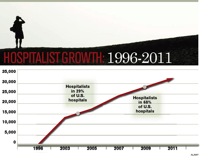
When HM emerged 15 years ago, advocates pitched the fledgling physician specialty as a model of efficient inpatient care, and subsequent findings that the concept led to reductions in length of stay encouraged more hospitals to bolster their staff with the newcomers. With a rising emphasis on quality and patient safety over the past decade, and the new era of pay-for-performance, the hospitalist model of care has expanded to embrace improved quality of care as a chief selling point.
Measuring quality is no easy task, however, and researchers still debate the relative merits of metrics like 30-day readmission rates and inpatient mortality. "Without question, quality measurement is an imperfect science, and all measures will contain some level of imprecision and bias," concluded a recent commentary in Health Affairs.1
Against that backdrop, relatively few studies have looked broadly at the contributions of hospital medicine. Most interventions have been individually tailored to a hospital or instituted at only a few sites, precluding large-scale, head-to-head comparisons.
And so the question remains: Has hospital medicine lived up to its promise on quality?
The Evidence
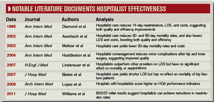
In one of the few national surveys of HM’s impact on patient care, a yearlong comparison of more than 3,600 hospitals found that the roughly 40% that employed hospitalists scored better on multiple Hospital Quality Alliance indicators. The 2009 Archives of Internal Medicine study suggested that hospitals with hospitalists outperformed their counterparts in quality metrics for acute myocardial infarction, pneumonia, overall disease treatment and diagnosis, and counseling and prevention. Congestive heart failure was the only category of the five reviewed that lacked a statistically significant difference.2
A separate editorial, however, argued that the study’s data were not persuasive enough to support the conclusion that hospitalists bring a higher quality of care to the table.3 And even less can be said about the national impact of HM on newly elevated metrics, such as readmission rates. The obligation to gather evidence, in fact, is largely falling upon hospitalists themselves, and the multitude of research abstracts from SHM’s annual meeting in May suggests that plenty of physician scientists are taking the responsibility seriously. Among the presentations, a study led by David Boyte, MD, assistant professor of medicine at Duke University and a hospitalist at Durham Regional Hospital, found that a multidisciplinary approach greatly improved one hospital unit’s 30-day readmission rates for heart failure patients. After a three-month pilot in the cardiac nursing unit, readmission rates fell to 10.7% from 27.6%.4
Although the multidisciplinary effort has included doctors, nurses, nutritionists, pharmacists, unit managers, and other personnel, Dr. Boyte says the involvement of hospitalists has been key to the project’s success. "We feel like we were the main participants who could see the whole picture from a patient-centered perspective," he says. "We were the glue; we were the center node of all the healthcare providers." Based on that dramatic improvement, Dr. Boyte says, the same interventional protocol has been rolled out in three other medical surgical units, and the hospital is using a similar approach to address AMI readmission rates.
SHM’s Project BOOST (Better Outcomes for Older Adults through Safe Transitions; www.hospitalmedicine.org/boost )—by far the largest study of how HM is impacting readmission rates—has amassed data from more than 20 hospitals, with more expected from a growing roster of participants. So far, however, the project has only released data from six pilot sites describing the six-month periods before and after the project’s start. Among those sites, initial results suggest that readmission rates fell by an average of more than 20%, to 11.2% from 14.2%.5
Though the early numbers are encouraging, experts say rates from a larger group of participants at the one-year mark will be more telling, as will direct comparisons between BOOST units and nonparticipating counterparts at the same hospitals. Principal investigator Mark Williams, MD, FHM, professor and chief of the division of hospital medicine at the Northwestern University Feinberg School of Medicine in Chicago, says researchers still need to clean up that data before they’re ready to share it publicly.
In the meantime, some individual BOOST case studies are suggesting that hospitalist-led changes could pay big dividends. To help create cohesiveness and a sense of ownership within its HM program, St. Mary’s Health Center in St. Louis started a 20-bed hospitalist unit in 2008. Philip Vaidyan, MD, FACP, head of the hospitalist program and practice group leader for IPC: The Hospitalist Company at St. Mary’s, says one unit, 3 West, has since functioned as a lab for testing new ideas that are then introduced hospitalwide.
One early change was to bring all of the unit’s care providers together, from doctors and nurses to the unit-based case manager and social worker, for 9 a.m. handoff meetings. "We have this collective brain to find unique solutions," Dr. Vaidyan says. After seeing positive trends on length of stay, 30-day readmission rates, and patient satisfaction scores, St. Mary’s upgraded to a 32-bed hospitalist unit in early 2009. That same year, the 525-bed community teaching hospital was accepted into the BOOST program.
The hospitalist unit’s improved quality scores continued under BOOST, leading to a 33% reduction in readmission rates from 2008 to 2010 (to 10.5% from 15.7%). Rates for a nonhospitalist unit, by contrast, hovered around 17%. "For reducing readmissions, people may think that you have to have a higher length of stay," Dr. Vaidyan says. But the unit trended toward a lower length of stay, in addition to its reduced 30-day readmissions and improved patient satisfaction scores.

Flush with success, the 10 physicians and four nurse practitioners in the hospitalist program have since begun spreading their best practices to the rest of the hospital units. "Hospitalists are in the best ‘sweet spot,’ " Dr. Vaidyan says, "partnering with all of the disciplines, bringing them together, and keeping everybody on the same page."
Ironically, pinpointing the contribution of hospitalists is harder when their changes produce an ecological effect throughout an entire institution, says Siddhartha Singh, MD, MS, associate chief medical officer of Medical College Physicians, the adult practice for Medical College of Wisconsin in Milwaukee. Even so, he stresses that the impact of the two dozen hospitalists at Medical College Physicians has been felt.
"Coinciding with and following the introduction of our hospitalist program in 2004, we have noticed dramatic decreases in our length of stay throughout medicine services," he says. The same has held true for inpatient mortality. "And that, we feel, is attributable to the standardization of processes introduced by the hospitalist group." Multidisciplinary rounds; whiteboards in patient rooms; and standardized admission orders, prophylactic treatments, and discharge processes—"all of this would’ve been impossible, absolutely impossible, without the hospitalist," he says.
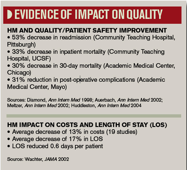
Over the past decade, Dr. Singh’s assessment has been echoed by several studies suggesting that individual hospitalist programs have brought significant improvements in quality measures, such as complication rates and inpatient mortality. In 2002, for example, Andrew Auerbach, MD, MPH, at the University of California San Francisco Medical Center, led a study that compared HM care with that of community physicians in a community-based teaching hospital. Patients cared for by hospitalists, the study found, had a lower risk of death during the hospitalization, as well as at 30 days and 60 days after discharge.6

A separate report by David Meltzer, MD, PhD, and colleagues at the University of Chicago found that an HM program in an academic general medicine service led to a 30% reduction in 30-day mortality rates during its second year of operation.7 And a 2004 study led by Jeanne Huddleston, MD, at the Mayo Clinic College of Medicine in Rochester, Minn., found that a hospitalist-orthopedic co-management model (versus care by orthopedic surgeons with medical consultation) led to more patients being discharged with no complications after elective hip or knee surgery.8 Hospitalist co-management also reduced the rate of minor complications, but had no effect on actual length of stay or cost.
A subsequent study by the same group, however, documented improved efficiency of care through the HM model, but no effect on the mortality of hip fracture patients up to one year after discharge.9 Multiple studies of hospitalist programs, in fact, have seen increased efficiency but little or no impact on inpatient mortality, leading researchers to broadly conclude that such programs can decrease resource use without compromising quality.
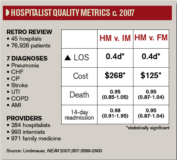
In 2007, a retrospective study of nearly 77,000 patients admitted to 45 hospitals with one of seven common diagnoses compared the care delivered by hospitalists, general internists, and family physicians.10 Although the study authors found that hospitalist care yielded a small drop in length of stay, they saw no difference in the inpatient mortality rates or 14-day readmission rates. More recently, mortality has become ensnared in controversy over its reliability as an accurate indicator of quality.

-Shai Gavi, DO, MPH, chief, section of hospital medicine, assistant professor, Stony Brook University School of Medicine, Brookhaven, N.Y.
Half of the Equation
Despite a lack of ideal metrics, another promising sign for HM might be the model’s exportability. Lee Kheng Hock, MMed, senior consultant and head of the Department of Family Medicine and Continuing Care at Singapore General Hospital, says the 1,600-bed hospital began experimenting with the hospitalist model when officials realized the existing care system wasn’t sustainable. Amid an aging population and increasingly complex and fragmented care, Hock views the hospitalist movement as a natural evolution of the healthcare system to meet the needs of a changing environment.
In a recent study, Hock and his colleagues used the hospital’s administrative database to examine the resource use and outcomes of patients cared for in 2008 by family medicine hospitalists or by specialists.11 The comparison, based on several standard metrics, found no significant improvements in quality, with similar inpatient mortality rates and 30-day, all-cause, unscheduled readmission rates regardless of the care delivery method. The study, though, revealed a significantly shorter hospital stay (4.4 days vs. 5.3 days) and lower costs per patient for those cared for by hospitalists ($2,250 vs. $2,500).11
Hock points out that, like his study, most analyses of hospitalist programs have shown an improvement in length of stay and cost of care without any increase in mortality and morbidity. If value equals quality divided by cost, he says, it stands to reason that quality must increase as overall value remains the same but costs decrease.
"The main difference is that the patients received undivided attention from a well-rounded generalist physician who is focused on providing holistic general medical care," Hock says, adding that "it is really a no-brainer that the outcome would be different."
Patients Rule
Other measures like the effectiveness of communication and seamlessness of handoffs often are assessed through their impacts on patient outcomes. But Sunil Kripalani, MD, MSc, SFHM, chief of the section of hospital medicine and an associate professor of medicine at Vanderbilt University Medical Center in Nashville, Tenn., says communication is now a primary focal point in Medicare’s new hospital value-based purchasing program (VBP). Within VBP’s Hospital Consumer Assessment of Healthcare Providers and Systems (HCAHPS) component, worth 30% of a hospital’s sum score, four of the 10 survey-based measures deal directly with communication. Patients’ overall rating and recommendation of hospitals likely will reflect their satisfaction with communication as well. Dr. Kripalani says it’s inevitable that hospitals—and hospitalists—will pay more attention to communication ratings as patients become judges of quality.
The expertise of hospitalists in handling challenging patients also leads to improved quality over time, says Shai Gavi, DO, MPH, chief of the section of hospital medicine and assistant professor of clinical medicine at Stony Brook University School of Medicine in Brookhaven, N.Y. Hospitalists, he says, excel in handling such high-stakes medical issues as gastrointestinal bleeding, pancreatitis, sepsis, and pain management that can quickly impact patient outcomes if not addressed properly and proficiently. "I think there’s significant value to having people who do this on a pretty frequent basis," he says.
And because of their broad day-to-day interactions, Dr. Gavi says, hospitalists are natural choices for committees focused on improving quality. "When we sit on committees, people often look to us for answers and directions because they know we’re on the front lines and we’ve interfaced with all of the services in the hospital," he says. "You have a good view of the whole hospital operation from A to Z, and I think that’s pretty unique to hospitalists."
The Verdict
In a recent issue brief by Lisa Sprague, principal policy analyst at the National Health Policy Forum, she asserts, "Hospitalists have the undeniable advantage of being there when a crisis occurs, when a patient is ready for discharge, and so on."12
So is "being there" the defining concept of hospital medicine, as she subsequently suggests?
Based on both scientific and anecdotal evidence, the contribution of hospitalists to healthcare quality might be better summarized as "being involved." Whether as innovators, navigators, physician champions, the "sweet spot" of interdepartmental partnerships, the "glue" of multidisciplinary teams, or the nuclei of performance committees, hospitalists are increasingly described as being in the middle of efforts to improve quality. On this basis, the discipline appears to be living up to expectations, though experts say more research is needed to better assess the impacts of HM on quality.
Dr. Vaidyan says hospitalists are particularly well positioned to understand what constitutes ideal care from the perspective of patients. "They want to be treated well: That’s patient satisfaction," he says. "They want to have their chief complaint—why they came to the hospital—properly addressed, so you need a coordinated care team. They want to go home early and don’t want come back: That’s low length of stay and a reduction in 30-day readmissions. And they don’t want any hospital-acquired complications."
Treating patients better, then, should be reflected by improved quality, even if the participation of hospitalists cannot be precisely quantified. "Being involved is something that may be difficult to measure," Dr. Gavi says, "but nonetheless, it has an important impact." TH
Bryn Nelson is a medical writer based in Seattle.
References
- Pronovost PJ, Lilford R. Analysis & commentary: A roadmap for improving the performance of performance measures. Health Aff (Millwood). 2011;30(4):569-73.
- López L, Hicks LS, Cohen AP, McKean S, Weissman JS. Hospitalists and the quality of care in hospitals. Arch Intern Med. 2009;169(15):1389-1394.
- Centor RM, Taylor BB. Do hospitalists improve quality? Arch Intern Med. 2009;169(15):1351-1352.
- Boyte D, Verma L, Wightman M. A multidisciplinary approach to reducing heart failure readmissions. J Hosp Med. 2011;6(4)Supp 2:S14.
- Williams MV, Hansen L, Greenwald J, Howell E, et al. BOOST: impact of a quality improvement project to reduce rehospitalizations. J Hosp Med. 2011;6(4) Supp 2:S88. BOOST: impact of a quality improvement project to reduce rehospitalizations.
- Auerbach AD, Wachter RM, Katz P, Showstack J, Baron RB, Goldman L. Implementation of a voluntary hospitalist service at a community teaching hospital: improved clinical efficiency and patient outcomes. Ann Intern Med. 2002;137(11):859-865.
- Meltzer D, Manning WG, Morrison J, et al. Effects of physician experience on costs and outcomes on an academic general medicine service: results of a trial of hospitalists. Ann Intern Med. 2002;137(1):866-874.
- Huddleston JM, Hall K, Naessens JM, et al. Medical and surgical comanagement after elective hip and knee arthroplasty. Ann Intern Med. 2004;141(1):28-38.
- Batsis JA, Phy MP, Melton LJ, et al. Effects of a hospitalist care model on mortality of elderly patients with hip fractures. J Hosp Med. 2007;2(4): 219–225.
- Lindenauer PK, Rothberg MB, Pekow PS, et al. Outcomes of care by hospitalists, general internists, and family physicians. N Eng J Med. 2007;357:2589-2600.
- Hock Lee K, Yang Y, Soong Yang K, Chi Ong B, Seong Ng H. Bringing generalists into the hospital: outcomes of a family medicine hospitalist model in Singapore. J Hosp Med. 2011;6(3):115-121.
- Sprague L. The hospitalist: better value in inpatient care? National Health Policy Forum website. Available at: www.nhpf.org/library/issue-briefs/IB842_Hospitalist_03-30-11.pdf. Accessed June 28, 2011.
One hospitalist-led pilot project produced a 61% decrease in heart failure readmission rates. Another resulted in a 33% drop in all-cause readmissions. The numbers might be impressive, but what do they really say about how hospitalists have influenced healthcare quality?
When HM emerged 15 years ago, advocates pitched the fledgling physician specialty as a model of efficient inpatient care, and subsequent findings that the concept led to reductions in length of stay encouraged more hospitals to bolster their staff with the newcomers. With a rising emphasis on quality and patient safety over the past decade, and the new era of pay-for-performance, the hospitalist model of care has expanded to embrace improved quality of care as a chief selling point.
Measuring quality is no easy task, however, and researchers still debate the relative merits of metrics like 30-day readmission rates and inpatient mortality. "Without question, quality measurement is an imperfect science, and all measures will contain some level of imprecision and bias," concluded a recent commentary in Health Affairs.1
Against that backdrop, relatively few studies have looked broadly at the contributions of hospital medicine. Most interventions have been individually tailored to a hospital or instituted at only a few sites, precluding large-scale, head-to-head comparisons.
And so the question remains: Has hospital medicine lived up to its promise on quality?
The Evidence
In one of the few national surveys of HM’s impact on patient care, a yearlong comparison of more than 3,600 hospitals found that the roughly 40% that employed hospitalists scored better on multiple Hospital Quality Alliance indicators. The 2009 Archives of Internal Medicine study suggested that hospitals with hospitalists outperformed their counterparts in quality metrics for acute myocardial infarction, pneumonia, overall disease treatment and diagnosis, and counseling and prevention. Congestive heart failure was the only category of the five reviewed that lacked a statistically significant difference.2
A separate editorial, however, argued that the study’s data were not persuasive enough to support the conclusion that hospitalists bring a higher quality of care to the table.3 And even less can be said about the national impact of HM on newly elevated metrics, such as readmission rates. The obligation to gather evidence, in fact, is largely falling upon hospitalists themselves, and the multitude of research abstracts from SHM’s annual meeting in May suggests that plenty of physician scientists are taking the responsibility seriously. Among the presentations, a study led by David Boyte, MD, assistant professor of medicine at Duke University and a hospitalist at Durham Regional Hospital, found that a multidisciplinary approach greatly improved one hospital unit’s 30-day readmission rates for heart failure patients. After a three-month pilot in the cardiac nursing unit, readmission rates fell to 10.7% from 27.6%.4
Although the multidisciplinary effort has included doctors, nurses, nutritionists, pharmacists, unit managers, and other personnel, Dr. Boyte says the involvement of hospitalists has been key to the project’s success. "We feel like we were the main participants who could see the whole picture from a patient-centered perspective," he says. "We were the glue; we were the center node of all the healthcare providers." Based on that dramatic improvement, Dr. Boyte says, the same interventional protocol has been rolled out in three other medical surgical units, and the hospital is using a similar approach to address AMI readmission rates.
SHM’s Project BOOST (Better Outcomes for Older Adults through Safe Transitions; www.hospitalmedicine.org/boost )—by far the largest study of how HM is impacting readmission rates—has amassed data from more than 20 hospitals, with more expected from a growing roster of participants. So far, however, the project has only released data from six pilot sites describing the six-month periods before and after the project’s start. Among those sites, initial results suggest that readmission rates fell by an average of more than 20%, to 11.2% from 14.2%.5
Though the early numbers are encouraging, experts say rates from a larger group of participants at the one-year mark will be more telling, as will direct comparisons between BOOST units and nonparticipating counterparts at the same hospitals. Principal investigator Mark Williams, MD, FHM, professor and chief of the division of hospital medicine at the Northwestern University Feinberg School of Medicine in Chicago, says researchers still need to clean up that data before they’re ready to share it publicly.
In the meantime, some individual BOOST case studies are suggesting that hospitalist-led changes could pay big dividends. To help create cohesiveness and a sense of ownership within its HM program, St. Mary’s Health Center in St. Louis started a 20-bed hospitalist unit in 2008. Philip Vaidyan, MD, FACP, head of the hospitalist program and practice group leader for IPC: The Hospitalist Company at St. Mary’s, says one unit, 3 West, has since functioned as a lab for testing new ideas that are then introduced hospitalwide.
One early change was to bring all of the unit’s care providers together, from doctors and nurses to the unit-based case manager and social worker, for 9 a.m. handoff meetings. "We have this collective brain to find unique solutions," Dr. Vaidyan says. After seeing positive trends on length of stay, 30-day readmission rates, and patient satisfaction scores, St. Mary’s upgraded to a 32-bed hospitalist unit in early 2009. That same year, the 525-bed community teaching hospital was accepted into the BOOST program.
The hospitalist unit’s improved quality scores continued under BOOST, leading to a 33% reduction in readmission rates from 2008 to 2010 (to 10.5% from 15.7%). Rates for a nonhospitalist unit, by contrast, hovered around 17%. "For reducing readmissions, people may think that you have to have a higher length of stay," Dr. Vaidyan says. But the unit trended toward a lower length of stay, in addition to its reduced 30-day readmissions and improved patient satisfaction scores.
Flush with success, the 10 physicians and four nurse practitioners in the hospitalist program have since begun spreading their best practices to the rest of the hospital units. "Hospitalists are in the best ‘sweet spot,’ " Dr. Vaidyan says, "partnering with all of the disciplines, bringing them together, and keeping everybody on the same page."
Ironically, pinpointing the contribution of hospitalists is harder when their changes produce an ecological effect throughout an entire institution, says Siddhartha Singh, MD, MS, associate chief medical officer of Medical College Physicians, the adult practice for Medical College of Wisconsin in Milwaukee. Even so, he stresses that the impact of the two dozen hospitalists at Medical College Physicians has been felt.
"Coinciding with and following the introduction of our hospitalist program in 2004, we have noticed dramatic decreases in our length of stay throughout medicine services," he says. The same has held true for inpatient mortality. "And that, we feel, is attributable to the standardization of processes introduced by the hospitalist group." Multidisciplinary rounds; whiteboards in patient rooms; and standardized admission orders, prophylactic treatments, and discharge processes—"all of this would’ve been impossible, absolutely impossible, without the hospitalist," he says.
Over the past decade, Dr. Singh’s assessment has been echoed by several studies suggesting that individual hospitalist programs have brought significant improvements in quality measures, such as complication rates and inpatient mortality. In 2002, for example, Andrew Auerbach, MD, MPH, at the University of California San Francisco Medical Center, led a study that compared HM care with that of community physicians in a community-based teaching hospital. Patients cared for by hospitalists, the study found, had a lower risk of death during the hospitalization, as well as at 30 days and 60 days after discharge.6
A separate report by David Meltzer, MD, PhD, and colleagues at the University of Chicago found that an HM program in an academic general medicine service led to a 30% reduction in 30-day mortality rates during its second year of operation.7 And a 2004 study led by Jeanne Huddleston, MD, at the Mayo Clinic College of Medicine in Rochester, Minn., found that a hospitalist-orthopedic co-management model (versus care by orthopedic surgeons with medical consultation) led to more patients being discharged with no complications after elective hip or knee surgery.8 Hospitalist co-management also reduced the rate of minor complications, but had no effect on actual length of stay or cost.
A subsequent study by the same group, however, documented improved efficiency of care through the HM model, but no effect on the mortality of hip fracture patients up to one year after discharge.9 Multiple studies of hospitalist programs, in fact, have seen increased efficiency but little or no impact on inpatient mortality, leading researchers to broadly conclude that such programs can decrease resource use without compromising quality.
In 2007, a retrospective study of nearly 77,000 patients admitted to 45 hospitals with one of seven common diagnoses compared the care delivered by hospitalists, general internists, and family physicians.10 Although the study authors found that hospitalist care yielded a small drop in length of stay, they saw no difference in the inpatient mortality rates or 14-day readmission rates. More recently, mortality has become ensnared in controversy over its reliability as an accurate indicator of quality.
-Shai Gavi, DO, MPH, chief, section of hospital medicine, assistant professor, Stony Brook University School of Medicine, Brookhaven, N.Y.
Half of the Equation
Despite a lack of ideal metrics, another promising sign for HM might be the model’s exportability. Lee Kheng Hock, MMed, senior consultant and head of the Department of Family Medicine and Continuing Care at Singapore General Hospital, says the 1,600-bed hospital began experimenting with the hospitalist model when officials realized the existing care system wasn’t sustainable. Amid an aging population and increasingly complex and fragmented care, Hock views the hospitalist movement as a natural evolution of the healthcare system to meet the needs of a changing environment.
In a recent study, Hock and his colleagues used the hospital’s administrative database to examine the resource use and outcomes of patients cared for in 2008 by family medicine hospitalists or by specialists.11 The comparison, based on several standard metrics, found no significant improvements in quality, with similar inpatient mortality rates and 30-day, all-cause, unscheduled readmission rates regardless of the care delivery method. The study, though, revealed a significantly shorter hospital stay (4.4 days vs. 5.3 days) and lower costs per patient for those cared for by hospitalists ($2,250 vs. $2,500).11
Hock points out that, like his study, most analyses of hospitalist programs have shown an improvement in length of stay and cost of care without any increase in mortality and morbidity. If value equals quality divided by cost, he says, it stands to reason that quality must increase as overall value remains the same but costs decrease.
"The main difference is that the patients received undivided attention from a well-rounded generalist physician who is focused on providing holistic general medical care," Hock says, adding that "it is really a no-brainer that the outcome would be different."
Patients Rule
Other measures like the effectiveness of communication and seamlessness of handoffs often are assessed through their impacts on patient outcomes. But Sunil Kripalani, MD, MSc, SFHM, chief of the section of hospital medicine and an associate professor of medicine at Vanderbilt University Medical Center in Nashville, Tenn., says communication is now a primary focal point in Medicare’s new hospital value-based purchasing program (VBP). Within VBP’s Hospital Consumer Assessment of Healthcare Providers and Systems (HCAHPS) component, worth 30% of a hospital’s sum score, four of the 10 survey-based measures deal directly with communication. Patients’ overall rating and recommendation of hospitals likely will reflect their satisfaction with communication as well. Dr. Kripalani says it’s inevitable that hospitals—and hospitalists—will pay more attention to communication ratings as patients become judges of quality.
The expertise of hospitalists in handling challenging patients also leads to improved quality over time, says Shai Gavi, DO, MPH, chief of the section of hospital medicine and assistant professor of clinical medicine at Stony Brook University School of Medicine in Brookhaven, N.Y. Hospitalists, he says, excel in handling such high-stakes medical issues as gastrointestinal bleeding, pancreatitis, sepsis, and pain management that can quickly impact patient outcomes if not addressed properly and proficiently. "I think there’s significant value to having people who do this on a pretty frequent basis," he says.
And because of their broad day-to-day interactions, Dr. Gavi says, hospitalists are natural choices for committees focused on improving quality. "When we sit on committees, people often look to us for answers and directions because they know we’re on the front lines and we’ve interfaced with all of the services in the hospital," he says. "You have a good view of the whole hospital operation from A to Z, and I think that’s pretty unique to hospitalists."
The Verdict
In a recent issue brief by Lisa Sprague, principal policy analyst at the National Health Policy Forum, she asserts, "Hospitalists have the undeniable advantage of being there when a crisis occurs, when a patient is ready for discharge, and so on."12
So is "being there" the defining concept of hospital medicine, as she subsequently suggests?
Based on both scientific and anecdotal evidence, the contribution of hospitalists to healthcare quality might be better summarized as "being involved." Whether as innovators, navigators, physician champions, the "sweet spot" of interdepartmental partnerships, the "glue" of multidisciplinary teams, or the nuclei of performance committees, hospitalists are increasingly described as being in the middle of efforts to improve quality. On this basis, the discipline appears to be living up to expectations, though experts say more research is needed to better assess the impacts of HM on quality.
Dr. Vaidyan says hospitalists are particularly well positioned to understand what constitutes ideal care from the perspective of patients. "They want to be treated well: That’s patient satisfaction," he says. "They want to have their chief complaint—why they came to the hospital—properly addressed, so you need a coordinated care team. They want to go home early and don’t want come back: That’s low length of stay and a reduction in 30-day readmissions. And they don’t want any hospital-acquired complications."
Treating patients better, then, should be reflected by improved quality, even if the participation of hospitalists cannot be precisely quantified. "Being involved is something that may be difficult to measure," Dr. Gavi says, "but nonetheless, it has an important impact." TH
Bryn Nelson is a medical writer based in Seattle.
References
- Pronovost PJ, Lilford R. Analysis & commentary: A roadmap for improving the performance of performance measures. Health Aff (Millwood). 2011;30(4):569-73.
- López L, Hicks LS, Cohen AP, McKean S, Weissman JS. Hospitalists and the quality of care in hospitals. Arch Intern Med. 2009;169(15):1389-1394.
- Centor RM, Taylor BB. Do hospitalists improve quality? Arch Intern Med. 2009;169(15):1351-1352.
- Boyte D, Verma L, Wightman M. A multidisciplinary approach to reducing heart failure readmissions. J Hosp Med. 2011;6(4)Supp 2:S14.
- Williams MV, Hansen L, Greenwald J, Howell E, et al. BOOST: impact of a quality improvement project to reduce rehospitalizations. J Hosp Med. 2011;6(4) Supp 2:S88. BOOST: impact of a quality improvement project to reduce rehospitalizations.
- Auerbach AD, Wachter RM, Katz P, Showstack J, Baron RB, Goldman L. Implementation of a voluntary hospitalist service at a community teaching hospital: improved clinical efficiency and patient outcomes. Ann Intern Med. 2002;137(11):859-865.
- Meltzer D, Manning WG, Morrison J, et al. Effects of physician experience on costs and outcomes on an academic general medicine service: results of a trial of hospitalists. Ann Intern Med. 2002;137(1):866-874.
- Huddleston JM, Hall K, Naessens JM, et al. Medical and surgical comanagement after elective hip and knee arthroplasty. Ann Intern Med. 2004;141(1):28-38.
- Batsis JA, Phy MP, Melton LJ, et al. Effects of a hospitalist care model on mortality of elderly patients with hip fractures. J Hosp Med. 2007;2(4): 219–225.
- Lindenauer PK, Rothberg MB, Pekow PS, et al. Outcomes of care by hospitalists, general internists, and family physicians. N Eng J Med. 2007;357:2589-2600.
- Hock Lee K, Yang Y, Soong Yang K, Chi Ong B, Seong Ng H. Bringing generalists into the hospital: outcomes of a family medicine hospitalist model in Singapore. J Hosp Med. 2011;6(3):115-121.
- Sprague L. The hospitalist: better value in inpatient care? National Health Policy Forum website. Available at: www.nhpf.org/library/issue-briefs/IB842_Hospitalist_03-30-11.pdf. Accessed June 28, 2011.
No Easy Task
In college, while most of her fellow students were staying up late and sleeping in, Alice Marshbanks, MD, FHM, was an early riser. Now she regularly works from 4 p.m. to 2 a.m., and she sleeps in most mornings. "I’m sleeping later and living more of a teenage lifestyle," she jokes. "I’m actually getting younger."
Dr. Marshbanks might be an anomaly among established hospitalists. A physician since 1989 and a hospitalist since 1995, she actually prefers working the swing shift, and she says she’s the only one in her group at WakeMed Hospital in Raleigh, N.C., who does. Although Dr. Marshbanks is not a true nocturnist—she doesn’t work the typical 7 p.m. to 7 a.m. graveyard shift—her contracted position provides valuable transition coverage for night admissions, which have increased as the HM program at WakeMed has grown.
Surveys indicate that HM groups continue to move toward in-house coverage models to provide 24/7 hospitalist responsiveness. In the 2011 SHM-MGMA State of Hospital Medicine report, which will be released next month, 81% of responding nonteaching hospitalist practices reported providing on-site care at night. That’s up from 68% of responding HM practices that reported furnishing that service in the 2010 report. Only 53% of HM groups reported providing on-site night hospitalists in the 2007-2008 State of Hospital Medicine survey, which was produced solely by SHM.
Kenneth R. Epstein, MD, MBA, FACP, FHM, chief medical officer for Hospitalist Consultants Inc., headquartered in Traverse City, Mich., has observed this trend first-hand. In academic hospitals, due to new Accreditation Council for Graduate Medical Education (ACGME) and Resident Review Committee (RRC) regulations, "the only safety valve to handle admissions after the house staff numbers are capped is the hospitalist."
The need for such a safety valve will increase again this summer, as new ACGME duty-hour regulations on resident hours and supervision kick in.
Nonteaching hospitals are not exempt from these pressures. To deal with increasing demands for night coverage, HM groups across the country are using a variety of practice models, such as hiring dedicated nocturnists or moonlighters to cover nights, rotating shifts among team members, or using midlevel providers (physician assistants or nurse practitioners) as night staffers. On-call or in-house coverage models are determined by a variety of factors, including the size of the HM group, patient volume and acuity, and staff availability. Sustainability continues to be a challenge for most groups; however, the in-house coverage model seems to increase nursing and ED satisfaction, most experts say, and is an added value for hospital administration, although financial returns vary.
Continuity of care is at the heart of the night-coverage issue. Some experts worry that patient outcomes will suffer if there isn’t an in-house presence, but studies looking at this issue have been inconclusive, asserts Patti VanDort, RN, MSN, NEA-BC, vice president of nursing and chief nursing officer at Holland Hospital in southwestern Michigan.
"You’ve got to have the same level and quality of care during nights and weekends that you have during the weekdays," she says. "It’s got to be the same for all."
That said, some hospitals don’t have the volume to justify in-house night staffing. Hospitalists and program directors have described the ways in which they handle night staffing, balancing demand, program size, and physician satisfaction.
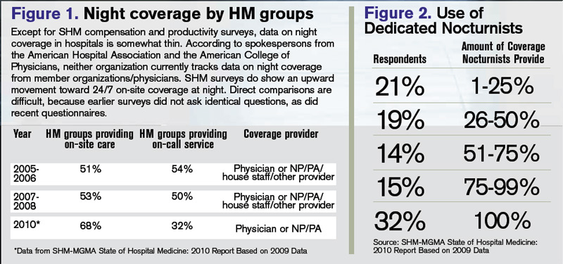
Tailored to Fit
"Hospitalist programs have different scale and scope depending on the needs of the institution," says Michael R. Humphrey, MD, vice president and chief clinical officer for Emergency and Ambulatory Services at St. Rita’s Medical Center in Lima, Ohio. A 365-bed community hospital, St. Rita’s employs nocturnists as part of its 24-hour hospitalist program. Dr. Humphrey still works as an ED physician and reports that the hospitalists are invaluable for admitting, providing cross-cover, covering the ICU, and handling code blue and rapid responses. "As a Level II trauma center, we can’t have ED physicians leave the department to run upstairs and do codes," he says. "They typically don’t get back within five minutes."
Holland Hospital, a 213-bed facility, provides around-the-clock hospitalist coverage in its eight-bed ICU, according to VanDort. That change was precipitated by the nursing staff’s decision to pursue Magnet Status, which was awarded in 2007 by the American Nurses Credentialing Center (ANCC). For inpatient coverage, the hospital-owned HM group Lakeshore Health Partners, headed by Bart D. Sak, MD, MA, FHM, maintains six FTE hospitalists on a rotating block schedule. Each night, one physician works from 4 p.m. until midnight, overlapping with a nonphysician provider (NPP), a member of the hospitalist group, who works a 7 p.m. to 7 a.m. shift.
"We have two providers in-house when admissions from the ED are heating up, and then we have an NPP in-house to cover the one to three additional admissions that may come in after midnight and to field floor calls," Dr. Sak says.
The physician who worked until midnight is on call for backup support and might come back to the hospital if things get too intense in the pre-dawn hours. "This arrangement works quite well for a program of our size," Dr. Sak says. "It takes a team-oriented approach and experienced NPPs who can work independently."
The Holland approach simply wouldn’t work at Kaiser Permanente’s East Bay site in Oakland, Calif., where Tom Baudendistel, MD, FACP, is part of a 50-member hospitalist group and director of the internal-medicine residency program. "Between codes, cross-cover, ICU, and floor admissions, there is simply too much acuity and volume," he says.
The peak hours for East Bay admissions are mid-afternoon to midnight. Two overnight hospitalist shifts (one from 8 a.m. to 8 p.m., another from 7 a.m. to 7 p.m.) are supplemented with two swing shifts (one from 2 to 10 p.m., another from 4 p.m. to midnight). Four full-time nocturnists cover 10 of the 14 overnight shifts per week, which allows for vacation and some protected administrative time. The balance of the overnight shifts are covered by the rest of the hospitalist group, which has 50 members.
The contracted nocturnists are incentivized with additional compensation at the end of the year, when the chief of hospitalists allocates bonuses. They also work fewer shifts a month than the other members of the group. "One thing our group agrees on is that the night docs should get a little more," Dr. Baudendistel says. "It’s a very fair tradeoff for everyone."
A Mile in Their Shoes
Medical directors must balance a variety of factors when scheduling around-the-clock coverage. From day one, the hospitalist program at Albany Memorial Hospital in New York, where John Krisa, MD, is medical director, has been an in-house 24/7 program. Dr. Krisa’s group uses per diem physicians or fellows on their days off to cover most of the nights. The other hospitalists on the team do not escape occasional night duty, and they cover what is left after plugging in the moonlighters. This leaves from zero to five nights per month for each full-time hospitalist. Even the medical director covers night shifts, something Dr. Krisa thinks is valuable to his leadership.
"You, as the leader, still have to walk a mile in that other person’s shoes," he says. "There are different challenges associated with both day and night shifts, so you have to appreciate what your colleagues are going through on the other shifts."
Hospitalist Consultants’ Dr. Epstein agrees with that concept.
"Whenever medical directors have personal experience of how the system is working, they are better able to recommend and make changes," he says.
It’s also valuable, Dr. Krisa explains, for the group leader to interact with ED staff and hear their concerns. Working night shifts helps avoid the night team versus day team schisms, which can lead to group disunity, he says.
Different Skill Set, Different Mindset?
The fact of the matter, though, is that pulling night shifts does not appeal to most established hospitalists. Sleep researchers have found that humans’ body clocks prefer office hours. Even if night-shift hours are consistent, those who work nights never really catch up on the sleep they need during the daytime.
Even so, some physicians embrace the graveyard shift. Working the night swing shift agrees with Dr. Marshbanks’ schedule. The hours are consistent, she works fewer shifts to qualify for FTE pay, and her shift is time-limited, as opposed to work-limited. She’s also filling a niche that others in her group eschew. "It’s a shift that most people with children don’t like because the hours are very disruptive to family life," she says.
The workload at night is different. Instead of the routine rounding typical in day shifts, her work is more urgent. She does more admissions because she works the busiest ED hours, covers acute-stroke codes, and provides cross-cover. And, she says, night staff tends to be "a solid group, so we interact more on a regular basis, since there are fewer of us."
The nocturnists at St. Rita’s Hospital are not held to the same meeting schedule as their daytime hospitalist colleagues, but they’re expected to read meeting minutes and to be responsible for any changes in guidelines or operational information, Dr. Humphrey says. Also stipulated in their hospitalist contracts is the requirement that they maintain competency in procedures, such as central-line placement and airway management.
What’s Better for Patients?
Experts have raised concerns that patient care can be compromised during off-hours, when staffing levels are reduced.1 The Leapfrog Group’s ICU Physician Safety (IPS) Standard argues for high-intensity ICU staffing to reduce patient mortality.2 A number of investigators have tried to determine whether patients admitted off-hours (weekends, nights, holidays) fare worse than those admitted during weekdays. Peter Cram, MD, MBA, acting director of the division of general internal medicine and associate professor of medicine at the Carver College of Medicine at the University of Iowa in Iowa City, found in a 2004 study that patients admitted to hospitals on weekends experienced slightly higher risk-adjusted mortality than did patients admitted on weekdays.3
But here’s the problem with studies such as this, says Dr. Cram: "Patients admitted on evenings and weekends are not the same as those admitted 9 to 5 on weekdays."
During weekdays, admissions combine patients with emergent issues and those scheduled for elective procedures. On weekends, "you get only emergencies—you don’t have low-risk patients," he points out. "So, even with optimal 24/7 staffing, you would still expect those patients coming in at night, and on holidays, to have worse outcomes because they are coming in with more acute problems. It remains an open question whether 24/7 staffing will improve off-hours outcomes." More research, Dr. Cram adds, is needed to establish whether full in-house staffing is the best solution.
Dr. Epstein has compared on-call versus in-house night staffing. In a 2007 study, he found no difference when using indicators such as length of stay, readmission rates, and patient satisfaction.4 However, he noticed positives from in-house coverage. "Although there are no data supporting the value of hospitalists on these parameters, having a nocturnist in-house increases nursing satisfaction, because they are responsive to pages when there is a question about a patient," he says. "It’s also a service to hospital medical staff, because they can handle rapid responses and codes."
There is some evidence that working nights can be deleterious to physicians’ and nurses’ health. One study found that interns were more likely to be involved in collisions after leaving extended night shifts; another found an increased risk of needle-stick injury at the end of a long night shift; and data from the long-running Nurses’ Health Study indicate that long-term night work can result in increased risk of colorectal and breast cancers.5,6,7,8 The increased risks of cancer could be related to lack of exposure to light at night and the body’s decreased production of melatonin, although this remains a topic of ongoing research.
"No Easy Answers"
VanDort, the nursing director, is "passionate" about having 24/7 coverage and reports that her nursing staff is happy with the hybrid model currently used at Holland Hospital. "I do envision a day when we’ll have physicians here around the clock," she says. "Patients are sick during the middle of the night, so you can’t staff your system one way during the daytime hours and your nighttime differently. It’s not fair to those patients."
Dr. Cram, who is a hospitalist, outcomes researcher, and division director, says that in an ideal world, it would make more business sense to have the hospital operating at full capacity around the clock, seven days a week. "But we don’t live in that world," he admits. "It is hard to find ways to achieve ’round-the-clock staffing at the levels we’d like."
He also concludes that there are "no easy answers" to the night-coverage conundrum. "But it might be prudent to think about incentives," he says. "Perhaps we should pay more for staffing weekends, evenings, and holidays, or we could reduce the annual number of shifts we expect our nocturnists to do, relative to those physicians who staff days."
Dr. Krisa says he, too, is biased toward an in-house coverage model, especially when programs reach a critical volume. "There is no substitute for the immediate ability to evaluate a sick patient," he explains. "My feeling is that an in-house, 24/7 presence will become the standard." TH
Gretchen Henkel is a freelance writer based in California.
References
- Wong HJ, Morra D. Excellent hospital care for all: open and operating 24/7. J Gen Intern Med. 2011.
- Pronovost PJ, Angus DC, Dorman T, Robinson KA, Dremsizov TT, Young TL. Physician staffing patterns and clinical outcomes in critically ill patients: a systemic review. JAMA. 2002;288(17):2151-2162.
- Cram P, Hillis SL, Barnett M, Rosenthal GE. Effects of weekend admission and hospital teaching status on in-hospital mortality. Am J Med. 2004;117(3):151-157.
- Epstein KR, Juarez E, Loya K, Gorman MJ, Singer A. The effect of 24-7 hospitalist coverage on clinical metrics. Presented May 2007, annual meeting, Society of Hospital Medicine, Dallas.
- Barger LK, Cade BE, Ayas NT, et al. Extended work shifts and the risk of motor vehicle crashes among interns. N Engl J Med. 2005;352:125-134.
- Ayas NT, Barger LK, Cade BE, et al. Extended work duration and the risk of self-reported percutaneous injuries in interns. JAMA. 2006;296(9):1055-1062.
- Schernhammer ES, Laden F, Speizer FE, et al. Night-shift work and risk of colorectal cancer in the nurses’ health study. J Natl Cancer Inst. 2003;95(11):825-828.
- Schernhammer ES, Laden F, Speizer FE, et al. Rotating night shifts and risk of breast cancer in women partici-pating in the nurses’ health study. J Natl Cancer Inst. 2001;93(20):1563-1568.
In college, while most of her fellow students were staying up late and sleeping in, Alice Marshbanks, MD, FHM, was an early riser. Now she regularly works from 4 p.m. to 2 a.m., and she sleeps in most mornings. "I’m sleeping later and living more of a teenage lifestyle," she jokes. "I’m actually getting younger."
Dr. Marshbanks might be an anomaly among established hospitalists. A physician since 1989 and a hospitalist since 1995, she actually prefers working the swing shift, and she says she’s the only one in her group at WakeMed Hospital in Raleigh, N.C., who does. Although Dr. Marshbanks is not a true nocturnist—she doesn’t work the typical 7 p.m. to 7 a.m. graveyard shift—her contracted position provides valuable transition coverage for night admissions, which have increased as the HM program at WakeMed has grown.
Surveys indicate that HM groups continue to move toward in-house coverage models to provide 24/7 hospitalist responsiveness. In the 2011 SHM-MGMA State of Hospital Medicine report, which will be released next month, 81% of responding nonteaching hospitalist practices reported providing on-site care at night. That’s up from 68% of responding HM practices that reported furnishing that service in the 2010 report. Only 53% of HM groups reported providing on-site night hospitalists in the 2007-2008 State of Hospital Medicine survey, which was produced solely by SHM.
Kenneth R. Epstein, MD, MBA, FACP, FHM, chief medical officer for Hospitalist Consultants Inc., headquartered in Traverse City, Mich., has observed this trend first-hand. In academic hospitals, due to new Accreditation Council for Graduate Medical Education (ACGME) and Resident Review Committee (RRC) regulations, "the only safety valve to handle admissions after the house staff numbers are capped is the hospitalist."
The need for such a safety valve will increase again this summer, as new ACGME duty-hour regulations on resident hours and supervision kick in.
Nonteaching hospitals are not exempt from these pressures. To deal with increasing demands for night coverage, HM groups across the country are using a variety of practice models, such as hiring dedicated nocturnists or moonlighters to cover nights, rotating shifts among team members, or using midlevel providers (physician assistants or nurse practitioners) as night staffers. On-call or in-house coverage models are determined by a variety of factors, including the size of the HM group, patient volume and acuity, and staff availability. Sustainability continues to be a challenge for most groups; however, the in-house coverage model seems to increase nursing and ED satisfaction, most experts say, and is an added value for hospital administration, although financial returns vary.
Continuity of care is at the heart of the night-coverage issue. Some experts worry that patient outcomes will suffer if there isn’t an in-house presence, but studies looking at this issue have been inconclusive, asserts Patti VanDort, RN, MSN, NEA-BC, vice president of nursing and chief nursing officer at Holland Hospital in southwestern Michigan.
"You’ve got to have the same level and quality of care during nights and weekends that you have during the weekdays," she says. "It’s got to be the same for all."
That said, some hospitals don’t have the volume to justify in-house night staffing. Hospitalists and program directors have described the ways in which they handle night staffing, balancing demand, program size, and physician satisfaction.
Tailored to Fit
"Hospitalist programs have different scale and scope depending on the needs of the institution," says Michael R. Humphrey, MD, vice president and chief clinical officer for Emergency and Ambulatory Services at St. Rita’s Medical Center in Lima, Ohio. A 365-bed community hospital, St. Rita’s employs nocturnists as part of its 24-hour hospitalist program. Dr. Humphrey still works as an ED physician and reports that the hospitalists are invaluable for admitting, providing cross-cover, covering the ICU, and handling code blue and rapid responses. "As a Level II trauma center, we can’t have ED physicians leave the department to run upstairs and do codes," he says. "They typically don’t get back within five minutes."
Holland Hospital, a 213-bed facility, provides around-the-clock hospitalist coverage in its eight-bed ICU, according to VanDort. That change was precipitated by the nursing staff’s decision to pursue Magnet Status, which was awarded in 2007 by the American Nurses Credentialing Center (ANCC). For inpatient coverage, the hospital-owned HM group Lakeshore Health Partners, headed by Bart D. Sak, MD, MA, FHM, maintains six FTE hospitalists on a rotating block schedule. Each night, one physician works from 4 p.m. until midnight, overlapping with a nonphysician provider (NPP), a member of the hospitalist group, who works a 7 p.m. to 7 a.m. shift.
"We have two providers in-house when admissions from the ED are heating up, and then we have an NPP in-house to cover the one to three additional admissions that may come in after midnight and to field floor calls," Dr. Sak says.
The physician who worked until midnight is on call for backup support and might come back to the hospital if things get too intense in the pre-dawn hours. "This arrangement works quite well for a program of our size," Dr. Sak says. "It takes a team-oriented approach and experienced NPPs who can work independently."
The Holland approach simply wouldn’t work at Kaiser Permanente’s East Bay site in Oakland, Calif., where Tom Baudendistel, MD, FACP, is part of a 50-member hospitalist group and director of the internal-medicine residency program. "Between codes, cross-cover, ICU, and floor admissions, there is simply too much acuity and volume," he says.
The peak hours for East Bay admissions are mid-afternoon to midnight. Two overnight hospitalist shifts (one from 8 a.m. to 8 p.m., another from 7 a.m. to 7 p.m.) are supplemented with two swing shifts (one from 2 to 10 p.m., another from 4 p.m. to midnight). Four full-time nocturnists cover 10 of the 14 overnight shifts per week, which allows for vacation and some protected administrative time. The balance of the overnight shifts are covered by the rest of the hospitalist group, which has 50 members.
The contracted nocturnists are incentivized with additional compensation at the end of the year, when the chief of hospitalists allocates bonuses. They also work fewer shifts a month than the other members of the group. "One thing our group agrees on is that the night docs should get a little more," Dr. Baudendistel says. "It’s a very fair tradeoff for everyone."
A Mile in Their Shoes
Medical directors must balance a variety of factors when scheduling around-the-clock coverage. From day one, the hospitalist program at Albany Memorial Hospital in New York, where John Krisa, MD, is medical director, has been an in-house 24/7 program. Dr. Krisa’s group uses per diem physicians or fellows on their days off to cover most of the nights. The other hospitalists on the team do not escape occasional night duty, and they cover what is left after plugging in the moonlighters. This leaves from zero to five nights per month for each full-time hospitalist. Even the medical director covers night shifts, something Dr. Krisa thinks is valuable to his leadership.
"You, as the leader, still have to walk a mile in that other person’s shoes," he says. "There are different challenges associated with both day and night shifts, so you have to appreciate what your colleagues are going through on the other shifts."
Hospitalist Consultants’ Dr. Epstein agrees with that concept.
"Whenever medical directors have personal experience of how the system is working, they are better able to recommend and make changes," he says.
It’s also valuable, Dr. Krisa explains, for the group leader to interact with ED staff and hear their concerns. Working night shifts helps avoid the night team versus day team schisms, which can lead to group disunity, he says.
Different Skill Set, Different Mindset?
The fact of the matter, though, is that pulling night shifts does not appeal to most established hospitalists. Sleep researchers have found that humans’ body clocks prefer office hours. Even if night-shift hours are consistent, those who work nights never really catch up on the sleep they need during the daytime.
Even so, some physicians embrace the graveyard shift. Working the night swing shift agrees with Dr. Marshbanks’ schedule. The hours are consistent, she works fewer shifts to qualify for FTE pay, and her shift is time-limited, as opposed to work-limited. She’s also filling a niche that others in her group eschew. "It’s a shift that most people with children don’t like because the hours are very disruptive to family life," she says.
The workload at night is different. Instead of the routine rounding typical in day shifts, her work is more urgent. She does more admissions because she works the busiest ED hours, covers acute-stroke codes, and provides cross-cover. And, she says, night staff tends to be "a solid group, so we interact more on a regular basis, since there are fewer of us."
The nocturnists at St. Rita’s Hospital are not held to the same meeting schedule as their daytime hospitalist colleagues, but they’re expected to read meeting minutes and to be responsible for any changes in guidelines or operational information, Dr. Humphrey says. Also stipulated in their hospitalist contracts is the requirement that they maintain competency in procedures, such as central-line placement and airway management.
What’s Better for Patients?
Experts have raised concerns that patient care can be compromised during off-hours, when staffing levels are reduced.1 The Leapfrog Group’s ICU Physician Safety (IPS) Standard argues for high-intensity ICU staffing to reduce patient mortality.2 A number of investigators have tried to determine whether patients admitted off-hours (weekends, nights, holidays) fare worse than those admitted during weekdays. Peter Cram, MD, MBA, acting director of the division of general internal medicine and associate professor of medicine at the Carver College of Medicine at the University of Iowa in Iowa City, found in a 2004 study that patients admitted to hospitals on weekends experienced slightly higher risk-adjusted mortality than did patients admitted on weekdays.3
But here’s the problem with studies such as this, says Dr. Cram: "Patients admitted on evenings and weekends are not the same as those admitted 9 to 5 on weekdays."
During weekdays, admissions combine patients with emergent issues and those scheduled for elective procedures. On weekends, "you get only emergencies—you don’t have low-risk patients," he points out. "So, even with optimal 24/7 staffing, you would still expect those patients coming in at night, and on holidays, to have worse outcomes because they are coming in with more acute problems. It remains an open question whether 24/7 staffing will improve off-hours outcomes." More research, Dr. Cram adds, is needed to establish whether full in-house staffing is the best solution.
Dr. Epstein has compared on-call versus in-house night staffing. In a 2007 study, he found no difference when using indicators such as length of stay, readmission rates, and patient satisfaction.4 However, he noticed positives from in-house coverage. "Although there are no data supporting the value of hospitalists on these parameters, having a nocturnist in-house increases nursing satisfaction, because they are responsive to pages when there is a question about a patient," he says. "It’s also a service to hospital medical staff, because they can handle rapid responses and codes."
There is some evidence that working nights can be deleterious to physicians’ and nurses’ health. One study found that interns were more likely to be involved in collisions after leaving extended night shifts; another found an increased risk of needle-stick injury at the end of a long night shift; and data from the long-running Nurses’ Health Study indicate that long-term night work can result in increased risk of colorectal and breast cancers.5,6,7,8 The increased risks of cancer could be related to lack of exposure to light at night and the body’s decreased production of melatonin, although this remains a topic of ongoing research.
"No Easy Answers"
VanDort, the nursing director, is "passionate" about having 24/7 coverage and reports that her nursing staff is happy with the hybrid model currently used at Holland Hospital. "I do envision a day when we’ll have physicians here around the clock," she says. "Patients are sick during the middle of the night, so you can’t staff your system one way during the daytime hours and your nighttime differently. It’s not fair to those patients."
Dr. Cram, who is a hospitalist, outcomes researcher, and division director, says that in an ideal world, it would make more business sense to have the hospital operating at full capacity around the clock, seven days a week. "But we don’t live in that world," he admits. "It is hard to find ways to achieve ’round-the-clock staffing at the levels we’d like."
He also concludes that there are "no easy answers" to the night-coverage conundrum. "But it might be prudent to think about incentives," he says. "Perhaps we should pay more for staffing weekends, evenings, and holidays, or we could reduce the annual number of shifts we expect our nocturnists to do, relative to those physicians who staff days."
Dr. Krisa says he, too, is biased toward an in-house coverage model, especially when programs reach a critical volume. "There is no substitute for the immediate ability to evaluate a sick patient," he explains. "My feeling is that an in-house, 24/7 presence will become the standard." TH
Gretchen Henkel is a freelance writer based in California.
References
- Wong HJ, Morra D. Excellent hospital care for all: open and operating 24/7. J Gen Intern Med. 2011.
- Pronovost PJ, Angus DC, Dorman T, Robinson KA, Dremsizov TT, Young TL. Physician staffing patterns and clinical outcomes in critically ill patients: a systemic review. JAMA. 2002;288(17):2151-2162.
- Cram P, Hillis SL, Barnett M, Rosenthal GE. Effects of weekend admission and hospital teaching status on in-hospital mortality. Am J Med. 2004;117(3):151-157.
- Epstein KR, Juarez E, Loya K, Gorman MJ, Singer A. The effect of 24-7 hospitalist coverage on clinical metrics. Presented May 2007, annual meeting, Society of Hospital Medicine, Dallas.
- Barger LK, Cade BE, Ayas NT, et al. Extended work shifts and the risk of motor vehicle crashes among interns. N Engl J Med. 2005;352:125-134.
- Ayas NT, Barger LK, Cade BE, et al. Extended work duration and the risk of self-reported percutaneous injuries in interns. JAMA. 2006;296(9):1055-1062.
- Schernhammer ES, Laden F, Speizer FE, et al. Night-shift work and risk of colorectal cancer in the nurses’ health study. J Natl Cancer Inst. 2003;95(11):825-828.
- Schernhammer ES, Laden F, Speizer FE, et al. Rotating night shifts and risk of breast cancer in women partici-pating in the nurses’ health study. J Natl Cancer Inst. 2001;93(20):1563-1568.
What’s Next for Hospital Medicine?
At the Medical University of South Carolina (MUSC) in Charleston, a familiar scene plays out in the hospitalist program. New hospitalists express an interest in a certain area and the university tries to accommodate them, making time for them to pursue additional training as they juggle the daily demands of treating patients, says Patrick Cawley, MD, MBA, SFHM, associate professor at the university and a former SHM president.
"We try to have a personal growth plan for each hospitalist that aligns with their interest," Dr. Cawley says. "So if we have a hospitalist that’s very, very interested in quality improvement, we’ll seek out opportunities to get that hospitalist experience, and start with smaller projects and then bigger projects."
As the field of HM hits a notable mark in its history—it’s been 15 years since the term "hospitalist" was coined—more advanced training will continue to emerge as a key issue and obstacle in the field, say experts who were asked to take a look into HM’s crystal ball.
They also predict continued growth of the field, with tens of thousands of new hospitalists emerging in the next decade or so. They also say that hospitalists will emerge as leaders in the application and use of new technology, and that there will be more demands placed on hospitalists to show their worth in hard data.
There also promises to be a growing presence of private management firms providing hospitalists to hospitals, which doctors both inside and outside of those firms say could have a beneficial effect on the overall quality of patient care.

-Patrick Cawley, MD, MBA, SFHM, associate professor, Medical University of South Carolina, Charleston, former SHM president
Father Time
For now, Dr. Cawley says, at MUSC and elsewhere, hospitalist programs are scrambling for time to enhance the skills needed to tend to increased demands.
"You have to carve out time. That’s literally what you have to do," he explains. "That’s expensive to take a doctor away from clinical service for a week, or an even an hour or two a week. I mean, somebody’s got to pay for that."
Training on hospitalist-specific management topics, he says, needs to evolve further. "I think there’s a recognition that this stuff is important and that hospitals and hospitalists need to get better aligned," he says. "This is something that will continue to mature over the next 10 years."
The range of tasks is growing ever broader for the hospitalist, and so the need for enhanced training is greater, says Larry Wellikson, MD, SFHM, CEO of SHM.
"They’re being asked to do bedside patient care, but they’re being asked to do more. They’re asked to be systems engineers, they’re asked to be safety experts, they’re asked to be the information manager, if you will, the IT guys," he says. "These skills they have not been trained to do and they need … either to say, ‘No, I can’t do that because I haven’t been trained,’ or they need to go and look where they can get that expertise.
"That’s what we try to do at SHM, with our Leadership Academy and our Practice Management Academy."
Frank Michota, MD, FHM, director of academic affairs in the Department of Hospital Medicine at The Cleveland Clinic, says that one of the biggest challenges the field needs to tackle over the next several years is to better standardize the education of hospitalists, saying there is "incredible inconsistency from hospitalist to hospitalist in terms of knowledge base, experience and … understanding the scope of practice."
"We continue to have significant variation in hospital practice models and the types of measurements that are available to those hospitalists for practice improvement," he says. "We continue to see significant turnover in the field with kind of a lack of maturity"—and not the kind of experience base that "you would like to see 15 years in."
"There is really no confidence that everyone at the base of that iceberg will ever make it to the tip because it’s still not viewed by many who entered the field as being a long-term career choice," Dr. Michota says. For many, he said, it is "a look-and-see proposition."
All of this, he says, points to the need for a full certification process by an HM board.
"I don’t want to make it sound like it has not been an impressive evolution to this point, but I think if we are going to meet the expectations, we do have to do more than we’re doing now," Dr. Michota says.
Some of the gaps in training might be able to be filled by private hospital management groups, which have training programs for their doctors that are made possible by their scale and whose presence is predicted to grow over the next 15 years.
Robert Bessler, MD, who in 2001 founded Tacoma, Wash.-based Sound Physicians, which has become one of the largest private hospitalist organizations in the country, says private companies are able to conduct training that is impossible for many hospitals to conduct themselves.
"You’re going to get good people who are all of good training and good knowledge, but they’re not all going to have experience," he says, "and so what are the hospitals that are employing 50% of the hospitalists in this country going to do about that? It’s pretty much nothing. They’re going to occasionally send some people to conferences and hope—because they don’t have that infrastructure."
At teaching institutes like those at private firms, the process is sped up, Dr. Bessler adds.
"That’s why we built our hospitalists’ institute at Sound—to turn really good, quality doctors into effective hospitalists in a much more rapid fashion," he says. "Because before we built this, it was just get them involved and hope after a couple of years they’ve really become efficient. Our hospital partners and the patients can’t wait that long."
Robert Reynolds, MD, founder of PrimeDoc, an Asheville, N.C.-based company that provides doctors for 12 hospitalist programs and employs about 100 doctors, says there needs to be more focus on teaching the "realistic side of the business of medicine," as well as on quality outcomes and patient satisfaction. But he also doubts there will be much change in training.
"[From] my cynical side and the voice of experience, I don’t see any change in the near future," he says. "What we’re seeing now is physicians come out of residency with a good clinical base, but really having no idea of how the healthcare system works in a bigger picture, how it works as an industry. So we’re having to spend a lot of time and effort training physicians to start thinking like practicing physicians."
The experts all agree that there will be an increase in hospitalists being provided by private corporations. Dr. Reynolds says that trend will continue in part due to healthcare reform’s emphasis on outcomes for reimbursement and a corporation’s ability to assist with physician training, as well as data and reporting needs.
"More and more hospital compensation and physician compensation is going to be based on actual data, performance data," he says. "And in order to really do a good job of capturing and reporting that kind of data, you need enough size to support an IT system and training systems that will produce and capture the kind of data that will be necessary."
Erin Fisher, MD, MHM, a pediatric hospitalist at Rady Children’s Hospital in San Diego, says a major goal of the future should be to change the reimbursement structure "so that you have something that is reasonable and encourages appropriate testing, treatments, and coordination of our healthcare system in a systematic way, rather than pieces." In such a system, hospitalists might see something to prompt them to intervene in a preventive way.
"The bigger question is, can our healthcare system, in five to 10 years, change itself enough that it uses every episode of care as an opportunity to do preventive care and coordinate care in the best way?" says Dr. Fisher, an SHM board member.
Continued Growth?
There is agreement that the field will continue to expand, with SHM predicting that the number of hospitalists in the U.S. will reach 40,000 in the next several years, up from today’s 30,000 figure.
Dr. Wellikson says that the figure could rise to as many as 70,000 or more if specialty hospitalists—such as surgical hospitalists, neuro-hospitalists, and laborists—are included. Those hospital-based specialties are now only in their infancy.
"Everything you can see shows that people are still flocking into hospital medicine," Dr. Wellikson adds.
Hospitalists numbered in the hundreds just 15 years ago, so growth has been explosive the past decade. Dr. Cawley, however, says the pace of growth might be starting to slow already, shifting to undeveloped or underserved areas. "Hospitalist programs are at almost all the large [hospitals] and really the growth has been at the smaller hospitals in the last several years," he says.
With the projected rise of Medicare beneficiaries due to the aging of the baby-boom generation, use of hospitals is expected to skyrocket, meaning more hospitalists will be needed, Dr. Bessler says. He also cites data from the National Rural Health Association noting that 25% of the U.S. population lives in areas considered rural, but that only 10% of the physicians live in those areas, indicating a potential growth area for hospitalists.
"That would tell me that demand will continue to outpace supply," he says.
Mike Tarwater, a member of the board of the American Hospital Association and CEO of Carolinas Medical Center in Charlotte, N.C., agrees with Dr. Bessler. Even with the move toward more outpatient care, Tarwater says, the aging of the population will mean a higher demand for hospitalists.

-Mike Tarwater, board member, American Hospital Association, CEO, Carolinas Medical Center, Charlotte, N.C.
"I think that the primary-care physicians—either because of their love for it or their belief that it’s the better way to go with the treatment of their patients—are going to be really stretched to keep that ambulatory practice going and to get to round on patients in the hospitals," he says. "I think there’s going to be a continued growth of the trend that we’ve seen over the last 15 years."
That growth also will mean a greater emphasis on technology use, whether it’s technology used for quick diagnostics like portable ultrasound or more widely used and refined electronic health records (EHR)—or, as Tarwater describes, "probably things we don’t imagine today."
"Our doctors, more than any other doctors, are tech-savvy; they’re early adopters," Dr. Wellikson says.
Hospitalists likely will emerge as leaders in the adoption of new technology, several experts predict.
Without a doubt, I think that hospitalists are going to be a driving force in the adaptation of the electronic [health] record to the clinical care within their hospitals," Dr. Michota says.
As the needs of HM grow, and the field grows more complex, there will inevitably be more divisions and departments of hospital medicine in places where it is now only a section, Dr. Cawley says.
"When you’re a division or a department, you have more autonomy over your own future, so I see this happening," he says. "I think more and more will carve themselves out of general internal medicine, and a lot of that will come because of a demand for more independence and greater autonomy." TH
Thomas R. Collins is a medical writer based in Florida.
More Value, More PATIENTs, More Technology
HM Pioneer Looks Into Crystal Ball

"We’re that camel’s nose."
With that in mind, what does Dr. Wachter predict for the next 15 years of HM? Here are his top three prognostications:
- A shift from the pressure to improve quality and safety to pressure to improve value, with more emphasis on cost and waste reduction.
"Hospitalist groups that are effective at [quality and safety] will continue to be popular in their organization, while hospitalist groups that aren’t will find that their standing is compromised," he says.
How that is done will vary from institution to institution. It will require a complex process of literature and creation of algorithms, he adds, "but also rolling up your sleeves and meeting with the right people, and working through the politics and diplomacy in order to get this work done."
- At teaching hospitals, a greater role for hospitalists to take care of patients who have traditionally been cared for by residents.
This is borne of the new Accreditation Council for Graduate Medical Education (ACGME) rules for residents’ work hours and supervision, which will require more hospitalists to fill voids in patient care and supervisory roles.
"If you like growth, that’s a great trend for hospitalists," Dr. Wachter says. "But if you’re in the business of trying to hire enough hospitalists to fill all your needs, it’s not that great because the demand curve is just tremendous; there’s a national shortage of hospitalists."
The trend also will affect what "a faculty job looks like," because there will be more clinical needs that will take time away from more traditionally academic work, he explains.
"Academic hospitals are not very good at creating satisfying, sustainable jobs that are largely clinical," Dr. Wachter says. "So how do you make sure that those people have fulfilling jobs, that they don’t feel like second-class citizens, that they can get promoted if they do what you’ve asked them to do well? I think that’s a huge challenge for the field, but it is a challenge borne of a new imperative."
- A revolutionary move from a pen-and-paper hospital environment to a technology-driven workplace.
"It will change the way we do our work," Dr. Wachter says, adding it will also mean other, more subtle changes.
"It takes away the importance of geography to some extent," he adds. "I can be off in the doctor’s lounge or in my house and still do my work.
"And so how do you retain or enhance the relationships that are so fundamental to providing good care? That’s not only between doctors and patients and families, but also between doctors and nurses, and each other."-TC
At the Medical University of South Carolina (MUSC) in Charleston, a familiar scene plays out in the hospitalist program. New hospitalists express an interest in a certain area and the university tries to accommodate them, making time for them to pursue additional training as they juggle the daily demands of treating patients, says Patrick Cawley, MD, MBA, SFHM, associate professor at the university and a former SHM president.
"We try to have a personal growth plan for each hospitalist that aligns with their interest," Dr. Cawley says. "So if we have a hospitalist that’s very, very interested in quality improvement, we’ll seek out opportunities to get that hospitalist experience, and start with smaller projects and then bigger projects."
As the field of HM hits a notable mark in its history—it’s been 15 years since the term "hospitalist" was coined—more advanced training will continue to emerge as a key issue and obstacle in the field, say experts who were asked to take a look into HM’s crystal ball.
They also predict continued growth of the field, with tens of thousands of new hospitalists emerging in the next decade or so. They also say that hospitalists will emerge as leaders in the application and use of new technology, and that there will be more demands placed on hospitalists to show their worth in hard data.
There also promises to be a growing presence of private management firms providing hospitalists to hospitals, which doctors both inside and outside of those firms say could have a beneficial effect on the overall quality of patient care.
-Patrick Cawley, MD, MBA, SFHM, associate professor, Medical University of South Carolina, Charleston, former SHM president
Father Time
For now, Dr. Cawley says, at MUSC and elsewhere, hospitalist programs are scrambling for time to enhance the skills needed to tend to increased demands.
"You have to carve out time. That’s literally what you have to do," he explains. "That’s expensive to take a doctor away from clinical service for a week, or an even an hour or two a week. I mean, somebody’s got to pay for that."
Training on hospitalist-specific management topics, he says, needs to evolve further. "I think there’s a recognition that this stuff is important and that hospitals and hospitalists need to get better aligned," he says. "This is something that will continue to mature over the next 10 years."
The range of tasks is growing ever broader for the hospitalist, and so the need for enhanced training is greater, says Larry Wellikson, MD, SFHM, CEO of SHM.
"They’re being asked to do bedside patient care, but they’re being asked to do more. They’re asked to be systems engineers, they’re asked to be safety experts, they’re asked to be the information manager, if you will, the IT guys," he says. "These skills they have not been trained to do and they need … either to say, ‘No, I can’t do that because I haven’t been trained,’ or they need to go and look where they can get that expertise.
"That’s what we try to do at SHM, with our Leadership Academy and our Practice Management Academy."
Frank Michota, MD, FHM, director of academic affairs in the Department of Hospital Medicine at The Cleveland Clinic, says that one of the biggest challenges the field needs to tackle over the next several years is to better standardize the education of hospitalists, saying there is "incredible inconsistency from hospitalist to hospitalist in terms of knowledge base, experience and … understanding the scope of practice."
"We continue to have significant variation in hospital practice models and the types of measurements that are available to those hospitalists for practice improvement," he says. "We continue to see significant turnover in the field with kind of a lack of maturity"—and not the kind of experience base that "you would like to see 15 years in."
"There is really no confidence that everyone at the base of that iceberg will ever make it to the tip because it’s still not viewed by many who entered the field as being a long-term career choice," Dr. Michota says. For many, he said, it is "a look-and-see proposition."
All of this, he says, points to the need for a full certification process by an HM board.
"I don’t want to make it sound like it has not been an impressive evolution to this point, but I think if we are going to meet the expectations, we do have to do more than we’re doing now," Dr. Michota says.
Some of the gaps in training might be able to be filled by private hospital management groups, which have training programs for their doctors that are made possible by their scale and whose presence is predicted to grow over the next 15 years.
Robert Bessler, MD, who in 2001 founded Tacoma, Wash.-based Sound Physicians, which has become one of the largest private hospitalist organizations in the country, says private companies are able to conduct training that is impossible for many hospitals to conduct themselves.
"You’re going to get good people who are all of good training and good knowledge, but they’re not all going to have experience," he says, "and so what are the hospitals that are employing 50% of the hospitalists in this country going to do about that? It’s pretty much nothing. They’re going to occasionally send some people to conferences and hope—because they don’t have that infrastructure."
At teaching institutes like those at private firms, the process is sped up, Dr. Bessler adds.
"That’s why we built our hospitalists’ institute at Sound—to turn really good, quality doctors into effective hospitalists in a much more rapid fashion," he says. "Because before we built this, it was just get them involved and hope after a couple of years they’ve really become efficient. Our hospital partners and the patients can’t wait that long."
Robert Reynolds, MD, founder of PrimeDoc, an Asheville, N.C.-based company that provides doctors for 12 hospitalist programs and employs about 100 doctors, says there needs to be more focus on teaching the "realistic side of the business of medicine," as well as on quality outcomes and patient satisfaction. But he also doubts there will be much change in training.
"[From] my cynical side and the voice of experience, I don’t see any change in the near future," he says. "What we’re seeing now is physicians come out of residency with a good clinical base, but really having no idea of how the healthcare system works in a bigger picture, how it works as an industry. So we’re having to spend a lot of time and effort training physicians to start thinking like practicing physicians."
The experts all agree that there will be an increase in hospitalists being provided by private corporations. Dr. Reynolds says that trend will continue in part due to healthcare reform’s emphasis on outcomes for reimbursement and a corporation’s ability to assist with physician training, as well as data and reporting needs.
"More and more hospital compensation and physician compensation is going to be based on actual data, performance data," he says. "And in order to really do a good job of capturing and reporting that kind of data, you need enough size to support an IT system and training systems that will produce and capture the kind of data that will be necessary."
Erin Fisher, MD, MHM, a pediatric hospitalist at Rady Children’s Hospital in San Diego, says a major goal of the future should be to change the reimbursement structure "so that you have something that is reasonable and encourages appropriate testing, treatments, and coordination of our healthcare system in a systematic way, rather than pieces." In such a system, hospitalists might see something to prompt them to intervene in a preventive way.
"The bigger question is, can our healthcare system, in five to 10 years, change itself enough that it uses every episode of care as an opportunity to do preventive care and coordinate care in the best way?" says Dr. Fisher, an SHM board member.
Continued Growth?
There is agreement that the field will continue to expand, with SHM predicting that the number of hospitalists in the U.S. will reach 40,000 in the next several years, up from today’s 30,000 figure.
Dr. Wellikson says that the figure could rise to as many as 70,000 or more if specialty hospitalists—such as surgical hospitalists, neuro-hospitalists, and laborists—are included. Those hospital-based specialties are now only in their infancy.
"Everything you can see shows that people are still flocking into hospital medicine," Dr. Wellikson adds.
Hospitalists numbered in the hundreds just 15 years ago, so growth has been explosive the past decade. Dr. Cawley, however, says the pace of growth might be starting to slow already, shifting to undeveloped or underserved areas. "Hospitalist programs are at almost all the large [hospitals] and really the growth has been at the smaller hospitals in the last several years," he says.
With the projected rise of Medicare beneficiaries due to the aging of the baby-boom generation, use of hospitals is expected to skyrocket, meaning more hospitalists will be needed, Dr. Bessler says. He also cites data from the National Rural Health Association noting that 25% of the U.S. population lives in areas considered rural, but that only 10% of the physicians live in those areas, indicating a potential growth area for hospitalists.
"That would tell me that demand will continue to outpace supply," he says.
Mike Tarwater, a member of the board of the American Hospital Association and CEO of Carolinas Medical Center in Charlotte, N.C., agrees with Dr. Bessler. Even with the move toward more outpatient care, Tarwater says, the aging of the population will mean a higher demand for hospitalists.
-Mike Tarwater, board member, American Hospital Association, CEO, Carolinas Medical Center, Charlotte, N.C.
"I think that the primary-care physicians—either because of their love for it or their belief that it’s the better way to go with the treatment of their patients—are going to be really stretched to keep that ambulatory practice going and to get to round on patients in the hospitals," he says. "I think there’s going to be a continued growth of the trend that we’ve seen over the last 15 years."
That growth also will mean a greater emphasis on technology use, whether it’s technology used for quick diagnostics like portable ultrasound or more widely used and refined electronic health records (EHR)—or, as Tarwater describes, "probably things we don’t imagine today."
"Our doctors, more than any other doctors, are tech-savvy; they’re early adopters," Dr. Wellikson says.
Hospitalists likely will emerge as leaders in the adoption of new technology, several experts predict.
Without a doubt, I think that hospitalists are going to be a driving force in the adaptation of the electronic [health] record to the clinical care within their hospitals," Dr. Michota says.
As the needs of HM grow, and the field grows more complex, there will inevitably be more divisions and departments of hospital medicine in places where it is now only a section, Dr. Cawley says.
"When you’re a division or a department, you have more autonomy over your own future, so I see this happening," he says. "I think more and more will carve themselves out of general internal medicine, and a lot of that will come because of a demand for more independence and greater autonomy." TH
Thomas R. Collins is a medical writer based in Florida.
More Value, More PATIENTs, More Technology
HM Pioneer Looks Into Crystal Ball
"We’re that camel’s nose."
With that in mind, what does Dr. Wachter predict for the next 15 years of HM? Here are his top three prognostications:
- A shift from the pressure to improve quality and safety to pressure to improve value, with more emphasis on cost and waste reduction.
"Hospitalist groups that are effective at [quality and safety] will continue to be popular in their organization, while hospitalist groups that aren’t will find that their standing is compromised," he says.
How that is done will vary from institution to institution. It will require a complex process of literature and creation of algorithms, he adds, "but also rolling up your sleeves and meeting with the right people, and working through the politics and diplomacy in order to get this work done."
- At teaching hospitals, a greater role for hospitalists to take care of patients who have traditionally been cared for by residents.
This is borne of the new Accreditation Council for Graduate Medical Education (ACGME) rules for residents’ work hours and supervision, which will require more hospitalists to fill voids in patient care and supervisory roles.
"If you like growth, that’s a great trend for hospitalists," Dr. Wachter says. "But if you’re in the business of trying to hire enough hospitalists to fill all your needs, it’s not that great because the demand curve is just tremendous; there’s a national shortage of hospitalists."
The trend also will affect what "a faculty job looks like," because there will be more clinical needs that will take time away from more traditionally academic work, he explains.
"Academic hospitals are not very good at creating satisfying, sustainable jobs that are largely clinical," Dr. Wachter says. "So how do you make sure that those people have fulfilling jobs, that they don’t feel like second-class citizens, that they can get promoted if they do what you’ve asked them to do well? I think that’s a huge challenge for the field, but it is a challenge borne of a new imperative."
- A revolutionary move from a pen-and-paper hospital environment to a technology-driven workplace.
"It will change the way we do our work," Dr. Wachter says, adding it will also mean other, more subtle changes.
"It takes away the importance of geography to some extent," he adds. "I can be off in the doctor’s lounge or in my house and still do my work.
"And so how do you retain or enhance the relationships that are so fundamental to providing good care? That’s not only between doctors and patients and families, but also between doctors and nurses, and each other."-TC
Maternity Management
Editor's Note: Second in a two-part series
Lest anyone forget, it is essential to support workers having children for one reason—the continuation of the human species, says Rachel Lovins, MD, SFHM, who directs the hospitalist program at Waterbury Hospital in Waterbury, Conn. For HM program directors, that means following pregnancy labor laws. But it also should involve reasonably accommodating hospitalists who are balancing their new baby’s needs with the demands of their profession, says Dr. Lovins and other HM leaders.
"As there are more women in medicine, everybody needs to be more aware of this issue. We don’t want to make good talent feel uncomfortable with the process of taking maternity leave and reducing time," says Michelle Marks, DO, FAAP, SFHM, director of the Center for Pediatric Hospital Medicine at the Cleveland Clinic.
All HM program directors need to be aware of such federal laws as the Pregnancy Discrimination Act and the Family and Medical Leave Act (www.eeoc.gov/laws/types/pregnancy.cfm), as well as the corresponding laws of the state in which they work. Directors can contact their human resources (HR) department for assistance.
"Calling them upfront will save a lot of headaches later on," says Jasen Gundersen, MD, MBA, CPE, SFHM, chief medical officer of the hospital medicine division in Fort Lauderdale, Fla., for Knoxville, Tenn.-based TeamHealth.
Here are some other recommendations on how HM directors can best manage pregnancy issues affecting their team:
The "R" in Relationship
There are many reasons why the director of a hospitalist group should develop a good relationship with the providers in their group, but one of them is that a hospitalist is more likely to tell her director sooner rather than later that she is pregnant, Dr. Marks says.
"Knowing your staff well and knowing them personally helps a lot, too, because you can gauge where they are going personally, as far as marriage, children, that type of thing," she adds.
The earlier a group leader knows a staff member is pregnant, the more time they have to plan for maternity leave. And the better the plan, the easier the leave is on the entire group, says Dr. Gundersen.
Generally, finding out that a physician is pregnant within three to five months of conception provides enough time to make adequate arrangements for coverage, Drs. Marks and Lovins note.

The Conversation
Before scheduling a meeting to discuss maternity leave and plans for returning to work with the hospitalist, the group leader should call HR to see if such a conversation is permissible, says Dr. Marks. A better approach might be to wait until the hospitalist broaches the subject.
"So many times the hospitalist will ask for counseling as far as what are her options of coming back," Dr. Marks says. "That opens the door for an open discussion."
Once the conversation starts, the group leader should gauge the length of maternity leave, her plans for coming back full time or part time, and the anticipated scheduling limitations or childcare considerations, Dr. Gundersen says.
"That’s not to say the pregnant woman can really predict all the time what’s going to happen," says Kerry Weiner, MD, MPH, chief clinical officer for North Hollywood, Calif.-based IPC: The Hospitalist Company, Inc. "Obviously, it’s a medical condition that can change and everyone understands that. It’s getting a feel of what you can actually know at the time."
If it’s the HM director’s intent to call the physician while she is on leave to see how she and the baby are doing and how the maternity leave is going, that should be discussed during the conversation, Dr. Gundersen says.
"If you establish upfront that you are going to make that phone call, I think that’s fine to do," he explains. "If you’re calling constantly and pressuring the person, I don’t think that that’s kosher at all."

—Rachel Lovins, MD, SFHM, director, hospitalist program, Waterbury (Conn.) Hospital
The Coverage Plan
Most maternity leaves are from eight to 12 weeks, although the length varies by HM program and individual. It is essential to have your group’s coverage plan outlined well in advance of the maternity leave.
In a private-practice model in which hospitalists work weekdays and have a call-coverage schedule for nights and weekends, a group leader can spread the extra work among the other hospitalists in the group because there are more hospitalists working during the day when patient census is higher, Dr. Weiner says.
Shifting the workload in other schedule models isn’t always as easy. "In the seven-day-on, seven-day-off model, because of that maximum patient-to-doctor ratio, I don’t think there’s any way to do it without hiring help," Dr. Lovins says. "It’s important to recruit per diems all the time. When you’re in a bind is the worst time to do it."
To limit the disruption to patient care and operations quality, the goal when using outside hospitalists is to contract with physicians who have worked with the group before and who know the community, hospital, systems, and patients, Dr. Weiner says.
For HM groups that use a flexible schedule, maternity coverage plans aren’t really needed, says Reuben Tovar, MD, chairman of Hospital Internists of Austin, a physician-owned and -managed hospitalist practice in Texas.
"We’re not salary, so that changes the dynamic completely. People who work more make more, and people who work less make less," he explains. "We are much more liberal about time off, because if a person is taking off to do what is important to them, like taking care of a child, then the rest of us feel better about doing extra work."
—Michelle Marks, DO, FAAP, SFHM, director, Center for Pediatric Hospital Medicine, Cleveland Clinic
Things Change
Plans discussed at the outset with a pregnant hospitalist can change after the child is born, HM group directors caution.
"Particularly for the first child, people say, ‘I’ll come back full blast. Don’t worry about it.’ And they figure out how hard all that is in the first couple of weeks, and then I get a different answer," Dr. Tovar says. "I think the whole mom/wife/doctor thing is tough. I recognize how hard that is. Even though I am not in that role, I can see it."
Dr. Gundersen suggests group directors have a backup plan, in case the maternity leave lasts longer than expected or the transition back to work is delayed. "It really prevents you from putting pressure on the physician," he says.
If a hospitalist who had planned to come back full time decides that she wants to work less, a director should check with HR to see what the process would entail.
"Generally, we have to negotiate a time frame for when they can drop down" to part-time hours, Dr. Marks says. "It usually takes three to four months for me to be able to adjust staffing to make it work."
Back to Work
Physicians can return from maternity leave in a reduced role, but they very rarely drop out of medicine entirely, Dr. Marks says.
"[They] have put in a lot of time to get where they are," she says. "Plus, women in medicine are usually high achievers and very interested in their careers."
Yet hospitalist leaders should recognize that returning to work after having a baby is stressful. It will take some time for the returning hospitalist to develop a rhythm between her duties as a mother and a doctor.
Directors can review the hospitalist’s nonclinical roles, help with priorities, and perhaps reassign some of the responsibilities to colleagues, Dr. Marks says. With more women breastfeeding, it is important to provide a convenient space with a door that locks for women to breast-pump at work, she and the other directors say.
"The best thing in the world is to have colleagues that you trust and can rely on," Dr. Lovins says. "That way, people can help each other out in emergencies, like if someone has to take their kid to the doctor. That’s the kind of program I want to have and would want to be part of."
Lisa Ryan is a freelance writer based in New Jersey.
Editor's Note: Second in a two-part series
Lest anyone forget, it is essential to support workers having children for one reason—the continuation of the human species, says Rachel Lovins, MD, SFHM, who directs the hospitalist program at Waterbury Hospital in Waterbury, Conn. For HM program directors, that means following pregnancy labor laws. But it also should involve reasonably accommodating hospitalists who are balancing their new baby’s needs with the demands of their profession, says Dr. Lovins and other HM leaders.
"As there are more women in medicine, everybody needs to be more aware of this issue. We don’t want to make good talent feel uncomfortable with the process of taking maternity leave and reducing time," says Michelle Marks, DO, FAAP, SFHM, director of the Center for Pediatric Hospital Medicine at the Cleveland Clinic.
All HM program directors need to be aware of such federal laws as the Pregnancy Discrimination Act and the Family and Medical Leave Act (www.eeoc.gov/laws/types/pregnancy.cfm), as well as the corresponding laws of the state in which they work. Directors can contact their human resources (HR) department for assistance.
"Calling them upfront will save a lot of headaches later on," says Jasen Gundersen, MD, MBA, CPE, SFHM, chief medical officer of the hospital medicine division in Fort Lauderdale, Fla., for Knoxville, Tenn.-based TeamHealth.
Here are some other recommendations on how HM directors can best manage pregnancy issues affecting their team:
The "R" in Relationship
There are many reasons why the director of a hospitalist group should develop a good relationship with the providers in their group, but one of them is that a hospitalist is more likely to tell her director sooner rather than later that she is pregnant, Dr. Marks says.
"Knowing your staff well and knowing them personally helps a lot, too, because you can gauge where they are going personally, as far as marriage, children, that type of thing," she adds.
The earlier a group leader knows a staff member is pregnant, the more time they have to plan for maternity leave. And the better the plan, the easier the leave is on the entire group, says Dr. Gundersen.
Generally, finding out that a physician is pregnant within three to five months of conception provides enough time to make adequate arrangements for coverage, Drs. Marks and Lovins note.
The Conversation
Before scheduling a meeting to discuss maternity leave and plans for returning to work with the hospitalist, the group leader should call HR to see if such a conversation is permissible, says Dr. Marks. A better approach might be to wait until the hospitalist broaches the subject.
"So many times the hospitalist will ask for counseling as far as what are her options of coming back," Dr. Marks says. "That opens the door for an open discussion."
Once the conversation starts, the group leader should gauge the length of maternity leave, her plans for coming back full time or part time, and the anticipated scheduling limitations or childcare considerations, Dr. Gundersen says.
"That’s not to say the pregnant woman can really predict all the time what’s going to happen," says Kerry Weiner, MD, MPH, chief clinical officer for North Hollywood, Calif.-based IPC: The Hospitalist Company, Inc. "Obviously, it’s a medical condition that can change and everyone understands that. It’s getting a feel of what you can actually know at the time."
If it’s the HM director’s intent to call the physician while she is on leave to see how she and the baby are doing and how the maternity leave is going, that should be discussed during the conversation, Dr. Gundersen says.
"If you establish upfront that you are going to make that phone call, I think that’s fine to do," he explains. "If you’re calling constantly and pressuring the person, I don’t think that that’s kosher at all."
—Rachel Lovins, MD, SFHM, director, hospitalist program, Waterbury (Conn.) Hospital
The Coverage Plan
Most maternity leaves are from eight to 12 weeks, although the length varies by HM program and individual. It is essential to have your group’s coverage plan outlined well in advance of the maternity leave.
In a private-practice model in which hospitalists work weekdays and have a call-coverage schedule for nights and weekends, a group leader can spread the extra work among the other hospitalists in the group because there are more hospitalists working during the day when patient census is higher, Dr. Weiner says.
Shifting the workload in other schedule models isn’t always as easy. "In the seven-day-on, seven-day-off model, because of that maximum patient-to-doctor ratio, I don’t think there’s any way to do it without hiring help," Dr. Lovins says. "It’s important to recruit per diems all the time. When you’re in a bind is the worst time to do it."
To limit the disruption to patient care and operations quality, the goal when using outside hospitalists is to contract with physicians who have worked with the group before and who know the community, hospital, systems, and patients, Dr. Weiner says.
For HM groups that use a flexible schedule, maternity coverage plans aren’t really needed, says Reuben Tovar, MD, chairman of Hospital Internists of Austin, a physician-owned and -managed hospitalist practice in Texas.
"We’re not salary, so that changes the dynamic completely. People who work more make more, and people who work less make less," he explains. "We are much more liberal about time off, because if a person is taking off to do what is important to them, like taking care of a child, then the rest of us feel better about doing extra work."
—Michelle Marks, DO, FAAP, SFHM, director, Center for Pediatric Hospital Medicine, Cleveland Clinic
Things Change
Plans discussed at the outset with a pregnant hospitalist can change after the child is born, HM group directors caution.
"Particularly for the first child, people say, ‘I’ll come back full blast. Don’t worry about it.’ And they figure out how hard all that is in the first couple of weeks, and then I get a different answer," Dr. Tovar says. "I think the whole mom/wife/doctor thing is tough. I recognize how hard that is. Even though I am not in that role, I can see it."
Dr. Gundersen suggests group directors have a backup plan, in case the maternity leave lasts longer than expected or the transition back to work is delayed. "It really prevents you from putting pressure on the physician," he says.
If a hospitalist who had planned to come back full time decides that she wants to work less, a director should check with HR to see what the process would entail.
"Generally, we have to negotiate a time frame for when they can drop down" to part-time hours, Dr. Marks says. "It usually takes three to four months for me to be able to adjust staffing to make it work."
Back to Work
Physicians can return from maternity leave in a reduced role, but they very rarely drop out of medicine entirely, Dr. Marks says.
"[They] have put in a lot of time to get where they are," she says. "Plus, women in medicine are usually high achievers and very interested in their careers."
Yet hospitalist leaders should recognize that returning to work after having a baby is stressful. It will take some time for the returning hospitalist to develop a rhythm between her duties as a mother and a doctor.
Directors can review the hospitalist’s nonclinical roles, help with priorities, and perhaps reassign some of the responsibilities to colleagues, Dr. Marks says. With more women breastfeeding, it is important to provide a convenient space with a door that locks for women to breast-pump at work, she and the other directors say.
"The best thing in the world is to have colleagues that you trust and can rely on," Dr. Lovins says. "That way, people can help each other out in emergencies, like if someone has to take their kid to the doctor. That’s the kind of program I want to have and would want to be part of."
Lisa Ryan is a freelance writer based in New Jersey.
Pediatric HM Literature

Background: The management of young febrile infants with UTIs is marked by uncertainty and variation. Although recent studies have demonstrated the safety and efficacy of oral antibiotics as primary treatment in infants younger than six months of age, younger infants tend to receive a longer duration of intravenous antibiotics. This might reflect a lack of clear delineation of the risk of adverse events and bacteremia in this population.
Study design: Retrospective chart review.
Setting: Twenty primarily tertiary-care EDs.
Synopsis: Infants aged 29 to 60 days with febrile UTIs were identified through laboratory and chart review at the participating centers. Bacteremia and adverse events (death, shock, bacterial meningitis, intensive care, surgical intervention, or other substantial clinical complications) were identified, as well as patients with a high-risk past medical history (PMH) or who were clinically ill on examination, based on a priori definitions of chart wording.
Adverse events occurred in 2.8% of the 1,895 patients; bacteremia occurred in 6.5%. Recursive partitioning analysis was used to identify a very-low-risk population for adverse events—those who were not clinically ill and without high-risk PMH (prediction model sensitivity 98% and negative predictive value 99.9%)—but it was not as successful in accurately identifying infants at very low risk for bacteremia.
Limitations of this study include the lack of a clear description of the adverse events identified (and their presumed relationship to the UTIs), the reliance on ED documentation, and conservative definitions of bacterial meningitis. Nonetheless, this is a study of significant magnitude in a population marked by uncertainty. Results of this study further strengthen data that support the feasibility of outpatient antibiotic therapy in well-appearing infants.
Bottom line: Well-appearing infants aged 29-60 days and without significant past medical history are at very low risk for adverse events.
Citation: Schnadower D, Kupperman N, Macias CG, et al. Febrile infants with urinary tract infections at very low risk for adverse events and bacteremia. Pediatrics. 2010;126:1074-1083.
ONLINE EXCLUSIVE: Listen to Pat Cawley and Frank Michota discuss what's next for HM
Click here to listen to Dr. Cawley
Click here to listen to Dr. Michota
Click here to listen to Dr. Cawley
Click here to listen to Dr. Michota
Click here to listen to Dr. Cawley
Click here to listen to Dr. Michota
ONLINE EXCLUSIVE: Listen to program directors discuss managing a group when a hospitalist is out on maternity leave
Click here to listen to Dr. Weiner
Click here to listen to Dr. Marks
Click here to listen to Dr. Weiner
Click here to listen to Dr. Marks
Click here to listen to Dr. Weiner
Click here to listen to Dr. Marks
ONLINE EXCLUSIVE: Listen to HM pioneers John Nelson, Bob Wachter, and Win Whitcomb ruminate on HM's 15-year anniversary
Click here to listen to Dr. Nelson
Click here to listen to Dr. Wachter
Click here to listen to Dr. Whitcomb
Click here to listen to Dr. Nelson
Click here to listen to Dr. Wachter
Click here to listen to Dr. Whitcomb
Click here to listen to Dr. Nelson
Click here to listen to Dr. Wachter
Click here to listen to Dr. Whitcomb