User login
PPIs With Warfarin Regimens: Balancing the Perks and Pitfalls
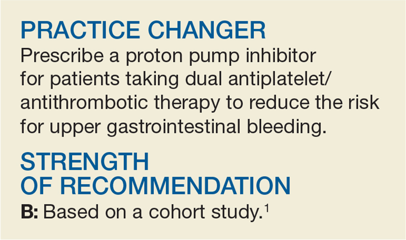
A 60-year-old man establishes care with you. He has well-controlled osteoarthritis (as long as he takes his low-dose daily aspirin) and chronic atrial fibrillation, for which he takes warfarin. His international normalized ratio (INR) is consistently within the recommended target range of 2 to 3. He feels well and has never had GERD or a gastrointestinal (GI) bleed. Should you recommend a proton pump inhibitor (PPI) to decrease the likelihood of a future upper GI bleed?
Anticoagulation therapy creates a dilemma—the need to balance the benefit of preventing embolization with the risk for serious bleeding. Concurrent use of NSAIDs, aspirin, and other antiplatelet agents further increases the latter risk.2
Clinicians have long used PPIs to treat upper GI bleeds. They prevent acid secretion and are the most effective drugs for healing peptic ulcers.3,4 But while previous case-control studies show that PPIs reduce the risk for upper GI bleeds in patients taking antiplatelet agents or NSAIDs, they do not show a statistically significant benefit for patients taking warfarin.5,6 What’s more, while one expert consensus report recommends that patients taking dual warfarin and antiplatelet agent/NSAID therapy take a PPI to decrease the risk for upper GI bleeding, other guidelines do not address this clinical question.2,7,8
STUDY SUMMARY
Study supports PPI use in a high-risk group
This retrospective cohort study sought to answer the question: “Does PPI co-therapy decrease the rate of serious upper GI bleeds in patients taking warfarin?” Researchers examined rates of hospitalization for upper GI bleeding in Medicare and Medicaid patients taking warfarin, with and without PPI co-therapy (tracked via prescription fill dates). They also evaluated concomitant use of NSAIDs and antiplatelet agents.
The authors excluded patients with a recent history of severe bleeding or certain illnesses that predispose patients to GI bleeding (eg, esophageal varices). Patients with risk factors for an upper GI bleed (eg, abdominal pain, peptic ulcer disease, anemia) were more likely to be taking PPI co-therapy. Researchers analyzed the effect of PPI co-therapy in patients with and without these additional risk factors.
Results. The study followed more than 75,000 person-years of active warfarin therapy (Medicaid, > 52,000 person-years; Medicare, > 23,000 person-years). Hospitalizations due to upper GI bleeding occurred at a rate of 127/10,000 person-years (incidence was similar in both the Medicaid and Medicare groups).
Among all patients taking warfarin (regardless of whether they were also taking an NSAID or antiplatelet agent), PPI co-therapy reduced the risk for hospitalization for upper GI bleeding by 24% (adjusted hazard ratio [HR], 0.76), which translates into 29 fewer hospitalizations per 10,000 person-years. The number needed to treat (NNT) was 345 person-years, meaning that 345 patients taking warfarin would have to take a PPI for one year to prevent one hospitalization for an upper GI bleed. As one might expect, PPI co-therapy did not significantly reduce the risk for lower GI, other GI, or non-GI bleeding.
In patients taking both warfarin and concurrent antiplatelet agents or NSAIDs, PPI co-therapy reduced the risk for hospitalization for upper GI bleeding by about half (HR, 0.55). Hospitalizations decreased by 128/10,000 person-years (NNT, 78 person-years). For patients taking warfarin but not antiplatelet agents or NSAIDs, PPI co-therapy did not significantly reduce the risk for hospitalization due to upper GI bleeding (HR, 0.86).
Additional risk factors for GI bleeds. Researchers also looked at patients who had additional risk factors for GI bleeds (other than the exclusion criteria). For patients taking both warfarin and an antiplatelet agent/NSAID, PPI co-therapy decreased the risk for upper GI bleeding regardless of whether the patients had other bleeding risk factors. Again, for patients who had additional bleeding risk factors, but were not taking an antiplatelet agent or NSAID, PPI therapy showed no statistically significant effect.
WHAT’S NEW
PPIs offer benefits, but not to warfarin-only patients
The statistically significant results in this large observational study suggest that PPI co-therapy is beneficial in reducing the risk for upper GI bleeding in patients taking warfarin plus an antiplatelet agent/NSAID, but that PPI co-therapy provides no benefit to patients taking warfarin exclusively.
CAVEATS
Not a randomized controlled trial
This study was observational, not a randomized control trial (RCT). Therefore, unknown confounding variables may have skewed results. For example, patients could have taken OTC medications that influenced or obscured results but were not included in the data analysis (misclassification bias).
At best, we can infer a correlation between PPIs and decreased risk for upper GI bleeds. We need RCTs to determine whether PPIs cause a risk reduction.
Don’t overlook the risks of PPIs. This study assessed the ability of PPIs to prevent bleeds but did not address the risks of long-term PPI therapy. Adverse effects of PPIs include increased risk for pneumonia, infection with Clostridium difficile, hip and spinal fractures, anemia, and possibly chronic kidney disease and dementia.9-11 In addition, cost-analysis studies of PPI therapy are limited and their results are inconsistent.12 Therefore, it’s best to make decisions regarding PPIs after discussing other risks and benefits.
What about DOACs? Another option is to prescribe a direct oral anticoagulant (DOAC; eg, dabigatran, rivaroxaban, or apixaban) instead of warfarin. DOACs are at least as effective as warfarin at preventing stroke in patients with atrial fibrillation and may even be safer.13 Dabigatran 110 mg causes fewer “major bleeding” events than warfarin.13 Compared to warfarin, rivaroxaban has been shown to result in fewer fatal bleeding events due to intracranial bleeds, although it is associated with more GI bleeding.13 Apixaban is associated with fewer GI bleeds and lower bleeding rates overall, compared with warfarin.13 Further research is warranted to determine if PPI therapy is beneficial to patients who are taking DOACs.
CHALLENGES TO IMPLEMENTATION
It’s still a balancing act
When long-term anticoagulation is necessary, providers and patients must attempt to prevent thrombotic events while minimizing the risk for GI bleeds. PPIs may be beneficial in preventing upper GI bleeds in patients taking dual warfarin and antiplatelet therapy, but the long-term consequences of PPI therapy should not be ignored.
ACKNOWLEDGEMENT
The PURLs Surveillance System was supported in part by Grant Number UL1RR024999 from the National Center For Research Resources, a Clinical Translational Science Award to the University of Chicago. The content is solely the responsibility of the authors and does not necessarily represent the official views of the National Center For Research Resources or the National Institutes of Health.
Copyright © 2017. The Family Physicians Inquiries Network. All rights reserved.
Reprinted with permission from the Family Physicians Inquiries Network and The Journal of Family Practice (2017;66[11]:694-696).
1. Ray WA, Chung CP, Murray KT, et al. Association of proton pump inhibitors with reduced risk of warfarin-related serious upper gastrointestinal bleeding. Gastroenterology. 2016;151:1105-1112.
2. Bhatt DL, Scheiman J, Abraham NS, et al. ACCF/ACG/AHA 2008 expert consensus document on reducing the gastrointestinal risks of antiplatelet therapy and NSAID use: a report of the American College of Cardiology Foundation Task Force on Clinical Expert Consensus Documents. J Am Coll Cardiol. 2008;52:1502-1517.
3. Salas M, Ward A, Caro J. Are proton pump inhibitors the first choice for acute treatment of gastric ulcers? A meta analysis of randomized clinical trials. BMC Gastroenterol. 2002;2:17.
4. Shin JM, Sachs G. Pharmacology of proton pump inhibitors. Curr Gastroenterol Rep. 2008;10:528-534.
5. Lanas A, García-Rodríguez LA, Arroyo MT, et al. Effect of antisecretory drugs and nitrates on the risk of ulcer bleeding associated with nonsteroidal anti-inflammatory drugs, antiplatelet agents, and anticoagulants. Am J Gastroenterol. 2007;102:507-515.
6. Lin KJ, Hernández-Díaz S, García Rodríguez LA. Acid suppressants reduce risk of gastrointestinal bleeding in patients on antithrombotic or anti-inflammatory therapy. Gastroenterology. 2011;141:71-79.
7. Ansell J, Hirsh J, Hylek E, et al. Pharmacology and management of the vitamin K antagonists: American College of Chest Physicians Evidence-Based Clinical Practice Guidelines (8th Edition). Chest. 2008;133(6 suppl):160S-198S.
8. Schulman S, Beyth RJ, Kearon C, et al. Hemorrhagic complications of anticoagulant and thrombolytic treatment: American College of Chest Physicians Evidence-Based Clinical Practice Guidelines (8th Edition). Chest. 2008;133(6 suppl):257S-298S.
9. Ament PW, Dicola DB, James ME. Reducing adverse effects of proton pump inhibitors. Am Fam Physician. 2012;86:66-70.
10. Gomm W, von Holt HK, Thomé F, et al. Association of proton pump inhibitors with risk of dementia: a pharmacoepidemiological claims data analysis. JAMA Neurol. 2016;73:410-416.
11. Lazarus B, Chen Y, Wilson FP, et al. Proton pump inhibitor use and the risk of chronic kidney disease. JAMA Intern Med. 2016;176:238-246.
12. Smeets HM, Hoes AW, de Wit NJ. Effectiveness and costs of implementation strategies to reduce acid suppressive drug prescriptions: a systematic review. BMC Health Serv Res. 2007;7:177.
13. Hanley CM, Kowey PR. Are the novel anticoagulants better than warfarin for patients with atrial fibrillation? J Thorac Dis. 2015;7:165-171.
A 60-year-old man establishes care with you. He has well-controlled osteoarthritis (as long as he takes his low-dose daily aspirin) and chronic atrial fibrillation, for which he takes warfarin. His international normalized ratio (INR) is consistently within the recommended target range of 2 to 3. He feels well and has never had GERD or a gastrointestinal (GI) bleed. Should you recommend a proton pump inhibitor (PPI) to decrease the likelihood of a future upper GI bleed?
Anticoagulation therapy creates a dilemma—the need to balance the benefit of preventing embolization with the risk for serious bleeding. Concurrent use of NSAIDs, aspirin, and other antiplatelet agents further increases the latter risk.2
Clinicians have long used PPIs to treat upper GI bleeds. They prevent acid secretion and are the most effective drugs for healing peptic ulcers.3,4 But while previous case-control studies show that PPIs reduce the risk for upper GI bleeds in patients taking antiplatelet agents or NSAIDs, they do not show a statistically significant benefit for patients taking warfarin.5,6 What’s more, while one expert consensus report recommends that patients taking dual warfarin and antiplatelet agent/NSAID therapy take a PPI to decrease the risk for upper GI bleeding, other guidelines do not address this clinical question.2,7,8
STUDY SUMMARY
Study supports PPI use in a high-risk group
This retrospective cohort study sought to answer the question: “Does PPI co-therapy decrease the rate of serious upper GI bleeds in patients taking warfarin?” Researchers examined rates of hospitalization for upper GI bleeding in Medicare and Medicaid patients taking warfarin, with and without PPI co-therapy (tracked via prescription fill dates). They also evaluated concomitant use of NSAIDs and antiplatelet agents.
The authors excluded patients with a recent history of severe bleeding or certain illnesses that predispose patients to GI bleeding (eg, esophageal varices). Patients with risk factors for an upper GI bleed (eg, abdominal pain, peptic ulcer disease, anemia) were more likely to be taking PPI co-therapy. Researchers analyzed the effect of PPI co-therapy in patients with and without these additional risk factors.
Results. The study followed more than 75,000 person-years of active warfarin therapy (Medicaid, > 52,000 person-years; Medicare, > 23,000 person-years). Hospitalizations due to upper GI bleeding occurred at a rate of 127/10,000 person-years (incidence was similar in both the Medicaid and Medicare groups).
Among all patients taking warfarin (regardless of whether they were also taking an NSAID or antiplatelet agent), PPI co-therapy reduced the risk for hospitalization for upper GI bleeding by 24% (adjusted hazard ratio [HR], 0.76), which translates into 29 fewer hospitalizations per 10,000 person-years. The number needed to treat (NNT) was 345 person-years, meaning that 345 patients taking warfarin would have to take a PPI for one year to prevent one hospitalization for an upper GI bleed. As one might expect, PPI co-therapy did not significantly reduce the risk for lower GI, other GI, or non-GI bleeding.
In patients taking both warfarin and concurrent antiplatelet agents or NSAIDs, PPI co-therapy reduced the risk for hospitalization for upper GI bleeding by about half (HR, 0.55). Hospitalizations decreased by 128/10,000 person-years (NNT, 78 person-years). For patients taking warfarin but not antiplatelet agents or NSAIDs, PPI co-therapy did not significantly reduce the risk for hospitalization due to upper GI bleeding (HR, 0.86).
Additional risk factors for GI bleeds. Researchers also looked at patients who had additional risk factors for GI bleeds (other than the exclusion criteria). For patients taking both warfarin and an antiplatelet agent/NSAID, PPI co-therapy decreased the risk for upper GI bleeding regardless of whether the patients had other bleeding risk factors. Again, for patients who had additional bleeding risk factors, but were not taking an antiplatelet agent or NSAID, PPI therapy showed no statistically significant effect.
WHAT’S NEW
PPIs offer benefits, but not to warfarin-only patients
The statistically significant results in this large observational study suggest that PPI co-therapy is beneficial in reducing the risk for upper GI bleeding in patients taking warfarin plus an antiplatelet agent/NSAID, but that PPI co-therapy provides no benefit to patients taking warfarin exclusively.
CAVEATS
Not a randomized controlled trial
This study was observational, not a randomized control trial (RCT). Therefore, unknown confounding variables may have skewed results. For example, patients could have taken OTC medications that influenced or obscured results but were not included in the data analysis (misclassification bias).
At best, we can infer a correlation between PPIs and decreased risk for upper GI bleeds. We need RCTs to determine whether PPIs cause a risk reduction.
Don’t overlook the risks of PPIs. This study assessed the ability of PPIs to prevent bleeds but did not address the risks of long-term PPI therapy. Adverse effects of PPIs include increased risk for pneumonia, infection with Clostridium difficile, hip and spinal fractures, anemia, and possibly chronic kidney disease and dementia.9-11 In addition, cost-analysis studies of PPI therapy are limited and their results are inconsistent.12 Therefore, it’s best to make decisions regarding PPIs after discussing other risks and benefits.
What about DOACs? Another option is to prescribe a direct oral anticoagulant (DOAC; eg, dabigatran, rivaroxaban, or apixaban) instead of warfarin. DOACs are at least as effective as warfarin at preventing stroke in patients with atrial fibrillation and may even be safer.13 Dabigatran 110 mg causes fewer “major bleeding” events than warfarin.13 Compared to warfarin, rivaroxaban has been shown to result in fewer fatal bleeding events due to intracranial bleeds, although it is associated with more GI bleeding.13 Apixaban is associated with fewer GI bleeds and lower bleeding rates overall, compared with warfarin.13 Further research is warranted to determine if PPI therapy is beneficial to patients who are taking DOACs.
CHALLENGES TO IMPLEMENTATION
It’s still a balancing act
When long-term anticoagulation is necessary, providers and patients must attempt to prevent thrombotic events while minimizing the risk for GI bleeds. PPIs may be beneficial in preventing upper GI bleeds in patients taking dual warfarin and antiplatelet therapy, but the long-term consequences of PPI therapy should not be ignored.
ACKNOWLEDGEMENT
The PURLs Surveillance System was supported in part by Grant Number UL1RR024999 from the National Center For Research Resources, a Clinical Translational Science Award to the University of Chicago. The content is solely the responsibility of the authors and does not necessarily represent the official views of the National Center For Research Resources or the National Institutes of Health.
Copyright © 2017. The Family Physicians Inquiries Network. All rights reserved.
Reprinted with permission from the Family Physicians Inquiries Network and The Journal of Family Practice (2017;66[11]:694-696).
1. Ray WA, Chung CP, Murray KT, et al. Association of proton pump inhibitors with reduced risk of warfarin-related serious upper gastrointestinal bleeding. Gastroenterology. 2016;151:1105-1112.
2. Bhatt DL, Scheiman J, Abraham NS, et al. ACCF/ACG/AHA 2008 expert consensus document on reducing the gastrointestinal risks of antiplatelet therapy and NSAID use: a report of the American College of Cardiology Foundation Task Force on Clinical Expert Consensus Documents. J Am Coll Cardiol. 2008;52:1502-1517.
3. Salas M, Ward A, Caro J. Are proton pump inhibitors the first choice for acute treatment of gastric ulcers? A meta analysis of randomized clinical trials. BMC Gastroenterol. 2002;2:17.
4. Shin JM, Sachs G. Pharmacology of proton pump inhibitors. Curr Gastroenterol Rep. 2008;10:528-534.
5. Lanas A, García-Rodríguez LA, Arroyo MT, et al. Effect of antisecretory drugs and nitrates on the risk of ulcer bleeding associated with nonsteroidal anti-inflammatory drugs, antiplatelet agents, and anticoagulants. Am J Gastroenterol. 2007;102:507-515.
6. Lin KJ, Hernández-Díaz S, García Rodríguez LA. Acid suppressants reduce risk of gastrointestinal bleeding in patients on antithrombotic or anti-inflammatory therapy. Gastroenterology. 2011;141:71-79.
7. Ansell J, Hirsh J, Hylek E, et al. Pharmacology and management of the vitamin K antagonists: American College of Chest Physicians Evidence-Based Clinical Practice Guidelines (8th Edition). Chest. 2008;133(6 suppl):160S-198S.
8. Schulman S, Beyth RJ, Kearon C, et al. Hemorrhagic complications of anticoagulant and thrombolytic treatment: American College of Chest Physicians Evidence-Based Clinical Practice Guidelines (8th Edition). Chest. 2008;133(6 suppl):257S-298S.
9. Ament PW, Dicola DB, James ME. Reducing adverse effects of proton pump inhibitors. Am Fam Physician. 2012;86:66-70.
10. Gomm W, von Holt HK, Thomé F, et al. Association of proton pump inhibitors with risk of dementia: a pharmacoepidemiological claims data analysis. JAMA Neurol. 2016;73:410-416.
11. Lazarus B, Chen Y, Wilson FP, et al. Proton pump inhibitor use and the risk of chronic kidney disease. JAMA Intern Med. 2016;176:238-246.
12. Smeets HM, Hoes AW, de Wit NJ. Effectiveness and costs of implementation strategies to reduce acid suppressive drug prescriptions: a systematic review. BMC Health Serv Res. 2007;7:177.
13. Hanley CM, Kowey PR. Are the novel anticoagulants better than warfarin for patients with atrial fibrillation? J Thorac Dis. 2015;7:165-171.
Emicizumab reduces bleeds in kids with hemophilia A and inhibitors

ATLANTA—Updated results from the HAVEN 2 trial have shown that emicizumab prophylaxis can reduce bleeds in children with hemophilia A and factor VIII inhibitors.
Sixty-five percent of all patients enrolled in HAVEN 2 had no bleeds while on emicizumab, and 95% had no treated bleeds.
Among patients who had been on emicizumab for at least 12 weeks, 35% had no bleeds, and 87% had no treated bleeds.
The most common adverse events (AEs) in this trial were viral upper respiratory tract infections and injection site reactions.
Guy Young, MD, of Children’s Hospital Los Angeles in California, presented these results at the 2017 ASH Annual Meeting (abstract 85). The trial was sponsored by Hoffmann-La Roche.
HAVEN 2 enrolled 60 patients, ages 1 to 17, who had hemophilia A and inhibitors. Most patients (95%) had severe hemophilia, 3.3% (n=2) had mild disease, and 1.7% (n=1) had moderate disease.
Nearly a quarter of patients (73.3%) had previously received prophylaxis, and 26.7% had previously received episodic treatment.
The median number of bleeds in the previous 24 weeks was 6.0 (range, 0-155), and 38.3% of patients had target joints.
Patients received emicizumab prophylaxis at 3 mg/kg/week for 4 weeks and 1.5 mg/kg/week thereafter. The median observation time was 9 weeks (range, 1.6 to 41.6 weeks).
Efficacy
The efficacy analysis included 57 patients who were younger than 12. The 3 older patients were only included in the safety analysis.
Of the 57 patients, 64.9% had 0 bleeds, 94.7% had 0 treated bleeds, and 98.2% had 0 treated spontaneous bleeds and 0 treated joint bleeds. None of the patients had treated target joint bleeds.
There were a total of 65 bleeds in 20 patients. Eight were joint bleeds, 2 were muscle bleeds, and the rest were classified as “other.” Of the 55 “other’’ bleeds, 26 (40.0%) were spontaneous, 36 (55.4%) were traumatic, and 3 (4.6%) were due to a procedure/surgery.
A subset of 23 patients received emicizumab for at least 12 weeks. They had a median treatment duration of 38.1 weeks (range, 12.7 to 41.6 weeks).
Of these patients, 34.8% had 0 bleeds, 87.0% had 0 treated bleeds, and 95.7% had 0 treated spontaneous bleeds and 0 treated joint bleeds. There were a total of 41 bleeds in 15 of these patients. Three bleeds (joint, muscle, and hip) were treated.
The median annualized bleeding rate (ABR) for the 23 patients was 1.5 for all bleeds and 0.0 for all types of treated bleeds.
There were 13 patients who had participated in a non-interventional study prior to enrolling in HAVEN 2, so these patients could serve as their own controls. The patients had an overall reduction in ABR of 99% with emicizumab.
Safety
All 60 patients were evaluated for safety. Forty patients had a total of 201 AEs. The most common AEs were viral upper respiratory tract infection (16.7%) and injection site reactions (16.7%)
There were 7 serious AEs in 6 patients—muscle hemorrhage (n=2), eye pain, catheter site injection, device-related infection, mouth hemorrhage, and appendicitis. None of these events were considered treatment-related.
There were no thromboembolic or thrombotic microangiopathy events, and none of the patients tested positive for anti-drug antibodies.
“The safety profile of emicizumab was favorable and well-tolerated,” Dr Young said. “And these updated results from the HAVEN 2 study confirm our prior efficacy results, presented at ISTH, that emicizumab successfully prevents or reduces bleeds.” ![]()
Type 2 diabetes remitted with low-calorie diet
Type 2 diabetes mellitus remitted without medication in 46% of subjects who followed a strict, calorie-controlled diet for 1 year, judging from the findings of an open-label, cluster-randomized trial.
Remission rates closely tracked weight loss, Michael E.J. Lean, MD, reported in the Dec. 5 online issue of the Lancet. Among those who lost 15 kg or more, 86% also normalized their hemoglobin A1c levels. Lesser weight losses were successful too, with diabetes remitting in 57% of those who lost 10-15 kg and 34% of those who lost 5-10 kg, reported Dr. Lean, who is chair of human nutrition at the University of Glasgow.
Weight loss conferred other benefits as well. Quality of life improved significantly, triglycerides declined, and about half of the subjects were able to discontinue both their antidiabetic and antihypertension prescriptions.
DiRECT (the Diabetes Remission Clinical Trial) didn’t include a strict exercise component – something that sets it apart from most dietary interventions, Dr. Lean noted in a press statement. Instead, the study’s “Counterweight-plus” diet intervention focused on very strict calorie control. Counterweight is a proprietary, subscription-based weight-loss program that costs about $570 for 1 year.
The paper offered few details about the intervention, which was supervised by a nurse and/or dietitian. For the first 3-5 months, patients consumed only Counterweight-branded soups and shakes, amounting to about 850 calories per day. After that, solid foods were reintroduced over 2-8 weeks. There was ongoing support for weight-loss maintenance, including cognitive-behavioral therapy, combined with strategies to increase physical activity. Activity strategies were confined to encouraging subjects to walk up to 15,000 steps per day in the second and third phase, but the investigators had little hope that this would actually occur.
“It was recognized that this target was unlikely to be achieved by many, and objectively measured physical activity showed no increase in physical activity in either group between baseline and 12 months, which underlines the difficulty this population has in maintaining increased activity,” they noted.
DiRECT enrolled 298 adults with type 2 diabetes recruited from 49 primary care practices across Scotland and England. They were about 54 years old, with a mean diabetes duration of about 3 years. Subjects were assigned to either the Counterweight-plus weight management program or best practice care under current guidelines. At baseline, subjects’ mean body mass index was 35 mg/m2. Their mean HbA1c was about 7.6%; about 75% were taking at least one antidiabetic medication, and 30% taking two or more. Hypertension was present in more than half.
In the active group, investigators withdrew all antidiabetic and antihypertensive medications when the diet commenced. Antihypertensives were restarted only if subjects experienced an increase in systolic blood pressure. Patients in the control group stayed on their medications.
At 12 months, the mean weight loss was significantly greater in the intervention group than the control group (10 kg vs. 1 kg). Weight loss of at least 15 kg occurred in 24% of the intervention group and none of the control group. It was most pronounced in the total diet replacement phase, falling by a mean of 14.5 kg; participants regained weigh during the food reintroduction phase (mean, 1 kg) and again during the maintenance phase (mean, 1.9 kg). Four subjects in the intervention group who experienced diabetes remission needed a “short rescue plan” on the total diet replacement phase because of weight regain within 60 days of the study’s end. The authors didn’t say how much weight these patients regained.
By the end of the study period, diabetes had remitted in 46% of the intervention group and 4% of the control group (odds ratio, 19.7) and was positively associated with the amount of weight loss.
At 12 months, 74% of the intervention group and 18% of the control group were off antidiabetic medications. HbA1c was significantly better in the intervention group (mean 6.4% vs. 7.2%). Antihypertensive drugs also were less common among the intervention group (32% vs. 61%) at 12 months. Despite the reduction in medication, there were no significant changes in blood pressure from baseline.
Nine serious adverse events occurred among seven intervention subjects. Two (biliary colic and abdominal pain) occurred in the same subject and were considered related to the diet, but they did not withdraw promptly.
Counterweight sponsored the trial, and several of the coinvestigators are stockholders and were company employees during the study. Dr. Lean reported financial remuneration from Counterweight.
SOURCE: Lean M et al. Lancet 2017 Dec 5; doi: 10.1016/ S0140-6736(17)33102-1.
The results of the DiRECT are “impressive and strongly support the view that type 2 diabetes is tightly associated with excessive fat mass in the body,” Matti Uusitupa, MD, wrote in an accompanying editorial.
Emerging data point at weight loss as the most effective treatment for type 2 diabetes, Dr. Uusitupa wrote. It confers a variety of benefits: improved insulin sensitivity in muscles and liver, decreased organ fat, and improved insulin secretion. Some studies suggest that fat loss also helps preserve beta cells in the pancreas.
Nevertheless, the study raises some questions. Without long-term data, it’s tough to know whether DiRECT should be a watershed moment in type 2 diabetes treatment, shifting efforts more toward weight loss and less toward medications.
“In view of the results of the DiRECT trial, a nonpharmacologic approach should be revived. In clinical practice, antidiabetic drugs seldom result in normalization of glucose metabolism if patients’ lifestyles remain unchanged. Mechanisms of action of some drugs for type 2 diabetes might not be in line with current knowledge of pathophysiology of disease, whereas intensive weight management along with physical activity and healthy diet is targeted therapy for type 2 diabetes.”
The best time to start a weight-loss war on type 2 diabetes is probably at the time of diagnosis, Dr. Uusitupa suggested, because patients are most highly motivated at that point.
“However, disease prevention should be maintained as the primary goal that requires both individual-level and population-based strategies.”
Dr. Uusitupa is an emeritus professor at the University of Eastern Finland, Kuopio.
Type 2 diabetes mellitus remitted without medication in 46% of subjects who followed a strict, calorie-controlled diet for 1 year, judging from the findings of an open-label, cluster-randomized trial.
Remission rates closely tracked weight loss, Michael E.J. Lean, MD, reported in the Dec. 5 online issue of the Lancet. Among those who lost 15 kg or more, 86% also normalized their hemoglobin A1c levels. Lesser weight losses were successful too, with diabetes remitting in 57% of those who lost 10-15 kg and 34% of those who lost 5-10 kg, reported Dr. Lean, who is chair of human nutrition at the University of Glasgow.
Weight loss conferred other benefits as well. Quality of life improved significantly, triglycerides declined, and about half of the subjects were able to discontinue both their antidiabetic and antihypertension prescriptions.
DiRECT (the Diabetes Remission Clinical Trial) didn’t include a strict exercise component – something that sets it apart from most dietary interventions, Dr. Lean noted in a press statement. Instead, the study’s “Counterweight-plus” diet intervention focused on very strict calorie control. Counterweight is a proprietary, subscription-based weight-loss program that costs about $570 for 1 year.
The paper offered few details about the intervention, which was supervised by a nurse and/or dietitian. For the first 3-5 months, patients consumed only Counterweight-branded soups and shakes, amounting to about 850 calories per day. After that, solid foods were reintroduced over 2-8 weeks. There was ongoing support for weight-loss maintenance, including cognitive-behavioral therapy, combined with strategies to increase physical activity. Activity strategies were confined to encouraging subjects to walk up to 15,000 steps per day in the second and third phase, but the investigators had little hope that this would actually occur.
“It was recognized that this target was unlikely to be achieved by many, and objectively measured physical activity showed no increase in physical activity in either group between baseline and 12 months, which underlines the difficulty this population has in maintaining increased activity,” they noted.
DiRECT enrolled 298 adults with type 2 diabetes recruited from 49 primary care practices across Scotland and England. They were about 54 years old, with a mean diabetes duration of about 3 years. Subjects were assigned to either the Counterweight-plus weight management program or best practice care under current guidelines. At baseline, subjects’ mean body mass index was 35 mg/m2. Their mean HbA1c was about 7.6%; about 75% were taking at least one antidiabetic medication, and 30% taking two or more. Hypertension was present in more than half.
In the active group, investigators withdrew all antidiabetic and antihypertensive medications when the diet commenced. Antihypertensives were restarted only if subjects experienced an increase in systolic blood pressure. Patients in the control group stayed on their medications.
At 12 months, the mean weight loss was significantly greater in the intervention group than the control group (10 kg vs. 1 kg). Weight loss of at least 15 kg occurred in 24% of the intervention group and none of the control group. It was most pronounced in the total diet replacement phase, falling by a mean of 14.5 kg; participants regained weigh during the food reintroduction phase (mean, 1 kg) and again during the maintenance phase (mean, 1.9 kg). Four subjects in the intervention group who experienced diabetes remission needed a “short rescue plan” on the total diet replacement phase because of weight regain within 60 days of the study’s end. The authors didn’t say how much weight these patients regained.
By the end of the study period, diabetes had remitted in 46% of the intervention group and 4% of the control group (odds ratio, 19.7) and was positively associated with the amount of weight loss.
At 12 months, 74% of the intervention group and 18% of the control group were off antidiabetic medications. HbA1c was significantly better in the intervention group (mean 6.4% vs. 7.2%). Antihypertensive drugs also were less common among the intervention group (32% vs. 61%) at 12 months. Despite the reduction in medication, there were no significant changes in blood pressure from baseline.
Nine serious adverse events occurred among seven intervention subjects. Two (biliary colic and abdominal pain) occurred in the same subject and were considered related to the diet, but they did not withdraw promptly.
Counterweight sponsored the trial, and several of the coinvestigators are stockholders and were company employees during the study. Dr. Lean reported financial remuneration from Counterweight.
SOURCE: Lean M et al. Lancet 2017 Dec 5; doi: 10.1016/ S0140-6736(17)33102-1.
FROM LANCET
Key clinical point: Diet alone may be enough to cause remission of type 2 diabetes.
Major finding: Type 2 diabetes remitted without medication in 46% of subjects who followed a strict, calorie-controlled diet for 1 year, according to results from a randomized, controlled trial.
Study details: The randomized study comprised 298 subjects.
Disclosures: Counterweight sponsored the trial, and several of the coinvestigators are stockholders and were company employees during the study. Dr. Lean reported financial remuneration from Counterweight.
Source: Lean M et al. Lancet. 2017. doi: 10.1016/ S0140-6736(17)33102-1.
Azacitidine maintenance improves PFS in older AML patients
ATLANTA – In older patients with acute myeloid leukemia (AML) in complete remission after intensive chemotherapy, the addition of maintenance therapy with azacitidine significantly improved disease-free survival (DFS), according to results of a randomized, placebo-controlled phase 3 study.
Compared with observation, DFS was significantly improved in the maintenance azacitidine arm, according to results from the 116-patient HOVON97 trial presented at the annual meeting of the American Society of Hematology.
Overall survival was not significantly different between arms, possibly because of an excess of allogeneic transplant in the observation arm, according to Geert Huls, MD, PhD, of the department of hematology, University Medical Center Groningen, the Netherlands.
“When censored for allogeneic transplant, maintenance with azacitidine improves overall survival,” Dr. Huls said during an oral presentation on the findings.
The randomized maintenance therapy trial was designed to include 126 patients aged 60 years or older who had a confirmed diagnosis of AML and refractory anemia with excess of blasts (RAEB, RAEB-t) and who were in complete remission or in complete remission with incomplete blood count recovery after two cycles of therapy.
Investigators randomly assigned 116 patients to maintenance versus observation. Researchers intended to assign a total of 126 patients, but the trial was stopped early because of slow accrual, Dr. Huls said.
Maintenance treatment with azacitidine was given until relapse for no more than 12 cycles, according to the study protocol. Disease-free survival, the primary endpoint, was measured from the date of randomization to relapse or death from any cause.
Azacitidine maintenance therapy significantly improved DFS (P = .03), Dr. Huls said. After researchers adjusted for poor risk cytogenetic abnormalities at diagnosis and platelet count at study entry, the DFS difference remained significant (hazard ratio, 0.61; 95% confidence interval, 0.4-0.92; P = .019).
Overall survival, a secondary endpoint of the trial, was not significantly different between arms, even after adjustment for cytogenetic abnormalities and platelet counts, Dr. Huls said.
However, investigators found an excess of allogeneic transplant in the observation arm (11 patients, vs. 3 in the azacitidine arm). After they censored those 14 patients, they saw a difference in overall survival favoring azacitidine maintenance that approached significance (P = .07).
Dr. Huls speculated that the excess of transplant may have been related to “the psychology of the doctors.” In the maintenance arm, the physician’s thought process may have been that “ ‘this patient has now had two lines of treatment and has a relapse, and we are done,’ and in the [observation] arm he says, ‘well, the patient has had one arm of treatment, let’s go for another,’ ” Dr. Huls said.
Tolerability data showed that 14 adverse events were reported in the azacitidine maintenance arm, versus 4 for observation. One serious adverse event of grade 3 was reported in the azacitidine arm. The proportion of patients without platelet transfusions during the study was 86% for azacitidine and 93% for observation, and the proportion of patients without red blood cell transfusions was similarly 86% and 92% for the azacitidine and observation arms, respectively.
Dr. Huls reported financial relationships with Janssen and Celgene.
SOURCE: Huls G et al. ASH 2017 Abstract 463.
AT ASH 2017
Key clinical point:
Major finding: Disease-free survival was significantly improved (HR, 0.61; 95% CI, 0.4-0.92; P = .019)
Data source: A randomized, multicenter phase 3 trial including 116 older patients (60 years or older) with AML and refractory anemia with excess of blasts (RAEB, RAEB-t).
Disclosures: Dr. Huls reported financial relationships with Janssen and Celgene.
Source: Huls G et al. ASH 2017 Abstract 463.
PANACEA: pembrolizumab overcomes trastuzumab resistance for some
SAN ANTONIO – The immune checkpoint inhibitor pembrolizumab overcomes trastuzumab resistance in HER2-positive advanced breast cancer provided that the tumor expresses programmed death ligand 1 (PD-L1), a trial reported at the San Antonio Breast Cancer Symposium suggests. But presence of immune cells in the tumor is a major additional determinant of benefit.

The single-arm phase 1b/2 trial, called PANACEA (also KEYNOTE-014), enrolled 58 patients with HER2-positive advanced breast cancer that had progressed on trastuzumab (Herceptin) or trastuzumab emtansine (Kadcyla). All were given pembrolizumab (Keytruda), which unleashes antitumor immunity by targeting the programmed death-1 receptor on immune cells, in combination with trastuzumab.
With a median follow-up of 13.6 months, the cohort of patients having tumors positive for PD-L1 achieved an overall response rate of 15.2% and a disease control rate of 24%,” Dr. Loi reported in a press briefing and session, on behalf of the International Breast Cancer Study Group and Breast International Group. In contrast, there were no responses in the PD-L1–negative cohort.
Within the PD-L1–positive cohort, stromal levels of TILs in the metastatic lesion – which were low overall – influenced likelihood of benefit. The response rate was almost eight times higher in patients who had at least 5% of the stromal area densely infiltrated with TILs.
“The PANACEA study met its primary endpoint in the PD-L1–positive cohort. For responders, this combination offers durable control without chemotherapy,” Dr. Loi summarized.
“Metastatic HER2-positive breast cancer in this [heavily pretreated] setting is poorly immunogenic, as evidenced by the majority of patients having low TILs in their metastatic lesions. Saying that, however, we did observe a higher response rate in this study as compared to the equivalent triple-negative breast cancer studied in KEYNOTE-086,” she noted. “Future directions in this disease space should focus on combinations with effective anti-HER2 therapy, particularly in low-TIL patients.”
Predicting benefit
The trial is noteworthy for its efforts to identify the subset of patients most likely to benefit from immune checkpoint inhibition, according to press briefing moderator Virginia Kaklamani, MD, a professor of medicine in the division of hematology/oncology at the University of Texas Health Science Center, San Antonio, and a leader of the Breast Cancer Program at the UT Health San Antonio Cancer Center.

In similar studies among patients with HER2-negative breast cancer, PD-L1 did not pan out as a strong predictive biomarker. “What do you think the difference is between that subset and the HER2-positive subset?” Dr. Kaklamani asked.
“First off, I think that there are technical issues with the PD-L1 assay. And we find that patients with high TILs or immune infiltration usually have high levels of PD-L1 expression on their TILs,” Dr. Loi replied. “So I think that PD-L1 can be expressed on the tumor as well as the TIL, and it certainly seems to be the TIL infiltrate that probably enriches for responders to a PD-L1 checkpoint inhibitor on its own or in this case with trastuzumab.”
Study details
In the PANACEA trial (additionally known as IBCSG 45-13 and BIG 4-13), the most common adverse event of any grade and type with the pembrolizumab-trastuzumab combination was fatigue, seen in 21% of patients, Dr. Loi reported. For immune-related adverse events specifically, 19.0% of patients experienced an event, 10.3% experienced an event of grade 3 or worse, and 6.9% stopped treatment because of these events.
“These frequencies are consistent with what has been reported in other solid tumor types with pembrolizumab,” she commented. There were no cardiac events reported.
Efficacy analyses were restricted largely to the PD-L1–positive cohort, given the lack of any response in the negative cohort.
Median duration of response in the positive cohort was 3.5 months, and median duration of disease control was 11.1 months. Five patients (10.8%) remain on treatment with no progression; three of them have completed 2 years of pembrolizumab.
Median progression-free and overall survival were 2.7 and 16.1 months, respectively; corresponding 12-month rates were 13% and 65%. “There is a tantalizing suggestion of a tail on the curve. ... Obviously, this requires further follow-up, and the numbers are small,” Dr. Loi commented.
The median baseline stromal TIL level in metastatic lesions was just 1%. “This is 20 times less than what we observe in primary HER2-positive breast cancers,” she pointed out.
Compared with the PD-L1–negative cohort, the PD-L1–positive cohort had higher TIL levels. Additionally, within that latter cohort, TIL level was higher among patients achieving response versus not (P = .006) and patients achieving disease control versus not (P = .0006).
“We then went on to try to identify a TIL cutoff that could enrich the population for responders. This has been done in other solid tumor types,” Dr. Loi explained.
Analyses in the PD-L1-positive cohort showed that TIL levels down to 5% predicted benefit. The 41% of patients having 5% or more TILs were dramatically more likely to have a response (39% vs. 5%) and disease control (47% vs. 5%).
TIL levels varied widely according to site of the metastasis, with higher levels seen in metastases from lung and lymph nodes, and lower levels seen in those from liver and skin.
“At this stage, we are not sure which is the chicken and the egg: Patients could have disease in their lung and their lymph nodes because their immune system is better controlling their disease,” Dr. Loi commented. “How we treat these patients is still an open question. In patients with liver metastases, perhaps we need to be more aggressive with the primary or tumor-control anti-HER2 therapy.”
Improving efficacy
Going forward, one strategy for improving pembrolizumab efficacy in this patient population might be priming the immune response, according to Dr. Loi.
“In HER2 disease, it’s very clear that oncogenic signaling is the driver, so targeting HER2 potently also will help relieve tumor-mediated immune suppression,” she elaborated. “In this particular context, targeting HER2 well is the key. Whether you need the addition of a little bit of chemo or some radiation, all this needs to be studied.”
Another strategy for improving pembrolizumab efficacy might be moving the drug to earlier disease settings, Dr. Loi proposed.
“By the time you get to advanced stage and have had multiple treatments, you actually have low levels of T-cell infiltration in your metastatic lesion, for whatever reasons – tumor burden, immunosuppression, multiple lines of treatment. That all reduces your chance of responding to pembrolizumab, for example, as monotherapy,” she elaborated. “We don’t know yet if chemotherapy in addition to pembrolizumab could change that tumor microenvironment. But still, I think the earlier in lines you go, the more chance you are going to have of preexisting effective antitumor immunity that can be reactivated with the addition of pembrolizumab.”
Dr. Loi disclosed that her institution receives research funding from Novartis, Pfizer, Merck, Genentech/Roche, and Puma. Merck provided study drug and support for PANACEA.
SOURCE: Loi S et al. SABCS 2017 Abstract GS2-06.
SAN ANTONIO – The immune checkpoint inhibitor pembrolizumab overcomes trastuzumab resistance in HER2-positive advanced breast cancer provided that the tumor expresses programmed death ligand 1 (PD-L1), a trial reported at the San Antonio Breast Cancer Symposium suggests. But presence of immune cells in the tumor is a major additional determinant of benefit.
The single-arm phase 1b/2 trial, called PANACEA (also KEYNOTE-014), enrolled 58 patients with HER2-positive advanced breast cancer that had progressed on trastuzumab (Herceptin) or trastuzumab emtansine (Kadcyla). All were given pembrolizumab (Keytruda), which unleashes antitumor immunity by targeting the programmed death-1 receptor on immune cells, in combination with trastuzumab.
With a median follow-up of 13.6 months, the cohort of patients having tumors positive for PD-L1 achieved an overall response rate of 15.2% and a disease control rate of 24%,” Dr. Loi reported in a press briefing and session, on behalf of the International Breast Cancer Study Group and Breast International Group. In contrast, there were no responses in the PD-L1–negative cohort.
Within the PD-L1–positive cohort, stromal levels of TILs in the metastatic lesion – which were low overall – influenced likelihood of benefit. The response rate was almost eight times higher in patients who had at least 5% of the stromal area densely infiltrated with TILs.
“The PANACEA study met its primary endpoint in the PD-L1–positive cohort. For responders, this combination offers durable control without chemotherapy,” Dr. Loi summarized.
“Metastatic HER2-positive breast cancer in this [heavily pretreated] setting is poorly immunogenic, as evidenced by the majority of patients having low TILs in their metastatic lesions. Saying that, however, we did observe a higher response rate in this study as compared to the equivalent triple-negative breast cancer studied in KEYNOTE-086,” she noted. “Future directions in this disease space should focus on combinations with effective anti-HER2 therapy, particularly in low-TIL patients.”
Predicting benefit
The trial is noteworthy for its efforts to identify the subset of patients most likely to benefit from immune checkpoint inhibition, according to press briefing moderator Virginia Kaklamani, MD, a professor of medicine in the division of hematology/oncology at the University of Texas Health Science Center, San Antonio, and a leader of the Breast Cancer Program at the UT Health San Antonio Cancer Center.
In similar studies among patients with HER2-negative breast cancer, PD-L1 did not pan out as a strong predictive biomarker. “What do you think the difference is between that subset and the HER2-positive subset?” Dr. Kaklamani asked.
“First off, I think that there are technical issues with the PD-L1 assay. And we find that patients with high TILs or immune infiltration usually have high levels of PD-L1 expression on their TILs,” Dr. Loi replied. “So I think that PD-L1 can be expressed on the tumor as well as the TIL, and it certainly seems to be the TIL infiltrate that probably enriches for responders to a PD-L1 checkpoint inhibitor on its own or in this case with trastuzumab.”
Study details
In the PANACEA trial (additionally known as IBCSG 45-13 and BIG 4-13), the most common adverse event of any grade and type with the pembrolizumab-trastuzumab combination was fatigue, seen in 21% of patients, Dr. Loi reported. For immune-related adverse events specifically, 19.0% of patients experienced an event, 10.3% experienced an event of grade 3 or worse, and 6.9% stopped treatment because of these events.
“These frequencies are consistent with what has been reported in other solid tumor types with pembrolizumab,” she commented. There were no cardiac events reported.
Efficacy analyses were restricted largely to the PD-L1–positive cohort, given the lack of any response in the negative cohort.
Median duration of response in the positive cohort was 3.5 months, and median duration of disease control was 11.1 months. Five patients (10.8%) remain on treatment with no progression; three of them have completed 2 years of pembrolizumab.
Median progression-free and overall survival were 2.7 and 16.1 months, respectively; corresponding 12-month rates were 13% and 65%. “There is a tantalizing suggestion of a tail on the curve. ... Obviously, this requires further follow-up, and the numbers are small,” Dr. Loi commented.
The median baseline stromal TIL level in metastatic lesions was just 1%. “This is 20 times less than what we observe in primary HER2-positive breast cancers,” she pointed out.
Compared with the PD-L1–negative cohort, the PD-L1–positive cohort had higher TIL levels. Additionally, within that latter cohort, TIL level was higher among patients achieving response versus not (P = .006) and patients achieving disease control versus not (P = .0006).
“We then went on to try to identify a TIL cutoff that could enrich the population for responders. This has been done in other solid tumor types,” Dr. Loi explained.
Analyses in the PD-L1-positive cohort showed that TIL levels down to 5% predicted benefit. The 41% of patients having 5% or more TILs were dramatically more likely to have a response (39% vs. 5%) and disease control (47% vs. 5%).
TIL levels varied widely according to site of the metastasis, with higher levels seen in metastases from lung and lymph nodes, and lower levels seen in those from liver and skin.
“At this stage, we are not sure which is the chicken and the egg: Patients could have disease in their lung and their lymph nodes because their immune system is better controlling their disease,” Dr. Loi commented. “How we treat these patients is still an open question. In patients with liver metastases, perhaps we need to be more aggressive with the primary or tumor-control anti-HER2 therapy.”
Improving efficacy
Going forward, one strategy for improving pembrolizumab efficacy in this patient population might be priming the immune response, according to Dr. Loi.
“In HER2 disease, it’s very clear that oncogenic signaling is the driver, so targeting HER2 potently also will help relieve tumor-mediated immune suppression,” she elaborated. “In this particular context, targeting HER2 well is the key. Whether you need the addition of a little bit of chemo or some radiation, all this needs to be studied.”
Another strategy for improving pembrolizumab efficacy might be moving the drug to earlier disease settings, Dr. Loi proposed.
“By the time you get to advanced stage and have had multiple treatments, you actually have low levels of T-cell infiltration in your metastatic lesion, for whatever reasons – tumor burden, immunosuppression, multiple lines of treatment. That all reduces your chance of responding to pembrolizumab, for example, as monotherapy,” she elaborated. “We don’t know yet if chemotherapy in addition to pembrolizumab could change that tumor microenvironment. But still, I think the earlier in lines you go, the more chance you are going to have of preexisting effective antitumor immunity that can be reactivated with the addition of pembrolizumab.”
Dr. Loi disclosed that her institution receives research funding from Novartis, Pfizer, Merck, Genentech/Roche, and Puma. Merck provided study drug and support for PANACEA.
SOURCE: Loi S et al. SABCS 2017 Abstract GS2-06.
REPORTING FROM SABCS 2017
Key clinical point:
Major finding: The PD-L1–positive cohort had an overall response rate of 15.2% and a disease control rate of 24%.
Data source: A single-arm phase 1b/2 trial among 58 women with trastuzumab-resistant HER2-positive advanced breast cancer (PANACEA study).
Disclosures: Dr. Loi disclosed that her institution receives research funding from Novartis, Pfizer, Merck, Genentech/Roche, and Puma. Merck provided study drug and support.
Source: Loi S et al. SABCS 2017 Abstract GS2-06.
Flu vaccine did not protect children with acute leukemia
said April Sykes of St. Jude Children’s Research Hospital in Carmel, Ind., and her associates.
Patients aged 1-21 years being treated for acute leukemia during three successive influenza seasons (2011-2012, 2012-2013, and 2013-2014) were identified by a retrospective review of EHRs; of those patients, 354 (71%) patients received TIV, and 98 (20%) received a booster dose of flu vaccine.
Also, whether the children and youth received one or two doses of flu vaccine made no difference in the rates of influenza (0.60 vs. 1.02; P = .107), the investigators reported.
These data suggest “that influenza vaccine may be ineffective in children receiving therapy for acute leukemia and that routine administration of TIV may not reflect high-value care,” the researchers said. “Until more immunogenic and protective vaccines are developed, efforts to prevent influenza in high-risk populations should focus on more general strategies, such as avoiding ill persons and practicing good respiratory hygiene in households and health care facilities.”
Read more in the Journal of Pediatrics (2017 Nov 21. doi: 10.1016/j.jpeds.2017.08.071).
FROM THE JOURNAL OF PEDIATRICS
Novel PARP inhibitor boosts PFS in HER2- breast cancer with BRCA mutations
SAN ANTONIO – In women with advanced HER2-negative breast cancer with germline BRCA mutations, an investigational oral PARP inhibitor talazoparib was associated with a near doubling in progression-free survival (PFS) when compared with single-agent chemotherapy, results of the phase 3 EMBRACA trial show.
After a median follow-up of 11.2 months, the median PFS by blinded central review – the primary endpoint – was 8.6 months for patients assigned to receive talazoparib, compared with 5.6 months for patients randomized to receive the physician’s choice of either capecitabine, eribulin, gemcitabine, or vinorelbine, reported Jennifer K. Litton, MD, from the University of Texas MD Anderson Cancer Center in Houston.

“Patients who were assigned to talazoparib had an improvement in their global health status versus patients who had deterioration when randomized.”
Talazoparib is an oral inhibitor of poly ADP-ribose polymerase (PARP) with a dual mechanism of action: It both inhibits the PARP enzyme directly and traps PARP on single-stranded DNA breaks, preventing repair of DNA damage and leading to the death of malignant cells.
In the phase 2 ABRAZO trial, the PARP inhibitor showed “encouraging” efficacy and safety in patients with germline BRCA1/BRCA2 mutations who had received platinum-based chemotherapy or at least three prior cytotoxic regimens.
Dr. Litton reported results of the EMBRACA trial, a phase 3 study in patients with locally advanced or metastatic HER2 negative breast cancer a germline BRCA1 or BRCA2 mutation. Patients were stratified by number of prior chemotherapy regimens, by having triple-negative breast cancer or hormone receptor-positive breast cancer, and by having a history of either central nervous system metastases or no CNS metastases; they were then randomized on a 2:1 basis to either oral talazoparib 1 mg daily (287 patients) or to the physician’s choice of therapy with one of the agents noted before.
The patient characteristics were generally well balanced, although there was a higher percentage of patients aged younger than 50 years in the talazoparib group than in the group treated with other agents (63.4% vs. 46.5%, respectively), slightly more CNS metastases (15% vs. 13.9%), and a higher percentage of patients with a disease-free interval (time from initial diagnosis to advanced breast cancer) shorter than 12 months (37.6% vs. 29.2%).
The primary endpoint of PFS by blinded central review showed the aforementioned significant benefit of talazoparib. A PFS by subgroup analysis showed that talazoparib was significantly better in all parameters except for patients who had previously received platinum-based therapy.
The trial was also powered to show overall survival as a secondary endpoint, but the data are not mature, Dr. Litton said. An interim OS analysis showed an apparent trend favoring the PARP inhibitor, with a median of 22.3 months, compared with 19.5 months with physician’s choice of treatment.
The 24- and 36-month probabilities of survival were 45% and 34% respectively for patients treated with talazoparib, compared with 37% and 0% for patients treated with other agents.
The objective response rate by investigator rating was 62.6% with talazoparib, compared with 27.2% for other drugs (odds ratio, 4.99; P less than .0001).
Anemia was the most common hematologic adverse event, with grade 3 or greater occurring in 39.2% of patients on the PARP inhibitor, compared with 4.8% of patients treated with other agents.
Talazoparib, unlike other PARP inhibitors, was also associated with grade 1 or 2 alopecia, which occurred in 25.2% of those patients, compared with 27.8% of those receiving the physician’s choice of treatment.
Grade 3 or 4 serious adverse events occurred in about 25.5% of patients in each study arm. Events leading to permanent drug discontinuation were more common with physician’s choice agents at 9.5%, compared with 7.7% of patients treated with talazoparib.

Kent Osborne, MD, the director of the Dan L. Duncan Cancer Center at Baylor College of Medicine, Houston, who moderated a briefing where Dr. Litton presented the data, commented that patients may not be as enthusiastic about the results as investigators seem to be.
“I’ve heard doctors like you and I say ‘This is really great, we’ve got some activity from a PARP inhibitor;’ patients look at it and say ‘Gee, a few more responses and a 3-month prolongation on average of my time to progression is not a very big advantage,’ ” he said to Dr. Litton.
“So what’s the next step in the development of these drugs? Are they going to be used in combinations? Are we going to come up with a mechanism of resistance that we can then overcome to extend the duration of their benefit?” he asked.
Dr. Litton replied that she was encouraged by fact that the tails of the survival curves appear to be separating and that some patients have complete responses and some have relatively durable responses.
“One of the things that we’re going to be looking at are the correlatives, trying to identify who these extraordinary responders are and the mechanisms of resistance as best we can,” she said.
This study was funded by Pfizer, which developed the inhibitor. Dr. Litton has disclosed research funding with EMD Serono, AstraZeneca, Pfizer, Genentech, and GlaxoSmithKline, and serves on advisory boards for Pfizer and AstraZeneca, all uncompensated.
SOURCE: Litton et al. SABCS 2017 Abstract GS6-07.
SAN ANTONIO – In women with advanced HER2-negative breast cancer with germline BRCA mutations, an investigational oral PARP inhibitor talazoparib was associated with a near doubling in progression-free survival (PFS) when compared with single-agent chemotherapy, results of the phase 3 EMBRACA trial show.
After a median follow-up of 11.2 months, the median PFS by blinded central review – the primary endpoint – was 8.6 months for patients assigned to receive talazoparib, compared with 5.6 months for patients randomized to receive the physician’s choice of either capecitabine, eribulin, gemcitabine, or vinorelbine, reported Jennifer K. Litton, MD, from the University of Texas MD Anderson Cancer Center in Houston.
“Patients who were assigned to talazoparib had an improvement in their global health status versus patients who had deterioration when randomized.”
Talazoparib is an oral inhibitor of poly ADP-ribose polymerase (PARP) with a dual mechanism of action: It both inhibits the PARP enzyme directly and traps PARP on single-stranded DNA breaks, preventing repair of DNA damage and leading to the death of malignant cells.
In the phase 2 ABRAZO trial, the PARP inhibitor showed “encouraging” efficacy and safety in patients with germline BRCA1/BRCA2 mutations who had received platinum-based chemotherapy or at least three prior cytotoxic regimens.
Dr. Litton reported results of the EMBRACA trial, a phase 3 study in patients with locally advanced or metastatic HER2 negative breast cancer a germline BRCA1 or BRCA2 mutation. Patients were stratified by number of prior chemotherapy regimens, by having triple-negative breast cancer or hormone receptor-positive breast cancer, and by having a history of either central nervous system metastases or no CNS metastases; they were then randomized on a 2:1 basis to either oral talazoparib 1 mg daily (287 patients) or to the physician’s choice of therapy with one of the agents noted before.
The patient characteristics were generally well balanced, although there was a higher percentage of patients aged younger than 50 years in the talazoparib group than in the group treated with other agents (63.4% vs. 46.5%, respectively), slightly more CNS metastases (15% vs. 13.9%), and a higher percentage of patients with a disease-free interval (time from initial diagnosis to advanced breast cancer) shorter than 12 months (37.6% vs. 29.2%).
The primary endpoint of PFS by blinded central review showed the aforementioned significant benefit of talazoparib. A PFS by subgroup analysis showed that talazoparib was significantly better in all parameters except for patients who had previously received platinum-based therapy.
The trial was also powered to show overall survival as a secondary endpoint, but the data are not mature, Dr. Litton said. An interim OS analysis showed an apparent trend favoring the PARP inhibitor, with a median of 22.3 months, compared with 19.5 months with physician’s choice of treatment.
The 24- and 36-month probabilities of survival were 45% and 34% respectively for patients treated with talazoparib, compared with 37% and 0% for patients treated with other agents.
The objective response rate by investigator rating was 62.6% with talazoparib, compared with 27.2% for other drugs (odds ratio, 4.99; P less than .0001).
Anemia was the most common hematologic adverse event, with grade 3 or greater occurring in 39.2% of patients on the PARP inhibitor, compared with 4.8% of patients treated with other agents.
Talazoparib, unlike other PARP inhibitors, was also associated with grade 1 or 2 alopecia, which occurred in 25.2% of those patients, compared with 27.8% of those receiving the physician’s choice of treatment.
Grade 3 or 4 serious adverse events occurred in about 25.5% of patients in each study arm. Events leading to permanent drug discontinuation were more common with physician’s choice agents at 9.5%, compared with 7.7% of patients treated with talazoparib.
Kent Osborne, MD, the director of the Dan L. Duncan Cancer Center at Baylor College of Medicine, Houston, who moderated a briefing where Dr. Litton presented the data, commented that patients may not be as enthusiastic about the results as investigators seem to be.
“I’ve heard doctors like you and I say ‘This is really great, we’ve got some activity from a PARP inhibitor;’ patients look at it and say ‘Gee, a few more responses and a 3-month prolongation on average of my time to progression is not a very big advantage,’ ” he said to Dr. Litton.
“So what’s the next step in the development of these drugs? Are they going to be used in combinations? Are we going to come up with a mechanism of resistance that we can then overcome to extend the duration of their benefit?” he asked.
Dr. Litton replied that she was encouraged by fact that the tails of the survival curves appear to be separating and that some patients have complete responses and some have relatively durable responses.
“One of the things that we’re going to be looking at are the correlatives, trying to identify who these extraordinary responders are and the mechanisms of resistance as best we can,” she said.
This study was funded by Pfizer, which developed the inhibitor. Dr. Litton has disclosed research funding with EMD Serono, AstraZeneca, Pfizer, Genentech, and GlaxoSmithKline, and serves on advisory boards for Pfizer and AstraZeneca, all uncompensated.
SOURCE: Litton et al. SABCS 2017 Abstract GS6-07.
REPORTING FROM SABCS 2017
Key clinical point: The investigational PARP inhibitor talazoparib extended progression-free survival of advanced HER2-negative breast cancer with germline BRCA mutations.
Major finding: Talazoparib was associated with a 46% reduction in risk for progression when compared with standard single agent therapies.
Data source: Randomized clinical trial in 431 patients with advanced, previously treated breast cancer with germline BRCA1 and BRCA2 mutations.
Disclosures: This study was funded by Pfizer, which developed the inhibitor. Dr. Litton disclosed that she has received research funding from EMD Serono, AstraZeneca, Pfizer, Genentech, and GlaxoSmithKline and that she serves on advisory boards for Pfizer and AstraZeneca, all uncompensated.
Source: Litton J et al. SABCS 2017 Abstract GS6-07.
Extra years of adjuvant bisphosphonate not needed in early breast cancer
SAN ANTONIO – When it comes to adjuvant bisphosphonate therapy following adjuvant chemotherapy for high-risk early breast cancer, more is not better than less, phase 3 data from the randomized SUCCESS A study suggest.
Among 3,421 patients randomized to adjuvant bisphosphonate therapy following chemotherapy, there was barely a speck of difference in either disease-free survival (DFS) or overall survival (OS) between patients randomized to either 2 years or 5 years of adjuvant bisphosphonate therapy with zoledronate, reported Wolfgang Janni, MD, from University Hospital Ulm (Germany).
“We conclude 5 years of adjuvant zoledronate treatment should not be considered currently in these patients in the absence of decreased bone density,” he said at the San Antonio Breast Cancer Symposium.
Adjuvant bisphosphonate therapy in patients with early breast cancer is associated with improved breast cancer–specific survival and reduced rates of breast cancer recurrence in bone, especially for postmenopausal patients, as shown in a meta-analysis from the Early Breast Cancer Trialists’ Collaborative Group, Dr. Janni noted.
German breast cancer guidelines state that postmenopausal women should be offered bisphosphonates as part of their adjuvant systemic therapy, but the optimal duration of therapy is uncertain, prompting the investigators to examine the issue in a randomized trial.
SUCCESS A was a multicenter, phase 3, randomized trial with a multifactorial 2 x 2 design, in patients with high-risk node-negative and node-positive disease. Patients were randomized to FEC100 chemotherapy followed by docetaxel with or without gemcitabine. Chemotherapy was followed by endocrine therapy with 2 years of tamoxifen followed by 3 years of anastrozole (Arimidex). At the start of endocrine therapy, patients were further randomized to receive either 2 or 5 years of adjuvant zoledronate, 4 mg intravenously every 3 months for 2 years, or the same schedule over 2 years, followed by 4 mg every 6 months for 3 years.
A total of 2,987 of the 3,421 patients randomized to a zoledronate schedule were available for inclusion in the analysis.
As noted, adapted DFS and OS, measured starting from 2 years after the start of zoledronate with a maximum observation time of 48 months, were virtually identical between the two treatment groups, with respective P values of .827 and .713. Similarly, in a multivariate regression analysis model adjusted for age, body mass index, menopausal status, tumor size, nodal stage, histological grade and type, hormone receptor status, HER2 status, surgery type, and chemotherapy regimen, the hazard ratio for 5 vs. 2 years was 0.97 for DFS and 0.98 for OS. Neither endpoint was significantly different between the groups.
Similarly, there was no significant differences in the number of bone recurrences as first distant recurrences or in premenopausal vs. postmenopausal women.
Adverse events of any grade were significantly higher with 5 years of bisphosphonate therapy (46.2% vs. 27.2%, P less than .001), including significantly higher grade 3 or greater adverse events (7.6% vs. 5.1%, P = .006).
Following presentation of the data in an oral session, moderator Sibylle Loibl, MD, PhD, of the German Breast Group in Neu-Isenburg, Germany, questioned whether the follow-up was long enough to detect a clinically meaningful difference.
“The negative result of this study might be due to the small observation time,” Dr. Janni conceded.”We have a quite intensive drug regimen for the first 2 years, so this might also be a contributing factor [as to why] we did not see any difference.”
The SUCCESS A study was supported by AstraZeneca, Chugai, Janssen Diagnostics, Lilly, Novartis, and Sanofi-Aventis. Dr. Janni has reported financial relationships with AstraZeneca, Chugai, Janssen, Lilly, Novartis, and Sanofi.
SOURCE: Janni et al. SABCS 2017 Abstract GS1-06
REPORTING FROM SABCS 2017
Key clinical point: Five years of adjuvant bisphosphonate therapy offered no survival advantages over 2 years of therapy for women with early breast cancers.
Major finding: Neither adapted disease-free survival nor overall survival were significantly better with 3 extra years of zoledronate therapy.
Data source: Randomized phase 3 trial.
Disclosures: The SUCCESS A study was supported by AstraZeneca, Chugai, Janssen Diagnostics, Lilly, Novartis, and Sanofi-Aventis. Dr. Janni has reported financial relationships with AstraZeneca, Chugai, Janssen, Lilly, Novartis, and Sanofi.
Source: Janni et al., SABCS 2017 abstract GS1-06
Make the diagnosis - January 2018
Cutaneous lupus erythematosus can be classified into acute, subacute, and chronic lesions. Chronic cutaneous lupus, or discoid lupus erythematosus (DLE), may occur independently of or in combination with systemic lupus erythematosus (SLE). They are one of the more common skin presentations seen in lupus. Young adults are typically affected, with a female-to-male ratio of 2:1. Progression from DLE to SLE is uncommon. However, patients with SLE will frequently develop discoid lesions.

The differential diagnosis includes: subacute cutaneous lupus, lichen planus, seborrheic dermatitis, Jessner’s lymphocytic infiltrate, polymorphous light eruption, rosacea, granuloma faciale, and sarcoidosis. Histology of DLE may reveal hyperkeratosis, a thin epidermis with effacement of the rete ridges, a lichenoid and vacuolar interface dermatitis, and follicular plugging. Damaged keratinocytes called colloid bodies may be present. Increased mucin and thickening of the basement membrane are commonly seen. Active lesions will exhibit more of an inflammatory infiltrate. Direct immunofluorescence of lesional skin is positive in more than 75% of cases.

This case and the photo were submitted by Dr. Bilu Martin.
Dr. Bilu Martin is a board-certified dermatologist in private practice at Premier Dermatology, MD, in Aventura, Fla. More diagnostic cases are available at edermatologynews.com. To submit a case for possible publication, send an email to dermnews@frontlinemedcom.com.
Cutaneous lupus erythematosus can be classified into acute, subacute, and chronic lesions. Chronic cutaneous lupus, or discoid lupus erythematosus (DLE), may occur independently of or in combination with systemic lupus erythematosus (SLE). They are one of the more common skin presentations seen in lupus. Young adults are typically affected, with a female-to-male ratio of 2:1. Progression from DLE to SLE is uncommon. However, patients with SLE will frequently develop discoid lesions.
The differential diagnosis includes: subacute cutaneous lupus, lichen planus, seborrheic dermatitis, Jessner’s lymphocytic infiltrate, polymorphous light eruption, rosacea, granuloma faciale, and sarcoidosis. Histology of DLE may reveal hyperkeratosis, a thin epidermis with effacement of the rete ridges, a lichenoid and vacuolar interface dermatitis, and follicular plugging. Damaged keratinocytes called colloid bodies may be present. Increased mucin and thickening of the basement membrane are commonly seen. Active lesions will exhibit more of an inflammatory infiltrate. Direct immunofluorescence of lesional skin is positive in more than 75% of cases.
This case and the photo were submitted by Dr. Bilu Martin.
Dr. Bilu Martin is a board-certified dermatologist in private practice at Premier Dermatology, MD, in Aventura, Fla. More diagnostic cases are available at edermatologynews.com. To submit a case for possible publication, send an email to dermnews@frontlinemedcom.com.
A 32-year-old male with no significant past medical history presented with a 2-year history of asymptomatic perioral lesions. On physical examination, multiple erythematous to hypopigmented atrophic plaques with peripheral hyperpigmentation were present.
High-intensity treadmill workouts preserved motor function in early-stage Parkinson’s
Six months of high-intensity treadmill walking largely preserved motor function in patients with recently diagnosed Parkinson’s disease, a phase 2 study has determined.
Patients randomized to the high-intensity exercise arm stayed very close to their baseline motor scores on the Unified Parkinson’s Disease Rating Scale (UPDRS), while the scores of those randomized to a wait list increased a mean of 3 points over 24 weeks. The scores of patients randomized to an intermediate-intensity treadmill workout increased 2 points over the study, which was not significantly different than the increase seen in the control group, Margaret Schenkman, PhD, and her colleagues wrote Dec. 11 in JAMA Neurology.
“A larger efficacy trial is warranted to determine whether exercising at 80%-85% maximum heart rate produces meaningful clinical benefits in de novo Parkinson disease,” wrote Dr. Schenkman and her colleagues. “Meanwhile, clinicians may safely prescribe exercise at this intensity level for this population.”
The Study in Parkinson Disease of Exercise (SPARX) randomized 128 patients with newly diagnosed Parkinson’s to 30-minute treadmill workouts, four times weekly, at either 80%-85% or 60%-65% maximum heart rate. They were compared against a control group of wait-listed patients.
Subjects were a mean of 64 years old and had been diagnosed for about 3-4 months before enrolling. Their mean total UPDRS score was 23.
Both active arms hit the gym a mean of 3 days/week and were able to exercise at their target heart rates, confirming the feasibility of treadmill workouts for this patient population.
The mean change in UPDRS motor score in the high-intensity group was an increase of 0.3 at 24 weeks, compared with an increase of 3.2 in the usual care group – a statistically significant difference. The mean change in the moderate-intensity group was an increase of 2.0, a nonsignificant difference.
Adverse events consisted largely of falls and musculoskeletal pain. There were 6 falls in the high-intensity group, 5 in the moderate-intensity group, and 11 in the control group. Of these falls, one in the high-intensity and one in the moderate-intensity group were considered serious.
SPARX was largely funded by a grant from the National Institute of Neurological Disorders and Stroke. None of the investigators reported having any financial conflicts.
SOURCE: Schenkman M et al. JAMA Neurol. 2017 Dec 11. doi: 10.1001/jamaneurol.2017.3517
FROM JAMA NEUROLOGY
Key clinical point:
Major finding: The high-intensity exercisers had a mean increase of 0.3 points on the UPDRS motor scale, compared with a 3.2-point increase in the control group.
Study details: The study randomized 128 patients to high-intensity or moderate-intensity treadmill exercise or to a wait list.
Disclosures: SPARX was largely funded by a grant from the National Institute of Neurological Disorders and Stroke. None of the investigators reported having any financial conflicts.
Source: Schenkman M et al. JAMA Neurol. 2017 Dec 11. doi: 10.1001/jamaneurol.2017.3517