User login
European Society for Human Reproduction and Embryology (ESHRE): Annual Meeting
Estrogen-containing OCs cloud measurement of ovarian reserve
MUNICH – Oral contraceptives do more than prevent unwanted pregnancy. They also make it hard to gauge a woman’s ovarian reserve, based on data from 833 women aged 19-46 years seen at a single Danish fertility clinic.
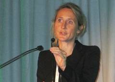
Study findings suggested that an accurate measure of a woman’s ovarian reserve can occur only after she has been off an estrogen-containing OC, probably for at least 3 months, Dr. Kathrine Birch Petersen reported at the annual meeting of the European Society of Human Reproduction and Embryology.

The impact of estrogen-containing OC use on reducing ovarian volume was especially pronounced in women under age 30, the reduction increased with longer durations of OC use, and the ability of OCs to mask a woman’s actual ovarian reserve was strong enough to potentially conceal a true case of premature ovarian insufficiency, said Dr. Birch Petersen, an ob.gyn. at the Fertility Assessment and Counseling Clinic at Rigshospitalet in Copenhagen.
"When we see a woman on an OC with impaired ovarian reserve, we would presume [based on these new findings] that her real ovarian reserve was about 30% higher than what we measure. We would advise her to be retested after she was off her OC for about 3 months," Dr. Birch Petersen said during a press conference before her presentation at the meeting.
The study included the first women seen at the clinic since it opened in 2011, excluding those who were pregnant or failed to supply adequate information. The cross-sectional cohort included 240 women on an estrogen-containing OC and 593 women with natural cycles.
The analysis focused on three parameters: blood level of anti-Müllerian hormone (AMH), antral follicle count (AFC), and ovarian volume. The multivariate, linear regression analysis adjusted for age, body mass index, smoking, age of maternal menopause, maternal smoking during pregnancy, preterm birth, and duration of OC use.
The analysis showed that compared with the women with natural cycles, those on an OC had a 19% relative reduction in their average blood level of AMH, a 16% relative reduction in average AFC, and a 47% relative reduction in average ovarian volume. The women on an OC also had smaller antral follicles. All three differences were statistically significant.
Seeing an effect from an estrogen-containing OC on all three measures makes sense because of their interrelatedness. The antral follicles produce AMH, and a reduction in antral follicle number as well as size would shrink the ovarian contents and result in reduced volume. These effects would not occur in women on a progestin-only OC, she said.
Dr. Birch Petersen had no disclosures.
On Twitter @mitchelzoler
MUNICH – Oral contraceptives do more than prevent unwanted pregnancy. They also make it hard to gauge a woman’s ovarian reserve, based on data from 833 women aged 19-46 years seen at a single Danish fertility clinic.
Study findings suggested that an accurate measure of a woman’s ovarian reserve can occur only after she has been off an estrogen-containing OC, probably for at least 3 months, Dr. Kathrine Birch Petersen reported at the annual meeting of the European Society of Human Reproduction and Embryology.
The impact of estrogen-containing OC use on reducing ovarian volume was especially pronounced in women under age 30, the reduction increased with longer durations of OC use, and the ability of OCs to mask a woman’s actual ovarian reserve was strong enough to potentially conceal a true case of premature ovarian insufficiency, said Dr. Birch Petersen, an ob.gyn. at the Fertility Assessment and Counseling Clinic at Rigshospitalet in Copenhagen.
"When we see a woman on an OC with impaired ovarian reserve, we would presume [based on these new findings] that her real ovarian reserve was about 30% higher than what we measure. We would advise her to be retested after she was off her OC for about 3 months," Dr. Birch Petersen said during a press conference before her presentation at the meeting.
The study included the first women seen at the clinic since it opened in 2011, excluding those who were pregnant or failed to supply adequate information. The cross-sectional cohort included 240 women on an estrogen-containing OC and 593 women with natural cycles.
The analysis focused on three parameters: blood level of anti-Müllerian hormone (AMH), antral follicle count (AFC), and ovarian volume. The multivariate, linear regression analysis adjusted for age, body mass index, smoking, age of maternal menopause, maternal smoking during pregnancy, preterm birth, and duration of OC use.
The analysis showed that compared with the women with natural cycles, those on an OC had a 19% relative reduction in their average blood level of AMH, a 16% relative reduction in average AFC, and a 47% relative reduction in average ovarian volume. The women on an OC also had smaller antral follicles. All three differences were statistically significant.
Seeing an effect from an estrogen-containing OC on all three measures makes sense because of their interrelatedness. The antral follicles produce AMH, and a reduction in antral follicle number as well as size would shrink the ovarian contents and result in reduced volume. These effects would not occur in women on a progestin-only OC, she said.
Dr. Birch Petersen had no disclosures.
On Twitter @mitchelzoler
AT ESHRE 2014
Key clinical point: Estrogen-containing oral contraceptive use is linked with significantly reduced levels of ovarian volume, antral follicle count, and anti-Müllerian hormone.
Major finding: Users of estrogen-containing OCs had an average 47% reduced ovarian volume, compared with similar women not taking an OC.
Data source: Review of 833 women aged 19-46 years seen at a Danish fertility clinic during 2011-2014.
Disclosures: Dr. Birch Petersen had no disclosures.

Hyperovulation before IVF linked to higher obstetric complication risk
MUNICH – Women who hyperrespond to ovarian stimulation have an increased risk for obstetric complication of a pregnancy started during the same oocyte retrieval cycle, based on data from more than 40,000 women who underwent assisted reproduction during 2001-2008.
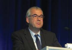
A retrospective review of 28,275 singleton pregnancies that followed in vitro fertilization or intracytoplasmic sperm injection found that hyperresponsive women, defined as those producing more than 15 oocytes following ovarian stimulation, had a relative 9% increased risk for having a low-birth-weight baby and a 10% relative increased rate of preterm birth, compared with women who produced a normal oocyte number, 4-15, Dr. Sesh K. Sunkara said at the annual meeting of the European Society of Human Reproduction and Embryology. These differences were statistically significant.

Other significant correlates of an increased risk for low birth weight and preterm birth were age, presence of an ovulatory disorder, and presence of a tubular disorder, said Dr. Sunkara, an obstetrician at Royal Marsden Hospital in London. After establishing these risk factors in this derivation cohort of more than 28,000 women, Dr. Sunkara and her associates tested the relationships with data collected from an additional 12,118 women drawn from the same U.K. registry during 2001-2008 and found identical relationships.
The findings highlight the importance of using ovarian stimulation methods that avoid hyperstimulation and hyperresponse, Dr. Sunkara said. In addition, future research should explore the physiologic processes that underlie the increased obstetric risk seen in the oocyte hyperresponders.
The study used data collected by the U.K. Human Fertilization and Embryology Authority. The overall live birth rate among the 28,275 women in the derivation cohort was 23%. The rate of preterm birth (less than 37 weeks’ gestation) and low-birth-weight babies (less than 2500 g) was 10% for each category. The rate of extreme preterm birth (less than 32 weeks) and the rate of extreme low birth weight (less than 1500 g) was 2% for each category.
The rate of extreme low birth rate was increased by a relative 16% among the hyperresponsive women, compared with those who produced 4-15 oocytes. The rate of extreme preterm birth increased by a relative 32%, compared with normally responsive women. Women with a poor response to ovarian stimulation, producing three or fewer oocytes, had similar rates of obstetric complications as normally responding women.
The analysis showed the highest combined rate of low birth weight plus preterm birth as 19% of all births in women younger than age 34 with hyperresponse to stimulation and both ovulatory and tubular disorders. In contrast, women aged 38 or older with a poor response to ovarian stimulation and neither an ovulatory nor a tubal disorder had a 10% combined rate of low-birth-weight and preterm birth pregnancies.
Dr. Sunkara had no disclosures.
On Twitter @mitchelzoler
MUNICH – Women who hyperrespond to ovarian stimulation have an increased risk for obstetric complication of a pregnancy started during the same oocyte retrieval cycle, based on data from more than 40,000 women who underwent assisted reproduction during 2001-2008.
A retrospective review of 28,275 singleton pregnancies that followed in vitro fertilization or intracytoplasmic sperm injection found that hyperresponsive women, defined as those producing more than 15 oocytes following ovarian stimulation, had a relative 9% increased risk for having a low-birth-weight baby and a 10% relative increased rate of preterm birth, compared with women who produced a normal oocyte number, 4-15, Dr. Sesh K. Sunkara said at the annual meeting of the European Society of Human Reproduction and Embryology. These differences were statistically significant.
Other significant correlates of an increased risk for low birth weight and preterm birth were age, presence of an ovulatory disorder, and presence of a tubular disorder, said Dr. Sunkara, an obstetrician at Royal Marsden Hospital in London. After establishing these risk factors in this derivation cohort of more than 28,000 women, Dr. Sunkara and her associates tested the relationships with data collected from an additional 12,118 women drawn from the same U.K. registry during 2001-2008 and found identical relationships.
The findings highlight the importance of using ovarian stimulation methods that avoid hyperstimulation and hyperresponse, Dr. Sunkara said. In addition, future research should explore the physiologic processes that underlie the increased obstetric risk seen in the oocyte hyperresponders.
The study used data collected by the U.K. Human Fertilization and Embryology Authority. The overall live birth rate among the 28,275 women in the derivation cohort was 23%. The rate of preterm birth (less than 37 weeks’ gestation) and low-birth-weight babies (less than 2500 g) was 10% for each category. The rate of extreme preterm birth (less than 32 weeks) and the rate of extreme low birth weight (less than 1500 g) was 2% for each category.
The rate of extreme low birth rate was increased by a relative 16% among the hyperresponsive women, compared with those who produced 4-15 oocytes. The rate of extreme preterm birth increased by a relative 32%, compared with normally responsive women. Women with a poor response to ovarian stimulation, producing three or fewer oocytes, had similar rates of obstetric complications as normally responding women.
The analysis showed the highest combined rate of low birth weight plus preterm birth as 19% of all births in women younger than age 34 with hyperresponse to stimulation and both ovulatory and tubular disorders. In contrast, women aged 38 or older with a poor response to ovarian stimulation and neither an ovulatory nor a tubal disorder had a 10% combined rate of low-birth-weight and preterm birth pregnancies.
Dr. Sunkara had no disclosures.
On Twitter @mitchelzoler
FROM ESHRE 2014
Key clinical point: Hyperresponders to ovarian stimulation are at increased risk for low-birth-weight and preterm birth deliveries.
Major finding: Hyperresponsive women had a 10% increase in relative risk of low birth weight and preterm birth as compared with normal responders.
Data source: Records review for 40,393 women pregnant after assisted reproduction during 2001-2008 collected by the U.K. Human Fertilization and Embryology Authority.
Disclosures: Dr. Sunkara had no disclosures.

Quick freezing oocytes and embryos transforms assisted reproduction
MUNICH – Vitrification, quick freezing oocytes or embryos with concentrated cyroprotectant and no ice crystal formation to better preserve viability, has been around clinically some 15 years, but now is gathering steam and may have already helped downshift the rate of triplets or higher pregnancies for women undergoing assisted reproduction.
While vitrification of all embryos fertilized in vitro has not proven superior to fresh embryo transfers and may even be inferior for older women whose eggs may not be as forgiving of cryopreservation, its use has grown. It offers the unique advantage over fresh embryos of easier coordination between the in vitro fertilization process and subsequent transfer to the woman who will carry the pregnancy. Vitrification has also made possible true oocyte banking, be it autologous for fertility preservation, or for oocyte donation.
"It is now possible to freeze embryos with a 90% survival rate; with slow freezing there was 50%-60% survival. Now we have a technique that is really good, and it may cause a paradigm shift toward elective freezing," said Dr. Anja Pinborg, a professor of ob.gyn. at Hvidovre (Denmark) Hospital.
Most clinicians still use the approach of placing an embryo during the same cycle when the oocyte was harvested, "the way it’s always been done, but at many clinics now they see that with vitrification they can get pregnancy rates that are similar to or even better than fresh embryo transfer. I think they will go more and more to frozen embryos," Dr. Pinborg said in an interview during the annual meeting of the European Society of Human Reproduction and Embryology.

For now the trend is specific for women younger than 35 years. Safety and efficacy of vitrification for embryos made from oocytes taken from older women has not yet been studied, cautioned Dr. Pinborg and others.
"All we know about cryopreservation is from young women with young oocytes. We don’t know what will happen with oocytes from women who are 41 or 42, who are the types of patients I see," said Dr. Richard J. Paulson, professor of ob.gyn. and chief of the division of reproductive endocrinology and infertility at the University of Southern California and director of USC Fertility in Los Angeles. "Vitrification is so good and embryos come out of it so well that the benefit from improved endometrial receptivity outweighs any change in embryo quality. The timing coordinates so much better."
The biggest impact of vitrification for embryo freezing may be its ability to successfully cryopreserve blastocysts. "We will see many more programs going to 5-day embryos" for implantation, and this will allow preimplantation genetic diagnosis (PGD), predicted Dr. Paulson during an interview at the meeting. "It looks like you can do pretty good trophectoderm biopsy on a vitrified blastocyst, so you can place only chromosomally normal embryos. That will reduce miscarriages." So far, only a handful of groups have presented case reports using this approach, with "very good numbers," he said.
Vitrification’s impact on embryo transfer number
Despite lingering questions about vitrification safety and limited knowledge of its efficacy for embryos begun from oocytes from older women, the most recent worldwide and European data on patterns of assisted reproductive technology show increases in frozen embryo use and decreases in triplets or greater multiples.
Worldwide, frozen embryo transfers grew from 10% of all in vitro fertilization or sperm injection procedures in 1991 to 28% in 2010, the most recent year with worldwide data available. Transfers of one or two embryos grew from 20% of all in vitro procedures done in North America in 1998 to 68% in 2010. In Australia in 2010, the rate of one- or two-embryo transfers stood at 98%.
"The trend is for more frozen transfers, and for reducing multiple rates. I think that is a very positive development," said Dr. David Adamson, a reproductive endocrinologist who practices in Palo Alto, Calif., and presented at the meeting worldwide data collected by the International Committee Monitoring Assisted Reproductive Technologies (ICMART). "The pregnancy rate is continuing to go up, but the multiple rate is dropping. We hope that will continue," he said in an interview.
"There is no question that because vitrification works so much better than slow freezing, clinics have more confidence to put in fewer embryos and still have viability," he said. "We’ll see more frozen embryo transfers in the next few years. When you decrease the number of embryos transferred, you increase the likelihood that you’ll need to follow with a frozen embryo transfer, but now you have higher-quality embryos."
A similar shift to fewer embryos transferred at a time appeared in the exclusively European registry with data derived from 33 countries that reported complete or partial data to the European IVF Monitoring Program, a project organized by ESHRE. The prevalence of triplet or greater deliveries from in vitro pregnancies fell in Europe from about 4% in 1999 to less than 1% in 2011. By 2011, roughly 28% of IVF or sperm-injected pregnancies involved placement of two embryos, and about 57% involved a single embryo, reported Dr. Markus S. Kupka, an ob.gyn. and reproductive medicine specialist who practices in Hamburg, Germany. Frozen embryos involved 31% of transfers in 2011 in Europe. European data for 2010 were included in the worldwide tallies reported by Dr. Adamson.
Vitrification’s safety
Despite growing use, some questions remain about vitrification’s impact on children, although the concern has not been strong enough to dim enthusiasm.
The most striking safety signal is a consistent, 50% increased rate of babies born following vitrification who are large for gestational age. Dr. Pinborg also cited a possibly increased rate of maternal preeclampsia.
"It’s only a 50% increased risk for large for gestational age with frozen embryo transfer. We should not pay too much attention to this, but it reminds us that when we freeze, it may have an effect on the children" Dr. Pinborg said in an interview.
"We can’t say that vitrified embryos do better than after fresh embryo transfer, but they have an altered risk profile." Frozen embryos have a reduced risk for preterm birth and small for gestational age, compared with fresh embryo transfers, but an increased risk of large for gestational age (LGA). "And the risk is small," stressed Dr. Pinborg. "When you stimulate ovulation, you harvest 10-fold as many oocytes than occur with a normal cycle, and the woman’s estrogen and progesterone levels are very high. So, you could ask: Why should we ever place an embryo during an oocyte harvesting cycle? We did that because we had poor freezing methods, but with vitrification we have a way to use preserved embryos and there is better cycle synchronization."
In addition, seeing increased rates of LGA babies following frozen embryo transfer may result from unadjusted confounding by, for example, the embryo’s age when cryopreservation occurred that could influence epigenetic changes that happen early after fertilization or maternal disorders such as polycystic ovary syndrome, Dr. Paulson said.
Safety aside, vitrification is very effective for assisted reproduction because any decrement in viability is counterbalanced by the advantage of placing embryos squarely during a natural cycle, these experts said.
Vitrification and oocytes
Vitrification does not stop with embryos. The impact it has had on oocyte banking has arguably been even greater.
Creating multiple embryos at one time is problematic, especially in the United States, Dr. Paulson said. "The obvious thing we should do is freeze oocytes. This approach would create another significant advantage. Routine vitrification at the oocyte stage rather than after embryos are made "would make donor oocytes a reality. You would be able to buy frozen oocytes the same way as sperm. Results from good randomized, controlled trials show that outcomes with cryopreserved oocytes are as good as with fresh oocytes, which was never possible with slow freezing."
"Vitrification created the ability to have oocyte banking. It has been a breakthrough in assisted reproductive technology," said Ana Cobo, Ph.D., director of the cryopreservation laboratory at the IVI Foundation in Valencia, Spain. Cryopreserving oocytes was essentially impossible before vitrification became available, she said in an interview. Studies run by her group showed that the vitrification process has no detectable effect on oocyte viability, embryo viability, or the health of the child. It also made oocyte donation much more feasible by eliminating the need for cycle synchrony between the donor and recipient.
The success rate of in vitro fertilization using a donated oocyte is generally much higher than for an autologous oocyte because "we use a highly selected population of young, healthy women with the best quality oocytes. In assisted reproductive technology, oocyte quality means a lot," Dr. Cobo said.
Dr. Pinborg said that she has received research grants from MSD and Ferring. Dr. Paulson said that he has been a speaker on behalf of Ferring and served on advisory boards for Origio and Cooper Surgical. Dr. Adamson said that he had received honoraria from or consulted for Bayer, Glycotype, and Ziva Medical, that he owns stock in Advanced Reproductive Care, and has received research support from Auxogyn and LabCorp. Dr. Kupka said he had no disclosures. Dr. Cobo had no disclosures.
On Twitter @mitchelzoler
MUNICH – Vitrification, quick freezing oocytes or embryos with concentrated cyroprotectant and no ice crystal formation to better preserve viability, has been around clinically some 15 years, but now is gathering steam and may have already helped downshift the rate of triplets or higher pregnancies for women undergoing assisted reproduction.
While vitrification of all embryos fertilized in vitro has not proven superior to fresh embryo transfers and may even be inferior for older women whose eggs may not be as forgiving of cryopreservation, its use has grown. It offers the unique advantage over fresh embryos of easier coordination between the in vitro fertilization process and subsequent transfer to the woman who will carry the pregnancy. Vitrification has also made possible true oocyte banking, be it autologous for fertility preservation, or for oocyte donation.
"It is now possible to freeze embryos with a 90% survival rate; with slow freezing there was 50%-60% survival. Now we have a technique that is really good, and it may cause a paradigm shift toward elective freezing," said Dr. Anja Pinborg, a professor of ob.gyn. at Hvidovre (Denmark) Hospital.
Most clinicians still use the approach of placing an embryo during the same cycle when the oocyte was harvested, "the way it’s always been done, but at many clinics now they see that with vitrification they can get pregnancy rates that are similar to or even better than fresh embryo transfer. I think they will go more and more to frozen embryos," Dr. Pinborg said in an interview during the annual meeting of the European Society of Human Reproduction and Embryology.
For now the trend is specific for women younger than 35 years. Safety and efficacy of vitrification for embryos made from oocytes taken from older women has not yet been studied, cautioned Dr. Pinborg and others.
"All we know about cryopreservation is from young women with young oocytes. We don’t know what will happen with oocytes from women who are 41 or 42, who are the types of patients I see," said Dr. Richard J. Paulson, professor of ob.gyn. and chief of the division of reproductive endocrinology and infertility at the University of Southern California and director of USC Fertility in Los Angeles. "Vitrification is so good and embryos come out of it so well that the benefit from improved endometrial receptivity outweighs any change in embryo quality. The timing coordinates so much better."
The biggest impact of vitrification for embryo freezing may be its ability to successfully cryopreserve blastocysts. "We will see many more programs going to 5-day embryos" for implantation, and this will allow preimplantation genetic diagnosis (PGD), predicted Dr. Paulson during an interview at the meeting. "It looks like you can do pretty good trophectoderm biopsy on a vitrified blastocyst, so you can place only chromosomally normal embryos. That will reduce miscarriages." So far, only a handful of groups have presented case reports using this approach, with "very good numbers," he said.
Vitrification’s impact on embryo transfer number
Despite lingering questions about vitrification safety and limited knowledge of its efficacy for embryos begun from oocytes from older women, the most recent worldwide and European data on patterns of assisted reproductive technology show increases in frozen embryo use and decreases in triplets or greater multiples.
Worldwide, frozen embryo transfers grew from 10% of all in vitro fertilization or sperm injection procedures in 1991 to 28% in 2010, the most recent year with worldwide data available. Transfers of one or two embryos grew from 20% of all in vitro procedures done in North America in 1998 to 68% in 2010. In Australia in 2010, the rate of one- or two-embryo transfers stood at 98%.
"The trend is for more frozen transfers, and for reducing multiple rates. I think that is a very positive development," said Dr. David Adamson, a reproductive endocrinologist who practices in Palo Alto, Calif., and presented at the meeting worldwide data collected by the International Committee Monitoring Assisted Reproductive Technologies (ICMART). "The pregnancy rate is continuing to go up, but the multiple rate is dropping. We hope that will continue," he said in an interview.
"There is no question that because vitrification works so much better than slow freezing, clinics have more confidence to put in fewer embryos and still have viability," he said. "We’ll see more frozen embryo transfers in the next few years. When you decrease the number of embryos transferred, you increase the likelihood that you’ll need to follow with a frozen embryo transfer, but now you have higher-quality embryos."
A similar shift to fewer embryos transferred at a time appeared in the exclusively European registry with data derived from 33 countries that reported complete or partial data to the European IVF Monitoring Program, a project organized by ESHRE. The prevalence of triplet or greater deliveries from in vitro pregnancies fell in Europe from about 4% in 1999 to less than 1% in 2011. By 2011, roughly 28% of IVF or sperm-injected pregnancies involved placement of two embryos, and about 57% involved a single embryo, reported Dr. Markus S. Kupka, an ob.gyn. and reproductive medicine specialist who practices in Hamburg, Germany. Frozen embryos involved 31% of transfers in 2011 in Europe. European data for 2010 were included in the worldwide tallies reported by Dr. Adamson.
Vitrification’s safety
Despite growing use, some questions remain about vitrification’s impact on children, although the concern has not been strong enough to dim enthusiasm.
The most striking safety signal is a consistent, 50% increased rate of babies born following vitrification who are large for gestational age. Dr. Pinborg also cited a possibly increased rate of maternal preeclampsia.
"It’s only a 50% increased risk for large for gestational age with frozen embryo transfer. We should not pay too much attention to this, but it reminds us that when we freeze, it may have an effect on the children" Dr. Pinborg said in an interview.
"We can’t say that vitrified embryos do better than after fresh embryo transfer, but they have an altered risk profile." Frozen embryos have a reduced risk for preterm birth and small for gestational age, compared with fresh embryo transfers, but an increased risk of large for gestational age (LGA). "And the risk is small," stressed Dr. Pinborg. "When you stimulate ovulation, you harvest 10-fold as many oocytes than occur with a normal cycle, and the woman’s estrogen and progesterone levels are very high. So, you could ask: Why should we ever place an embryo during an oocyte harvesting cycle? We did that because we had poor freezing methods, but with vitrification we have a way to use preserved embryos and there is better cycle synchronization."
In addition, seeing increased rates of LGA babies following frozen embryo transfer may result from unadjusted confounding by, for example, the embryo’s age when cryopreservation occurred that could influence epigenetic changes that happen early after fertilization or maternal disorders such as polycystic ovary syndrome, Dr. Paulson said.
Safety aside, vitrification is very effective for assisted reproduction because any decrement in viability is counterbalanced by the advantage of placing embryos squarely during a natural cycle, these experts said.
Vitrification and oocytes
Vitrification does not stop with embryos. The impact it has had on oocyte banking has arguably been even greater.
Creating multiple embryos at one time is problematic, especially in the United States, Dr. Paulson said. "The obvious thing we should do is freeze oocytes. This approach would create another significant advantage. Routine vitrification at the oocyte stage rather than after embryos are made "would make donor oocytes a reality. You would be able to buy frozen oocytes the same way as sperm. Results from good randomized, controlled trials show that outcomes with cryopreserved oocytes are as good as with fresh oocytes, which was never possible with slow freezing."
"Vitrification created the ability to have oocyte banking. It has been a breakthrough in assisted reproductive technology," said Ana Cobo, Ph.D., director of the cryopreservation laboratory at the IVI Foundation in Valencia, Spain. Cryopreserving oocytes was essentially impossible before vitrification became available, she said in an interview. Studies run by her group showed that the vitrification process has no detectable effect on oocyte viability, embryo viability, or the health of the child. It also made oocyte donation much more feasible by eliminating the need for cycle synchrony between the donor and recipient.
The success rate of in vitro fertilization using a donated oocyte is generally much higher than for an autologous oocyte because "we use a highly selected population of young, healthy women with the best quality oocytes. In assisted reproductive technology, oocyte quality means a lot," Dr. Cobo said.
Dr. Pinborg said that she has received research grants from MSD and Ferring. Dr. Paulson said that he has been a speaker on behalf of Ferring and served on advisory boards for Origio and Cooper Surgical. Dr. Adamson said that he had received honoraria from or consulted for Bayer, Glycotype, and Ziva Medical, that he owns stock in Advanced Reproductive Care, and has received research support from Auxogyn and LabCorp. Dr. Kupka said he had no disclosures. Dr. Cobo had no disclosures.
On Twitter @mitchelzoler
EXPERT ANALYSIS FROM ESHRE 2014

ESHRE writes first premature ovarian insufficiency guidelines
MUNICH – A working group of the European Society of Human Reproduction and Embryology drafted the first international guidelines for managing premature ovarian insufficiency.
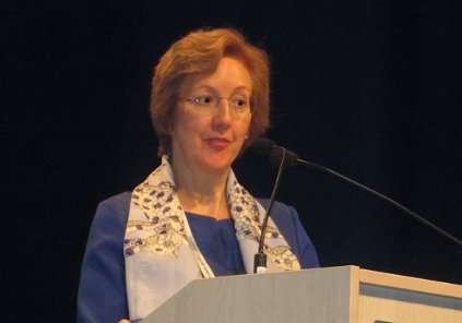
Once formally released, by late this year or soon after, the guidelines should "improve clinical practice and awareness of the condition," which affects roughly 1% of women, said Dr. Melanie Davies, a gynecologist at University College London Hospital and a working-group member who presented a few key items from the draft guidelines during a session at the society’s annual meeting.

Women with premature ovarian insufficiency (POI) sometimes are treated for one aspect of POI by a physician "without full realization of the condition and its implications," Dr. Davies said in an interview. For example, "there have been fatal cases of women with Turner syndrome who received donor oocytes," the disorder posing the greatest risk to women receiving donor eggs. "This is a good example of where the guidelines may increase awareness," she said.
One draft excerpt Dr. Davies presented advises physicians and patients that candidates for donor oocytes suspected of having POI should undergo prior to receiving eggs a full investigation for thyroid and adrenal function and karyotype. "Pregnancy in some women can be of such high risk that clinicians may consider egg donation life threatening and inappropriate," she said.
For women with Turner syndrome, the guidelines call for assessment by a cardiologist with a special interest in adult congenital heart disease, as well as a general medical and endocrine examination prior to pregnancy. During pregnancy, these women have an elevated risk for obstetric complications and should be managed in an obstetric unit designed for high-risk pregnancies with involvement of a cardiologist.
The draft defines POI as depletion of follicular activity before age 40 years with menstrual disturbance, raised gonadotropin levels, and low estradiol levels. These women should understand that no interventions are proven to reliably increase ovarian activity and their natural conception rate. But physicians also can reassure these women that spontaneous pregnancy after idiopathic POI or most forms of chemotherapy has no higher obstetric or neonatal risk than in the general population.
Pregnancy in women who received cancer treatment is increasingly common, Dr. Davies said in her talk at the meeting. Women who received ovarian irradiation face an increased risk of pregnancy complications and should be managed accordingly. Women who received treatment with an anthracycline, high-dose cyclophosphamide, or cardiac irradiation should undergo echocardiography before pregnancy, and prenatal care should include cardiology assessment.
The draft guidelines contain a total of 13 chapters addressing 31 key issues, ranging from symptoms and diagnosis, complications including bone and cardiovascular health, sexual function, and quality of life. The 17-member working group included a patient representative as well as experts from psychology, cardiology, endocrinology, internal medicine, and ob.gyn. In mid-August, the ESHRE website said that posting of the draft version should occur later in August. Once posted, ESHRE will open a 6-week comment period. The final version should appear by late 2014, Dr. Davies said.
She said she had no relevant financial disclosures.
On Twitter @mitchelzoler
MUNICH – A working group of the European Society of Human Reproduction and Embryology drafted the first international guidelines for managing premature ovarian insufficiency.
Once formally released, by late this year or soon after, the guidelines should "improve clinical practice and awareness of the condition," which affects roughly 1% of women, said Dr. Melanie Davies, a gynecologist at University College London Hospital and a working-group member who presented a few key items from the draft guidelines during a session at the society’s annual meeting.
Women with premature ovarian insufficiency (POI) sometimes are treated for one aspect of POI by a physician "without full realization of the condition and its implications," Dr. Davies said in an interview. For example, "there have been fatal cases of women with Turner syndrome who received donor oocytes," the disorder posing the greatest risk to women receiving donor eggs. "This is a good example of where the guidelines may increase awareness," she said.
One draft excerpt Dr. Davies presented advises physicians and patients that candidates for donor oocytes suspected of having POI should undergo prior to receiving eggs a full investigation for thyroid and adrenal function and karyotype. "Pregnancy in some women can be of such high risk that clinicians may consider egg donation life threatening and inappropriate," she said.
For women with Turner syndrome, the guidelines call for assessment by a cardiologist with a special interest in adult congenital heart disease, as well as a general medical and endocrine examination prior to pregnancy. During pregnancy, these women have an elevated risk for obstetric complications and should be managed in an obstetric unit designed for high-risk pregnancies with involvement of a cardiologist.
The draft defines POI as depletion of follicular activity before age 40 years with menstrual disturbance, raised gonadotropin levels, and low estradiol levels. These women should understand that no interventions are proven to reliably increase ovarian activity and their natural conception rate. But physicians also can reassure these women that spontaneous pregnancy after idiopathic POI or most forms of chemotherapy has no higher obstetric or neonatal risk than in the general population.
Pregnancy in women who received cancer treatment is increasingly common, Dr. Davies said in her talk at the meeting. Women who received ovarian irradiation face an increased risk of pregnancy complications and should be managed accordingly. Women who received treatment with an anthracycline, high-dose cyclophosphamide, or cardiac irradiation should undergo echocardiography before pregnancy, and prenatal care should include cardiology assessment.
The draft guidelines contain a total of 13 chapters addressing 31 key issues, ranging from symptoms and diagnosis, complications including bone and cardiovascular health, sexual function, and quality of life. The 17-member working group included a patient representative as well as experts from psychology, cardiology, endocrinology, internal medicine, and ob.gyn. In mid-August, the ESHRE website said that posting of the draft version should occur later in August. Once posted, ESHRE will open a 6-week comment period. The final version should appear by late 2014, Dr. Davies said.
She said she had no relevant financial disclosures.
On Twitter @mitchelzoler
EXPERT OPINION FROM ESHRE 2014
VIDEO: Vitrification makes oocyte banking a reality
MUNICH – Quick freezing of oocytes without ice formation–vitrification–has been a "breakthrough" for oocyte banking for both autologous fertility preservation and for oocyte donations, Ana Cobo, Ph.D., said in a video interview during the annual meeting of the European Society of Human Reproduction and Embryology.
Results of studies done by Dr. Cobo and her associates have shown that children born from vitrified oocytes have similar outcomes compared with children born from fresh oocytes. Many women who receive embryos made from donated oocytes are older and may have age-related obstetric complications, but these have no relationship to vitrification itself. Nor are their complications exacerbated by vitrification, said Dr. Cobo, director of the cryopreservation laboratory at the Instituto Valenciano de Infertilidad in Valencia, Spain.
A major advantage of using vitrified oocytes instead of fresh is that cryopreservation makes it much easier to synchronize placement of an appropriately aged embryo with the optimal period of receptivity during the recipient’s cycle, Dr. Cobo said.
Dr. Cobo had no disclosures.
The video associated with this article is no longer available on this site. Please view all of our videos on the MDedge YouTube channel
On Twitter @mitchelzoler
AT ESHRE 2014

Fertility treatment was associated with limited cancer risks
MUNICH – Hormones and drugs used for fertility treatment of women caused no overall excess of cancers in either the treated women or their children during relatively long follow-up in two large, separate epidemiologic studies.
But results from each of the two studies also showed some evidence of an increased incidence of a few specific types of cancers from two ovarian-stimulating agents, suggesting a need for caution and continued surveillance.

One study, which used a case-control design to analyze data from several Danish national registries, showed that children born to women treated with progesterone had a significantly elevated incidence of leukemias in general, of acute lymphoblastic leukemia specifically, and also of sympathetic nervous system tumors such as neuroblastomas, Marie Hargreave, Ph.D., said at the annual meeting of the European Society of Human Reproduction and Embryology.
"Taking into account the biological plausibility that fertility drugs could cause cancer in children, these results are strong enough to cause concern," said Dr. Hargreave, a researcher at the Danish Cancer Society Research Center.
The second report, a retrospective analysis of cancer cases among nearly 10,000 women who underwent ovulation stimulation at any of five U.S. fertility centers and were followed for 30 years, showed that two very specific subgroups of woman had a significantly increased number of cancers associated with treatment by the ovulation-stimulating drug clomiphene citrate, a selective estrogen-receptor modulator. Nulligravid women treated with clomiphene who then remained nulligravid through follow-up had a significantly increased rate of ovarian cancer, but not breast or endometrial cancer, reported Dr. Humberto Scoccia, professor of ob.gyn. and medical director of the in vitro fertilization program at the University of Illinois in Chicago.
In addition, the small group of 31 women who developed invasive breast cancer during follow-up showed a statistically significant increased rate of this cancer among the women who received clomiphene for 12 or more ovulatory cycles.
However, women in the study received treatment during 1965-1988, an era when clomiphene was used more often and at higher dosages than is typical today, when the maximum number of clomiphene-treatment cycles usually tops out at six, Dr. Scoccia said.
The Danish study used data collected from several national registries on nearly 91,000 children born to mothers who had fertility treatment during 1964-2006. The study focused on 148 of these children who developed cancer as children or young adults, and compared the treatments that their mothers had received with 1,289 cancer-free children born to mothers who had fertility treatment.
The overall findings showed no significantly increased risk for any type of cancer linked with any drug or hormone exposure the mothers received – "quite reassuring results," Dr. Hargreave said.
However, a more granular analysis revealed a few cancers linked to maternal progesterone treatment. Children born to mothers who received progesterone during three or more ovulatory cycles had a statistically significant, fourfold increased rate of all types of leukemias. Cases of acute lymphoblastic leukemia specifically occurred at a significantly increased rate in children whose mothers had received any progesterone exposure, and among children whose mothers received the hormone during three or more cycles, the rate was nearly 10-fold higher than the rate seen in children whose mothers had no progesterone.
In addition, any maternal progesterone exposure also was linked to a significant, nearly sixfold increased rate of sympathetic nervous system tumors in the children. No other drug or hormones used – including clomiphene and various gonadotropins – showed any significant links to offspring cancers.

"This is the largest study reported to date with data on specific fertility drugs, as it had an appropriate reference group," children who remained cancer free but were born to mothers who underwent fertility treatment, Dr. Hargreave noted. She also cautioned that the subanalyses for specific cancer and treatment types involved relatively small numbers of cases.
"Some of the numbers in the subgroup analyses are so small that the findings need to be interpreted with caution," Dr. Scoccia commented during the session. "Multiple prior studies looked at progesterone and did not see a problem," he noted.
"More study of progesterone is needed, as this is the first large epidemiologic study to show this effect," Dr. Hargreave said. "What is important in this study is that we saw no overall increase in cancer rates."
The U.S. study reported by Dr. Scoccia reviewed 9,892 women treated at any of five U.S. fertility clinics during 1965-1988 who also completed a questionnaire in 2000, resulting in follow-up for a median of 30 years from when they received ovulation stimulation. During follow-up, 749 women developed breast cancer, 118 developed endometrial cancer, and 85 developed ovarian cancer.
Among women who received clomiphene, the analysis showed no overall, significantly increased risk of any cancer. But when the analysis broke out the incidence of these three cancer types individually, it showed that the subgroup of women who were nulligravid at treatment and remained nulligravid throughout follow-up had a significant, 3.6-fold increased risk of ovarian cancer, compared with women who did not receive clomiphene. But Dr. Scoccia cautioned that only 13 women were in this subgroup.
A second subgroup analysis that looked specifically at the 31 women who developed invasive breast cancer showed that women who received clomiphene during 12 or more ovulatory cycles had a 69% higher rate of invasive breast cancer than did those who never received clomiphene.
Dr. Scoccia also found that none of the analyses of women who had received a gonadotropin showed any subgroup with an increased rate of cancer.
"Overall, these findings are reassuring," he said, but cautioned that because the women averaged 30 years of age at the time they received fertility treatment, even after 30 years of follow-up these women generally remained younger than the peak age for incidence of these cancer types; hence, further follow-up of the cohort will help further define whether they face increased lifetime cancer rates. He also noted that the higher cancer rates seen with increased clomiphene treatment may link to more resistant infertility among these women rather to than the clomiphene they received, but added "we don’t think it is explained by more resistant infertility alone."
Dr. Hargreave and Dr. Scoccia had no disclosures.
On Twitter @mitchelzoler
MUNICH – Hormones and drugs used for fertility treatment of women caused no overall excess of cancers in either the treated women or their children during relatively long follow-up in two large, separate epidemiologic studies.
But results from each of the two studies also showed some evidence of an increased incidence of a few specific types of cancers from two ovarian-stimulating agents, suggesting a need for caution and continued surveillance.
One study, which used a case-control design to analyze data from several Danish national registries, showed that children born to women treated with progesterone had a significantly elevated incidence of leukemias in general, of acute lymphoblastic leukemia specifically, and also of sympathetic nervous system tumors such as neuroblastomas, Marie Hargreave, Ph.D., said at the annual meeting of the European Society of Human Reproduction and Embryology.
"Taking into account the biological plausibility that fertility drugs could cause cancer in children, these results are strong enough to cause concern," said Dr. Hargreave, a researcher at the Danish Cancer Society Research Center.
The second report, a retrospective analysis of cancer cases among nearly 10,000 women who underwent ovulation stimulation at any of five U.S. fertility centers and were followed for 30 years, showed that two very specific subgroups of woman had a significantly increased number of cancers associated with treatment by the ovulation-stimulating drug clomiphene citrate, a selective estrogen-receptor modulator. Nulligravid women treated with clomiphene who then remained nulligravid through follow-up had a significantly increased rate of ovarian cancer, but not breast or endometrial cancer, reported Dr. Humberto Scoccia, professor of ob.gyn. and medical director of the in vitro fertilization program at the University of Illinois in Chicago.
In addition, the small group of 31 women who developed invasive breast cancer during follow-up showed a statistically significant increased rate of this cancer among the women who received clomiphene for 12 or more ovulatory cycles.
However, women in the study received treatment during 1965-1988, an era when clomiphene was used more often and at higher dosages than is typical today, when the maximum number of clomiphene-treatment cycles usually tops out at six, Dr. Scoccia said.
The Danish study used data collected from several national registries on nearly 91,000 children born to mothers who had fertility treatment during 1964-2006. The study focused on 148 of these children who developed cancer as children or young adults, and compared the treatments that their mothers had received with 1,289 cancer-free children born to mothers who had fertility treatment.
The overall findings showed no significantly increased risk for any type of cancer linked with any drug or hormone exposure the mothers received – "quite reassuring results," Dr. Hargreave said.
However, a more granular analysis revealed a few cancers linked to maternal progesterone treatment. Children born to mothers who received progesterone during three or more ovulatory cycles had a statistically significant, fourfold increased rate of all types of leukemias. Cases of acute lymphoblastic leukemia specifically occurred at a significantly increased rate in children whose mothers had received any progesterone exposure, and among children whose mothers received the hormone during three or more cycles, the rate was nearly 10-fold higher than the rate seen in children whose mothers had no progesterone.
In addition, any maternal progesterone exposure also was linked to a significant, nearly sixfold increased rate of sympathetic nervous system tumors in the children. No other drug or hormones used – including clomiphene and various gonadotropins – showed any significant links to offspring cancers.
"This is the largest study reported to date with data on specific fertility drugs, as it had an appropriate reference group," children who remained cancer free but were born to mothers who underwent fertility treatment, Dr. Hargreave noted. She also cautioned that the subanalyses for specific cancer and treatment types involved relatively small numbers of cases.
"Some of the numbers in the subgroup analyses are so small that the findings need to be interpreted with caution," Dr. Scoccia commented during the session. "Multiple prior studies looked at progesterone and did not see a problem," he noted.
"More study of progesterone is needed, as this is the first large epidemiologic study to show this effect," Dr. Hargreave said. "What is important in this study is that we saw no overall increase in cancer rates."
The U.S. study reported by Dr. Scoccia reviewed 9,892 women treated at any of five U.S. fertility clinics during 1965-1988 who also completed a questionnaire in 2000, resulting in follow-up for a median of 30 years from when they received ovulation stimulation. During follow-up, 749 women developed breast cancer, 118 developed endometrial cancer, and 85 developed ovarian cancer.
Among women who received clomiphene, the analysis showed no overall, significantly increased risk of any cancer. But when the analysis broke out the incidence of these three cancer types individually, it showed that the subgroup of women who were nulligravid at treatment and remained nulligravid throughout follow-up had a significant, 3.6-fold increased risk of ovarian cancer, compared with women who did not receive clomiphene. But Dr. Scoccia cautioned that only 13 women were in this subgroup.
A second subgroup analysis that looked specifically at the 31 women who developed invasive breast cancer showed that women who received clomiphene during 12 or more ovulatory cycles had a 69% higher rate of invasive breast cancer than did those who never received clomiphene.
Dr. Scoccia also found that none of the analyses of women who had received a gonadotropin showed any subgroup with an increased rate of cancer.
"Overall, these findings are reassuring," he said, but cautioned that because the women averaged 30 years of age at the time they received fertility treatment, even after 30 years of follow-up these women generally remained younger than the peak age for incidence of these cancer types; hence, further follow-up of the cohort will help further define whether they face increased lifetime cancer rates. He also noted that the higher cancer rates seen with increased clomiphene treatment may link to more resistant infertility among these women rather to than the clomiphene they received, but added "we don’t think it is explained by more resistant infertility alone."
Dr. Hargreave and Dr. Scoccia had no disclosures.
On Twitter @mitchelzoler
AT ESHRE 2014
Key clinical point: Results from two large epidemiologic studies suggest fertility treatment posed a limited cancer risk to women and their children.
Major finding: Clomiphene treatment linked with a 3.6-fold increased risk for ovarian cancer, and prolonged use linked with a 69% higher rate of invasive breast cancer. Progesterone treatment was linked with increased leukemias and sympathetic nervous system cancers in the children of treated mothers.
Data source: One study reviewed 9,892 women treated at five U.S. centers during 1965-1988. A second study ran a case-control analysis of 1,437 Danish children born to mothers who underwent fertility treatment.
Disclosures: Dr. Hargreave and Dr. Scoccia had no disclosures.

Children’s Psychiatric Disorders Linked With Moms’ Fertility Problems
MUNICH – Children born to mothers who required fertility treatment had a significantly higher rate of psychiatric disorders, compared with children born to mothers without fertility problems, based on population-wide registry data collected in Denmark.
Although psychiatric disorders occurred a third more often in children born to mothers treated at fertility clinics, this "modest" increase should not deter women with fertility problems from seeking treatment and becoming pregnant, Allan Jensen, Ph.D., said in a video interview at the annual meeting of the European Society of Human Reproduction and Embryology.
The increased risk of psychiatric disorders probably springs from the underlying infertility of these women and damaged genes that they may carry, rather than because of any fertility treatments they received, said Dr. Jensen, a senior researcher at the Danish Cancer Society Research Center, Copenhagen.
Dr. Jensen said that he had no disclosures.
The video associated with this article is no longer available on this site. Please view all of our videos on the MDedge YouTube channel
On Twitter @mitchelzoler
AT ESHRE 2014

VIDEO: Children’s psychiatric disorders linked with moms’ fertility problems
MUNICH – Children born to mothers who required fertility treatment had a significantly higher rate of psychiatric disorders, compared with children born to mothers without fertility problems, based on population-wide registry data collected in Denmark.
Although psychiatric disorders occurred a third more often in children born to mothers treated at fertility clinics, this "modest" increase should not deter women with fertility problems from seeking treatment and becoming pregnant, Allan Jensen, Ph.D., said in a video interview at the annual meeting of the European Society of Human Reproduction and Embryology.
The increased risk of psychiatric disorders probably springs from the underlying infertility of these women and damaged genes that they may carry, rather than because of any fertility treatments they received, said Dr. Jensen, a senior researcher at the Danish Cancer Society Research Center, Copenhagen.
Dr. Jensen said that he had no disclosures.
The video associated with this article is no longer available on this site. Please view all of our videos on the MDedge YouTube channel
On Twitter @mitchelzoler
AT ESHRE 2014

Children’s psychiatric disorders linked with moms’ infertility
MUNICH – Children born to mothers who had fertility problems had a 33% increased incidence of psychiatric disorders as children or young adults, compared with the offspring from mothers without fertility problems, a population-based Danish study found.
While clinicians and patients should be aware of this "modest" increased risk, it should be "balanced against the physical and psychological benefits of pregnancy," Allan Jensen, Ph.D., said at the annual meeting of the European Society of Human Reproduction and Embryology.

"I think this is an acceptable risk" from the perspective of both individual women and society, said Dr. Jensen, a senior researcher at the Danish Cancer Society Research Center in Copenhagen. The findings "should not discourage women from receiving fertility treatment," he said in an interview. "I think this is a small risk."
Dr. Jensen estimated that about 2% of psychiatric disorders that exist today among Danish children and adults are attributable to mothers who underwent fertility treatment. Although his study has not yet analyzed whether any specific treatments used during assisted reproduction seemed to link with subsequent psychiatric disorders in offspring, he speculated that the primary mechanism is likely defective genes carried by women with fertility problems.
The Danish infertility cohort contained information on more than 109,000 women who underwent a fertility consultation from 1963 to 2009, and these data were used along with Danish birth records starting from 1969 and Danish national medical records for psychiatric disorders that also dated from 1969. The analysis identified 7,860 children or adults who had received a psychiatric diagnosis and were born to women treated for fertility problems.
Results from a multivariate, regression analysis that controlled for year of birth, maternal age at birth, sex, and birth order, showed that children of mothers who had received fertility treatment had a statistically significant, 33% increased rate of having a psychiatric disorder, compared with people born to mothers without a fertility problem.
Additional analysis showed statistically significant increases across a broad swath of psychiatric disorders. For example, children born to mothers who had fertility treatment had a 47% higher rate of attention-deficit/hyperactivity disorder, a 43% higher rate of personality and behavior disorders, a 32% increase in affective disorders, a 28% higher rate of mental retardation, a 27% higher rate of schizophrenia, and a 12% higher rate of autism-spectrum disorders.
The analysis did not adjust for fertility treatment the women received or for child factors such as birth weight and prematurity. But the analysis did find that a statistically increased rate of various psychiatric disorders existed independently in both children and in young adults.
The study had the advantages of using the largest population ever examined for this issue, including a long follow-up, and avoiding selection bias or recall bias by being population- and registry-based, with virtually complete ascertainment of psychiatric cases. It had the shortcoming of being unable to distinguish cause from infertility in the women or from the treatments they received, Dr. Jensen said.
Dr. Jensen said that he had no disclosures.
On Twitter @mitchelzoler
MUNICH – Children born to mothers who had fertility problems had a 33% increased incidence of psychiatric disorders as children or young adults, compared with the offspring from mothers without fertility problems, a population-based Danish study found.
While clinicians and patients should be aware of this "modest" increased risk, it should be "balanced against the physical and psychological benefits of pregnancy," Allan Jensen, Ph.D., said at the annual meeting of the European Society of Human Reproduction and Embryology.
"I think this is an acceptable risk" from the perspective of both individual women and society, said Dr. Jensen, a senior researcher at the Danish Cancer Society Research Center in Copenhagen. The findings "should not discourage women from receiving fertility treatment," he said in an interview. "I think this is a small risk."
Dr. Jensen estimated that about 2% of psychiatric disorders that exist today among Danish children and adults are attributable to mothers who underwent fertility treatment. Although his study has not yet analyzed whether any specific treatments used during assisted reproduction seemed to link with subsequent psychiatric disorders in offspring, he speculated that the primary mechanism is likely defective genes carried by women with fertility problems.
The Danish infertility cohort contained information on more than 109,000 women who underwent a fertility consultation from 1963 to 2009, and these data were used along with Danish birth records starting from 1969 and Danish national medical records for psychiatric disorders that also dated from 1969. The analysis identified 7,860 children or adults who had received a psychiatric diagnosis and were born to women treated for fertility problems.
Results from a multivariate, regression analysis that controlled for year of birth, maternal age at birth, sex, and birth order, showed that children of mothers who had received fertility treatment had a statistically significant, 33% increased rate of having a psychiatric disorder, compared with people born to mothers without a fertility problem.
Additional analysis showed statistically significant increases across a broad swath of psychiatric disorders. For example, children born to mothers who had fertility treatment had a 47% higher rate of attention-deficit/hyperactivity disorder, a 43% higher rate of personality and behavior disorders, a 32% increase in affective disorders, a 28% higher rate of mental retardation, a 27% higher rate of schizophrenia, and a 12% higher rate of autism-spectrum disorders.
The analysis did not adjust for fertility treatment the women received or for child factors such as birth weight and prematurity. But the analysis did find that a statistically increased rate of various psychiatric disorders existed independently in both children and in young adults.
The study had the advantages of using the largest population ever examined for this issue, including a long follow-up, and avoiding selection bias or recall bias by being population- and registry-based, with virtually complete ascertainment of psychiatric cases. It had the shortcoming of being unable to distinguish cause from infertility in the women or from the treatments they received, Dr. Jensen said.
Dr. Jensen said that he had no disclosures.
On Twitter @mitchelzoler
AT ESHRE 2014
Key clinical point: Women with fertility problems who became pregnant through assisted technology had a significantly increased rate of children with psychiatric disorders.
Major finding: Women with fertility problems had a 33% increased rate of having children with a psychiatric disorder.
Data source: Danish national medical registry data for the 2.4 million children born in Denmark during 1969-2006 and for 109,000 women who had fertility treatment during the period.
Disclosures: Dr. Jensen said that he had no disclosures.

Donated IVF eggs linked to gestational hypertension
MUNICH – Women pregnant with an embryo produced by in vitro fertilization of a donated egg developed gestational hypertension at nearly four times the rate of matched women who were pregnant following IVF with a self egg, in a multicenter, case-control study with 580 participants.
In addition, preeclampsia was more than four times higher in the women who had received a donor egg as part of an IVF procedure, compared with women who used a self egg for their IVF, Dr. Hélène Letur-Könirsch reported at the annual meeting of the European Society of Human Reproduction and Embryology.

Although the mechanism underlying this association is not known, one possible explanation is that when a woman carries an embryo that is completely allogenic, it triggers modified immune tolerance that leads to impaired trophoblast implantation. Other candidate hypotheses are that defective genes in the mother trigger abnormal metabolic pathways that cause hypertension and preeclampsia, and that ovarian insufficiency features reduced ovarian steroid production, causing vascular and immunologic changes that lead to pregnancy-induced hypertension.
Results from several prior studies also had shown a link between use of a donor egg for IVF and an increased rate of gestational hypertension and preeclampsia, but most prior studies had been smaller and failed to adequately control for possible confounders, said Dr. Letur-Könirsch, an endocrinologist at the Fertility Center of the Institut Mutualiste Montsouris in Paris.
She and her associates from seven IVF centers in France pooled data from their entire series of IVF cases that involved donated eggs during the period from February 2005 to September 2012, a total of 217 pregnant women. The analysis matched these cases with 363 control women who had IVF pregnancies during the study period using self eggs by parameters such as age, parity, time of pregnancy, and whether the IVF procedure involved transfer of a fresh or frozen embryo.
Women in the study averaged 35 years of age, with 82% aged 39 years or younger. Average body mass index was virtually identical in both groups, about 24 kg/m2, and the live-birth rate was 79% in both groups. All women in both groups were normotensive prior to their IVF procedure.
The incidence of pregnancy-induced hypertension, defined as blood pressure that reached at least 140/90 mm Hg when measured in office on both arms with two separate measurements, was about 29% in the women who received a donated egg and 14% in those who used a self egg. Development of preeclampsia, defined as persistent gestational hypertension plus proteinuria of at least 0.3 g/day, occurred in about 18% of women who received a donated egg and 7% in those who used a self egg.
In a multivariate analysis that controlled for age, pregnancy history, and use of a fresh or frozen embryo, women who used a donated egg had a 3.9-fold increased rate of pregnancy-induced hypertension and a 4.6-fold increased rate of preeclampsia, compared with women who used a self egg – both statistically significant differences.
The findings suggested that before a woman embarks on pregnancy using a donated egg, she should undergo thorough evaluation for preexisting risk factors for gestational hypertension, including hypertension, obesity, diabetes, renal disease, chronic infection, autoimmune diseases such as systemic lupus erythematosus, a family history of preeclampsia, and living at a high altitude. If these risk factors are present, they should be controlled if possible. Women who have one or more of these risk factors may also be candidates for prophylactic treatment with aspirin to prevent development of preeclampsia, especially if they develop pregnancy-induced hypertension.
During pregnancy, women who received a donor egg should undergo regular and frequent blood pressure measurement, as well as assessment for other possible abnormalities such as Doppler ultrasound examination of uterine arteries and measuring serum and urine markers.
"Physicians and patients must be aware of the risk [from donor eggs] to assure that these women get adequate monitoring and management during pregnancy," Dr. Letur-Könirsch said.
Dr. Letur-Könirsch had no disclosures.
On Twitter @mitchelzoler
MUNICH – Women pregnant with an embryo produced by in vitro fertilization of a donated egg developed gestational hypertension at nearly four times the rate of matched women who were pregnant following IVF with a self egg, in a multicenter, case-control study with 580 participants.
In addition, preeclampsia was more than four times higher in the women who had received a donor egg as part of an IVF procedure, compared with women who used a self egg for their IVF, Dr. Hélène Letur-Könirsch reported at the annual meeting of the European Society of Human Reproduction and Embryology.
Although the mechanism underlying this association is not known, one possible explanation is that when a woman carries an embryo that is completely allogenic, it triggers modified immune tolerance that leads to impaired trophoblast implantation. Other candidate hypotheses are that defective genes in the mother trigger abnormal metabolic pathways that cause hypertension and preeclampsia, and that ovarian insufficiency features reduced ovarian steroid production, causing vascular and immunologic changes that lead to pregnancy-induced hypertension.
Results from several prior studies also had shown a link between use of a donor egg for IVF and an increased rate of gestational hypertension and preeclampsia, but most prior studies had been smaller and failed to adequately control for possible confounders, said Dr. Letur-Könirsch, an endocrinologist at the Fertility Center of the Institut Mutualiste Montsouris in Paris.
She and her associates from seven IVF centers in France pooled data from their entire series of IVF cases that involved donated eggs during the period from February 2005 to September 2012, a total of 217 pregnant women. The analysis matched these cases with 363 control women who had IVF pregnancies during the study period using self eggs by parameters such as age, parity, time of pregnancy, and whether the IVF procedure involved transfer of a fresh or frozen embryo.
Women in the study averaged 35 years of age, with 82% aged 39 years or younger. Average body mass index was virtually identical in both groups, about 24 kg/m2, and the live-birth rate was 79% in both groups. All women in both groups were normotensive prior to their IVF procedure.
The incidence of pregnancy-induced hypertension, defined as blood pressure that reached at least 140/90 mm Hg when measured in office on both arms with two separate measurements, was about 29% in the women who received a donated egg and 14% in those who used a self egg. Development of preeclampsia, defined as persistent gestational hypertension plus proteinuria of at least 0.3 g/day, occurred in about 18% of women who received a donated egg and 7% in those who used a self egg.
In a multivariate analysis that controlled for age, pregnancy history, and use of a fresh or frozen embryo, women who used a donated egg had a 3.9-fold increased rate of pregnancy-induced hypertension and a 4.6-fold increased rate of preeclampsia, compared with women who used a self egg – both statistically significant differences.
The findings suggested that before a woman embarks on pregnancy using a donated egg, she should undergo thorough evaluation for preexisting risk factors for gestational hypertension, including hypertension, obesity, diabetes, renal disease, chronic infection, autoimmune diseases such as systemic lupus erythematosus, a family history of preeclampsia, and living at a high altitude. If these risk factors are present, they should be controlled if possible. Women who have one or more of these risk factors may also be candidates for prophylactic treatment with aspirin to prevent development of preeclampsia, especially if they develop pregnancy-induced hypertension.
During pregnancy, women who received a donor egg should undergo regular and frequent blood pressure measurement, as well as assessment for other possible abnormalities such as Doppler ultrasound examination of uterine arteries and measuring serum and urine markers.
"Physicians and patients must be aware of the risk [from donor eggs] to assure that these women get adequate monitoring and management during pregnancy," Dr. Letur-Könirsch said.
Dr. Letur-Könirsch had no disclosures.
On Twitter @mitchelzoler
AT ESHRE 2014
Key clinical point: Women who became pregnant by in vitro fertilization with a donated egg had a substantially increased risk of pregnancy-induced hypertension and preeclampsia.
Major finding: Women with IVF pregnancy using donated eggs had a 3.9-fold increased rate of gestational hypertension, compared with women using self eggs.
Data source: A case-control study with 580 women who underwent IVF at any of seven French fertility clinics.
Disclosures: Dr. Letur-Könirsch had no disclosures.
