User login
Digestive Disease Week (DDW 2015)
Postbleed Blood Thinners up Rebleeding Risk, Lower Death Risk
WASHINGTON – Early resumption of antiplatelet agents or anticoagulants after a major gastrointestinal bleeding event is clearly associated with an increased risk of rebleeding, but a decreased risk of death, results from an observational study show.
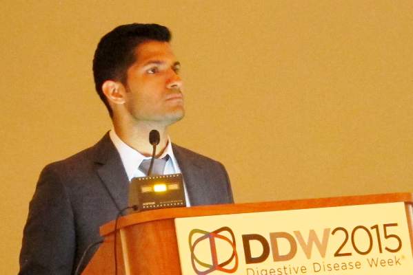
Furthermore, anticoagulant treatment “is associated with a higher risk of rebleeding and death compared with antiplatelet treatment after a previous GI event,” Dr. Angel Lanas said to an overflow crowd at the annual Digestive Disease Week.
In a separate case-control study, Dr. Lanas and his associates recently reported that the risk of GI bleeding was twofold higher for anticoagulants than for low-dose aspirin in patients hospitalized for GI bleeding (Clin. Gastroenterol. Hepatol. 2015 May;13:906-12.e2. [doi:10.1016/j.cgh.2014.11.007])
The current study examined adverse events in a cohort of 160 patients who developed a major gastrointestinal bleed (GIB) while using anticoagulants and/or antiplatelet therapy between March 2008 and July 2013. Long-term interruption or short-term resumption of these treatments has important clinical implications and differences in the intrinsic risks between antiplatelet or anticoagulant users after drug resumption are not well established, said Dr. Lanas of the University of Zaragoza, Spain.
Drug use information was prospectively collected during the GIB event, with data during the follow-up period obtained from two different Spanish databases.
Treatment during the index bleeding event was continued without interruption in 11 patients and interrupted in 149 patients (93%). Among those whose therapy was interrupted, 21 (14%) never resumed therapy and 128 (86%) resumed therapy (118 patients within 15 days and 10 patients after 15 days). The 86% treatment resumption rate is much higher than the 40%-66% rates reported in previous studies, indicating that Spanish physicians restarted treatment quite early, Dr. Lanas observed.
The mean age at baseline was 76.6 years, 61.3% of patients were men, and half had a Charlson index score > 4. Median follow-up was 21.5 months (range 1-63 months).
Ischemic events did not differ between patients who did or did not restart anticoagulants or antiplatelets (16.4% vs. 14.3%; P value = .806). However, rebleeding occurred in 32% of patients who resumed therapy versus none who did not (P = .002), but deaths were higher in those who did not restart therapy (38.1% vs. 12.5%; P = .003), Dr. Lanas said.
These differences remain significant in Kaplan-Meier survival curves for death (P = .021) and rebleeding (P = .004).
A comparison of early therapy resumption (≤ 15 days) vs. delayed (mean delay 62 days) or no resumption revealed similar results. Early resumption was associated with a higher rate of rebleeding (32.2% vs. 9.7%; P = .012), but a lower rate of death (11% vs. 35.5%; P = .001), with no difference in ischemic events (17% vs. 13%; P = .586), Dr. Lanas said.
Again, the differences remain significant in Kaplan-Meier survival curves for death (P = .011) and rebleeding (P = .013).
When the investigators looked at rebleeding according to drug use, patients receiving anticoagulants vs. antiplatelets had significantly higher rates of rebleeding (34.7% vs. 20.5%; P = .043), death (22.2% vs. 10.2%; P = .038), and any event (68.1% vs. 52.3%; P = .043).
After adjustment for gender, age, Charlson index, diabetes, and arterial hypertension, the risk of rebleeding was more than threefold higher for dual antiplatelet and anticoagulant users than for antiplatelet-alone users (odds ratio, 3.45; P = .025) and was twofold higher for anticoagulant vs. antiplatelet users (OR, 2.07; P = .045), Dr. Lanas said.
Finally, an analysis of the cause of bleeding suggests the cause of rebleeding may be different from the original event and that there is a shift toward the lower GI tract, he added.
The index bleeding event was caused largely by an upper GI peptic ulcer in 48% of all 160 patients, with 43.7% of events due to lower GI diverticulosis, vascular lesions, ischemic, or other lesions. In contrast, peptic ulcers accounted for only 7% of rebleeding events, while lower GI events accounted for 72%. Proton pump inhibition use was evenly distributed in upper and lower GI bleeding, although effective endoscopic treatment may have influenced upper GI bleeds, Dr. Lanas said.
“The importance of this is that we may have very good therapy tools for the upper GI, but still we have problems controlling the bleeding from the lower GI,” he added.
During a discussion of the study, an audience member asked how many days clinicians should wait to restart anticoagulants or antiplatelets.
“In those patients with peptic ulcer bleeding, it’s better to just give the antiplatelet therapy soon after the bleeding event or just to not interrupt the aspirin because the morality at 30 days was higher in those who were interrupted,” Dr. Lanas advised. “...I think for the cutoff point to show differences for patients with a worse outcome versus those with a better outcome, you shouldn’t restart anticoagulant therapy before day 15 after the bleeding event.”
Dr. Lanas received consulting fees, speaking and teaching fees, other financial support, and grant and research support from Bayer.
EXPERT ANALYSIS FROM DDW 2015
DDW: Postbleed blood thinners up rebleeding risk, lower death risk
WASHINGTON – Early resumption of antiplatelet agents or anticoagulants after a major gastrointestinal bleeding event is clearly associated with an increased risk of rebleeding, but a decreased risk of death, results from an observational study show.
Furthermore, anticoagulant treatment “is associated with a higher risk of rebleeding and death compared with antiplatelet treatment after a previous GI event,” Dr. Angel Lanas said to an overflow crowd at the annual Digestive Disease Week.
In a separate case-control study, Dr. Lanas and his associates recently reported that the risk of GI bleeding was twofold higher for anticoagulants than for low-dose aspirin in patients hospitalized for GI bleeding (Clin. Gastroenterol. Hepatol. 2015 May;13:906-12.e2. [doi:10.1016/j.cgh.2014.11.007])
The current study examined adverse events in a cohort of 160 patients who developed a major gastrointestinal bleed (GIB) while using anticoagulants and/or antiplatelet therapy between March 2008 and July 2013. Long-term interruption or short-term resumption of these treatments has important clinical implications and differences in the intrinsic risks between antiplatelet or anticoagulant users after drug resumption are not well established, said Dr. Lanas of the University of Zaragoza, Spain.
Drug use information was prospectively collected during the GIB event, with data during the follow-up period obtained from two different Spanish databases.
Treatment during the index bleeding event was continued without interruption in 11 patients and interrupted in 149 patients (93%). Among those whose therapy was interrupted, 21 (14%) never resumed therapy and 128 (86%) resumed therapy (118 patients within 15 days and 10 patients after 15 days). The 86% treatment resumption rate is much higher than the 40%-66% rates reported in previous studies, indicating that Spanish physicians restarted treatment quite early, Dr. Lanas observed.
The mean age at baseline was 76.6 years, 61.3% of patients were men, and half had a Charlson index score > 4. Median follow-up was 21.5 months (range 1-63 months).
Ischemic events did not differ between patients who did or did not restart anticoagulants or antiplatelets (16.4% vs. 14.3%; P value = .806). However, rebleeding occurred in 32% of patients who resumed therapy versus none who did not (P = .002), but deaths were higher in those who did not restart therapy (38.1% vs. 12.5%; P = .003), Dr. Lanas said.
These differences remain significant in Kaplan-Meier survival curves for death (P = .021) and rebleeding (P = .004).
A comparison of early therapy resumption (≤ 15 days) vs. delayed (mean delay 62 days) or no resumption revealed similar results. Early resumption was associated with a higher rate of rebleeding (32.2% vs. 9.7%; P = .012), but a lower rate of death (11% vs. 35.5%; P = .001), with no difference in ischemic events (17% vs. 13%; P = .586), Dr. Lanas said.
Again, the differences remain significant in Kaplan-Meier survival curves for death (P = .011) and rebleeding (P = .013).
When the investigators looked at rebleeding according to drug use, patients receiving anticoagulants vs. antiplatelets had significantly higher rates of rebleeding (34.7% vs. 20.5%; P = .043), death (22.2% vs. 10.2%; P = .038), and any event (68.1% vs. 52.3%; P = .043).
After adjustment for gender, age, Charlson index, diabetes, and arterial hypertension, the risk of rebleeding was more than threefold higher for dual antiplatelet and anticoagulant users than for antiplatelet-alone users (odds ratio, 3.45; P = .025) and was twofold higher for anticoagulant vs. antiplatelet users (OR, 2.07; P = .045), Dr. Lanas said.
Finally, an analysis of the cause of bleeding suggests the cause of rebleeding may be different from the original event and that there is a shift toward the lower GI tract, he added.
The index bleeding event was caused largely by an upper GI peptic ulcer in 48% of all 160 patients, with 43.7% of events due to lower GI diverticulosis, vascular lesions, ischemic, or other lesions. In contrast, peptic ulcers accounted for only 7% of rebleeding events, while lower GI events accounted for 72%. Proton pump inhibition use was evenly distributed in upper and lower GI bleeding, although effective endoscopic treatment may have influenced upper GI bleeds, Dr. Lanas said.
“The importance of this is that we may have very good therapy tools for the upper GI, but still we have problems controlling the bleeding from the lower GI,” he added.
During a discussion of the study, an audience member asked how many days clinicians should wait to restart anticoagulants or antiplatelets.
“In those patients with peptic ulcer bleeding, it’s better to just give the antiplatelet therapy soon after the bleeding event or just to not interrupt the aspirin because the morality at 30 days was higher in those who were interrupted,” Dr. Lanas advised. “...I think for the cutoff point to show differences for patients with a worse outcome versus those with a better outcome, you shouldn’t restart anticoagulant therapy before day 15 after the bleeding event.”
Dr. Lanas received consulting fees, speaking and teaching fees, other financial support, and grant and research support from Bayer.
On Twitter @pwendl
EXPERT ANALYSIS FROM DDW 2015
Key clinical point: Early resumption of antiplatelet agents or anticoagulants after a major gastrointestinal bleeding event is clearly associated with an increased risk of rebleeding, but a decreased risk of death.
Major finding: Rebleeding occurred in 32% of patients who resumed therapy versus none who did not (P = .002), but deaths were higher in those who did not restart therapy (38.1% vs. 12.5%; P = .003).
Data source: Retrospective, observational cohort study of 160 patients who developed GI bleeding while on antiplatelet or anticoagulant therapy.
Disclosures: Dr. Lanas received consulting fees, speaking and teaching fees, other financial support, and grant and research support from Bayer.
DDW: Antibiotic rifaximin eases functional dyspepsia symptoms
WASHINGTON – Two weeks of antibiotic therapy with rifaximin provided relief from functional dyspepsia symptoms in a phase III double-blind, randomized trial.
“This is the first study that demonstrates that rifaximin is efficacious in the treatment of functional dyspepsia, particularly for global dyspeptic symptoms, bloating, and possibly belching. Our finding may suggest a role for the gut microbiota in the pathogenesis of functional dyspepsia,” Dr. Victoria Tan said at the annual Digestive Disease Week.
Rifaximin (Xifaxan) works by reducing or altering bacteria in the gut and has been shown to be efficacious in the treatment of diarrhea-predominant irritable bowel syndrome. It is approved to treat traveler’s diarrhea caused by Escherichia coli and to prevent hepatic encephalopathy.
The study randomly assigned 95 consecutive adults with functional dyspepsia as per ROME III criteria who had a normal gastroscopy within the last 2 years, had active symptoms in the month prior to enrollment, and were Helicobacter pylori negative, to rifaximin 400 mg or placebo three times a day for 2 weeks. In all, 33 rifaximin and 39 placebo patients were evaluable for the primary efficacy outcome of adequate relief of global dyspeptic symptoms (either no or mild dyspeptic symptoms) at any of the follow-up time points.
At baseline, 77% of patients had moderate to severe global dyspepsia symptoms, 74% of the placebo group and 55%% of the rifaximin group had moderate to severe belching, and roughly half of all patients were not on any GI medications. Mean age of the patients was 52 years.
Global dyspepsia symptoms improved with rifaximin beginning at week 2 and significantly favored rifaximin by week 8, with 23.5% of rifaximin patients reporting moderate to severe symptoms compared with 47.4% given placebo (P value = .04), said Dr. Tan of the University of Hong Kong.
Rates of moderate to severe belching were significantly improved with rifaximin at week 4 compared with placebo (14.3% vs. 35.7%; P = .03), but this difference was no longer significant at week 8 (26.5% vs. 29%).
The story was similar for moderate to severe bloating: Rates declined significantly with rifaximin at week 4 (20% vs. 43%; P = .03), but were no longer significant at week 8 (26.5% vs. 34.2%), she said.
A subgroup analysis of female patients showed significant improvements in moderate to severe global dyspeptic symptoms with rifaximin compared with placebo at week 4 (20.8% vs. 59.4%; P = .006) and week 8 (20% vs. 48.4%; P = .048).
Treatment response was not reflected in change in hydrogen breath response, Dr. Tan said. Results of a 3-hour hydrogen breath test performed after a 12-hour overnight fast showed no differences between the rifaximin and placebo groups for H2 peak above baseline (2.94 ppm vs. 0.11 ppm; P = .29), H2 area under the curve (+43.64 ppm vs. –49.71 ppm; P = .76), and oro-cecal transit time (24.23 minutes vs. 16.5 minutes; P = .68).
Adverse events were very similar between the two groups at both 4 and 8 weeks, Dr. Tan said. Only one major event occurred, a severe case of acute hepatitis in a woman in the placebo arm who also took traditional Chinese herbs.
On Twitter @pwendl
AT DDW 2015
Key clinical point: A 2-week course of rifaximin is efficacious in the treatment of functional dyspepsia.
Major finding: At week 8, 23.5% of patients treated with rifaximin vs. 47.4% given placebo reported moderate to severe global dyspepsia symptoms (P = .04).
Data source: Phase III placebo-controlled trial of 95 consecutive patients with functional dyspepsia as per ROME III criteria who had a normal gastroscopy within the last 2 years, had active symptoms in the month prior to enrollment, and were H. pylori negative.
Disclosures: Chong Lap Pharmaceuticals supplied the study drugs, but had no input into the study. Dr. Tan reported having no financial disclosures.
Is It IBS? Blood Test May Offer Conclusive Answer
WASHINGTON – A new blood test could conclusively determine if a patient with chronic diarrhea has diarrhea-predominant irritable bowel syndrome (D-IBS).
The IBSchek blood test detects the presence of antibodies to cytolethal distending toxin B and vinculin. In a study presented at the annual Digestive Disease Week and published in PLoS ONE, the positive predictive value for D-IBS of just one of the antibodies was greater than 98%, explained study lead author Dr. Mark Pimentel of Cedars-Sinai Medical Center, Los Angeles. If the test is positive for both antibodies, “the post-test probability is 95% that you have IBS.”
In a video interview, Dr. Pimentel discussed the study’s findings and the potential impact for physicians and patients. The search for diagnostic answers leads to “a lot of doctor-shopping, certainly a lot of colonoscopies and unnecessary testing that are always negative with these patients,” he noted. “Maybe this will put an end to that.
“People used to think this is all psychological,” Dr. Pimentel added. “Now we can say, No, it’s organic. There’s something real going on; I’ve got a test that proves that.”
Dr. Pimentel has received consulting fees from Commonwealth Laboratories, which makes the IBSchek blood test.
The video associated with this article is no longer available on this site. Please view all of our videos on the MDedge YouTube channel
AT DDW 2015
VIDEO: Is it IBS? Blood test may offer conclusive answer
WASHINGTON – A new blood test could conclusively determine if a patient with chronic diarrhea has diarrhea-predominant irritable bowel syndrome (D-IBS).
The IBSchek blood test detects the presence of antibodies to cytolethal distending toxin B and vinculin. In a study presented at the annual Digestive Disease Week and published in PLoS ONE, the positive predictive value for D-IBS of just one of the antibodies was greater than 98%, explained study lead author Dr. Mark Pimentel of Cedars-Sinai Medical Center, Los Angeles. If the test is positive for both antibodies, “the post-test probability is 95% that you have IBS.”
In a video interview, Dr. Pimentel discussed the study’s findings and the potential impact for physicians and patients. The search for diagnostic answers leads to “a lot of doctor-shopping, certainly a lot of colonoscopies and unnecessary testing that are always negative with these patients,” he noted. “Maybe this will put an end to that.
“People used to think this is all psychological,” Dr. Pimentel added. “Now we can say, No, it’s organic. There’s something real going on; I’ve got a test that proves that.”
Dr. Pimentel has received consulting fees from Commonwealth Laboratories, which makes the IBSchek blood test.
The video associated with this article is no longer available on this site. Please view all of our videos on the MDedge YouTube channel
AT DDW 2015
DDW: Ozanimod active in moderate-severe UC, without cardiac signal in TOUCHSTONE trial
WASHINGTON– The oral S1P receptor modulator ozanimod is clinically active and well tolerated in patients with moderate to severe ulcerative colitis (UC), phase II study results show.
Importantly, no notable cardiac, opthalmologic, or infectious treatment-related adverse events were observed, Dr. William Sandborn said at the annual Digestive Disease Week.
The first sphingosine-1-phosphate (S1P) receptor modulator, Fingolimod (Gilenya), carries a warning for first-dose cardiac effects, liver function test elevations, and macular edema and targets S1P receptors 1, 3, 4, and 5. Ozanimod is a next-generation S1P receptor modulator that has increased selectivity for the S1P receptors 1 and 5, compared with receptor 3, which may be related to safety concerns with fingolimod, said Dr. Sandborn, chief of gastroenterology at the University of California-San Diego.
He reported on the double-blind, phase II TOUCHSTONE trial involving 197 adults with moderate to severe UC who were receiving oral aminosalicylates and/or prednisone, and were randomly assigned to receive ozanimod 0.5 mg (n = 65) or 1 mg (n = 67) or placebo (n = 65). Doses were titrated through week 1, followed by 8 weeks of full-dose therapy.
The study’s primary efficacy endpoint was the proportion of patients in clinical remission at week 8, defined as a Mayo score of 2 or less, with no subscore of more than 1.
At week 8, 16.4% of patients on ozanimod 1 mg were in remission vs. 6.2% on placebo (P = .0482) and 13.8% on ozanimod 0.5 mg (P = .1422), Dr. Sandborn reported.
Rates of clinical response at 8 weeks were 56.7% with the high-dose ozanimod, 53.8% with the low dose, and 37% with placebo. Once again, the between-group difference was significant only for high-dose ozanimod (P = .0207).
Mucosal improvement was significantly more common with either high-dose ozanimod (34.3% vs. 12.3% placebo; P = .0023) or low-dose ozanimod (27.7% vs. 12.3% placebo; P = .0348), he said.
The most common adverse events were anemia/decreased hemoglobin, occurring in four patients in the placebo and low-dose ozanimod groups, and worsening of UC, occurring in three patients on placebo, two on low-dose ozanimod, and one on high-dose ozanimod.
Serious treatment-related adverse events were reported in four patients on placebo, one on ozanimod 0.5 mg (hyperpyrexia), and one on ozanimod 1 mg (UC).
The overall incidence of cardiac events was low, with two palpitations reported in the placebo group, one sinus bradycardia and one first-degree AV block in the ozanimod 0.5-mg group, and none in the 1-mg group.
“Ozanimod was well tolerated with a favorable benefit-risk profile supporting the planned phase III trial in ulcerative colitis and the phase II study in Crohn’s disease,” said Dr. Sandborn, who also reported the results earlier this year in Europe.
On Twitter@pwendl
AT DDW 2015
Key clinical point: Ozanimod 1 mg induced clinical remission at week 8 in patients with moderate to severe ulcerative colitis.
Major finding: At week 8, 16.4% of patients on ozanimod 1 mg were in remission vs. 6.2% on placebo (P = .0482) and 13.8% on ozanimod 0.5 mg (P = .1422).
Data source: Randomized, double-blind trial of 197 patients with moderate to severe ulcerative colitis.
Disclosures: Dr. Sandborn reported financial relationships with numerous firms including Receptos, which funded the study.
DDW: Gestational diabetes linked to increased NAFLD risk in middle age
WASHINGTON – Gestational diabetes was identified as a significant, independent risk factor for developing non-alcoholic fatty liver disease later in life, according to a study of more than 1,000 women followed for 25 years.
This study “is the first to show a strong association between gestational diabetes in young adulthood and non-alcoholic fatty liver disease in middle-age,” Dr. Veeral Ajmera of the department of gastroenterology at the University of California, San Francisco, reported at the annual Digestive Disease Week.
The study evaluated 1,115 women from the Coronary Artery Risk Development in Young Adults (CARDIA) study, who had at least one delivery, did not have a diabetes diagnosis before pregnancy, and had a CT evaluation for hepatic steatosis at the 25-year visit, in 2010 and 2011. Women were excluded if they did not have a complete CT scan at that time, they had more than 14 drinks of alcohol per week, used medications associated with steatosis, or had chronic viral hepatitis or HIV infections.
At baseline the median age was 25-26 years, about 55%-56% were black, and the prevalence of hypertension (2%-3%) and dyslipidemia were similar. The CARDIA study enrolled about 5,100 men and women aged 18-30 years at four U.S. medical centers in the mid 1980s. Patients had 7 study visits over a 25-year follow-up period.
Of the 1,115 women evaluated, 124 (11%) went on to develop GDM and 75 (7%) met the CT definition for NAFLD (liver attenuation less than or equal to 40 Hounsfield units on CT scan) by the 25-year follow-up, when they were a median age of 50-51 years.
At 25 years, 14% of those with a history of GDM had NAFLD, compared with 5.8% of those who did not have GDM, for an unadjusted odds ratio of 2.56 (p <0.01), Dr. Ajmera said.
In the statistical analysis, the researchers determined that baseline homeostatic model assessment of insulin resistance (HOMA-IR) and baseline triglycerides were also ”strongly associated” with NAFLD at year 25.
“Importantly,” he noted, the measure of the association between GDM and NAFLD remained statistically significant, after adjusting for these covariates (OR, 2.29). At the start of the study, women who went on to develop GDM had significantly higher body mass index (BMI), HOMA-IR, waist circumference, and triglycerides than those who did not develop GDM, but the magnitude of those differences was small.
Additionally, after adjustment for a diagnosis of diabetes at year 25, the association between a history of GDM and NAFLD was stronger (OR, 1.99) than the association between a diagnosis of diabetes at year 25 and NAFLD (OR, 1.5), he said.
The researchers added BMI to their multivariate analysis to determine whether women with a gestational diabetes history gained more weight, and whether that weight explained the increased prevalence of NAFLD, “and found that the association between gestational diabetes and non-alcoholic fatty liver disease remained,” Dr. Ajmera added.
Evaluations of race, baseline BMI, and baseline HOMA-IR as effect modifiers of the relationship between GDM and NAFLD were not significant.
“Gestational diabetes represents insulin resistance unmasked by the stress of pregnancy, and offers the unique opportunity to identify those at risk of NAFLD at a young age,” Dr. Ajmera concluded.
Limitations of the study included not being able to determine if the women had NAFLD before they were diagnosed with GDM. However, based on the participants’ young age at enrollment, that is unlikely, he said. In addition, the researchers had no information on liver biochemistry tests, which, however, are not sensitive or specific for diagnosing NAFLD. The strengths of the study include the length of follow-up of a large biracial population, with measurement of well-characterized metabolic covariates measured, he added.
Dr. Ajmera reported having no relevant financial disclosures. The CARDIA study is supported by the National Heart, Lung, and Blood Institute, part of the National Institutes of Health.
Nonalcoholic fatty liver disease is the most common and fastest growing liver disease worldwide. Identifying individuals at high risk for NAFLD remains challenging. Dr. Ajmera and his colleagues highlight the association between gestational diabetes mellitus (GDM) and NAFLD. Their work confirms and extends earlier studies that reported increased hepatic fat content and a substantially increased risk for NAFLD, in women with a prior history of GDM. In the present study, a more racially diverse U.S.-based population was followed for a much longer duration than was previous cohorts. After adjusted multivariate analysis, GDM remained a strong, independent predictor of future NAFLD. Indeed, GDM was a a better NAFLD predictor than the diagnosis of diabetes alone was. It is unclear if prepregnancy NAFLD might have affected the risk for GDM, as subjects were not assessed for hepatic steatosis or liver test elevations at baseline. Also, the percentage of 50-year-old women with NAFLD was much lower in this cohort (7%) than in the general U.S. population (at least 25%), perhaps because the study population was enriched with individuals who are typically at low risk for NAFLD (i.e., African Americans, people with low body mass index, and/or low serum triglycerides).
Additional research is needed to clarify these issues and to investigate how maternal GDM affected the health of their children. Intrauterine exposure to metabolic stresses increases the risk for obesity and other metabolic disorders later in life, suggesting that offspring of mothers with GDM may require specialized surveillance or other precautions during childhood. Given that more young women are entering their reproductive years affected by diabetes and obesity, increased attention to these issues will be required to curb the growing public health burden of NAFLD.
Dr. Cynthia A. Moylan, M.H.S., is assistant professor of medicine, Duke University Medical Center and Veterans Affairs Medical Center, Durham, N.C. She is has no relevant financial conflicts of interests.
WASHINGTON – Gestational diabetes was identified as a significant, independent risk factor for developing non-alcoholic fatty liver disease later in life, according to a study of more than 1,000 women followed for 25 years.
This study “is the first to show a strong association between gestational diabetes in young adulthood and non-alcoholic fatty liver disease in middle-age,” Dr. Veeral Ajmera of the department of gastroenterology at the University of California, San Francisco, reported at the annual Digestive Disease Week.
The study evaluated 1,115 women from the Coronary Artery Risk Development in Young Adults (CARDIA) study, who had at least one delivery, did not have a diabetes diagnosis before pregnancy, and had a CT evaluation for hepatic steatosis at the 25-year visit, in 2010 and 2011. Women were excluded if they did not have a complete CT scan at that time, they had more than 14 drinks of alcohol per week, used medications associated with steatosis, or had chronic viral hepatitis or HIV infections.
At baseline the median age was 25-26 years, about 55%-56% were black, and the prevalence of hypertension (2%-3%) and dyslipidemia were similar. The CARDIA study enrolled about 5,100 men and women aged 18-30 years at four U.S. medical centers in the mid 1980s. Patients had 7 study visits over a 25-year follow-up period.
Of the 1,115 women evaluated, 124 (11%) went on to develop GDM and 75 (7%) met the CT definition for NAFLD (liver attenuation less than or equal to 40 Hounsfield units on CT scan) by the 25-year follow-up, when they were a median age of 50-51 years.
At 25 years, 14% of those with a history of GDM had NAFLD, compared with 5.8% of those who did not have GDM, for an unadjusted odds ratio of 2.56 (p <0.01), Dr. Ajmera said.
In the statistical analysis, the researchers determined that baseline homeostatic model assessment of insulin resistance (HOMA-IR) and baseline triglycerides were also ”strongly associated” with NAFLD at year 25.
“Importantly,” he noted, the measure of the association between GDM and NAFLD remained statistically significant, after adjusting for these covariates (OR, 2.29). At the start of the study, women who went on to develop GDM had significantly higher body mass index (BMI), HOMA-IR, waist circumference, and triglycerides than those who did not develop GDM, but the magnitude of those differences was small.
Additionally, after adjustment for a diagnosis of diabetes at year 25, the association between a history of GDM and NAFLD was stronger (OR, 1.99) than the association between a diagnosis of diabetes at year 25 and NAFLD (OR, 1.5), he said.
The researchers added BMI to their multivariate analysis to determine whether women with a gestational diabetes history gained more weight, and whether that weight explained the increased prevalence of NAFLD, “and found that the association between gestational diabetes and non-alcoholic fatty liver disease remained,” Dr. Ajmera added.
Evaluations of race, baseline BMI, and baseline HOMA-IR as effect modifiers of the relationship between GDM and NAFLD were not significant.
“Gestational diabetes represents insulin resistance unmasked by the stress of pregnancy, and offers the unique opportunity to identify those at risk of NAFLD at a young age,” Dr. Ajmera concluded.
Limitations of the study included not being able to determine if the women had NAFLD before they were diagnosed with GDM. However, based on the participants’ young age at enrollment, that is unlikely, he said. In addition, the researchers had no information on liver biochemistry tests, which, however, are not sensitive or specific for diagnosing NAFLD. The strengths of the study include the length of follow-up of a large biracial population, with measurement of well-characterized metabolic covariates measured, he added.
Dr. Ajmera reported having no relevant financial disclosures. The CARDIA study is supported by the National Heart, Lung, and Blood Institute, part of the National Institutes of Health.
AT DDW® 2015
Key clinical point: Gestational diabetes could be used to identify young women at increased risk of having non-alcoholic fatty liver disease (NAFLD) in middle age.
Major finding: Having been diagnosed with gestational diabetes was associated with about a two-fold increased risk of having NAFLD in middle age.
Data source: The research involved 1,115 women enrolled in the 25-year Coronary Artery Risk Development in Young Adults (CARDIA) study.
Disclosures: Dr. Ajmera reported having no relevant financial disclosures. The CARDIA study is supported by the National Heart, Lung, and Blood Institute.
DDW: Recurrent C. difficile infections take heavy toll on IBD patients
WASHINGTON – Patients with inflammatory bowel disease had a one-third higher risk for having a recurrent Clostridium difficile infection than did the general population, and a 20-fold higher risk for needing a total colectomy because of the infection, a study showed.
Risk factors for recurrent C. difficile infection (rCDI) in patients with inflammatory bowel disease (IBD) include a recent hospitalization, immunosuppressive drugs, and antibiotics, but which drugs are most culpable is unclear, according to Dr. Roshan Razik of Mount Sinai Hospital, Toronto, and the University of Toronto.
“The drugs that we’re using do place patients at higher risk for recurrent C. difficile, but it’s yet to come out which drugs within drug categories – immunomodulators, antibiotics, biologics – pose a higher risk, although we are beginning to see evidence, for example, that azathioprine is higher risk than methotrexate, infliximab might be higher risk than adalimumab, so we have to continue to parse through the data,” he said in an interview.
He and his colleagues conducted two retrospective studies to assess the effects of rCDI on patients with IBD.
The first study used a case-control design, including patients with IBD who had two or more documented instances of rCDI from 2010 through 2013 as cases, and IBD patients with only one infection as controls.
The second study used a retrospective cohort to calculate the incidence of rCDI in patients with IBD, compared with patients without IBD, Dr. Razik reported at the annual Digestive Disease Week.
There were a total of 503 patients who tested positive for CDI included in the studies: 110 patients with IBD (49% with Crohn’s disease and 51% with ulcerative colitis) and 393 without. The mean age was 58.8 years, and 61.4% were female.
Compared with patients without IBD, patients with Crohn’s disease or ulcerative colitis developed CDI at a younger age (39 years vs. 64 years, P < .001), used more steroids (39.1% vs. 12%, P < .001), used more immunosuppressive agents (42.7% vs. 13.2%, P < .001), and were more likely to have a prior bowel resection (28.2% vs. 11.5%, P < .001).
In all, 32% of patients with IBD had a recurrent CDI, compared with 24% of non-IBD patients (P < .01). There were no significant differences between the groups in the number of hospitalizations due to CDI, but patients with IBD were significantly more likely to require colectomy because of the infections (6.4% vs. 0.3%, P < .001).
In a multivariate analysis, risk factors for rCDI in patients with IBD included nonileal Crohn’s disease (odds ratio, 2.59; P < .001), recent antibiotic therapy (OR, 2.60; P < .001), use of a 5-aminosalicylic acid drug (OR, 3.06; P < .001), steroid use (OR, 2.94; P < .001), biologic therapy (OR, 2.50; P = .001), a recent hospitalization (OR, 2.62; P < .001), and no previous bowel resections (OR, 1.72; P = .020).
Clinicians need to look beyond the usual suspects, antibiotics, as causative agents for rCDI. Immunomodulators and biologic agents also were strongly associated with rCDI in the study, Dr. Razik noted.
The study source was not disclosed. He reported having no relevant financial conflicts of interest.
AT DDW 2015
Key clinical point: Patients with inflammatory bowel disease are at higher risk for recurrent Clostridium difficile infections.
Major finding: 32% of patients with IBD had a recurrent CDI, compared with 24% of non-IBD patients
Data source: Retrospective case-control studies with 503 patients, comparing patients with IBD with and without recurrent C. difficile infections and comparing patients with IBD with non-IBD patients.
Disclosures: The study source was not disclosed. Dr. Razik reported having no relevant financial conflicts of interest.
DDW: Menopausal hormone therapy increases major GI bleed risk
Menopausal hormone therapy is associated with an increased risk of major gastrointestinal bleeding, particularly in the lower gastrointestinal tract, that is associated with duration of use, a study has found.
Analysis of data from 73,863 women enrolled in the Nurses’ Health Study II in 1989 showed that current users of menopausal hormone therapy had a 46% increase in the risk of a major gastrointestinal bleed and a more than twofold increase in the risk of a lower GI bleed or ischemic colitis, compared with never users, said Dr. Prashant Singh of Massachusetts General Hospital, Boston.
Past users showed a much smaller increase risk of bleeding, while increasing duration of hormone therapy was significantly associated with increasing risk of major and low gastrointestinal bleeding.
“Although our findings show that menopausal hormone therapy may increase the risk of major GI bleeding, especially in the lower GI tract, it is important for these patients to know that this therapy is still an effective treatment; however, both clinician and patient should be more cautious in using this therapy in some cases, such as with patients who have a history of ischemic colitis,” Dr. Singh said at the annual Digestive Disease Week.
Dr. Singh does not have any relevant financial or other relationship with any manufacturer or provider of commercial products or services that he discussed during the presentation.
FROM DDW 2015
Key clinical point: Menopausal hormone therapy is associated with an increased risk of major gastrointestinal bleeding, particularly in the lower gastrointestinal tract.
Major finding: Current users of menopausal hormone therapy had a 46% increase in the risk of a major gastrointestinal bleed and a more than twofold increase in the risk of a lower GI bleed or ischemic colitis.
Data source: Analysis of data from 73,863 women enrolled in the Nurses’ Health Study II.
Disclosures: No conflicts of interest were disclosed.
DDW: Urinary enzymes hint at gastric cancer
WASHINGTON – A simple urine test could detect gastric cancer even at an early stage, the test’s developers say.
The test, which looks for the presence of two metalloprotease enzymes labeled ADAM 12 and MMP-9/NGAL had 77.1% sensitivity and 82.9% specificity for gastric cancer when tested in 35 patients with the malignancy and an equal number of healthy controls, reported Dr. Takaya Shimura from the department of surgery at Boston Children’s Hospital and Harvard Medical School in Boston.
“This study represents the first demonstration of the presence of ADAM 12 and MMO-9/NGAL complex in the urine of gastric cancer patients,” he said at the annual Digestive Disease Week.
Dr. Stephen J. Meltzer of Johns Hopkins University, Baltimore, commented in an interview that the findings are convincing but preliminary.
A randomized clinical trial enrolling a larger number of patients and controls would be required before he would consider screening patients for the enzymes, said Dr. Meltzer, who was not involved in the study and comoderated the meeting session where the results were presented.
ADAM 12 (a disintegrin and metalloprotease 12) and MMP-9 (matrix metalloprotease 9) are both members of a family of enzymes involved in cellular adhesion, invasion, growth, and angiogenesis, Dr. Shimura explained. MMP-9, when complexed with NGAL (neutrophil gelatinase associated lipocalin) is protected from autodegradation.
The investigators, from the lab of Dr. Marsha A. Moses at Boston Children’s Hospital, and their collaborators in Japan had previously reported that MMPs in urine were independent predictors of both organ-confined and metastatic cancer.
Urinary assays are noninvasive, using easily accessed tissues that can be handled simply and inexpensively, making them ideal for cancer detection, Dr, Shimura said.
Current tests for gastric cancer, such as carcinoembryonic antigen (CEA) and cancer antigens (CA) 19-9 and 72-4, have poor sensitivity for detecting advanced disease, and are even worse at spotting early disease, he noted.
To see whether they could improve on the current lot of tests, the investigators enrolled 106 patients in a case-control study, settling eventually, after age and sex matching, on a cohort of 70 patients: 35 with primarily early-stage gastric cancer, and 35 healthy controls.
After screening the urine of participants for about 50 different antigenic proteins, they found that the patients with gastric cancer had significantly higher levels in their urine of both ADAM 12 (P < .001) and the MMP-9/NGAL complex (P = .020).
In a multivariate analysis, they showed that both enzymes were strong, independent predictors of gastric cancer, with an odds ratio for urinary MMO-9/NGAL of 6.71 (P = .002), and an OR of 15.4 for ADAM 12 (P = .002). In contrast, Helicobacter pylori infection was associated with a nonsignificant OR of 2.54.
In a receiver operating characteristic (ROC) analysis, they also found that MMP-9/NGAL was associated with an area-under-the curve (AUC) of 0.657 (P = .024), ADAM 12 was associated with an AUC of 0.757 (P < .001), and that the two combined had an AUC of 0.825 (P < .001).
As noted before, the sensitivity of the combined enzymes was 77%, and the specificity was 83%.
Finally, using immunohistochemical analysis, the investigators were able to show that gastric cancer tissues had high levels of coexpression of MMP-9 and NGAL (P <.001) and high expression levels of ADAM 12 (P < .001), compared with adjacent normal tissues.
AT DDW 2015
Key clinical point: Urinary levels of two metalloproteases were significantly elevated in the urine of patients with gastric cancer, compared with controls.
Major finding: High expression of ADAM 12 and MMP-9/NGAL complex had a 77% sensitivity and 83% specificity for gastric cancer.
Data source: Case-control study of 35 patients with gastric cancer and 35 controls.
Disclosures: The study was supported by the Advanced Medical Research Foundation in the United States and the Research Fellowship of the Uehara Memorial Foundation, Japan. Dr. Shimura reported having no conflicts of interest.

