User login
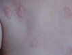
Vitiliginous Lesions Induced by Amyl Nitrite Exposure
Managing Postinflammatory Hyperpigmentation
Pigmentation Concerns: Assessment and Treatment [editorial]
Cutaneous Mastocytosis: A Case of Bullous Urticaria Pigmentosa
Cosmetic tattooing and ethnic skin
Cosmetic tattooing, also known as micropigmentation or permanent makeup, is a technique in which tattooing is performed to address cosmetic skin imperfections. It is often used to create eyeliner or lip liner, but it can also be used to camouflage stable patches of vitiligo, to create eyebrows on those who have lost them due to alopecia areata or chemotherapy, to create areolas for women who have had mastectomies, or to correct the shape of a reconstructed cleft lip. Cosmetic tattooing is also useful in women who want to wear makeup, but who have trouble applying it due to visual deficits, tremor, stroke, multiple sclerosis, or Parkinson’s disease.
In Asian cultures, cosmetic tattooing is not uncommon. Many women have cosmetic tattooing procedures to create permanent eyeliner and eyebrows, since many Asian women have sparse brows at baseline that often become thinner with aging. Cosmetic tattooing of eyeliner also enhances the natural almond shape of the eyes.
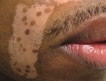
In darker ethnic skin types, where vitiligo is more visible, stable patches can be effectively camouflaged by cosmetic tattooing. However, cosmetic tattooing is not recommended unless these patches have been stable for several years and the patient has failed other therapies. The best candidate would be the darker-skinned patient with long-standing segmental vitiligo, for whom pigment grafting would also be highly considered.
Individuals who wish to perform cosmetic tattooing can receive training and certification in micropigmentology. Many also undergo apprenticeships to receive more hands-on training. I have firsthand knowledge of this process because my mother received this training and performed cosmetic tattooing on her clients when I was growing up. Good training is key, as not every tattoo ink will have the same result in every skin tone. For example, brown eyeliner might eventually turn pink on skin that has a red undertone. On someone with yellow undertones or olive skin, black pigment liner might turn greenish. So the experienced practitioner will often use different color hues depending on the person’s underlying skin color and tone to prevent this discoloration. The best results of cosmetic tattooing are achieved when others can’t tell that the work has been done.
Pitfalls with cosmetic tattooing, as with tattooing in general, include infection, allergic reaction to the tattoo ink, scarring, photocytotoxicity, and cosmetic disfigurement if the tattoo is placed improperly. Delayed granulomatous response has also been reported in cases of permanent eyebrow tattooing. In addition to typical skin infections caused by staphylococcus or streptococcus, cases of mycobacterium infection with tattooing have been reported (although such infections have been reported more often with traditional tattooing than with permanent makeup).
Red ink is the more commonly reported allergen. Titanium dioxide (TiO2) is widely used in tattoo inks to achieve certain colors. When TiO2 is exposed to certain wavelengths of light, including UV light and certain lasers, hydroxyl radicals can form, leading to photocytotoxicity (also called paradoxical darkening), which often results in a change or darkening of the pigment color. This condition is more common with pink, peach, or white tattoo colors where TiO2 is used in the color. Q-switched lasers are the most effective at removing tattoos.
If performed correctly by a properly trained person, cosmetic tattooing can be a useful aesthetic solution to various cosmetic and medical skin concerns.
This column, "Skin of Color," regularly appears in Dermatology News, a publication of Frontline Medical Communications. Dr. Wesley practices dermatology in Beverly Hills, Calif.
Do you have questions about treating patients with dark skin? If so, send them to sknews@elsevier.com.
Cosmetic tattooing, also known as micropigmentation or permanent makeup, is a technique in which tattooing is performed to address cosmetic skin imperfections. It is often used to create eyeliner or lip liner, but it can also be used to camouflage stable patches of vitiligo, to create eyebrows on those who have lost them due to alopecia areata or chemotherapy, to create areolas for women who have had mastectomies, or to correct the shape of a reconstructed cleft lip. Cosmetic tattooing is also useful in women who want to wear makeup, but who have trouble applying it due to visual deficits, tremor, stroke, multiple sclerosis, or Parkinson’s disease.
In Asian cultures, cosmetic tattooing is not uncommon. Many women have cosmetic tattooing procedures to create permanent eyeliner and eyebrows, since many Asian women have sparse brows at baseline that often become thinner with aging. Cosmetic tattooing of eyeliner also enhances the natural almond shape of the eyes.
In darker ethnic skin types, where vitiligo is more visible, stable patches can be effectively camouflaged by cosmetic tattooing. However, cosmetic tattooing is not recommended unless these patches have been stable for several years and the patient has failed other therapies. The best candidate would be the darker-skinned patient with long-standing segmental vitiligo, for whom pigment grafting would also be highly considered.
Individuals who wish to perform cosmetic tattooing can receive training and certification in micropigmentology. Many also undergo apprenticeships to receive more hands-on training. I have firsthand knowledge of this process because my mother received this training and performed cosmetic tattooing on her clients when I was growing up. Good training is key, as not every tattoo ink will have the same result in every skin tone. For example, brown eyeliner might eventually turn pink on skin that has a red undertone. On someone with yellow undertones or olive skin, black pigment liner might turn greenish. So the experienced practitioner will often use different color hues depending on the person’s underlying skin color and tone to prevent this discoloration. The best results of cosmetic tattooing are achieved when others can’t tell that the work has been done.
Pitfalls with cosmetic tattooing, as with tattooing in general, include infection, allergic reaction to the tattoo ink, scarring, photocytotoxicity, and cosmetic disfigurement if the tattoo is placed improperly. Delayed granulomatous response has also been reported in cases of permanent eyebrow tattooing. In addition to typical skin infections caused by staphylococcus or streptococcus, cases of mycobacterium infection with tattooing have been reported (although such infections have been reported more often with traditional tattooing than with permanent makeup).
Red ink is the more commonly reported allergen. Titanium dioxide (TiO2) is widely used in tattoo inks to achieve certain colors. When TiO2 is exposed to certain wavelengths of light, including UV light and certain lasers, hydroxyl radicals can form, leading to photocytotoxicity (also called paradoxical darkening), which often results in a change or darkening of the pigment color. This condition is more common with pink, peach, or white tattoo colors where TiO2 is used in the color. Q-switched lasers are the most effective at removing tattoos.
If performed correctly by a properly trained person, cosmetic tattooing can be a useful aesthetic solution to various cosmetic and medical skin concerns.
This column, "Skin of Color," regularly appears in Dermatology News, a publication of Frontline Medical Communications. Dr. Wesley practices dermatology in Beverly Hills, Calif.
Do you have questions about treating patients with dark skin? If so, send them to sknews@elsevier.com.

Treating Epidermal Melasma With a 4% Hydroquinone Skin Care System Plus Tretinoin Cream 0.025%
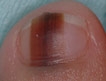
Dots and Lines: A Dermoscopic Sign of Regression of Longitudinal Melanonychia in Children
Innovative Methods of UV Protection
One of the main reasons patients with darker skin don't apply sunscreen is because they believe they are at low or no risk for ultraviolet damage, according to the results of a survey that I conducted with Dr. Brooke Jackson and Dr. Chikoti Mibenge.
Our findings were presented in a poster at the American Academy of Dermatology's Summer Academy Meeting in Boston.
The study, conducted by surveying 105 patients in Chicago, revealed that 60% of black patients do not wear sunscreen regularly. Additionally, many darker skinned patients reported not liking the whitish or chalky appearance that sunscreens often leave.
However, sunscreen manufacturers are making more elegant formulations of both chemical and physical blockers that do not leave a whitish hue on darker skin.
Sun protective clothing, hats, parasols or umbrellas, avoiding peak hours of sun intensity, and avoiding tanning are all common methods we recommend to patients to protect themselves. Lesser known methods that we can also recommend to our patients include:
Heliocare
Heliocare tablets contain Polypodium leucotomos extract, a fern native to Central and South America rich in antioxidants which protect against formation of free radicals from UV exposure, particularly UVA. The science is based off of the fact that the fern, which was once aquatic, adapted to life on land and created its own protection from UV rays. The recommended dose is 1 tablet each morning or 2 tablets before intense sun exposure. The effect begins 30 minutes after consumption and is still active 2.5 hours after consumption. Total elimination is estimated to be about 8 hours, but pharmacokinetics for elimination have not been published. Numerous published studies have reported its benefits with regards to UV protection. A head-to-head study of UV protection from heliocare versus other powerful antioxidant supplements would be interesting.
Algae and Coral
At King's College in London, research is being done on the photoprotective effect of coral. In a press release last year, Dr. Paul Long reported that algae living within coral produces a sunscreen-like compound that not only protects the algae and coral from UV damage, but also the fish that feed on the coral. The part the algae play is thought to be part of the shikimate pathway found only in microbes and plants. A sunscreen tablet with this ingredient for human use is in the works.
Strawberries

Strawberries, as well as other darker colored berries, are known to contain polyphenols, which are antioxidants. Researchers in Italy and Spain tested a strawberry extract on cultured human fibroblasts to see if there was a photoprotective effect. They added strawberry extract in different concentrations to all but the control group. They then exposed the samples to a dose of UV light "equivalent to 90 minutes of midday summer sun in the French Riviera," said lead investigator Maurizio Battino. The results demonstrated that strawberry extract, especially at a concentration of 0.5 mg/ml, provided UVA protection. It not only boosted cell survival and viability, but also minimized DNA damage when compared with control cells.
Perhaps there will be topical sunscreens that contain strawberry extract in the future. Other foods high in antioxidants that may have sun protective benefits include:
Colored peppers and yellow squash (high in carotenoids).
Tomatoes and watermelon (high in lycopene).
Dark berries, such as blueberries, acai, blackberries, cranberries (rich in anthocyanin).
Turmeric root (curcumin).
Pomegranate (ellegic acid).
Green and black tea (catechins).
Dark cocoa (flavanols).
Green leafy vegetables, such as spinach and kale (xanthophylls, oxygenated carotenoids).
Fish, such as mackerel, salmon, trout, herring, and sardines (omega 3 fatty acids).
These are not a replacement for the more common methods of sun protection, but they may certainly provide an added benefit.
- Naissan Wesley, M.D.
Do you have questions about treating patients with darker skin? If so, send them to sknews@elsevier.com.
One of the main reasons patients with darker skin don't apply sunscreen is because they believe they are at low or no risk for ultraviolet damage, according to the results of a survey that I conducted with Dr. Brooke Jackson and Dr. Chikoti Mibenge.
Our findings were presented in a poster at the American Academy of Dermatology's Summer Academy Meeting in Boston.
The study, conducted by surveying 105 patients in Chicago, revealed that 60% of black patients do not wear sunscreen regularly. Additionally, many darker skinned patients reported not liking the whitish or chalky appearance that sunscreens often leave.
However, sunscreen manufacturers are making more elegant formulations of both chemical and physical blockers that do not leave a whitish hue on darker skin.
Sun protective clothing, hats, parasols or umbrellas, avoiding peak hours of sun intensity, and avoiding tanning are all common methods we recommend to patients to protect themselves. Lesser known methods that we can also recommend to our patients include:
Heliocare
Heliocare tablets contain Polypodium leucotomos extract, a fern native to Central and South America rich in antioxidants which protect against formation of free radicals from UV exposure, particularly UVA. The science is based off of the fact that the fern, which was once aquatic, adapted to life on land and created its own protection from UV rays. The recommended dose is 1 tablet each morning or 2 tablets before intense sun exposure. The effect begins 30 minutes after consumption and is still active 2.5 hours after consumption. Total elimination is estimated to be about 8 hours, but pharmacokinetics for elimination have not been published. Numerous published studies have reported its benefits with regards to UV protection. A head-to-head study of UV protection from heliocare versus other powerful antioxidant supplements would be interesting.
Algae and Coral
At King's College in London, research is being done on the photoprotective effect of coral. In a press release last year, Dr. Paul Long reported that algae living within coral produces a sunscreen-like compound that not only protects the algae and coral from UV damage, but also the fish that feed on the coral. The part the algae play is thought to be part of the shikimate pathway found only in microbes and plants. A sunscreen tablet with this ingredient for human use is in the works.
Strawberries
Strawberries, as well as other darker colored berries, are known to contain polyphenols, which are antioxidants. Researchers in Italy and Spain tested a strawberry extract on cultured human fibroblasts to see if there was a photoprotective effect. They added strawberry extract in different concentrations to all but the control group. They then exposed the samples to a dose of UV light "equivalent to 90 minutes of midday summer sun in the French Riviera," said lead investigator Maurizio Battino. The results demonstrated that strawberry extract, especially at a concentration of 0.5 mg/ml, provided UVA protection. It not only boosted cell survival and viability, but also minimized DNA damage when compared with control cells.
Perhaps there will be topical sunscreens that contain strawberry extract in the future. Other foods high in antioxidants that may have sun protective benefits include:
Colored peppers and yellow squash (high in carotenoids).
Tomatoes and watermelon (high in lycopene).
Dark berries, such as blueberries, acai, blackberries, cranberries (rich in anthocyanin).
Turmeric root (curcumin).
Pomegranate (ellegic acid).
Green and black tea (catechins).
Dark cocoa (flavanols).
Green leafy vegetables, such as spinach and kale (xanthophylls, oxygenated carotenoids).
Fish, such as mackerel, salmon, trout, herring, and sardines (omega 3 fatty acids).
These are not a replacement for the more common methods of sun protection, but they may certainly provide an added benefit.
- Naissan Wesley, M.D.
Do you have questions about treating patients with darker skin? If so, send them to sknews@elsevier.com.
Cryolipolysis Appears Safe for All Skin Types
ATLANTA – Cryolipolysis appears as safe and effective in dark skin types as in lighter skin types, according to a review of outcomes in 396 patients.
The primary goal of the study was to assess whether this technology is safe in patients with dark skin types, given their increased risk for developing postinflammatory hyperpigmentation with cold exposure and other treatments such as laser treatments, Dr. Ava Shamban said at the annual meeting of the American Society for Dermatologic Surgery.
However, no differences were seen in efficacy or safety based on skin type or ethnicity in the patients who participated in the multicenter study, she said.
"Cryolipolysis is, indeed, a color-blind technology."
In fact, patients with darker skin types had slightly higher satisfaction with the procedure, raising the question of whether the treatment might even be more effective in these patients, said Dr. Shamban, a dermatologist in private practice in Santa Monica, Calif.
The patients were treated on the back, flank, and/or abdomen, with most having multiple areas treated. Fitzpatrick skin types II-VI were represented: 85 had type II, 185 had type III, 104 had type IV, 18 had type V, and 4 had type VI.
Numerous ethnicities also were represented: 7 patients were African American, 38 were Asian, 295 were white, 37 were Latino, 5 were Mediterranean, and 14 were of Middle Eastern descent.
Women comprised 84% of the study population, and patients ranged in age from 24 to 74 years, with most in their late 40s to early 50s, Dr. Shamban noted.
No major adverse events occurred. Some patients did, however, experience minor effects, including bruising in 16%, swelling in 20%, and both bruising and swelling in 4%, she said, adding that the incidence of these effects did not differ between those with Fitzpatrick skin types II-III and those with type IV-VI.
Satisfaction with the procedure and outcomes also did not differ between those groups in 201 patients who completed a patient-satisfaction assessment. Only 7% of 122 patients with skin types II-III and 4% of 79 patients with skin types IV-VI were unsatisfied with the results, Dr. Shamban said.
"Cryolipolysis is, indeed, a color-blind technology, and it is a safe and effective method to reduce fat and thickening in patients of all skin types and ethnicities," she concluded.
Dr. Shamban had no disclosures to report. Her coauthor, Dr. Vic Narurkar, reported serving as a consultant for Zeltiq, the maker of the CoolSculpting cryolipolysis system used in this study.
AT THE ANNUAL MEETING OF THE AMERICAN SOCIETY FOR DERMATOLOGIC SURGERY
Major Finding: No differences were seen in efficacy or safety based on skin type or ethnicity.
Data Source: This review of outcomes involved 396 patients.
Disclosures: Dr. Shamban had no disclosures to report. Her coauthor, Dr. Vic Narurkar, reported serving as a consultant for Zeltiq, the maker of the CoolSculpting cryolipolysis system used in this study.
Differences in the Stratum Corneum Barrier
Despite a growing worldwide population, there is still limited data on the physiology and structural differences in ethnic skin. Although the increased presence of melanin in skin of color confers a photo-protective effect, there are various differences in the stratum corneum that will influence skin physiology in different racial groups.
Barrier function of the skin depends on the structure of the corneocytes, lipid content, and transepidermal water loss.
Compared to white skin, black skin has more corneocyte layers and a more compact stratum corneum with greater intercellular cohesiveness (Semin. Cutan. Med. Surg. 2009;28:115-29). The epidermal barrier in darker skin has been shown to be stronger when exposed to mechanical or chemical challenge. Although the size of the individual corneocytes is the same in black and white skin, the desquamation rate in certain locations is higher in black skin. This is likely because of increased desquamatory enzyme levels, such as cathepsin L2 in the lamellar granules of darker pigmented individuals, leading to an "ashy" manifestation.
Black skin has the highest sebum content of all ethnicities but also the lowest ceramide level, and is, therefore, the most susceptible to transepidermal water loss and xerosis of the skin, when compared with other ethnic groups.
Asian skin has similar corneocyte architecture and transepidermal water loss when compared to white skin, but has decreased corneocyte cohesion, and, thus, a weaker barrier function when exposed to mechanical and chemical stimuli. Compared to white and black patients, Asian patients have the highest amount of ceramides and the least amount of transepidermal water loss.
Although no large, multiethnic group studies have been performed looking at all of the skin barrier physiologic properties and their relation to clinical signs of disease, several small studies do shed light on some of the ethnic variations in skin barrier function. In clinical practice, these small variations should play a role in ethnic-specific treatment regimens for common conditions, such as acne and atopic dermatitis.
In my practice, black patients with acne often have a high sebum content but cannot tolerate drying medications such as benzoyl peroxide. These patients often present with ashy, dry skin in certain areas and oily acne-prone skin in other areas, leading to more complex skin care regimens. Understanding these concepts can help tailor skin treatments for ethnic patients.
- Lily Talakoub, M.D.
Do you have questions about treating patients with darker skin? If so, send them to sknews@elsevier.com.