User login
Spitz Nevi Do Not Predict Pediatric Melanoma
BOCA RATON, FLA. – Pigmented Spitz nevi, and even atypical Spitzoid melanocytic tumors, are unlikely to be melanoma in children.
"Boy, does this create controversy," noted Dr. Seth J. Orlow at the meeting of the Florida Society of Dermatology and Dermatologic Surgery.

Pigmented Spitz nevi and atypical Spitzoid melanocytic tumors (ASMT) can appear suddenly and grow rapidly in children. Part of the problem is that these lesions have been shown to be associated with microscopic foci in draining lymph nodes in children who have been subjected to sentinel lymph node biopsy.
In some cases, this has been used as a basis for a diagnosis of melanoma.
"It turns logic upside down, but it's no proof of anything – only that the child has Spitz nevus," said Dr. Orlow, chair of dermatology and the Samuel Weinberg professor of pediatric dermatology at New York University.
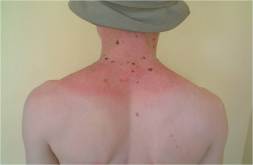
Spitz nevi are common, and typically appear as pink-red papules on the face that are less than 1 cm in diameter. However, they can also develop as pigmented lesions, particularly on the limbs.
Although a pigmented lesion in a child is likely a Spitz nevus, the dermatoscope is particularly helpful for confirming the diagnosis, as Spitz nevi have a very characteristic starburst or peripheral globule appearance.
"This is one of those examples where having a dermatoscope really changes things I do," Dr. Orlow said, explaining that although he tends to assume that a pigmented lesion in a child is a Spitz nevus, seeing the typical pattern that is diagnostic of a Spitz nevus is reassuring for both him and the patient’s parents.
Even with ASMT (a subset of Spitzlike growths with features such as mitoses that are associated with melanoma), the risk of melanoma is minimal, he noted.
Although interobserver agreement on diagnosis and prognosis is poor with these lesions, data show that the prognosis is favorable in patients both with and without positive lymph nodes. In one recent study of 11 children with ASMT and positive lymph nodes, all remained disease free at an average of 47 months’ follow-up. By comparison, two of five children younger than age 18 years with histologically unambiguous melanoma and positive sentinel lymph nodes died from metastatic melanoma (Am. J. Surg. Pathol. 2009;33:1386-95).
This finding is not surprising, because these are not melanomas, Dr. Orlow said.
What are pediatric dermatologists' attitudes about Spitz nevi and ASMTs?
Findings from a recent Web-based survey of 350 pediatric dermatologists conducted by Dr. Orlow and his associates showed that academic pediatric dermatologists are most comfortable following Spitz nevi clinically, and are more likely to do so than are those in private practice. It also showed that both never seeing an unambiguous melanoma in a child and believing that Spitz nevi are not precursors to melanoma are – not unexpectedly – predictors of following patients clinically, Dr. Orlow said.
Also, clinicians observe both involution and evolution of Spitz nevi to more banal nevus subtypes as evidence of their benign nature, he said.
Of all those surveyed, only two (who were in private practice) had reported that a death resulting from melanoma had occurred in a patient of theirs.
"Not a single pediatric dermatologist at any academic medical center had ever seen a death from an atypical Spitz nevus in their entire career" he concluded.
Dr. Orlow had no disclosures relevant to his presentation.
BOCA RATON, FLA. – Pigmented Spitz nevi, and even atypical Spitzoid melanocytic tumors, are unlikely to be melanoma in children.
"Boy, does this create controversy," noted Dr. Seth J. Orlow at the meeting of the Florida Society of Dermatology and Dermatologic Surgery.
Pigmented Spitz nevi and atypical Spitzoid melanocytic tumors (ASMT) can appear suddenly and grow rapidly in children. Part of the problem is that these lesions have been shown to be associated with microscopic foci in draining lymph nodes in children who have been subjected to sentinel lymph node biopsy.
In some cases, this has been used as a basis for a diagnosis of melanoma.
"It turns logic upside down, but it's no proof of anything – only that the child has Spitz nevus," said Dr. Orlow, chair of dermatology and the Samuel Weinberg professor of pediatric dermatology at New York University.
Spitz nevi are common, and typically appear as pink-red papules on the face that are less than 1 cm in diameter. However, they can also develop as pigmented lesions, particularly on the limbs.
Although a pigmented lesion in a child is likely a Spitz nevus, the dermatoscope is particularly helpful for confirming the diagnosis, as Spitz nevi have a very characteristic starburst or peripheral globule appearance.
"This is one of those examples where having a dermatoscope really changes things I do," Dr. Orlow said, explaining that although he tends to assume that a pigmented lesion in a child is a Spitz nevus, seeing the typical pattern that is diagnostic of a Spitz nevus is reassuring for both him and the patient’s parents.
Even with ASMT (a subset of Spitzlike growths with features such as mitoses that are associated with melanoma), the risk of melanoma is minimal, he noted.
Although interobserver agreement on diagnosis and prognosis is poor with these lesions, data show that the prognosis is favorable in patients both with and without positive lymph nodes. In one recent study of 11 children with ASMT and positive lymph nodes, all remained disease free at an average of 47 months’ follow-up. By comparison, two of five children younger than age 18 years with histologically unambiguous melanoma and positive sentinel lymph nodes died from metastatic melanoma (Am. J. Surg. Pathol. 2009;33:1386-95).
This finding is not surprising, because these are not melanomas, Dr. Orlow said.
What are pediatric dermatologists' attitudes about Spitz nevi and ASMTs?
Findings from a recent Web-based survey of 350 pediatric dermatologists conducted by Dr. Orlow and his associates showed that academic pediatric dermatologists are most comfortable following Spitz nevi clinically, and are more likely to do so than are those in private practice. It also showed that both never seeing an unambiguous melanoma in a child and believing that Spitz nevi are not precursors to melanoma are – not unexpectedly – predictors of following patients clinically, Dr. Orlow said.
Also, clinicians observe both involution and evolution of Spitz nevi to more banal nevus subtypes as evidence of their benign nature, he said.
Of all those surveyed, only two (who were in private practice) had reported that a death resulting from melanoma had occurred in a patient of theirs.
"Not a single pediatric dermatologist at any academic medical center had ever seen a death from an atypical Spitz nevus in their entire career" he concluded.
Dr. Orlow had no disclosures relevant to his presentation.
EXPERT ANALYSIS FROM THE ANNUAL MEETING OF THE FLORIDA SOCIETY OF DERMATOLOGY AND DERMATOLOGIC SURGERY
Point/Counterpoint: Is Mohs Surgery Being Overutilized?
Yes - Surgeons Are Softening Criteria for Who Gets Mohs.
By David S. Becker, M.D.
The practice of Mohs micrographic surgery is in some cases drifting away from the classic indications that used to make up most of our practice.
There are different published criteria for cases indicated for Mohs surgery, but these often include patients with recurrent or incompletely excised skin cancers, aggressive histology, or poorly defined margins. Other classic indications include tumors larger than 0.4 cm in high-risk H-zone locations, larger than 1 cm elsewhere on the face, or larger than 2 cm on the trunk or extremities.
Some of these listed criteria would not include a 3-mm basal cell carcinoma on the nose or ear, an 8-mm basal cell carcinoma on the cheek or forehead, or a 1.5-cm lesion on the pretibial skin, but I often have had each of these referred to me for Mohs surgery.
What's driving us to soften these traditional indications for Mohs surgery? Patients, referring dermatologists, dermatopathologists, and Mohs surgeons each may play a role.
Patients love Mohs surgery for many reasons. For one, we do a good job. Perhaps they've had previous Mohs surgery with good results or their friends have, or they have looked online and found that Mohs has the best cure rate. Or, they may have had a bad experience with a prior treatment.
It's not uncommon for patients to do research, and then want to play a role in determining their medical care. If they demand Mohs surgery, and I don't think it's indicated, it can lead to a contentious consultation.
One young man with a squamous cell carcinoma on his chest recently told me that he had researched the cure rates of treatments and he wanted Mohs surgery. He was almost certainly correct that if the sole criteria for therapy were best cure rate, Mohs would be the choice.
There are only two downsides to Mohs versus excision in a case like this: One, the patient has to spend more time in the office for Mohs surgery; and two, it is perhaps overutilization of medical resources. Good luck trying to explain that to patients. They don't care about overutilization of resources.
Who else drives this drift toward enhanced Mohs utilization? Referring dermatologists. They may have an interest in maximizing utilization of a Mohs surgeon in their practice, while some dermatologists have truly been burned, that is, had a patient with a metastatic squamous cell carcinoma or difficult recurrences, and they want the greater certitude of cure from Mohs surgery.
Sometimes the referring dermatologist says the patient is a VIP or relative and asks me to make an exception. If you start making exceptions, pretty soon you're seeing a lot of patients who don't necessarily meet the classic criteria for Mohs surgery.
The next set of people who are driving this drift in overutilization is dermatopathologists. We have dermatopathologists being overly cautious and prudent in ways they may not have been 10-15 years ago. Something that might have been diagnosed as actinic keratosis 20 years ago might now be called actinic keratosis with extension to the base, "squamous cell carcinoma cannot be excluded," or superficially invasive squamous cell carcinoma.
Mohs surgeons also drive the drift in utilization. We do it because that's what we like to do, or because we work for someone and we want to do what they tell us to do, or we have a referring dermatologist whom we want to please. And we want to keep our patients happy too.
It may be time to reconceptualize the indications for Mohs surgery. Should patient demand or anxiety be an indication? Should the referring dermatologist's or Mohs surgeon's instincts about a given lesion be an indication? Should any small lesion on the face be an indication?
Finally, who should decide when to do Mohs surgery? Should it be patients? They want some autonomy and to play a role in managing their own care, and are often educated in doing so.
Should it be the referring dermatologist? They understand the history of a given lesion and know the patient and the response to prior therapies.
Should the Mohs surgeon decide? We have the most experience with cutaneous malignancies, and in many cases we're the best suited to decide who needs Mohs and who doesn't.
Or, finally, should it be the government or insurers? I think that because we have allowed this drift in utilization to go forward, that may be who ultimately is deciding.
Dr. Becker is a Mohs surgeon in private practice in New York. He said he has no relevant conflicts of interest. His comments were presented at the American College of Mohs Surgery meeting.
No - Mohs Surgery Is Being Properly Utilized.
By Gary Monheit, M.D.
Mohs micrographic surgery is not overutilized, it's increasingly utilized.
A recent study reported a twofold increase in the proportion of Mohs surgery being done in Medicare patients from 2001 to 2006, while the rate of excisions has remained stable. So what's driving this increased utilization of Mohs surgery?
Is it greedy surgeons applying Mohs micrographic surgery to actinic keratoses on the arms and trunk? The few surgeons who are utilizing it inappropriately need to be controlled, but I don't believe this is the main reason for the increased utilization.
In most cases, increased Mohs utilization is appropriate. Mohs is a superior and much more cost-effective modality than excision and it has a greater cure rate, which is now being recognized.
The vision of Dr. Frederic E. Mohs was a change in the treatment of skin cancer. He was tired of seeing recurrences with excision. He realized that if things were accurately mapped out, physicians would have a better understanding of how to treat tumors, spare tissue, and reduce costs.
Brilliantly, it was a merger of surgery and histopathology controlled by one practitioner, the Mohs surgeon. Results of surgical treatment approach 100% the first time.
Should this be done for all nonmelanoma skin cancers at all locations? Should patients be cured the first time rather than risk recurrences? Is this cost effective for most tumors?
Twenty years ago, 50% or more of the Mohs surgeries that I did were on patients with tumors that had undergone two surgical excisions with recurrences that were covered up with flaps. I'm very happy to say that now I'm curing patients with a primary basal cell carcinoma the first time I operate.
We are having a cancer epidemic. The annual incidence of nonmelanoma skin cancer is a staggering 3.5 million, according to a recent study involving epidemiologic databases and the National Cancer Institute (Arch. Dermatol. 2010;146:283-7). And, I think the incidence is much higher.
The same study reported a 4.2% annual growth rate. That would predict a 23% growth rate in Mohs micrographic surgery from 2001 to 2006. Instead, we had a 70% growth rate.
What accounts for this? I believe the high growth rate is driven by what I call the "miracle of Mohs surgery." Five-year recurrence rates after Mohs are 1% for primary basal cell carcinomas, compared with 10% for surgical excision in well-controlled studies, and 4% after Mohs surgery for recurrent basal cell carcinomas, as opposed to 17% for excision.
Studies have found that 18%-32% of facial basal cell carcinomas are incompletely excised and require a second procedure. Repeat excisions create larger defects and worse cosmetic outcomes. Complete re-excisions are challenging after flap repairs. But with Mohs, one procedure does it all.
Studies have shown that Mohs surgery is superior financially because it is more cost effective than traditional excision if you include the costs of re-excision and pathology, and re-repair and treatment of recurrences after excisions.
Patients also are twice as satisfied with Mohs surgery, another study found (Dermatol. Surg. 2009;35:1041-9).
We've got an epidemic of cancer, and we have recognition that Mohs is the best technique for treatment. Patients with tumors that would have been excised and recurred are being referred initially for Mohs surgery. Simple primary lesions that are in the right areas should be treated with Mohs.
Does increased utilization of Mohs surgery translate to greater expense in treating skin cancer? The data show that for primary lesions and recurrences, the cost savings is greater with Mohs surgery. Insurance companies are starting to recognize that, but we can't let insurance companies govern simply by cost.
We really don't know for sure what's driving the increase in Mohs micrographic surgery utilization. However, I do believe that the superiority of the technique, which is becoming more recognized and established, and the epidemic of skin cancer have produced this high rate of utilization.
The growth rate reflects recognition of the unparalleled value of Mohs micrographic surgery as we confront the skin cancer epidemic.
Dr. Monheit is a Mohs surgeon in private practice in Birmingham, Ala., and has directed Mohs fellowship programs for 25 years. He said he has no other relevant conflicts of interest. These remarks were presented at the American College of Mohs Surgery meeting.
Risk for Melanoma in Kids Not Clear Cut
BOCA RATON, FLA. – The risk factors for melanoma are well established in adults, as is the well-known ABCD method for self-evaluation for the disease, but assessing risk and recognizing melanoma in children are far less clear cut.
The typical risk factors, such as a history of changing moles, white race, and sun sensitivity, don't seem to apply in children to the degree they do in adults, and although the ABCDs (asymmetry, border irregularity, color variations, diameter over 6 mm) have withstood the test of time in adults, for whom they were developed, they don’t necessarily apply in children, Dr. Seth J. Orlow said at the meeting of the Florida Society of Dermatology and Dermatologic Surgery.
The relatively recent addition of an E to the ABCD method adds an element that is applicable to assessment of suspected melanoma in children, noted Dr. Orlow, chair of dermatology and the Samuel Weinberg professor of pediatric dermatology at New York University.
The E stands for evolving, and it refers to any unexpected change in a mole. It is the word "unexpected" that is important when it comes to moles in children, because in many cases – such as with Spitz nevi – some change is normal, he noted.
Adding to the challenge of predicting and diagnosing melanoma in children is the fact that the disease is very rare in this population, he said.
Based on information from the National Cancer Institute's Surveillance, Epidemiology and End Results (SEER) Program database from 1973 to 2001, only 95 of 140,206 cases of melanoma were in children under age 10 years. Compared with older children with melanoma, those in this age group who had melanoma were more likely to be nonwhite; have metastatic disease; show nodular histology; have primary tumors on the head, face, and neck; and have a history of other cancer (J. Clin. Oncol. 2005;23:4735-41).
Among the melanoma patients younger than 10 years old, survival was about 90%, which was comparable to that in individuals aged 10-19 years and 20-24 years. Among those under age 20, survival was 100% if the melanoma was in situ, and 96%, 77%, and 57% if it was localized, regional, or distant, respectively.

Dr. Orlow noted that findings from other series over the years have also underscored the rarity of pediatric melanomas: Children's Hospital of Boston reported only 23 cases over 36 years, a Montreal hospital reported only 13 cases over 22 years, and Istituto Nazionale Tumor reported only 33 cases over 25 years. Many of the cases included nodular or amelanotic disease, reinforcing other data suggesting that children tend to have a different presentation than adults. For example, studies have shown that about 20% of melanomas are nodular in adults, compared with 30%-40% in children, and about 10%-20% of melanomas in adults are amelanotic, compared with about 30% in children.
Dr. Orlow noted that his own experience over 21 years underscores the rarity and differences of melanoma in children, compared with adults.
He has encountered only five authentic cases (excluding giant congenital melanocytic nevi), including one that he said "made the most sense" because it involved a 12-year-old boy from Chernobyl, the site of a 1986 large-scale explosion at a nuclear power plant in Ukraine. Radiation levels at that site remain high to this day. The child had a lesion on his lower back and innumerable dysplastic nevi. The boy's mother also had a history of melanoma as a child.
The other cases included one involving a 12-year old Ashkenazic Jewish girl with a less than 1-mm-thick lesion on her shoulder, one involving a 14-year-old Peruvian Indian girl with deeply pigmented skin and a 1-mm-thick lesion on her fingertip, one involving a 15-year old Ashkenazic/Sephardic boy with xeroderma pigmentosum C and two melanomas in situ, and one involving a 16-year-old Jamaican girl with very deeply pigmented skin who had an 8-mm-deep lesion on her posterior thigh and who died of her disease within 3 months.
"I think this list is typical in that it shows you that these are not people you would necessarily predict to have a problem," he said.
Children seem to differ from adults not only in terms of who gets melanoma, but also in terms of how they develop the disease.
Molecular alterations appear to differ in children, Dr. Orlow noted. While BRAF/NRAS pathway genetic mutations are present in 50%-70% of adults with superficial spreading melanomas, and mutations in the c-KIT gene are present in acral lentiginous and mucosal melanomas in adults, in children melanomas appear to have an increased incidence of deletion of c-KIT as well as mutations in the CDKN2A gene, according to one study (J. Invest. Dermatol. 2009;129:1759-68).
The workup and treatment of children with pediatric melanoma are well established. The prognostic value of sentinel lymph node biopsy – which is well established in adults – is also believed to apply in children. There is no evidence regarding lymph node dissection in either adults or children, although it is used frequently. Risks of lymph node dissection include infection and lymphedema, he noted.
As for treatment, adjuvant therapy includes high-dose interferon-alpha, which is approved for use in adults but not children. It has been used in several case series in children. Risks include fever, malaise, neutropenia, and abnormal liver function tests.
While there remains a great deal to learn about melanoma in children, there are several fallacies about the disease that dermatologists should know. The top five falacies, according to Dr. Orlow, are:
• Any new mole that appears suddenly in a child, or a mole that has grown over the past 6 months, should prompt concern about melanoma just as it does in adults. Reality: The appearance of new nevi in children is normal, and it is also normal for nevi to grow and evolve until they reach their zenith.
• Nevi on the scalp are particularly worrisome and should be removed because they are difficult to follow. Reality: The scalp is a common site for nevi to arise in white children. Such nevi will often have a targetlike appearance or resemble a fried egg. Many resolve by adulthood.
• All atypical/dysplastic nevi must be removed. Reality: Even in patients with the highest risk, such as those with familial atypical mole melanoma syndrome, more than 50% of melanomas will not have any evidence of a preexisting nevus.
• If melanocytic cells are found in a sentinel lymph node biopsy, it must be melanoma. Reality: Such cells can be found in the lymph nodes of children with Spitz nevi, atypical spitzoid melanocytic tumors, and blue nevi.
• We can prevent most prepubertal melanomas by applying our experience with melanoma and melanoma risk in adults. Reality: Such melanomas are unusual both clinically and with respect to the patients in whom they arise.
Dr. Orlow had no disclosures relevant to his presentation.
BOCA RATON, FLA. – The risk factors for melanoma are well established in adults, as is the well-known ABCD method for self-evaluation for the disease, but assessing risk and recognizing melanoma in children are far less clear cut.
The typical risk factors, such as a history of changing moles, white race, and sun sensitivity, don't seem to apply in children to the degree they do in adults, and although the ABCDs (asymmetry, border irregularity, color variations, diameter over 6 mm) have withstood the test of time in adults, for whom they were developed, they don’t necessarily apply in children, Dr. Seth J. Orlow said at the meeting of the Florida Society of Dermatology and Dermatologic Surgery.
The relatively recent addition of an E to the ABCD method adds an element that is applicable to assessment of suspected melanoma in children, noted Dr. Orlow, chair of dermatology and the Samuel Weinberg professor of pediatric dermatology at New York University.
The E stands for evolving, and it refers to any unexpected change in a mole. It is the word "unexpected" that is important when it comes to moles in children, because in many cases – such as with Spitz nevi – some change is normal, he noted.
Adding to the challenge of predicting and diagnosing melanoma in children is the fact that the disease is very rare in this population, he said.
Based on information from the National Cancer Institute's Surveillance, Epidemiology and End Results (SEER) Program database from 1973 to 2001, only 95 of 140,206 cases of melanoma were in children under age 10 years. Compared with older children with melanoma, those in this age group who had melanoma were more likely to be nonwhite; have metastatic disease; show nodular histology; have primary tumors on the head, face, and neck; and have a history of other cancer (J. Clin. Oncol. 2005;23:4735-41).
Among the melanoma patients younger than 10 years old, survival was about 90%, which was comparable to that in individuals aged 10-19 years and 20-24 years. Among those under age 20, survival was 100% if the melanoma was in situ, and 96%, 77%, and 57% if it was localized, regional, or distant, respectively.
Dr. Orlow noted that findings from other series over the years have also underscored the rarity of pediatric melanomas: Children's Hospital of Boston reported only 23 cases over 36 years, a Montreal hospital reported only 13 cases over 22 years, and Istituto Nazionale Tumor reported only 33 cases over 25 years. Many of the cases included nodular or amelanotic disease, reinforcing other data suggesting that children tend to have a different presentation than adults. For example, studies have shown that about 20% of melanomas are nodular in adults, compared with 30%-40% in children, and about 10%-20% of melanomas in adults are amelanotic, compared with about 30% in children.
Dr. Orlow noted that his own experience over 21 years underscores the rarity and differences of melanoma in children, compared with adults.
He has encountered only five authentic cases (excluding giant congenital melanocytic nevi), including one that he said "made the most sense" because it involved a 12-year-old boy from Chernobyl, the site of a 1986 large-scale explosion at a nuclear power plant in Ukraine. Radiation levels at that site remain high to this day. The child had a lesion on his lower back and innumerable dysplastic nevi. The boy's mother also had a history of melanoma as a child.
The other cases included one involving a 12-year old Ashkenazic Jewish girl with a less than 1-mm-thick lesion on her shoulder, one involving a 14-year-old Peruvian Indian girl with deeply pigmented skin and a 1-mm-thick lesion on her fingertip, one involving a 15-year old Ashkenazic/Sephardic boy with xeroderma pigmentosum C and two melanomas in situ, and one involving a 16-year-old Jamaican girl with very deeply pigmented skin who had an 8-mm-deep lesion on her posterior thigh and who died of her disease within 3 months.
"I think this list is typical in that it shows you that these are not people you would necessarily predict to have a problem," he said.
Children seem to differ from adults not only in terms of who gets melanoma, but also in terms of how they develop the disease.
Molecular alterations appear to differ in children, Dr. Orlow noted. While BRAF/NRAS pathway genetic mutations are present in 50%-70% of adults with superficial spreading melanomas, and mutations in the c-KIT gene are present in acral lentiginous and mucosal melanomas in adults, in children melanomas appear to have an increased incidence of deletion of c-KIT as well as mutations in the CDKN2A gene, according to one study (J. Invest. Dermatol. 2009;129:1759-68).
The workup and treatment of children with pediatric melanoma are well established. The prognostic value of sentinel lymph node biopsy – which is well established in adults – is also believed to apply in children. There is no evidence regarding lymph node dissection in either adults or children, although it is used frequently. Risks of lymph node dissection include infection and lymphedema, he noted.
As for treatment, adjuvant therapy includes high-dose interferon-alpha, which is approved for use in adults but not children. It has been used in several case series in children. Risks include fever, malaise, neutropenia, and abnormal liver function tests.
While there remains a great deal to learn about melanoma in children, there are several fallacies about the disease that dermatologists should know. The top five falacies, according to Dr. Orlow, are:
• Any new mole that appears suddenly in a child, or a mole that has grown over the past 6 months, should prompt concern about melanoma just as it does in adults. Reality: The appearance of new nevi in children is normal, and it is also normal for nevi to grow and evolve until they reach their zenith.
• Nevi on the scalp are particularly worrisome and should be removed because they are difficult to follow. Reality: The scalp is a common site for nevi to arise in white children. Such nevi will often have a targetlike appearance or resemble a fried egg. Many resolve by adulthood.
• All atypical/dysplastic nevi must be removed. Reality: Even in patients with the highest risk, such as those with familial atypical mole melanoma syndrome, more than 50% of melanomas will not have any evidence of a preexisting nevus.
• If melanocytic cells are found in a sentinel lymph node biopsy, it must be melanoma. Reality: Such cells can be found in the lymph nodes of children with Spitz nevi, atypical spitzoid melanocytic tumors, and blue nevi.
• We can prevent most prepubertal melanomas by applying our experience with melanoma and melanoma risk in adults. Reality: Such melanomas are unusual both clinically and with respect to the patients in whom they arise.
Dr. Orlow had no disclosures relevant to his presentation.
EXPERT ANALYSIS FROM THE ANNUAL MEETING OF THE FLORIDA SOCIETY OF DERMATOLOGY AND DERMATOLOGIC SURGERY
Blog: Hats Off to Tanzania Albinism Project
In Tanzania, where some of the oldest human fossils have been found, and where Mt. Kilimanjaro rises above the clouds, a group of international dermatologists are hoping to help a very vulnerable population.
The region has one of the highest incidences of albinism in the world.
Although the condition is rare in the western world, it is quite common in sub-Saharan Africa (Dermatol. Clin. 2011;29:79-87). The rate is as high as 1 in 1,400 people, compared with about 1 in 37,000 in the U.S.
Patients with albinism are subject to discrimination, stigma, and even murder.
But, another important concern is the health of patients with albinism whose pink skin is exposed to the African sunshine, and where many occupations are outdoors and in the field.
Many of the locals with albinism die of cancer before age 40; in fact, fewer than 2% make it to their 40th birthday. And almost all of the children with albinism show signs of sun damage before age 10.
Due to lack of funding, many can't afford hats to protect themselves; due to lack of education, many don't know the linkage between sun damage and skin cancer, according to Dr. David McLean, the secretary-general of the International League of Dermatological Societies (ILDS), a nongovernmental organization affiliated with the World Health Organization.
(An audio interview with Dr. McLean is available at the end of this blog.)
Dr. McLean has been visiting the region for the past two decades, helping to establish and grow the Regional Dermatology Training Center (RDTC), an ILDS program, in the town of Moshi in Tanzania.
He is also part of a group who recently spearheaded a project to make hats with a 7.5-cm rim available to the albinism population in Tanzania.
Called "Hats on for Skin Health," a collaboration between the ILDS and Stiefel, the project is a global effort to raise funds for the purchase of hats and other protective items for patients with albinism in Tanzania.

The items will be distributed by the RDTC, which manages a mobile skin care clinic that regularly visits people with albinism living in the region and educates the locals, especially parents, about albinism. The lesson they try to get across, said Dr. McLean, is to let their children play outdoors, but cover them up first.
The group located a hat manufacturer in Moshi, which is currently producing template models for children and adults. Many of the workers, said Dr. McLean, have albinism. "We think that's definitely part of the solution going forward," he said.
The cost of manufacturing a hat in Africa? Less than $2.50, and the hats are expected to last for at least 10 years.
To start the campaign, Stiefel, a subsidiary of GlaxoSmithKline, donated $25,000, and Dr. McLean hopes that dermatologists, other professionals, and even the public, will get involved with the campaign.
"Our people are on the ground there. We know what happens to every donated dollar," said Dr. McLean.
The group expects to have hand out at least 15,000 hats in the next year. Visit www.hatsonforskinhealth.org to learn more.
— Naseem S. Miller (@NaseemSMiller)
In Tanzania, where some of the oldest human fossils have been found, and where Mt. Kilimanjaro rises above the clouds, a group of international dermatologists are hoping to help a very vulnerable population.
The region has one of the highest incidences of albinism in the world.
Although the condition is rare in the western world, it is quite common in sub-Saharan Africa (Dermatol. Clin. 2011;29:79-87). The rate is as high as 1 in 1,400 people, compared with about 1 in 37,000 in the U.S.
Patients with albinism are subject to discrimination, stigma, and even murder.
But, another important concern is the health of patients with albinism whose pink skin is exposed to the African sunshine, and where many occupations are outdoors and in the field.
Many of the locals with albinism die of cancer before age 40; in fact, fewer than 2% make it to their 40th birthday. And almost all of the children with albinism show signs of sun damage before age 10.
Due to lack of funding, many can't afford hats to protect themselves; due to lack of education, many don't know the linkage between sun damage and skin cancer, according to Dr. David McLean, the secretary-general of the International League of Dermatological Societies (ILDS), a nongovernmental organization affiliated with the World Health Organization.
(An audio interview with Dr. McLean is available at the end of this blog.)
Dr. McLean has been visiting the region for the past two decades, helping to establish and grow the Regional Dermatology Training Center (RDTC), an ILDS program, in the town of Moshi in Tanzania.
He is also part of a group who recently spearheaded a project to make hats with a 7.5-cm rim available to the albinism population in Tanzania.
Called "Hats on for Skin Health," a collaboration between the ILDS and Stiefel, the project is a global effort to raise funds for the purchase of hats and other protective items for patients with albinism in Tanzania.
The items will be distributed by the RDTC, which manages a mobile skin care clinic that regularly visits people with albinism living in the region and educates the locals, especially parents, about albinism. The lesson they try to get across, said Dr. McLean, is to let their children play outdoors, but cover them up first.
The group located a hat manufacturer in Moshi, which is currently producing template models for children and adults. Many of the workers, said Dr. McLean, have albinism. "We think that's definitely part of the solution going forward," he said.
The cost of manufacturing a hat in Africa? Less than $2.50, and the hats are expected to last for at least 10 years.
To start the campaign, Stiefel, a subsidiary of GlaxoSmithKline, donated $25,000, and Dr. McLean hopes that dermatologists, other professionals, and even the public, will get involved with the campaign.
"Our people are on the ground there. We know what happens to every donated dollar," said Dr. McLean.
The group expects to have hand out at least 15,000 hats in the next year. Visit www.hatsonforskinhealth.org to learn more.
— Naseem S. Miller (@NaseemSMiller)
Video of the Week: More Melanoma Treatments on the Way?
A closely watched experimental drug has excited melanoma oncologists and patients with a 63% reduction in the relative risk of death from metastatic melanoma when compared with standard therapy in a phase III trial that had enrolled 675 newly diagnosed patients. Vemurafenib (better known as PLX4032) targets the BRAF V600E mutation found in 40%-60% of melanoma patients. It is only the second melanoma drug to extend the lives of melanoma patients in a randomized clinical study.
The first such agent, ipilimumab (Yervoy), was approved earlier this year, and the melanoma community expects the Food and Drug Administration will award an indication to vemurafenib based on the new data from the BRIM-3 trial. We talked with Dr. Paul Chapman – lead author of the BRIM-3 study — about vemurafenib. He also hypothesized how clinicians would decide which drug — vemurafenib (assuming approval) vs. ipilimumab to use for their patients.
Real-Time Monitoring of Melanoma Markers Predicts Relapse
SEOUL, SOUTH KOREA – Serial monitoring of melanoma tumor marker levels in peripheral blood using a novel quantitative real-time reverse-transcriptase polymerase chain reaction method after surgical resection of melanoma has shown promise for the early detection of patients at high risk for disease progression.
The real-time polymerase chain reaction (PCR) assay measures circulating levels of five markers unique to melanoma cells: glycoprotein 100 (gp100), melanoma antigen gene-3 (MAGE-3), tyrosinase, melanoma marker A (Melan-A), and melanoma inhibitory activity (MIA) protein, Dr. Spyridon Gkalpakiotis explained at the World Congress of Dermatology.
He reported on 65 patients who underwent peripheral blood testing and analysis of the five markers every 3 months for the first 2 years after resection of their stage II or III melanoma, for a total of 2,925 PCR assays.
Twenty-six patients experienced elevated test results. All 26 relapsed during 5 years of follow-up; the 5-year survival rate in this group was 65%.
In contrast, only 1 of 39 patients with consistently negative real-time PCR assays experienced disease progression; 5-year survival in PCR-negative patients was 97%, reported Dr. Gkalpakiotis of Charles University in Prague.
MAGE-3 was expressed in 21 patients with disease progression. The next most sensitive markers of melanoma progression were MIA and gp100.
Dr. Gkalpakiotis declared having no financial conflicts.
FROM THE WORLD CONGRESS OF DERMATOLOGY
Melanoma Treatment Ipilimumab Approved Europe-Wide
The European Commission has approved ipilimumab, a novel immunotherapy, for the second-line treatment of adult patients with previously treated, advanced melanoma, the medicine’s manufacturer said July 14.
Ipilimumab (Bristol-Myers Squibb’s Yervoy) was recommended in May by the European Medicines Agency for the indication, and was approved in March by the U.S. Food and Drug Administration. Ipilimumab, a monoclonal antibody, causes tumor cell death by blocking the inhibitory signal of CTLA-4, resulting in T-cell activation, proliferation, and lymphocyte infiltration into tumors.
The medication was shown in one manufacturer-funded, randomized, controlled trial to prolong overall survival for an unprecedented amount of time for people with metastasized unresectable melanoma (N. Engl. J. Med. 2010; 363:711-23).
Patients in the trial (n = 676) received ipilimumab plus glycoprotein 100 (gp100), gp100 alone, or ipilimumab alone, in four intravenous infusions at 3 mg/kg of body weight over 12 weeks. Patients who received the combination therapy or ipilimumab alone saw a median overall survival of about 10 months, compared with 6.4 months for those receiving only gp100. Gp100 was not found to improve the efficacy of ipilimumab.
Responding patients could receive additional courses of ipilimumab, and more than a fifth of patients in the ipilimumab arms survived 2 years or longer.
Adverse reactions to ipilimumab were mostly immune related, and included enterocolitis, hepatitis, dermatitis (including toxic epidermal necrolysis), neuropathy, and endocrinopathy.
European Commission approval means that ipilimumab will be available to patients throughout the European Union. However, pricing may differ, depending on procurement and patient-access schemes in each country. In the United States, ipilimumab is estimated to cost $120,000 per course of treatment.
Cancer-Protective Effects Seen for Type IV Allergies
Patients with a history of contact allergies may be less likely to develop nonmelanoma skin cancer, brain cancer, and breast cancer, a study has shown.
In research published online July 11 in BMJ Open (doi:10.1136/bmjopen-2011-000084), investigators conversely found diagnoses of bladder cancer more likely in people with a history of contact allergies.
Any protective effects could possibly be explained by the stimulation of natural killer T (NKT) cells through contact allergic reactions, the observational study’s lead author, Kaare Engkilde, Ph.D., of the national allergy research center at Copenhagen University Hospital, said in an interview. "You would probably think that [contact allergy] could lead to skin cancer, but that is not what we found," Dr. Engkilde said.
Contact allergic reactions are delayed reactions caused by chemicals small enough to penetrate the skin – triggers can include perfumes, hair dyes, cobalt, nickel, and formaldehyde. Animal studies have shown that contact allergens have the ability to increase the number of NKT cells, at least temporarily, Dr. Engkilde said.
For their research, Dr. Engkilde and his colleagues used a database of 16,922 Danish patients patch tested for type IV allergies between 1984 and 2008. Of these, 6,065 (35.8%) had at least one positive reaction (26.1% of men, 41.4% of women). Linkage with the Danish Cancer Registry showed 3,200, or 18.9%, of dermatitis patients had a benign tumor, a malignant cancer diagnosis, or both, and 1,207 (37.7%) of these patients had had a positive patch test reaction.
Dr. Engkilde and his colleagues looked specifically at 15 types of cancers, all of which affected at least 40 people in the study population.
The investigators found an inverse association between diagnosed contact allergy and nonmelanoma skin cancer (OR, 0.82) along with breast cancer (OR, 0.80) among both men and women. Dr. Engkilde and his colleagues also noted an inverse trend for brain cancer among women with contact allergies, though the P value was above.05 (OR, 0.36).
The findings related to nonmelanoma skin cancers were on some level expected, Dr. Engkilde said; however, the researchers said they were surprised not to see any protective effect against melanomas.
Dr. Engkilde said his group had no hypothesis for the inverse breast cancer finding, but noted that an earlier study had found that self-reported perfume allergy, a type IV allergy, was related inversely to brain cancer incidence (Am. J. Epidemiol. 2007;166:941-50).
The positive association between contact allergy and bladder cancer (OR, 1.44) was more likely related to accumulations of chemical metabolites of type IV allergens in the bladder than to an effect on NKT cells or another immune response, the investigators said.
They cautioned that the relationships between type IV reactions and cancers were "uncertain and not necessarily the result of causality," and that future analyses would have to adjust for social class and smoking, as the latter can increase the risk of developing nickel contact allergies and several types of cancer.
Also, they wrote, "studies focusing on specific chemical exposures are required to further our understanding of the role of contact allergies in the development of cancer." However, if the relationships are in fact etiologic, the findings have "implications for understanding how contact allergy can affect cancer development and vice versa," they said.
Dr. Engkilde and his coauthors received research support from Aage Bang’s Foundation and the Capital Region’s Research Foundation, and disclosed no competing interests.
FROM BMJ OPEN
Major Finding: The investigators found an inverse association between diagnosed contact allergy and nonmelanoma skin cancer (OR, 0.82) along with breast cancer (OR 0.80) among both men and women.
Data Source: A database of 16,922 Danish patients patch tested for type IV allergies between 1984 and 2008.
Disclosures: Dr. Engkilde and his coauthors received research support from Aage Bang’s Foundation and the Capital Region’s Research Foundation, and disclosed no competing interests.
Steaming Veins: The Skinny Podcast
In this month's podcast, Skin & Allergy News' reporters discuss cancer, wrinkles, and veins.
Positive clinical trial results for new melanoma therapies were presented at the American Society of Clinical Oncology. Jeff Evans interviews Dr. Lynn Schuchter about the findings, and what they mean for patients.
Also in this episode, Heidi Splete offers a report on the link between wrinkles and bone health, which may have you sending your wrinkled patients to the bone doctor.
Then, Sherry Boschert talks with Dr. Rene Milleret about a new endovenous ablation technique for varicose veins that uses steam; the procedure is catching on in Europe.
In this month's Cosmetic Counter segment, Dr. Lily Talakoub tackles nanotechnology.
And finally, Dr. Alan Rockoff assures everyone that, contrary to any rumors, he has not kicked the bucket.
Purse String Stitch Handy for Lip Defects
LAS VEGAS – A simple purse string stitch can provide an elegant closure of challenging lip defects after Mohs surgery.
"It's quite easy to perform. It's useful for numerous areas of the lip. It's low-risk, with low morbidity, and requires little down time," yet provides an excellent aesthetic outcome, Dr. Kenny J. Omlin said at the annual meeting of the American College of Mohs Surgery.
The keys to reconstruction using the purse string stitch start with undermining the entire surgical wound in the subdermal plane to decrease sheering forces.
Next, uniformly place an absorbable suture in the deep dermis using a small needle, with circumferential tissue advancement to distribute the tension uniformly, explained Dr. Omlin, chief of Mohs surgery for Kaiser Permanente Napa-Solano County, Vacaville, Calif.
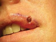
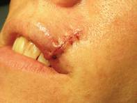
"There is a uniform stitch all the way around the perimeter" of the wound with particular attention to precisely aligning the vermilion/cutaneous lip junction, he said.
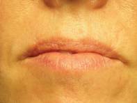
The purse string stitch creates a trestle-like framework that supports normal wound healing.
As with any reconstructions on the lower cosmetic subunits of the face, he tells patients to practice a "ventriloquist act" while healing and not talk much or move their mouths much.
One of his patients provided an excellent case-control comparison of wound closures. She initially presented with a basal cell carcinoma that intersected both the cutaneous and vermilion margins of her left upper lip.
After Mohs surgery, Dr. Omlin did a standard, complex linear closure, followed later by two pulsed-dye laser treatments. At 1-year follow-up, the patient was satisfied with an acceptable cosmetic outcome.
Six months later, she presented with a nearly identical basal cell carcinoma on the right upper lip. This time, Dr. Omlin used a purse string stitch after Mohs surgery. "It takes all of 5-10 minutes," he noted. The aesthetic result was "nearly perfect" a month later, said Dr. Omlin, also of the University of California, Davis.
On the central upper lip, "a lot of our older patients tend to have absent philtral columns or an absent Cupid's bow. Again, this is an excellent place for a purse string stitch," he said.
For patients on warfarin, the purse string stitch is great for hemostasis when repairing Mohs defects of the lip, Dr. Omlin added.
He also likes to use it for defects at the oral commissure. "Sure, you can use an elaborate cross-lip commissuroplasty or other elaborate techniques," but a simple purse string stitch reproduces the accordion-like structure of the oral commissure.
After wound granulation and healing, at 3 months it's hard to tell that a defect was ever there, he said.
In an interview after his presentation, he said he was pleased by the enthusiasm expressed by other attendees at the meeting for his simple surgical pearl.
Dr. Omlin said he has no relevant financial disclosures.
LAS VEGAS – A simple purse string stitch can provide an elegant closure of challenging lip defects after Mohs surgery.
"It's quite easy to perform. It's useful for numerous areas of the lip. It's low-risk, with low morbidity, and requires little down time," yet provides an excellent aesthetic outcome, Dr. Kenny J. Omlin said at the annual meeting of the American College of Mohs Surgery.
The keys to reconstruction using the purse string stitch start with undermining the entire surgical wound in the subdermal plane to decrease sheering forces.
Next, uniformly place an absorbable suture in the deep dermis using a small needle, with circumferential tissue advancement to distribute the tension uniformly, explained Dr. Omlin, chief of Mohs surgery for Kaiser Permanente Napa-Solano County, Vacaville, Calif.
"There is a uniform stitch all the way around the perimeter" of the wound with particular attention to precisely aligning the vermilion/cutaneous lip junction, he said.
The purse string stitch creates a trestle-like framework that supports normal wound healing.
As with any reconstructions on the lower cosmetic subunits of the face, he tells patients to practice a "ventriloquist act" while healing and not talk much or move their mouths much.
One of his patients provided an excellent case-control comparison of wound closures. She initially presented with a basal cell carcinoma that intersected both the cutaneous and vermilion margins of her left upper lip.
After Mohs surgery, Dr. Omlin did a standard, complex linear closure, followed later by two pulsed-dye laser treatments. At 1-year follow-up, the patient was satisfied with an acceptable cosmetic outcome.
Six months later, she presented with a nearly identical basal cell carcinoma on the right upper lip. This time, Dr. Omlin used a purse string stitch after Mohs surgery. "It takes all of 5-10 minutes," he noted. The aesthetic result was "nearly perfect" a month later, said Dr. Omlin, also of the University of California, Davis.
On the central upper lip, "a lot of our older patients tend to have absent philtral columns or an absent Cupid's bow. Again, this is an excellent place for a purse string stitch," he said.
For patients on warfarin, the purse string stitch is great for hemostasis when repairing Mohs defects of the lip, Dr. Omlin added.
He also likes to use it for defects at the oral commissure. "Sure, you can use an elaborate cross-lip commissuroplasty or other elaborate techniques," but a simple purse string stitch reproduces the accordion-like structure of the oral commissure.
After wound granulation and healing, at 3 months it's hard to tell that a defect was ever there, he said.
In an interview after his presentation, he said he was pleased by the enthusiasm expressed by other attendees at the meeting for his simple surgical pearl.
Dr. Omlin said he has no relevant financial disclosures.
EXPERT ANALYSIS FROM THE ANNUAL MEETING OF THE AMERICAN COLLEGE OF MOHS SURGERY