User login
MULTIMEDIA: Vemurafenib -- Melanoma Drug Cuts Death Risk
This drug works by attacking a specific genetic mutation, BRAF V600E, that accelerates tumor growth.
This drug works by attacking a specific genetic mutation, BRAF V600E, that accelerates tumor growth.
This drug works by attacking a specific genetic mutation, BRAF V600E, that accelerates tumor growth.
From Approval to Treatment, the History of Ipilimumab
In conjunction with the Food and Drug Administration's impending approval of another drug for melanoma treatment, vemurafenib, we present this look on the late-stage melanoma drug ipilimumab, how it was approved and the response that followed.
In conjunction with the Food and Drug Administration's impending approval of another drug for melanoma treatment, vemurafenib, we present this look on the late-stage melanoma drug ipilimumab, how it was approved and the response that followed.
In conjunction with the Food and Drug Administration's impending approval of another drug for melanoma treatment, vemurafenib, we present this look on the late-stage melanoma drug ipilimumab, how it was approved and the response that followed.
Melanoma Heads Down the Genetic Pathway
That might happen any day now. Reuters said on August 9 that the FDA’s approval of vemurafenib could get announced soon. Roche/Genentech submitted their NDA for vemurafenib to the FDA in May, and in June came impressive efficacy results in a phase 3 study that got reported at both ASCO and in a NEJM article.
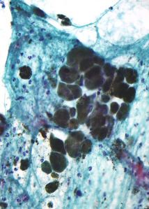
Last week, I covered the American Academy of Dermatology’s summer meeting in New York, and melanoma specialist Richard D. Carvajal from Memorial Sloan-Kettering told me that once vemurafenib was on the market genetic analysis of advanced melanomas to find BRAF mutations would suddenly become standard of care. Once testing for one mutation starts, several more genes can easily piggyback onto the assay, which will help to further flesh out the range of genetic mutations that can exist in metastatic melanoma and provide potential targets for new drugs.
It’s becoming a well-trodden path that’s rapidly driving the treatment of advanced cancers of all kinds into the molecular-profiling era. Just a couple of weeks ago, I blogged here on how it had already transformed advanced lung cancer management. Breast cancer and colorectal cancer have an even longer history of genetic assessment, and more cancers will likely follow this route soon.
When I wrote my blog post about lung cancer in late July, I focused on the challenge to successfully treating late-stage cancer, and I said that better alternatives were lung cancer prevention, by not smoking, and earlier diagnosis, with CT screening.
Melanoma does not pose the same screening issues as lung cancer. It’s a lot easier to survey the skin than to peer into a person’s lungs. And the idea of melanoma prevention by sunlight avoidance and protection has transformed the way many Americans approach outdoor activity. Over the last generation or so, SPF has become a cultural touchstone.
Despite that, cases of advanced melanoma are inevitable. Last week at the AAD meeting, Darrell S. Rigel from New York University showed evidence of a troubling, new wrinkle in melanoma epidemiology: An appreciable blip in cases that first appeared about 10 years ago in American women aged 30-34, a shift that Dr. Rigel attributed to an increased use of tanning salons among teenage girls that started in the 1980s.
Even if advanced stage tumors, melanoma or lung cancers, are unstable and unlikely to respond to even the best targeted of drugs for more than a few years, those extra years of cancer control with good quality of life can make a big difference to each patient who responds to a genetically-targeted treatment, Dr. Carvajal told me.
—Mitchel Zoler (on Twitter @mitchelzoler)
That might happen any day now. Reuters said on August 9 that the FDA’s approval of vemurafenib could get announced soon. Roche/Genentech submitted their NDA for vemurafenib to the FDA in May, and in June came impressive efficacy results in a phase 3 study that got reported at both ASCO and in a NEJM article.
Last week, I covered the American Academy of Dermatology’s summer meeting in New York, and melanoma specialist Richard D. Carvajal from Memorial Sloan-Kettering told me that once vemurafenib was on the market genetic analysis of advanced melanomas to find BRAF mutations would suddenly become standard of care. Once testing for one mutation starts, several more genes can easily piggyback onto the assay, which will help to further flesh out the range of genetic mutations that can exist in metastatic melanoma and provide potential targets for new drugs.
It’s becoming a well-trodden path that’s rapidly driving the treatment of advanced cancers of all kinds into the molecular-profiling era. Just a couple of weeks ago, I blogged here on how it had already transformed advanced lung cancer management. Breast cancer and colorectal cancer have an even longer history of genetic assessment, and more cancers will likely follow this route soon.
When I wrote my blog post about lung cancer in late July, I focused on the challenge to successfully treating late-stage cancer, and I said that better alternatives were lung cancer prevention, by not smoking, and earlier diagnosis, with CT screening.
Melanoma does not pose the same screening issues as lung cancer. It’s a lot easier to survey the skin than to peer into a person’s lungs. And the idea of melanoma prevention by sunlight avoidance and protection has transformed the way many Americans approach outdoor activity. Over the last generation or so, SPF has become a cultural touchstone.
Despite that, cases of advanced melanoma are inevitable. Last week at the AAD meeting, Darrell S. Rigel from New York University showed evidence of a troubling, new wrinkle in melanoma epidemiology: An appreciable blip in cases that first appeared about 10 years ago in American women aged 30-34, a shift that Dr. Rigel attributed to an increased use of tanning salons among teenage girls that started in the 1980s.
Even if advanced stage tumors, melanoma or lung cancers, are unstable and unlikely to respond to even the best targeted of drugs for more than a few years, those extra years of cancer control with good quality of life can make a big difference to each patient who responds to a genetically-targeted treatment, Dr. Carvajal told me.
—Mitchel Zoler (on Twitter @mitchelzoler)
Blog: Melanoma Heads Down the Genetic Pathway
That might happen any day now. Reuters said on August 9 that the FDA's approval of vemurafenib could be announced soon. Roche/Genentech submitted their NDA for vemurafenib to the FDA in May, and in June came impressive efficacy results in a phase 3 study that was reported at both ASCO and in a NEJM article.
Last week, I covered the American Academy of Dermatology's Summer Academy meeting in New York, where melanoma specialist Richard D. Carvajal from Memorial Sloan-Kettering said that once vemurafenib was on the market genetic analysis of advanced melanomas to find BRAF mutations would suddenly become standard of care. Once testing for one mutation starts, several more genes can easily piggyback onto the assay, which will help to further flesh out the range of genetic mutations that can exist in metastatic melanoma and provide potential targets for new drugs.
It's becoming a well-trodden path that's rapidly driving the treatment of advanced cancers of all kinds into the molecular-profiling era.
Just a couple of weeks ago, I blogged about how it had already transformed advanced lung cancer management. Breast cancer and colorectal cancer have an even longer history of genetic assessment, and more cancers will likely follow this route soon.
When I wrote my blog post about lung cancer in late July, I focused on the challenge to successfully treating late-stage cancer, and I said that better alternatives were lung cancer prevention, by not smoking, and earlier diagnosis, with CT screening.
Melanoma does not pose the same screening issues as lung cancer. It's a lot easier to survey the skin than to peer into a person's lungs. And the idea of melanoma prevention by sunlight avoidance and protection has transformed the way many Americans approach outdoor activity. Over the last generation or so, SPF has become a cultural touchstone.
Despite that, cases of advanced melanoma are inevitable.
At the AAD meeting, Darrell S. Rigel from New York University showed evidence of a troubling, new wrinkle in melanoma epidemiology: An appreciable blip in cases that first appeared about 10 years ago in American women aged 30-34, a shift that Dr. Rigel attributed to an increased use of tanning salons among teenage girls that started in the 1980s.
Even if advanced stage tumors, melanoma or lung cancers, are unstable and unlikely to respond to even the best targeted of drugs for more than a few years, those extra years of cancer control with good quality of life can make a big difference to each patient who responds to a genetically-targeted treatment, Dr. Carvajal told me.
—Mitchel Zoler (on Twitter @mitchelzoler)
Love and Denture Cream: The Skinny Podcast
In this month's Skinny podcast, we talk with dermatologist and AIDS expert Dr. Marcus Conant on why he believes universal HIV screening is the only way to curb the AIDS epidemic.
Also, Dr. Robert Weiss discusses the new FDA approved filler, Laviv, which uses a patient's own fibroblasts to treat wrinkles.
President of the American College of Mohs Surgery, Dr. Brett Coldiron, gives us his dire predictions about Medicare payments.
In this month's Cosmetic Counter segment, Dr. Lily Talakoub talks about a new rival to Botox and Dysport, which could hit the market next year.
And finally, Dr. Alan Rockoff says it's never too late for a little romance, even if it does involve denture cream.
Laser Therapy of Nonmelanoma Cancers Continues to Grow
BOCA RATON, FLA. – Experience with various lasers for the treatment and prophylaxis of nonmelanoma skin cancers is increasing, and the outcomes are promising.
One setting where lasers have particular potential is in patients with multiple basal cell carcinomas, Dr. Keyvan Nouri said at the annual meeting of the Florida Society of Dermatology and Dermatologic Surgery.

BCCs tend to have telangiectasias, and by specifically targeting the tumor vasculature, the BCC burden can be decreased or eliminated by laser treatment with little damage to surrounding skin. An ablative approach can also eliminate premalignant cells, said Dr. Nouri, professor of dermatology and otolaryngology and director of Mohs, dermatologic, and laser surgery at the University of Miami.
Studies using this approach have been conducted over the past several years using CO2, Er:YAG, pulsed-dye, and alexandrite lasers.
The 10,600-nm CO2 laser achieves a depth of 15-200 mcm in a single pass, which is adequate for coagulating the epidermis and superficial dermis, and for eliminating squamous cells at risk for evolution to actinic keratosis or squamous cell carcinoma. It also is partially effective for eliminating cells en route to basal cell carcinoma, he said, noting that deeper treatment can be achieved with multiple passes or protocol modifications.
In one study using this CO2 laser, 14 patients with diffuse facial AKs underwent resurfacing and experienced significant long-term reduction in AK burden (Dermatol. Surg. 1997;23:885-9). In another report of two patients with a history of multiple nonmelanoma skin cancer who were treated with two-pass CO2 resurfacing, no new lesions developed in the treated area at 33 and 52 months (Dermatol. Surg. 1999;25:513-16).
In a randomized controlled study of 34 patients, no differences in outcomes were seen at 5 years regardless of whether patients were treated with the CO2 laser, a 30% trichloroacetic acid peel, or 5% fluorouracil cream twice daily for 3 weeks. All of the treatments reduced AK counts by 83%-92%, and reduced the incidence of nonmelanoma skin cancers, compared with control groups (Dermatol. Surg. 1999;25:729-32), Dr. Nouri said.
In three of his patients with basal cell nevus syndrome, the Ultrapulse CO2 laser also proved effective for treatment. Postoperative Mohs micrographic surgical sections verified complete histologic clearance, and the patients had minimal scarring at follow-up (Dermatol. Surg. 2002;28:287-90).
A high-energy pulsed CO2 laser has also been used to treat a squamous cell carcinoma in situ on the nose with one pass (at 250 mJ, 8 W). In that case, no recurrence had occurred at 5 months’ follow-up, and the cosmetic result was excellent, Dr. Nouri said.
In a series of patients with nodular and superficial BCCs, a combination of curettage and one or two passes with the high-energy pulsed CO2 laser was also effective, with no histologic evidence or residual tumor and no known recurrences.
In another study, the pulsed CO2 laser was effective for treating 30 neoplasms, including 17 BCCs and 13 SCCs (Arch. Dermatol. 1998;134:1247-52). Two or three passes were used (at 500 mJ and 2-4 W). Treatment with a 4-mm margin and three passes is recommended for complete vaporization of neoplasms using this laser, Dr. Nouri said. Also, one SCC in situ in that study was incompletely ablated after three passes, so this modality alone is not recommended for thick or keratotic lesions, he said.
More recently, fractional laser resurfacing and pulsed-dye laser treatment have also shown promise for nonmelanoma skin cancers. In a pilot study of 28 patients with mild to moderate actinic damage who were treated with a fractional Er:YAG laser (2,940 nm) with one to four treatments at 4-week intervals, 75% had excellent results and the other 25% had good results. The effects were maintained at up to 9 months (Dermatol. Surg. 2008;34:1048-53).
And the 595-nm pulsed-dye laser has been used successfully to target tumor vasculature in glottal dysplasia and SCCs. Depending on fluence and spot size, the 595-nm pulsed-dye laser can penetrate skin to thicknesses ranging from 0.75 to 1.25 mm, which encompasses most BCCs, Dr. Nouri said.
In one study, a single treatment on seven BCCs resulted in histologic clearance of only one tumor (Lasers Med. Sci. 2003;18:125-6), but in two other studies of patients with superficial BCCs, this laser was associated with no clinical recurrences at a minimum follow-up of 1 year in 16 of 20 BCCs (Lasers Med. Sci. 2005;20:147-8; Dermatol. Ther. 2008; 21:402-5).
In another study, 20 BCCs treated with four 595-nm pulsed-dye laser treatments at 2-week intervals showed that nearly all BCCs less than 1.5 cm responded completely, compared with about 17% of controls. Larger BCCs had a complete response rate of 25% vs. 0% in controls.
Tumors with an incomplete response showed a significant reduction in tumor burden following treatment, with residual histologic tumor burden ranging from less than 1% to 29% of the original clinical tumor diameter, compared with 13%-68% residual tumor burden for the corresponding controls.
The investigators took the study one step further, treating 14 patients with a total of 20 BCCs on the trunk or extremities with four treatments at 3- to 4-week intervals; 19 of 20 BCCs had a complete clinical response, regardless of size and histological subtype (Lasers Surg. Med. 2009; 41:417-22). The investigators concluded that the 595-nm pulsed-dye laser is a "novel, quick, and relatively nonpainful treatment" for BCCs when used in the appropriate clinical setting, Dr. Nouri said.
As for the long-pulse alexandrite laser, it is selective like the pulsed-dye laser, but has deeper penetration, and may be helpful in significantly reducing tumor burden with a single treatment, he said.
In 15 of 18 patients with basal cell nevus syndrome who were treated in one study, a complete clinical response was seen at follow-ups of 2 and 7 months (Lasers Surg. Med. 2010;42:68-71).
No particular laser has emerged as the ideal tool for the treatment or prevention of skin cancer, but work is ongoing. More research is needed to confirm the observations made thus far, as well as to optimize treatment parameters, but the findings to date are encouraging, Dr. Nouri said, noting that he is confident that laser and light sources will eventually be used more for both medical and oncologic purposes.
Dr. Nouri disclosed that he has received grants or research support from Aesthera, CureLight, and Omnilux.
BOCA RATON, FLA. – Experience with various lasers for the treatment and prophylaxis of nonmelanoma skin cancers is increasing, and the outcomes are promising.
One setting where lasers have particular potential is in patients with multiple basal cell carcinomas, Dr. Keyvan Nouri said at the annual meeting of the Florida Society of Dermatology and Dermatologic Surgery.
BCCs tend to have telangiectasias, and by specifically targeting the tumor vasculature, the BCC burden can be decreased or eliminated by laser treatment with little damage to surrounding skin. An ablative approach can also eliminate premalignant cells, said Dr. Nouri, professor of dermatology and otolaryngology and director of Mohs, dermatologic, and laser surgery at the University of Miami.
Studies using this approach have been conducted over the past several years using CO2, Er:YAG, pulsed-dye, and alexandrite lasers.
The 10,600-nm CO2 laser achieves a depth of 15-200 mcm in a single pass, which is adequate for coagulating the epidermis and superficial dermis, and for eliminating squamous cells at risk for evolution to actinic keratosis or squamous cell carcinoma. It also is partially effective for eliminating cells en route to basal cell carcinoma, he said, noting that deeper treatment can be achieved with multiple passes or protocol modifications.
In one study using this CO2 laser, 14 patients with diffuse facial AKs underwent resurfacing and experienced significant long-term reduction in AK burden (Dermatol. Surg. 1997;23:885-9). In another report of two patients with a history of multiple nonmelanoma skin cancer who were treated with two-pass CO2 resurfacing, no new lesions developed in the treated area at 33 and 52 months (Dermatol. Surg. 1999;25:513-16).
In a randomized controlled study of 34 patients, no differences in outcomes were seen at 5 years regardless of whether patients were treated with the CO2 laser, a 30% trichloroacetic acid peel, or 5% fluorouracil cream twice daily for 3 weeks. All of the treatments reduced AK counts by 83%-92%, and reduced the incidence of nonmelanoma skin cancers, compared with control groups (Dermatol. Surg. 1999;25:729-32), Dr. Nouri said.
In three of his patients with basal cell nevus syndrome, the Ultrapulse CO2 laser also proved effective for treatment. Postoperative Mohs micrographic surgical sections verified complete histologic clearance, and the patients had minimal scarring at follow-up (Dermatol. Surg. 2002;28:287-90).
A high-energy pulsed CO2 laser has also been used to treat a squamous cell carcinoma in situ on the nose with one pass (at 250 mJ, 8 W). In that case, no recurrence had occurred at 5 months’ follow-up, and the cosmetic result was excellent, Dr. Nouri said.
In a series of patients with nodular and superficial BCCs, a combination of curettage and one or two passes with the high-energy pulsed CO2 laser was also effective, with no histologic evidence or residual tumor and no known recurrences.
In another study, the pulsed CO2 laser was effective for treating 30 neoplasms, including 17 BCCs and 13 SCCs (Arch. Dermatol. 1998;134:1247-52). Two or three passes were used (at 500 mJ and 2-4 W). Treatment with a 4-mm margin and three passes is recommended for complete vaporization of neoplasms using this laser, Dr. Nouri said. Also, one SCC in situ in that study was incompletely ablated after three passes, so this modality alone is not recommended for thick or keratotic lesions, he said.
More recently, fractional laser resurfacing and pulsed-dye laser treatment have also shown promise for nonmelanoma skin cancers. In a pilot study of 28 patients with mild to moderate actinic damage who were treated with a fractional Er:YAG laser (2,940 nm) with one to four treatments at 4-week intervals, 75% had excellent results and the other 25% had good results. The effects were maintained at up to 9 months (Dermatol. Surg. 2008;34:1048-53).
And the 595-nm pulsed-dye laser has been used successfully to target tumor vasculature in glottal dysplasia and SCCs. Depending on fluence and spot size, the 595-nm pulsed-dye laser can penetrate skin to thicknesses ranging from 0.75 to 1.25 mm, which encompasses most BCCs, Dr. Nouri said.
In one study, a single treatment on seven BCCs resulted in histologic clearance of only one tumor (Lasers Med. Sci. 2003;18:125-6), but in two other studies of patients with superficial BCCs, this laser was associated with no clinical recurrences at a minimum follow-up of 1 year in 16 of 20 BCCs (Lasers Med. Sci. 2005;20:147-8; Dermatol. Ther. 2008; 21:402-5).
In another study, 20 BCCs treated with four 595-nm pulsed-dye laser treatments at 2-week intervals showed that nearly all BCCs less than 1.5 cm responded completely, compared with about 17% of controls. Larger BCCs had a complete response rate of 25% vs. 0% in controls.
Tumors with an incomplete response showed a significant reduction in tumor burden following treatment, with residual histologic tumor burden ranging from less than 1% to 29% of the original clinical tumor diameter, compared with 13%-68% residual tumor burden for the corresponding controls.
The investigators took the study one step further, treating 14 patients with a total of 20 BCCs on the trunk or extremities with four treatments at 3- to 4-week intervals; 19 of 20 BCCs had a complete clinical response, regardless of size and histological subtype (Lasers Surg. Med. 2009; 41:417-22). The investigators concluded that the 595-nm pulsed-dye laser is a "novel, quick, and relatively nonpainful treatment" for BCCs when used in the appropriate clinical setting, Dr. Nouri said.
As for the long-pulse alexandrite laser, it is selective like the pulsed-dye laser, but has deeper penetration, and may be helpful in significantly reducing tumor burden with a single treatment, he said.
In 15 of 18 patients with basal cell nevus syndrome who were treated in one study, a complete clinical response was seen at follow-ups of 2 and 7 months (Lasers Surg. Med. 2010;42:68-71).
No particular laser has emerged as the ideal tool for the treatment or prevention of skin cancer, but work is ongoing. More research is needed to confirm the observations made thus far, as well as to optimize treatment parameters, but the findings to date are encouraging, Dr. Nouri said, noting that he is confident that laser and light sources will eventually be used more for both medical and oncologic purposes.
Dr. Nouri disclosed that he has received grants or research support from Aesthera, CureLight, and Omnilux.
EXPERT ANALYSIS FROM THE ANNUAL MEETING OF THE FLORIDA SOCIETY OF DERMATOLOGY AND DERMATOLOGIC SURGERY
Thulium Laser Yields 'Dramatic' Resolution of AKs
SEOUL, SOUTH KOREA – The nonablative fractionated thulium laser at 1,927-nm wavelength is a promising new noninvasive therapy for actinic keratoses.
The laser was actually developed for superficial skin resurfacing, an application for which it is particularly well suited because the 1,927-nm wavelength minimizes patient discomfort. But while investigating the device for improvement of pigmentation, Dr. Roy G. Geronemus noted incidentally that patients were also achieving "a rather dramatic resolution" of multiple facial actinic keratoses (AKs). So he decided to conduct a formal examination of the laser’s performance for this purpose, he said at the World Congress of Dermatology
To date, in 15 patients followed for 1-6 months after the last of several thulium laser treatment sessions for multiple facial AKs, the mean clearance of the lesions was 84%-91%.
"This compares very favorably to other modalities that are out there, including the topical chemotherapies, immunomodulatory agents, and photodynamic therapy. The advantage of this is not only do you improve the AKs, but you’re also getting the cosmetic benefit simultaneously," said Dr. Geronemus, medical director of the Laser and Skin Surgery Center of New York.
Patients received up to four treatments at 2- to 6-week intervals. The laser setting was 5-20 mJ, with 30%-70% coverage per session. Topical anesthetic was utilized for 1 hour, supplemented as needed by intramuscular ketorolac.
After a single treatment a mean of 63% of AKs were cleared. After two, 84%, and after three, 85%.
The laser therapy was well tolerated. The average pain score during treatment was 2.7 on a 0-9 scale. No scarring or infections have occurred. Mild redness and peeling typically lasted 4-5 days.
Dr. Geronemus said he and his colleagues have also found that the 1,927-nm fractionated thulium laser brings about "dramatic improvement" in actinic cheilitis, and is also highly effective for the thorny problem of enlarged facial pore size.
Dr. Geronemus is a shareholder in Solta Medical, which markets the 1,927-nm fractionated thulium laser. He also is on the advisory boards of numerous dermatologic laser manufacturers.
FROM THE WORLD CONGRESS OF DERMATOLOGY
Current Therapies for AK and Precancerous Lesions Subpar
DANA POINT, CALIF. – Despite significant advances in dermatology in recent years, little progress has been made in reducing actinic keratoses and skin cancers, Dr. Mark G. Rubin said at the SDEF Summit in Aesthetic Medicine.
"The take-home message to me in all this is, don’t get actinic keratoses," said Dr. Rubin, who practices dermatology in Beverly Hills, Calif. "We really don’t have a great therapy, so it’s important for your patients and for yourself that you limit the amount of ultraviolet light that you get, because we don’t have a wonderful treatment option for patients at this point."

That message is especially important for immunosuppressed organ transplant patients, who face an incidence of skin cancer 64-250 times higher than that of the general population.
"Immunosuppressed patients grow four times as many squamous cells than basal cells, which is the reverse of the ratio of these cancers in immunocompetent patients," he said. "Those particular tumors are much more aggressive and have a higher incidence of metastasis. Sun protection is tremendously important in these patients."
In a randomized trial of 120 transplant patients, 60 patients applied 2 mg/cm2 of sunscreen with an SPF greater than 50 to the head, neck, forearms, and hands daily for 24 months, while 60 patients in the control group did not apply any sunscreen (Br. J. Derm. 2009;161 [Suppl. 3]:78-84). Both groups of patients had an equal number of AKs at the start of the trial, but at the end of 24 months, 82 new AKs developed in the control group compared with none in the sunscreen treatment group. In addition, eight patients in the control group developed squamous cell carcinoma, compared with none in the sunscreen group, while nine patients in the control group developed basal cell epithelioma, compared with two in the sunscreen group.
While fluorouracil in the form of Efudex 2% and 5%, Fluoroplex 1%, and Carac 0.5% has been a mainstay of AK treatment, imiquimod in the form of Aldara Cream 5% and Zyclara Cream 3.75% "has probably been the most popular recently," said Dr. Rubin, also of the University of California, San Diego. Another treatment option is diclofenac in the form of Solaraze 3%.
"None of these are fun therapies for patients," he said. "It’s hard to get patients to apply 5-FU more than once. They’ll do it once, hate the experience, and ask, ‘What do you have now doc, because I’m never facing that again.’ It’s made some of the other products like Solaraze, Carac, and Aldara more popular because they’re a little less brutal for the patient. But it’s important to realize that any of these products are going to impact your patients’ daily life. They’re all going to cause some redness, swelling, crusting, stinging, and burning that will go on for a period of weeks if not months, depending on the product that you use and the protocol that you follow."
A review of multiple trials suggests that these medical therapies show complete clearing of AKs in 36%-58% of patients within 1-4 months post treatment.
"It’s important to differentiate between reducing the overall bulk of precancer and eradication, or complete clearance," Dr. Rubin added. "You really want to look at complete clearance, because if you just improved it and the keratosis is smaller, 6 months later it will be back and look like you never touched it. Unless you’ve eradicated the lesion, you’re wasting your time."
Systemic retinoids such as Acitretin and Etretinate are another treatment option, yet they are not a long-term solution given their propensity to cause multiple side effects, including chapped lips, dry eyes, headaches, and hyperlipidemia.
"It’s almost unfair to put a patient on these because they’ll love it for awhile, and then you have to take it away, and then they’ll do horribly – unless you’re doing this with a second plan where you put the patient on it for 6 months to stabilize them before moving on to another treatment option, such as photodynamic therapy, which may be reasonable," Dr. Rubin said.
Chemical peels have some value for decreasing AKs in immunocompetent patients, but the results are no better than with topical medications, he said. The deeper the destruction, the better the result.
"Some actinic keratoses and squamous cell in situ go down the hair follicles," Dr. Rubin said. "If you’re not chasing it down the hair follicle you’re leaving the root behind in a lot of these patients, and that’s what creates a lot of these recurrences. Medical therapies are moderately effective if you look at them a couple of months later. But response rates are not wonderful, and the relapse rates are really terrible."
Dr. Rubin disclosed that he has received research support and consulting fees from Medicis. SDEF and this news organization are owned by Elsevier.
DANA POINT, CALIF. – Despite significant advances in dermatology in recent years, little progress has been made in reducing actinic keratoses and skin cancers, Dr. Mark G. Rubin said at the SDEF Summit in Aesthetic Medicine.
"The take-home message to me in all this is, don’t get actinic keratoses," said Dr. Rubin, who practices dermatology in Beverly Hills, Calif. "We really don’t have a great therapy, so it’s important for your patients and for yourself that you limit the amount of ultraviolet light that you get, because we don’t have a wonderful treatment option for patients at this point."
That message is especially important for immunosuppressed organ transplant patients, who face an incidence of skin cancer 64-250 times higher than that of the general population.
"Immunosuppressed patients grow four times as many squamous cells than basal cells, which is the reverse of the ratio of these cancers in immunocompetent patients," he said. "Those particular tumors are much more aggressive and have a higher incidence of metastasis. Sun protection is tremendously important in these patients."
In a randomized trial of 120 transplant patients, 60 patients applied 2 mg/cm2 of sunscreen with an SPF greater than 50 to the head, neck, forearms, and hands daily for 24 months, while 60 patients in the control group did not apply any sunscreen (Br. J. Derm. 2009;161 [Suppl. 3]:78-84). Both groups of patients had an equal number of AKs at the start of the trial, but at the end of 24 months, 82 new AKs developed in the control group compared with none in the sunscreen treatment group. In addition, eight patients in the control group developed squamous cell carcinoma, compared with none in the sunscreen group, while nine patients in the control group developed basal cell epithelioma, compared with two in the sunscreen group.
While fluorouracil in the form of Efudex 2% and 5%, Fluoroplex 1%, and Carac 0.5% has been a mainstay of AK treatment, imiquimod in the form of Aldara Cream 5% and Zyclara Cream 3.75% "has probably been the most popular recently," said Dr. Rubin, also of the University of California, San Diego. Another treatment option is diclofenac in the form of Solaraze 3%.
"None of these are fun therapies for patients," he said. "It’s hard to get patients to apply 5-FU more than once. They’ll do it once, hate the experience, and ask, ‘What do you have now doc, because I’m never facing that again.’ It’s made some of the other products like Solaraze, Carac, and Aldara more popular because they’re a little less brutal for the patient. But it’s important to realize that any of these products are going to impact your patients’ daily life. They’re all going to cause some redness, swelling, crusting, stinging, and burning that will go on for a period of weeks if not months, depending on the product that you use and the protocol that you follow."
A review of multiple trials suggests that these medical therapies show complete clearing of AKs in 36%-58% of patients within 1-4 months post treatment.
"It’s important to differentiate between reducing the overall bulk of precancer and eradication, or complete clearance," Dr. Rubin added. "You really want to look at complete clearance, because if you just improved it and the keratosis is smaller, 6 months later it will be back and look like you never touched it. Unless you’ve eradicated the lesion, you’re wasting your time."
Systemic retinoids such as Acitretin and Etretinate are another treatment option, yet they are not a long-term solution given their propensity to cause multiple side effects, including chapped lips, dry eyes, headaches, and hyperlipidemia.
"It’s almost unfair to put a patient on these because they’ll love it for awhile, and then you have to take it away, and then they’ll do horribly – unless you’re doing this with a second plan where you put the patient on it for 6 months to stabilize them before moving on to another treatment option, such as photodynamic therapy, which may be reasonable," Dr. Rubin said.
Chemical peels have some value for decreasing AKs in immunocompetent patients, but the results are no better than with topical medications, he said. The deeper the destruction, the better the result.
"Some actinic keratoses and squamous cell in situ go down the hair follicles," Dr. Rubin said. "If you’re not chasing it down the hair follicle you’re leaving the root behind in a lot of these patients, and that’s what creates a lot of these recurrences. Medical therapies are moderately effective if you look at them a couple of months later. But response rates are not wonderful, and the relapse rates are really terrible."
Dr. Rubin disclosed that he has received research support and consulting fees from Medicis. SDEF and this news organization are owned by Elsevier.
EXPERT ANALYSIS FROM THE SDEF SUMMIT IN AESTHETIC MEDICINE
Giant Congenital Melanocytic Nevi May Signal Melanoma in Kids
BOCA RATON, FLA. – Pediatric melanoma can be difficult to predict, as the known risk factors in adults typically don't apply in children, but one exception is in the setting of giant congenital melanocytic nevi.
Giant congenital melanocytic nevi (CMN) are rare, occurring in fewer than 1 in 100,000 live births, but the estimated incidence of soft tissue melanoma in association with such nevi, based on a number of retrospective studies, is about 4%, said Dr. Seth J. Orlow at the annual meeting of the Florida Society of Dermatology and Dermatologic Surgery.

Interestingly, the risk of melanoma of the central nervous system or other internal site is slightly higher at about 5% (J. Am. Acad. Dermatol. 1979;1:123-300) , said Dr. Orlow, chair of dermatology and a professor of pediatric dermatology at New York University.
The risk of melanoma in association with giant CMN across the lifetime is up to 10%. The risk appears based on findings from several prospective trials that half of that risk is concentrated in the first 5 years of life, he said.
The greatest risk, in his experience, is in children with giant CMN involving the scalp or posterior axial location, and in particular in those with satellite congenital nevi.
"The child who is born with multiple small congenital nevi – in fact, even if they don't have a giant congenital nevus with multiple small congenital nevi – has a risk factor," he said. Giant CMN by itself doesn't seem to be much of an issue, he noted.
To date there have been no reports of melanoma developing within a satellite nevus, despite some of these patients having up to 100 congenital nevi for years, he said.
When melanomas do arise in patients with giant CMN, they can be very difficult to recognize early, he added. In addition to those that arise in the central nervous system, some develop in the dermis or below, and in some cases no primary site can be found.
The risk of melanoma in children who have medium-sized CMN is much lower than with giant CMN and is estimated at less than 0.1% over the lifetime, he noted.
In one study of 227 patients with medium CMN followed for nearly 7 years, no melanomas developed (J. Am. Acad. Dermatol. 1998;39:428-33). Findings from another study involving information from the National Cancer Institute’s Surveillance, Epidemiology, and End Results (SEER) Program database showed that the risk is about 1 in 10,000 patients before puberty, and 1 in 4,000 patients over the lifetime (Pediatr. Dermatol. 1994;11:204-8).
Thus it appears that most of the medium CMN occur after puberty, and they tend to arise at the dermal-epidermal junction, Dr. Orlow said.
The risk with small CMN can't be quantified, but is "much, much, much lower," he said.
Dr. Orlow had no disclosures relevant to his presentation.
BOCA RATON, FLA. – Pediatric melanoma can be difficult to predict, as the known risk factors in adults typically don't apply in children, but one exception is in the setting of giant congenital melanocytic nevi.
Giant congenital melanocytic nevi (CMN) are rare, occurring in fewer than 1 in 100,000 live births, but the estimated incidence of soft tissue melanoma in association with such nevi, based on a number of retrospective studies, is about 4%, said Dr. Seth J. Orlow at the annual meeting of the Florida Society of Dermatology and Dermatologic Surgery.
Interestingly, the risk of melanoma of the central nervous system or other internal site is slightly higher at about 5% (J. Am. Acad. Dermatol. 1979;1:123-300) , said Dr. Orlow, chair of dermatology and a professor of pediatric dermatology at New York University.
The risk of melanoma in association with giant CMN across the lifetime is up to 10%. The risk appears based on findings from several prospective trials that half of that risk is concentrated in the first 5 years of life, he said.
The greatest risk, in his experience, is in children with giant CMN involving the scalp or posterior axial location, and in particular in those with satellite congenital nevi.
"The child who is born with multiple small congenital nevi – in fact, even if they don't have a giant congenital nevus with multiple small congenital nevi – has a risk factor," he said. Giant CMN by itself doesn't seem to be much of an issue, he noted.
To date there have been no reports of melanoma developing within a satellite nevus, despite some of these patients having up to 100 congenital nevi for years, he said.
When melanomas do arise in patients with giant CMN, they can be very difficult to recognize early, he added. In addition to those that arise in the central nervous system, some develop in the dermis or below, and in some cases no primary site can be found.
The risk of melanoma in children who have medium-sized CMN is much lower than with giant CMN and is estimated at less than 0.1% over the lifetime, he noted.
In one study of 227 patients with medium CMN followed for nearly 7 years, no melanomas developed (J. Am. Acad. Dermatol. 1998;39:428-33). Findings from another study involving information from the National Cancer Institute’s Surveillance, Epidemiology, and End Results (SEER) Program database showed that the risk is about 1 in 10,000 patients before puberty, and 1 in 4,000 patients over the lifetime (Pediatr. Dermatol. 1994;11:204-8).
Thus it appears that most of the medium CMN occur after puberty, and they tend to arise at the dermal-epidermal junction, Dr. Orlow said.
The risk with small CMN can't be quantified, but is "much, much, much lower," he said.
Dr. Orlow had no disclosures relevant to his presentation.
EXPERT ANALYSIS FROM THE ANNUAL MEETING OF THE FLORIDA SOCIETY OF DERMATOLOGY AND DERMATOLOGIC SURGERY
Real-Time Monitoring of Melanoma Markers Predicts Relapse
SEOUL, SOUTH KOREA – Serial monitoring of melanoma tumor marker levels in peripheral blood using a novel quantitative real-time reverse-transcriptase polymerase chain reaction method after surgical resection of melanoma has shown promise for the early detection of patients at high risk for disease progression.
The real-time polymerase chain reaction (PCR) assay measures circulating levels of five markers unique to melanoma cells: glycoprotein 100 (gp100), melanoma antigen gene-3 (MAGE-3), tyrosinase, melanoma marker A (Melan-A), and melanoma inhibitory activity (MIA) protein, Dr. Spyridon Gkalpakiotis explained at the World Congress of Dermatology.
He reported on 65 patients who underwent peripheral blood testing and analysis of the five markers every 3 months for the first 2 years after resection of their stage II or III melanoma, for a total of 2,925 PCR assays.
Twenty-six patients experienced elevated test results. All 26 relapsed during 5 years of follow-up; the 5-year survival rate in this group was 65%.
In contrast, only 1 of 39 patients with consistently negative real-time PCR assays experienced disease progression; 5-year survival in PCR-negative patients was 97%, reported Dr. Gkalpakiotis of Charles University in Prague.
MAGE-3 was expressed in 21 patients with disease progression. The next most sensitive markers of melanoma progression were MIA and gp100.
Dr. Gkalpakiotis declared having no financial conflicts.
FROM THE WORLD CONGRESS OF DERMATOLOGY
Major Finding: Twenty-six patients experienced
elevated test results. All 26 relapsed during 5 years of follow-up.
Data Source: Patients (n = 65) who underwent peripheral blood testing and analysis of the
five markers every 3 months for the first 2 years after resection of
their stage II or III melanoma, for a total of 2,925 PCR assays.
Disclosures: Dr. Gkalpakiotis declared having no financial conflicts.