User login
Scalp Cooling Protects Against Chemotherapy-Induced Alopecia
CHICAGO – Wearing a scalp-cooling cap can reduce hair loss in women receiving chemotherapy for breast cancer, the results of a small prospective cohort study suggest.
Among women who used the cooling headgear starting 20 minutes before chemotherapy and continuing for 60-90 minutes after the infusion, 24% did not wear a wig or headband upon completion of chemotherapy, compared with 4% of a control group that did not have access to the device, investigators reported.

Further, patient satisfaction scores were higher than these numbers in a blinded assessment, according to Dr. Julie Lemieux of Laval University in Quebec City and her coinvestigators.
To grade the results with and without the cooling device, a hairdresser looked at before and after photos of women in the study, and was not told which women were in the scalp-cooling group. The criteria for successful hair preservation was characterization of hair loss as "not at all," "a little," or "moderate" from the beginning to the end of chemotherapy. The procedure was deemed a failure if the reviewer rated hair loss as "a lot," or "all," or "hair shaved."
The hairdresser graded the hair loss intervention as successful in 34% of the scalp-cooling group – as did 49% of the women who wore the caps. Only 9% of the control group received a successful grade from the hairdresser; even fewer, 4%, agreed they had not had substantial hair loss.
In all, 69% of women who tried scalp cooling said the advantages outweighed the disadvantages, and 78% said they would recommend it to other women receiving the same chemotherapy for breast cancer.
"When you look at patient evaluations, they are ... more optimistic than the hairdresser evaluations. They were more satisfied," Dr. Lemieux said in a poster-side interview at the annual meeting of the American Society of Clinical Oncology, where she displayed the results.

Scalp-cooling systems are approved for the reduction of alopecia in Canada, she said, but controversy persists among oncologists over safety and impact, if any, on the effectiveness of chemotherapy.
"If you cool the scalp there is vasoconstriction, so there is less blood that goes in the scalp ... that is the main mechanism," Dr. Lemieux explained. One concern is that scalp metastases could increase; another is that patients might receive less chemotherapy as a result.
Dr. Lemieux and her colleagues reviewed seven randomized trials of hair-cooling studies and found no safety signals. In all, 260 women were enrolled, and the studies covered a variety of chemotherapy regimens, including at least one that is not known to cause alopecia.
They also did a retrospective cohort study, and found that the incidence of scalp metastases was about 1% whether women used scalp cooling or not (Breast Cancer Res. Treat. 2009;118:547-52). Subsequently, they reported on two cases where the scalp was the first metastatic site, with metastases occurring 7 and 9 years after cooling (Breast Cancer Res. Treat. 2011;128:563-6).
At the San Antonio Breast Cancer Symposium, Dr. Lemieux and her associates reported on a retrospective study that found no difference in survival between patients who used scalp cooling and those who did not.
For the current study, the researchers compared outcomes in 110 patients at Centre des Maladies du Sein Deschênes-Fabia in Quebec City, which uses scalp cooling routinely, with those in 26 patients at the Centre Hospitalier Universitaire de Montréal, where scalp cooling is not available. The median patient age was in the early 50s, and most of the women had stage I or II, hormone receptor–positive breast cancer. A variety of neoadjuvant and adjuvant regimens were used.
The system tested in the study used a cap that is placed in a freezer and changed every 20-30 minutes, starting 20 minutes before chemotherapy and continuing for 60-90 minutes afterward. A new generation of scalp-cooling systems uses a compressor that circulates cold fluid in the cap, and it does not have to be changed.
Dr. Lemieux said the researchers conceived the study as a pilot for a larger randomized controlled trial that will address efficacy, cost, and quality of life issues. They are seeking to raise funds, as the companies that make the systems are too small to sponsor a large trial.
Cost is a concern, she noted, because of the additional time the women spend in the infusion room. "So you have to have that time available in the chemotherapy room," she said. "We also want to look at the cost of the system, of the extra time that women are in hospital, and at quality of life, too."
The trial was funded by the Fondations des Hôpitaux Enfant-Jésus et Saint-Sacrement, the Canadian Breast Cancer Research Alliance, and Sanofi-Aventis. Dr. Lemieux received a research grant from the Fonds de la Recherche en Santé du Québec.
CHICAGO – Wearing a scalp-cooling cap can reduce hair loss in women receiving chemotherapy for breast cancer, the results of a small prospective cohort study suggest.
Among women who used the cooling headgear starting 20 minutes before chemotherapy and continuing for 60-90 minutes after the infusion, 24% did not wear a wig or headband upon completion of chemotherapy, compared with 4% of a control group that did not have access to the device, investigators reported.
Further, patient satisfaction scores were higher than these numbers in a blinded assessment, according to Dr. Julie Lemieux of Laval University in Quebec City and her coinvestigators.
To grade the results with and without the cooling device, a hairdresser looked at before and after photos of women in the study, and was not told which women were in the scalp-cooling group. The criteria for successful hair preservation was characterization of hair loss as "not at all," "a little," or "moderate" from the beginning to the end of chemotherapy. The procedure was deemed a failure if the reviewer rated hair loss as "a lot," or "all," or "hair shaved."
The hairdresser graded the hair loss intervention as successful in 34% of the scalp-cooling group – as did 49% of the women who wore the caps. Only 9% of the control group received a successful grade from the hairdresser; even fewer, 4%, agreed they had not had substantial hair loss.
In all, 69% of women who tried scalp cooling said the advantages outweighed the disadvantages, and 78% said they would recommend it to other women receiving the same chemotherapy for breast cancer.
"When you look at patient evaluations, they are ... more optimistic than the hairdresser evaluations. They were more satisfied," Dr. Lemieux said in a poster-side interview at the annual meeting of the American Society of Clinical Oncology, where she displayed the results.
Scalp-cooling systems are approved for the reduction of alopecia in Canada, she said, but controversy persists among oncologists over safety and impact, if any, on the effectiveness of chemotherapy.
"If you cool the scalp there is vasoconstriction, so there is less blood that goes in the scalp ... that is the main mechanism," Dr. Lemieux explained. One concern is that scalp metastases could increase; another is that patients might receive less chemotherapy as a result.
Dr. Lemieux and her colleagues reviewed seven randomized trials of hair-cooling studies and found no safety signals. In all, 260 women were enrolled, and the studies covered a variety of chemotherapy regimens, including at least one that is not known to cause alopecia.
They also did a retrospective cohort study, and found that the incidence of scalp metastases was about 1% whether women used scalp cooling or not (Breast Cancer Res. Treat. 2009;118:547-52). Subsequently, they reported on two cases where the scalp was the first metastatic site, with metastases occurring 7 and 9 years after cooling (Breast Cancer Res. Treat. 2011;128:563-6).
At the San Antonio Breast Cancer Symposium, Dr. Lemieux and her associates reported on a retrospective study that found no difference in survival between patients who used scalp cooling and those who did not.
For the current study, the researchers compared outcomes in 110 patients at Centre des Maladies du Sein Deschênes-Fabia in Quebec City, which uses scalp cooling routinely, with those in 26 patients at the Centre Hospitalier Universitaire de Montréal, where scalp cooling is not available. The median patient age was in the early 50s, and most of the women had stage I or II, hormone receptor–positive breast cancer. A variety of neoadjuvant and adjuvant regimens were used.
The system tested in the study used a cap that is placed in a freezer and changed every 20-30 minutes, starting 20 minutes before chemotherapy and continuing for 60-90 minutes afterward. A new generation of scalp-cooling systems uses a compressor that circulates cold fluid in the cap, and it does not have to be changed.
Dr. Lemieux said the researchers conceived the study as a pilot for a larger randomized controlled trial that will address efficacy, cost, and quality of life issues. They are seeking to raise funds, as the companies that make the systems are too small to sponsor a large trial.
Cost is a concern, she noted, because of the additional time the women spend in the infusion room. "So you have to have that time available in the chemotherapy room," she said. "We also want to look at the cost of the system, of the extra time that women are in hospital, and at quality of life, too."
The trial was funded by the Fondations des Hôpitaux Enfant-Jésus et Saint-Sacrement, the Canadian Breast Cancer Research Alliance, and Sanofi-Aventis. Dr. Lemieux received a research grant from the Fonds de la Recherche en Santé du Québec.
CHICAGO – Wearing a scalp-cooling cap can reduce hair loss in women receiving chemotherapy for breast cancer, the results of a small prospective cohort study suggest.
Among women who used the cooling headgear starting 20 minutes before chemotherapy and continuing for 60-90 minutes after the infusion, 24% did not wear a wig or headband upon completion of chemotherapy, compared with 4% of a control group that did not have access to the device, investigators reported.
Further, patient satisfaction scores were higher than these numbers in a blinded assessment, according to Dr. Julie Lemieux of Laval University in Quebec City and her coinvestigators.
To grade the results with and without the cooling device, a hairdresser looked at before and after photos of women in the study, and was not told which women were in the scalp-cooling group. The criteria for successful hair preservation was characterization of hair loss as "not at all," "a little," or "moderate" from the beginning to the end of chemotherapy. The procedure was deemed a failure if the reviewer rated hair loss as "a lot," or "all," or "hair shaved."
The hairdresser graded the hair loss intervention as successful in 34% of the scalp-cooling group – as did 49% of the women who wore the caps. Only 9% of the control group received a successful grade from the hairdresser; even fewer, 4%, agreed they had not had substantial hair loss.
In all, 69% of women who tried scalp cooling said the advantages outweighed the disadvantages, and 78% said they would recommend it to other women receiving the same chemotherapy for breast cancer.
"When you look at patient evaluations, they are ... more optimistic than the hairdresser evaluations. They were more satisfied," Dr. Lemieux said in a poster-side interview at the annual meeting of the American Society of Clinical Oncology, where she displayed the results.
Scalp-cooling systems are approved for the reduction of alopecia in Canada, she said, but controversy persists among oncologists over safety and impact, if any, on the effectiveness of chemotherapy.
"If you cool the scalp there is vasoconstriction, so there is less blood that goes in the scalp ... that is the main mechanism," Dr. Lemieux explained. One concern is that scalp metastases could increase; another is that patients might receive less chemotherapy as a result.
Dr. Lemieux and her colleagues reviewed seven randomized trials of hair-cooling studies and found no safety signals. In all, 260 women were enrolled, and the studies covered a variety of chemotherapy regimens, including at least one that is not known to cause alopecia.
They also did a retrospective cohort study, and found that the incidence of scalp metastases was about 1% whether women used scalp cooling or not (Breast Cancer Res. Treat. 2009;118:547-52). Subsequently, they reported on two cases where the scalp was the first metastatic site, with metastases occurring 7 and 9 years after cooling (Breast Cancer Res. Treat. 2011;128:563-6).
At the San Antonio Breast Cancer Symposium, Dr. Lemieux and her associates reported on a retrospective study that found no difference in survival between patients who used scalp cooling and those who did not.
For the current study, the researchers compared outcomes in 110 patients at Centre des Maladies du Sein Deschênes-Fabia in Quebec City, which uses scalp cooling routinely, with those in 26 patients at the Centre Hospitalier Universitaire de Montréal, where scalp cooling is not available. The median patient age was in the early 50s, and most of the women had stage I or II, hormone receptor–positive breast cancer. A variety of neoadjuvant and adjuvant regimens were used.
The system tested in the study used a cap that is placed in a freezer and changed every 20-30 minutes, starting 20 minutes before chemotherapy and continuing for 60-90 minutes afterward. A new generation of scalp-cooling systems uses a compressor that circulates cold fluid in the cap, and it does not have to be changed.
Dr. Lemieux said the researchers conceived the study as a pilot for a larger randomized controlled trial that will address efficacy, cost, and quality of life issues. They are seeking to raise funds, as the companies that make the systems are too small to sponsor a large trial.
Cost is a concern, she noted, because of the additional time the women spend in the infusion room. "So you have to have that time available in the chemotherapy room," she said. "We also want to look at the cost of the system, of the extra time that women are in hospital, and at quality of life, too."
The trial was funded by the Fondations des Hôpitaux Enfant-Jésus et Saint-Sacrement, the Canadian Breast Cancer Research Alliance, and Sanofi-Aventis. Dr. Lemieux received a research grant from the Fonds de la Recherche en Santé du Québec.
AT THE ANNUAL MEETING OF THE AMERICAN SOCIETY OF CLINICAL ONCOLOGY
A Mending Ozone: The Skinny Podcast
In this month's program, Dr. Axel Hauschild discusses the results of a study that showed a 70% improvement in progression-free survival in patients with melanoma randomized to dabrafenib in the open-label break-3 trial.
Then, our reporter Heidi Splete reviews a study showing that an at-home hair removal device may work no better than standard shaving.

The Environment Protection Agency has reported that the ozone layer is improving, thanks to the worldwide ban of chlorofluorocarbons in 1989. We interviewed Drusilla Hufford at the EPA to find out more.
Cosmetic Counter host, Dr. Lily Talakoub, offers tips for educating patients about retinoids and anti-wrinkle creams. And lastly, Dr. Alan Rockoff tells a story that may induce contractions.
Don’t miss another episode of The Skinny Podcast; subscribe for free on iTunes!
In this month's program, Dr. Axel Hauschild discusses the results of a study that showed a 70% improvement in progression-free survival in patients with melanoma randomized to dabrafenib in the open-label break-3 trial.
Then, our reporter Heidi Splete reviews a study showing that an at-home hair removal device may work no better than standard shaving.
The Environment Protection Agency has reported that the ozone layer is improving, thanks to the worldwide ban of chlorofluorocarbons in 1989. We interviewed Drusilla Hufford at the EPA to find out more.
Cosmetic Counter host, Dr. Lily Talakoub, offers tips for educating patients about retinoids and anti-wrinkle creams. And lastly, Dr. Alan Rockoff tells a story that may induce contractions.
Don’t miss another episode of The Skinny Podcast; subscribe for free on iTunes!
In this month's program, Dr. Axel Hauschild discusses the results of a study that showed a 70% improvement in progression-free survival in patients with melanoma randomized to dabrafenib in the open-label break-3 trial.
Then, our reporter Heidi Splete reviews a study showing that an at-home hair removal device may work no better than standard shaving.
The Environment Protection Agency has reported that the ozone layer is improving, thanks to the worldwide ban of chlorofluorocarbons in 1989. We interviewed Drusilla Hufford at the EPA to find out more.
Cosmetic Counter host, Dr. Lily Talakoub, offers tips for educating patients about retinoids and anti-wrinkle creams. And lastly, Dr. Alan Rockoff tells a story that may induce contractions.
Don’t miss another episode of The Skinny Podcast; subscribe for free on iTunes!
Vemurafenib's Efficacy in Melanoma Wanes Over Time
CHICAGO – Vemurafenib has a persistent survival benefit when given as first-line therapy for advanced BRAF-mutant melanoma, but the magnitude of benefit diminishes over time, according to an update of the BRIM-3 randomized trial.
With a median follow-up of about a year, vemurafenib was associated with a 30% relative reduction in the risk of death, relative to dacarbazine, presenting author Dr. Paul Chapman reported at the annual meeting of the American Society of Clinical Oncology. This is roughly half of the 63% relative risk reduction seen at a median follow-up of about 3 months, as reported at last year’s meeting.

The safety profile of vemurafenib was largely consistent with the earlier experience; the main high-grade adverse effects were skin toxicity (seen in about a third of patients) and liver toxicity (seen in about a tenth). But a few patients have developed second primary melanomas.
The observed temporal trend for overall survival benefit suggests "perhaps that most of the benefit in overall survival may be due to decreasing early deaths," said Dr. Chapman, an oncologist at Memorial Sloan-Kettering Cancer Center in New York.
Discussant Dr. Michael B. Atkins of the Lombardi Comprehensive Cancer Center at Georgetown University in Washington maintained that "the mature overall survival data [confirm] that vemurafenib is better than dacarbazine," and noted that it was "reassuring" that findings were similar, whether patients who crossed over to the vemurafenib arm were censored or not, "probably the most stringent test of survival."
However, he called attention to the fact that the median survival of 13-plus months with vemurafenib seen in BRIM-3 falls short of the 16-plus months seen in BRIM-2, a phase II trial in patients with previously treated advanced melanoma. "Did first-line patients have more aggressive disease and therefore fall off early?" he wondered. "Alternatively, was there less continuation of vemurafenib after progressive disease in the BRIM-3 trial relative to the BRIM-2 trial? If that was the case, some sort of confirmatory analysis should be done to see whether that can impact overall survival."
The decrease in vemurafenib efficacy seen over time might have several explanations, Dr. Atkins proposed. "Is it that the early, more robust hazard ratio was a manifestation of the so-called Lazarus effect, that these targeted therapies can cause patients who are really in bad shape and would have a short survival to have their survival prolonged, even though it was less than the survival of patients with less-aggressive tumors?" he wondered, echoing Dr. Chapman’s suggestion. Alternatively, maturation of the survival data might have contributed to the change in efficacy in patients with stage IIIC, M1a, and M1b disease who did not appear to benefit from vemurafenib.
In the larger context, the findings raise an important question pertaining to the clinical trials process generally, according to Dr. Atkins: "What does this say about the early analysis of trials like this, and reporting forever more that the hazard ratio is 0.35 or so, rather than what the more accurate 0.70 is?"
A Genentech spokesperson said that "due to differences in the study populations and trial designs, the results from these trials cannot be directly compared. BRIM3 was a global, randomized, Phase III trial comparing Zelboraf (vemurafenib) to DTIC chemotherapy in people with newly diagnosed BRAF V600 mutation-positive metastatic melanoma, while BRIM2 was a Phase II study that evaluated patients with previously treated disease."
The 675 patients in BRIM-3 had stage IIIC or IV melanoma harboring the BRAF V600E mutation. They were assigned evenly to intravenous dacarbazine or oral vemurafenib, the only targeted agent approved specifically for treating BRAF-mutant melanoma.
Large proportions of patients in both the vemurafenib and dacarbazine arms (36% and 44%, respectively) received other anticancer therapies after coming off the trial, Dr. Chapman reported. About a fifth of all patients in each arm went on to receive ipilimumab, and a fourth in the dacarbazine arm crossed over to vemurafenib.
Updated results – after a median follow-up of 9.5 months in the dacarbazine arm and 12.5 months in the vemurafenib arm – showed persistence of a progression-free survival benefit in favor of vemurafenib with censoring of patients at crossover (6.9 vs. 1.6 months; hazard ratio, 0.38; P less than .001).
There was also still an improvement in overall survival, regardless of whether patients were censored at crossover (13.6 vs. 9.7 months; HR, 0.70; P less than .001) or not (13.6 vs. 10.3 months; HR, 0.76; P less than .01).
The leading grade 3 or worse adverse events with vemurafenib remained cutaneous squamous cell carcinomas (seen in 19% of patients), keratoacanthomas (10%), and elevations of liver function tests (10%). In addition, eight patients developed new primary melanomas.
Dr. Chapman disclosed that he receives honoraria and research funding from Roche/Genentech. The trial was sponsored by Hoffman-LaRoche. Dr. Atkins disclosed that he is a consultant to Bristol-Myers Squibb, Celgene, Curetech, Genentech, Merck, Novartis, and Prometheus.
This article was updated on July 3, 2012.
CHICAGO – Vemurafenib has a persistent survival benefit when given as first-line therapy for advanced BRAF-mutant melanoma, but the magnitude of benefit diminishes over time, according to an update of the BRIM-3 randomized trial.
With a median follow-up of about a year, vemurafenib was associated with a 30% relative reduction in the risk of death, relative to dacarbazine, presenting author Dr. Paul Chapman reported at the annual meeting of the American Society of Clinical Oncology. This is roughly half of the 63% relative risk reduction seen at a median follow-up of about 3 months, as reported at last year’s meeting.
The safety profile of vemurafenib was largely consistent with the earlier experience; the main high-grade adverse effects were skin toxicity (seen in about a third of patients) and liver toxicity (seen in about a tenth). But a few patients have developed second primary melanomas.
The observed temporal trend for overall survival benefit suggests "perhaps that most of the benefit in overall survival may be due to decreasing early deaths," said Dr. Chapman, an oncologist at Memorial Sloan-Kettering Cancer Center in New York.
Discussant Dr. Michael B. Atkins of the Lombardi Comprehensive Cancer Center at Georgetown University in Washington maintained that "the mature overall survival data [confirm] that vemurafenib is better than dacarbazine," and noted that it was "reassuring" that findings were similar, whether patients who crossed over to the vemurafenib arm were censored or not, "probably the most stringent test of survival."
However, he called attention to the fact that the median survival of 13-plus months with vemurafenib seen in BRIM-3 falls short of the 16-plus months seen in BRIM-2, a phase II trial in patients with previously treated advanced melanoma. "Did first-line patients have more aggressive disease and therefore fall off early?" he wondered. "Alternatively, was there less continuation of vemurafenib after progressive disease in the BRIM-3 trial relative to the BRIM-2 trial? If that was the case, some sort of confirmatory analysis should be done to see whether that can impact overall survival."
The decrease in vemurafenib efficacy seen over time might have several explanations, Dr. Atkins proposed. "Is it that the early, more robust hazard ratio was a manifestation of the so-called Lazarus effect, that these targeted therapies can cause patients who are really in bad shape and would have a short survival to have their survival prolonged, even though it was less than the survival of patients with less-aggressive tumors?" he wondered, echoing Dr. Chapman’s suggestion. Alternatively, maturation of the survival data might have contributed to the change in efficacy in patients with stage IIIC, M1a, and M1b disease who did not appear to benefit from vemurafenib.
In the larger context, the findings raise an important question pertaining to the clinical trials process generally, according to Dr. Atkins: "What does this say about the early analysis of trials like this, and reporting forever more that the hazard ratio is 0.35 or so, rather than what the more accurate 0.70 is?"
A Genentech spokesperson said that "due to differences in the study populations and trial designs, the results from these trials cannot be directly compared. BRIM3 was a global, randomized, Phase III trial comparing Zelboraf (vemurafenib) to DTIC chemotherapy in people with newly diagnosed BRAF V600 mutation-positive metastatic melanoma, while BRIM2 was a Phase II study that evaluated patients with previously treated disease."
The 675 patients in BRIM-3 had stage IIIC or IV melanoma harboring the BRAF V600E mutation. They were assigned evenly to intravenous dacarbazine or oral vemurafenib, the only targeted agent approved specifically for treating BRAF-mutant melanoma.
Large proportions of patients in both the vemurafenib and dacarbazine arms (36% and 44%, respectively) received other anticancer therapies after coming off the trial, Dr. Chapman reported. About a fifth of all patients in each arm went on to receive ipilimumab, and a fourth in the dacarbazine arm crossed over to vemurafenib.
Updated results – after a median follow-up of 9.5 months in the dacarbazine arm and 12.5 months in the vemurafenib arm – showed persistence of a progression-free survival benefit in favor of vemurafenib with censoring of patients at crossover (6.9 vs. 1.6 months; hazard ratio, 0.38; P less than .001).
There was also still an improvement in overall survival, regardless of whether patients were censored at crossover (13.6 vs. 9.7 months; HR, 0.70; P less than .001) or not (13.6 vs. 10.3 months; HR, 0.76; P less than .01).
The leading grade 3 or worse adverse events with vemurafenib remained cutaneous squamous cell carcinomas (seen in 19% of patients), keratoacanthomas (10%), and elevations of liver function tests (10%). In addition, eight patients developed new primary melanomas.
Dr. Chapman disclosed that he receives honoraria and research funding from Roche/Genentech. The trial was sponsored by Hoffman-LaRoche. Dr. Atkins disclosed that he is a consultant to Bristol-Myers Squibb, Celgene, Curetech, Genentech, Merck, Novartis, and Prometheus.
This article was updated on July 3, 2012.
CHICAGO – Vemurafenib has a persistent survival benefit when given as first-line therapy for advanced BRAF-mutant melanoma, but the magnitude of benefit diminishes over time, according to an update of the BRIM-3 randomized trial.
With a median follow-up of about a year, vemurafenib was associated with a 30% relative reduction in the risk of death, relative to dacarbazine, presenting author Dr. Paul Chapman reported at the annual meeting of the American Society of Clinical Oncology. This is roughly half of the 63% relative risk reduction seen at a median follow-up of about 3 months, as reported at last year’s meeting.
The safety profile of vemurafenib was largely consistent with the earlier experience; the main high-grade adverse effects were skin toxicity (seen in about a third of patients) and liver toxicity (seen in about a tenth). But a few patients have developed second primary melanomas.
The observed temporal trend for overall survival benefit suggests "perhaps that most of the benefit in overall survival may be due to decreasing early deaths," said Dr. Chapman, an oncologist at Memorial Sloan-Kettering Cancer Center in New York.
Discussant Dr. Michael B. Atkins of the Lombardi Comprehensive Cancer Center at Georgetown University in Washington maintained that "the mature overall survival data [confirm] that vemurafenib is better than dacarbazine," and noted that it was "reassuring" that findings were similar, whether patients who crossed over to the vemurafenib arm were censored or not, "probably the most stringent test of survival."
However, he called attention to the fact that the median survival of 13-plus months with vemurafenib seen in BRIM-3 falls short of the 16-plus months seen in BRIM-2, a phase II trial in patients with previously treated advanced melanoma. "Did first-line patients have more aggressive disease and therefore fall off early?" he wondered. "Alternatively, was there less continuation of vemurafenib after progressive disease in the BRIM-3 trial relative to the BRIM-2 trial? If that was the case, some sort of confirmatory analysis should be done to see whether that can impact overall survival."
The decrease in vemurafenib efficacy seen over time might have several explanations, Dr. Atkins proposed. "Is it that the early, more robust hazard ratio was a manifestation of the so-called Lazarus effect, that these targeted therapies can cause patients who are really in bad shape and would have a short survival to have their survival prolonged, even though it was less than the survival of patients with less-aggressive tumors?" he wondered, echoing Dr. Chapman’s suggestion. Alternatively, maturation of the survival data might have contributed to the change in efficacy in patients with stage IIIC, M1a, and M1b disease who did not appear to benefit from vemurafenib.
In the larger context, the findings raise an important question pertaining to the clinical trials process generally, according to Dr. Atkins: "What does this say about the early analysis of trials like this, and reporting forever more that the hazard ratio is 0.35 or so, rather than what the more accurate 0.70 is?"
A Genentech spokesperson said that "due to differences in the study populations and trial designs, the results from these trials cannot be directly compared. BRIM3 was a global, randomized, Phase III trial comparing Zelboraf (vemurafenib) to DTIC chemotherapy in people with newly diagnosed BRAF V600 mutation-positive metastatic melanoma, while BRIM2 was a Phase II study that evaluated patients with previously treated disease."
The 675 patients in BRIM-3 had stage IIIC or IV melanoma harboring the BRAF V600E mutation. They were assigned evenly to intravenous dacarbazine or oral vemurafenib, the only targeted agent approved specifically for treating BRAF-mutant melanoma.
Large proportions of patients in both the vemurafenib and dacarbazine arms (36% and 44%, respectively) received other anticancer therapies after coming off the trial, Dr. Chapman reported. About a fifth of all patients in each arm went on to receive ipilimumab, and a fourth in the dacarbazine arm crossed over to vemurafenib.
Updated results – after a median follow-up of 9.5 months in the dacarbazine arm and 12.5 months in the vemurafenib arm – showed persistence of a progression-free survival benefit in favor of vemurafenib with censoring of patients at crossover (6.9 vs. 1.6 months; hazard ratio, 0.38; P less than .001).
There was also still an improvement in overall survival, regardless of whether patients were censored at crossover (13.6 vs. 9.7 months; HR, 0.70; P less than .001) or not (13.6 vs. 10.3 months; HR, 0.76; P less than .01).
The leading grade 3 or worse adverse events with vemurafenib remained cutaneous squamous cell carcinomas (seen in 19% of patients), keratoacanthomas (10%), and elevations of liver function tests (10%). In addition, eight patients developed new primary melanomas.
Dr. Chapman disclosed that he receives honoraria and research funding from Roche/Genentech. The trial was sponsored by Hoffman-LaRoche. Dr. Atkins disclosed that he is a consultant to Bristol-Myers Squibb, Celgene, Curetech, Genentech, Merck, Novartis, and Prometheus.
This article was updated on July 3, 2012.
AT THE ANNUAL MEETING OF THE AMERICAN SOCIETY OF CLINICAL ONCOLOGY
Major Finding: At a median follow-up of 9.5-12.5 months, vemurafenib conferred a 30% reduction in the risk of death vs. dacarbazine, about half the 63% reduction reported at a median follow-up of 2.3-3.8 months.
Data Source: The phase III, randomized BRIM-3 trial randomized 675 patients with V600E BRAF-mutant melanoma.
Disclosures: Dr. Chapman disclosed that he receives honoraria and research funding from Roche/Genentech. The trial was sponsored by Hoffman-LaRoche. Dr. Atkins disclosed that he is a consultant to Bristol-Myers Squibb, Celgene, Curetech, Genentech, Merck, Novartis, and Prometheus.
Last Case
Dear Intern:
Well, your elective is over. So is medical school, since this is your last rotation. I get nostalgic this time of year, having graduated 40 years ago, which I’m sure you’ll agree is a ridiculously long time. Because you matched at your first choice, you are now well and truly launched.
One of my recurring sentimental notions is to note a student’s last case in medical school. Will this encounter be noteworthy; or will the whole thing end, so to speak, not with a bang but a whimper?
Looking for significance in a last case is similar to quoting people’s last words. The famous ones you hear about are usually edited, interpolated from some earlier occasion, or made up altogether. If you’ve seen people breathe their last, you know that most are not able to say much of anything, nor are their families, should they happen to be there, in any frame of mind to absorb enduring wisdom.
But lo and behold – our last encounter together on this elective has indeed turned out to be unusual, perhaps even memorable. We should discuss it.
A biopsy report just came back via fax, with the heading, "Crucial Clinical Information." We saw this patient together yesterday. Truong was the Vietnamese man in his mid-20s. A mole on his back started to grow. It got itchy, he said. It bled twice.
We examined the mole in question, along with the rest of him. As you recall, he had several moles, all black like the one in question. The irritated one was regular in outline, except for a scab in the center, consistent with having been scratched.
You remember that I was not very concerned. I said that raised moles often worry patients but aren’t usually a problem. Bleeding, moreover, is mostly a sign of minor, unrecalled trauma, a scratch while sleeping or something like that. As you’ve seen this month, patients troop in daily to complain that this mole is itchy or that one has bled. I often take these lesions off to reduce anxiety as to make the diagnosis. As a prospective primary physician, you will have to make judgments about itchy moles all the time.
I asked Truong how much the irritation bothered him. He waffled. I did the biopsy.
Here is the report: malignant melanoma, level IV, thickness at least 2.1 mm.
This is what runs through my head as I read this: At this point in my career, I am as good at judging moles as I am ever going to be. I am as clinically astute as I am ever going to be. And yet, here I am, still managing to almost miss a diagnosis in which the patient’s well-being, even his life, depends on my getting it right. In how many other cases have I (and the patient) not been so lucky? How would I know?
What lessons can I draw from this experience, and which ones can I pass on to you at your very different stage of clinical life? That because certainty is unattainable, we should take off every mole just to be sure? Every mole a patient points to? If we did that, would we be better doctors? Would we be treating patients, or our own insecurity?
It has always seemed to me that in medical practice and, in general, it is best to avoid the extremes, to be neither cavalier nor paranoid. Every passerby may assault you, every neighbor (or patient) may sue you, but it’s no good going through life treating everybody as a potential mugger or litigant. You won’t have many friends if you do that, and your patients will have a lot of unnecessary scars on their skin.
There really is something to the old saying: smart is good, lucky is better. Difficult or atypical cases can make the clever and diligent doctor look like a fool, or worse.
So perhaps your last case in medical school will come to mind from time to time as your career advances. Work hard, study hard, pay attention; become as good as you possibly can. But don’t ever get too comfortable. We try our best, but nobody always gets it right.
Good luck!
To respond to this column, e-mail Dr. Rockoff at our editorial offices at sknews@elsevier.com.
Dear Intern:
Well, your elective is over. So is medical school, since this is your last rotation. I get nostalgic this time of year, having graduated 40 years ago, which I’m sure you’ll agree is a ridiculously long time. Because you matched at your first choice, you are now well and truly launched.
One of my recurring sentimental notions is to note a student’s last case in medical school. Will this encounter be noteworthy; or will the whole thing end, so to speak, not with a bang but a whimper?
Looking for significance in a last case is similar to quoting people’s last words. The famous ones you hear about are usually edited, interpolated from some earlier occasion, or made up altogether. If you’ve seen people breathe their last, you know that most are not able to say much of anything, nor are their families, should they happen to be there, in any frame of mind to absorb enduring wisdom.
But lo and behold – our last encounter together on this elective has indeed turned out to be unusual, perhaps even memorable. We should discuss it.
A biopsy report just came back via fax, with the heading, "Crucial Clinical Information." We saw this patient together yesterday. Truong was the Vietnamese man in his mid-20s. A mole on his back started to grow. It got itchy, he said. It bled twice.
We examined the mole in question, along with the rest of him. As you recall, he had several moles, all black like the one in question. The irritated one was regular in outline, except for a scab in the center, consistent with having been scratched.
You remember that I was not very concerned. I said that raised moles often worry patients but aren’t usually a problem. Bleeding, moreover, is mostly a sign of minor, unrecalled trauma, a scratch while sleeping or something like that. As you’ve seen this month, patients troop in daily to complain that this mole is itchy or that one has bled. I often take these lesions off to reduce anxiety as to make the diagnosis. As a prospective primary physician, you will have to make judgments about itchy moles all the time.
I asked Truong how much the irritation bothered him. He waffled. I did the biopsy.
Here is the report: malignant melanoma, level IV, thickness at least 2.1 mm.
This is what runs through my head as I read this: At this point in my career, I am as good at judging moles as I am ever going to be. I am as clinically astute as I am ever going to be. And yet, here I am, still managing to almost miss a diagnosis in which the patient’s well-being, even his life, depends on my getting it right. In how many other cases have I (and the patient) not been so lucky? How would I know?
What lessons can I draw from this experience, and which ones can I pass on to you at your very different stage of clinical life? That because certainty is unattainable, we should take off every mole just to be sure? Every mole a patient points to? If we did that, would we be better doctors? Would we be treating patients, or our own insecurity?
It has always seemed to me that in medical practice and, in general, it is best to avoid the extremes, to be neither cavalier nor paranoid. Every passerby may assault you, every neighbor (or patient) may sue you, but it’s no good going through life treating everybody as a potential mugger or litigant. You won’t have many friends if you do that, and your patients will have a lot of unnecessary scars on their skin.
There really is something to the old saying: smart is good, lucky is better. Difficult or atypical cases can make the clever and diligent doctor look like a fool, or worse.
So perhaps your last case in medical school will come to mind from time to time as your career advances. Work hard, study hard, pay attention; become as good as you possibly can. But don’t ever get too comfortable. We try our best, but nobody always gets it right.
Good luck!
To respond to this column, e-mail Dr. Rockoff at our editorial offices at sknews@elsevier.com.
Dear Intern:
Well, your elective is over. So is medical school, since this is your last rotation. I get nostalgic this time of year, having graduated 40 years ago, which I’m sure you’ll agree is a ridiculously long time. Because you matched at your first choice, you are now well and truly launched.
One of my recurring sentimental notions is to note a student’s last case in medical school. Will this encounter be noteworthy; or will the whole thing end, so to speak, not with a bang but a whimper?
Looking for significance in a last case is similar to quoting people’s last words. The famous ones you hear about are usually edited, interpolated from some earlier occasion, or made up altogether. If you’ve seen people breathe their last, you know that most are not able to say much of anything, nor are their families, should they happen to be there, in any frame of mind to absorb enduring wisdom.
But lo and behold – our last encounter together on this elective has indeed turned out to be unusual, perhaps even memorable. We should discuss it.
A biopsy report just came back via fax, with the heading, "Crucial Clinical Information." We saw this patient together yesterday. Truong was the Vietnamese man in his mid-20s. A mole on his back started to grow. It got itchy, he said. It bled twice.
We examined the mole in question, along with the rest of him. As you recall, he had several moles, all black like the one in question. The irritated one was regular in outline, except for a scab in the center, consistent with having been scratched.
You remember that I was not very concerned. I said that raised moles often worry patients but aren’t usually a problem. Bleeding, moreover, is mostly a sign of minor, unrecalled trauma, a scratch while sleeping or something like that. As you’ve seen this month, patients troop in daily to complain that this mole is itchy or that one has bled. I often take these lesions off to reduce anxiety as to make the diagnosis. As a prospective primary physician, you will have to make judgments about itchy moles all the time.
I asked Truong how much the irritation bothered him. He waffled. I did the biopsy.
Here is the report: malignant melanoma, level IV, thickness at least 2.1 mm.
This is what runs through my head as I read this: At this point in my career, I am as good at judging moles as I am ever going to be. I am as clinically astute as I am ever going to be. And yet, here I am, still managing to almost miss a diagnosis in which the patient’s well-being, even his life, depends on my getting it right. In how many other cases have I (and the patient) not been so lucky? How would I know?
What lessons can I draw from this experience, and which ones can I pass on to you at your very different stage of clinical life? That because certainty is unattainable, we should take off every mole just to be sure? Every mole a patient points to? If we did that, would we be better doctors? Would we be treating patients, or our own insecurity?
It has always seemed to me that in medical practice and, in general, it is best to avoid the extremes, to be neither cavalier nor paranoid. Every passerby may assault you, every neighbor (or patient) may sue you, but it’s no good going through life treating everybody as a potential mugger or litigant. You won’t have many friends if you do that, and your patients will have a lot of unnecessary scars on their skin.
There really is something to the old saying: smart is good, lucky is better. Difficult or atypical cases can make the clever and diligent doctor look like a fool, or worse.
So perhaps your last case in medical school will come to mind from time to time as your career advances. Work hard, study hard, pay attention; become as good as you possibly can. But don’t ever get too comfortable. We try our best, but nobody always gets it right.
Good luck!
To respond to this column, e-mail Dr. Rockoff at our editorial offices at sknews@elsevier.com.
Melanoma in the Skin of Color Population
Dr. Rossi discusses melanoma in the skin of color population including the incidence and presentation of melanoma as well as factors contributing to delayed diagnosis in these patients. He also gives recommendations to physicians to help educate their patients. For more information, read Dr. Rossi's article in the May 2012 issue, "Melanoma in Skin of Color."

Dr. Rossi discusses melanoma in the skin of color population including the incidence and presentation of melanoma as well as factors contributing to delayed diagnosis in these patients. He also gives recommendations to physicians to help educate their patients. For more information, read Dr. Rossi's article in the May 2012 issue, "Melanoma in Skin of Color."
Dr. Rossi discusses melanoma in the skin of color population including the incidence and presentation of melanoma as well as factors contributing to delayed diagnosis in these patients. He also gives recommendations to physicians to help educate their patients. For more information, read Dr. Rossi's article in the May 2012 issue, "Melanoma in Skin of Color."

Rhode Island Passes Tan Ban
Rhode Island joins California and Vermont to become the third state to institute restrictions on the use of tanning beds by minors.
There had been an effort to completely ban the use of tanning beds by anyone under the age of 18, but a compromise was struck. In the final bill, which passed into law without the governor's signature, minors cannot use indoor tanning beds without a prescription from a physician for ultraviolet radiation therapy or a signed consent from a parent or guardian. The parental consent has to be signed in the presence of a tanning salon employee, and it essentially has the parent acknowledge that UV radiation is a carcinogen and that exposure increases the probability of developing melanoma.
The American Academy of Dermatology applauded the state's action, and noted that in Rhode Island the incidence of melanoma in women 15 years and older increased by 24% between 2004 and 2008.
"Prevention is one of the most valuable tools that we have as dermatologists. We need to continue educating patients about the risks of indoor tanning and encouraging healthy decisions to help prevent skin cancer," Dr. Daniel M. Siegel, president of the AAD, said in a statement.
"This law is similar to bans in California and Vermont that will protect children and adolescents from the health hazards of indoor tanning and sends a strong message from the state that tanning is a dangerous behavior and should be avoided," said Dr. Siegel.
Rhode Island joins California and Vermont to become the third state to institute restrictions on the use of tanning beds by minors.
There had been an effort to completely ban the use of tanning beds by anyone under the age of 18, but a compromise was struck. In the final bill, which passed into law without the governor's signature, minors cannot use indoor tanning beds without a prescription from a physician for ultraviolet radiation therapy or a signed consent from a parent or guardian. The parental consent has to be signed in the presence of a tanning salon employee, and it essentially has the parent acknowledge that UV radiation is a carcinogen and that exposure increases the probability of developing melanoma.
The American Academy of Dermatology applauded the state's action, and noted that in Rhode Island the incidence of melanoma in women 15 years and older increased by 24% between 2004 and 2008.
"Prevention is one of the most valuable tools that we have as dermatologists. We need to continue educating patients about the risks of indoor tanning and encouraging healthy decisions to help prevent skin cancer," Dr. Daniel M. Siegel, president of the AAD, said in a statement.
"This law is similar to bans in California and Vermont that will protect children and adolescents from the health hazards of indoor tanning and sends a strong message from the state that tanning is a dangerous behavior and should be avoided," said Dr. Siegel.
Rhode Island joins California and Vermont to become the third state to institute restrictions on the use of tanning beds by minors.
There had been an effort to completely ban the use of tanning beds by anyone under the age of 18, but a compromise was struck. In the final bill, which passed into law without the governor's signature, minors cannot use indoor tanning beds without a prescription from a physician for ultraviolet radiation therapy or a signed consent from a parent or guardian. The parental consent has to be signed in the presence of a tanning salon employee, and it essentially has the parent acknowledge that UV radiation is a carcinogen and that exposure increases the probability of developing melanoma.
The American Academy of Dermatology applauded the state's action, and noted that in Rhode Island the incidence of melanoma in women 15 years and older increased by 24% between 2004 and 2008.
"Prevention is one of the most valuable tools that we have as dermatologists. We need to continue educating patients about the risks of indoor tanning and encouraging healthy decisions to help prevent skin cancer," Dr. Daniel M. Siegel, president of the AAD, said in a statement.
"This law is similar to bans in California and Vermont that will protect children and adolescents from the health hazards of indoor tanning and sends a strong message from the state that tanning is a dangerous behavior and should be avoided," said Dr. Siegel.
Don't Disregard High-Dose Brachytherapy for Skin Cancer
ORLANDO – Although Mohs surgery remains a mainstay of skin cancer treatment, some patients benefit from targeted brachytherapy of their basal or squamous cell carcinoma lesions, according to Dr. Michael E. Kasper.
Patients who are elderly, infirm, or on blood thinners are good candidates for noninvasive brachytherapy using high-dose, small surface applicators, Dr. Kasper said. This therapeutic strategy also works well for treating lesions in anatomic locations at risk for delayed surgical healing.
"We are treating various small lesions with these surface applicators," Dr. Kasper said. For example, superficial squamous cell carcinoma (SCC) lesions up to 2 cm can be targeted "where we feel comfortable about the visible margins," he said at the annual meeting of the Florida Society of Dermatology and Dermatologic Surgery.
"We also see patients who are tired of [invasive resection] or who are poor candidates for surgery, and that is really the bulk of our patients," Dr. Kasper said. He assured meeting attendees that his goal as a radiation oncologist is not to take skin cancer patients away from dermatologists. "We’re not interested in treating 40- or 50-year-olds and really competing," said Dr. Kasper of Lynn Cancer Institute at Boca Raton (Fla.) Regional Hospital. "We are interested in working with dermatologists and really helping you with those patients who might be neglected or who are at high risk of developing a serious recurrence."
Neglected patients may include nursing home residents who do not get medical attention for their skin cancer in its earlier stages, he said.
Available data point to good local control and cosmesis for a majority of patients, Dr. Kasper said. A typical patient might experience acute effects such as crusting and some mild erythema about 10 days to 2 weeks after brachytherapy of a well differentiated SCC lesion of the lower extremity. More brisk erythema also occurs in about 10%-15% of patients, Dr. Kasper said.
Late hypopigmentation also develops in about 10% of patients, he said. "We are also seeing a few telangiectasias, but it’s fairly mild."
Interpret postradiation therapy biopsies with caution, Dr. Kasper warned. Of the 240 patients treated to date at his institution over about 6.5 years, there were three documented recurrences, including a couple at 3 and 4 months. "While we are counting those as recurrences, they probably aren’t. That’s way too early to biopsy these lesions." Use discretion and ideally wait until you see clear progression prior to performing a postradiation biopsy, he said. "There certainly are false positives that occur due to delayed tumor regression. The cancer cells die when they reproduce and many of these are slow growing tumors. So we would not expect them to all be completely resolved at 3 or 4 months although clinically, on the surface, they can appear that way."
Historically brachytherapy was delivered as a low-dose treatment over a long period of time at many sessions. Low-dose brachytherapy is typically in the 0.4 to 2.0 Gy/hr range, medium dose is greater than 2 and up to 12 Gy/hr, and the high dose exceeds 12 Gy/hr.
Availability of high-dose rate brachytherapy was a "major breakthrough" because patients no longer had to lie in the hospital all weekend to receive treatment. "It was a bit controversial at first, but now there are really good data to show there are radiobiologic reasons why the high dose rate may actually be advantageous in killing cancer cells compared to this low trickle effect that we were using with low-dose regimens."
Dr. Kasper determined that up to 30 sessions of low-dose rate brachytherapy can be a real impediment to patient compliance and worked to design a safe and effective regimen delivered in fewer sessions. "We wanted to be at six treatments and worked backwards." Striking a balance between the dose-response rate and the potential late side effects was another consideration.
A meeting attendee asked about the relative cost of brachytherapy, compared with other treatment modalities. Brachytherapy is generally more expensive than Mohs surgery, Dr. Kasper replied. The avoidance of cancer recurrences will hopefully justify the higher initial costs, he said. He added that brachytherapy costs are coming down, more so in outpatient centers, compared with hospital settings.
Dr. Kasper said he receives consulting fees from Nucletron/Elekta.
ORLANDO – Although Mohs surgery remains a mainstay of skin cancer treatment, some patients benefit from targeted brachytherapy of their basal or squamous cell carcinoma lesions, according to Dr. Michael E. Kasper.
Patients who are elderly, infirm, or on blood thinners are good candidates for noninvasive brachytherapy using high-dose, small surface applicators, Dr. Kasper said. This therapeutic strategy also works well for treating lesions in anatomic locations at risk for delayed surgical healing.
"We are treating various small lesions with these surface applicators," Dr. Kasper said. For example, superficial squamous cell carcinoma (SCC) lesions up to 2 cm can be targeted "where we feel comfortable about the visible margins," he said at the annual meeting of the Florida Society of Dermatology and Dermatologic Surgery.
"We also see patients who are tired of [invasive resection] or who are poor candidates for surgery, and that is really the bulk of our patients," Dr. Kasper said. He assured meeting attendees that his goal as a radiation oncologist is not to take skin cancer patients away from dermatologists. "We’re not interested in treating 40- or 50-year-olds and really competing," said Dr. Kasper of Lynn Cancer Institute at Boca Raton (Fla.) Regional Hospital. "We are interested in working with dermatologists and really helping you with those patients who might be neglected or who are at high risk of developing a serious recurrence."
Neglected patients may include nursing home residents who do not get medical attention for their skin cancer in its earlier stages, he said.
Available data point to good local control and cosmesis for a majority of patients, Dr. Kasper said. A typical patient might experience acute effects such as crusting and some mild erythema about 10 days to 2 weeks after brachytherapy of a well differentiated SCC lesion of the lower extremity. More brisk erythema also occurs in about 10%-15% of patients, Dr. Kasper said.
Late hypopigmentation also develops in about 10% of patients, he said. "We are also seeing a few telangiectasias, but it’s fairly mild."
Interpret postradiation therapy biopsies with caution, Dr. Kasper warned. Of the 240 patients treated to date at his institution over about 6.5 years, there were three documented recurrences, including a couple at 3 and 4 months. "While we are counting those as recurrences, they probably aren’t. That’s way too early to biopsy these lesions." Use discretion and ideally wait until you see clear progression prior to performing a postradiation biopsy, he said. "There certainly are false positives that occur due to delayed tumor regression. The cancer cells die when they reproduce and many of these are slow growing tumors. So we would not expect them to all be completely resolved at 3 or 4 months although clinically, on the surface, they can appear that way."
Historically brachytherapy was delivered as a low-dose treatment over a long period of time at many sessions. Low-dose brachytherapy is typically in the 0.4 to 2.0 Gy/hr range, medium dose is greater than 2 and up to 12 Gy/hr, and the high dose exceeds 12 Gy/hr.
Availability of high-dose rate brachytherapy was a "major breakthrough" because patients no longer had to lie in the hospital all weekend to receive treatment. "It was a bit controversial at first, but now there are really good data to show there are radiobiologic reasons why the high dose rate may actually be advantageous in killing cancer cells compared to this low trickle effect that we were using with low-dose regimens."
Dr. Kasper determined that up to 30 sessions of low-dose rate brachytherapy can be a real impediment to patient compliance and worked to design a safe and effective regimen delivered in fewer sessions. "We wanted to be at six treatments and worked backwards." Striking a balance between the dose-response rate and the potential late side effects was another consideration.
A meeting attendee asked about the relative cost of brachytherapy, compared with other treatment modalities. Brachytherapy is generally more expensive than Mohs surgery, Dr. Kasper replied. The avoidance of cancer recurrences will hopefully justify the higher initial costs, he said. He added that brachytherapy costs are coming down, more so in outpatient centers, compared with hospital settings.
Dr. Kasper said he receives consulting fees from Nucletron/Elekta.
ORLANDO – Although Mohs surgery remains a mainstay of skin cancer treatment, some patients benefit from targeted brachytherapy of their basal or squamous cell carcinoma lesions, according to Dr. Michael E. Kasper.
Patients who are elderly, infirm, or on blood thinners are good candidates for noninvasive brachytherapy using high-dose, small surface applicators, Dr. Kasper said. This therapeutic strategy also works well for treating lesions in anatomic locations at risk for delayed surgical healing.
"We are treating various small lesions with these surface applicators," Dr. Kasper said. For example, superficial squamous cell carcinoma (SCC) lesions up to 2 cm can be targeted "where we feel comfortable about the visible margins," he said at the annual meeting of the Florida Society of Dermatology and Dermatologic Surgery.
"We also see patients who are tired of [invasive resection] or who are poor candidates for surgery, and that is really the bulk of our patients," Dr. Kasper said. He assured meeting attendees that his goal as a radiation oncologist is not to take skin cancer patients away from dermatologists. "We’re not interested in treating 40- or 50-year-olds and really competing," said Dr. Kasper of Lynn Cancer Institute at Boca Raton (Fla.) Regional Hospital. "We are interested in working with dermatologists and really helping you with those patients who might be neglected or who are at high risk of developing a serious recurrence."
Neglected patients may include nursing home residents who do not get medical attention for their skin cancer in its earlier stages, he said.
Available data point to good local control and cosmesis for a majority of patients, Dr. Kasper said. A typical patient might experience acute effects such as crusting and some mild erythema about 10 days to 2 weeks after brachytherapy of a well differentiated SCC lesion of the lower extremity. More brisk erythema also occurs in about 10%-15% of patients, Dr. Kasper said.
Late hypopigmentation also develops in about 10% of patients, he said. "We are also seeing a few telangiectasias, but it’s fairly mild."
Interpret postradiation therapy biopsies with caution, Dr. Kasper warned. Of the 240 patients treated to date at his institution over about 6.5 years, there were three documented recurrences, including a couple at 3 and 4 months. "While we are counting those as recurrences, they probably aren’t. That’s way too early to biopsy these lesions." Use discretion and ideally wait until you see clear progression prior to performing a postradiation biopsy, he said. "There certainly are false positives that occur due to delayed tumor regression. The cancer cells die when they reproduce and many of these are slow growing tumors. So we would not expect them to all be completely resolved at 3 or 4 months although clinically, on the surface, they can appear that way."
Historically brachytherapy was delivered as a low-dose treatment over a long period of time at many sessions. Low-dose brachytherapy is typically in the 0.4 to 2.0 Gy/hr range, medium dose is greater than 2 and up to 12 Gy/hr, and the high dose exceeds 12 Gy/hr.
Availability of high-dose rate brachytherapy was a "major breakthrough" because patients no longer had to lie in the hospital all weekend to receive treatment. "It was a bit controversial at first, but now there are really good data to show there are radiobiologic reasons why the high dose rate may actually be advantageous in killing cancer cells compared to this low trickle effect that we were using with low-dose regimens."
Dr. Kasper determined that up to 30 sessions of low-dose rate brachytherapy can be a real impediment to patient compliance and worked to design a safe and effective regimen delivered in fewer sessions. "We wanted to be at six treatments and worked backwards." Striking a balance between the dose-response rate and the potential late side effects was another consideration.
A meeting attendee asked about the relative cost of brachytherapy, compared with other treatment modalities. Brachytherapy is generally more expensive than Mohs surgery, Dr. Kasper replied. The avoidance of cancer recurrences will hopefully justify the higher initial costs, he said. He added that brachytherapy costs are coming down, more so in outpatient centers, compared with hospital settings.
Dr. Kasper said he receives consulting fees from Nucletron/Elekta.
EXPERT ANALYSIS FROM THE ANNUAL MEETING OF THE FLORIDA SOCIETY OF DERMATOLOGY AND DERMATOLOGIC SURGERY
Most Cancers Not Elevated in Vasculitis
BERLIN – Nonmelanoma skin cancer was the only type of malignancy found to occur at an increased rate in new 5-year follow-up data on 535 patients treated for systemic small-vessel vasculitis in four major randomized trials sponsored by the European Vasculitis Society.
With 2,650 person-years of prospective observation of 281 clinical trial participants treated for granulomatosis with polyangiitis (formerly called Wegener’s granulomatosis) and 254 treated for microscopic polyangiitis, patients with circulating antineutrophil cytoplasmic antibody (ANCA)-associated vasculitis had a 2.78-fold greater incidence of nonmelanoma skin cancer than did the matched general population, based upon data from the European cancer registry. But no other type of cancer was significantly increased, Dr. Kerstin W.A. Westman reported at the annual European Congress of Rheumatology.
This finding came as a pleasant surprise. Earlier publications by other investigators have reported broadly elevated malignancy risks in the years following administration of the various toxic, largely cyclophosphamide-based therapeutic regimens for ANCA-associated vasculitis, noted Dr. Westman, a nephrologist at Lund (Sweden) University.
For example, Danish investigators reported significantly increased rates of bladder and prostate cancer and acute myeloid leukemia as well as nonmelanoma skin cancer in a study featuring 2,121 person-years of follow-up (J. Rheumatol.2008;35:100-5). However, the Danish patients were treated during 1973-1999, while participants in the four European Vasculitis Society randomized trials underwent treatment as recently as 2007, at which time, the research focus had shifted to trying to decrease the burden of immunosuppression while maintaining effectiveness, Dr. Westman observed. For this reason, she and her coworkers are cautiously optimistic about the limited cancer risk seen to date in their analysis.
"This may be caused by too short a period of time of follow-up or else, as we would like to think, maybe we have improved the treatment by lowering the cumulative dose of cyclophosphamide, making it less toxic. But that we don’t really know yet," she said.
Several audience members rose to relate that their own clinical experience has been that lymphoma, bladder cancer, and other malignancies crop up not within 5 years post treatment for ANCA-associated vasculitis, but 10-15 years later or even more.
"I completely agree," Dr. Westman replied. "It’s important for us to arrange longer-term follow-up to say anything conclusive about malignancy. We’re aiming for 10-year follow-up in these patients."
Still, she added, there is reason to be hopeful that some of the more recently evaluated treatment regimens are less oncogenic. For one thing, the earlier Danish study found increased rates of several types of cancer within 5 years.
Also, she cited a recent report on 445 patients with granulomatosis with polyangiitis treated during the past 4 decades at a single large German academic rheumatology center. The German investigators found no increase in malignancies. Moreover, the standardized mortality ratio – essentially, the risk of all-cause mortality compared to that of the matched general population – fell from 2.1 in patients treated during 1966-1993, to 1.41 in those treated in 1994-1998, to 1.03, meaning no increased risk, in patients treated in 1999-2002. Relapse rates during those three time periods dropped from 61% to 51% to 35% (Arthritis Rheum. 2011;63:257-66).
The four-study European Vasculitis Society cohort followed prospectively for 5 years had a median age of 61 years at the time of enrollment in the randomized trials. Of the 535 patients, 53% had antiproteinase-3 antibody positive ANCA (PR3-ANCA) and 38% had antimyeloperoxidase-positive ANCA.
Overall survival at 2 and 5 years was 85% and 78%, respectively. Mortality was 2.6-fold higher than in the age- and sex-matched general population. The mortality risk was greatest during the first year of follow up, with deaths mainly from infection or active vasculitis. After year 1, most deaths were caused by infection, cardiovascular events, or cancer.
Relapse, defined as new or worsened manifestations of ANCA-associated vasculitis requiring a change in therapy, occurred in 38% of patients. The two major risk factors for relapse were being PR3-ANCA positive or having cardiovascular involvement at enrollment.
The best 5-year overall survival was in patients younger than age 50 years at enrollment in their randomized trial. Survival was significantly worse in stepwise fashion in 50- to 60-year-olds, worse yet in 60- to 70-year-olds, and lowest of all in patients above age 70 years.
"Age matters," Dr. Westman declared.
One or more cardiovascular events occurred in 14% of the subjects during 5 years of follow-up. There were 32 cardiovascular deaths, 25 nonfatal strokes, and 42 nonfatal myocardial infarctions or revascularization procedures. Not surprisingly, older age was an independent risk factor for cardiovascular events. But PR3-ANCA–positive status proved to be an independent protector against cardiovascular events; it was associated with a 59% reduction in risk.
Also, in a new analysis of the CYCLOPS study, the most recent of the four European Vasculitis Society clinical trials, the risk of relapse was independently related to a participant’s PR3-ANCA status. The lowest relapse rate occurred in PR3-ANCA–negative patients randomized to daily oral cyclophosphamide for remission induction, while the worst relapse rate was in PR3-ANCA–positive patients assigned to pulse cyclophosphamide (Ann. Rheum. Dis. 2012;71:955-60). These findings may bring closer the day when individualized tailoring of immunosuppression might become possible.
Of patients with granulomatosis with polyangiitis, 49% experienced otolaryngologic involvement during 5 years of follow up, and in 65% of affected patients the otolaryngologic damage appeared to be permanent. An important study observation was that patients with frequent relapses were at increased risk of permanent otolaryngologic damage, Dr. Westman continued.
The four European Vasculitis Society randomized trials that tested various induction and maintenance-of-remission treatment regimens were known as NORAM, CYCAZAREM, MEPEX, and CYCLOPS.
The 5-year follow-up analysis of the four trials was funded by the European League Against Rheumatism. Dr. Westman reported having no financial conflicts.
BERLIN – Nonmelanoma skin cancer was the only type of malignancy found to occur at an increased rate in new 5-year follow-up data on 535 patients treated for systemic small-vessel vasculitis in four major randomized trials sponsored by the European Vasculitis Society.
With 2,650 person-years of prospective observation of 281 clinical trial participants treated for granulomatosis with polyangiitis (formerly called Wegener’s granulomatosis) and 254 treated for microscopic polyangiitis, patients with circulating antineutrophil cytoplasmic antibody (ANCA)-associated vasculitis had a 2.78-fold greater incidence of nonmelanoma skin cancer than did the matched general population, based upon data from the European cancer registry. But no other type of cancer was significantly increased, Dr. Kerstin W.A. Westman reported at the annual European Congress of Rheumatology.
This finding came as a pleasant surprise. Earlier publications by other investigators have reported broadly elevated malignancy risks in the years following administration of the various toxic, largely cyclophosphamide-based therapeutic regimens for ANCA-associated vasculitis, noted Dr. Westman, a nephrologist at Lund (Sweden) University.
For example, Danish investigators reported significantly increased rates of bladder and prostate cancer and acute myeloid leukemia as well as nonmelanoma skin cancer in a study featuring 2,121 person-years of follow-up (J. Rheumatol.2008;35:100-5). However, the Danish patients were treated during 1973-1999, while participants in the four European Vasculitis Society randomized trials underwent treatment as recently as 2007, at which time, the research focus had shifted to trying to decrease the burden of immunosuppression while maintaining effectiveness, Dr. Westman observed. For this reason, she and her coworkers are cautiously optimistic about the limited cancer risk seen to date in their analysis.
"This may be caused by too short a period of time of follow-up or else, as we would like to think, maybe we have improved the treatment by lowering the cumulative dose of cyclophosphamide, making it less toxic. But that we don’t really know yet," she said.
Several audience members rose to relate that their own clinical experience has been that lymphoma, bladder cancer, and other malignancies crop up not within 5 years post treatment for ANCA-associated vasculitis, but 10-15 years later or even more.
"I completely agree," Dr. Westman replied. "It’s important for us to arrange longer-term follow-up to say anything conclusive about malignancy. We’re aiming for 10-year follow-up in these patients."
Still, she added, there is reason to be hopeful that some of the more recently evaluated treatment regimens are less oncogenic. For one thing, the earlier Danish study found increased rates of several types of cancer within 5 years.
Also, she cited a recent report on 445 patients with granulomatosis with polyangiitis treated during the past 4 decades at a single large German academic rheumatology center. The German investigators found no increase in malignancies. Moreover, the standardized mortality ratio – essentially, the risk of all-cause mortality compared to that of the matched general population – fell from 2.1 in patients treated during 1966-1993, to 1.41 in those treated in 1994-1998, to 1.03, meaning no increased risk, in patients treated in 1999-2002. Relapse rates during those three time periods dropped from 61% to 51% to 35% (Arthritis Rheum. 2011;63:257-66).
The four-study European Vasculitis Society cohort followed prospectively for 5 years had a median age of 61 years at the time of enrollment in the randomized trials. Of the 535 patients, 53% had antiproteinase-3 antibody positive ANCA (PR3-ANCA) and 38% had antimyeloperoxidase-positive ANCA.
Overall survival at 2 and 5 years was 85% and 78%, respectively. Mortality was 2.6-fold higher than in the age- and sex-matched general population. The mortality risk was greatest during the first year of follow up, with deaths mainly from infection or active vasculitis. After year 1, most deaths were caused by infection, cardiovascular events, or cancer.
Relapse, defined as new or worsened manifestations of ANCA-associated vasculitis requiring a change in therapy, occurred in 38% of patients. The two major risk factors for relapse were being PR3-ANCA positive or having cardiovascular involvement at enrollment.
The best 5-year overall survival was in patients younger than age 50 years at enrollment in their randomized trial. Survival was significantly worse in stepwise fashion in 50- to 60-year-olds, worse yet in 60- to 70-year-olds, and lowest of all in patients above age 70 years.
"Age matters," Dr. Westman declared.
One or more cardiovascular events occurred in 14% of the subjects during 5 years of follow-up. There were 32 cardiovascular deaths, 25 nonfatal strokes, and 42 nonfatal myocardial infarctions or revascularization procedures. Not surprisingly, older age was an independent risk factor for cardiovascular events. But PR3-ANCA–positive status proved to be an independent protector against cardiovascular events; it was associated with a 59% reduction in risk.
Also, in a new analysis of the CYCLOPS study, the most recent of the four European Vasculitis Society clinical trials, the risk of relapse was independently related to a participant’s PR3-ANCA status. The lowest relapse rate occurred in PR3-ANCA–negative patients randomized to daily oral cyclophosphamide for remission induction, while the worst relapse rate was in PR3-ANCA–positive patients assigned to pulse cyclophosphamide (Ann. Rheum. Dis. 2012;71:955-60). These findings may bring closer the day when individualized tailoring of immunosuppression might become possible.
Of patients with granulomatosis with polyangiitis, 49% experienced otolaryngologic involvement during 5 years of follow up, and in 65% of affected patients the otolaryngologic damage appeared to be permanent. An important study observation was that patients with frequent relapses were at increased risk of permanent otolaryngologic damage, Dr. Westman continued.
The four European Vasculitis Society randomized trials that tested various induction and maintenance-of-remission treatment regimens were known as NORAM, CYCAZAREM, MEPEX, and CYCLOPS.
The 5-year follow-up analysis of the four trials was funded by the European League Against Rheumatism. Dr. Westman reported having no financial conflicts.
BERLIN – Nonmelanoma skin cancer was the only type of malignancy found to occur at an increased rate in new 5-year follow-up data on 535 patients treated for systemic small-vessel vasculitis in four major randomized trials sponsored by the European Vasculitis Society.
With 2,650 person-years of prospective observation of 281 clinical trial participants treated for granulomatosis with polyangiitis (formerly called Wegener’s granulomatosis) and 254 treated for microscopic polyangiitis, patients with circulating antineutrophil cytoplasmic antibody (ANCA)-associated vasculitis had a 2.78-fold greater incidence of nonmelanoma skin cancer than did the matched general population, based upon data from the European cancer registry. But no other type of cancer was significantly increased, Dr. Kerstin W.A. Westman reported at the annual European Congress of Rheumatology.
This finding came as a pleasant surprise. Earlier publications by other investigators have reported broadly elevated malignancy risks in the years following administration of the various toxic, largely cyclophosphamide-based therapeutic regimens for ANCA-associated vasculitis, noted Dr. Westman, a nephrologist at Lund (Sweden) University.
For example, Danish investigators reported significantly increased rates of bladder and prostate cancer and acute myeloid leukemia as well as nonmelanoma skin cancer in a study featuring 2,121 person-years of follow-up (J. Rheumatol.2008;35:100-5). However, the Danish patients were treated during 1973-1999, while participants in the four European Vasculitis Society randomized trials underwent treatment as recently as 2007, at which time, the research focus had shifted to trying to decrease the burden of immunosuppression while maintaining effectiveness, Dr. Westman observed. For this reason, she and her coworkers are cautiously optimistic about the limited cancer risk seen to date in their analysis.
"This may be caused by too short a period of time of follow-up or else, as we would like to think, maybe we have improved the treatment by lowering the cumulative dose of cyclophosphamide, making it less toxic. But that we don’t really know yet," she said.
Several audience members rose to relate that their own clinical experience has been that lymphoma, bladder cancer, and other malignancies crop up not within 5 years post treatment for ANCA-associated vasculitis, but 10-15 years later or even more.
"I completely agree," Dr. Westman replied. "It’s important for us to arrange longer-term follow-up to say anything conclusive about malignancy. We’re aiming for 10-year follow-up in these patients."
Still, she added, there is reason to be hopeful that some of the more recently evaluated treatment regimens are less oncogenic. For one thing, the earlier Danish study found increased rates of several types of cancer within 5 years.
Also, she cited a recent report on 445 patients with granulomatosis with polyangiitis treated during the past 4 decades at a single large German academic rheumatology center. The German investigators found no increase in malignancies. Moreover, the standardized mortality ratio – essentially, the risk of all-cause mortality compared to that of the matched general population – fell from 2.1 in patients treated during 1966-1993, to 1.41 in those treated in 1994-1998, to 1.03, meaning no increased risk, in patients treated in 1999-2002. Relapse rates during those three time periods dropped from 61% to 51% to 35% (Arthritis Rheum. 2011;63:257-66).
The four-study European Vasculitis Society cohort followed prospectively for 5 years had a median age of 61 years at the time of enrollment in the randomized trials. Of the 535 patients, 53% had antiproteinase-3 antibody positive ANCA (PR3-ANCA) and 38% had antimyeloperoxidase-positive ANCA.
Overall survival at 2 and 5 years was 85% and 78%, respectively. Mortality was 2.6-fold higher than in the age- and sex-matched general population. The mortality risk was greatest during the first year of follow up, with deaths mainly from infection or active vasculitis. After year 1, most deaths were caused by infection, cardiovascular events, or cancer.
Relapse, defined as new or worsened manifestations of ANCA-associated vasculitis requiring a change in therapy, occurred in 38% of patients. The two major risk factors for relapse were being PR3-ANCA positive or having cardiovascular involvement at enrollment.
The best 5-year overall survival was in patients younger than age 50 years at enrollment in their randomized trial. Survival was significantly worse in stepwise fashion in 50- to 60-year-olds, worse yet in 60- to 70-year-olds, and lowest of all in patients above age 70 years.
"Age matters," Dr. Westman declared.
One or more cardiovascular events occurred in 14% of the subjects during 5 years of follow-up. There were 32 cardiovascular deaths, 25 nonfatal strokes, and 42 nonfatal myocardial infarctions or revascularization procedures. Not surprisingly, older age was an independent risk factor for cardiovascular events. But PR3-ANCA–positive status proved to be an independent protector against cardiovascular events; it was associated with a 59% reduction in risk.
Also, in a new analysis of the CYCLOPS study, the most recent of the four European Vasculitis Society clinical trials, the risk of relapse was independently related to a participant’s PR3-ANCA status. The lowest relapse rate occurred in PR3-ANCA–negative patients randomized to daily oral cyclophosphamide for remission induction, while the worst relapse rate was in PR3-ANCA–positive patients assigned to pulse cyclophosphamide (Ann. Rheum. Dis. 2012;71:955-60). These findings may bring closer the day when individualized tailoring of immunosuppression might become possible.
Of patients with granulomatosis with polyangiitis, 49% experienced otolaryngologic involvement during 5 years of follow up, and in 65% of affected patients the otolaryngologic damage appeared to be permanent. An important study observation was that patients with frequent relapses were at increased risk of permanent otolaryngologic damage, Dr. Westman continued.
The four European Vasculitis Society randomized trials that tested various induction and maintenance-of-remission treatment regimens were known as NORAM, CYCAZAREM, MEPEX, and CYCLOPS.
The 5-year follow-up analysis of the four trials was funded by the European League Against Rheumatism. Dr. Westman reported having no financial conflicts.
AT THE ANNUAL EUROPEAN CONGRESS OF RHEUMATOLOGY
Major Finding: Patients with circulating antineutrophil cytoplasmic antibody (ANCA)-associated vasculitis had a 2.78-fold greater incidence of nonmelanoma skin cancer than did the matched general population, based upon European cancer registry data. But no other type of cancer was significantly increased in patients treated for ANCA-associated vasculitis. Patients in four European Vasculitis Society–sponsored randomized trials had an increased incidence of only one type of malignancy – nonmelanoma skin cancer – during 5 years of prospective follow-up.
Data Source: This finding is based on an analysis of data from a prospective follow-up of 281 patients with granulomatosis with polyangiitis and 254 with microscopic polyangiitis.
Disclosures: The long-term follow-up was funded by the European League Against Rheumatism. The investigators reported having no financial conflicts.
Baggy Eye Solutions: The Skinny Podcast
In this month's program, Miami dermatologist Dr. Jill Waibel shares her excitement about the effectiveness of fractional lasers to treat burn scars.
Then, Dr. Brett Coldiron, president of the American College of Mohs Surgery, explains why Mohs surgery rates are on the rise.

Health and Human Services Secretary Kathleen Sebelius asks for help and support in implementing the affordable care act.
And finally, Cosmetic Counter host Dr. Lily Talakoub offers tips for treating under-eye circles.
Don't miss another episode of The Skinny Podcast; subscribe on iTunes!
In this month's program, Miami dermatologist Dr. Jill Waibel shares her excitement about the effectiveness of fractional lasers to treat burn scars.
Then, Dr. Brett Coldiron, president of the American College of Mohs Surgery, explains why Mohs surgery rates are on the rise.
Health and Human Services Secretary Kathleen Sebelius asks for help and support in implementing the affordable care act.
And finally, Cosmetic Counter host Dr. Lily Talakoub offers tips for treating under-eye circles.
Don't miss another episode of The Skinny Podcast; subscribe on iTunes!
In this month's program, Miami dermatologist Dr. Jill Waibel shares her excitement about the effectiveness of fractional lasers to treat burn scars.
Then, Dr. Brett Coldiron, president of the American College of Mohs Surgery, explains why Mohs surgery rates are on the rise.
Health and Human Services Secretary Kathleen Sebelius asks for help and support in implementing the affordable care act.
And finally, Cosmetic Counter host Dr. Lily Talakoub offers tips for treating under-eye circles.
Don't miss another episode of The Skinny Podcast; subscribe on iTunes!
CTCL Proves Challenging in Skin of Color Patients
Over the past 4 decades, cutaneous T-cell lymphoma diagnoses have been on the rise, especially among black and other skin of color patients. Unfortunately, this form of skin cancer can be a diagnostic challenge.
Regarding diagnosis, cutaneous T-cell lymphoma (CTCL) is the wild card of dermatology; it is a great mimicker of many skin disorders, and can look like almost anything. However, the most striking feature of the condition – and the key to diagnosis in ethnic skin – is its polymorphous pigmentation that is unique to skin of color.
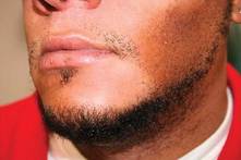
CTCL lesions may be flat or raised, and may mimic other skin disorders associated with pigment change. These can include tinea versicolor, vitiligo, pityriasis rosea, and psoriasis. Lichen planus pigmentosus and progressive macular hypomelanosis are other important CTCL mimickers. Consider all of these conditions in the differential diagnosis.
An excellent resource for photos and descriptions of CTCL, as well as CTCL look-alikes, is an article by Dr. Ginette A. Hinds and Dr. Peter Heald (J. Am. Acad. Dermatol. 2009;60:359-75). The article also highlights the clinical variants within the mycosis fungoides subtype, the most common form of CTCL among blacks.
For example, look out for the "herald patch" of unilesional mycosis fungoides, which takes the form of a single large, dyspigmented patch. According to the authors, this variant has an excellent prognosis, possibly because of an active immune response that limits the initial spread of disease and contributes to preventing relapse.
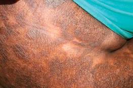
Another variant is pigmented purpuric mycosis fungoides, which can often be confused with the benign pigmented purpura syndromes of Schamberg’s disease (progressive pigmented purpuric dermatitis), Gougerot-Blum syndrome (pigmented purpuric lichenoid dermatitis), Majocchi’s disease (purpura annularis telangiectodes), Doucas and Kapetanakis (lymphocytic capillaritis of unknown cause), and lichen aureus. The key to diagnosis in this case is recalling that "the lesions of the benign syndromes rarely assume the morphology and distribution found with mycosis fungoides," Dr. Hinds and Dr. Heald wrote.
Not only is mycosis fungoides difficult to diagnose, but it is especially prevalent among black patients. For instance, a 1988 study looking at Surveillance, Epidemiology, and End Results (SEER) data from 1973 through 1984 found that the incidence among black patients was twice that of white patients: 0.52/100,000 vs. 0.26 (JAMA 1988;260:42-46).
Similarly, in another study of 132 black patients with skin cancer, mycosis fungoides represented 12.1% of all skin neoplasms (Dermatol. Clin. 1988;6:397-405).
Mycosis fungoides is the fourth most common skin cancer among Japanese patients, representing approximately 5% of all skin malignancies in the population.

The incidence of CTCL and its subtypes is only increasing. A 2007 study that extended the SEER data mentioned above from 1973 to 2002 and included all CTCL cases found an incidence rate of 9.0 for black patients, per 1 million person-years, compared with 6.1 for white patients (Arch. Dermatol. 2007;143:854-9).
As in many skin cancers, early diagnosis can make the difference in successful outcomes. So add CTCL, and especially the mycosis fungoides subtype, to your differential when confronting pigmentation disorders in skin of color patients.
Dr. Roberts is past president of the Women’s Dermatologic Society (WDS) as well as past president of the California Society of Dermatology and Dermatologic Surgery (Calderm). She was a founding director of dermatopathology at the Loma Linda University Medical Center (Calif.). She currently runs a private practice in Rancho Mirage, Calif.
She disclosed financial relationships with Allergan, L’Oréal/La Roche Posay, Skin Medica, and Valeant Pharmaceuticals.
Over the past 4 decades, cutaneous T-cell lymphoma diagnoses have been on the rise, especially among black and other skin of color patients. Unfortunately, this form of skin cancer can be a diagnostic challenge.
Regarding diagnosis, cutaneous T-cell lymphoma (CTCL) is the wild card of dermatology; it is a great mimicker of many skin disorders, and can look like almost anything. However, the most striking feature of the condition – and the key to diagnosis in ethnic skin – is its polymorphous pigmentation that is unique to skin of color.
CTCL lesions may be flat or raised, and may mimic other skin disorders associated with pigment change. These can include tinea versicolor, vitiligo, pityriasis rosea, and psoriasis. Lichen planus pigmentosus and progressive macular hypomelanosis are other important CTCL mimickers. Consider all of these conditions in the differential diagnosis.
An excellent resource for photos and descriptions of CTCL, as well as CTCL look-alikes, is an article by Dr. Ginette A. Hinds and Dr. Peter Heald (J. Am. Acad. Dermatol. 2009;60:359-75). The article also highlights the clinical variants within the mycosis fungoides subtype, the most common form of CTCL among blacks.
For example, look out for the "herald patch" of unilesional mycosis fungoides, which takes the form of a single large, dyspigmented patch. According to the authors, this variant has an excellent prognosis, possibly because of an active immune response that limits the initial spread of disease and contributes to preventing relapse.
Another variant is pigmented purpuric mycosis fungoides, which can often be confused with the benign pigmented purpura syndromes of Schamberg’s disease (progressive pigmented purpuric dermatitis), Gougerot-Blum syndrome (pigmented purpuric lichenoid dermatitis), Majocchi’s disease (purpura annularis telangiectodes), Doucas and Kapetanakis (lymphocytic capillaritis of unknown cause), and lichen aureus. The key to diagnosis in this case is recalling that "the lesions of the benign syndromes rarely assume the morphology and distribution found with mycosis fungoides," Dr. Hinds and Dr. Heald wrote.
Not only is mycosis fungoides difficult to diagnose, but it is especially prevalent among black patients. For instance, a 1988 study looking at Surveillance, Epidemiology, and End Results (SEER) data from 1973 through 1984 found that the incidence among black patients was twice that of white patients: 0.52/100,000 vs. 0.26 (JAMA 1988;260:42-46).
Similarly, in another study of 132 black patients with skin cancer, mycosis fungoides represented 12.1% of all skin neoplasms (Dermatol. Clin. 1988;6:397-405).
Mycosis fungoides is the fourth most common skin cancer among Japanese patients, representing approximately 5% of all skin malignancies in the population.
The incidence of CTCL and its subtypes is only increasing. A 2007 study that extended the SEER data mentioned above from 1973 to 2002 and included all CTCL cases found an incidence rate of 9.0 for black patients, per 1 million person-years, compared with 6.1 for white patients (Arch. Dermatol. 2007;143:854-9).
As in many skin cancers, early diagnosis can make the difference in successful outcomes. So add CTCL, and especially the mycosis fungoides subtype, to your differential when confronting pigmentation disorders in skin of color patients.
Dr. Roberts is past president of the Women’s Dermatologic Society (WDS) as well as past president of the California Society of Dermatology and Dermatologic Surgery (Calderm). She was a founding director of dermatopathology at the Loma Linda University Medical Center (Calif.). She currently runs a private practice in Rancho Mirage, Calif.
She disclosed financial relationships with Allergan, L’Oréal/La Roche Posay, Skin Medica, and Valeant Pharmaceuticals.
Over the past 4 decades, cutaneous T-cell lymphoma diagnoses have been on the rise, especially among black and other skin of color patients. Unfortunately, this form of skin cancer can be a diagnostic challenge.
Regarding diagnosis, cutaneous T-cell lymphoma (CTCL) is the wild card of dermatology; it is a great mimicker of many skin disorders, and can look like almost anything. However, the most striking feature of the condition – and the key to diagnosis in ethnic skin – is its polymorphous pigmentation that is unique to skin of color.
CTCL lesions may be flat or raised, and may mimic other skin disorders associated with pigment change. These can include tinea versicolor, vitiligo, pityriasis rosea, and psoriasis. Lichen planus pigmentosus and progressive macular hypomelanosis are other important CTCL mimickers. Consider all of these conditions in the differential diagnosis.
An excellent resource for photos and descriptions of CTCL, as well as CTCL look-alikes, is an article by Dr. Ginette A. Hinds and Dr. Peter Heald (J. Am. Acad. Dermatol. 2009;60:359-75). The article also highlights the clinical variants within the mycosis fungoides subtype, the most common form of CTCL among blacks.
For example, look out for the "herald patch" of unilesional mycosis fungoides, which takes the form of a single large, dyspigmented patch. According to the authors, this variant has an excellent prognosis, possibly because of an active immune response that limits the initial spread of disease and contributes to preventing relapse.
Another variant is pigmented purpuric mycosis fungoides, which can often be confused with the benign pigmented purpura syndromes of Schamberg’s disease (progressive pigmented purpuric dermatitis), Gougerot-Blum syndrome (pigmented purpuric lichenoid dermatitis), Majocchi’s disease (purpura annularis telangiectodes), Doucas and Kapetanakis (lymphocytic capillaritis of unknown cause), and lichen aureus. The key to diagnosis in this case is recalling that "the lesions of the benign syndromes rarely assume the morphology and distribution found with mycosis fungoides," Dr. Hinds and Dr. Heald wrote.
Not only is mycosis fungoides difficult to diagnose, but it is especially prevalent among black patients. For instance, a 1988 study looking at Surveillance, Epidemiology, and End Results (SEER) data from 1973 through 1984 found that the incidence among black patients was twice that of white patients: 0.52/100,000 vs. 0.26 (JAMA 1988;260:42-46).
Similarly, in another study of 132 black patients with skin cancer, mycosis fungoides represented 12.1% of all skin neoplasms (Dermatol. Clin. 1988;6:397-405).
Mycosis fungoides is the fourth most common skin cancer among Japanese patients, representing approximately 5% of all skin malignancies in the population.
The incidence of CTCL and its subtypes is only increasing. A 2007 study that extended the SEER data mentioned above from 1973 to 2002 and included all CTCL cases found an incidence rate of 9.0 for black patients, per 1 million person-years, compared with 6.1 for white patients (Arch. Dermatol. 2007;143:854-9).
As in many skin cancers, early diagnosis can make the difference in successful outcomes. So add CTCL, and especially the mycosis fungoides subtype, to your differential when confronting pigmentation disorders in skin of color patients.
Dr. Roberts is past president of the Women’s Dermatologic Society (WDS) as well as past president of the California Society of Dermatology and Dermatologic Surgery (Calderm). She was a founding director of dermatopathology at the Loma Linda University Medical Center (Calif.). She currently runs a private practice in Rancho Mirage, Calif.
She disclosed financial relationships with Allergan, L’Oréal/La Roche Posay, Skin Medica, and Valeant Pharmaceuticals.