User login
Data support subcutaneous bortezomib in multiple myeloma
CHICAGO – Subcutaneous and intravenous bortezomib performed comparably well in patients with newly diagnosed multiple myeloma, according to a review of 372 cases.
The findings, which showed a trend toward higher cumulative dosage and improved outcomes with the subcutaneous route, are consistent with those from a phase III study (Lancet Oncology. 2011;12[5]:431-40) of patients with relapsed or refractory multiple myeloma, Dr. Robert M. Rifkin reported in a poster at the American Society of Hematology Meeting on Hematologic Malignancies.
Median treatment duration in 248 patients who received subcutaneous bortezomib and 124 who received intravenous bortezomib was similar at 4.4 and 4.1 months, respectively. Dose reductions were required in the first 16 weeks in 10% and 13% of patients in the groups, respectively, and median time to dose reduction was 49 days in both groups, said Dr. Rifkin of the Rocky Mountain Cancer Center, McKesson Specialty Health/US Oncology Network, Denver.
Median relative dose intensities were the same in the two groups, but the subcutaneous group had a higher median cumulative dose (25.8 mg/m2 vs. 22.9 mg/m2). When dichotomized by median cumulative dose, those in the subcutaneous group were more likely to have received the higher dose (52% vs. 48%), he said, noting that a higher cumulative dose was associated with longer improved response and progression-free survival (PFS) at 12 weeks. The odds ratio for improved objective response with greater vs. less than 24.7 mg/m2 was 2.20 and the hazard ratio for PFS was 0.34.
Two-year overall survival was similar at 81% and 78%; 2-year PFS was 75% and 70% in the groups, respectively.
Among 204 patients who were evaluable with respect to response, the complete response/very good partial response rates at a median of 19.1 and 20.8 months were16% and 22% in the subcutaneous and intravenous groups, respectively, Dr. Rifkin said.
Adverse events included neuropathy in 34% and 38%, thrombocytopenia in 13% and 10%, neutropenia in 10% and 5%, and anemia in 25% and 24% of patients in the groups, respectively.
The previously observed noninferiority of subcutaneous vs. intravenous bortezomib has the potential to affect treatment outcomes in the clinical setting, but there was a lack of comparative effectiveness data on the two treatment approaches as initial therapy in newly diagnosed multiple myeloma patients, he noted.
The current findings, based on adults treated between Jan. 1 and Dec. 31, 2012, within the McKesson Specialty Health/US Oncology Network, demonstrate a trend toward higher cumulative dose in the subcutaneous group, which was associated with improved response and PFS. The findings support extended therapy for higher cumulative dosage and better treatment outcomes, he concluded.
Dr. Rifkin is a board or advisory committee member for Celgene, Millenium: The Takeda Oncology Company, and Onyx.
AT MHM 2015
Key clinical point: Subcutaneous and intravenous bortezomib performed comparably well in patients with newly diagnosed multiple myeloma, according to a review of 372 cases.
Major finding: 2-year overall survival and progression-free survival were 81% and 78%, and 75% and 70% in the subcutaneous and intravenous groups, respectively.
Data source: A review of 372 newly diagnosed multiple myeloma cases.
Disclosures: Dr. Rifkin is a board or advisory committee member for Celgene, Millenium: The Takeda Oncology Company, and Onyx.
Pregnant cancer patients: Start treatment ASAP

Photo by Nina Matthews
VIENNA—Women who are pregnant when diagnosed with cancer should carry their child to term but start cancer treatment immediately, according to researchers.
A study of young children suggested that exposure to cancer treatment in utero did not have detrimental effects on a child’s mental development or heart function.
Premature delivery, on the other hand, was associated with delayed cognitive development.
“Our results show that fear of cancer treatment is no reason to terminate a pregnancy, that maternal treatment should not be delayed, and that chemotherapy can be given,” said Frederic Amant, MD, PhD, of University Hospitals Leuven in Belgium.
“The study also shows that children suffer more from prematurity than from chemotherapy, so avoiding prematurity is more important than avoiding chemotherapy.”
Dr Amant presented these findings at the 2015 European Cancer Congress. The study was also published in NEJM.
The study included 129 children born to mothers with cancer, matched with 129 children of the same gestational age who were born to mothers unaffected by cancer.
The most common malignancies were breast (n=69) and hematologic cancers. This included acute myeloid leukemia (n=4), acute lymphoblastic leukemia (n=1), chronic myeloid leukemia (n=1), Hodgkin lymphoma (n=8), and non-Hodgkin lymphoma (n=6).
The researchers assessed the children’s general health and mental development when they were 18 months and 3 years old. At the age of 3, 47 of the children also had their heart function checked with electrocardiograms and echocardiography.
Ninety-six children (74.4%) were exposed to chemotherapy (alone or in combination with other treatment) before birth, 11 children (8.5%) were exposed to radiotherapy (alone or in combination), 13 (10.1%) were exposed to surgery alone, and 2 (1.6%) were exposed to drugs other than chemotherapeutic agents. Fourteen (10.9%) mothers did not receive cancer treatment during pregnancy.
Mental development
“Compared to the control group of children, we found no significant differences in mental development among children exposed to chemotherapy, radiotherapy, surgery alone, or no treatment,” Dr Amant said. “Nor was the number of chemotherapy cycles during pregnancy, which ranged from 1 to 10, related to the outcome of the children.”
To measure cognitive development, the researchers used the Bayley Scales of Infant Development. The median score was 101 (range, 56-145) in children exposed to cancer treatment and 100 (range, 50-145) in unexposed children.
When compared to controls, there was no significant difference in Bayley II or III score for all children born to mothers with cancer (P=0.08), children exposed to any chemotherapy (P=0.43), children exposed to anthracyclines (P=0.43), children exposed to taxanes (P=0.57), children exposed to platinum derivatives (P=0.95), children exposed to radiotherapy (P=0.69), children exposed to surgery alone (P=0.13), and children whose mothers did not undergo treatment (P=0.08).
Premature birth
Conversely, Bayley scores tended to increase by an average of 2.9 points for every week in gestational age. This was after the researchers controlled for a child’s age, gender, country, ethnicity, and parental education level.
“Delayed development of mental processes appeared to be related to premature birth,” Dr Amant said.
Premature birth was more frequent among children born to mothers with cancer, regardless of whether or not they received prenatal treatment, than in the general population in the countries participating in this study (Belgium, The Netherlands, Italy, and the Czech Republic).
The children born to mothers with cancer had a median gestational age of 36 weeks, ranging from 27 to 41 weeks. Seventy-nine (61.2%) children were born preterm, compared to 7% to 8% in the general population.
“In most cases, they were born prematurely due to a medical decision to induce preterm so as to continue cancer treatment after the delivery,” Dr Amant said.
“In some cases, preterm delivery was spontaneous, and it is possible that cancer treatment plays a role in this. But we do not know what exactly triggers preterm delivery. It could be that chemotherapy induces preterm contractions or vaginal inflammation with preterm rupture of the membranes.”
Cardiac function
The researchers assessed cardiac function in 47 three-year-olds whose mothers had cancer and 47 control children.
There were no significant differences between the exposed and control children for most measures of cardiac function, such as heart rate, ejection fraction, fractional shortening, global longitudinal strain, and circumferential strain.
The only exceptions were diastolic blood pressure, which was higher among exposed children (P=0.001), and tissue Doppler imaging measurements of the basal segment of the interventricular septum. There were higher mean peak systolic and early diastolic velocities in the control group than the exposed group (P=0.003 for both comparisons).
The researchers noted, however, that the differences in tissue Doppler velocities were not present when comparing the control group and the 26 children who were exposed to anthracyclines.
Next steps
Last year, Dr Amant reported similarly favorable results in 54 children exposed to chemotherapy or radiation in utero. The new report is a continuation of this work.
“These latest results are, again, reassuring,” Dr Amant said. “But given that we have a larger group of children . . . , the current data are much more robust.”
However, he also pointed out that this study has some limitations.
“Our data include many types of chemotherapy, but we cannot guarantee that all types of chemotherapy are safe,” Dr Amant said. “We need to look at larger numbers of children and larger numbers exposed to each drug in order to be able to document the potential effects of individual drugs.”
“In addition, we cannot extrapolate to newer drugs, including targeted drugs. We need longer follow-up to see if there are any long-term toxic effects in cases where cisplatin was administered before birth.”
“For these reasons, we will continue to follow these children until the age of 18 years, and we will enlarge the group. This will allow us to document longer-term effects and to draw conclusions for specific drugs. In addition, we will investigate to what extent anticancer drugs are diluted in the body during pregnancy and also [examine] the psycho-emotional needs of mothers and their partners.” ![]()
Variations in blood cancer survival across Europe

chemotherapy
Photo by Rhoda Baer
VIENNA—Results of the EUROCARE-5 study have revealed regional differences in survival for European patients with hematologic malignancies.
The data showed regional variations in 5-year relative survival rates for a number of cancers.
But the differences were particularly pronounced for leukemias, non-Hodgkin lymphomas (NHLs), and plasma cell neoplasms (PCNs).
Milena Sant, MD, of the Fondazione IRCCS Istituto Nazionale dei Tumori in Milan, Italy, presented these results at the 2015 European Cancer Congress (LBA 1).
Data from this study have also been published in several articles in the October 2015 issue of the European Journal of Cancer.
EUROCARE-5 includes records from 22 million cancer patients diagnosed between 1978 and 2007. The latest data encompass more than 10 million patients (ages 15 and older) diagnosed from 1995 to 2007 and followed up to 2008.
The data came from 107 cancer registries in 29 countries. The researchers estimated 5-year relative survival and trends from 1999 to 2007 according to region—Ireland/UK, Northern Europe, Central Europe, Southern Europe, and Eastern Europe.
“In general, 5-year relative survival—survival that is adjusted for causes of death other than cancer—increased steadily over time in Europe, particularly in Eastern Europe, for most cancers,” Dr Sant said.
“However, the most dramatic geographical variations were observed for cancers of the blood where there have been recent advances in treatment, such as chronic myeloid and lymphocytic leukemias, non-Hodgkin lymphoma and 2 of its subtypes (follicular and diffuse large B-cell lymphoma), and multiple myeloma. Hodgkin lymphoma was the exception, with smaller regional variations and a fairly good prognosis in most countries.”
Hodgkin lymphoma and NHL
Of all the hematologic malignancies, 5-year relative survival was highest for Hodgkin lymphoma, at 80.8% (40,625 cases). Five-year survival was 79.4% in Ireland and the UK, 85% in Northern countries, and 74.3% in Eastern Europe, which was significantly below the European average (P<0.0001).
For NHL, the 5-year relative survival was 59.4% (329,204 cases). Survival rates for NHL patients ranged from 49.7% in Eastern Europe to 63.3% in Northern Europe.
CLL/SLL
For chronic lymphocytic leukemia/small lymphocytic lymphoma (CLL/SLL), the 5-year relative survival was 70.4% (81,914 cases). CLL/SLL survival ranged from 58% in Eastern Europe to about 74% in Central and Northern Europe.
The researchers noted that between-country variations in CLL/SLL survival were high in all regions. Outliers that were significantly below the regional average were Austria (67%), Croatia (52%), and Bulgaria (45.5%).
PCNs
PCNs included multiple myeloma, plasmacytoma, and plasma cell leukemias. The 5-year relative survival for all PCNs was 39.2% (94,024 cases).
PCN survival rates were lowest in Eastern Europe (31.7%), slightly higher in the UK/Ireland (35.9%), and between 39.1% and 42% in the rest of Europe.
Myeloid leukemias
Of all the hematologic malignancies, 5-year relative survival was poorest for patients with acute myeloid leukemia (AML), at 17.1% (57,026 cases).
AML survival rates in Ireland/UK (15.0%) and Eastern Europe (13.0%) were significantly below the European average. But AML survival in Sweden, Belgium, France, and Germany was significantly higher than the average (P<0.005).
Five-year relative survival for chronic myeloid leukemia (CML) was 52.9% (17,713 cases).
Of all the hematologic malignancies, the survival gap between Eastern Europe and the rest of Europe was highest for CML. Five-year survival for CML patients was 33% in Eastern Europe and ranged from 51% to 58% in the rest of Europe.
The researchers also said there were striking survival variations by country in all areas. They found significant deviations from the regional average in Sweden (69.7%), Scotland (64.6%), France (71.7%), Austria (48.2%), Croatia (37.8%), Estonia (48.9%), Czech Republic (45.2%), and Latvia (22.1%).
“Results from EUROCARE can help to identify regions of low survival where action is needed to improve patients’ outcomes,” Dr Sant noted.
“Population-based survival information is essential for physicians, policy-makers, administrators, researchers, and patient organizations who deal with the needs of cancer patients, as well as with the issue of the growing expenditure on healthcare.” ![]()
NICE backs discounted idelalisib for CLL

The UK’s National Institute for Health and Care Excellence (NICE) has issued a final draft guidance recommending that the PI3Kδ inhibitor idelalisib (Zydelig) be made available on the National Health Service (NHS) for some adults with chronic lymphocytic leukemia (CLL).
NICE is recommending idelalisib in combination with rituximab for adults with previously untreated CLL who have a 17p deletion or TP53 mutation and for adults with CLL who have relapsed within 24 months of their previous treatment.
This decision follows a preliminary decision earlier this year, when NICE asked Gilead Sciences, the company developing idelalisib, to provide further information on the cost-effectiveness of the drug.
Gilead responded by submitting new economic analyses and a simple discount agreement to the list price of idelalisib.
NICE’s recommendation for idelalisib is contingent upon the company providing the agreed upon discount.
NICE’s draft guidance is now with consultees, who have the opportunity to appeal against it. Until NICE issues a final guidance, NHS bodies should make decisions locally on the funding of specific treatments.
Patients whose treatment with idelalisib is not recommended in this NICE guidance but was started within the NHS before this guidance was published should be able to continue treatment until they and their NHS clinician consider it appropriate to stop.
Clinical effectiveness
The committee advising NICE concluded that idelalisib could not be recommended for patients whose disease had relapsed more than 24 months after previous treatment, as no evidence was submitted for this patient group.
For the other populations, the clinical effectiveness data from Study 116 showed that idelalisib plus rituximab produced a significant improvement in progression-free survival and overall survival, compared with rituximab alone, for patients with high-risk, relapsed or refractory CLL.
Cost-effectiveness
Idelalisib is priced at £3114.75 for sixty 150-mg tablets (British national formulary 2015). The mean cost of a 1-year treatment course is £37,922.
Gilead’s agreement provides a discount to the list price of idelalisib, but the level of the discount is currently confidential.
Analyses suggested that, at the discount agreement price, idelalisib plus rituximab was associated with higher costs and greater quality-adjusted life-year (QALY) gains when compared with rituximab alone.
The deterministic incremental cost-effectiveness ratio (ICER) for idelalisib plus rituximab compared with rituximab alone was £13,634 per QALY gained (incremental costs £26,128; incremental QALYs 1.92).
Compared with best supportive care, the ICER for idelalisib plus rituximab was £20,461 per QALY gained (incremental costs £39,211; incremental QALYs 1.92). And compared with ofatumumab, the ICER was £1527 per QALY gained (incremental costs £2926; incremental QALYs 1.92). ![]()
Reducing side effects of CAR T-cell therapy

Photo courtesy of UCSF
Researchers have reported progress in developing an “on/off switch” to temper the over-active immune response and severe toxicities that can result from chimeric antigen receptor (CAR) T-cell therapy.
The team created CAR T cells that are “off” by default, homing to CD19-expressing cancer cells but remaining inactive until a small molecule is administered.
This system effectively targeted leukemia and lymphoma cells in preclinical experiments.
But the researchers said it’s not ready for clinical testing, as the small-molecule “trigger” is expensive and lasts only 4 hours.
Still, the team believes this type of CAR T-cell therapy could eventually help doctors gradually increase the immune response to treatment and therefore avoid toxicities such as cytokine release syndrome and tumor lysis syndrome.
Wendell Lim, PhD, of University of California, San Francisco, and his colleagues described this work in Science.
“T cells are really powerful beasts, and they can be lethal when they’re activated,” Dr Lim said. “We’ve needed a remote control system that retains the power of these engineered T cells but allows us to communicate specifically with them and manage them while they’re in the body.”
To that end, he and his colleagues created a CAR that requires both an antigen and a small molecule for activation. They dubbed it the “ON-switch CAR.”
ON-switch CAR
The researchers explained that the ON-switch CAR consists of 2 parts that assemble in a small molecule-dependent manner.
Part 1 consists of a CD8α signal sequence, Myc epitope, anti-CD19 single-chain variable fragment, CD8α hinge and transmembrane domain, 4-1BB costimulatory motif, and FK506 Binding Protein (FKBP) domain for heterodimerization.
Part 2 consists of the ectodomain of DNAX-activating protein 10 (DAP10) for homodimerization, CD8α transmembrane domain for membrane anchoring, 4-1BB costimulatory motif, T2089L mutant of FKBP-rapamycin binding (FRB*) domain, T-cell receptor CD3ζ signaling chain, and mCherry tag.
The FKBP and FRB* domains heterodimerize in the presence of the rapamycin analog AP21967, referred to as the “rapalog.”
The researchers conducted in vitro experiments with this ON-switch CAR in cells expressing CD19 (K562, Raji, and Daudi).
The ON-switch CAR T cells homed to CD19-expressing cells but did nothing else until the rapalog was added. Once the rapalog was added, CD19-expressing cells were killed off in a dose-dependent manner.
The team observed similar results in mice with leukemia. Leukemia cells (K562) were selectively eliminated by the ON-switch CARs only after the rapalog had been administered.
Dr Lim stressed that this work should be considered a proof of principle, as the rapalog has too short a half-life to be clinically useful. Nevertheless, he believes the research provides the foundation for practical remote control of CAR T cells.
Members of his lab are exploring other techniques to accomplish this goal, such as controlling CAR T-cell activation with light.
The team is also working to reduce side effects of CAR T-cell therapy by introducing multiple CARs into T cells so the cells will respond to multiple characteristics that are distinctive to an individual patient’s tumor, rather than to a single protein that may also be found on normal cells.
“That we can engineer CAR T cells to have slightly different, quite powerful effects—even if for a subset of patients or for certain types of cancer—is really remarkable,” Dr Lim said. “And this is just the tip of the iceberg.” ![]()
Childhood cancer increases material hardship

Photo by Bill Branson
Results of a small study reveal the material hardships families experience when a child is undergoing cancer treatment.
Researchers surveyed 99 families of children with cancer.
Six months after the child’s diagnosis, 29% of the families reported having at least one household material hardship, such as food, housing, or energy insecurity.
Twenty percent of the families had reported having such hardships at the time of the child’s diagnosis.
Kira Bona, MD, of Dana-Farber/Boston Children’s Cancer and Blood Disorders Center in Massachusetts, and her colleagues reported results from this survey in Pediatric Blood & Cancer.
The researchers surveyed 99 families of pediatric cancer patients treated at Dana-Farber/Boston Children’s, first within a month of diagnosis and then 6 months later.
At diagnosis, 20% of the families were low-income, which was defined as 200% of the federal poverty level. Six months later, an additional 12% suffered income losses that pushed them into the low-income group.
At 6 months, 25% of the families said they had lost more than 40% of their household income due to treatment-related work disruptions. A total of 56% of adults who supported their families experienced a disruption of their work.
This included 15% of parents who either quit their jobs or were laid off as a result of their child’s illness, as well as 37% of respondents who cut their hours or took a leave of absence. Thirty-four percent of these individuals were paid during their leave.
At 6 months, 29% of families said they had at least one material hardship. Twenty percent reported food insecurity, 17% reported energy insecurity, and 8% reported housing insecurity.*
These findings surprised researchers, who said they expected lower levels of need at their center because it provides psychosocial support for patients and has resource specialists to help families facing financial difficulties.
“What it says is that even at a well-resourced, large referral center, about a third of families are reporting food, housing, or energy insecurity 6 months into treatment,” Dr Bona said. “If anything, the numbers in our study are an underestimate of what might be seen at less well-resourced institutions, which was somewhat surprising to us.”
By focusing on specific material hardships, which can be addressed through governmental or philanthropic support, the researchers hope they have identified variables that are easier for clinicians to ameliorate than overall income.
Dr Bona said subsequent research will examine whether material hardship has the same effect on patient outcomes as low-income status.
“If household material hardship is linked to poorer outcomes in pediatric oncology, just like income is, then we can design interventions to fix food, housing, and energy insecurity,” she said. “It’s not clear what you do about income in a clinical setting.” ![]()
*Definitions for household material hardships were as follows.
Food insecurity was measured via the US Household Food Security Survey Module: Six-Item Short Form, which includes questions to asses if respondents:
- sometimes/often do not have enough food to eat
- sometimes/often cannot afford to eat balanced meals
- sometimes/often worry about having enough money to buy food, etc.
Families met the definition for housing insecurity if they reported any of the following:
- crowding (defined as >2 people per bedroom in the home)
- multiple moves (>1 move in the prior year)
- doubling up (having to live with other people, even temporarily, because of financial difficulties in the past 6 months).
Families met the definition for energy insecurity if, in the prior 6 months, they had experienced any of the following:
- received a letter threatening to shut off the gas/electricity/oil to their house because they had not paid the bills
- had the gas/electric/oil company shut off electricity or refused to deliver oil/gas because they had not paid the bills
- had any days that their home was not heated/cooled because they couldn’t pay the bills
- had ever used a cooking stove to heat their home because they couldn’t pay the bills.
Topical resiquimod effective for early-stage cutaneous T-cell lymphoma
Topical resiquimod was effective and well tolerated in patients with early-stage cutaneous T-cell lymphoma (CTCL), in some cases inducing regression in both treated and untreated lesions, according to researchers.
The mean number of prior unsuccessful therapies among the patients was 6, yet the majority of patients (11 of 12) experienced significant improvement, and 2 patients had complete clinical responses with no evidence of disease after treatment. One patient, despite a 15-year history of disease and 11 unsuccessful treatments, experienced a complete resolution of both treated and untreated skin lesions.

The open-label, phase I trial evaluated 12 patients with early-stage CTCL. Patients experienced minor adverse effects (all grade 1), which were primarily skin irritation. The trial evaluated 0.03% and 0.06% resiquimod, with complete and more rapid responses occurring at the higher dose. Both doses were equally well tolerated.
“These studies support further trials of this medication in early-stage, skin-limited CTCL and suggest resiquimod might also be useful as an adjuvant therapy in the treatment of more advanced CTCL,” wrote Dr. Alain Rook of the Department of Dermatology and the Center for Clinical Biostatistics and Epidemiology, Perelman School of Medicine, Philadelphia, and colleagues.
Arising from T cells that traffic to the skin, CTCLs are non-Hodgkin lymphomas whose only potential cure is stem cell transplantation. Studies suggest that host antitumor immunity plays an important role in the disease, and in this study, high responders showed recruitment and expansion of benign T-cell clones and activation of T cells and natural killer cells in the skin.
In the absence of cell-surface markers to distinguish malignant from benign T cells in the lesion, the team used high throughput screening of the T-cell receptor–beta gene to quantify malignant cells and monitor response to therapy. Of the 10 patients with identified malignant cells, biopsied lesions showed that most had reduction of malignant T-cell clones and 3 had complete eradication. The results may not reflect responses in nonbiopsied lesions.
Resiquimod recruits T cells and other immune cells to the skin, causing inflammation that the researchers observed persisted after complete or nearly complete malignant T-cell eradication. Study results suggested that activation of CD4+ cells and expansion of tumor-specific T cells is critical for effectiveness of resiquimod.
Topical resiquimod was effective and well tolerated in patients with early-stage cutaneous T-cell lymphoma (CTCL), in some cases inducing regression in both treated and untreated lesions, according to researchers.
The mean number of prior unsuccessful therapies among the patients was 6, yet the majority of patients (11 of 12) experienced significant improvement, and 2 patients had complete clinical responses with no evidence of disease after treatment. One patient, despite a 15-year history of disease and 11 unsuccessful treatments, experienced a complete resolution of both treated and untreated skin lesions.
The open-label, phase I trial evaluated 12 patients with early-stage CTCL. Patients experienced minor adverse effects (all grade 1), which were primarily skin irritation. The trial evaluated 0.03% and 0.06% resiquimod, with complete and more rapid responses occurring at the higher dose. Both doses were equally well tolerated.
“These studies support further trials of this medication in early-stage, skin-limited CTCL and suggest resiquimod might also be useful as an adjuvant therapy in the treatment of more advanced CTCL,” wrote Dr. Alain Rook of the Department of Dermatology and the Center for Clinical Biostatistics and Epidemiology, Perelman School of Medicine, Philadelphia, and colleagues.
Arising from T cells that traffic to the skin, CTCLs are non-Hodgkin lymphomas whose only potential cure is stem cell transplantation. Studies suggest that host antitumor immunity plays an important role in the disease, and in this study, high responders showed recruitment and expansion of benign T-cell clones and activation of T cells and natural killer cells in the skin.
In the absence of cell-surface markers to distinguish malignant from benign T cells in the lesion, the team used high throughput screening of the T-cell receptor–beta gene to quantify malignant cells and monitor response to therapy. Of the 10 patients with identified malignant cells, biopsied lesions showed that most had reduction of malignant T-cell clones and 3 had complete eradication. The results may not reflect responses in nonbiopsied lesions.
Resiquimod recruits T cells and other immune cells to the skin, causing inflammation that the researchers observed persisted after complete or nearly complete malignant T-cell eradication. Study results suggested that activation of CD4+ cells and expansion of tumor-specific T cells is critical for effectiveness of resiquimod.
FROM BLOOD
Key clinical point: Topical resiquimod was effective and well tolerated in patients with early-stage cutaneous T-cell lymphoma (CTCL).
Major finding: In total, 11 of 12 patients had significant improvement: 2 had resolution of all evidence of disease, and 9 experienced improvement greater than or equal to 50%.
Data source: The open-label, phase I trial evaluated 12 patients with early stage CTCL.
Disclosures: Dr. Rook and one coauthor have patents pending on HTS in cutaneous lymphoma. His coauthor is employed by Adaptive Biotechnologies.
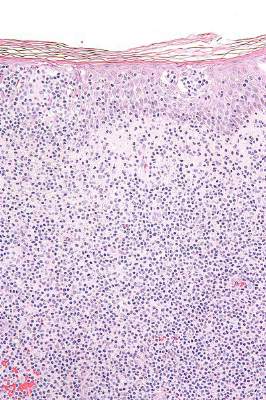
Hospital readmission following transplantation: identifying risk factors and designing preventive measures
Background About 1 in 7 of all hospitalized patients is readmitted within 30 days of discharge. The cost of readmissions is significant, with Medicare readmissions alone costing the health care system an estimated $28 billion a year.
Objective To identify the rates of and causes for readmission within 100 days of patients receiving a hematopoietic stem cell transplant.
Methods We performed a retrospective review of 235 consecutive transplant recipients (autologous, n = 144; allogeneic, n = 91) to determine rates and causes for readmission within 100 days of patients receiving a transplant. Medical records and hospital readmissions were reviewed for each patient.
Results 36 allogeneic patients accounted for 56 readmissions. 23 autologous patients accounted for 26 readmissions. Autologous transplant recipients were most commonly readmitted for the development of a fever (n = 15 patients) or cardiopulmonary issues (n = 4). The most prevalent reasons for readmission in the allogeneic recipients included a fever (n = 21) or the development or exacerbation of graft-versus-host disease (n = 5). The readmission length of stay was 6 days (median range, 1-91 days) for allogeneic patients and 4 days (median range, 1-22 days) for autologous patients. There was no difference in survival between the readmitted and the non-readmitted cohorts (P = .55 for allogeneic patients; P = .24 for autologous patients). Although allogeneic graft recipients demonstrated a higher readmission rate (39.6%) compared with autologous recipients (16%), none of the variables examined, including age, gender, performance status, diagnosis, remission status at the time of transplant, comorbidities, type of preparative chemotherapy regimen or donor type, identified patients at increased risk for readmission.
Limitations Variations in clinical care, physician practices, and patient characteristics need to be considered when examining readmission rates. Most of the allogeneic patient population included unrelated donor recipients (65%) who received nonmyeloablative conditioning regimens (81% of allogeneic recipients). These features may not be characteristic of other centers.
Conclusions In these high-risk patients, readmissions following a transplant are common. Enhanced predischarge education by nurses and pharmacists, along with ongoing outpatient education and rigorous outpatient follow-up through phone calls or social media may decrease readmission rates.
Click on the PDF icon at the top of this introduction to read the full article.
Immunotherapy: Inject locally, treat globally?
NEW YORK – “Inject locally, treat globally” may become a new mantra for cancer immunotherapy. Dr. Ronald Levy, professor and chief of the department of oncology at Stanford (Calif.) University, discussed impressive and durable systemic results from local treatment of tumors, reviewing his group’s recent work and discussing ongoing early stage clinical trials.
In the hope of triggering an immune response that induces a systemic CD8 T-cell response, Dr. Levy and other investigators began experimenting with intralesional injections of immunotherapies, with and without adjunctive radiation or chemotherapy. The principle has been evaluated in a mouse model, in which up to 80% of mice receiving intratumoral immunotherapy were cured. Now, human trials are underway for solid tumors as well as lymphoma.
An early clinical trial involving 15 patients with recurrent low-grade B-cell lymphoma combined targeted low-dose radiation of a single tumor site with injection of CpG, immune-boosting snippets of DNA, into the same site. Looking for partial or complete regression, investigators saw promising results, though some patients who were responders didn’t see full effect until 24 weeks after injection (J Clin Oncol. 2010 Oct 1;28[28]:4324-32). The overall objective response rate was 27%, although 80% of patients (12 of 15) had stable disease or partial or complete response through a median follow-up of 33.7 months. Some individual patients had marked regression or disappearance of bulky tumors at distant sites.
CpG – motifs of cytosines and guanines – were strung together, said Dr. Levy, with a sulfur rather than a phosphate backbone to make them more stable for injection. These bits of DNA, which are present in both bacteria and vertebrates, are an agonist for toll-like receptor 9, activating B cells and dendritic cells, and then tumor-specific T-cells.
Dr. Levy and his collaborators used a two-tumor mouse model to track intralesional injection effectiveness for a variety of immunotherapies. Mice seeded with tumor cells bilaterally over the abdomen were injected with CpG and two other immune therapies at a single abdominal tumor site, and bilateral regression, if any, was tracked in comparison to systemic immunotherapy (J Clin Invest. 2013;123[6]:2447-63).
Though both treatment arms had good initial response, 70% of the systemically treated mice relapsed by 150 days after injection, compared with just 10% of those receiving intratumoral therapy (P = .002).
Of the 23 mice whose therapy consisted of intratumoral administration of CpG together with anti-CTLA4 and anti-OX40 antibodies (aCTLA4 and aOX40), 21 (91%) were alive 50 days after treatment. These mice fared better than did those receiving any other intratumoral treatment combination (P = .004, compared with CpG+aOX40; P = .03, compared with aCTLA4), suggesting a synergistic benefit to the triple combination.
Somewhat surprisingly, even murine models that also had tumor seeding into brain tissue saw marked reduction or even resolution of brain tumors, showing that the blood-brain barrier does not impede the effect within the CNS.
What didn’t work? PD-1 inhibitors were not particularly effective at provoking a systemic effect when injected into tumors. Peritumoral injection, though theoretically taking advantage of some aspects of the tumor microenvironment, also did not show global effect.
Based on the human CpG + radiation trials and the mouse experiments, the research group is proceeding with phase I and II clinical trials of CpG in combinations with aCTLA4 and aOX40. These, Dr. Levy said, were more likely to be available clinically, and human intratumoral T regulatory cells are known to express both CTLA4 and OX40.
All agents and combinations had an enhancing effect, said Dr. Levy. “We like this treatment and trial design,” he said, noting that everyone sees benefit at the local injection site, and some see global results.
Dr. Levy discussed multiple studies, and he disclosed receiving grant support from Pfizer, Dynavax, and Bristol-Myers Squibb. He also has served as a consultant to Five Prime, Kite, BeiGene, Innate Pharma, Bullet Biotech, and Immune Design.
EXPERT ANALYSIS FROM THE FIRST INTERNATIONAL CANCER IMMUNOTHERAPY CONFERENCE
Insights from the 5-year follow-up of CTL019 in CLL
Photo courtesy of the
University of Pennsylvania
NEW YORK—The 5-year follow-up of the phase 1 trial of CTL019 in relapsed or refractory chronic lymphoblastic leukemia (CLL) is allowing investigators to define more clearly who will respond to chimeric antigen receptor (CAR) T cells directed against CD19.
One thing investigators have determined is that persistence of the CARs is essential for long-term responses.
In the first 2 patients who achieved a complete remission (CR), CAR T cells persisted for more than 4 years. In addition, no patient in CR has relapsed to date.
Of the 14 patients enrolled in the trial, 4 (28%) achieved a CR, 4 (28%) achieved a partial response, and 6 (43%) had no response, for an overall response rate of 57%.
These results were recently published in Science Translational Medicine.
Carl June, MD, of the University of Pennsylvania in Philadelphia, shared some insights into the research with attendees at the inaugural CRI-CIMT-EATI-AACR International Cancer Immunotherapy Conference.
Dr June explained that CTL019 is a CD19-directed single chain variable fragment with a 4-1BB signaling module that transduces T cells with a lentiviral vector. The technology was developed at the University of Pennsylvania and subsequently licensed to Novartis.
In the phase 1 trial of CTL019 in CLL, patients who achieved complete remission have very high levels of CARs—100% of the circulating T cells—but the non-responders don’t. The CARs engrafted in non-responders but did not proliferate.
“So the biomarker correlate of success is persistence and proliferation, in CLL at least,” Dr June said.
The investigators performed IGH next-generation sequencing and found no detectable CLL clones in the complete responders, including 1 patient at 3.5 years and another at 4 years post-infusion.
“There was no clinically evident disease in these patients,” Dr June said, “and so the responses are durable.”
The team also believes that at least a subset of the cells remains functional because the patients still had B-cell aplasia.
The investigators have not observed a CD19 loss in any CLL patient who responded.
“Patients who have gone into remission stay in remission,” Dr June added.
Kinetics of delayed CR
Dr June discussed in detail Patient 10, whose response was somewhat different from the other complete responders. Patient 10 achieved a CR, but the response was delayed. It took 51 days after infusion, compared to about 10 days in the other complete responders.
Patient 10 was initially scored as a failure at the 28-day evaluation. Eventually, he had marked improvement, and, by a year, he was in CR.
He required hospitalization for tumor lysis syndrome and treatment with tocilizumab for cytokine release syndrome.
Patient 10 had a single cell that investigators surmise could have been responsible for the tumor elimination.
“In fact, on day 28, when he still had tumor, his CARs were polyclonal,” Dr June said. “So we stained and isolated his CARs by sorting, and, at time of tumor elimination, he had descendants of 1 CAR.”
Nevertheless, Patient 10’s response is durable. He is now 81 years old and remains engrafted with CAR19 cells.
Investigators hypothesize that the kinetics and CAR proliferation were so different in Patient 10 because Tet2 was disrupted by the integration of the CAR into the intronic region.
“What we don’t know is whether Tet2 was a passenger or a driver here,” Dr June observed. “Did it actually aid the function of the CAR cells or was it just a marker?”
He noted that Tet2 has been shown in acute myeloid leukemia to increase stem cell renewal, “and it may well have done that in this patient.” ![]()