User login
VIDEO – Microneedling: Simple procedure offers good results for wrinkles, pores, and more
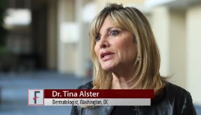
WASHINGTON – Microneedling is one of the hottest new trends in aesthetic dermatology and is easy to incorporate into any practice, according to Washington dermatologist Dr. Tina Alster.
Microneedling involves using an updated microneedle device to create small wounds on a patient’s area of concern. Perioral wrinkles, large pores on the nose, certain scars, and stretch marks all respond well to the procedure, Dr. Alster said at the annual meeting of the American Academy of Dermatology.
“I’ve been blown away by the fact that microneedling works as well as it does,” Dr. Alster added, noting that it’s not hard to do, it’s not hard to heal from, and it offers good results.
In this video interview, Dr. Alster explains how to do this microneedling procedures and how to incorporate the device into practice.
On Twitter @denisefulton
AT AAD 16
Cellfina 3-year data will show sustained effects on cellulite, researcher says
WAIKOLOA, HAWAII – Of the plethora of cellulite treatment devices on the market, the Food and Drug Administration has deemed only two capable of providing long-term improvement in the appearance of cellulite on the buttocks and thighs, Dr. Michael S. Kaminer said at the Hawaii Dermatology Seminar provided by Global Academy for Medical Education/Skin Disease Education Foundation.
These two devices are Cellulaze, a 1440 nm Nd:YAG laser typically utilized in conjunction with liposuction and marketed by Cynosure, and Cellfina, a semiautomated system for precise dermal undermining beneath cellulite dimples and marketed by Merz. Both devices target the structural problem that causes cellulite: subdermal fibrous septae that are tethered to the dermis, pulling down on the skin with resultant fat herniation and creation of cellulite dimples. The devices have the same mechanism of benefit, which entails cutting and thereby releasing the fibrous septae, albeit through two different technologies. Cellulaze cuts the fibrous septae with a laser fiber, while Cellfina uses a rapidly moving, piston-driven 18-gauge blade.

The superior efficacy of these two devices settles a long-standing controversy regarding the cause of cellulite, according to Dr. Kaminer, a dermatologist at Yale University, New Haven, Conn., and SkinCare Physicians of Chestnut Hill, Mass. “I think we now know what causes cellulite ... that if you cut the fibrous septae, things will improve.”
“There are a ton of devices (for treating cellulite) out there, but if you look at the literature none of them except Cellulaze and Cellfina have been shown to work,” he said. “Arguably the other options have, at most, limited efficacy.”
He cited a recent systematic evidence-based literature review led by Stefanie Luebberding, Ph.D., of the Rosenpark Clinic in Darmstadt, Germany. She and her coinvestigators concluded that “no clear evidence of good efficacy could be identified in any of the evaluated cellulite treatments,” which included shock wave and other forms of mechanical stimulation, massage, topical agents designed to plump up the skin, high- and low-energy laser therapies, infrared light, and radiofrequency (Am J Clin Dermatol. 2015 Aug;16[4]:243-56).
In U.S. clinical trials of Cellulaze, 68% of subjects had significant improvement at 1 year based on photographic evaluation and 65% based on Vectra three-dimensional surface imaging. Good to excellent results were reported by 76% of patients and 69% of physicians. On the downside, the Cellulaze device costs more than $100,000, recovery time is long, and the use of liposuction is falling in the United States, Dr. Kaminer observed.
He was the lead investigator in the multicenter study of Cellfina, in which 96% of patients in the open-label trial said they were satisfied or very satisfied with their results at 2 years post procedure.
From a mean baseline score of 3.4 on a 0-5 Cellulite Severity Scale, the average improvement was 2.0 points at both 1- and 2-year follow-up as assessed by independent physicians examining standardized professional photographs. Of 55 study participants, 2 experienced mild, transient adverse events (Dermatol Surg. 2015 Mar;41[3]:336-47).
Based on that trial, a single treatment session provides improvement lasting up to 1 year in the appearance of cellulite. Similar findings were noted 2 years after the procedure, said Dr. Kaminer, and he and his coinvestigators are now preparing to submit to the FDA the 3-year follow-up data, which show “essentially the same” sustained improvement.
Before the Cellfina procedure begins, the target cellulite dimples are marked on the standing patient. Roughly 20-25 dimples can be treated per session. The patient then assumes a prone position. A vacuum device is placed over the target area to control the depth and area of treatment. Tumescent anesthesia is injected, and then the piston-driven 18-gauge blade is introduced. The vacuum suction precisely controls the cutting depth at either 6 or 10 mm.
Dr. Kaminer noted that, potentially, the cellulite treatment market is colossal. One study concluded that an estimated 85% of American women age 24-54 have cellulite, and 37% of them – nearly 24 million women – say they are interested in a minimally invasive procedure to get rid of it.
“The definition of cellulite depends on what women you ask. Based on what I hear, 100% of women believe that they have cellulite and a lot of them are interested in having their cellulite treated,” he remarked.
Dr. Kaminer reported serving as a consultant to Merz, the maker of Cellfina, and receiving research funding from Cabochon, the maker of a system for improving the appearance of cellulite.
The SDEF and this news organization are owned by the same parent company.
WAIKOLOA, HAWAII – Of the plethora of cellulite treatment devices on the market, the Food and Drug Administration has deemed only two capable of providing long-term improvement in the appearance of cellulite on the buttocks and thighs, Dr. Michael S. Kaminer said at the Hawaii Dermatology Seminar provided by Global Academy for Medical Education/Skin Disease Education Foundation.
These two devices are Cellulaze, a 1440 nm Nd:YAG laser typically utilized in conjunction with liposuction and marketed by Cynosure, and Cellfina, a semiautomated system for precise dermal undermining beneath cellulite dimples and marketed by Merz. Both devices target the structural problem that causes cellulite: subdermal fibrous septae that are tethered to the dermis, pulling down on the skin with resultant fat herniation and creation of cellulite dimples. The devices have the same mechanism of benefit, which entails cutting and thereby releasing the fibrous septae, albeit through two different technologies. Cellulaze cuts the fibrous septae with a laser fiber, while Cellfina uses a rapidly moving, piston-driven 18-gauge blade.
The superior efficacy of these two devices settles a long-standing controversy regarding the cause of cellulite, according to Dr. Kaminer, a dermatologist at Yale University, New Haven, Conn., and SkinCare Physicians of Chestnut Hill, Mass. “I think we now know what causes cellulite ... that if you cut the fibrous septae, things will improve.”
“There are a ton of devices (for treating cellulite) out there, but if you look at the literature none of them except Cellulaze and Cellfina have been shown to work,” he said. “Arguably the other options have, at most, limited efficacy.”
He cited a recent systematic evidence-based literature review led by Stefanie Luebberding, Ph.D., of the Rosenpark Clinic in Darmstadt, Germany. She and her coinvestigators concluded that “no clear evidence of good efficacy could be identified in any of the evaluated cellulite treatments,” which included shock wave and other forms of mechanical stimulation, massage, topical agents designed to plump up the skin, high- and low-energy laser therapies, infrared light, and radiofrequency (Am J Clin Dermatol. 2015 Aug;16[4]:243-56).
In U.S. clinical trials of Cellulaze, 68% of subjects had significant improvement at 1 year based on photographic evaluation and 65% based on Vectra three-dimensional surface imaging. Good to excellent results were reported by 76% of patients and 69% of physicians. On the downside, the Cellulaze device costs more than $100,000, recovery time is long, and the use of liposuction is falling in the United States, Dr. Kaminer observed.
He was the lead investigator in the multicenter study of Cellfina, in which 96% of patients in the open-label trial said they were satisfied or very satisfied with their results at 2 years post procedure.
From a mean baseline score of 3.4 on a 0-5 Cellulite Severity Scale, the average improvement was 2.0 points at both 1- and 2-year follow-up as assessed by independent physicians examining standardized professional photographs. Of 55 study participants, 2 experienced mild, transient adverse events (Dermatol Surg. 2015 Mar;41[3]:336-47).
Based on that trial, a single treatment session provides improvement lasting up to 1 year in the appearance of cellulite. Similar findings were noted 2 years after the procedure, said Dr. Kaminer, and he and his coinvestigators are now preparing to submit to the FDA the 3-year follow-up data, which show “essentially the same” sustained improvement.
Before the Cellfina procedure begins, the target cellulite dimples are marked on the standing patient. Roughly 20-25 dimples can be treated per session. The patient then assumes a prone position. A vacuum device is placed over the target area to control the depth and area of treatment. Tumescent anesthesia is injected, and then the piston-driven 18-gauge blade is introduced. The vacuum suction precisely controls the cutting depth at either 6 or 10 mm.
Dr. Kaminer noted that, potentially, the cellulite treatment market is colossal. One study concluded that an estimated 85% of American women age 24-54 have cellulite, and 37% of them – nearly 24 million women – say they are interested in a minimally invasive procedure to get rid of it.
“The definition of cellulite depends on what women you ask. Based on what I hear, 100% of women believe that they have cellulite and a lot of them are interested in having their cellulite treated,” he remarked.
Dr. Kaminer reported serving as a consultant to Merz, the maker of Cellfina, and receiving research funding from Cabochon, the maker of a system for improving the appearance of cellulite.
The SDEF and this news organization are owned by the same parent company.
EXPERT ANALYSIS FROM SDEF HAWAII DERMATOLOGY SEMINAR

Chestnut extract
Known as sweet chestnut, Castanea sativa is a member of the Fagaceae family, and is found in abundance in Southern and Southeastern Europe and Asia.1 In traditional medicine, chestnut tree flower preparations have been used for various indications.2 Chestnut has been used in French folk medicine as a tea to treat severe cough, colds, and bronchitis as well as diarrhea.2-6 In modern times, C. sativa leaf extract has been described as having the capacity to scavenge various free radicals associated with oxidative stress induced by ultraviolet exposure.7
Traditional uses
A 2014 study of the therapeutic and traditional uses of the plants native to the Western Italian Alps revealed that C. sativa has long been important in the region, typically for food and wood.8 But medical uses have been uncovered in that region as well. In fact, ancient Romans found C. sativa to exhibit antibacterial, astringent, antitoxic, and tonic qualities, with chestnut honey used then to dress chronic wounds, burns, and skin ulcers.9 A 2014 study by Carocho et al. of the phytochemical profile and antioxidant activity of C. sativa flowers is noteworthy for buttressing the reported health benefits of the use of chestnut flower infusions and decoctions in traditional medicine.2
Antioxidant activity
In 2005, Calliste et al. investigated the antioxidant potential of C. sativa leaf to act against the stable free radical 2,2-diphenyl-1-pycrylhydrazyl, superoxide anion, and hydroxyl radical. Using electronic spin resonance, the investigators showed that C. sativa exhibited high antioxidant potential equivalent to reference antioxidants quercetin and vitamin E.3

Three years later, Almeida et al. conducted an in vitro assessment of an ethanol/water (7:3) extract from C. sativa leaves and an ethanol/water (2:3) extract from Quercus robur (English oak) leaves, finding that both plants demonstrated a high potency to scavenge various reactive oxygen and nitrogen species. The researchers concluded that these findings supported the burgeoning interest in these extracts for use in topical antioxidant formulations.4 An in vivo investigation using an ethanol/water (7:3) extract from C. sativa conducted by the same team later in the year yielded similar results, with the researchers concluding that chestnut extract has the potential to confer benefits against photoaging and other oxidative stress–mediated conditions when included in an appropriately formulated topical antioxidant preparation.6 Subsequently, Barreira et al. demonstrated that chestnut skin and leaves exhibited sufficient antioxidant potency to warrant use in novel antioxidant formulations.10
In 2015, Almeida et al. characterized an antioxidant semisolid surfactant-free topical formulation featuring C. sativa leaf extract. In the process of ascertaining the physical, functional, and microbiologic stability of the antioxidant formulation, the investigators identified a hydrating effect and good skin tolerance, which they concluded suggested a capacity to prevent or treat cutaneous conditions in which oxidative stress plays a role.11
Photoprotective potential
In 2010, Sapkota et al. evaluated the antioxidant and antimelanogenic characteristics of several prebloom and full-bloom chestnut flower extracts, finding that a prebloom methanol extract and an ethanol extract evinced the greatest levels of phenolic and flavonoid compounds. These extracts also displayed the best radical scavenging and mushroom tyrosinase–inhibiting activities. Notably, the prebloom extract was effective in protecting the skin from the deleterious impact of UV radiation. The investigators also observed that all of the tested extracts lowered the tyrosinase activity and melanin formation of SK-MEL-2 cells similarly to arbutin. They ascribed the antimelanogenic effects of chestnut flower extracts to their antioxidant-mediated inhibitory effects on tyrosinase. They concluded that chestnut flower extracts have considerable potential as cosmetic agents.12

Recently, Almeida et al. studied the protective effects in a human keratinocyte cell line of C. sativa extract at various concentrations (0.001-, 0.01-, 0.05-, and 0.1-mcg/mL) against UV-induced DNA damage. They found that the chestnut extract concentration dependently protected against UV-mediated DNA damage, with the 0.1-mcg/mL concentration affording maximum protection (66.4%). This result was considered to be a direct antioxidant effect attributed to various phenolic antioxidants present in C. sativa. In addition, the investigators observed no phototoxic or genotoxic effects on HaCaT cells incubated with up to 0.1 mcg/mL of chestnut leaf extract. They concluded that C. sativa leaf extract has the potential to prevent or mitigate UV-induced harm to the skin.7
Other benefits and bioactivity
Assessments of C. sativa by-products have shown a favorable profile of bioactive constituents that demonstrate antioxidant, anticarcinogenic, and cardioprotective activity. Braga et al. conducted a 2015 review that concluded these compounds, as part of agro-industrial waste, offer value to the pharmaceutical, cosmetics, and food industries, with the potential to lower pollution costs and raise profits while enhancing social, economic, and environmental sustainability in growing regions.1
A related chestnut species also has been linked to dermatologic uses. In East Asia, a skin firming/antiwrinkle formulation features the inner shell of Castanea crenata as an active ingredient.13 In 2002, Chi et al. showed that the chestnut inner shell extract improved cell-associated expression of the adhesion molecules fibronectin and vitronectin. They also found that scoparone (6,7-dimethoxycoumarin) isolated from the chestnut extract exhibited comparable qualities. The investigators concluded that the enhanced expression of adhesion molecules imparted by the chestnut inner shell extract may account for the prevention of cell detachment and the manifestation of antiaging effects.13
Allergy
It is worth noting that chestnut is one of the many allergens associated with the latex-fruit syndrome.14 However, in a patch test investigation of the skin irritation potential of C. sativa leaf extract in 20 volunteers, Almeida et al. identified five phenolic compounds in the extract (chlorogenic acid, ellagic acid, rutin, isoquercitrin, and hyperoside) and found it safe for topical application.6 Chestnut is considered to pose a low to moderate risk of inducing allergic reactions.9
Conclusion
Recent research appears to suggest the in vitro antioxidant activity of sweet chestnut and potential for use in topical formulations. There remains a paucity of in vivo evidence, however. While much more research is necessary to determine whether it has a place in the dermatologic armamentarium, current data are intriguing.
References
1. Nat Prod Res. 2015;29(1):1-18
2. Biomed Res Int. 2014;2014:232956
3. J Agric Food Chem. 2005 Jan 26;53(2):282-8
4. J Photochem Photobiol B. 2008 May 29;91(2-3):87-95
5. A Modern Herbal (vol. I). New York: Dover Publications, 1971, p. 195
6. Basic Clin Pharmacol Toxicol. 2008 Nov;103(5):461-7
7. J Photochem Photobiol B. 2015 Mar;144C:28-34
8. J Ethnopharmacol. 2014 Aug 8;155(1):463-84
9. J Sci Food Agric. 2010 Aug 15;90(10):1578-89
10. Food Sci Technol Int. 2010 June;16(3):209-16
11. Drug Dev Ind Pharm. 2015 Jan;41(1):148-55
12. Biosci Biotechnol Biochem. 2010;74(8):1527-33
13. Arch Pharm Res. 2002 Aug;25(4):469-74
14. Allergy. 2007 Nov;62(11):1277-81
Dr. Baumann is chief executive officer of the Baumann Cosmetic & Research Institute in the Design District in Miami. She founded the Cosmetic Dermatology Center at the University of Miami in 1997. Dr. Baumann wrote the textbook “Cosmetic Dermatology: Principles and Practice” (New York: McGraw-Hill, 2002), and a book for consumers, “The Skin Type Solution” (New York: Bantam Dell, 2006). Her latest book, “Cosmeceuticals and Cosmetic Ingredients,” was published in November 2014. Dr. Baumann has received funding for clinical grants from Allergan, Aveeno, Avon Products, Evolus, Galderma, GlaxoSmithKline, Kythera Biopharmaceuticals, Mary Kay, Medicis Pharmaceuticals, Neutrogena, Philosophy, Topix Pharmaceuticals, and Unilever.
Known as sweet chestnut, Castanea sativa is a member of the Fagaceae family, and is found in abundance in Southern and Southeastern Europe and Asia.1 In traditional medicine, chestnut tree flower preparations have been used for various indications.2 Chestnut has been used in French folk medicine as a tea to treat severe cough, colds, and bronchitis as well as diarrhea.2-6 In modern times, C. sativa leaf extract has been described as having the capacity to scavenge various free radicals associated with oxidative stress induced by ultraviolet exposure.7
Traditional uses
A 2014 study of the therapeutic and traditional uses of the plants native to the Western Italian Alps revealed that C. sativa has long been important in the region, typically for food and wood.8 But medical uses have been uncovered in that region as well. In fact, ancient Romans found C. sativa to exhibit antibacterial, astringent, antitoxic, and tonic qualities, with chestnut honey used then to dress chronic wounds, burns, and skin ulcers.9 A 2014 study by Carocho et al. of the phytochemical profile and antioxidant activity of C. sativa flowers is noteworthy for buttressing the reported health benefits of the use of chestnut flower infusions and decoctions in traditional medicine.2
Antioxidant activity
In 2005, Calliste et al. investigated the antioxidant potential of C. sativa leaf to act against the stable free radical 2,2-diphenyl-1-pycrylhydrazyl, superoxide anion, and hydroxyl radical. Using electronic spin resonance, the investigators showed that C. sativa exhibited high antioxidant potential equivalent to reference antioxidants quercetin and vitamin E.3
Three years later, Almeida et al. conducted an in vitro assessment of an ethanol/water (7:3) extract from C. sativa leaves and an ethanol/water (2:3) extract from Quercus robur (English oak) leaves, finding that both plants demonstrated a high potency to scavenge various reactive oxygen and nitrogen species. The researchers concluded that these findings supported the burgeoning interest in these extracts for use in topical antioxidant formulations.4 An in vivo investigation using an ethanol/water (7:3) extract from C. sativa conducted by the same team later in the year yielded similar results, with the researchers concluding that chestnut extract has the potential to confer benefits against photoaging and other oxidative stress–mediated conditions when included in an appropriately formulated topical antioxidant preparation.6 Subsequently, Barreira et al. demonstrated that chestnut skin and leaves exhibited sufficient antioxidant potency to warrant use in novel antioxidant formulations.10
In 2015, Almeida et al. characterized an antioxidant semisolid surfactant-free topical formulation featuring C. sativa leaf extract. In the process of ascertaining the physical, functional, and microbiologic stability of the antioxidant formulation, the investigators identified a hydrating effect and good skin tolerance, which they concluded suggested a capacity to prevent or treat cutaneous conditions in which oxidative stress plays a role.11
Photoprotective potential
In 2010, Sapkota et al. evaluated the antioxidant and antimelanogenic characteristics of several prebloom and full-bloom chestnut flower extracts, finding that a prebloom methanol extract and an ethanol extract evinced the greatest levels of phenolic and flavonoid compounds. These extracts also displayed the best radical scavenging and mushroom tyrosinase–inhibiting activities. Notably, the prebloom extract was effective in protecting the skin from the deleterious impact of UV radiation. The investigators also observed that all of the tested extracts lowered the tyrosinase activity and melanin formation of SK-MEL-2 cells similarly to arbutin. They ascribed the antimelanogenic effects of chestnut flower extracts to their antioxidant-mediated inhibitory effects on tyrosinase. They concluded that chestnut flower extracts have considerable potential as cosmetic agents.12
Recently, Almeida et al. studied the protective effects in a human keratinocyte cell line of C. sativa extract at various concentrations (0.001-, 0.01-, 0.05-, and 0.1-mcg/mL) against UV-induced DNA damage. They found that the chestnut extract concentration dependently protected against UV-mediated DNA damage, with the 0.1-mcg/mL concentration affording maximum protection (66.4%). This result was considered to be a direct antioxidant effect attributed to various phenolic antioxidants present in C. sativa. In addition, the investigators observed no phototoxic or genotoxic effects on HaCaT cells incubated with up to 0.1 mcg/mL of chestnut leaf extract. They concluded that C. sativa leaf extract has the potential to prevent or mitigate UV-induced harm to the skin.7
Other benefits and bioactivity
Assessments of C. sativa by-products have shown a favorable profile of bioactive constituents that demonstrate antioxidant, anticarcinogenic, and cardioprotective activity. Braga et al. conducted a 2015 review that concluded these compounds, as part of agro-industrial waste, offer value to the pharmaceutical, cosmetics, and food industries, with the potential to lower pollution costs and raise profits while enhancing social, economic, and environmental sustainability in growing regions.1
A related chestnut species also has been linked to dermatologic uses. In East Asia, a skin firming/antiwrinkle formulation features the inner shell of Castanea crenata as an active ingredient.13 In 2002, Chi et al. showed that the chestnut inner shell extract improved cell-associated expression of the adhesion molecules fibronectin and vitronectin. They also found that scoparone (6,7-dimethoxycoumarin) isolated from the chestnut extract exhibited comparable qualities. The investigators concluded that the enhanced expression of adhesion molecules imparted by the chestnut inner shell extract may account for the prevention of cell detachment and the manifestation of antiaging effects.13
Allergy
It is worth noting that chestnut is one of the many allergens associated with the latex-fruit syndrome.14 However, in a patch test investigation of the skin irritation potential of C. sativa leaf extract in 20 volunteers, Almeida et al. identified five phenolic compounds in the extract (chlorogenic acid, ellagic acid, rutin, isoquercitrin, and hyperoside) and found it safe for topical application.6 Chestnut is considered to pose a low to moderate risk of inducing allergic reactions.9
Conclusion
Recent research appears to suggest the in vitro antioxidant activity of sweet chestnut and potential for use in topical formulations. There remains a paucity of in vivo evidence, however. While much more research is necessary to determine whether it has a place in the dermatologic armamentarium, current data are intriguing.
References
1. Nat Prod Res. 2015;29(1):1-18
2. Biomed Res Int. 2014;2014:232956
3. J Agric Food Chem. 2005 Jan 26;53(2):282-8
4. J Photochem Photobiol B. 2008 May 29;91(2-3):87-95
5. A Modern Herbal (vol. I). New York: Dover Publications, 1971, p. 195
6. Basic Clin Pharmacol Toxicol. 2008 Nov;103(5):461-7
7. J Photochem Photobiol B. 2015 Mar;144C:28-34
8. J Ethnopharmacol. 2014 Aug 8;155(1):463-84
9. J Sci Food Agric. 2010 Aug 15;90(10):1578-89
10. Food Sci Technol Int. 2010 June;16(3):209-16
11. Drug Dev Ind Pharm. 2015 Jan;41(1):148-55
12. Biosci Biotechnol Biochem. 2010;74(8):1527-33
13. Arch Pharm Res. 2002 Aug;25(4):469-74
14. Allergy. 2007 Nov;62(11):1277-81
Dr. Baumann is chief executive officer of the Baumann Cosmetic & Research Institute in the Design District in Miami. She founded the Cosmetic Dermatology Center at the University of Miami in 1997. Dr. Baumann wrote the textbook “Cosmetic Dermatology: Principles and Practice” (New York: McGraw-Hill, 2002), and a book for consumers, “The Skin Type Solution” (New York: Bantam Dell, 2006). Her latest book, “Cosmeceuticals and Cosmetic Ingredients,” was published in November 2014. Dr. Baumann has received funding for clinical grants from Allergan, Aveeno, Avon Products, Evolus, Galderma, GlaxoSmithKline, Kythera Biopharmaceuticals, Mary Kay, Medicis Pharmaceuticals, Neutrogena, Philosophy, Topix Pharmaceuticals, and Unilever.

AACS: Less is more when using liquid silicone for lip enhancement
HOLLYWOOD, FL. – Less may be more when it comes to liquid silicone for lip enhancement.
Tiny droplets of silicone, placed judiciously and built up gradually, can give a soft, natural, permanent effect, according to Dr. Robert Shumway, president of the American Academy of Cosmetic Surgery.
“Liquid silicone is one of the most awesome fillers you can use, as long as you use it correctly,” said Dr. Shumway at the annual meeting of the American Academy of Cosmetic Surgery. He discussed silicone lip enhancement at the meeting jointly with Dr. Salvador Renteria, his colleague in private practice in La Jolla, Calif.
Using strategically placed microdroplets can produce “permanent, soft, and amazing” results with the correct technique, said Dr. Shumway. The only equipment needed is a 27 gauge needle and a 1 cc syringe, together with the purified liquid silicone material, technically termed polydimethylsiloxane. Silicone is also relatively cost effective for all involved, he said.
Free liquid silicone is only currently approved by the FDA for ophthalmic use, and it’s this formulation that is also used off-label for cosmetic procedures. The fact that cosmetic injection of liquid silicone is off label can present a marketing barrier, and physicians must be careful to adhere to state regulations regarding advertising off-label procedures.
For Dr. Renteria and Dr. Shumway, it also means that a special patient consent form must be used, and carefully reviewed with patients. “We go over this form with them about all the complications that can occur, that it is permanent, that it’s going to require more than one session,” said Dr. Renteria.
The injection technique involves beginning at the lateral commissures and proceeding toward the philtrum in an outline fashion with microdroplets, then adding some volume to the lips after the outline has been enhanced. “In particular, you can augment the philtrum, which gives anterior projection,” said Dr. Renteria. The total amount of liquid silicone injected per session is usually about 0.5 to 0.8 cc, he said.
Managing patient expectations is also important, said Dr. Renteria. “We have a fairly lengthy discussion about what their desires are,” he said.
The interval between injection sessions should be six to eight weeks, and multiple sessions may be required. This interval between treatment sessions allows fibroplasia to occur, so the tissue surrounding the silicone has a similar texture to the injected material. “You’re never going to try to achieve the goal in one session,” said Dr. Shumway.
“Each droplet’s going to grow, which is why you give it 6 to 8 weeks for that growth to occur and encapsulate around each droplet. That’s important, because you don’t get the final result for at least 2 months,” added Dr. Renteria.
Patient compliance with this schedule is essential: “You are the one who has to be in control” of the injection schedule, said Dr. Shumway. “If you’re patient, and your patient is patient, you can get excellent results.”
Dr. Shumway and Dr. Renteria reported no relevant financial disclosures.
On Twitter @karioakes
EXPERT ANALYSIS FROM THE AACS ANNUAL MEETING
Cosmetic Corner: Dermatologists Weigh in on Facial Sunscreens
To improve patient care and outcomes, leading dermatologists offered their recommendations on facial sunscreens. Consideration must be given to:
- Anthelios SX
La Roche-Posay Laboratoire Dermatologique
"This medium-weight facial moisturizing cream with broad-spectrum sunscreen seems to be a widely accepted option for daily patient use, and I use it myself.”
—Lorraine L. Rosamilia, MD, State College, Pennsylvania
- Anthelios 50
La Roche-Posay Laboratoire Dermatologique
Recommended by Gary Goldenberg, MD, New York, New York
- Elizabeth Arden Pro Triple Protection Factor SPF 50
Elizabeth Arden, Inc.
“This is a tinted, chemical-free SPF 50 sunscreen that looks, feels, and smells like its made by a cosmetic company that understands what people want in a skin care product. Additionally, it has several antioxidants and DNA repair enzyme. So not only is it protecting the skin from UV damage, but it’s trying to reverse some of that damage as well.”
—Mark G. Rubin, MD, Beverly Hills, California
- EltaMD UV Clear Broad-Spectrum SPF 46
EltaMD
“This sunscreen has an elegant feel upon application and leaves little residue, making it a nice product for daily facial application.”
—Neil Fernandes, MD, Phoenix, Arizona
- Neutrogena Age Shield Face Lotion Sunscreen
Johnson & Johnson Consumer Inc
“This sunscreen has broad UV spectrum coverage that blocks against harmful UVA and UVB rays. It has the added benefit of having antioxidants that may slow and reverse photoaging.”
—Shari Lipner, MD, PhD, New York, New York
- Sheer Physical UV Defense SPF 50
SkinCeuticals
“It is very lightweight, especially for a physical block, and is noncomedogenic and sheer but still provides broad-spectrum coverage with both titanium dioxide and zinc oxide. There is a tinted version as well that I often recommend for women.”
—Monica Schadlow, MD, New York, New York
Cutis invites readers to send us their recommendations. Hand creams, scar treatments, and body scrubs will be featured in upcoming editions of Cosmetic Corner. Please e-mail your recommendation(s) to the Editorial Office.
Disclaimer: Opinions expressed herein do not necessarily reflect those of Cutis or Frontline Medical Communications Inc. and shall not be used for product endorsement purposes. Any reference made to a specific commercial product does not indicate or imply that Cutis or Frontline Medical Communications Inc. endorses, recommends, or favors the product mentioned. No guarantee is given to the effects of recommended products.
Skin Disorders During Menopause
In 1983 the Brazilian Ministry of Health launched the Program for Integrated Women’s Health Care following a worldwide trend to adopt multidisciplinary approaches that consider the complexity of women’s health.1 Although menopause may have the greatest impact on women’s health among all the stages of life, research on this topic is limited.2 Due to the aging general population, both the proportion of women who are menopausal and the total population of menopausal women have increased.2 On average, women in developed countries spend one-third of their lives in menopause; thus, the physiology of menopause has become a matter of public health. In a survey of 87 women attending a specialist menopause clinic, more than 64% reported prior skin problems.3 Despite the high frequency of dermatologic signs and symptoms associated with menopause, few studies have been conducted on the subject.3,4 In this article, we review some of the common skin disorders that occur during menopause and assess possible therapeutic and preventive skin care approaches.
Stages of Menopause
During perimenopause, irregular menstrual cycles and a series of clinical manifestations occur5 that may precede menopause by 2 to 8 years.6 The term menopausal transition is used by the World Health Organization to describe the phase of perimenopause prior to the end of menstrual periods.7 The World Health Organization also suggests that the term climacterium should be substituted for perimenopause in the period ranging from just before the onset of menopause to 1 year after menopause. Climacterium is the period of transition between the last years of the reproductive stage and postreproductive life, which begins with the gradual disappearance of ovarian function.8
Menopause is the cessation of menstrual periods due to the loss of ovarian function and is a normal physiologic process in women when it occurs after the fifth decade of life. The mean age at menopause is 51 years, and the clinical criterion used to establish the diagnosis is complete absence of menstrual periods for 12 months.6
Throughout a woman’s life, the total number of primordial ovarian follicles decreases and most become refractory to the actions of pituitary gonadotropins. As a result, the circulating level of estradiol progressively decreases and progesterone production by the corpus luteum becomes irregular and subsequently ceases.8 Increased production of follicle-stimulating hormone and luteinizing hormone occurs as a consequence. Conversely, the changes in circulating androgens are more complex and controversial.9 It has been documented that testosterone production is lower in postmenopausal patients and that sex hormone–binding globulin decreases and the free androgen index increases.Dehydroepiandrosterone sulfate linearly declines as a function of age, but it lacks an obvious relationship with ovarian function.10
The Importance of Hormones on the Skin
Ovarian failure and the resulting hormonal changes during menopause affect almost all aspects of women’s health and may present with signs and symptoms in nearly every body system.5 Symptoms are experienced differently according to ethnic, educational, and sociocultural variability. Asian American women report a low frequency of physical, psychological, and psychosomatic symptoms compared with black women.11 Brazilian women have a higher prevalence of vasomotor symptoms compared to women in other developed Western countries.12 Also, medications used during perimenopause to prevent and treat osteoporosis are capable of inducing hot flashes.13
Estrogens are essential for skin hydration because they increase production of glycosaminoglycans, promote an increased production of sebum, increase water retention, improve barrier function of the stratum corneum, and optimize the surface area of corneocytes. As a result, concerns about dry skin are more frequent among menopausal women who are not taking hormone replacement therapy (HRT).2 Decreased estrogen reduces the polymerization of glycosaminoglycans, while elastin experiences granular degeneration and fragmentation, forming cystic spaces. In addition, there is a reduction in the microvasculature and thinning of the epidermis.14,15
Albright et al16 noted that the skin of menopausal women with osteoporosis showed considerable atrophy, a finding subsequently supported by a study from Brincat et al.17 In menopausal women, the decrease in estrogen promotes a reduction in type I and type III collagen and a reduction in the type III collagen to type I collagen ratio compared with nonmenopausal women.18 Healthy skin is made up of type I collagen (80%, responsible for strength) to type III collagen (15%, responsible for elasticity).2 However, a decrease in androgens is partially responsible for the reduction in sebum secretion, xerosis, and skin thinning or atrophy, accompanied by a reduction in blood vessels, oxygenation, and nutrition of the skin, as well as increased transepidermal water loss.19,20 Regarding skin annexes, the decrease in estrogen causes a reduction in axillary and pubic hair. The reduction in elastic fibers results in a loss of firmness and elasticity. Moreover, with a relative predominance of androgenic hormones, vellus hair may be replaced by thicker hair.21
Anagen hairs have estrogen receptors in both sexes. In contrast to the α-receptor, the β-receptor largely is expressed in the papillary dermis and the hair’s bulb region; this expression could account for the occurrence of androgenetic alopecia in menopausal women. These receptors are not expressed in telogen hairs, and their role in regulating the hair cycle is unknown.20 The aging of the follicular unit, resulting from the reduction of active melanocytes, promotes the appearance of gray hair. It is estimated that in 50% of men and women, half of their hair will be gray by 50 years of age.21 The age of onset for graying hair appears to be influenced by heredity and ethnicity. Unlike the skin, hair aging is more affected by intrinsic than extrinsic factors.22,23
In women, hormonal changes during menopause are the main source of alterations in hair characteristics.24 The identification of high concentrations of hydrogen peroxide and low levels of catalase in the stems of gray hairs have shed light on the biochemistry of hair whitening and opened new possibilities for its prevention and treatment. A change in the balance of oxidation/reduction reactions may lead to DNA damage and melanocyte apoptosis.22,25
Osteoporosis and Vitamin D
Concerns about the worsening of or induction of osteoporosis after menopause due to the excessive use of sunscreens and vitamin D (VD) deficiency are controversial. Middle-aged women with low serum 25-hydroxyvitamin D levels (<20 ng/mL) have an increased risk of fracture during menopausal transition.26 A study that measured the UV index in São Paulo, Brazil, demonstrated that environmental levels ensure sufficient production of VD from unintentional sun exposure throughout the course of the year.27 Thus, concerns about the use of sunscreen affecting VD levels are not justified.27,28
In a study that specifically focused on postmenopausal women in Recife, Brazil (which is located 10º south of the equator), a considerable prevalence of VD deficiency was found, ranging from 30% to 83% depending on age. Despite the abundance of sunlight, the researchers emphasized that the VD prevalence rates found in the study were similar to those observed in nontropical countries, such as the United States and Canada; however, the period of intentional exposure to the sun was not assessed.29 Moreover, the lack of consensus on the appropriate levels of sun exposure makes it difficult to compare different countries, and thus it is recommended that minimum normal limits be regionally established.29,30
Although it has been suggested that the use of sun protection factor 15 could, in theory, promote a 99% reduction in the synthesis of VD, other studies have failed to identify such an insufficiency.31,32 In practice, the disparity may be explained by the large variation in the amount of sunscreen applied, by the body areas to which it is applied, and by the fact that duration of sun exposure usually is greater when using sunscreen.31
Considering all the evidence and taking into account that the safe limit for sun exposure that allows maximum synthesis of VD without an increased risk for skin cancer remains unknown, the American Academy of Dermatology states that intentional exposure to the sun should not be considered a main source of sun exposure and the use of sunscreen should not be discouraged. Instead, the Academy recommends using dietary sources of VD or artificial VD supplementation at doses that vary by age: between 1 and 70 years, a dose of 600 IU daily is recommended; older than 70 years, 800 IU daily.33
Primary Skin Disorders of Menopause
Pruritus
Pruritus is the primary skin concern in women older than 65 years. Given that xerosis is the most prominent cause of pruritus, consider the possible role of menopause-related transepidermal water loss.19,34 Regardless of the underlying cause, however, some general measures are recommended for managing pruritus in menopausal women such as using low-pH moisturizers daily, preferably after bathing; keeping nails short; wearing loose and light clothing; maintaining a comfortable ambient temperature; using humidifiers or air-conditioning devices; restricting bathing time; and avoiding hot water and high-pH sanitizers.34
Hyperhidrosis
Night sweats, hyperhidrosis, and hot flashes (flushing) are common concerns in 35% to 50% of perimenopausal women and in 30% to 80% of postmenopausal women.Menopausal hyperhidrosis is classified as secondary hyperhidrosis, the symptoms of which may be alleviated by HRT, suggesting that the cause is decreasing levels of estrogen.35
In addition to HRT, other treatments such as gabapentin, serotonin-norepinephrine reuptake inhibitors, and acupuncture are used to treat menopausal hyperhidrosis. One study evaluated the use of oxybutynin for 3 months in 21 patients with menopausal hyperhidrosis, and the authors concluded that the drug was effective and well tolerated in women who were nonresponsive to HRT.36
Senile Alopecia
Starting at 50 years of age, scalp hairs show varying degrees of change in pigmentation, growth, and diameter. Despite the normal ratio of telogen to anagen hair, there may be a considerable reduction in follicular density. The clinical distinction between senile alopecia and androgenetic alopecia can be challenging, and the conditions may coexist.24
Androgenetic Alopecia
Up to 50% of women experience androgenetic alopecia, or female pattern hair loss (FPHL), during their lives.24 It is the main cause of hair loss in women, and women in perimenopause are the most affected. Hair regrowth is difficult when treatment is not instituted early in perimenopausal FPHL.24 The pathogenesis involves a progressive reduction in the hair cycle, resulting in shrinkage of the hair follicles.37 Unlike the pathogenesis of androgenetic alopecia in men, little is known about the role of androgens in FPHL.37 The measurement of androgen levels is not recommended in the absence of symptoms of virilization or in the absence of abnormal clinical patterns or progression.24
Three clinical forms of FPHL have been described: (1) Ludwig classification (diffuse central thinning concentrated in the parieto-occipital region with the frontal hairline intact), (2) Olsen classification (thinning of the central line and a consequent Christmas tree pattern), and (3) Hamilton classification (frontotemporal or vertex recession, which is seen less often than the other 2 forms). Female pattern hair loss primarily is treated with a 2% to 5% minoxidil solution,38 which is able to interrupt hair loss or induce mild to moderate regrowth in 60% of patients with FPHL.37 The effectiveness of the treatment should only be assessed after 1 year of use.37 Contact dermatitis is the main adverse effect, but its incidence may be reduced by up to 82% by using vehicles that do not contain propylene glycol.39 If the use of minoxidil solution is not possible, good results also have been reported with antiandrogen medications, such as spironolactone.40 These drugs are especially useful in cases of hyperandrogenism.37
Conventional doses of finasteride 1 mg daily, as used in men, have shown discrepant results in menopausal women.41-45 Improvement of FPHL has been shown in studies using doses of 2.5 mg or higher for a minimum of 12 months.42-45 The use of dutasteride, an inhibitor of 5α-reductases I and II, promotes greater inhibition (100%) of dihydrotestosterone activity than finasteride (70%) in men; however, it has not yet been approved by the US Food and Drug Administration for treatment in women.46
Impaired Wound Healing
Wound healing also is affected by aging. Delays in healing may be more closely related to the decrease in estrogen levels than to intrinsic aging. A comparison between the expression of genes associated with healing in young and elderly men showed that most of the genes are regulated exclusively by estrogen, which could explain the higher incidence of chronic ulcers in elderly men compared to women.47 However, menopausal women also are at risk for development of chronic ulcers.48 Ashcroft et al49 showed that the use of topical estrogen accelerates the healing of acute incisional wounds by increasing transforming growth factor β.
Healing of the oral mucosa is associated with a higher rate of complications and longer recovery time in women than in men. Estrogens produce anti-inflammatory effects, whereas progesterone demonstrates a proinflammatory effect. Testosterone has anti-inflammatory effects and is able to modify the proinflammatory state in the oral mucosae of menopausal women. Wound healing in menopausal women who are not receiving HRT tends to be slower than in those who are receiving HRT. Age is not necessarily an important factor in wound healing. Premenopausal and younger women have shown no notable differences in healing. Nevertheless, after menopause, differences in wound healing have been found, indicating that hormonal status may be more crucial to wound healing than age.50
Common Dermatoses With No Hormonal Associations
Brittle Nail Syndrome
Brittle nail syndrome (BNS) affects 20% of the population with a female-to-male ratio of 2:1.The pathogenesis of BNS involves factors that affect the adhesion of corneocytes to the nail plate and alter nail formation from its matrix; the former process produces onychoschizia, whereas the latter leads to onychorrhexis.51
The normal nail contains approximately 18% water, and nails with less than 16% water content are more likely to develop weakness.52 Nail water content appears to be negatively influenced by repetitive occupational exposure to water, and its increase is proportional to the frequency of moisturizer use. The use of certain nail polishes and cuticle removers is considered one of the main reasons for nail weakness in those who have frequent manicures.53
Management of BNS requires the correction of the precipitating cause by hydration of the nail blade, cuticle, and proximal nail folds, preferably under occlusion. Supplementation with biotin is considered highly effective by many researchers.54,55 In a retrospective study, the use of biotin for 6 months improved BNS in 63% (22/35) of patients.56 Recommended doses generally are more than 2.5 mg daily.57 The use of 10% urea in nail polish once or twice daily showed that both regimens improved the morphology, consistency, and reflectiveness of the nail plate.52
The use of nail polish containing hydroxypropyl chitosan, Equisetum arvense extract, and methylsulfonylmethane has been reported as a treatment of dystrophic and fragile fingernails. The treatment was evaluated in patients with nail psoriasis and it was shown to be effective in decreasing dystrophy.58
Although women are affected twice as frequently as men,51 there are no known studies comparing the prevalence of BNS in premenopausal versus menopausal women, despite the fact that the ratio of women to men affected has been shown to increase with age.51,52 In our clinical practice, BNS predominates among menopausal women. We believe that low estrogen levels may lead to dehydration of the nail plate.
Frontal Fibrosing Alopecia
Frontal fibrosing alopecia has a tendency to affect menopausal women.59 Frontal fibrosing alopecia is a slow, progressive, lymphocytic cicatricial alopecia that produces symmetrical frontal or temporal recession but rarely affects other areas of the scalp. It often is associated with nonscarring alopecia of body hair or eyebrows. The cicatricial area is atrophic, pale, and surrounded by hyperpigmented skin due to long-term sun damage.60,61
Many investigators believe it is a variant of lichen planopilaris.62,63 Others suggest the possibility that hormonal changes characteristic of perimenopause contribute to triggering the disease. Some cases show a partial response to finasteride or dutasteride.64 Furthermore, the lymphocytic inflammatory component of the disorder has been treated with immunomodulators, topical and intralesional corticosteroids, and hydroxychloroquine.60,63
Telogen Effluvium
Telogen effluvium (TE) is the premature transformation of hair from the anagen phase to the telogen phase. Considered a symptom of an underlying condition (eg, endocrine, nutritional, and autoimmune disorders) rather than a full diagnosis in itself,65 TE is characterized by diffuse hair loss confirmed by a pull test in which more than 5 hairs are removed from the scalp on tugging a section of 25 to 50 hairs.66 If there is concurrent TE in women with androgenetic alopecia, more severe hair loss has been reported.24,66 There may be concerns of dysesthesia of the scalp (trichodynia), especially in patients with emotional stress.66
Most often diagnosed in women, TE in its acute form is even more common in menopausal women and lasts less than 6 months.24 The acute form of TE is secondary to hemorrhage, high fever, surgery, drug use, systemic diseases, diet, or great psychological stress and typically occurs 1 to 3 months after the primary event.24,66 The most common cause of iron deficiency at menopausal transition is malabsorption or chronic gastrointestinal bleeding. Ferritin levels below 40 µg/L are associated withhair loss with a 98% specificity and sensitivity.24 Low serum levels of vitamin B12 or VD also are considered important factors.24,65,66
Chronic TE (ie, lasting more than 6 months) predominantly occurs in women aged 40 to 60 years, and its onset is abrupt. Chronic TE is considered a diagnosis of exclusion.24 In 30% of cases of chronic diffuse hair loss lasting longer than 6 months, the cause is unknown.67 The pathogenesis is poorly understood, though it is assumed to result from a reduced duration of the anagen growth phase in the absence of shrinking hair follicles.37,68
Patient education is the most important aspect of TE management. The aim of treatment is to reduce hair loss and correct the precipitating factors. Even if the underlying cause is corrected, hair loss may continue for up to 6 months with the desired cosmetic regrowth occurring after only 12 to 18 months.37,65 In acute secondary TE, the course of the disease is self-limited, and correction of the causal factor is sufficient. In chronic diffuse loss, identification of causal factors is more difficult and treatment involves adequate nutrition (ie, at least 1200 calories daily including 9.8 mg/kg body weight of protein) and multivitamin supplementation, minoxidil, and even antiandrogen medications.37,65-67
Trichotillomania
Trichotillomania is the compulsive behavior of plucking strands of hair and is considered to be a poor adaptive response to stress. Although trichotillomania most commonly occurs in children, adolescents, and young adults, in older adults it is more often associated with psychopathology and is markedly more common in women.69 The condition usually is refractory to treatment, and although the scalp usually is the primary focus of the behavior, eventually patients may pluck body hair. Menopausal women also may present with trichoteiromania in which hair loss is secondary to repeated friction that has fractured the hair shaft; this condition often is associated with scalp dysesthesia.24 Trichotillomania is considered an obsessive-compulsive disorder, whereas trichoteiromania needs further investigation because it can occur secondary to many psychiatric disorders. The specific psychotherapeutic and pharmacologic treatments likely will depend on the underlying cause of the disease.70
Treatment of Skin Disorders in Menopausal Women
Classic HRT
Several studies have used histologic analysis or ultrasonography to show that estrogens used in HRT thicken the skin or increase collagen content, whether given orally, topically, or transdermally.71-75 In a randomized, double-blind study comparing topical estrogen versus glycolic acid, 6 months of estrogen use on only one side of the face promoted a 23% increase in epidermal thickness (P=.00458), and the use of glycolic acid stimulated a 27% increase (P=.00467). The combined use of estrogen and glycolic acid prompted a 38% increase in epidermal thickness (P=.000181), with significant differences observed for all groups compared with the controls for the reversal of histologic markers of skin aging.76
Finally, collagen synthesis also is increased as inferred by the increase in procollagen type I and II terminal peptides.75 Hormone replacement therapy also affects the skin’s ability to retain water and leads to a reduction in skin wrinkling; however, the effects of HRT on dyschromic alterations have not been well studied.77 The numerous adverse effects of HRT, such as an increased incidence of cancer and cardiovascular morbidity, limit its use.
Isoflavones
Estrogen use is capable of causing morphologic changes in the aged skin of menopausal women.19,77 Given that HRT is contraindicated for some women and can cause adverse effects or pose unacceptable risks for others, Accorsi-Neto et al15 studied the possibility of achieving the beneficial effects of estrogen with plant hormones. Oral isoflavones given to rats that had been irradiated with UV light inhibited the increased expression of UV-induced metalloproteinases, reducing collagen degradation.78
Among the phytoestrogens, genistein, an isoflavone, is notable for its selectivity, with a high affinity for estrogen receptor β and low affinity for estrogen receptor α, which is found in the uterus and breasts. Accorsi-Neto et al15 assessed whether soy isoflavones also would reduce skin aging in women, as observed in the aforementioned rat study. After 6 months of using 100 mg of concentrated soy extract daily, the investigators noted increased thickness of the dermis and epidermis, increased dermal vasculature, an increased number of collagen and elastic fibers, and an increased papillary index. In rats, genistein increases antioxidant enzymes, such as superoxide dismutase, catalase, and glutathione.78,79 Topical phytoestrogens also were evaluated, with promising results for increased skin thickness. In animals, the use of isoflavones also offers protection against carcinogenesis in sun-damaged skin.15
Some investigators believe that a better understanding of the mechanism of action and possible side effects of phytoestrogens is essential to allow their use as a promising antiaging alternative.80 There is no evidence that estrogens (eg, HRT) possess antioxidant or photoprotective properties.78 Moreover, it is possible that new selective estrogen receptor modulators will specifically affect the skin without the expected systemic effects of existing estrogens.80
Conclusion
Although often overlooked, skin disorders are quite common during menopause. Understanding the physiology of this important period in a woman’s life is essential for developing an early and effective preventive therapeutic approach. Use of sunscreens has been questioned due to a concern about osteoporosis, but studies have not shown a connection between sunscreen use and reduced VD levels. Intentional sun exposure should not be considered a source of VD; instead, recommend dietary or artificial supplementation. Although studies have shown HRT to positively affect wound healing, reduce signs of aging, increase hydration, and yield other benefits, its use is not recommended for treating skin disorders. Isoflavones could be promising alternatives to estrogen; however, further studies are needed before their use can be recommended.
- Osis MJMD. The Program for Integrated Women’s Health Care [in Portuguese]. Cad Saúde Pública. 1998;14(suppl 1):S25-S32.
- Shah MG, Maibach HI. Estrogen and skin. an overview. Am J Clin Dermatol. 2001;2:143-150.
- Leitch C, Doherty V, Gebbie A. Women’s perceptions of the effects of menopause and hormone replacement therapy on skin. Menopause Int. 2011;17:11-13.
- Wolff E, Pal L, Altun T, et al. Skin wrinkles and rigidity in early postmenopausal women vary by race/ethnicity: baseline characteristics of the skin ancillary study of the KEEPS trial. Fertil Steril. 2011;95:658-662.
- Prior JC. Perimenopause: the complex endocrinology of the menopausal transition. Endocr Rev. 1998;19:397-428.
- Greendale GA, Lee NP, Arriola ER. The menopause. Lancet. 1999;353:571-580.
- McKinlay SM. The normal menopause transition: an overview. Maturitas. 1996;23:137-145.
- Guthrie JR, Dennerstein L, Hopper JL, et al. Hot flushes, menstrual status, and hormone levels in a population-based sample of midlife women. Obstet Gynecol. 1996;88:437-442.
- Schwenkhagen A. Hormonal changes in menopause and implications on sexual health. J Sex Med. 2007;4(suppl 3):220-226.
- Burger HG, Dudley EC, Hopper JL, et al. Prospectively measured levels of serum follicle-stimulating hormone, estradiol, and the dimeric inhibins during the menopausal transition in a population-based cohort of women. J Clin Endocrinol Metab. 1999;84:4025-4030.
- Im EO. Ethnic differences in symptoms experienced during the menopausal transition. Health Care Women Int. 2009;30:339-355.
- Pedro AO, Pinto-Neto AM, Costa-Paiva LH, et al. Climacteric syndrome: a population-based study in Campinas, SP, Brazil [in Portuguese]. Rev Saude Publica. 2003;37:735-742.
- Kulak J Jr, Urbanetz AA, Kulak CA, et al. Serum androgen concentrations and bone mineral density in postmenopausal ovariectomized and non-ovariectomized women [in Portuguese]. Arq Bras Endocrinol Metabol. 2009;53:1033-1039.
- Gilhar A, Ullmann Y, Karry R, et al. Ageing of human epidermis: the role of apoptosis, Fas and telomerase. Br J Dermatol. 2004;150:56-63.
- Accorsi-Neto A, Haidar M, Simões R, et al. Effects of isoflavones on the skin of postmenopausal women: a pilot study. Clinics (Sao Paulo). 2009;64:505-510.
- Albright F, Smith PH, Richardson AM. Postmenopausal osteoporosis. its clinical features. JAMA. 1941;116:2465-2474.
- Brincat M, Kabalan S, Studd J W, et al. A study of the decrease of skin collagen content, skin thickness, and bone mass in the postmenopausal women. Obstet Gynecol. 1987;70:840-845.
- Affinito P, Palomba S, Sorrentino C, et al. Effects of postmenopausal hypoestrogenism on skin collagen. Maturitas. 1999;33:239-247.
- Pérez-López FR. Androgens in menopausal women [in Spanish]. Med Clin (Barc). 2003;120:31-36.
- Verdier-Sévrain S, Bonté F, Gilchrest B. Biology of estrogens in skin: implications for skin aging. Exp Dermatol. 2006;15:83-94.
- Al-Azzawi F, Palacios S. Hormonal changes during menopause [published online April 15, 2009]. Maturitas. 2009;63:135-137.
- Slominski A, Wortsman J, Plonka PM, et al. Hair follicle pigmentation. J Invest Dermatol. 2005;124:13-21.
- Van Neste D, Tobin DJ. Hair cycle and hair pigmentation: dynamic interactions and changes associated with aging. Micron. 2004;35:193-200.
- Chen W, Yang CC, Todorova A, et al. Hair loss in elderly women. Eur J Dermatol. 2010;20:145-151.
- Wood JM, Decker H, Hartmann H, et al. Senile hair graying: H2O2-mediated oxidative stress affects human hair color by blunting methionine sulfoxide repair. FASEB J. 2009;23:2065-2075.
- Reichrath J. The challenge resulting from positive and negative effects of sunlight: how much solar UV exposure is appropriate to balance between risks of vitamin D deficiency and skin cancer? Prog Biophys Mol Biol. 2006;92:9-16.
- De Paula Corrêa M, Ceballos JC. Solar ultraviolet radiation measurements in one of the most populous cities of the world: aspects related to skin cancer cases and vitamin D availability. Photochem Photobiol. 2010;86:438-444.
- Maia M, Maeda SS, Marcon C. Correlation between photoprotection and 25 hydroxyvitamin D and parathyroid levels [in Portuguese]. An Bras Dermatol. 2007;82:233-237.
- Bandeira F, Griz L, Freese E, et al. Vitamin D deficiency and its relationship with bone mineral density among postmenopausal women living in the tropics. Arq Bras Endocrinol Metabol. 2010;54:227-232.
- de Gruijl FR. Sufficient vitamin D from casual sun exposure [published online April 6, 2011]? Photochem Photobiol. 2011;87:598-601.
- Diehl JW, Chiu MW. Effects of ambient sunlight and photoprotection on vitamin D status. Dermatol Ther. 2010;23:48-60
- Springbett P, Buglass S, Young AR. Photoprotection and vitamin D status. J Photochem Photobiol B. 2010;101:160-168.
- American Academy of Dermatology. Position statement on vitamin D. https://www.aad.org/forms/policies/uploads/ps/ps-vitamin%20d%20postition%20statement.pdf. Updated December 22, 2010. Accessed February 2, 2016.
- Patel T, Yosipovitch G. The management of chronic pruritus in the elderly. Skin Therapy Lett. 2010;15:5-9.
- Paisley AN, Buckler HM. Investigating secondary hyperhidrosis. BMJ. 2010;341:c4475.
- Kim WO, Kil HK, Yoon KB, et al. Treatment of generalized hyperhidrosis with oxybutynin in post-menopausal patients. Acta Derm Venereol. 2010;90:291-293.
- Shrivastava SB. Diffuse hair loss in an adult female: approach to diagnosis and management. Indian J Dermatol Venereol Leprol. 2009;75:20-27.
- Rivera R, Guerra-Tapia A. Management of androgenetic alopecia in postmenopausal women [in Spanish]. Actas Dermosifiliogr. 2008;99:257-261.
- Leffel DJ, Herrick C, eds. A Dermatology Foundation Publication. Dermatology focus. DF clinical symposia proceedings 2006—part II. http://dermatologyfoundation.org/pdf/pubs/DF_Summer_2006.pdf. Published 2006. Accessed August 8, 2012.
- Adamopoulos DA, Karamertzanis M, Nicopoulou S, et al. Beneficial effect of spironolactone on androgenic alopecia. Clin Endocrinol (Oxf). 1997;47:759-760.
- Price VH, Roberts JL, Hordinsky M, et al. Lack of efficacy of finasteride in postmenopausal women with androgenetic alopecia. J Am Acad Dermatol. 2000;43(5, pt 1):768-776.
- Trüeb RM; Swiss Trichology Study Group. Finasteride treatment of patterned hair loss in normoandrogenic postmenopausal women. Dermatology. 2004;209:202-207.
- Iorizzo M, Vincenzi C, Voudouris S, et al. Finasteride treatment of female pattern hair loss. Arch Dermatol. 2006;142:298-302.
- Yeon JH, Jung JY, Choi JW, et al. 5 mg/day finasteride treatment for normoandrogenic Asian women with female pattern hair loss. J Eur Acad Dermatol Venereol. 2011;25:211-214.
- Keene S, Goren A. Therapeutic hotline. genetic variations in the androgen receptor gene and finasteride response in women with androgenetic alopecia mediated by epigenetics. Dermatol Ther. 2011;24:296-300.
- Dinh QQ, Sinclair R. Female pattern hair loss: current treatment concepts. Clin Interv Aging. 2007;2:189-199.
- Hardman MJ, Ashcroft GS. Estrogen, not intrinsic aging, is the major regulator of delayed human wound healing in the elderly. Genome Biol. 2008;9:R80.
- Campbell L, Emmerson E, Davies F, et al. Estrogen promotes cutaneous wound healing via estrogen receptor β independent of its antiinflammatory activities. J Exp Med. 2010;207:1825-1833.
- Ashcroft GS, Dodsworth J, Boxtel EV, et al. Estrogen accelerates cutaneous wound healing associated with an increase in TGF-β1 levels. Nature Med. 1997;3:1209-1215.
- Engeland CG, Sabzehei B, Marucha PT. Sex hormones and mucosal wound healing. Brain Behav Immun. 2009;23:629-635.
- Van de Kerkhof PC, Pasch MC, Scher RK, et al. Brittle nail syndrome: a pathogenesis-based approach with a proposed grading system. J Am Acad Dermatol. 2005;53:644-651.
- Krüger N, Reuther T, Williams S, et al. Effect of urea nail lacquer on nail quality. clinical evaluation and biophysical measurements [in German]. Hautarzt. 2006;57:1089-1094.
- Stern DK, Diamantis S, Smith E, et al. Water content and other aspects of brittle versus normal fingernails. J Am Acad Dermatol. 2007;57:31-36.
- Iorizzo M, Pazzaglia M, Piraccini BM, et al. Brittle nails. J Cosmet Dermatol. 2004;3:138-144.
- Singh G, Haneef NS, Uday A. Nail changes and disorders among the elderly. Indian J Dermatol Venerol Leprol. 2005;71:386-392.
- Hochman LG, Scher RK, Meyerson MS. Brittle nails: response to daily biotin supplementation. Cutis. 1995;51:303-305.
- Scheinfeld N, Dahdah MJ, Scher R. Vitamins and minerals: their role in nail health and disease. J Drugs Dermatol. 2007;6:782-787.
- Cantoresi F, Sorgi P, Arcese A, et al. Improvement of psoriatic onychodystrophy by a water-soluble nail lacquer. J Eur Acad Dermatol Venerol. 2009;23:832-834.
- Kossard S. Postmenopausal frontal fibrosing alopecia. scarring alopecia in a pattern distribution. Arch Dermatol. 1994;130:770-774.
- Smirdale DN, Seidl M, Silva RC. Frontal fibrosing alopecia: case report. An Bras Dermatol. 2010;85:879-882.
- Fiorucci MC, Cozzani E, Parodi A, et al. Frontal fibrosing alopecia. Eur J Dermatol. 2003;13:203-204.
- Faulkner CF, Wilson NJ, Jones SK. Frontal fibrosing alopecia associated with cutaneous lichen planus in a premenopausal woman. Australas J Dermatol. 2002;43:65-67.
- Kossard S, Lee MS, Wilkinson B. Postmenopausal frontal fibrosing alopecia: a frontal variant of lichen planopilaris. J Am Acad Dermatol. 1997;36:59-66.
- Katoulis A, Georgala, Bozi E, et al. Frontal fibrosing alopecia: treatment with oral dutasteride and topical pimecrolimus. J Eur Acad Dermatol Venereol. 2009;23:580-582.
- Bergfeld WF, Mulinari-Brenner F. Shedding: how to manage a common cause of hair loss. Cleve Clin J Med. 2001;68:256-261.
- Headington JT. Telogen effluvium. new concepts and review. Arch Dermatol. 1993;129:356-363.
- García-Hernández MJ, Camacho FM. Chronic telogen effluvium: incidence, clinical biochemical features, and treatment. Arch Dermatol. 1999;135:1123-1124.
- Whiting DA. Chronic telogen effluvium: increased scalp hair shedding in middle-aged women. J Am Acad Dermatol. 1996;35:899-906.
- Sah DE, Koo J, Price VH. Trichotillomania. Dermatol Ther. 2008;21:13-21.
- Reich S, Trüeb RM. Trichoteiromania [in German]. J Dtsch Dermatol Ges. 2003;1:22-28.
- Castelo-Branco C, Duran M, Gonzáles-Merlo J. Skin collagen changes related to age and hormone replacement therapy. Maturitas. 1992;15:113-119.
- Callens A, Vaillant L, Lecomte P, et al. Does hormonal skin aging exist? a study of the influence of different hormone therapy regimens on the skin of postmenopausal women using non-invasive measurement techniques. Dermatology. 1996;193:289-294.
- Maheux R, Naud F, Rioux M, et al. A randomized, double-blind, placebo-controlled study on the effect of conjugated estrogens on skin thickness. Am J Obstet Gynecol. 1994;170:642-649.
- Sauerbronn AV, Fonseca AM, Bagnoli VR, et al. The effects of systemic hormone replacement therapy on the skin of the postmenopausal women. Int J Gynaecol Obstet. 2000;68:35-41.
- Varila E, Rantala I, Oikarinen A, et al. The effect of topical oestradiol on skin collagen of postmenopausal women. Br J Obstet Gynaecol. 1995;102:985-989.
- Fuchs KO, Solis O, Tapawan R, et al. The effects of an estrogen and glycolic acid cream on the facial skin of postmenopausal women: a randomized histologic study. Cutis. 2003;71:481-488.
- Verdier-Sévrain S, Bonté F, Gilchrest B. Biology of estrogens in skin: implications for skin aging. Exp Dermatol. 2006;15:83-94.
- Kim SY, Kim SJ, Lee JY, et al. Protective effects of dietary soy isoflavones against UV-induced skin-aging in hairless mouse model. J Am Coll Nutr. 2004;23:157-162.
- Cai Q, Wei H. Effec<hl name="2"/>t of dietary genistein on antioxidant enzyme activities in SENCAR mice. Nutr Cancer. 1996;25:1-7.
- Verdier-Sévrain S. Effect of estrogens on skin aging and the potential role of selective estrogen receptor modulators. Climacteric. 2007;10:289-297.
In 1983 the Brazilian Ministry of Health launched the Program for Integrated Women’s Health Care following a worldwide trend to adopt multidisciplinary approaches that consider the complexity of women’s health.1 Although menopause may have the greatest impact on women’s health among all the stages of life, research on this topic is limited.2 Due to the aging general population, both the proportion of women who are menopausal and the total population of menopausal women have increased.2 On average, women in developed countries spend one-third of their lives in menopause; thus, the physiology of menopause has become a matter of public health. In a survey of 87 women attending a specialist menopause clinic, more than 64% reported prior skin problems.3 Despite the high frequency of dermatologic signs and symptoms associated with menopause, few studies have been conducted on the subject.3,4 In this article, we review some of the common skin disorders that occur during menopause and assess possible therapeutic and preventive skin care approaches.
Stages of Menopause
During perimenopause, irregular menstrual cycles and a series of clinical manifestations occur5 that may precede menopause by 2 to 8 years.6 The term menopausal transition is used by the World Health Organization to describe the phase of perimenopause prior to the end of menstrual periods.7 The World Health Organization also suggests that the term climacterium should be substituted for perimenopause in the period ranging from just before the onset of menopause to 1 year after menopause. Climacterium is the period of transition between the last years of the reproductive stage and postreproductive life, which begins with the gradual disappearance of ovarian function.8
Menopause is the cessation of menstrual periods due to the loss of ovarian function and is a normal physiologic process in women when it occurs after the fifth decade of life. The mean age at menopause is 51 years, and the clinical criterion used to establish the diagnosis is complete absence of menstrual periods for 12 months.6
Throughout a woman’s life, the total number of primordial ovarian follicles decreases and most become refractory to the actions of pituitary gonadotropins. As a result, the circulating level of estradiol progressively decreases and progesterone production by the corpus luteum becomes irregular and subsequently ceases.8 Increased production of follicle-stimulating hormone and luteinizing hormone occurs as a consequence. Conversely, the changes in circulating androgens are more complex and controversial.9 It has been documented that testosterone production is lower in postmenopausal patients and that sex hormone–binding globulin decreases and the free androgen index increases.Dehydroepiandrosterone sulfate linearly declines as a function of age, but it lacks an obvious relationship with ovarian function.10
The Importance of Hormones on the Skin
Ovarian failure and the resulting hormonal changes during menopause affect almost all aspects of women’s health and may present with signs and symptoms in nearly every body system.5 Symptoms are experienced differently according to ethnic, educational, and sociocultural variability. Asian American women report a low frequency of physical, psychological, and psychosomatic symptoms compared with black women.11 Brazilian women have a higher prevalence of vasomotor symptoms compared to women in other developed Western countries.12 Also, medications used during perimenopause to prevent and treat osteoporosis are capable of inducing hot flashes.13
Estrogens are essential for skin hydration because they increase production of glycosaminoglycans, promote an increased production of sebum, increase water retention, improve barrier function of the stratum corneum, and optimize the surface area of corneocytes. As a result, concerns about dry skin are more frequent among menopausal women who are not taking hormone replacement therapy (HRT).2 Decreased estrogen reduces the polymerization of glycosaminoglycans, while elastin experiences granular degeneration and fragmentation, forming cystic spaces. In addition, there is a reduction in the microvasculature and thinning of the epidermis.14,15
Albright et al16 noted that the skin of menopausal women with osteoporosis showed considerable atrophy, a finding subsequently supported by a study from Brincat et al.17 In menopausal women, the decrease in estrogen promotes a reduction in type I and type III collagen and a reduction in the type III collagen to type I collagen ratio compared with nonmenopausal women.18 Healthy skin is made up of type I collagen (80%, responsible for strength) to type III collagen (15%, responsible for elasticity).2 However, a decrease in androgens is partially responsible for the reduction in sebum secretion, xerosis, and skin thinning or atrophy, accompanied by a reduction in blood vessels, oxygenation, and nutrition of the skin, as well as increased transepidermal water loss.19,20 Regarding skin annexes, the decrease in estrogen causes a reduction in axillary and pubic hair. The reduction in elastic fibers results in a loss of firmness and elasticity. Moreover, with a relative predominance of androgenic hormones, vellus hair may be replaced by thicker hair.21
Anagen hairs have estrogen receptors in both sexes. In contrast to the α-receptor, the β-receptor largely is expressed in the papillary dermis and the hair’s bulb region; this expression could account for the occurrence of androgenetic alopecia in menopausal women. These receptors are not expressed in telogen hairs, and their role in regulating the hair cycle is unknown.20 The aging of the follicular unit, resulting from the reduction of active melanocytes, promotes the appearance of gray hair. It is estimated that in 50% of men and women, half of their hair will be gray by 50 years of age.21 The age of onset for graying hair appears to be influenced by heredity and ethnicity. Unlike the skin, hair aging is more affected by intrinsic than extrinsic factors.22,23
In women, hormonal changes during menopause are the main source of alterations in hair characteristics.24 The identification of high concentrations of hydrogen peroxide and low levels of catalase in the stems of gray hairs have shed light on the biochemistry of hair whitening and opened new possibilities for its prevention and treatment. A change in the balance of oxidation/reduction reactions may lead to DNA damage and melanocyte apoptosis.22,25
Osteoporosis and Vitamin D
Concerns about the worsening of or induction of osteoporosis after menopause due to the excessive use of sunscreens and vitamin D (VD) deficiency are controversial. Middle-aged women with low serum 25-hydroxyvitamin D levels (<20 ng/mL) have an increased risk of fracture during menopausal transition.26 A study that measured the UV index in São Paulo, Brazil, demonstrated that environmental levels ensure sufficient production of VD from unintentional sun exposure throughout the course of the year.27 Thus, concerns about the use of sunscreen affecting VD levels are not justified.27,28
In a study that specifically focused on postmenopausal women in Recife, Brazil (which is located 10º south of the equator), a considerable prevalence of VD deficiency was found, ranging from 30% to 83% depending on age. Despite the abundance of sunlight, the researchers emphasized that the VD prevalence rates found in the study were similar to those observed in nontropical countries, such as the United States and Canada; however, the period of intentional exposure to the sun was not assessed.29 Moreover, the lack of consensus on the appropriate levels of sun exposure makes it difficult to compare different countries, and thus it is recommended that minimum normal limits be regionally established.29,30
Although it has been suggested that the use of sun protection factor 15 could, in theory, promote a 99% reduction in the synthesis of VD, other studies have failed to identify such an insufficiency.31,32 In practice, the disparity may be explained by the large variation in the amount of sunscreen applied, by the body areas to which it is applied, and by the fact that duration of sun exposure usually is greater when using sunscreen.31
Considering all the evidence and taking into account that the safe limit for sun exposure that allows maximum synthesis of VD without an increased risk for skin cancer remains unknown, the American Academy of Dermatology states that intentional exposure to the sun should not be considered a main source of sun exposure and the use of sunscreen should not be discouraged. Instead, the Academy recommends using dietary sources of VD or artificial VD supplementation at doses that vary by age: between 1 and 70 years, a dose of 600 IU daily is recommended; older than 70 years, 800 IU daily.33
Primary Skin Disorders of Menopause
Pruritus
Pruritus is the primary skin concern in women older than 65 years. Given that xerosis is the most prominent cause of pruritus, consider the possible role of menopause-related transepidermal water loss.19,34 Regardless of the underlying cause, however, some general measures are recommended for managing pruritus in menopausal women such as using low-pH moisturizers daily, preferably after bathing; keeping nails short; wearing loose and light clothing; maintaining a comfortable ambient temperature; using humidifiers or air-conditioning devices; restricting bathing time; and avoiding hot water and high-pH sanitizers.34
Hyperhidrosis
Night sweats, hyperhidrosis, and hot flashes (flushing) are common concerns in 35% to 50% of perimenopausal women and in 30% to 80% of postmenopausal women.Menopausal hyperhidrosis is classified as secondary hyperhidrosis, the symptoms of which may be alleviated by HRT, suggesting that the cause is decreasing levels of estrogen.35
In addition to HRT, other treatments such as gabapentin, serotonin-norepinephrine reuptake inhibitors, and acupuncture are used to treat menopausal hyperhidrosis. One study evaluated the use of oxybutynin for 3 months in 21 patients with menopausal hyperhidrosis, and the authors concluded that the drug was effective and well tolerated in women who were nonresponsive to HRT.36
Senile Alopecia
Starting at 50 years of age, scalp hairs show varying degrees of change in pigmentation, growth, and diameter. Despite the normal ratio of telogen to anagen hair, there may be a considerable reduction in follicular density. The clinical distinction between senile alopecia and androgenetic alopecia can be challenging, and the conditions may coexist.24
Androgenetic Alopecia
Up to 50% of women experience androgenetic alopecia, or female pattern hair loss (FPHL), during their lives.24 It is the main cause of hair loss in women, and women in perimenopause are the most affected. Hair regrowth is difficult when treatment is not instituted early in perimenopausal FPHL.24 The pathogenesis involves a progressive reduction in the hair cycle, resulting in shrinkage of the hair follicles.37 Unlike the pathogenesis of androgenetic alopecia in men, little is known about the role of androgens in FPHL.37 The measurement of androgen levels is not recommended in the absence of symptoms of virilization or in the absence of abnormal clinical patterns or progression.24
Three clinical forms of FPHL have been described: (1) Ludwig classification (diffuse central thinning concentrated in the parieto-occipital region with the frontal hairline intact), (2) Olsen classification (thinning of the central line and a consequent Christmas tree pattern), and (3) Hamilton classification (frontotemporal or vertex recession, which is seen less often than the other 2 forms). Female pattern hair loss primarily is treated with a 2% to 5% minoxidil solution,38 which is able to interrupt hair loss or induce mild to moderate regrowth in 60% of patients with FPHL.37 The effectiveness of the treatment should only be assessed after 1 year of use.37 Contact dermatitis is the main adverse effect, but its incidence may be reduced by up to 82% by using vehicles that do not contain propylene glycol.39 If the use of minoxidil solution is not possible, good results also have been reported with antiandrogen medications, such as spironolactone.40 These drugs are especially useful in cases of hyperandrogenism.37
Conventional doses of finasteride 1 mg daily, as used in men, have shown discrepant results in menopausal women.41-45 Improvement of FPHL has been shown in studies using doses of 2.5 mg or higher for a minimum of 12 months.42-45 The use of dutasteride, an inhibitor of 5α-reductases I and II, promotes greater inhibition (100%) of dihydrotestosterone activity than finasteride (70%) in men; however, it has not yet been approved by the US Food and Drug Administration for treatment in women.46
Impaired Wound Healing
Wound healing also is affected by aging. Delays in healing may be more closely related to the decrease in estrogen levels than to intrinsic aging. A comparison between the expression of genes associated with healing in young and elderly men showed that most of the genes are regulated exclusively by estrogen, which could explain the higher incidence of chronic ulcers in elderly men compared to women.47 However, menopausal women also are at risk for development of chronic ulcers.48 Ashcroft et al49 showed that the use of topical estrogen accelerates the healing of acute incisional wounds by increasing transforming growth factor β.
Healing of the oral mucosa is associated with a higher rate of complications and longer recovery time in women than in men. Estrogens produce anti-inflammatory effects, whereas progesterone demonstrates a proinflammatory effect. Testosterone has anti-inflammatory effects and is able to modify the proinflammatory state in the oral mucosae of menopausal women. Wound healing in menopausal women who are not receiving HRT tends to be slower than in those who are receiving HRT. Age is not necessarily an important factor in wound healing. Premenopausal and younger women have shown no notable differences in healing. Nevertheless, after menopause, differences in wound healing have been found, indicating that hormonal status may be more crucial to wound healing than age.50
Common Dermatoses With No Hormonal Associations
Brittle Nail Syndrome
Brittle nail syndrome (BNS) affects 20% of the population with a female-to-male ratio of 2:1.The pathogenesis of BNS involves factors that affect the adhesion of corneocytes to the nail plate and alter nail formation from its matrix; the former process produces onychoschizia, whereas the latter leads to onychorrhexis.51
The normal nail contains approximately 18% water, and nails with less than 16% water content are more likely to develop weakness.52 Nail water content appears to be negatively influenced by repetitive occupational exposure to water, and its increase is proportional to the frequency of moisturizer use. The use of certain nail polishes and cuticle removers is considered one of the main reasons for nail weakness in those who have frequent manicures.53
Management of BNS requires the correction of the precipitating cause by hydration of the nail blade, cuticle, and proximal nail folds, preferably under occlusion. Supplementation with biotin is considered highly effective by many researchers.54,55 In a retrospective study, the use of biotin for 6 months improved BNS in 63% (22/35) of patients.56 Recommended doses generally are more than 2.5 mg daily.57 The use of 10% urea in nail polish once or twice daily showed that both regimens improved the morphology, consistency, and reflectiveness of the nail plate.52
The use of nail polish containing hydroxypropyl chitosan, Equisetum arvense extract, and methylsulfonylmethane has been reported as a treatment of dystrophic and fragile fingernails. The treatment was evaluated in patients with nail psoriasis and it was shown to be effective in decreasing dystrophy.58
Although women are affected twice as frequently as men,51 there are no known studies comparing the prevalence of BNS in premenopausal versus menopausal women, despite the fact that the ratio of women to men affected has been shown to increase with age.51,52 In our clinical practice, BNS predominates among menopausal women. We believe that low estrogen levels may lead to dehydration of the nail plate.
Frontal Fibrosing Alopecia
Frontal fibrosing alopecia has a tendency to affect menopausal women.59 Frontal fibrosing alopecia is a slow, progressive, lymphocytic cicatricial alopecia that produces symmetrical frontal or temporal recession but rarely affects other areas of the scalp. It often is associated with nonscarring alopecia of body hair or eyebrows. The cicatricial area is atrophic, pale, and surrounded by hyperpigmented skin due to long-term sun damage.60,61
Many investigators believe it is a variant of lichen planopilaris.62,63 Others suggest the possibility that hormonal changes characteristic of perimenopause contribute to triggering the disease. Some cases show a partial response to finasteride or dutasteride.64 Furthermore, the lymphocytic inflammatory component of the disorder has been treated with immunomodulators, topical and intralesional corticosteroids, and hydroxychloroquine.60,63
Telogen Effluvium
Telogen effluvium (TE) is the premature transformation of hair from the anagen phase to the telogen phase. Considered a symptom of an underlying condition (eg, endocrine, nutritional, and autoimmune disorders) rather than a full diagnosis in itself,65 TE is characterized by diffuse hair loss confirmed by a pull test in which more than 5 hairs are removed from the scalp on tugging a section of 25 to 50 hairs.66 If there is concurrent TE in women with androgenetic alopecia, more severe hair loss has been reported.24,66 There may be concerns of dysesthesia of the scalp (trichodynia), especially in patients with emotional stress.66
Most often diagnosed in women, TE in its acute form is even more common in menopausal women and lasts less than 6 months.24 The acute form of TE is secondary to hemorrhage, high fever, surgery, drug use, systemic diseases, diet, or great psychological stress and typically occurs 1 to 3 months after the primary event.24,66 The most common cause of iron deficiency at menopausal transition is malabsorption or chronic gastrointestinal bleeding. Ferritin levels below 40 µg/L are associated withhair loss with a 98% specificity and sensitivity.24 Low serum levels of vitamin B12 or VD also are considered important factors.24,65,66
Chronic TE (ie, lasting more than 6 months) predominantly occurs in women aged 40 to 60 years, and its onset is abrupt. Chronic TE is considered a diagnosis of exclusion.24 In 30% of cases of chronic diffuse hair loss lasting longer than 6 months, the cause is unknown.67 The pathogenesis is poorly understood, though it is assumed to result from a reduced duration of the anagen growth phase in the absence of shrinking hair follicles.37,68
Patient education is the most important aspect of TE management. The aim of treatment is to reduce hair loss and correct the precipitating factors. Even if the underlying cause is corrected, hair loss may continue for up to 6 months with the desired cosmetic regrowth occurring after only 12 to 18 months.37,65 In acute secondary TE, the course of the disease is self-limited, and correction of the causal factor is sufficient. In chronic diffuse loss, identification of causal factors is more difficult and treatment involves adequate nutrition (ie, at least 1200 calories daily including 9.8 mg/kg body weight of protein) and multivitamin supplementation, minoxidil, and even antiandrogen medications.37,65-67
Trichotillomania
Trichotillomania is the compulsive behavior of plucking strands of hair and is considered to be a poor adaptive response to stress. Although trichotillomania most commonly occurs in children, adolescents, and young adults, in older adults it is more often associated with psychopathology and is markedly more common in women.69 The condition usually is refractory to treatment, and although the scalp usually is the primary focus of the behavior, eventually patients may pluck body hair. Menopausal women also may present with trichoteiromania in which hair loss is secondary to repeated friction that has fractured the hair shaft; this condition often is associated with scalp dysesthesia.24 Trichotillomania is considered an obsessive-compulsive disorder, whereas trichoteiromania needs further investigation because it can occur secondary to many psychiatric disorders. The specific psychotherapeutic and pharmacologic treatments likely will depend on the underlying cause of the disease.70
Treatment of Skin Disorders in Menopausal Women
Classic HRT
Several studies have used histologic analysis or ultrasonography to show that estrogens used in HRT thicken the skin or increase collagen content, whether given orally, topically, or transdermally.71-75 In a randomized, double-blind study comparing topical estrogen versus glycolic acid, 6 months of estrogen use on only one side of the face promoted a 23% increase in epidermal thickness (P=.00458), and the use of glycolic acid stimulated a 27% increase (P=.00467). The combined use of estrogen and glycolic acid prompted a 38% increase in epidermal thickness (P=.000181), with significant differences observed for all groups compared with the controls for the reversal of histologic markers of skin aging.76
Finally, collagen synthesis also is increased as inferred by the increase in procollagen type I and II terminal peptides.75 Hormone replacement therapy also affects the skin’s ability to retain water and leads to a reduction in skin wrinkling; however, the effects of HRT on dyschromic alterations have not been well studied.77 The numerous adverse effects of HRT, such as an increased incidence of cancer and cardiovascular morbidity, limit its use.
Isoflavones
Estrogen use is capable of causing morphologic changes in the aged skin of menopausal women.19,77 Given that HRT is contraindicated for some women and can cause adverse effects or pose unacceptable risks for others, Accorsi-Neto et al15 studied the possibility of achieving the beneficial effects of estrogen with plant hormones. Oral isoflavones given to rats that had been irradiated with UV light inhibited the increased expression of UV-induced metalloproteinases, reducing collagen degradation.78
Among the phytoestrogens, genistein, an isoflavone, is notable for its selectivity, with a high affinity for estrogen receptor β and low affinity for estrogen receptor α, which is found in the uterus and breasts. Accorsi-Neto et al15 assessed whether soy isoflavones also would reduce skin aging in women, as observed in the aforementioned rat study. After 6 months of using 100 mg of concentrated soy extract daily, the investigators noted increased thickness of the dermis and epidermis, increased dermal vasculature, an increased number of collagen and elastic fibers, and an increased papillary index. In rats, genistein increases antioxidant enzymes, such as superoxide dismutase, catalase, and glutathione.78,79 Topical phytoestrogens also were evaluated, with promising results for increased skin thickness. In animals, the use of isoflavones also offers protection against carcinogenesis in sun-damaged skin.15
Some investigators believe that a better understanding of the mechanism of action and possible side effects of phytoestrogens is essential to allow their use as a promising antiaging alternative.80 There is no evidence that estrogens (eg, HRT) possess antioxidant or photoprotective properties.78 Moreover, it is possible that new selective estrogen receptor modulators will specifically affect the skin without the expected systemic effects of existing estrogens.80
Conclusion
Although often overlooked, skin disorders are quite common during menopause. Understanding the physiology of this important period in a woman’s life is essential for developing an early and effective preventive therapeutic approach. Use of sunscreens has been questioned due to a concern about osteoporosis, but studies have not shown a connection between sunscreen use and reduced VD levels. Intentional sun exposure should not be considered a source of VD; instead, recommend dietary or artificial supplementation. Although studies have shown HRT to positively affect wound healing, reduce signs of aging, increase hydration, and yield other benefits, its use is not recommended for treating skin disorders. Isoflavones could be promising alternatives to estrogen; however, further studies are needed before their use can be recommended.
- Osis MJMD. The Program for Integrated Women’s Health Care [in Portuguese]. Cad Saúde Pública. 1998;14(suppl 1):S25-S32.
- Shah MG, Maibach HI. Estrogen and skin. an overview. Am J Clin Dermatol. 2001;2:143-150.
- Leitch C, Doherty V, Gebbie A. Women’s perceptions of the effects of menopause and hormone replacement therapy on skin. Menopause Int. 2011;17:11-13.
- Wolff E, Pal L, Altun T, et al. Skin wrinkles and rigidity in early postmenopausal women vary by race/ethnicity: baseline characteristics of the skin ancillary study of the KEEPS trial. Fertil Steril. 2011;95:658-662.
- Prior JC. Perimenopause: the complex endocrinology of the menopausal transition. Endocr Rev. 1998;19:397-428.
- Greendale GA, Lee NP, Arriola ER. The menopause. Lancet. 1999;353:571-580.
- McKinlay SM. The normal menopause transition: an overview. Maturitas. 1996;23:137-145.
- Guthrie JR, Dennerstein L, Hopper JL, et al. Hot flushes, menstrual status, and hormone levels in a population-based sample of midlife women. Obstet Gynecol. 1996;88:437-442.
- Schwenkhagen A. Hormonal changes in menopause and implications on sexual health. J Sex Med. 2007;4(suppl 3):220-226.
- Burger HG, Dudley EC, Hopper JL, et al. Prospectively measured levels of serum follicle-stimulating hormone, estradiol, and the dimeric inhibins during the menopausal transition in a population-based cohort of women. J Clin Endocrinol Metab. 1999;84:4025-4030.
- Im EO. Ethnic differences in symptoms experienced during the menopausal transition. Health Care Women Int. 2009;30:339-355.
- Pedro AO, Pinto-Neto AM, Costa-Paiva LH, et al. Climacteric syndrome: a population-based study in Campinas, SP, Brazil [in Portuguese]. Rev Saude Publica. 2003;37:735-742.
- Kulak J Jr, Urbanetz AA, Kulak CA, et al. Serum androgen concentrations and bone mineral density in postmenopausal ovariectomized and non-ovariectomized women [in Portuguese]. Arq Bras Endocrinol Metabol. 2009;53:1033-1039.
- Gilhar A, Ullmann Y, Karry R, et al. Ageing of human epidermis: the role of apoptosis, Fas and telomerase. Br J Dermatol. 2004;150:56-63.
- Accorsi-Neto A, Haidar M, Simões R, et al. Effects of isoflavones on the skin of postmenopausal women: a pilot study. Clinics (Sao Paulo). 2009;64:505-510.
- Albright F, Smith PH, Richardson AM. Postmenopausal osteoporosis. its clinical features. JAMA. 1941;116:2465-2474.
- Brincat M, Kabalan S, Studd J W, et al. A study of the decrease of skin collagen content, skin thickness, and bone mass in the postmenopausal women. Obstet Gynecol. 1987;70:840-845.
- Affinito P, Palomba S, Sorrentino C, et al. Effects of postmenopausal hypoestrogenism on skin collagen. Maturitas. 1999;33:239-247.
- Pérez-López FR. Androgens in menopausal women [in Spanish]. Med Clin (Barc). 2003;120:31-36.
- Verdier-Sévrain S, Bonté F, Gilchrest B. Biology of estrogens in skin: implications for skin aging. Exp Dermatol. 2006;15:83-94.
- Al-Azzawi F, Palacios S. Hormonal changes during menopause [published online April 15, 2009]. Maturitas. 2009;63:135-137.
- Slominski A, Wortsman J, Plonka PM, et al. Hair follicle pigmentation. J Invest Dermatol. 2005;124:13-21.
- Van Neste D, Tobin DJ. Hair cycle and hair pigmentation: dynamic interactions and changes associated with aging. Micron. 2004;35:193-200.
- Chen W, Yang CC, Todorova A, et al. Hair loss in elderly women. Eur J Dermatol. 2010;20:145-151.
- Wood JM, Decker H, Hartmann H, et al. Senile hair graying: H2O2-mediated oxidative stress affects human hair color by blunting methionine sulfoxide repair. FASEB J. 2009;23:2065-2075.
- Reichrath J. The challenge resulting from positive and negative effects of sunlight: how much solar UV exposure is appropriate to balance between risks of vitamin D deficiency and skin cancer? Prog Biophys Mol Biol. 2006;92:9-16.
- De Paula Corrêa M, Ceballos JC. Solar ultraviolet radiation measurements in one of the most populous cities of the world: aspects related to skin cancer cases and vitamin D availability. Photochem Photobiol. 2010;86:438-444.
- Maia M, Maeda SS, Marcon C. Correlation between photoprotection and 25 hydroxyvitamin D and parathyroid levels [in Portuguese]. An Bras Dermatol. 2007;82:233-237.
- Bandeira F, Griz L, Freese E, et al. Vitamin D deficiency and its relationship with bone mineral density among postmenopausal women living in the tropics. Arq Bras Endocrinol Metabol. 2010;54:227-232.
- de Gruijl FR. Sufficient vitamin D from casual sun exposure [published online April 6, 2011]? Photochem Photobiol. 2011;87:598-601.
- Diehl JW, Chiu MW. Effects of ambient sunlight and photoprotection on vitamin D status. Dermatol Ther. 2010;23:48-60
- Springbett P, Buglass S, Young AR. Photoprotection and vitamin D status. J Photochem Photobiol B. 2010;101:160-168.
- American Academy of Dermatology. Position statement on vitamin D. https://www.aad.org/forms/policies/uploads/ps/ps-vitamin%20d%20postition%20statement.pdf. Updated December 22, 2010. Accessed February 2, 2016.
- Patel T, Yosipovitch G. The management of chronic pruritus in the elderly. Skin Therapy Lett. 2010;15:5-9.
- Paisley AN, Buckler HM. Investigating secondary hyperhidrosis. BMJ. 2010;341:c4475.
- Kim WO, Kil HK, Yoon KB, et al. Treatment of generalized hyperhidrosis with oxybutynin in post-menopausal patients. Acta Derm Venereol. 2010;90:291-293.
- Shrivastava SB. Diffuse hair loss in an adult female: approach to diagnosis and management. Indian J Dermatol Venereol Leprol. 2009;75:20-27.
- Rivera R, Guerra-Tapia A. Management of androgenetic alopecia in postmenopausal women [in Spanish]. Actas Dermosifiliogr. 2008;99:257-261.
- Leffel DJ, Herrick C, eds. A Dermatology Foundation Publication. Dermatology focus. DF clinical symposia proceedings 2006—part II. http://dermatologyfoundation.org/pdf/pubs/DF_Summer_2006.pdf. Published 2006. Accessed August 8, 2012.
- Adamopoulos DA, Karamertzanis M, Nicopoulou S, et al. Beneficial effect of spironolactone on androgenic alopecia. Clin Endocrinol (Oxf). 1997;47:759-760.
- Price VH, Roberts JL, Hordinsky M, et al. Lack of efficacy of finasteride in postmenopausal women with androgenetic alopecia. J Am Acad Dermatol. 2000;43(5, pt 1):768-776.
- Trüeb RM; Swiss Trichology Study Group. Finasteride treatment of patterned hair loss in normoandrogenic postmenopausal women. Dermatology. 2004;209:202-207.
- Iorizzo M, Vincenzi C, Voudouris S, et al. Finasteride treatment of female pattern hair loss. Arch Dermatol. 2006;142:298-302.
- Yeon JH, Jung JY, Choi JW, et al. 5 mg/day finasteride treatment for normoandrogenic Asian women with female pattern hair loss. J Eur Acad Dermatol Venereol. 2011;25:211-214.
- Keene S, Goren A. Therapeutic hotline. genetic variations in the androgen receptor gene and finasteride response in women with androgenetic alopecia mediated by epigenetics. Dermatol Ther. 2011;24:296-300.
- Dinh QQ, Sinclair R. Female pattern hair loss: current treatment concepts. Clin Interv Aging. 2007;2:189-199.
- Hardman MJ, Ashcroft GS. Estrogen, not intrinsic aging, is the major regulator of delayed human wound healing in the elderly. Genome Biol. 2008;9:R80.
- Campbell L, Emmerson E, Davies F, et al. Estrogen promotes cutaneous wound healing via estrogen receptor β independent of its antiinflammatory activities. J Exp Med. 2010;207:1825-1833.
- Ashcroft GS, Dodsworth J, Boxtel EV, et al. Estrogen accelerates cutaneous wound healing associated with an increase in TGF-β1 levels. Nature Med. 1997;3:1209-1215.
- Engeland CG, Sabzehei B, Marucha PT. Sex hormones and mucosal wound healing. Brain Behav Immun. 2009;23:629-635.
- Van de Kerkhof PC, Pasch MC, Scher RK, et al. Brittle nail syndrome: a pathogenesis-based approach with a proposed grading system. J Am Acad Dermatol. 2005;53:644-651.
- Krüger N, Reuther T, Williams S, et al. Effect of urea nail lacquer on nail quality. clinical evaluation and biophysical measurements [in German]. Hautarzt. 2006;57:1089-1094.
- Stern DK, Diamantis S, Smith E, et al. Water content and other aspects of brittle versus normal fingernails. J Am Acad Dermatol. 2007;57:31-36.
- Iorizzo M, Pazzaglia M, Piraccini BM, et al. Brittle nails. J Cosmet Dermatol. 2004;3:138-144.
- Singh G, Haneef NS, Uday A. Nail changes and disorders among the elderly. Indian J Dermatol Venerol Leprol. 2005;71:386-392.
- Hochman LG, Scher RK, Meyerson MS. Brittle nails: response to daily biotin supplementation. Cutis. 1995;51:303-305.
- Scheinfeld N, Dahdah MJ, Scher R. Vitamins and minerals: their role in nail health and disease. J Drugs Dermatol. 2007;6:782-787.
- Cantoresi F, Sorgi P, Arcese A, et al. Improvement of psoriatic onychodystrophy by a water-soluble nail lacquer. J Eur Acad Dermatol Venerol. 2009;23:832-834.
- Kossard S. Postmenopausal frontal fibrosing alopecia. scarring alopecia in a pattern distribution. Arch Dermatol. 1994;130:770-774.
- Smirdale DN, Seidl M, Silva RC. Frontal fibrosing alopecia: case report. An Bras Dermatol. 2010;85:879-882.
- Fiorucci MC, Cozzani E, Parodi A, et al. Frontal fibrosing alopecia. Eur J Dermatol. 2003;13:203-204.
- Faulkner CF, Wilson NJ, Jones SK. Frontal fibrosing alopecia associated with cutaneous lichen planus in a premenopausal woman. Australas J Dermatol. 2002;43:65-67.
- Kossard S, Lee MS, Wilkinson B. Postmenopausal frontal fibrosing alopecia: a frontal variant of lichen planopilaris. J Am Acad Dermatol. 1997;36:59-66.
- Katoulis A, Georgala, Bozi E, et al. Frontal fibrosing alopecia: treatment with oral dutasteride and topical pimecrolimus. J Eur Acad Dermatol Venereol. 2009;23:580-582.
- Bergfeld WF, Mulinari-Brenner F. Shedding: how to manage a common cause of hair loss. Cleve Clin J Med. 2001;68:256-261.
- Headington JT. Telogen effluvium. new concepts and review. Arch Dermatol. 1993;129:356-363.
- García-Hernández MJ, Camacho FM. Chronic telogen effluvium: incidence, clinical biochemical features, and treatment. Arch Dermatol. 1999;135:1123-1124.
- Whiting DA. Chronic telogen effluvium: increased scalp hair shedding in middle-aged women. J Am Acad Dermatol. 1996;35:899-906.
- Sah DE, Koo J, Price VH. Trichotillomania. Dermatol Ther. 2008;21:13-21.
- Reich S, Trüeb RM. Trichoteiromania [in German]. J Dtsch Dermatol Ges. 2003;1:22-28.
- Castelo-Branco C, Duran M, Gonzáles-Merlo J. Skin collagen changes related to age and hormone replacement therapy. Maturitas. 1992;15:113-119.
- Callens A, Vaillant L, Lecomte P, et al. Does hormonal skin aging exist? a study of the influence of different hormone therapy regimens on the skin of postmenopausal women using non-invasive measurement techniques. Dermatology. 1996;193:289-294.
- Maheux R, Naud F, Rioux M, et al. A randomized, double-blind, placebo-controlled study on the effect of conjugated estrogens on skin thickness. Am J Obstet Gynecol. 1994;170:642-649.
- Sauerbronn AV, Fonseca AM, Bagnoli VR, et al. The effects of systemic hormone replacement therapy on the skin of the postmenopausal women. Int J Gynaecol Obstet. 2000;68:35-41.
- Varila E, Rantala I, Oikarinen A, et al. The effect of topical oestradiol on skin collagen of postmenopausal women. Br J Obstet Gynaecol. 1995;102:985-989.
- Fuchs KO, Solis O, Tapawan R, et al. The effects of an estrogen and glycolic acid cream on the facial skin of postmenopausal women: a randomized histologic study. Cutis. 2003;71:481-488.
- Verdier-Sévrain S, Bonté F, Gilchrest B. Biology of estrogens in skin: implications for skin aging. Exp Dermatol. 2006;15:83-94.
- Kim SY, Kim SJ, Lee JY, et al. Protective effects of dietary soy isoflavones against UV-induced skin-aging in hairless mouse model. J Am Coll Nutr. 2004;23:157-162.
- Cai Q, Wei H. Effec<hl name="2"/>t of dietary genistein on antioxidant enzyme activities in SENCAR mice. Nutr Cancer. 1996;25:1-7.
- Verdier-Sévrain S. Effect of estrogens on skin aging and the potential role of selective estrogen receptor modulators. Climacteric. 2007;10:289-297.
Practice Points
- Frontal fibrosing alopecia may respond to finasteride or dutasteride.
- Acute and chronic telogen effluvium may be associated with iron deficiency, mostly related to malabsorption or chronic gastrointestinal bleeding, during perimenopause.
- Oral and topical isoflavones may reduce skin aging in menopausal women.
- The use of estrogens as hormone replacement therapy in menopausal women promotes an increase in skin thickness and/or collagen content.

Subcision: The benefits of a classic technique
We’re always working toward medical breakthroughs so we can provide the most effective treatments for our patients with cutting-edge technology; however, there is a lot to be said about the techniques that have paved the way for new medical devices.
For certain conditions, the efficacy of classic procedures often cannot be matched by their modern successors. Subcision for treatment of deep depressed scars, for example, is often a more effective option than microneedling and can produce results with less healing time and fewer treatments, and at a more cost-effective price.

Both subcision and microneedling improve the appearance of scars by creating wounds in an effort to break up scar tissue and trigger collagen regrowth. Microneedling involves the use of a microneedling pen with several small needles that glide across the skin at different depths and speeds. Subcision is achieved with one larger gauge needle that is injected into scars at different angles and depths to break up scar tissue. Microneedling needles yield more epidermal damage than does subcision, causing more bleeding and ultimately lengthening the healing time.
The mechanism of subcising deeper scar tissue also seems to be more effective than that of microneedling. It often takes fewer subcision treatments than microneedling treatments to achieve comparable improvement of depressed scars. Microneedling needles are limited to penetrating at best 2.5 mm beneath the skin surface, while subcision allows the freedom to penetrate deeper into the dermis to reach deeper dermal scars. Subcising also creates larger channels within the scar tissue, which create more space for collagen regrowth, while microneedling does not.
A technique that has shown to improve treatment outcomes is the use of a 26- or 30-gauge needle, moving back and forth in a fanning pattern under the scar tissue while simultaneously injecting lidocaine or saline in those channels. The injection of a fluid component, particularly that of lidocaine, can both decrease the pain as well as inflate the scar in question, allowing more collagen regrowth and wound growth factors to fill the “gaps” created.
Unless scars have a significant epidermal component in addition to their dermal component, subcising the scar is a more effective and has faster healing times. Both procedures can cause bruising , edema, and erythema. However, the epidermal damage that can occur in microneedling has significantly more downtime.
In addition, subcision is a more cost-effective treatment than microneedling. The required materials for subcision are limited to materials that are readily used within practices: needles, syringes, saline, and lidocaine. Microneedling, on the other hand, requires purchase of expensive tools, including microneedling pens, sterile single-use microneedling tips, and protective sleeves for the device, in addition to topical skin care products to apply after the treatment to promote safe healing.
While microneedling is remarkably effective for treatment of superficial scars, fine lines, and hypopigmentation, subcision tends to be more effective for the treatment of deeper scars such as box-car acne scars.
We love new technology in our practices; however, sometimes our tried and true procedures may prove to be a better option in the appropriate patient.
Dr. Wesley and Dr. Talakoub are co-contributors to this column. Dr. Talakoub is in private practice in McLean, Va. Dr. Wesley practices dermatology in Beverly Hills, Calif. This month’s column is by Dr. Talakoub.
We’re always working toward medical breakthroughs so we can provide the most effective treatments for our patients with cutting-edge technology; however, there is a lot to be said about the techniques that have paved the way for new medical devices.
For certain conditions, the efficacy of classic procedures often cannot be matched by their modern successors. Subcision for treatment of deep depressed scars, for example, is often a more effective option than microneedling and can produce results with less healing time and fewer treatments, and at a more cost-effective price.
Both subcision and microneedling improve the appearance of scars by creating wounds in an effort to break up scar tissue and trigger collagen regrowth. Microneedling involves the use of a microneedling pen with several small needles that glide across the skin at different depths and speeds. Subcision is achieved with one larger gauge needle that is injected into scars at different angles and depths to break up scar tissue. Microneedling needles yield more epidermal damage than does subcision, causing more bleeding and ultimately lengthening the healing time.
The mechanism of subcising deeper scar tissue also seems to be more effective than that of microneedling. It often takes fewer subcision treatments than microneedling treatments to achieve comparable improvement of depressed scars. Microneedling needles are limited to penetrating at best 2.5 mm beneath the skin surface, while subcision allows the freedom to penetrate deeper into the dermis to reach deeper dermal scars. Subcising also creates larger channels within the scar tissue, which create more space for collagen regrowth, while microneedling does not.
A technique that has shown to improve treatment outcomes is the use of a 26- or 30-gauge needle, moving back and forth in a fanning pattern under the scar tissue while simultaneously injecting lidocaine or saline in those channels. The injection of a fluid component, particularly that of lidocaine, can both decrease the pain as well as inflate the scar in question, allowing more collagen regrowth and wound growth factors to fill the “gaps” created.
Unless scars have a significant epidermal component in addition to their dermal component, subcising the scar is a more effective and has faster healing times. Both procedures can cause bruising , edema, and erythema. However, the epidermal damage that can occur in microneedling has significantly more downtime.
In addition, subcision is a more cost-effective treatment than microneedling. The required materials for subcision are limited to materials that are readily used within practices: needles, syringes, saline, and lidocaine. Microneedling, on the other hand, requires purchase of expensive tools, including microneedling pens, sterile single-use microneedling tips, and protective sleeves for the device, in addition to topical skin care products to apply after the treatment to promote safe healing.
While microneedling is remarkably effective for treatment of superficial scars, fine lines, and hypopigmentation, subcision tends to be more effective for the treatment of deeper scars such as box-car acne scars.
We love new technology in our practices; however, sometimes our tried and true procedures may prove to be a better option in the appropriate patient.
Dr. Wesley and Dr. Talakoub are co-contributors to this column. Dr. Talakoub is in private practice in McLean, Va. Dr. Wesley practices dermatology in Beverly Hills, Calif. This month’s column is by Dr. Talakoub.

Consider laser therapy as an option for removing, minimizing scars
ORLANDO – For patients who want to get rid of scars, consider using laser treatment for speedy and effective removal, said Dr. Joel L. Cohen, a dermatologist practicing in Englewood, Colo.
“I do spend a fair amount of my time doing [laser treatments] and I do use a lot of these laser modalities,” Dr. Cohen said at the Orlando Dermatology Aesthetic and Clinical Conference. “We have them in aesthetic practices, and we have them for other things we can do, such as trying to minimize scars.”

For laser removal of a recent surgical scar, Dr. Cohen recommended the 585-nm pulsed dye laser, citing a 2010 study of 20 adults, which found that both long- and short-pulse dye lasers had no significant difference in their ability to effectively improve the appearance of surgical scars. After sutures were removed, sections of the postoperative scar of each patient were treated with the short-pulse 585-nm laser and the long-pulse 585-nm laser once a month for 3 months, and one section was left untreated to serve as controls. The investigators found that the sections treated with the laser demonstrated a statistically significant overall average improvement in appearance, as measured by the Vancouver Scar Scale, of 92% and 89%, vs. 67% for the untreated areas (Lasers Med Sci. 2010 Jan;25[1]:121-6).
For atrophic scars, fractional laser photothermolysis is considered the preferred method in a number of studies. In a 2007 study of 53 patients with mild to moderate atrophic facial acne scars, a 1,550-nm erbium-doped fiber laser was found to improve the appearance of scars safely and effectively in most of the patients (Dermatol Surg. 2007 Mar;33[3]:295-9). After three monthly treatments, almost 90% of the patients has clinical improvements averaging 51%-75%, according to the study results.
In addition, for traumatic scars, optimal treatment is via use of ablative fractional laser resurfacing, Dr. Cohen said. This approach was also mentioned in a 2014 consensus statement, which said that laser scar therapy, particularly ablative laser resurfacing, was a promising but underused tool (JAMA Dermatol. 2014;150[2]:187-93).
Importantly, Dr. Cohen advised that laser treatment can be used after traditional grafting, if surgeons and their patients are not satisfied with the aesthetic outcome of an operation.
“We don’t always get great closures,” said Dr. Cohen. “I think we’ve seen some really nice reconstructions done [but] things don’t always go great, and so this is an option” when more traditional approaches, like flaps, are simply not viable.
Preventing or minimizing scar formation
Dr. Cohen also referred to data on preventing or minimizing scar formation with approaches that include botulinum toxin.
“There are some data about using botulinum toxin to chemo-immobilize the area,” he noted, pointing to a 2006 blinded, prospective, randomized clinical trial of 31 patients, which found that botulinum toxin-induced immobilization enhanced healing of forehead wounds and improved the eventual appearance of scars (Mayo Clin Proc. 2006 Aug;81[8]:1023-8). In the study, botulinum toxin or placebo injections were administered into the muscle surrounding the wound within 24 hours after the wound was closed.
Another study, a retrospective chart review of 18 patients published in 2009, had similar results, showing that in patients who undergo facial reconstructions, botulinum toxin can help with wound healing regardless of which type (A or B) is used. When administered intraoperatively during reconstruction post Mohs micrographic surgery, botulinum toxin types A and B were effective in helping wound healing, according to the researchers (Dermatol Surg. 2009 Feb;35[2]:182-8).
Botulinum toxin type A also has been scrutinized in preclinical studies, including one that looked at its effects on the synthesis of cytokines, “in a cell culture model of cutaneous scarring.” This study found no effect of botulinum type A on cell proliferation or on cytokines and growth factors. The researchers concluded that the results did not provide evidence “to suggest a significant therapeutic role of botulinum toxin A injections for cutaneous wound healing beyond chemoimmobilization” (Arch Facial Plast Surg. 2012 Mar-Apr;14[2]:122-6).
Results of another study that looked at the effect of botulinum type A on transforming growth factor beta-1 on fibroblasts that were isolated from a hypertrophic scar suggested that it may be worth studying further as a treatment for hypertrophic scars. (Aesthetic Plast Surg. 2010 Aug;34[4]:424-7).
Dr. Cohen did not report any relevant financial disclosures.
ORLANDO – For patients who want to get rid of scars, consider using laser treatment for speedy and effective removal, said Dr. Joel L. Cohen, a dermatologist practicing in Englewood, Colo.
“I do spend a fair amount of my time doing [laser treatments] and I do use a lot of these laser modalities,” Dr. Cohen said at the Orlando Dermatology Aesthetic and Clinical Conference. “We have them in aesthetic practices, and we have them for other things we can do, such as trying to minimize scars.”
For laser removal of a recent surgical scar, Dr. Cohen recommended the 585-nm pulsed dye laser, citing a 2010 study of 20 adults, which found that both long- and short-pulse dye lasers had no significant difference in their ability to effectively improve the appearance of surgical scars. After sutures were removed, sections of the postoperative scar of each patient were treated with the short-pulse 585-nm laser and the long-pulse 585-nm laser once a month for 3 months, and one section was left untreated to serve as controls. The investigators found that the sections treated with the laser demonstrated a statistically significant overall average improvement in appearance, as measured by the Vancouver Scar Scale, of 92% and 89%, vs. 67% for the untreated areas (Lasers Med Sci. 2010 Jan;25[1]:121-6).
For atrophic scars, fractional laser photothermolysis is considered the preferred method in a number of studies. In a 2007 study of 53 patients with mild to moderate atrophic facial acne scars, a 1,550-nm erbium-doped fiber laser was found to improve the appearance of scars safely and effectively in most of the patients (Dermatol Surg. 2007 Mar;33[3]:295-9). After three monthly treatments, almost 90% of the patients has clinical improvements averaging 51%-75%, according to the study results.
In addition, for traumatic scars, optimal treatment is via use of ablative fractional laser resurfacing, Dr. Cohen said. This approach was also mentioned in a 2014 consensus statement, which said that laser scar therapy, particularly ablative laser resurfacing, was a promising but underused tool (JAMA Dermatol. 2014;150[2]:187-93).
Importantly, Dr. Cohen advised that laser treatment can be used after traditional grafting, if surgeons and their patients are not satisfied with the aesthetic outcome of an operation.
“We don’t always get great closures,” said Dr. Cohen. “I think we’ve seen some really nice reconstructions done [but] things don’t always go great, and so this is an option” when more traditional approaches, like flaps, are simply not viable.
Preventing or minimizing scar formation
Dr. Cohen also referred to data on preventing or minimizing scar formation with approaches that include botulinum toxin.
“There are some data about using botulinum toxin to chemo-immobilize the area,” he noted, pointing to a 2006 blinded, prospective, randomized clinical trial of 31 patients, which found that botulinum toxin-induced immobilization enhanced healing of forehead wounds and improved the eventual appearance of scars (Mayo Clin Proc. 2006 Aug;81[8]:1023-8). In the study, botulinum toxin or placebo injections were administered into the muscle surrounding the wound within 24 hours after the wound was closed.
Another study, a retrospective chart review of 18 patients published in 2009, had similar results, showing that in patients who undergo facial reconstructions, botulinum toxin can help with wound healing regardless of which type (A or B) is used. When administered intraoperatively during reconstruction post Mohs micrographic surgery, botulinum toxin types A and B were effective in helping wound healing, according to the researchers (Dermatol Surg. 2009 Feb;35[2]:182-8).
Botulinum toxin type A also has been scrutinized in preclinical studies, including one that looked at its effects on the synthesis of cytokines, “in a cell culture model of cutaneous scarring.” This study found no effect of botulinum type A on cell proliferation or on cytokines and growth factors. The researchers concluded that the results did not provide evidence “to suggest a significant therapeutic role of botulinum toxin A injections for cutaneous wound healing beyond chemoimmobilization” (Arch Facial Plast Surg. 2012 Mar-Apr;14[2]:122-6).
Results of another study that looked at the effect of botulinum type A on transforming growth factor beta-1 on fibroblasts that were isolated from a hypertrophic scar suggested that it may be worth studying further as a treatment for hypertrophic scars. (Aesthetic Plast Surg. 2010 Aug;34[4]:424-7).
Dr. Cohen did not report any relevant financial disclosures.
AT THE ODAC CONFERENCE

When the Doctor Is Not a Doctor

It is now common for patients to arrive in a physician office and never see the physician. Instead, patients are seen by so-called physician extenders. As our population ages, the need for medical care continues to grow beyond the capacity of the 900,000 US physicians that provide required services, particularly in the first level (primary care). The response to the physician shortage has entailed a variety of strategies. There has been a major immigration of foreign physicians, particularly from India; US medical schools have been encouraged to increase enrollment; and new medical schools have been inaugurated. Physicians have been pushed to adopt electronic medical records to permit increased throughput of patients in office practices. These multiple approaches have had an effect, though sometimes the results are undesirable. For example, complicated computer programs often detract from the physician-patient relationship.
One of the early solutions offered to deal with the doctor shortage in primary care was the concept of physician extenders (PEs), also called mid-level practitioners, who are professionals trained to take on a number of the simpler tasks performed by physicians. There are 2 basic classes of PEs: nurse practitioners and physician assistants. Nurse practitioners are originally trained to perform nursing but then undertake a course of study including scientific courses and clinical exposure to various parts of medicine. Physician assistants receive similar training. The duration of training for PEs usually is 18 to 24 months, whereas physicians attend medical school for 4 years. Unlike physicians, mid-level practitioners do not enter physician postgraduate residency training programs, which last many years.
The original concept was that PEs would work side by side with physicians who would supervise the care provided by the PEs. This team concept was designed to free physicians from the more mundane aspects of medical care and allow them to focus on the more challenging diagnostic and therapeutic issues presented by individual patients. In an era in which the burden of documentation has become increasingly onerous, the assistance of paraprofessionals can spare physicians the entry of redundant details in electronic databases that do not contribute to patient welfare.
However, research suggests that the concept of mid-level providers undertaking first-level care side by side with physicians has diverged from the original goal. An article by Coldiron and Ratnarathorn (JAMA Dermatol. 2014;150:1153-1159) studied Medicare billing data. The authors discovered that a variety of activities, many with higher reimbursement than primary care, were billed directly by PEs without apparent physician involvement, including a large number of complex invasive procedures, more than half in dermatology. Their article focused on dermatologic procedures, such as the destruction of skin cancers and advanced surgical repairs, but they listed many other procedures that are typically in the domain of highly trained physicians, including radiologic interpretations such as mammography and joint injections such as spinal injections. The data they presented were substantiated by publications in the medical literature suggesting that mid-level providers at certain hospitals even perform heart catheterizations and gastrointestinal endoscopies.
There have been no apologies for the unsupervised conduct of physician activities by nonphysicians. On the contrary, many PEs claim to be as well trained and proficient as medical doctors. Coldiron and Ratnarathorn argued otherwise. They pointed out that physicians receive an average of 10,000 hours of training compared to 2000 hours for mid-level practitioners, and they raised concerns about misdiagnoses, complications, and unnecessary procedures performed by PEs without supervision. In an editorial, Jalian and Avram (JAMA Dermatol. 2014;150:1149-1151) pointed out that a disproportionate number of cases of lawsuits for laser-induced injuries are related to performance by nonphysicians.
The pressures to allow nonphysicians to practice medicine independently are increasing. There is a shortage of physicians, especially in states such as Massachusetts that have substantial governmental limitation of physician reimbursement. In Massachusetts, regulations encourage mid-level practitioners to practice without physician supervision and even call themselves “doctors.” Furthermore, hospitals have faced residency funding cuts by Medicare and have had regulatory limitation of work hours by medical doctors in residency training. As a result, many institutions have turned to PEs to perform procedures that are typically performed by medical doctors.
Perhaps the greatest pressure favoring use of nonphysicians is financial. Mid-level practitioners receive lower salaries, typically 45% less, than medical doctors. In an era in which lowering costs has supplanted the goal of offering the best medical care possible, the attraction of replacement of a physician by a professional with less training becomes irresistible. It also is of concern that many physicians ignore the requirement to supervise the work of mid-level practitioners to maximize profit. Physicians often hire a mid-level provider rather than finding another physician to partner in their practice. Patients referred to a dermatologist often are seen by a PE and never even see the physician.
The concept of PEs working in a team with physicians remains an excellent approach to remedying the shortage of medical doctors, but we need to return to the original plan. Physician extenders should perform primary care rather than complex and lucrative subspecialties. There must be adequate supervision and definitely participation by physicians in rendering care.
All of the authors in the articles cited argue for greater regulation of unsupervised PEs to prevent performance of procedures where they lack expertise. Although the regulatory approach is sensible, it is more important to ensure that patients choose who gives them their medical care. They should not be obligated to see mid-level practitioners if they want to see a medical doctor. Above all, patients must be informed of the qualifications of those who provide their medical care. They should not be blindsided when they arrive for an appointment with their physician and find themselves shunted to a PE. We must not allow financial considerations to override the integrity of the medical care process.
What do you think is the optimal and safest role for PEs in a dermatology practice?
We want to know your views! Tell us what you think.

OK to mix energy and dermal fillers on the face
HOLLYWOOD, FLA. – Many physicians use microfocused ultrasound (MFU-V) or other energy devices along with neurotoxins and injectable fillers, but how these therapies should be sequenced, and how closely they can be grouped, remains an open question. One physician’s look at his own and colleagues’ data found a surprising answer: it doesn’t seem to matter.
“We have had this ongoing debate for years and years on when you should use an energy device, on when you should inject a toxin, on when you should inject a filler – and maybe more importantly, on when you shouldn’t use a toxin or a filler in proximity to the energy device,” said Dr. Phillip Werschler, a cosmetic surgeon in private practice in Spokane, Wash.
There are theoretical concerns that applying heat energy to recently placed fillers or neurotoxins may alter the protein structure of the injected material, and also about injecting skin recently treated by ultrasound or other energy-based therapies, Dr. Werschler said at the annual meeting of the American Academy of Cosmetic Surgery.
Citing a paucity of evidence for best practices, Dr. Werschler and his colleagues conducted a small retrospective study – a multicenter chart review – to examine whether microfocused ultrasound (MFU-V) is safe when used within 6 months of the administration of neurotoxins and/or dermal fillers.
The study’s primary outcome measure was the incidence of adverse events in patients who were treated with both MFU-V and toxins or fillers within 6 months’ proximity. Dr. Werschler and his colleagues examined the charts of 101 patients who had received MFU-V and either a neurotoxin or a dermal filler in the previous 2 years, and for whom the injectable therapy was administered within 6 months of the energy-based treatment. “One subject did not provide gender data. 101 subjects were enrolled. 4 males, 96 females, & 1 without gender provided.”
Men and women aged 25-70 years were included in the review. Mean age was 55.3 years (range, 32-72); 4 males, and 96 females were enrolled (gender information was not provided for one patient).
All 101 patients received MFU-V treatments, with transducer depth during treatment at 1.5 mm (n = 69 treatments), 3.0 mm (n = 99), or 4.5 mm (n = 84). Some patients received treatments in more than one area, so 48 subjects received a full-face treatment, 58 received neck treatment, 45 received partial facial or other area treatment, and 7 received décolleté treatment.
Filler treatment was administered to 83 subjects, with 57 of those (69%) receiving hyaluronic acid, and 26 (31%) receiving calcium hydroxylapatite. Depth of injection varied: 59 areas had intradermal injections, 56 had subdermal injections, and 11 injections targeted the periosteal layer.
Of the 83 patients receiving fillers, 16 received MFU-V on the same treatment day, and one patient had neurotoxin.
Twenty patients received neurotoxin treatment at the muscle (48 areas) and intradermal (10 areas) layers. Of these, four also received MFU-V on the same day.
The majority of patients treated with multiple modalities received toxin or filler within 90 days before or after the MFU-V treatment (n = 48 for filler + MFU-V and n = 12 for filler + toxin).
Of the 101 enrollees, seven patients each had one adverse event. Four of these events resolved without sequelae, while the outcomes for the other three are unknown. Most of the adverse events were mild bruising, purpura, or swelling; one patient had a herpes simplex virus outbreak, one patient had paresthesias, and one patient reported moderate bruising and swelling.
“What we didn’t see is … for example, granuloma formation in those patients that were treated previously with a filler, either a particulate or a natural filler, and then heat energy was applied over it in the form of microfocused ultrasound. We didn’t see melting of the fillers.”
The retrospective chart review supports the safety of combination treatments, said Dr. Werschler. “So what does all this mean to your practice? It’s another piece of evidence – a small piece – that it’s OK to put energy over toxins and fillers,” said Dr. Werschler.
Dr. Werschler disclosed that he is a consultant, investigator and speaker for Ulthera, a division of Merz Aesthetics. He did not receive compensation for this study.
On Twitter @karioakes
AT THE AACS ANNUAL MEETING
Key clinical point: Microfocused ultrasound (MFU-V), dermal fillers, and neurotoxins may be used in close temporal proximity.
Major finding: Adverse events were mild with no filler complications when MFU-V and injectables were used within 6 months of each other.
Data source: Multicenter retrospective chart review of 101 patients who received MFU-V within 6 months of neurotoxin and/or dermal filler injections.
Disclosures: Dr. Werschler disclosed that he is a consultant, investigator, and speaker for Ulthera, a division of Merz Aesthetics. He did not receive compensation for this study.