User login
Fat Transfer Boosts HIV Patients' Quality of Life
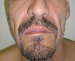
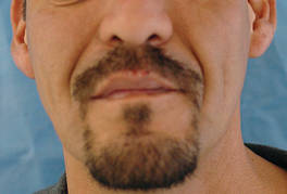
SANTA BARBARA, Calif. - Fat transfer for lipoatrophy is safe and cost effective, and provides excellent long-term results, lasting over a year in some cases, said Dr. Ilya Reyter.
Dr. Reyter, along with Dr. David Sawcer, launched a lipodystrophy clinic in the department of dermatology at the University of Southern California's Keck School of Medicine in early 2007. The clinic is dedicated to treating HIV-associated lipodystrophy and lipoatrophy using tumescent liposuction and fat transfer.

Dr. Reyter, an assistant clinical professor of dermatology at the university, also has extensive experience with fat transfer and liposuction for non-HIV cosmetic improvement, and performs the procedure in his private practice in Beverly Hills, Calif.
Lipodystrophy is marked by central fat deposition on the abdomen, dorsocervical area, salivary glands, breasts, face, and neck. "Before we started this clinic, a lot of our patients were being sent to plastic surgeons, especially for excision of lipodystrophy, particularly in the dorsocervical area," Dr. Reyter said. The dystrophic fat accumulations can be removed more simply using local anesthesia with tumescent liposuction, thereby avoiding the risks of surgery with general anesthesia, he said.
Patients are referred to the HIV lipodystrophy clinic by the HIV dermatology clinic and by other physicians working with HIV patients at the medical school. Dr. Reyter has treated numerous patients for non-HIV cosmetic fat transfer, "but in the HIV clinic, we have treated a few dozen patients," he said. "We select the patients carefully, to make sure that they are appropriate candidates. Not everyone with facial atrophy is a candidate for fat transfer, or fillers, for that matter."
He went on to describe his methods for treating lipoatrophy using fat transfer. Lipoatrophy, also know as fat wasting, particularly affects the face, and is marked by prominent zygomata, reduction of Bichat’s fat pads, sunken eyeballs, and a cachectic appearance.
HIV-associated lipoatrophy was recognized shortly after the first protease inhibitors were approved in 1995. "You can see some of these features in patients who are HIV positive but not on any medication, but it's a really small subset, only about 3% of them," said Dr. Reyter.
He said that lipoatrophy has replaced Kaposi’s sarcoma as the "scarlet letter" of HIV disease, "and it can lead to noncompliance with therapy. It has been shown that correction of the problem can lead to physical and psychological improvement. Lipoatrophy is increasing in prevalence, because HIV is becoming a chronic disease. The longer patients are on medications for their disease, the more of this we will see. The patients have nowhere else to go. As dermatologists we are the experts of skin and subcutaneous fat. If we don't treat it, who will?"
The exact causes of lipoatrophy and lipodystrophy remain unclear, but they are most likely related to multiple factors, he said, including a decrease in retinoic acid receptors, a decrease in triglyceride uptake, inhibition of mitochondrial DNA, inhibition of lipid metabolism, and prevention of adenocyte development.
For small volume areas that need treatment, Dr. Reyter prefers hyaluronic acid fillers such as Restylane and Juvéderm, but for large volume areas such as the cheeks, fillers "don't last a very long time, and they cost a lot. To me, those are prohibitive features for being widely used in HIV. It was not a cost-effective model to be doing this about every 6 months, using 10-14 syringes per side on a patient's face. It just made no sense," he said.
Other filler options include poly-L-lactic acid (Sculptra) and calcium hydroxylapatite (Radiesse), but Dr. Reyter said that he has seen patients with and without HIV develop complications including granulomas after being treated with these agents. "I don't feel as comfortable offering these particular fillers," he said, even though they are Food and Drug Administration approved. "I think it's up to physicians to evaluate whether or not a filler is something they would want to use on their own patients."
Fat transfer wins out for larger volumes, he said, if donor fat is available. "Fat is very cheap to harvest; most patients have adequate fat reserves, and it can be done safely."
In one study of 38 HIV patients who were treated with fat transfer, researchers graded the results at 1 year on a scale from 1 to 4, with 4 being excellent. The mean score reported by the patients was 3.7 and the mean score reported by the surgeon who performed the procedure was 3.2 (Plast. Reconstr. Surg. 2004;114:551-5). No serious adverse effects were observed.
In a separate study of 33 HIV patients who were treated with fat transfer and surveyed 1 year later, 93% of patients reported being satisfied (14 reported being very satisfied and 17 reported being partly satisfied). Furthermore, 81% (27 patients) reported an improved quality of life.
Three independent evaluators reported a 52% improvement in the area treated at 1 year (Arch. Dermatol. 2005;141:1220-4). No significant complications were observed.
In Dr. Reyter's clinical experience, some patients might need another fat transfer at 1 year, "but even so, a year is a significant improvement, especially for a procedure that didn't entail a lot of risk, didn’t have a lot of cost of consumables, and resulted in a benefit for a person."
In another study, researchers used computed tomography analysis and volume calculating software to evaluate the effects of fat transfer in 26 patients (18 men and 8 women aged 34-59 years) with HIV (Aesthetic Surg. J. 2008;28:380-6). The investigators observed increasing volumes of fat after 1 year, leading some to speculate that fat transfer may involve the transfer of stem cells.
"Are stem cells somehow influencing this result?" Dr. Reyter asked, adding that the data on stem cells in fat transfer are inconclusive. "Stem cells have been shown to be present in fat, and they have been shown to be transferred."
According to Dr. Reyter, his pretreatment protocol is the same as for tumescent liposuction: a CBC test; CD4 measurement; a viral load test; confirmation serum transaminases levels are not elevated; and clearance from the primary care physician.
"An important step is marking," he added. "I like to view the sites of deficit as triangles on the face. I make a topographical overlay. After we numb up the area, a lot of that numbing will distort the facial architecture. So if youdidn’t do a good marking job beforehand, your landmarks will be distorted."
For the fat transfer procedure Dr. Reyter uses small, blunt microcannulas. To harvest donor fat, he uses a straight Coleman harvesting cannula connected to a 10-cc syringe.
"I harvest on manual pressure, and then I let the fat sit to allow it to separate" he said. "There's a lot of debate as to whether or not you should spin the fat. I prefer to do nothing that would introduce trauma to the fat or to expose the fat to contamination."
He uses a 1-cc syringe attached to a blunt-tipped 18-gauge cannula to re-inject the fat, injecting 0.1-0.2 cc per pass on withdrawal.
The donor harvesting of fat takes about 20-30 minutes, allowing the fat to sit takes about 10-12 minutes, and reinjecting the fat takes about 30 minutes. "The whole procedure can take an hour to an hour and 15 minutes to do 20-30 ccs of fat per side, which I think is pretty efficient," he remarked.
Typical filling volumes are 10-20 cc for each cheek, 5-8 ccs for the temple, and 5-10 ccs for nasolabial folds. "Edema during and after fat transfer is common," said Dr. Reyter. "Because of this you have to overfill by 25%-50%. That’s where the skill comes in."
The procedures appear to change the overlying skin texture, "producing a global rejuvenation effect," he said. "It brings people back to speculating what the role of stem cells is."
To date there have been no serious complications since the lipodystrophy clinic opened its doors, said Dr. Reyter, who estimated that 25%-50% of his current clinical work involves fat transfer.
"There is a high degree of patient satisfaction at 6-12 months, and very few touch-ups are necessary," he said.
The responses from patients who have gone through the fat transfer procedure "have been overwhelmingly positive," he added. "So many patients tell us that their lives have been transformed."
For example, one patient with severe facial atrophy and longstanding unemployment was able to finally find employment after the fat transfer – "because he no longer looked so ill," Dr. Reyter said. "Another patient just wrote 'thank you for my new face. ... I look so healthy!' The doctors in the clinic regularly receive thank you notes and tokens of appreciation from the patients – in my experience, much more than we typically get in the course of providing any other medical care."
Dr. Reyter said that he had no relevant financial disclosures to make.
SANTA BARBARA, Calif. - Fat transfer for lipoatrophy is safe and cost effective, and provides excellent long-term results, lasting over a year in some cases, said Dr. Ilya Reyter.
Dr. Reyter, along with Dr. David Sawcer, launched a lipodystrophy clinic in the department of dermatology at the University of Southern California's Keck School of Medicine in early 2007. The clinic is dedicated to treating HIV-associated lipodystrophy and lipoatrophy using tumescent liposuction and fat transfer.
Dr. Reyter, an assistant clinical professor of dermatology at the university, also has extensive experience with fat transfer and liposuction for non-HIV cosmetic improvement, and performs the procedure in his private practice in Beverly Hills, Calif.
Lipodystrophy is marked by central fat deposition on the abdomen, dorsocervical area, salivary glands, breasts, face, and neck. "Before we started this clinic, a lot of our patients were being sent to plastic surgeons, especially for excision of lipodystrophy, particularly in the dorsocervical area," Dr. Reyter said. The dystrophic fat accumulations can be removed more simply using local anesthesia with tumescent liposuction, thereby avoiding the risks of surgery with general anesthesia, he said.
Patients are referred to the HIV lipodystrophy clinic by the HIV dermatology clinic and by other physicians working with HIV patients at the medical school. Dr. Reyter has treated numerous patients for non-HIV cosmetic fat transfer, "but in the HIV clinic, we have treated a few dozen patients," he said. "We select the patients carefully, to make sure that they are appropriate candidates. Not everyone with facial atrophy is a candidate for fat transfer, or fillers, for that matter."
He went on to describe his methods for treating lipoatrophy using fat transfer. Lipoatrophy, also know as fat wasting, particularly affects the face, and is marked by prominent zygomata, reduction of Bichat’s fat pads, sunken eyeballs, and a cachectic appearance.
HIV-associated lipoatrophy was recognized shortly after the first protease inhibitors were approved in 1995. "You can see some of these features in patients who are HIV positive but not on any medication, but it's a really small subset, only about 3% of them," said Dr. Reyter.
He said that lipoatrophy has replaced Kaposi’s sarcoma as the "scarlet letter" of HIV disease, "and it can lead to noncompliance with therapy. It has been shown that correction of the problem can lead to physical and psychological improvement. Lipoatrophy is increasing in prevalence, because HIV is becoming a chronic disease. The longer patients are on medications for their disease, the more of this we will see. The patients have nowhere else to go. As dermatologists we are the experts of skin and subcutaneous fat. If we don't treat it, who will?"
The exact causes of lipoatrophy and lipodystrophy remain unclear, but they are most likely related to multiple factors, he said, including a decrease in retinoic acid receptors, a decrease in triglyceride uptake, inhibition of mitochondrial DNA, inhibition of lipid metabolism, and prevention of adenocyte development.
For small volume areas that need treatment, Dr. Reyter prefers hyaluronic acid fillers such as Restylane and Juvéderm, but for large volume areas such as the cheeks, fillers "don't last a very long time, and they cost a lot. To me, those are prohibitive features for being widely used in HIV. It was not a cost-effective model to be doing this about every 6 months, using 10-14 syringes per side on a patient's face. It just made no sense," he said.
Other filler options include poly-L-lactic acid (Sculptra) and calcium hydroxylapatite (Radiesse), but Dr. Reyter said that he has seen patients with and without HIV develop complications including granulomas after being treated with these agents. "I don't feel as comfortable offering these particular fillers," he said, even though they are Food and Drug Administration approved. "I think it's up to physicians to evaluate whether or not a filler is something they would want to use on their own patients."
Fat transfer wins out for larger volumes, he said, if donor fat is available. "Fat is very cheap to harvest; most patients have adequate fat reserves, and it can be done safely."
In one study of 38 HIV patients who were treated with fat transfer, researchers graded the results at 1 year on a scale from 1 to 4, with 4 being excellent. The mean score reported by the patients was 3.7 and the mean score reported by the surgeon who performed the procedure was 3.2 (Plast. Reconstr. Surg. 2004;114:551-5). No serious adverse effects were observed.
In a separate study of 33 HIV patients who were treated with fat transfer and surveyed 1 year later, 93% of patients reported being satisfied (14 reported being very satisfied and 17 reported being partly satisfied). Furthermore, 81% (27 patients) reported an improved quality of life.
Three independent evaluators reported a 52% improvement in the area treated at 1 year (Arch. Dermatol. 2005;141:1220-4). No significant complications were observed.
In Dr. Reyter's clinical experience, some patients might need another fat transfer at 1 year, "but even so, a year is a significant improvement, especially for a procedure that didn't entail a lot of risk, didn’t have a lot of cost of consumables, and resulted in a benefit for a person."
In another study, researchers used computed tomography analysis and volume calculating software to evaluate the effects of fat transfer in 26 patients (18 men and 8 women aged 34-59 years) with HIV (Aesthetic Surg. J. 2008;28:380-6). The investigators observed increasing volumes of fat after 1 year, leading some to speculate that fat transfer may involve the transfer of stem cells.
"Are stem cells somehow influencing this result?" Dr. Reyter asked, adding that the data on stem cells in fat transfer are inconclusive. "Stem cells have been shown to be present in fat, and they have been shown to be transferred."
According to Dr. Reyter, his pretreatment protocol is the same as for tumescent liposuction: a CBC test; CD4 measurement; a viral load test; confirmation serum transaminases levels are not elevated; and clearance from the primary care physician.
"An important step is marking," he added. "I like to view the sites of deficit as triangles on the face. I make a topographical overlay. After we numb up the area, a lot of that numbing will distort the facial architecture. So if youdidn’t do a good marking job beforehand, your landmarks will be distorted."
For the fat transfer procedure Dr. Reyter uses small, blunt microcannulas. To harvest donor fat, he uses a straight Coleman harvesting cannula connected to a 10-cc syringe.
"I harvest on manual pressure, and then I let the fat sit to allow it to separate" he said. "There's a lot of debate as to whether or not you should spin the fat. I prefer to do nothing that would introduce trauma to the fat or to expose the fat to contamination."
He uses a 1-cc syringe attached to a blunt-tipped 18-gauge cannula to re-inject the fat, injecting 0.1-0.2 cc per pass on withdrawal.
The donor harvesting of fat takes about 20-30 minutes, allowing the fat to sit takes about 10-12 minutes, and reinjecting the fat takes about 30 minutes. "The whole procedure can take an hour to an hour and 15 minutes to do 20-30 ccs of fat per side, which I think is pretty efficient," he remarked.
Typical filling volumes are 10-20 cc for each cheek, 5-8 ccs for the temple, and 5-10 ccs for nasolabial folds. "Edema during and after fat transfer is common," said Dr. Reyter. "Because of this you have to overfill by 25%-50%. That’s where the skill comes in."
The procedures appear to change the overlying skin texture, "producing a global rejuvenation effect," he said. "It brings people back to speculating what the role of stem cells is."
To date there have been no serious complications since the lipodystrophy clinic opened its doors, said Dr. Reyter, who estimated that 25%-50% of his current clinical work involves fat transfer.
"There is a high degree of patient satisfaction at 6-12 months, and very few touch-ups are necessary," he said.
The responses from patients who have gone through the fat transfer procedure "have been overwhelmingly positive," he added. "So many patients tell us that their lives have been transformed."
For example, one patient with severe facial atrophy and longstanding unemployment was able to finally find employment after the fat transfer – "because he no longer looked so ill," Dr. Reyter said. "Another patient just wrote 'thank you for my new face. ... I look so healthy!' The doctors in the clinic regularly receive thank you notes and tokens of appreciation from the patients – in my experience, much more than we typically get in the course of providing any other medical care."
Dr. Reyter said that he had no relevant financial disclosures to make.
EXPERT ANALYSIS FROM THE ANNUAL MEETING OF THE CALIFORNIA SOCIETY OF DERMATOLOGY AND DERMATOLOGIC SURGERY
Never Say Never: Surgical Errors Remain a Concern
The frequency of surgical complications involving a wrong site or wrong patient remains high, even in the era of the Universal Protocol.
The Joint Commission introduced the Universal Protocol to ensure the correct patient, site, and procedure. Although it became effective July 1, 2004, there still exists a lack of data about the true incidence of wrong-patient and wrong-site operations, called "never events," according to new research reported in the October issue of Archives of Surgery.
To determine the frequency, root causes, and outcomes of these never events, Dr. Philip F. Stahel of Denver Health Medical Center and the University of Colorado School of Medicine, and colleagues performed a retrospective analysis of the Colorado Physician Insurance Company's (COPIC's) comprehensive database (Arch. Surg. 2010;145:978-84).
Dr. Stahel and his colleagues screened 27,370 physician self-reported adverse occurrences between Jan. 1, 2002, and June 1, 2008. The researchers initially found 119 wrong-site and 29 wrong-patient procedures, but eliminated cases they could not classify as being a factual wrong site or wrong patient. The final analysis consisted of 107 wrong-site and 25 wrong-patient procedures.
Analysis of root causes found errors in:
Diagnosis, a root cause for 14 (56.0%) wrong-patient and 13 (12.1%) wrong-site procedures.
Communication, 25 (100%) wrong-patient and 52 (48.6%) wrong-site procedures.
Judgment, 2 (8.0%) wrong-patient and 91 (85.0%) wrong-site procedures.
Treatment, 22 (88.0%) wrong-patient and 9 (92.5%) wrong-site procedures.
In addition, system issues were a root cause in 21 (84.0%) wrong-patient procedures and 78 (72.9%) wrong-site procedures. This category included time-out not being performed in 77 (72%) wrong-site cases.
Wrong-patient cases often were due to a mix-up of patients’ medical records, radiographs, and laboratory or biopsy samples, as well as errors in communication.
Next, the researchers looked at outcomes, namely:
Death, which occurred in 1 patient (0.9%) secondary to a wrong-site procedure.
Significant harm, which occurred in 5 (20%) wrong-patient and 38 (35.5%) wrong-site cases.
Minimal harm or functional impairment, which occurred in 8 (32%) wrong-patient and 65 (60.7%) wrong-site cases.
No-harm event, which occurred in 9 (36%) wrong-patient and 3 (2.8%) wrong-site cases.
The most frequent specialties involved in wrong-patient procedures were internal medicine (24.0% of cases) as well as family or general practice, pathology, urology, obstetrics-gynecology, and pediatrics (8.0% each). The most frequent specialties involved in wrong-site occurrences were orthopedic surgery (22.4% of cases), general surgery (16.8%), and anesthesiology (12.1%).
Overall, nonsurgical specialties were involved in 14 (48.3%) wrong-patient and 29 (27.1%) wrong-site cases.
"The findings from the present study emphasize a continuing and concerning occurrence of wrong-site and wrong-patient procedures in the current era of the Universal Protocol, leading to frequent patient harm and, rarely, patient death," the researchers said. "Shockingly, nonsurgical disciplines equally contribute to patient injuries related to wrong-site procedures."
The researchers believe these findings warrant expansion of the Universal Protocol to nonsurgical specialties.
Limitations of the study include the restricted coverage of the COPIC database to about 6,000 physicians in Colorado; the potential for subjective bias in determining root causes; and the designation of inadequate planning for the procedure, which represents a generic category.
Coauthors on the research analysis reported the following conflicts: Dr.Ted J. Clarke is the chief executive officer of Colorado Physician Insurance Company (COPIC); Dr. Jeffrey Varnell and Dr. Alan Lembitz are employed by COPIC; and Dr. Michael S. Victoroff and Dr. Dennis J. Boyle are consultants for COPIC.
Although compliance with the Universal Protocol is important, "it is not the magic wand of Merlin." Consider: The Universal Protocol has been in place since 2004, yet Dr. Philip F. Stahel and colleagues found that preventable errors, or "never events," exist at alarming rates. Further, the number of wrong-site procedures this study cites more likely reflect the number of errors reported rather than the actual rates of events. So, the number of wrong-site procedures is probably much higher than reflected here.
Perhaps a more accurate measurement comes from the complication rates and safety culture scores under the National Surgical Quality Improvement Program, or NSQIP. Safety culture scores reflect the comfort level of hospital employees about speaking up about safety concerns. To improve public reporting and benchmarking, hospitals should be required to publicly report their NSQIP outcomes and culture scores.
Finally, the Universal Protocol, while important, does not relieve hospital systems from emphasizing individual responsibility in preventing surgical errors.
Martin A. Makary, M.D., M.P.H., is with the department of surgery at Johns Hopkins University, Baltimore. His remarks were made in an accompanying commentary to the article (Arch. Surg. 2010;145:984). He had no conflicts to disclose.
The frequency of surgical complications involving a wrong site or wrong patient remains high, even in the era of the Universal Protocol.
The Joint Commission introduced the Universal Protocol to ensure the correct patient, site, and procedure. Although it became effective July 1, 2004, there still exists a lack of data about the true incidence of wrong-patient and wrong-site operations, called "never events," according to new research reported in the October issue of Archives of Surgery.
To determine the frequency, root causes, and outcomes of these never events, Dr. Philip F. Stahel of Denver Health Medical Center and the University of Colorado School of Medicine, and colleagues performed a retrospective analysis of the Colorado Physician Insurance Company's (COPIC's) comprehensive database (Arch. Surg. 2010;145:978-84).
Dr. Stahel and his colleagues screened 27,370 physician self-reported adverse occurrences between Jan. 1, 2002, and June 1, 2008. The researchers initially found 119 wrong-site and 29 wrong-patient procedures, but eliminated cases they could not classify as being a factual wrong site or wrong patient. The final analysis consisted of 107 wrong-site and 25 wrong-patient procedures.
Analysis of root causes found errors in:
Diagnosis, a root cause for 14 (56.0%) wrong-patient and 13 (12.1%) wrong-site procedures.
Communication, 25 (100%) wrong-patient and 52 (48.6%) wrong-site procedures.
Judgment, 2 (8.0%) wrong-patient and 91 (85.0%) wrong-site procedures.
Treatment, 22 (88.0%) wrong-patient and 9 (92.5%) wrong-site procedures.
In addition, system issues were a root cause in 21 (84.0%) wrong-patient procedures and 78 (72.9%) wrong-site procedures. This category included time-out not being performed in 77 (72%) wrong-site cases.
Wrong-patient cases often were due to a mix-up of patients’ medical records, radiographs, and laboratory or biopsy samples, as well as errors in communication.
Next, the researchers looked at outcomes, namely:
Death, which occurred in 1 patient (0.9%) secondary to a wrong-site procedure.
Significant harm, which occurred in 5 (20%) wrong-patient and 38 (35.5%) wrong-site cases.
Minimal harm or functional impairment, which occurred in 8 (32%) wrong-patient and 65 (60.7%) wrong-site cases.
No-harm event, which occurred in 9 (36%) wrong-patient and 3 (2.8%) wrong-site cases.
The most frequent specialties involved in wrong-patient procedures were internal medicine (24.0% of cases) as well as family or general practice, pathology, urology, obstetrics-gynecology, and pediatrics (8.0% each). The most frequent specialties involved in wrong-site occurrences were orthopedic surgery (22.4% of cases), general surgery (16.8%), and anesthesiology (12.1%).
Overall, nonsurgical specialties were involved in 14 (48.3%) wrong-patient and 29 (27.1%) wrong-site cases.
"The findings from the present study emphasize a continuing and concerning occurrence of wrong-site and wrong-patient procedures in the current era of the Universal Protocol, leading to frequent patient harm and, rarely, patient death," the researchers said. "Shockingly, nonsurgical disciplines equally contribute to patient injuries related to wrong-site procedures."
The researchers believe these findings warrant expansion of the Universal Protocol to nonsurgical specialties.
Limitations of the study include the restricted coverage of the COPIC database to about 6,000 physicians in Colorado; the potential for subjective bias in determining root causes; and the designation of inadequate planning for the procedure, which represents a generic category.
Coauthors on the research analysis reported the following conflicts: Dr.Ted J. Clarke is the chief executive officer of Colorado Physician Insurance Company (COPIC); Dr. Jeffrey Varnell and Dr. Alan Lembitz are employed by COPIC; and Dr. Michael S. Victoroff and Dr. Dennis J. Boyle are consultants for COPIC.
from archives of surgery
Major Finding: Between Jan. 1, 2002, and June 1, 2008, there were 107 wrong-site and 25 wrong-patient surgical procedures.
Data Source: Retrospective analysis of a prospective database of 27,370 physician self-reported adverse events.
Disclosures: Coauthors on the research analysis reported the following conflicts: Dr.Ted J. Clarke is the chief executive officer of Colorado Physician Insurance Company (COPIC); Dr. Jeffrey Varnell and Dr. Alan Lembitz are employed by COPIC; and Dr. Michael S. Victoroff and Dr. Dennis J. Boyle are consultants for COPIC.
Dermatologic Surgery Site Specifics Point to Antibiotic Choices
CHICAGO – Infections from dermatologic surgery are rare, but possible, especially in high-risk patients, such as the immunosuppressed and those with joint or cardiac prostheses.
"Data from the four largest studies on this topic suggest an infective bacteremia of about 1.9% associated with all derm surgeries, which is actually less than you see with normal daily activities, like brushing your teeth and flossing," Christopher C. Gasbarre, D.O. said at the meeting . "Add to that the fact that the American Heart Association guidelines note that antibiotic prophylaxis doesn’t decrease the risk of infection to 0%," and the decision of whether to give the drugs becomes problematic.
Patients who are at high risk of postoperative infections, however, should receive preoperative or immediately postoperative antibiotics, said Dr. Gasbarre, a dermatologist at the Cleveland Clinic. "It’s important to remember that infective endocarditis and prosthetic joint infections secondary to dermatologic surgery are a well-known phenomenon, and a distant skin infection is the leading cause of prosthetic joint infections."
The American Heart Association reviewed and updated its surgical antibiotic guidelines in 2007, "making a significant shift in focus compared to their previous guidelines," Dr. Gasbarre said. The guidelines now recommend prophylaxis for patients with any prosthetic implanted material including cardiac valve replacements; cardiac patients with valvular disease; those with a history of infective pericarditis; anyone with unrepaired cyanotic congenital heart disease or a defect repaired within the past 6 months; and any congenital heart disease with a residual defect.
The American Dental Association updated its surgical antibiotic guidelines recently as well. "The guidelines recommend prophylaxis for high-risk patients undergoing oral procedures with a high risk of bleeding," he said.
Guidelines issued by the American Association of Orthopaedic Surgeons identify similar high-risk groups that should receive presurgical antibiotic therapy, including anyone who is less than 2 years out from a joint replacement, who is immunosuppressed, and who has hemophilia. "In dermatologic surgery, there are no studies on antibiotic prophylaxis, so we have to make do with these other recommendations," Dr. Gasbarre said.
Preventing wound infections is a major goal of surgical antibiotic treatment, he said. “In the dermatologic literature, the numbers range from 0.7% to 8%, depending on what study you’re looking at.”
Surgical site is a major consideration in anticipating postsurgical infections. "Ears, the nose, and lower leg below the knee have a greater risk of infection. Flaps and grafts have a higher risk, especially flaps on the nose. And a major cosmetic reconstruction that results in necrosis and scarring should also be a consideration."
The Mayo Clinic’s 2008 surgical antibiotic prophylaxis guidelines suggest antibiotics for dermatologic surgeries of the lower extremities, groin, and perineum; wedge excisions of the lip or ear; and other procedures with reported infection rates of at least 5%. Again, patients with immunosuppression require specific attention, as do those with inflammatory skin diseases, “who may carry a higher staphylococcus load on the skin,” Dr. Gasbarre said.
The literature suggests other high-risk groups might be those who undergo several procedures in 1 day, have high-tension wound closures, and have surgery on the hands, he added.
"The one thing that gets the most debate is whether to give antibiotics for a wound that’s exposed more than 3 hours," Dr. Gasbarre said. "I often do, although there are no guidelines. I take into consideration comorbidities, but certainly any surgery of the nasal mucosa and long procedures are worthy of consideration."
Culture is important for high-risk patients. "Culture for both community- and hospital-acquired methicillin-resistant Staphylococcus aureus and jump on those infections and eradicate them as quickly as possible."
Timing is another consideration. "Ideally we give antibiotics 30-60 minutes before the procedure to get enough antibiotic in the coagulum. But the new AHA guidelines say you can give them up to 2 hours after the procedure if not given before."
Select antibiotics carefully. "Beta hemolytic streptococcus and S. aureus are most likely to cause endocarditis from skin sites. You see a lot of gram-negative infections in the lower extremities and perineum. Pseudomonas can show up in the ear, but more than 90% of cultured ear isolates are staph and strep, not pseudomonas."
If the patient is not allergic to penicillin, Dr. Gasbarre prefers 2 g oral cephalexin or treatment with dicloxacillin or amoxicillin. "For oral mucosa, amoxicillin is the drug of choice. For patients who are allergic, I stick with 600 mg of clindamycin or 500 mg of azithromycin."
Ciprofloxacin, trimethoprim, and levofloxacin are choice for the ear, groin, and lower extremities.
"If patients can’t take anything orally, ceftriaxone 1 g given intramuscularly or intravenously is acceptable, with or without gentamicin for gram-negative coverage," Dr. Gasbarre said.
Dr. Gasbarre said he had no relevant financial conflicts.
FROM THE AMERICAN ACADEMY OF DERMATOLOGY’S ACADEMY 2010 MEETING
Skin of Color: Postinflammatory Hypopigmentation and Hypopigmented Scars
Hypopigmentation is a result of inflammatory conditions or trauma in patients with darker skin. While there are many treatments for hyperpigmentation, there are fewer options for hypo.
Some of the clearly defined options that exist for hypopigmentation include using anti-inflammatories for conditions such as vitiligo and pityriasis alba (topical corticosteroids, tacrolimus, or pimecrolimus).
For hypopigmentation from tinea versicolor, repigmentation after treatment with antifungals does occur although it can be delayed for months. But for postinflammatory hypopigmentation or hypopigmented scars, what can be done?
Pharmaceutical treatments
Puvasol, topical psoralen, and ultraviolet light (natural sunlight or UVA phototherapy), may help speed the repigmentation of hypopigmented patches, particularly in cases caused by laser therapy.
In patients with postinflammatory hypopigmentation following laser hair removal, the repigmentation may also recur on its own if the initial insult from the laser was superficial, although repigmentation can take months. Topical psoralen can be applied to affected areas, and should be done 20-30 minutes before the patient sits in direct sunlight (for just a few minutes). Caution must be taken to avoid blistering and sunburn.
V-Tar, a 30% standardized, water-soluble, coal tar topical, has been used successfully in many patients with vitiligo and other hypopigmentary disorders. It is available by prescription, and is applied once daily (Skin & Allergy News, March 2002, pg. 39).
Laser Treatments
The 308-nm excimer laser has been studied as an effective treatment for localized hypopigmented scars and striae alba. In one study, minimal erythema dose tests were performed for 31 patients at 100, 150, 200, 250, 300, and 350 mJ/cm2. Each patient was then treated with his or her minimal erythema dose minus 50 mJ/cm2. Subsequent treatments were performed biweekly until 50%-75% pigment correction was achieved, and every 2 weeks thereafter for a maximum of 10 treatments, a 75% increase in colorimetric measurements, or 100% visual pigment correction. However, maintenance treatment every 1-4 months was required to sustain cosmetic benefit (Arch. Dermatol. 2004;140:955-60).
In another study, a 1550-nm Fraxel laser was used to treat seven patients with hypopigmented scars on the face. Energy settings ranged from 7 mJ to 20 mJ and a total density of 1,000-2,500 microthermal zones/cm2. Six patients experienced 51%-75% improvement in hypopigmentation after two to four consecutive treatments spaced 4 weeks apart. One patient had a 26%-50% improvement (Dermatol. Surg. 2007;33:289-94).
Fractionated resurfacing is also helpful in evening out skin tone when there is a mixture of hypo- and hyperpigmentation from chronic UV exposure or poikiloderma. In addition, cultured epithelial autografts have been used to treat hypopigmentation secondary to burns.
Dyspigmentation is a common concern among patients with skin of color. Hypopigmentation caused by disease or scarring can be ameliorated by treatments using psoralens, tar, and lasers; however, more data and additional treatment options are needed to address the common and often difficult-to-treat dyschromia we see in this patient population.