User login
Ablative Laser Effectively Remodeled Scar Collagen
KISSIMMEE, FLA. – Treatment with a 2,940 Er:YAG ablative fractional laser led to a clinically significant reduction of third-degree burn scars in a prospective study of 11 patients.
Clinical and histologic findings from the laser study provide important clues as to the mechanism of action and appropriate treatment intervals, Dr. Jill S. Waibel reported at the meeting. Scars from fire or thermal injury are among the worst that are seen in clinical practice, and although fractional laser therapy is emerging as the preferred treatment for these injuries, a better understanding of the optimal laser wavelengths, clinical response patterns, scar tissue response, and histologic changes will improve outcomes.
Patients in this study underwent three ablative treatments at 4-week intervals, and – based on blinded evaluation by independent investigators – were found to have an overall modified Manchester score of 2.3 out of 3 points, indicating moderate to excellent improvement. The scores for dyschromia, hypertrophy reduction, vascularity, and scar texture improvement were 1.7, 1.9, 2.0, and 2.0, respectively, indicating mild to moderate improvement, said Dr. Waibel, a physician in private practice in Miami.
The patients, who had third-degree burn scars over an average of 80% of their body, were treated with one pass at depths ranging from 400 to 800 microns, depending on scar thickness and density.
Biopsies were performed at baseline and at 3 months after the final treatment. On biopsies taken at baseline, the scars were noted to have thickened, homogenized, dystrophic collagen structures; and on biopsies taken 3 months after the final treatment, a decrease in these dystrophic fibers was seen, she said.
Dr. Waibel hypothesized that scar improvement resulted from the complete replacement of ablated zones by newly synthesized collagen and that the collagen remodeling led to more normal-appearing skin.
She noted that the erbium laser is "very powerful" and caused a great deal of erythema in two patients, leading her to "turn the power down" over the course of the study. Although no worsening of scars was seen, the increased vascularity in the two patients suggested that intervals longer than 4 weeks between treatments – perhaps to between 2 and 3 months - may be warranted.
Although additional study is needed, the findings suggest that the 2,940 Er:YAG laser is effective for clinically improving burn scars through vaporization and by inducing an organized wound-healing response; this histologic healing response may lead to the improvement that is seen clinically, she concluded.
This study was funded by Sciton. Dr. Waibel said she had no other relevant financial disclosures.
FROM THE ANNUAL MEETING OF THE AMERICAN SOCIETY FOR LASER MEDICINE AND SURGERY
Major Finding: Based on blinded evaluation by independent investigators, patients had an overall modified Manchester score of 2.27 out of 3 points, indicating moderate to excellent improvement.
Data Source: A prospective study of 11 patients with third-degree burn scars who underwent three ablative treatments at 4-week intervals.
Disclosures: This study was funded by Sciton. Dr. Waibel said she had no other relevant financial disclosures.
Repeat Treatment, Massage Improve Abdominal Cryolipolysis Results
KISSIMMEE, FLA. – Two new studies have found safe and effective means for improving the results of lower abdomen cryolipolysis.
In the first study, 20 patients underwent one lower abdomen cryolipolysis treatment for 60 minutes, followed by a second treatment 2 months later. A consistent and discernable decrease in the thickness of the fat layer was noted in an independent photo review 4 months following the initial treatment.
Blinded independent reviewers, including two plastic surgeons and one dermatologist, compared the follow-up photos with those taken at baseline, and correctly identified the posttreatment image 86% of the time, Dr. Flor Mayoral reported at the annual meeting of the American Society for Laser Medicine and Surgery.
"Massage appears to be a safe, effective way to further reduce the fat layer following a cryolipolysis procedure."
Patient satisfaction was also high, with 75% of study participants reporting a "somewhat to very visible" change in the treated region, said Dr. Mayoral, a dermatologist in private practice in Coral Gables, Fla.
"Patients didn’t have the advantage of looking at their own photographs to determine whether they looked better or not," she noted.
Study patients were treated using CoolSculpting, by Zeltiq Aesthetics, at a cooling intensity factor of 42. No adverse events were reported.
Additional treatment following initial cryolipolysis of the abdomen appeared to be safe and effective, Dr. Mayoral concluded.
In a separate single-center study, Dr. Gerald Boey, a dermatologist in private practice in Vancouver, B.C., found that performing manual massage to the abdominal area following cryolipolysis led to improved outcomes, compared with cryolipolysis without the manual massage.
Massage was performed on one randomly selected side of the abdomen immediately following treatment of the entire lower abdomen in 10 patients, and outcomes on each side were compared using photos taken at baseline and at the 2-month follow-up. Improvement occurred on both sides, but greater improvement was noted on the massaged side in all patients, and ultrasound measurement confirmed that the fat reduction on the massaged side was, on average, 68% greater than on the control side, Dr. Boey said.
In a separate set of seven patients, biopsies performed at baseline and at 3, 8, 13, 30, 60, and 120 days following massage demonstrated that massage was not associated with any evidence of necrosis or fibrosis.
As in Dr. Mayoral’s study, cryolipolysis in this study was performed using CoolSculpting at a cooling intensity factor of 42 for 60 minutes. Massage was performed for 1 minute using a vigorous kneading motion, followed by 1 minute using a circular motion with the pads of the fingers.
"Massage appears to be a safe, effective way to further reduce the fat layer following a cryolipolysis procedure," Dr. Boey said. As for why the massage helps, one possible explanation is that manipulation of the fat while it is in the crystalline state induced by cryolipolysis may cause an accelerated reperfusion injury.
Dr. Mayoral reported that her study was funded by Zeltiq Aesthetics. She had no other disclosures to report. Dr. Boey had no disclosures to report.
FROM THE ANNUAL MEETING OF THE AMERICAN SOCIETY FOR LASER MEDICINE AND SURGERY
Photodamage Effectively Treated With Quasi-Ablative Approach
KISSIMMEE, FLA. – The nonablative 1540-nm fractional laser can be safely converted to a quasi-ablative device for treating facial actinic keratoses and photodamage, according to Dr. Moshe Lapidoth.
By slightly modifying technique, a 50%-75% improvement in actinic keratoses and photodamage was noted in 17 patients who underwent two to three treatments at 4-week intervals, Dr. Lapidoth reported at the annual meeting of the American Society for Laser Medicine and Surgery. The laser was used in noncontact mode hovering 5-10 mm above the skin rather than in contact mode.
The patients had actinic keratoses and photodamage requiring ablative treatment of the epidermis. Treatment was applied using a fluence of 75 J/cm2, a 15-ms pulse duration, and a 10-mm spot size. Two blinded assessors and the participants evaluated clinical improvement at 3 months after the final treatment using a quartile grading scale (Lasers Med. Sci. 2012 April 27 [doi: 10.1007/s10103-012-1103-6])
A score of 0 was associated with no improvement; a score of 1 with 1%-25% improvement, a score of 2 with 26%-50% improvement, a score of 3 with 51%-75% improvement, and a score of 4 with 76%-100% improvement. The mean score for actinic keratoses was 3.4, and for skin appearance was 3.2, said Dr. Lapidoth, head of the laser department at Rabin Medical Center in Israel.
Side effects after each treatment included erythema, mild edema, erosion in two patients, and mild desquamation, but no scarring or postinflammatory pigmentary changes occurred, he said.
Although more current fractional laser devices are designed to be either ablative or nonablative, the 1540-nm fractional laser was designed to be nonablative.
"The question is, ‘Can you take a 1540-nm laser, which is totally nonablative, and turn it to be ablative or quasi-ablative?’ and the answer is, ‘yes,’ Dr. Lapidoth said.
This treatment approach can be used in conjunction with contact mode, he added, explaining that the nonablative contact mode can be used first to target the dermis for treatment of fine wrinkles and scars, followed by the ablative noncontact mode to treat actinic keratoses and resistant lentigines and other lesions.
Dr. Lapidoth reported having no disclosures.
FROM THE ANNUAL MEETING OF THE AMERICAN SOCIETY FOR LASER MEDICINE AND SURGERY
Major Finding: A quasi-ablative technique using a nonablative 1540-nm fractional laser resulted in a 50%-75% improvement in actinic keratoses and skin appearance in 17 patients.
Data Source: This was a prospective study of 17 patients treated.
Disclosures: Dr. Lapidoth reported having no disclosures.
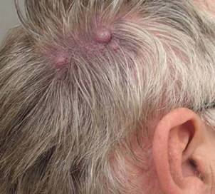
Port Wine Stains: Hypertrophy Risk Rises With Age
KISSIMMEE, FLA. – Hypertrophy of port wine stains is more common than not in the majority of patients older than age 50 with the vascular anomaly, and is most often associated with red and purple lesions, according to findings from a retrospective patient-questionnaire study.
"By the age of 50, over 70% had hypertrophy of their port wine stain," said Dr. Anne Margreet van Drooge of the Netherlands Institute of Pigment Disorders at the University of Amsterdam.
Hypertrophy is known to be fairly common in port wine stains, but data are lacking on its pathogenesis and development. In this study, the incidence of hypertrophy in port wine stains was about 20% in all patients, and both color and hypertrophy were frequently reported as reasons for seeking treatment.
Only 4% of all hypertrophy cases were associated with pink lesions, Dr. van Drooge said at the annual meeting of the American Society for Laser Medicine and Surgery.
Of the 335 patients included in the review, 68 had hypertrophy, classified as thickening, nodular, or both. Cases were divided almost equally into age groups of under 18 years, 18-30 years, and greater than 30 years. The median age of those with hypertrophic port wine stains was 50 years, and the median age at the time of hypertrophic development was 31 years, although the ages of onset of nodular hypertrophy and diffuse thickening were 39 years and 12 years, respectively. Nodular hypertrophy was rarely seen in those younger than 18 years.
In all study patients, most of the port wine stains were on the face. Half were red in color, and 44% were purple. No differences were seen between those with and without hypertrophy with respect to gender, lesion size, or lesion location, she noted.
Patients included in the study had been referred to a single outpatient clinic between 2005 and 2009. Medical records and photographs were examined to identify and characterize hypertrophic port wine stains.
The findings of different ages at onset for different types of hypertrophy suggest that unique pathomechanics may exist for each type, Dr. van Drooge said. "There is definitely a correlation between the color of the port wine stain and the risk for the development of hypertrophy. In a multivariate analysis of all patient characteristics, we found that both darker color and older age were significantly associated with hypertrophy, independent from each other."
Improved understanding of hypertrophy in port wine stains is important given the adverse psychosocial effects these lesions can have on patients; in this study, 5% of patients said they avoided social interaction because of their port wine stain, she noted, adding that the findings have implications for treatment of port wine stains.
Although this study provided no direct support for the notion that treatment may prevent hypertrophy, some studies have suggested that it may, adding that treatment guidelines should be adapted to include more emphasis on hypertrophy and its prevention, keeping in mind the findings of this study with respect to age and lesion color.
"However, I don’t think it is possible to treat only darker port wine stains in the effort to prevent hypertrophy, since pink port wine stains – particularly in younger patients – eventually can darken, and with that, the risk for hypertrophy increases," she noted.
Dr. van Drooge had no disclosures to report.
FROM THE ANNUAL MEETING OF THE AMERICAN SOCIETY FOR LASER MEDICINE AND SURGERY
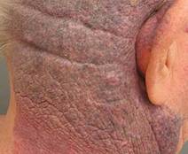
Optimized Pulsed Light 'Impressively' Clears Port Wine Stains
KISSIMMEE, FLA. – Optimized intense pulsed light was found to safely enable effective clearance of port wine stains and capillary malformations in 16 patients.
The outcomes were so impressive in two patients, who achieved 80-100% improvement after only one treatment with this "new class of filtered IPL," that additional planned treatments were canceled, Dr. Maurice Adatto reported at the annual meeting of the American Society for Laser Medicine and Surgery.
The 16 study patients from two centers had Fitzpatrick skin types II-IV and a mean age of 40 years (range, 11-71 years). All patients experienced improvement with one to four treatments, with 10 of the 16 patients achieving greater than 50% improvement, said Dr. Adatto, a dermatologist in private practice in Geneva, Switzerland.
The 10 females and 6 males had port wine stains of the face, neck, trunk, or extremities, with the exception of two patients who had capillary malformations.
Treatments were applied using Palomar’s MaxG IPL system with a spot size of 10 x 15 mm. Patients were treated using either one pass at a fluence of 50 J/cm2 and a 10-ms pulse duration, or two passes, with the first pass at 34-36 J/cm2 and a 10-ms pulse duration and the second at 20-28 J/cm2 and a 5-ms pulse duration.
Outcomes were assessed on the basis of clinical images generated using Miravex’s Antera 3D system to evaluate hemoglobin clearance. Photos were taken between 48 and 96 hours after each treatment and also at 2 months after each treatment, Dr. Adatto said.
The only reported side effects were limited or slight transitory purpura for 3-5 days, and mild to moderate edema for 1-3 days.
Lasers are more often used to treat port wine stains, but the higher peak power and the short pulse widths available with the new optimized pulsed light technology appear to allow for safe and effective clearance, Dr. Adatto said. Further optimization of the parameters and safety profile of the optimized pulsed light device is possible, and another study of its use in pediatric patients is currently underway, he noted.
Dr. Adatto disclosed that the equipment from Palomar used in the study was purchased at a discount. He had no other disclosures to report.
FROM THE ANNUAL MEETING OF THE AMERICAN SOCIETY FOR LASER MEDICINE AND SURGERY
Photoepilation System Painlessly Delivers Results
KISSIMMEE, FLA. – A combined 800-nm diode laser and vacuum system for photoepilation resulted in high patient satisfaction and was safe and virtually painless for more than 2,000 treated patients.
Photoepilation is among the most popular aesthetic treatments worldwide, and is the second most popular aesthetic treatment in Latin America, where most patients have darker skin types that can be prone to greater risk of adverse outcomes, Dr. Rafael Nunes said at the annual meeting of the American Society for Laser Medicine and Surgery. On the basis of this study, however, the LightSheer Duet laser system appears to be a safe method for successful photoepilation even in darker-skinned patients.
Of the 2,158 study patients, 1,845 were women and 313 were men; they had a mean age of 36 years (range, 18-63 years) and skin types I-IV. Photoepilation was performed on various body and facial areas at one of four participating laser centers in Brazil over an 18-month period, with a mean of six treatments every 28-45 days. The investigators treated patients with a Lumenis 800-nm LightSheer Duet diode laser and vacuum-assist technology at a fluence of 6-12 J/cm2, a spot size of 22 x 35 mm, a pulse width of 30-100 ms, and a single pass, said Dr. Nunes, a plastic surgeon in private practice in Rio de Janeiro.
At the 3- to 6-month follow-up, after completion of 4-10 treatment sessions, 92% of patients rated their outcomes as "good" or "great."
The system requires no pretreatment ice packs, creams, or anesthesia, and the vacuum system should be used only on the low setting to prevent injury – particularly in areas where there is skin laxity.
No permanent or severe adverse effects occurred in any of the patients. The only adverse effect was transient erythema in six patients (0.3%). Treatment tolerance was also impressive, with patients experiencing only a small sensation of heat at the treatment site, and "no discomfort whatsoever" following treatment, Dr. Nunes said.
Dr. Nunes is a speaker or consultant for Allergan, Alma Lasers, Q-MED (Galderma), Lumenis, MedixSysteme, Palomar, and Solta Medical.
FROM THE ANNUAL MEETING OF THE AMERICAN SOCIETY FOR LASER MEDICINE AND SURGERY
Red Light for Acne
Several light devices based on red light–emitting diodes (LEDs) have made their way to the market in recent years. Although red light is not truly a cosmeceutical, it is a significant emerging adjuvant therapy that, like blue light, has been studied in comparison to and in conjunction with topical options primarily to treat acne. Of course, acne is the most common skin disorder prompting visits to the dermatologist, with an estimated 85% of adolescents affected, many into adulthood (J. Am. Acad. Dermatol. 2008;58:56-9).
This discussion will consider red light when used alone and when used in combination with blue light. Blue light is the most effective light to target Propionibacterium acnes (specifically at wavelengths of 407-420 nm). However, many devices are utilizing red light because it has a purported anti-inflammatory effect and penetrates deeper into the skin (Dermatol. Ther. 2005;18:253-66).
Erythrasma
Darras-Vercambre et al. evaluated the effects of red light for the treatment of erythrasma (a superficial skin infection provoked by Corynebacterium minutissimum) in 13 patients. One treatment (80 J/cm2) by red light (broadband, peak at 635 nm) without exogenous photosensitizing molecules was administered to each subject. Therapy was effective and well tolerated, with three patients experiencing complete recovery, and significant reduction of lesions in most other cases. The authors noted that the key to their study, given the absence of an exogenous photosensitizing agent, was capitalizing on the presence of porphyrins in the lesions. They concluded that the use of red light alone for this localized infection is easy and inexpensive, but that an optimal method has not yet been established (Photodermatol. Photoimmunol. Photomed. 2006;22:153-6).
Acne
In 2007, Na and Suh evaluated the efficacy of red light phototherapy with a portable device in 28 volunteers with mild to moderate acne in a split-face randomized trial. Phototherapy was performed twice daily for 15 minutes for a total of 8 weeks to one side of the face. The investigators concluded that red light phototherapy alone is an effective therapeutic option for acne, as they noted significant reductions in noninflammatory and inflammatory lesion counts on the treated side versus the untreated side, a drop from 3.9 to 1.9 in the visual analog scale on the treatment side, and significant disparities between the treatment and control sides after 8 weeks (Dermatol. Surg. 2007;33:1228-33, discussion 1233).
A 2006 article in the British Journal of Dermatology reported good clinical results from acne treatment with photodynamic therapy (PDT) using methyl aminolevulinate (MAL) and red light, but there were adverse side effects that prompted 7 of 19 subjects to discontinue the study (Br. J. Dermatol. 2006;154:969-76). In response to the study, Mavilia et al. wrote a letter to the journal acknowledging their more effective combination therapy using a lower concentration of MAL and low doses of red light. All 16 patients completed the study, in which the count of inflammatory lesions fell an average of 66% with mild but tolerable side effects, including a subtle sensation of heat, then minimal erythema during the procedure and slight scaling that began 3 days after treatment (Br. J. Dermatol. 2007;157:810-1).
In a small study of patients with moderate facial acne, Zane et al. exposed 15 women to 20 J/cm2 of broadband red light (600-750 nm) twice weekly for 4 weeks. They also measured skin sebum, pH, hydration, and transepidermal water loss (TEWL). Untreated lesions of the trunk served as controls. The investigators found the treatment safe, well tolerated, and effective, with significant improvement in acne lesions and reduction of sebum excretion and TEWL after 4 weeks of therapy and at the 3-month follow-up visit. They speculated that the improvement was due to the decreased colonization of P. acnes, decimated by photoactivated endogenous porphyrin, and concluded that this inexpensive therapy warrants inclusion among treatment options for moderate acne (Photodermatol. Photoimmunol. Photomed. 2008;24:244-8).
In a 2009 study of 19 patients with moderate to severe facial acne who received a single treatment of low-dose, red-light PDT on the left cheek and MAL 3 hours before red light on the right cheek, both therapies yielded significant reductions in acne score. Red light was found to be as effective as MAL-PDT (Acta Derm. Venereol. 2009;89:372-8).
Combined Blue and Red Light Phototherapy
In 2006, Goldberg and Russell evaluated the combination of blue (415 nm) and red (633 nm) LED phototherapy in 24 patients with Fitzpatrick skin types II-V and mild to severe symmetric facial acne. Twenty-two patients completed the trial, which included two sessions per week (separated by 3 days) alternating between blue and red light for a total of eight sessions. Mild microdermabrasion was used at the start of each session. The mean decrease in lesion count was significant after 4 weeks (46%) and 12 weeks (81%). Inflammatory lesions responded better than did noninflammatory ones, and severe acne responded slightly better than mild acne. The investigators concluded that the combination of blue and red LED phototherapy is free of side effects and pain, and exhibits great potential for the treatment of mild to severe acne (J. Cosmet. Laser Ther. 2006;8:71-5).
In 2007, Lee et al. set out to examine the efficacy of combining blue and red LED phototherapy for acne in a study of 24 patients with mild to moderately severe facial acne. Twice weekly for 4 weeks, patients were treated with quasi-monochromatic LED devices, alternating blue (415 nm) and red (633 nm) light. Fourteen patients self-reported improvements in skin tone and texture. Improvements in noninflammatory and inflammatory lesions were substantial (34.28% and 77.93%, respectively). The researchers concluded that combined blue and red LED phototherapy is a safe and effective option, especially for papulopustular acne (Lasers Surg. Med. 2007;39:180-8).
In 2009, Sadick evaluated the efficacy of the combination of blue (415 nm) and near-infrared (830 nm) LED therapy for moderate acne in 13 females and 4 males ranging in skin type from II to VI and in Burton acne grade at baseline from 1 to 5. Twice-weekly 20-minute sessions were conducted for 4 weeks, alternating between blue and near-infrared light. Eleven patients exhibited improvement ranging from 0% to 83.3%, and 6 patients discontinued the study. A decreasing trend was observed in the Burton grade. Noninflammatory lesion counts improved in seven patients but increased in four. Sadick noted that these results paled in comparison to the effectiveness of the blue and red combination at lowering inflammatory lesions seen previously, but encouraged the study of the combination phototherapy in a much larger population (J. Cosmet. Laser Ther. 2009;11:125-8).
Several recent reviews have found that red light–activated MAL-PDT, the combination of blue and red light, and aminolevulinic acid as a photosensitizing agent before treatment with blue light, red light, or the 595-nm pulsed dye laser are among the most promising evidence-based laser- and light-based therapies for acne (Semin. Cutan. Med. Surg. 2008;27:207-11; J. Eur. Acad. Dermatol. Venereol. 2008;22:267-78; Dermatol. Surg. 2007;33:1005-26).
In a systematic literature review of randomized controlled trials of light and laser therapies for acne vulgaris (using the Cochrane Central Register of Controlled Trials, MEDLINE, Embase, CINAHL, PsycINFO, LILACS, ISI Science Citation Index, and Dissertation Abstracts International), Hamilton et al. found that trials of blue light, blue-red light, and infrared radiation were more successful, especially when multiple treatments were used. Notably, blue-red light demonstrated better short-term effectiveness than did topical 5% benzoyl peroxide cream (Br. J. Dermatol. 2009;160:1273-85).
Kim and Armstrong have noted that blue light has been demonstrated to photoinactivate P. acnes, but it does not penetrate deeply into the skin. It is believed to work synergistically, however, with red light, which is less effective than blue light at exciting porphyrins but can reach deeper sebaceous glands and may impart an anti-inflammatory effect by inciting cytokine release from macrophages (Dermatol. Surg. 2007;33:1005-26). Indeed, Kim and Armstrong found that combined blue-red light therapy was more effective at lowering the number of inflammatory acne lesions than were benzoyl peroxide monotherapy and blue light monotherapy (Lasers Surg. Med. 1989;9:497-505).
Conclusions
A lengthy review of the literature and personal experience treating patients have convinced me that blue light is an effective treatment for acne. P. acnes is most susceptible to the blue light wavelengths of 407-420 nm. Addition of red light may help speed resolution of inflammatory lesions through an anti-inflammatory effect. Blue and red light devices are efficacious when used in the office if the devices deliver enough joules.
Many at-home devices and iPhone apps have hit the market. These are a great alternative to irritating topicals and antibiotics, and they may help increase compliance. However, many at-home light devices are too weak (do not emit enough joules), or emit a broad range of light (rather than 407-420 nm). The manufacturers of some of these products claim that the heat produced by the devices improves acne, but there is a paucity of research proving this point. In my opinion, using an at-home device twice a day that delivers 407-420 nm (with or without the addition of red light), and delivers enough joules (at least 25 J/cm2), is an effective method of treating acne. For comparison purposes, the in-office Omnilux delivers around 49 J/cm2 but is used only two or three times per week. Know your wavelengths and joules when trying to decide which device to sell in your practice or recommend to patients.
Eflornithine + Laser 99% Effective for Pseudofolliculitis Barbae
WAIKOLOA, HAWAII – The use of eflornithine cream may increase the effectiveness of laser hair removal for treating pseudofolliculitis barbae, according to Dr. Andrew F. Alexis.
Laser hair removal has proved to be a game changer in the treatment of pseudofolliculitis barbae, a common chronic, inflammatory dermatosis that’s often been a difficult therapeutic challenge, Dr. Alexis said at the seminar sponsored by Skin Disease Education Foundation (SDEF). And the use of adjunctive eflornithine cream makes laser therapy even more effective, based on a recent study, which is one of the few rigorous studies conducted in pseudofolliculitis barbae (PFB) patients, noted Dr. Alexis, director of the skin of color center at St. Luke’s–Roosevelt Hospital and a dermatologist at Columbia University, New York.
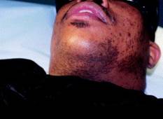
The double-blind placebo controlled study was carried out by U.S. military physicians. PFB, which is predominantly a disorder of black men, has at times been a source of racial tension in the military because the simplest treatment for PFB is to stop shaving and grow a beard, a form of individual expression at odds with regulations.
The study included 27 men with PFB. They received laser therapy once every 4 weeks for 16 weeks. In addition, they applied eflornithine cream to one side of their bearded neck region and placebo to the other side twice daily.
At 16 weeks, the laser plus eflornithine cream side produced a median 99.5% reduction in hair count and inflammatory papules. This was a significantly better result than the median 85% reduction on the laser plus placebo–treated side (J. Am. Acad. Dermatol. 2012 Jan. 8; in press).
The PFB study follows an earlier study by other investigators who demonstrated that eflornithine cream as an adjunct to laser hair removal for facial hirsutism in women was more effective than laser therapy alone (J. Am. Acad. Dermatol. 2007;57:54-9).
Dr. Alexis said performing laser hair removal safely in darker-skinned patients with PFB requires attention to several key principles: longer wavelengths, lower fluences, longer pulse durations, and plenty of epidermal cooling.
"The No. 1 thing is to use longer wavelengths, because the goal is deeper penetration to maximize the ratio of the temperature in the bulb of the follicle to the temperature in the epidermis," he explained.
The long-pulsed 1,064-nm Nd:YAG laser has the lowest rate of associated epidermal burns, hypopigmentation, and other adverse events in darker-skinned patients, as has been shown in a review of a wide assortment of lasers (J. Drugs Dermatol. 2007;6:40-6). It is clearly the safest laser option in patients with skin types IV-VI. The 810-nm diode laser is a reasonable alternative in skin types IV-V, Dr. Alexis said.

In treating patients for PFB with the 1,064-nm Nd:YAG laser, he said that he typically starts with a fluence of 20 J/cm2 and a pulse duration of 20-30 milliseconds. After several sessions, as he makes inroads into the initially dense follicular distribution, he said that he might increase the fluence to a maximum of 50 J/cm2 in the setting of skin type VI, and as high as 100 J/cm2 in skin types IV or V.
Longer pulse durations allow for more efficient epidermal cooling. This minimizes heat injury to melanin-containing epidermal cells. For the 810-nm diode laser, Dr. Alexis said he uses pulse durations of 100 or 400 milliseconds.
Epidermal cooling can be accomplished in several ways. His preferred method is to utilize contact cooling via a sapphire tip or chilled copper plate attached to the laser; the cooling is done before delivering the laser pulse. Alternatively, the epidermal cooling can be done using cold gels, forced air, or spray cooling, although dyschromia can occur in darker skin types if the spray technique isn’t optimal. Another option is to apply an ice pack for 5-10 minutes post treatment.
Laser therapy is expensive, so Dr. Alexis said he likes to give his patients a range of therapeutic options. These include growing a beard, chemical depilation with barium sulfide or calcium thioglycolate every 2-4 days, modification of shaving practices, and salicylic acid chemical peels.
"It’s kind of a long conversation," he said.
Whatever form of therapy the patient decides upon, it’s important that the patient stops tweezing to remove ingrown hairs. This is a common practice that induces trauma and worsens postinflammatory hyperpigmentation.
Dr. Alexis reported that he serves as a consultant to Schick and is on the advisory board for Allergan. SDEF and this news organization are owned by Elsevier.
WAIKOLOA, HAWAII – The use of eflornithine cream may increase the effectiveness of laser hair removal for treating pseudofolliculitis barbae, according to Dr. Andrew F. Alexis.
Laser hair removal has proved to be a game changer in the treatment of pseudofolliculitis barbae, a common chronic, inflammatory dermatosis that’s often been a difficult therapeutic challenge, Dr. Alexis said at the seminar sponsored by Skin Disease Education Foundation (SDEF). And the use of adjunctive eflornithine cream makes laser therapy even more effective, based on a recent study, which is one of the few rigorous studies conducted in pseudofolliculitis barbae (PFB) patients, noted Dr. Alexis, director of the skin of color center at St. Luke’s–Roosevelt Hospital and a dermatologist at Columbia University, New York.
The double-blind placebo controlled study was carried out by U.S. military physicians. PFB, which is predominantly a disorder of black men, has at times been a source of racial tension in the military because the simplest treatment for PFB is to stop shaving and grow a beard, a form of individual expression at odds with regulations.
The study included 27 men with PFB. They received laser therapy once every 4 weeks for 16 weeks. In addition, they applied eflornithine cream to one side of their bearded neck region and placebo to the other side twice daily.
At 16 weeks, the laser plus eflornithine cream side produced a median 99.5% reduction in hair count and inflammatory papules. This was a significantly better result than the median 85% reduction on the laser plus placebo–treated side (J. Am. Acad. Dermatol. 2012 Jan. 8; in press).
The PFB study follows an earlier study by other investigators who demonstrated that eflornithine cream as an adjunct to laser hair removal for facial hirsutism in women was more effective than laser therapy alone (J. Am. Acad. Dermatol. 2007;57:54-9).
Dr. Alexis said performing laser hair removal safely in darker-skinned patients with PFB requires attention to several key principles: longer wavelengths, lower fluences, longer pulse durations, and plenty of epidermal cooling.
"The No. 1 thing is to use longer wavelengths, because the goal is deeper penetration to maximize the ratio of the temperature in the bulb of the follicle to the temperature in the epidermis," he explained.
The long-pulsed 1,064-nm Nd:YAG laser has the lowest rate of associated epidermal burns, hypopigmentation, and other adverse events in darker-skinned patients, as has been shown in a review of a wide assortment of lasers (J. Drugs Dermatol. 2007;6:40-6). It is clearly the safest laser option in patients with skin types IV-VI. The 810-nm diode laser is a reasonable alternative in skin types IV-V, Dr. Alexis said.
In treating patients for PFB with the 1,064-nm Nd:YAG laser, he said that he typically starts with a fluence of 20 J/cm2 and a pulse duration of 20-30 milliseconds. After several sessions, as he makes inroads into the initially dense follicular distribution, he said that he might increase the fluence to a maximum of 50 J/cm2 in the setting of skin type VI, and as high as 100 J/cm2 in skin types IV or V.
Longer pulse durations allow for more efficient epidermal cooling. This minimizes heat injury to melanin-containing epidermal cells. For the 810-nm diode laser, Dr. Alexis said he uses pulse durations of 100 or 400 milliseconds.
Epidermal cooling can be accomplished in several ways. His preferred method is to utilize contact cooling via a sapphire tip or chilled copper plate attached to the laser; the cooling is done before delivering the laser pulse. Alternatively, the epidermal cooling can be done using cold gels, forced air, or spray cooling, although dyschromia can occur in darker skin types if the spray technique isn’t optimal. Another option is to apply an ice pack for 5-10 minutes post treatment.
Laser therapy is expensive, so Dr. Alexis said he likes to give his patients a range of therapeutic options. These include growing a beard, chemical depilation with barium sulfide or calcium thioglycolate every 2-4 days, modification of shaving practices, and salicylic acid chemical peels.
"It’s kind of a long conversation," he said.
Whatever form of therapy the patient decides upon, it’s important that the patient stops tweezing to remove ingrown hairs. This is a common practice that induces trauma and worsens postinflammatory hyperpigmentation.
Dr. Alexis reported that he serves as a consultant to Schick and is on the advisory board for Allergan. SDEF and this news organization are owned by Elsevier.
EXPERT ANALYSIS FROM THE SDEF HAWAII DERMATOLOGY SEMINAR
Microsecond Nd:YAG Safe, Effective for Facial Telangiectasia
KISSIMMEE, FLA. – A microsecond 1,064-nm Nd:YAG laser was found to be effective for treating facial telangiectasias without the scarring and discomfort that can occur with millisecond pulse systems.
A prospective single-center study enrolled 20 patients aged 35-70 years with Fitzpatrick skin types I-III. After undergoing one treatment per month for 2 months with the 650-microsecond LightPod Neo 1,064-nm Nd:YAG laser (manufactured by Aerolase), the patients experienced significant, and even "dramatic," improvements in the appearance of facial telangiectasias on the nose and cheek, Dr. David J. Goldberg reported at the annual meeting of the American Society for Laser Medicine and Surgery.

"We saw significant improvements in the appearance of telangiectasia with minimal discomfort, presumably because of the microsecond pulse duration," said Dr. Goldberg, a dermatologist at Mount Sinai School of Medicine, New York.
The treatments were applied at a fluence of 199 J/cm2, a pulse duration of 650 microseconds, and a 2-mm spot size. The multisetting device was set at treatment level 6. Evaluation of results was based on clinical assessment of digital photographs and both patient and investigator assessment of the reduction in the size and appearance of telangiectasias based on a 5-point scale.
"Most people know that facial telangiectasias can be treated with a variety of wavelengths, and the 532-nm laser has traditionally been effective for small vessels," Dr. Goldberg said.
However, longer wavelength lasers, such as the 1,064-nm Nd:YAG with a millisecond pulse duration (3-30 milliseconds), which have also been used to treat larger vessels, are associated with "well-documented scarring and greater discomfort," he noted.
These adverse effects were not seen with the use of the microsecond system, he said, adding: "I think this is really the most important part of the study because of the previous documentation of problems with Nd:YAG laser systems for the treatment of patients with telangiectasias."
The study was supported by a research grant from Aerolase. Dr. Goldberg had no other disclosures to report.
KISSIMMEE, FLA. – A microsecond 1,064-nm Nd:YAG laser was found to be effective for treating facial telangiectasias without the scarring and discomfort that can occur with millisecond pulse systems.
A prospective single-center study enrolled 20 patients aged 35-70 years with Fitzpatrick skin types I-III. After undergoing one treatment per month for 2 months with the 650-microsecond LightPod Neo 1,064-nm Nd:YAG laser (manufactured by Aerolase), the patients experienced significant, and even "dramatic," improvements in the appearance of facial telangiectasias on the nose and cheek, Dr. David J. Goldberg reported at the annual meeting of the American Society for Laser Medicine and Surgery.
"We saw significant improvements in the appearance of telangiectasia with minimal discomfort, presumably because of the microsecond pulse duration," said Dr. Goldberg, a dermatologist at Mount Sinai School of Medicine, New York.
The treatments were applied at a fluence of 199 J/cm2, a pulse duration of 650 microseconds, and a 2-mm spot size. The multisetting device was set at treatment level 6. Evaluation of results was based on clinical assessment of digital photographs and both patient and investigator assessment of the reduction in the size and appearance of telangiectasias based on a 5-point scale.
"Most people know that facial telangiectasias can be treated with a variety of wavelengths, and the 532-nm laser has traditionally been effective for small vessels," Dr. Goldberg said.
However, longer wavelength lasers, such as the 1,064-nm Nd:YAG with a millisecond pulse duration (3-30 milliseconds), which have also been used to treat larger vessels, are associated with "well-documented scarring and greater discomfort," he noted.
These adverse effects were not seen with the use of the microsecond system, he said, adding: "I think this is really the most important part of the study because of the previous documentation of problems with Nd:YAG laser systems for the treatment of patients with telangiectasias."
The study was supported by a research grant from Aerolase. Dr. Goldberg had no other disclosures to report.
FROM THE ANNUAL MEETING OF THE AMERICAN SOCIETY FOR LASER MEDICINE AND SURGERY
Major Finding: Twenty patients experienced significant improvements at 6 months after undergoing one treatment per month for 2 months.
Data Source: This was a prospective single-center study of patients with facial telangiectasias, aged 35-70 years with Fitzpatrick skin types I-III, who underwent treatment with the 650-microsecond LightPod Neo 1,064-nm Nd:YAG laser.
Disclosures: The study was sponsored by Aerolase. Dr. Goldberg had no other disclosures to report.
Skin of Color: Classifying Undereye Circles
How many times a week do you get asked by your patients how to get rid of the "dark circles" under their eyes? The term is a catch-all used by physicians and patients to refer to problems that have a vast range of genetic, environmental, and skin-related causes. It is a common and frustrating problem, with little structure in its definition and few full-proof treatments.
Below is my proposed classification system for the definition of dark circles and clinical pearls for their treatment. Most patients, however, have a combination of each type and multifactorial causes that need to be addressed.

Infraorbital fat pad protrusion. Also known as "eye bags."
Blepharoplasty is the best, and for now the only, solution for severe fat pad prominence. Referral to a board certified plastic surgeon or dermatologic surgeon is recommended.
If the protrusion is mild and tear troughs are prominent, fillers may be injected into the tear trough area to help mask the protrusion. My favorites for this area are hyalauronic acid fillers like Juvéderm Ultra or Restlyane, sometimes double diluted with normal saline or injected with a 32-gauge needle.
For loose skin with "bags," radiofrequency lasers can provide some benefit. The Thermage eyelid tip produces results over 3-6 months, with repeat treatment possible at 6 months. I always advise patients that this treatment is not a replacement for surgery but can provide some benefit in those who are not surgical candidates or who do not want surgery.
Infraorbital edema. Also known as "puffiness."
The infraorbital skin is very thin and highly sensitive to fluid compartmentalization. Seasonal allergies, sinus infections, crying or water retention from high blood pressure or eating high sodium foods are some of the reasons the loose, thin epidermis becomes edematous.
Treat seasonal allergies with over-the-counter allergy medications or prescription medications for resistant allergies or possible sinus infections.
Advise patients to switch their sleep position. Sleep position can be contributing to undereye bags through gravity. Sleeping on the side or stomach can encourage fluids to collect under the eyes. If patients report being a side sleeper, you may notice a heavier bag on the side they report sleeping on. Patients who wake up with puffy eyes can sleep on their back and add an extra pillow under their head.
Also advise patients to avoid rubbing their eyes, going to bed with makeup on, and using harsh cleansers. Anything that irritates the eyes can cause fluids to pool. Sleeping in eye makeup can irritate eyes, causing undereye edema.
Eye bags could be a sign of an underlying medical condition, especially if bags appear suddenly and none of the above conditions apply. Thyroid, cardiovascular, or kidney problems can cause undereye fluid retention and patients will need to see their primary care doctors for further evaluation.
Patients can place an ice pack, slices of cucumbers, chilled tea bags, refrigerated eye gels, or even a package of frozen peas on their eyes. This can constrict leaky blood vessels and lessen the periorbital edema.
Periorbital hyperpigmentation. Also known as "dark circles."
Pigmentation of the periorbital skin is very common in skin of color because of the increased melanin content. Genetics, rubbing, and inflammatory skin diseases such as eczema may play a role in exacerbating the pigmentation of the thin undereye skin.
Again, advise patients to avoid rubbing the area. Chronic rubbing and the development of lichen simplex chronicus can lead to dark, thickened undereye skin.
Retinoic acid creams can help slough the dark pigmented skin. It should, however, be used in very small amounts that increase over a few weeks to avoid severe irritation.
Skin lightening creams with azaleic acid, kojic acid, and glycolic acid can be found in varying strengths. Hydroquinone creams have been successful in lightening undereye hyperpigmentation. Strengths in over-the-counter preparations start at 1-2% and in prescription strength can be compounded to higher than 4%, but caution should be used to avoid further irritation and potential post-inflammatory pigment from these products.
Light chemical peels can assist in lightening dark undereye pigmentation. Peels with hydroquinone or retinoic acid can be used for an added lightening benefit.
Intense pulse light can help minimize undereye pigmentation, particularly UV-induced pigmentation. Q-switched lasers have also been reported to be effective.
Infraorbital tear trough depression.
Most often, dark circles aren't about changes in the color of the skin. Instead, they're created by a loss of volume in the area around the eye, exposing the orbital bone and creating a hollow trough that shows up as a dark circle. These changes are often genetic, but significant weigh loss can also expose undereye tear trough depressions.
- The best way to treat this problem is with a small amount of a hyaluronic acid filler placed by a dermatologist in the trough. Very small aliquots are needed in even the deepest trough but can give outstanding results. Use caution, however; this is a highly technical and injector-dependent procedure.
There are crucial vascular structures around the eye that need to be avoided, and over-filled troughs will give patients a puffy appearance and may pose a worse and more difficult problem to fix. Hyaluronic acid fillers are not FDA approved to treat undereye depressions, so patients should be knowledgeable to the risks and benefits prior to undergoing these procedures.
With age, the skin around the eye becomes thinner, exposing the small capillaries and venules just below the thin epidermal layer. Vascular prominence can leave a bluish undertone to the infraorbital skin, which can cast dark shadows and make the area appear dark or sallow.
Eye creams that contain caffeine can constrict the underlying blood vessels and temporarily diminish small vessel prominence.
For large blue veins, vascular lasers such as a long pulse Nd:YAG laser can be recommended. However, in darker skin types these lasers can cause hyperpigmented scars if not used with adequate skin cooling techniques.
Periorbital static and dynamic rhytids.
Botulinum toxin placed in small aliquots around the orbital rim will reduce the dynamic rhytids in this area. Treatments spaced 3-4 months apart will ensure long lasting benefits and because botulinum toxin wears off, repeat treatments are needed.
Laser resurfacing with CO2 or fractionated CO2 lasers provide excellent benefit for periocular rhytides. A traditional CO2 laser may require repeat treatment in 6-12 months. Fractionated CO2 lasers typically require 4-6 treatments spaced about 4 weeks apart to provide benefit.
Overall tips:
For most of the types of infraorbital issues, makeup can help conceal or mask some imperfections. Patients should choose a concealer that matches or is slightly lighter than their skin tone. If they have mild discoloration, advise or help them pick a liquid formula for more prominent imperfections. A cream, full coverage concealer works best.
Encourage patients to quit smoking, which dehydrates the skin and causes premature aging and collagen degradation.
Always remind patients to apply sunscreen around the eye area. Hyperpigmentation and tear troughs can accentuate with UV-induced skin pigmentation.
- Advise patients to apply a moisturizer to the eye area nightly to keep the skin from becoming dry, irritated, and dehydrated.
- Lily Talakoub, M.D.
Do you have questions about treating patients with darker skin? If so, send them to sknews@elsevier.com.
How many times a week do you get asked by your patients how to get rid of the "dark circles" under their eyes? The term is a catch-all used by physicians and patients to refer to problems that have a vast range of genetic, environmental, and skin-related causes. It is a common and frustrating problem, with little structure in its definition and few full-proof treatments.
Below is my proposed classification system for the definition of dark circles and clinical pearls for their treatment. Most patients, however, have a combination of each type and multifactorial causes that need to be addressed.
Infraorbital fat pad protrusion. Also known as "eye bags."
Blepharoplasty is the best, and for now the only, solution for severe fat pad prominence. Referral to a board certified plastic surgeon or dermatologic surgeon is recommended.
If the protrusion is mild and tear troughs are prominent, fillers may be injected into the tear trough area to help mask the protrusion. My favorites for this area are hyalauronic acid fillers like Juvéderm Ultra or Restlyane, sometimes double diluted with normal saline or injected with a 32-gauge needle.
For loose skin with "bags," radiofrequency lasers can provide some benefit. The Thermage eyelid tip produces results over 3-6 months, with repeat treatment possible at 6 months. I always advise patients that this treatment is not a replacement for surgery but can provide some benefit in those who are not surgical candidates or who do not want surgery.
Infraorbital edema. Also known as "puffiness."
The infraorbital skin is very thin and highly sensitive to fluid compartmentalization. Seasonal allergies, sinus infections, crying or water retention from high blood pressure or eating high sodium foods are some of the reasons the loose, thin epidermis becomes edematous.
Treat seasonal allergies with over-the-counter allergy medications or prescription medications for resistant allergies or possible sinus infections.
Advise patients to switch their sleep position. Sleep position can be contributing to undereye bags through gravity. Sleeping on the side or stomach can encourage fluids to collect under the eyes. If patients report being a side sleeper, you may notice a heavier bag on the side they report sleeping on. Patients who wake up with puffy eyes can sleep on their back and add an extra pillow under their head.
Also advise patients to avoid rubbing their eyes, going to bed with makeup on, and using harsh cleansers. Anything that irritates the eyes can cause fluids to pool. Sleeping in eye makeup can irritate eyes, causing undereye edema.
Eye bags could be a sign of an underlying medical condition, especially if bags appear suddenly and none of the above conditions apply. Thyroid, cardiovascular, or kidney problems can cause undereye fluid retention and patients will need to see their primary care doctors for further evaluation.
Patients can place an ice pack, slices of cucumbers, chilled tea bags, refrigerated eye gels, or even a package of frozen peas on their eyes. This can constrict leaky blood vessels and lessen the periorbital edema.
Periorbital hyperpigmentation. Also known as "dark circles."
Pigmentation of the periorbital skin is very common in skin of color because of the increased melanin content. Genetics, rubbing, and inflammatory skin diseases such as eczema may play a role in exacerbating the pigmentation of the thin undereye skin.
Again, advise patients to avoid rubbing the area. Chronic rubbing and the development of lichen simplex chronicus can lead to dark, thickened undereye skin.
Retinoic acid creams can help slough the dark pigmented skin. It should, however, be used in very small amounts that increase over a few weeks to avoid severe irritation.
Skin lightening creams with azaleic acid, kojic acid, and glycolic acid can be found in varying strengths. Hydroquinone creams have been successful in lightening undereye hyperpigmentation. Strengths in over-the-counter preparations start at 1-2% and in prescription strength can be compounded to higher than 4%, but caution should be used to avoid further irritation and potential post-inflammatory pigment from these products.
Light chemical peels can assist in lightening dark undereye pigmentation. Peels with hydroquinone or retinoic acid can be used for an added lightening benefit.
Intense pulse light can help minimize undereye pigmentation, particularly UV-induced pigmentation. Q-switched lasers have also been reported to be effective.
Infraorbital tear trough depression.
Most often, dark circles aren't about changes in the color of the skin. Instead, they're created by a loss of volume in the area around the eye, exposing the orbital bone and creating a hollow trough that shows up as a dark circle. These changes are often genetic, but significant weigh loss can also expose undereye tear trough depressions.
- The best way to treat this problem is with a small amount of a hyaluronic acid filler placed by a dermatologist in the trough. Very small aliquots are needed in even the deepest trough but can give outstanding results. Use caution, however; this is a highly technical and injector-dependent procedure.
There are crucial vascular structures around the eye that need to be avoided, and over-filled troughs will give patients a puffy appearance and may pose a worse and more difficult problem to fix. Hyaluronic acid fillers are not FDA approved to treat undereye depressions, so patients should be knowledgeable to the risks and benefits prior to undergoing these procedures.
With age, the skin around the eye becomes thinner, exposing the small capillaries and venules just below the thin epidermal layer. Vascular prominence can leave a bluish undertone to the infraorbital skin, which can cast dark shadows and make the area appear dark or sallow.
Eye creams that contain caffeine can constrict the underlying blood vessels and temporarily diminish small vessel prominence.
For large blue veins, vascular lasers such as a long pulse Nd:YAG laser can be recommended. However, in darker skin types these lasers can cause hyperpigmented scars if not used with adequate skin cooling techniques.
Periorbital static and dynamic rhytids.
Botulinum toxin placed in small aliquots around the orbital rim will reduce the dynamic rhytids in this area. Treatments spaced 3-4 months apart will ensure long lasting benefits and because botulinum toxin wears off, repeat treatments are needed.
Laser resurfacing with CO2 or fractionated CO2 lasers provide excellent benefit for periocular rhytides. A traditional CO2 laser may require repeat treatment in 6-12 months. Fractionated CO2 lasers typically require 4-6 treatments spaced about 4 weeks apart to provide benefit.
Overall tips:
For most of the types of infraorbital issues, makeup can help conceal or mask some imperfections. Patients should choose a concealer that matches or is slightly lighter than their skin tone. If they have mild discoloration, advise or help them pick a liquid formula for more prominent imperfections. A cream, full coverage concealer works best.
Encourage patients to quit smoking, which dehydrates the skin and causes premature aging and collagen degradation.
Always remind patients to apply sunscreen around the eye area. Hyperpigmentation and tear troughs can accentuate with UV-induced skin pigmentation.
- Advise patients to apply a moisturizer to the eye area nightly to keep the skin from becoming dry, irritated, and dehydrated.
- Lily Talakoub, M.D.
Do you have questions about treating patients with darker skin? If so, send them to sknews@elsevier.com.