User login
Bringing you the latest news, research and reviews, exclusive interviews, podcasts, quizzes, and more.
Powered by CHEST Physician, Clinician Reviews, MDedge Family Medicine, Internal Medicine News, and The Journal of Clinical Outcomes Management.
Pandemic Flu Highlights Racial/Ethnic Disparities Worldwide
ATLANTA – Surveillance data from Minnesota and Wellington, New Zealand, underscore the extent of the racial and ethnic disparities in the rates of influenza and hospitalization for influenza that occurred during the 2009 H1N1 influenza pandemic, and some of those data demonstrate that these disparities persisted at similar levels in 2010-2011.
The findings underscore the need for improved public health strategies that alleviate the socioeconomic factors that contribute to the disparities, the investigators agreed.

The combined incidence of hospitalization for influenza per 100,000 population in Minnesota during the pandemic influenza season in 2009 and the subsequent influenza season was 60.8 for blacks, 42.8 for Native Americans, 35.3 for Hispanics, 23.7 for Asians, and 17.8 for whites. Nonwhites accounted for 31% of cases during the pandemic period, and 23% during the following season, Craig Morin of the Minnesota Department of Health reported in a poster at the International Conference on Emerging Infectious Diseases.
The median age of hospitalized patients during the 2009 pandemic was 28.8 years; the median age in the 2010-2011 season was 52.6 years.
"After adjustment for age, the relative rates associated with nonwhite vs. white non-Hispanic race was 2.46 during the 2009 pandemic period, and 2.52 during the 2010-2011 season," Mr. Morin wrote. The differences were statistically significant.
Cases included in this study were laboratory-confirmed, hospitalized influenza cases. During the pandemic season, cases included Minnesota residents hospitalized between April 2009 and April 2010 for confirmed A (H1N1) pmd09 influenza. During the subsequent season, cases included Minnesota residents hospitalized between October 2010 and April 2011 for laboratory-confirmed A H3 or A (H1N1) pmd09 influenza.
Ethnicity information was available for 1,703 cases during the pandemic period and for 467 cases during the subsequent season.
The findings indicate that racial and ethnic disparities occurred to a similar degree during both seasons, and suggest that more study is needed to further delineate the relationship between race/ethnicity and socioeconomic factors, vaccine coverage, prevalence of chronic illnesses, and access to health care. A better understanding of these relationships would be helpful for designing public health interventions most likely to alleviate the disparities, Mr. Morin concluded.
Another study presented at the conference also demonstrated disparities in influenza rates and outcomes based on socioeconomic status. These findings among residents of New Zealand during the pandemic season also highlighted a need for public health interventions to reduce the factors contributing to disparities.
During the pandemic season, rates of infection were highest among the most socioeconomically deprived groups. For example, the rates were significantly higher among Pacific peoples than among European/other individuals (adjusted risk ratio, 1.49). Also, after the researchers adjusted for infection rates, they found that hospitalization rates were higher for Maori (adjusted risk ratio, 2.44), Pacific peoples (adjusted risk ratio, 4.16), and the most deprived socioeconomic status quintile of the population (adjusted risk ratio, 1.37). Mortality risk was also significantly higher for Pacific peoples (adjusted risk ratio, 3.28), Dr. Michael G. Baker of the University of Otaga, Wellington, reported at the conference.
Furthermore, general practitioner consultation rates were inversely associated with disease risk, and the rates of consultation were significantly lower for Maori and Pacific peoples and for those in more deprived quintiles after adjustment for infection rates, Dr. Baker said.
The findings reinforce the importance of identifying factors that contribute to elevated risk, reducing socioeconomic inequality, and improving access to primary care services for disadvantaged populations, he concluded.
Mr. Morin’s study was supported by the Centers for Disease Control and Prevention Emerging Infections Program. Neither Mr. Morin nor Dr. Baker had disclosures to report.
ATLANTA – Surveillance data from Minnesota and Wellington, New Zealand, underscore the extent of the racial and ethnic disparities in the rates of influenza and hospitalization for influenza that occurred during the 2009 H1N1 influenza pandemic, and some of those data demonstrate that these disparities persisted at similar levels in 2010-2011.
The findings underscore the need for improved public health strategies that alleviate the socioeconomic factors that contribute to the disparities, the investigators agreed.
The combined incidence of hospitalization for influenza per 100,000 population in Minnesota during the pandemic influenza season in 2009 and the subsequent influenza season was 60.8 for blacks, 42.8 for Native Americans, 35.3 for Hispanics, 23.7 for Asians, and 17.8 for whites. Nonwhites accounted for 31% of cases during the pandemic period, and 23% during the following season, Craig Morin of the Minnesota Department of Health reported in a poster at the International Conference on Emerging Infectious Diseases.
The median age of hospitalized patients during the 2009 pandemic was 28.8 years; the median age in the 2010-2011 season was 52.6 years.
"After adjustment for age, the relative rates associated with nonwhite vs. white non-Hispanic race was 2.46 during the 2009 pandemic period, and 2.52 during the 2010-2011 season," Mr. Morin wrote. The differences were statistically significant.
Cases included in this study were laboratory-confirmed, hospitalized influenza cases. During the pandemic season, cases included Minnesota residents hospitalized between April 2009 and April 2010 for confirmed A (H1N1) pmd09 influenza. During the subsequent season, cases included Minnesota residents hospitalized between October 2010 and April 2011 for laboratory-confirmed A H3 or A (H1N1) pmd09 influenza.
Ethnicity information was available for 1,703 cases during the pandemic period and for 467 cases during the subsequent season.
The findings indicate that racial and ethnic disparities occurred to a similar degree during both seasons, and suggest that more study is needed to further delineate the relationship between race/ethnicity and socioeconomic factors, vaccine coverage, prevalence of chronic illnesses, and access to health care. A better understanding of these relationships would be helpful for designing public health interventions most likely to alleviate the disparities, Mr. Morin concluded.
Another study presented at the conference also demonstrated disparities in influenza rates and outcomes based on socioeconomic status. These findings among residents of New Zealand during the pandemic season also highlighted a need for public health interventions to reduce the factors contributing to disparities.
During the pandemic season, rates of infection were highest among the most socioeconomically deprived groups. For example, the rates were significantly higher among Pacific peoples than among European/other individuals (adjusted risk ratio, 1.49). Also, after the researchers adjusted for infection rates, they found that hospitalization rates were higher for Maori (adjusted risk ratio, 2.44), Pacific peoples (adjusted risk ratio, 4.16), and the most deprived socioeconomic status quintile of the population (adjusted risk ratio, 1.37). Mortality risk was also significantly higher for Pacific peoples (adjusted risk ratio, 3.28), Dr. Michael G. Baker of the University of Otaga, Wellington, reported at the conference.
Furthermore, general practitioner consultation rates were inversely associated with disease risk, and the rates of consultation were significantly lower for Maori and Pacific peoples and for those in more deprived quintiles after adjustment for infection rates, Dr. Baker said.
The findings reinforce the importance of identifying factors that contribute to elevated risk, reducing socioeconomic inequality, and improving access to primary care services for disadvantaged populations, he concluded.
Mr. Morin’s study was supported by the Centers for Disease Control and Prevention Emerging Infections Program. Neither Mr. Morin nor Dr. Baker had disclosures to report.
ATLANTA – Surveillance data from Minnesota and Wellington, New Zealand, underscore the extent of the racial and ethnic disparities in the rates of influenza and hospitalization for influenza that occurred during the 2009 H1N1 influenza pandemic, and some of those data demonstrate that these disparities persisted at similar levels in 2010-2011.
The findings underscore the need for improved public health strategies that alleviate the socioeconomic factors that contribute to the disparities, the investigators agreed.
The combined incidence of hospitalization for influenza per 100,000 population in Minnesota during the pandemic influenza season in 2009 and the subsequent influenza season was 60.8 for blacks, 42.8 for Native Americans, 35.3 for Hispanics, 23.7 for Asians, and 17.8 for whites. Nonwhites accounted for 31% of cases during the pandemic period, and 23% during the following season, Craig Morin of the Minnesota Department of Health reported in a poster at the International Conference on Emerging Infectious Diseases.
The median age of hospitalized patients during the 2009 pandemic was 28.8 years; the median age in the 2010-2011 season was 52.6 years.
"After adjustment for age, the relative rates associated with nonwhite vs. white non-Hispanic race was 2.46 during the 2009 pandemic period, and 2.52 during the 2010-2011 season," Mr. Morin wrote. The differences were statistically significant.
Cases included in this study were laboratory-confirmed, hospitalized influenza cases. During the pandemic season, cases included Minnesota residents hospitalized between April 2009 and April 2010 for confirmed A (H1N1) pmd09 influenza. During the subsequent season, cases included Minnesota residents hospitalized between October 2010 and April 2011 for laboratory-confirmed A H3 or A (H1N1) pmd09 influenza.
Ethnicity information was available for 1,703 cases during the pandemic period and for 467 cases during the subsequent season.
The findings indicate that racial and ethnic disparities occurred to a similar degree during both seasons, and suggest that more study is needed to further delineate the relationship between race/ethnicity and socioeconomic factors, vaccine coverage, prevalence of chronic illnesses, and access to health care. A better understanding of these relationships would be helpful for designing public health interventions most likely to alleviate the disparities, Mr. Morin concluded.
Another study presented at the conference also demonstrated disparities in influenza rates and outcomes based on socioeconomic status. These findings among residents of New Zealand during the pandemic season also highlighted a need for public health interventions to reduce the factors contributing to disparities.
During the pandemic season, rates of infection were highest among the most socioeconomically deprived groups. For example, the rates were significantly higher among Pacific peoples than among European/other individuals (adjusted risk ratio, 1.49). Also, after the researchers adjusted for infection rates, they found that hospitalization rates were higher for Maori (adjusted risk ratio, 2.44), Pacific peoples (adjusted risk ratio, 4.16), and the most deprived socioeconomic status quintile of the population (adjusted risk ratio, 1.37). Mortality risk was also significantly higher for Pacific peoples (adjusted risk ratio, 3.28), Dr. Michael G. Baker of the University of Otaga, Wellington, reported at the conference.
Furthermore, general practitioner consultation rates were inversely associated with disease risk, and the rates of consultation were significantly lower for Maori and Pacific peoples and for those in more deprived quintiles after adjustment for infection rates, Dr. Baker said.
The findings reinforce the importance of identifying factors that contribute to elevated risk, reducing socioeconomic inequality, and improving access to primary care services for disadvantaged populations, he concluded.
Mr. Morin’s study was supported by the Centers for Disease Control and Prevention Emerging Infections Program. Neither Mr. Morin nor Dr. Baker had disclosures to report.
FROM THE INTERNATIONAL CONFERENCE ON EMERGING INFECTIOUS DISEASES
Intensivist Service Reduced Infections, Ventilator Days
SAN DIEGO – Converting some conventional hospitalists to intensive care unit hospitalists reduced the average length of stay, duration on ventilators, and rate of catheter-related bloodstream infections, worth an estimated $1.45 million per year in savings at one community hospital.
After a year with the intensivist hospitalist team in action, the rate of catheter-related bloodstream infections decreased by 75%, the average length of ICU stay declined by 22%, ventilator days decreased by 35%, and the rate of ventilator-associated pneumonia was brought down to 0%. In addition, 30% more general ward patients were discharged before noon by non-ICU hospitalists, Dr. Min Hlaing and Dr. Rod Felber reported in a joint presentation at the annual meeting of the Society of Hospital Medicine.

Surveys of other hospital staff suggested that having the intensivist hospitalist team improved communication between physician and nurses and between hospitalists and subspecialists. ICU nursing staff, respiratory technicians, and the sole pulmonologist reported being very satisfied with the ICU hospitalist care, said Dr. Hlaing and Dr. Felber of Lodi (Calif.) Memorial Hospital. The center has 270 beds.
The reductions in length of stay, ventilator days, and infections alone represent a savings to the hospital of $1.45 million per year, the speakers and their associates estimated.
Patient- and family-satisfaction scores increased after institution of the ICU hospitalist service. Job satisfaction among nurses improved, which increased retention of nurses in both the ICU and general ward. The hospital found it easier to recruit and retain specialists and easier to recruit general ward hospitalists because they no longer needed to have ICU skills. Satisfaction and retention among hospitalists as a whole improved at the hospital, said Dr. Felber, medical director of the hospitalist program.
To start the trial, Dr. Hlaing, who is an associate director of the hospitalist program, identified four of his hospitalists who were comfortable in caring for critically ill patients. He assigned them and himself to manage the 10-bed ICU and closed the ICU to other hospitalists. Team members became credentialed to perform procedures such as ultrasound-guided central venous catheter placement, arterial catheter insertion, lumbar puncture, paracentesis, endotracheal intubation, and ventilator management. They completed a course in the fundamentals of critical care support, and utilized evidence-based standardized order sets that were developed for common ICU conditions.
The ICU hospitalists had dedicated times for multidisciplinary rounds, family meetings, ICU-specific committees, and education of nurses. An emphasis on continuity of care enabled ICU patients to be admitted, rounded on daily in the ICU, followed after transfer to the medical/surgical floor, and discharged home by the same hospitalist.
After a year of the ICU hospitalist service in action, the investigators conducted a 360-degree evaluation and review of the major hospital stakeholders.
The workload of caring for an ICU patient was considered to be 1.5 times that of medical/surgical floor patients, so the intensivist hospitalists saw fewer patients than other hospitalists. The ICU hospitalists were paid $5 per hour more than other hospitalists. The Relative Value Units for ICU patient care and performance of procedures also led to higher compensation compared with medical/surgical hospitalists, Dr. Felber said.

During the year, the hospital administration requested expansion of the ICU hospitalist model from 12 hours per day of coverage to 24 hours per day.
"If you have hospitalists who are capable of doing it, an ICU hospitalist model is one of the most sustainable and economically viable options to provide quality care to our most critically ill patients," Dr. Felber said.
Previous studies suggest that, in general, only 23% of critically ill patients are seen by intensivists, the speakers said. ICUs consume a quarter of hospital budgets on average. The aging U.S. population will increase demand for intensivist services by 38%. Strategies proposed for hospitals to cope with this evolution include hiring more intensivists, greater use of telemedicine, and partnering with hospitalists, as in the current study.
Dr. Hlaing and Dr. Felber did not report financial disclosures.
SAN DIEGO – Converting some conventional hospitalists to intensive care unit hospitalists reduced the average length of stay, duration on ventilators, and rate of catheter-related bloodstream infections, worth an estimated $1.45 million per year in savings at one community hospital.
After a year with the intensivist hospitalist team in action, the rate of catheter-related bloodstream infections decreased by 75%, the average length of ICU stay declined by 22%, ventilator days decreased by 35%, and the rate of ventilator-associated pneumonia was brought down to 0%. In addition, 30% more general ward patients were discharged before noon by non-ICU hospitalists, Dr. Min Hlaing and Dr. Rod Felber reported in a joint presentation at the annual meeting of the Society of Hospital Medicine.
Surveys of other hospital staff suggested that having the intensivist hospitalist team improved communication between physician and nurses and between hospitalists and subspecialists. ICU nursing staff, respiratory technicians, and the sole pulmonologist reported being very satisfied with the ICU hospitalist care, said Dr. Hlaing and Dr. Felber of Lodi (Calif.) Memorial Hospital. The center has 270 beds.
The reductions in length of stay, ventilator days, and infections alone represent a savings to the hospital of $1.45 million per year, the speakers and their associates estimated.
Patient- and family-satisfaction scores increased after institution of the ICU hospitalist service. Job satisfaction among nurses improved, which increased retention of nurses in both the ICU and general ward. The hospital found it easier to recruit and retain specialists and easier to recruit general ward hospitalists because they no longer needed to have ICU skills. Satisfaction and retention among hospitalists as a whole improved at the hospital, said Dr. Felber, medical director of the hospitalist program.
To start the trial, Dr. Hlaing, who is an associate director of the hospitalist program, identified four of his hospitalists who were comfortable in caring for critically ill patients. He assigned them and himself to manage the 10-bed ICU and closed the ICU to other hospitalists. Team members became credentialed to perform procedures such as ultrasound-guided central venous catheter placement, arterial catheter insertion, lumbar puncture, paracentesis, endotracheal intubation, and ventilator management. They completed a course in the fundamentals of critical care support, and utilized evidence-based standardized order sets that were developed for common ICU conditions.
The ICU hospitalists had dedicated times for multidisciplinary rounds, family meetings, ICU-specific committees, and education of nurses. An emphasis on continuity of care enabled ICU patients to be admitted, rounded on daily in the ICU, followed after transfer to the medical/surgical floor, and discharged home by the same hospitalist.
After a year of the ICU hospitalist service in action, the investigators conducted a 360-degree evaluation and review of the major hospital stakeholders.
The workload of caring for an ICU patient was considered to be 1.5 times that of medical/surgical floor patients, so the intensivist hospitalists saw fewer patients than other hospitalists. The ICU hospitalists were paid $5 per hour more than other hospitalists. The Relative Value Units for ICU patient care and performance of procedures also led to higher compensation compared with medical/surgical hospitalists, Dr. Felber said.
During the year, the hospital administration requested expansion of the ICU hospitalist model from 12 hours per day of coverage to 24 hours per day.
"If you have hospitalists who are capable of doing it, an ICU hospitalist model is one of the most sustainable and economically viable options to provide quality care to our most critically ill patients," Dr. Felber said.
Previous studies suggest that, in general, only 23% of critically ill patients are seen by intensivists, the speakers said. ICUs consume a quarter of hospital budgets on average. The aging U.S. population will increase demand for intensivist services by 38%. Strategies proposed for hospitals to cope with this evolution include hiring more intensivists, greater use of telemedicine, and partnering with hospitalists, as in the current study.
Dr. Hlaing and Dr. Felber did not report financial disclosures.
SAN DIEGO – Converting some conventional hospitalists to intensive care unit hospitalists reduced the average length of stay, duration on ventilators, and rate of catheter-related bloodstream infections, worth an estimated $1.45 million per year in savings at one community hospital.
After a year with the intensivist hospitalist team in action, the rate of catheter-related bloodstream infections decreased by 75%, the average length of ICU stay declined by 22%, ventilator days decreased by 35%, and the rate of ventilator-associated pneumonia was brought down to 0%. In addition, 30% more general ward patients were discharged before noon by non-ICU hospitalists, Dr. Min Hlaing and Dr. Rod Felber reported in a joint presentation at the annual meeting of the Society of Hospital Medicine.
Surveys of other hospital staff suggested that having the intensivist hospitalist team improved communication between physician and nurses and between hospitalists and subspecialists. ICU nursing staff, respiratory technicians, and the sole pulmonologist reported being very satisfied with the ICU hospitalist care, said Dr. Hlaing and Dr. Felber of Lodi (Calif.) Memorial Hospital. The center has 270 beds.
The reductions in length of stay, ventilator days, and infections alone represent a savings to the hospital of $1.45 million per year, the speakers and their associates estimated.
Patient- and family-satisfaction scores increased after institution of the ICU hospitalist service. Job satisfaction among nurses improved, which increased retention of nurses in both the ICU and general ward. The hospital found it easier to recruit and retain specialists and easier to recruit general ward hospitalists because they no longer needed to have ICU skills. Satisfaction and retention among hospitalists as a whole improved at the hospital, said Dr. Felber, medical director of the hospitalist program.
To start the trial, Dr. Hlaing, who is an associate director of the hospitalist program, identified four of his hospitalists who were comfortable in caring for critically ill patients. He assigned them and himself to manage the 10-bed ICU and closed the ICU to other hospitalists. Team members became credentialed to perform procedures such as ultrasound-guided central venous catheter placement, arterial catheter insertion, lumbar puncture, paracentesis, endotracheal intubation, and ventilator management. They completed a course in the fundamentals of critical care support, and utilized evidence-based standardized order sets that were developed for common ICU conditions.
The ICU hospitalists had dedicated times for multidisciplinary rounds, family meetings, ICU-specific committees, and education of nurses. An emphasis on continuity of care enabled ICU patients to be admitted, rounded on daily in the ICU, followed after transfer to the medical/surgical floor, and discharged home by the same hospitalist.
After a year of the ICU hospitalist service in action, the investigators conducted a 360-degree evaluation and review of the major hospital stakeholders.
The workload of caring for an ICU patient was considered to be 1.5 times that of medical/surgical floor patients, so the intensivist hospitalists saw fewer patients than other hospitalists. The ICU hospitalists were paid $5 per hour more than other hospitalists. The Relative Value Units for ICU patient care and performance of procedures also led to higher compensation compared with medical/surgical hospitalists, Dr. Felber said.
During the year, the hospital administration requested expansion of the ICU hospitalist model from 12 hours per day of coverage to 24 hours per day.
"If you have hospitalists who are capable of doing it, an ICU hospitalist model is one of the most sustainable and economically viable options to provide quality care to our most critically ill patients," Dr. Felber said.
Previous studies suggest that, in general, only 23% of critically ill patients are seen by intensivists, the speakers said. ICUs consume a quarter of hospital budgets on average. The aging U.S. population will increase demand for intensivist services by 38%. Strategies proposed for hospitals to cope with this evolution include hiring more intensivists, greater use of telemedicine, and partnering with hospitalists, as in the current study.
Dr. Hlaing and Dr. Felber did not report financial disclosures.
FROM THE ANNUAL MEETING OF THE SOCIETY OF HOSPITAL MEDICINE
GERD Associated With Worse Infant Respiratory Disease
ORLANDO – Gastroesophageal reflux disease was associated with prior wheezing and greater severity of infant acute respiratory illness in a study involving 430 term, healthy infants presenting with acute respiratory illness due to bronchiolitis or upper respiratory infection.
New-onset gastroesophageal reflux disease (GERD) during bronchiolitis in previously health infants has been shown to increase the risk for respiratory failure (Acta Paediatr. 2007;96:1025-9). Bronchiolitis is a prominent risk factor for childhood asthma, and GERD is associated with worsened asthma severity (J. Asthma 2011;48:366-73). As yet unknown is whether preexisting GERD (clinically significant reflux) in infants modulates the severity of infant bronchiolitis or the diagnosis of childhood asthma, Dr. Robert S. Valet of Vanderbilt University, Nashville, Tenn., and his associates said in a poster at the annual meeting of the American Academy of Allergy, Asthma, and Immunology.
The 430 infants, aged 0-12 months, were enrolled in the Tennessee Children\'s Respiratory Initiative. The presence of infant GERD was defined by parental report with an answer of "yes" to the question, "Does your child have a history of gastroesophageal reflux disease?"
In all, 45 infants (10%) were reported to have GERD. They were more likely than those without GERD to be white and to have a family history of allergic rhinitis, but did not differ by age at illness, secondhand smoke (SHS) exposure, or family history of asthma. The group with GERD was significantly more likely to have previous treatment for wheeze (42% vs. 25%), and to have bronchiolitis (84% vs. 71%).
The median bronchiolitis severity score (range, 0-12; higher score indicates more severe disease, with a difference of 0.5 being clinically meaningful) in univariate analysis was 5.5 in the GERD group vs. 4.0 in infants without GERD. Adjusting for age, race, sex, and SHS, the increased bronchiolitis severity score in the GERD group persisted (odds ratio, 1.90). Although not significant, 11% of infants with GERD vs. 5% of non-GERD infants needed ICU care, Dr. Valet and his associates reported.
Preexisting GERD was associated with increased bronchiolitis severity and increased recurrent wheezing illness at 2 years, at which time 69% of infants with GERD and 46% of those without GERD had experienced wheeze since initial study enrollment (OR, 2.35; P = .006). And although not statistically significant, 52% with GERD vs. 40% without had used albuterol in this time period (P = .13). However, there was no difference in asthma diagnosis at age 2 years; 24% of infants with GERD were diagnosed with asthma by age 2, compared with 22% of those without GERD (P = .76).
This is the first time that preexisting infant GERD has been associated with increased bronchiolitis severity, Dr. Valet noted. Future study could investigate whether treating infant GERD decreases both bronchiolitis and asthma severity. "These findings should be replicated using a more rigorous assessment of GERD," he concluded.
The study was funded by the Thrasher Research Fund. Dr. Valet said he had no relevant financial disclosures.
ORLANDO – Gastroesophageal reflux disease was associated with prior wheezing and greater severity of infant acute respiratory illness in a study involving 430 term, healthy infants presenting with acute respiratory illness due to bronchiolitis or upper respiratory infection.
New-onset gastroesophageal reflux disease (GERD) during bronchiolitis in previously health infants has been shown to increase the risk for respiratory failure (Acta Paediatr. 2007;96:1025-9). Bronchiolitis is a prominent risk factor for childhood asthma, and GERD is associated with worsened asthma severity (J. Asthma 2011;48:366-73). As yet unknown is whether preexisting GERD (clinically significant reflux) in infants modulates the severity of infant bronchiolitis or the diagnosis of childhood asthma, Dr. Robert S. Valet of Vanderbilt University, Nashville, Tenn., and his associates said in a poster at the annual meeting of the American Academy of Allergy, Asthma, and Immunology.
The 430 infants, aged 0-12 months, were enrolled in the Tennessee Children\'s Respiratory Initiative. The presence of infant GERD was defined by parental report with an answer of "yes" to the question, "Does your child have a history of gastroesophageal reflux disease?"
In all, 45 infants (10%) were reported to have GERD. They were more likely than those without GERD to be white and to have a family history of allergic rhinitis, but did not differ by age at illness, secondhand smoke (SHS) exposure, or family history of asthma. The group with GERD was significantly more likely to have previous treatment for wheeze (42% vs. 25%), and to have bronchiolitis (84% vs. 71%).
The median bronchiolitis severity score (range, 0-12; higher score indicates more severe disease, with a difference of 0.5 being clinically meaningful) in univariate analysis was 5.5 in the GERD group vs. 4.0 in infants without GERD. Adjusting for age, race, sex, and SHS, the increased bronchiolitis severity score in the GERD group persisted (odds ratio, 1.90). Although not significant, 11% of infants with GERD vs. 5% of non-GERD infants needed ICU care, Dr. Valet and his associates reported.
Preexisting GERD was associated with increased bronchiolitis severity and increased recurrent wheezing illness at 2 years, at which time 69% of infants with GERD and 46% of those without GERD had experienced wheeze since initial study enrollment (OR, 2.35; P = .006). And although not statistically significant, 52% with GERD vs. 40% without had used albuterol in this time period (P = .13). However, there was no difference in asthma diagnosis at age 2 years; 24% of infants with GERD were diagnosed with asthma by age 2, compared with 22% of those without GERD (P = .76).
This is the first time that preexisting infant GERD has been associated with increased bronchiolitis severity, Dr. Valet noted. Future study could investigate whether treating infant GERD decreases both bronchiolitis and asthma severity. "These findings should be replicated using a more rigorous assessment of GERD," he concluded.
The study was funded by the Thrasher Research Fund. Dr. Valet said he had no relevant financial disclosures.
ORLANDO – Gastroesophageal reflux disease was associated with prior wheezing and greater severity of infant acute respiratory illness in a study involving 430 term, healthy infants presenting with acute respiratory illness due to bronchiolitis or upper respiratory infection.
New-onset gastroesophageal reflux disease (GERD) during bronchiolitis in previously health infants has been shown to increase the risk for respiratory failure (Acta Paediatr. 2007;96:1025-9). Bronchiolitis is a prominent risk factor for childhood asthma, and GERD is associated with worsened asthma severity (J. Asthma 2011;48:366-73). As yet unknown is whether preexisting GERD (clinically significant reflux) in infants modulates the severity of infant bronchiolitis or the diagnosis of childhood asthma, Dr. Robert S. Valet of Vanderbilt University, Nashville, Tenn., and his associates said in a poster at the annual meeting of the American Academy of Allergy, Asthma, and Immunology.
The 430 infants, aged 0-12 months, were enrolled in the Tennessee Children\'s Respiratory Initiative. The presence of infant GERD was defined by parental report with an answer of "yes" to the question, "Does your child have a history of gastroesophageal reflux disease?"
In all, 45 infants (10%) were reported to have GERD. They were more likely than those without GERD to be white and to have a family history of allergic rhinitis, but did not differ by age at illness, secondhand smoke (SHS) exposure, or family history of asthma. The group with GERD was significantly more likely to have previous treatment for wheeze (42% vs. 25%), and to have bronchiolitis (84% vs. 71%).
The median bronchiolitis severity score (range, 0-12; higher score indicates more severe disease, with a difference of 0.5 being clinically meaningful) in univariate analysis was 5.5 in the GERD group vs. 4.0 in infants without GERD. Adjusting for age, race, sex, and SHS, the increased bronchiolitis severity score in the GERD group persisted (odds ratio, 1.90). Although not significant, 11% of infants with GERD vs. 5% of non-GERD infants needed ICU care, Dr. Valet and his associates reported.
Preexisting GERD was associated with increased bronchiolitis severity and increased recurrent wheezing illness at 2 years, at which time 69% of infants with GERD and 46% of those without GERD had experienced wheeze since initial study enrollment (OR, 2.35; P = .006). And although not statistically significant, 52% with GERD vs. 40% without had used albuterol in this time period (P = .13). However, there was no difference in asthma diagnosis at age 2 years; 24% of infants with GERD were diagnosed with asthma by age 2, compared with 22% of those without GERD (P = .76).
This is the first time that preexisting infant GERD has been associated with increased bronchiolitis severity, Dr. Valet noted. Future study could investigate whether treating infant GERD decreases both bronchiolitis and asthma severity. "These findings should be replicated using a more rigorous assessment of GERD," he concluded.
The study was funded by the Thrasher Research Fund. Dr. Valet said he had no relevant financial disclosures.
FROM THE ANNUAL MEETING OF THE AMERICAN ACADEMY OF ALLERGY, ASTHMA, AND IMMUNOLOGY
Major Finding: At 2 years, 69% of infants with GERD and 46% of those without GERD had experienced wheeze since initial study enrollment (OR, 2.35; P = .006).
Data Source: The 2-year data come from a study of 430 infants aged 0-12 months who were enrolled in the Tennessee Children’s Respiratory Initiative.
Disclosures: The study was funded by the Thrasher Research Fund. Dr. Valet reported having no relevant financial disclosures.
Statins' Effect on Pneumonia Mortality Murky in Meta-Analysis
SAN DIEGO – Statins may reduce mortality after pneumonia, but this possible benefit may weaken or disappear in important subgroups of patients, results of a double meta-analysis suggest.
The analyses of data from 13 studies including 254,950 adults with pneumonia found a significant 38% decreased likelihood of dying in patients who were on statins compared with non-statin users, before adjusting for confounding factors, Dr. Vineet Chopra and his associates reported at the annual meeting of the Society of Hospital Medicine.

The investigators then pooled adjusted data from the studies accounting for the effects of various confounders, and found a combined reduction in the risk for death after pneumonia of 34% in statin users compared with nonusers, which also was statistically significant, said Dr. Chopra, a hospitalist at the University of Michigan, Ann Arbor.
He and his associates took it a step further, however, and conducted their own subgroup analyses of the studies’ data because of the heterogeneity of the studies and their patient populations, and because most studies used administrative data sets, which often don’t include the severity of illness and other important factors influencing mortality from pneumonia. Authors of 7 of the 13 studies provided unpublished data.
The subgroup analyses done by Dr. Chopra and his colleagues adjusted for the effects of smoking, vaccination status, severity of pneumonia, and whether the studies featured a score for the propensity of a patient to receive statins. They also looked at whether the studies included "uncommon covariates" such as time to antibiotic delivery, the presence of bacteremia, the presence of advance directives, the use of home health services, residence in a nursing home, and measures of frailty other than comorbidity indices.
"When we started teasing out from each model whether or not they adjusted for" any of these potential confounders and pooled the effects separately for each confounder, "any effect of statin use on mortality after pneumonia was completely eliminated," Dr. Chopra said.
A randomized controlled trial will be needed to understand the effects of statins on outcomes in patients with pneumonia, he said. The findings of the current meta-analyses are to be published in the American Journal of Medicine in October, he added.
The meta-analyses included 10 cohort studies (7 retrospective and 3 prospective), 2 case-control studies, and 1 randomized controlled trial published only as an abstract by Korean investigators who did not respond to requests for more information. "I have some doubts about this study," Dr. Chopra said.
The smallest study included 67 patients, and the largest had 121,254 patients. ("This is a nightmare to deal with" in a meta-analysis, he said.) Eight studies focused on hospitalized patients and five on outpatients. Four studies reported only in-hospital mortality, six reported 30-day mortality, two reported 60-day mortality, and one reported mortality 6 months after pneumonia diagnosis.
The variety in study designs and models limits the significance of the meta-analyses’ findings. The observational nature of meta-analyses and the source of data being observational studies also were limitations. In addition, restrictive inclusion criteria were a constraint: The meta-analyses excluded studies of influenza or ventilator-associated pneumonia because the investigators "wanted a pure cohort of community-acquired pneumonia," he said. Sepsis studies in which mortality could not be linked directly to pneumonia also were excluded.
Pneumonia is the eighth-leading cause of death in hospitalized patients. For hospitalists, "pneumonia is our bread and butter," Dr. Chopra said.
The advent of antibiotics greatly reduced mortality from pneumonia, but the drugs have nearly no effect on mortality in the first 5 days of treatment, he said. Sixty-six percent of deaths from pneumonia happen within 7 days of illness.
Inflammation may be an important factor. A recent study found a 15% reduction in the probability of hospital-acquired pneumonia after chest trauma if patients received hydrocortisone therapy (JAMA 2011;305:1242-3).
Statins have anti-inflammatory properties, and several recent studies have reported that statin users had lower risks of developing pneumonia or complications of pneumonia, or of dying of pneumonia (Respir. Res. 2005;6:82; Chest 2007;131:1006-12; Am. J. Med. 2008;121:1002-7; Pharmacoepidemiol. Drug Saf. 2009;18:697-703).
One study of 8,652 veterans found a 46% lower risk of 30-day mortality after pneumonia in statin users compared with nonusers (Eur. Respir. J. 2008;31:611-7).
Some experts have argued that these perceived benefits could all be due to confounding factors and methodological constraints in the studies, prompting the current study.
Dr. Chopra reported having no financial disclosures.
SAN DIEGO – Statins may reduce mortality after pneumonia, but this possible benefit may weaken or disappear in important subgroups of patients, results of a double meta-analysis suggest.
The analyses of data from 13 studies including 254,950 adults with pneumonia found a significant 38% decreased likelihood of dying in patients who were on statins compared with non-statin users, before adjusting for confounding factors, Dr. Vineet Chopra and his associates reported at the annual meeting of the Society of Hospital Medicine.
The investigators then pooled adjusted data from the studies accounting for the effects of various confounders, and found a combined reduction in the risk for death after pneumonia of 34% in statin users compared with nonusers, which also was statistically significant, said Dr. Chopra, a hospitalist at the University of Michigan, Ann Arbor.
He and his associates took it a step further, however, and conducted their own subgroup analyses of the studies’ data because of the heterogeneity of the studies and their patient populations, and because most studies used administrative data sets, which often don’t include the severity of illness and other important factors influencing mortality from pneumonia. Authors of 7 of the 13 studies provided unpublished data.
The subgroup analyses done by Dr. Chopra and his colleagues adjusted for the effects of smoking, vaccination status, severity of pneumonia, and whether the studies featured a score for the propensity of a patient to receive statins. They also looked at whether the studies included "uncommon covariates" such as time to antibiotic delivery, the presence of bacteremia, the presence of advance directives, the use of home health services, residence in a nursing home, and measures of frailty other than comorbidity indices.
"When we started teasing out from each model whether or not they adjusted for" any of these potential confounders and pooled the effects separately for each confounder, "any effect of statin use on mortality after pneumonia was completely eliminated," Dr. Chopra said.
A randomized controlled trial will be needed to understand the effects of statins on outcomes in patients with pneumonia, he said. The findings of the current meta-analyses are to be published in the American Journal of Medicine in October, he added.
The meta-analyses included 10 cohort studies (7 retrospective and 3 prospective), 2 case-control studies, and 1 randomized controlled trial published only as an abstract by Korean investigators who did not respond to requests for more information. "I have some doubts about this study," Dr. Chopra said.
The smallest study included 67 patients, and the largest had 121,254 patients. ("This is a nightmare to deal with" in a meta-analysis, he said.) Eight studies focused on hospitalized patients and five on outpatients. Four studies reported only in-hospital mortality, six reported 30-day mortality, two reported 60-day mortality, and one reported mortality 6 months after pneumonia diagnosis.
The variety in study designs and models limits the significance of the meta-analyses’ findings. The observational nature of meta-analyses and the source of data being observational studies also were limitations. In addition, restrictive inclusion criteria were a constraint: The meta-analyses excluded studies of influenza or ventilator-associated pneumonia because the investigators "wanted a pure cohort of community-acquired pneumonia," he said. Sepsis studies in which mortality could not be linked directly to pneumonia also were excluded.
Pneumonia is the eighth-leading cause of death in hospitalized patients. For hospitalists, "pneumonia is our bread and butter," Dr. Chopra said.
The advent of antibiotics greatly reduced mortality from pneumonia, but the drugs have nearly no effect on mortality in the first 5 days of treatment, he said. Sixty-six percent of deaths from pneumonia happen within 7 days of illness.
Inflammation may be an important factor. A recent study found a 15% reduction in the probability of hospital-acquired pneumonia after chest trauma if patients received hydrocortisone therapy (JAMA 2011;305:1242-3).
Statins have anti-inflammatory properties, and several recent studies have reported that statin users had lower risks of developing pneumonia or complications of pneumonia, or of dying of pneumonia (Respir. Res. 2005;6:82; Chest 2007;131:1006-12; Am. J. Med. 2008;121:1002-7; Pharmacoepidemiol. Drug Saf. 2009;18:697-703).
One study of 8,652 veterans found a 46% lower risk of 30-day mortality after pneumonia in statin users compared with nonusers (Eur. Respir. J. 2008;31:611-7).
Some experts have argued that these perceived benefits could all be due to confounding factors and methodological constraints in the studies, prompting the current study.
Dr. Chopra reported having no financial disclosures.
SAN DIEGO – Statins may reduce mortality after pneumonia, but this possible benefit may weaken or disappear in important subgroups of patients, results of a double meta-analysis suggest.
The analyses of data from 13 studies including 254,950 adults with pneumonia found a significant 38% decreased likelihood of dying in patients who were on statins compared with non-statin users, before adjusting for confounding factors, Dr. Vineet Chopra and his associates reported at the annual meeting of the Society of Hospital Medicine.
The investigators then pooled adjusted data from the studies accounting for the effects of various confounders, and found a combined reduction in the risk for death after pneumonia of 34% in statin users compared with nonusers, which also was statistically significant, said Dr. Chopra, a hospitalist at the University of Michigan, Ann Arbor.
He and his associates took it a step further, however, and conducted their own subgroup analyses of the studies’ data because of the heterogeneity of the studies and their patient populations, and because most studies used administrative data sets, which often don’t include the severity of illness and other important factors influencing mortality from pneumonia. Authors of 7 of the 13 studies provided unpublished data.
The subgroup analyses done by Dr. Chopra and his colleagues adjusted for the effects of smoking, vaccination status, severity of pneumonia, and whether the studies featured a score for the propensity of a patient to receive statins. They also looked at whether the studies included "uncommon covariates" such as time to antibiotic delivery, the presence of bacteremia, the presence of advance directives, the use of home health services, residence in a nursing home, and measures of frailty other than comorbidity indices.
"When we started teasing out from each model whether or not they adjusted for" any of these potential confounders and pooled the effects separately for each confounder, "any effect of statin use on mortality after pneumonia was completely eliminated," Dr. Chopra said.
A randomized controlled trial will be needed to understand the effects of statins on outcomes in patients with pneumonia, he said. The findings of the current meta-analyses are to be published in the American Journal of Medicine in October, he added.
The meta-analyses included 10 cohort studies (7 retrospective and 3 prospective), 2 case-control studies, and 1 randomized controlled trial published only as an abstract by Korean investigators who did not respond to requests for more information. "I have some doubts about this study," Dr. Chopra said.
The smallest study included 67 patients, and the largest had 121,254 patients. ("This is a nightmare to deal with" in a meta-analysis, he said.) Eight studies focused on hospitalized patients and five on outpatients. Four studies reported only in-hospital mortality, six reported 30-day mortality, two reported 60-day mortality, and one reported mortality 6 months after pneumonia diagnosis.
The variety in study designs and models limits the significance of the meta-analyses’ findings. The observational nature of meta-analyses and the source of data being observational studies also were limitations. In addition, restrictive inclusion criteria were a constraint: The meta-analyses excluded studies of influenza or ventilator-associated pneumonia because the investigators "wanted a pure cohort of community-acquired pneumonia," he said. Sepsis studies in which mortality could not be linked directly to pneumonia also were excluded.
Pneumonia is the eighth-leading cause of death in hospitalized patients. For hospitalists, "pneumonia is our bread and butter," Dr. Chopra said.
The advent of antibiotics greatly reduced mortality from pneumonia, but the drugs have nearly no effect on mortality in the first 5 days of treatment, he said. Sixty-six percent of deaths from pneumonia happen within 7 days of illness.
Inflammation may be an important factor. A recent study found a 15% reduction in the probability of hospital-acquired pneumonia after chest trauma if patients received hydrocortisone therapy (JAMA 2011;305:1242-3).
Statins have anti-inflammatory properties, and several recent studies have reported that statin users had lower risks of developing pneumonia or complications of pneumonia, or of dying of pneumonia (Respir. Res. 2005;6:82; Chest 2007;131:1006-12; Am. J. Med. 2008;121:1002-7; Pharmacoepidemiol. Drug Saf. 2009;18:697-703).
One study of 8,652 veterans found a 46% lower risk of 30-day mortality after pneumonia in statin users compared with nonusers (Eur. Respir. J. 2008;31:611-7).
Some experts have argued that these perceived benefits could all be due to confounding factors and methodological constraints in the studies, prompting the current study.
Dr. Chopra reported having no financial disclosures.
FROM THE ANNUAL MEETING OF THE SOCIETY OF HOSPITAL MEDICINE
Heeding the Need to Feed Kids with CF
MIAMI – Physicians can optimize quality of life and health for children and adolescents with cystic fibrosis by helping them overcome some considerable nutritional challenges, according to a pediatric gastroenterologist.
Following a diagnosis of cystic fibrosis (which can come as early as the newborn period, with a failure to thrive), "nutritional issues are very common. They are the cornerstone of the deficiencies," Dr. William I. Muinos said.

Parents and patients will need your help to comply with a high-fat diet, for example, or to dose the right amount of pancreatic enzyme therapy as the infant grows through childhood and beyond.
Supporting an appropriate diet is the simplest and most obvious intervention, Dr. Muinos said. However, compliance with a diet in which 35%-40% of total calories come from dietary fat can be challenging. "It sounds easy: Eat more fat! But try to get a picky toddler to comply. He may not want to eat in the hypermetabolic state."
Even with dietary supplements and calorie-dense foods, "it is very difficult, especially in growing children, to consume this amount of dietary fat," Dr. Muinos said at a pediatric update sponsored by Miami Children’s Hospital.
A hypermetabolic state adds to the difficulty. "Think of a long-distance runner trying to eat a Whopper and french fries and drink a milk shake while he’s running a 5-minute mile for 26 miles, and trying to do the work to absorb this food while he’s running. It’s very difficult to do that," he said.
Ideally, nutritional deficiencies should be addressed before they contribute to significant morbidity. Cystic fibrosis adversely affects multiple systems, including many gastrointestinal organs. The high-fat dietary requirements stem in part from deficiencies in pancreatic enzymes that break down fat in the intestine, combined with malabsorption along many portions of the GI tract.
"Manifestations in the gastrointestinal tract are what I deal with on a day-to-day basis," said Dr. Muinos, a pediatric gastroenterologist at Miami Children’s Hospital.
Pancreatic enzyme therapy, therefore, is a cornerstone of cystic fibrosis treatment. Primary care physicians can help families because it can be very difficult to calculate the amount a growing infant, child, or teenager should receive. In general, patients will need 500-4,000 lipase units per gram of fat ingested per day, Dr. Muinos said.
"Children and parents are asked to figure out how many enzymes to give, based on how much fat they are ingesting." Both weight-based and age-based dosing regimens are challenging, he added.
Another issue is that most parents do not know all the fat content in the food," Dr. Muinos said. "How many of you know how much fat was in the doughnut you ate this morning?"
Enzyme therapy also presents some practical hurdles that vary by age. "Infants might spit them up. Toddlers are toddlers and might refuse to take them. Older kids don’t want to be different; they don’t want to take the enzymes in front of their friends."
Watch for adverse events. Colonic strictures, for example, have been reported with high-dose enzyme therapy. Refer the patient to a gastroenterology specialist if adverse effects or symptoms of nutritional malabsorption persist, Dr. Muinos said.
Because of the fat metabolism and absorption challenges, clinical deficiencies in vitamins A, D, E, and K are common. "The high incidence of fat-soluble vitamin deficiencies contribute to the significant morbidity and mortality for our patients," Dr. Muinos said. For example, vitamin A deficiencies can lead to blindness; vitamin D deficiency can cause immune system dysfunction; vitamin E deficiency can cause truncal ataxia; and vitamin K deficiency can significantly affect coagulation factors.
This means the child will be taking more pills, Dr. Muinos said. In addition to the enzyme pills, they have to take fat-soluble vitamins on a daily basis.
"If things fail, we always have tubes in gastroenterology." Tube feeding can address high energy requirements, poor absorption, and/or poor intake. Tube feeding is generally indicated for children with cystic fibrosis whose height is below the 85th percentile and who demonstrates no weight gain for 2-3 months (if aged younger than 5 years) or no weight gain for 6 months or weight loss for 2-3 months (if aged 5 years or older).
"The challenges in treating these patients are incredible," Dr. Muinos said. He added that the only way to truly improve quality of life and outcomes for these patients is to find a cure.
Dr. Muinos reported that he had no relevant financial disclosures.
MIAMI – Physicians can optimize quality of life and health for children and adolescents with cystic fibrosis by helping them overcome some considerable nutritional challenges, according to a pediatric gastroenterologist.
Following a diagnosis of cystic fibrosis (which can come as early as the newborn period, with a failure to thrive), "nutritional issues are very common. They are the cornerstone of the deficiencies," Dr. William I. Muinos said.
Parents and patients will need your help to comply with a high-fat diet, for example, or to dose the right amount of pancreatic enzyme therapy as the infant grows through childhood and beyond.
Supporting an appropriate diet is the simplest and most obvious intervention, Dr. Muinos said. However, compliance with a diet in which 35%-40% of total calories come from dietary fat can be challenging. "It sounds easy: Eat more fat! But try to get a picky toddler to comply. He may not want to eat in the hypermetabolic state."
Even with dietary supplements and calorie-dense foods, "it is very difficult, especially in growing children, to consume this amount of dietary fat," Dr. Muinos said at a pediatric update sponsored by Miami Children’s Hospital.
A hypermetabolic state adds to the difficulty. "Think of a long-distance runner trying to eat a Whopper and french fries and drink a milk shake while he’s running a 5-minute mile for 26 miles, and trying to do the work to absorb this food while he’s running. It’s very difficult to do that," he said.
Ideally, nutritional deficiencies should be addressed before they contribute to significant morbidity. Cystic fibrosis adversely affects multiple systems, including many gastrointestinal organs. The high-fat dietary requirements stem in part from deficiencies in pancreatic enzymes that break down fat in the intestine, combined with malabsorption along many portions of the GI tract.
"Manifestations in the gastrointestinal tract are what I deal with on a day-to-day basis," said Dr. Muinos, a pediatric gastroenterologist at Miami Children’s Hospital.
Pancreatic enzyme therapy, therefore, is a cornerstone of cystic fibrosis treatment. Primary care physicians can help families because it can be very difficult to calculate the amount a growing infant, child, or teenager should receive. In general, patients will need 500-4,000 lipase units per gram of fat ingested per day, Dr. Muinos said.
"Children and parents are asked to figure out how many enzymes to give, based on how much fat they are ingesting." Both weight-based and age-based dosing regimens are challenging, he added.
Another issue is that most parents do not know all the fat content in the food," Dr. Muinos said. "How many of you know how much fat was in the doughnut you ate this morning?"
Enzyme therapy also presents some practical hurdles that vary by age. "Infants might spit them up. Toddlers are toddlers and might refuse to take them. Older kids don’t want to be different; they don’t want to take the enzymes in front of their friends."
Watch for adverse events. Colonic strictures, for example, have been reported with high-dose enzyme therapy. Refer the patient to a gastroenterology specialist if adverse effects or symptoms of nutritional malabsorption persist, Dr. Muinos said.
Because of the fat metabolism and absorption challenges, clinical deficiencies in vitamins A, D, E, and K are common. "The high incidence of fat-soluble vitamin deficiencies contribute to the significant morbidity and mortality for our patients," Dr. Muinos said. For example, vitamin A deficiencies can lead to blindness; vitamin D deficiency can cause immune system dysfunction; vitamin E deficiency can cause truncal ataxia; and vitamin K deficiency can significantly affect coagulation factors.
This means the child will be taking more pills, Dr. Muinos said. In addition to the enzyme pills, they have to take fat-soluble vitamins on a daily basis.
"If things fail, we always have tubes in gastroenterology." Tube feeding can address high energy requirements, poor absorption, and/or poor intake. Tube feeding is generally indicated for children with cystic fibrosis whose height is below the 85th percentile and who demonstrates no weight gain for 2-3 months (if aged younger than 5 years) or no weight gain for 6 months or weight loss for 2-3 months (if aged 5 years or older).
"The challenges in treating these patients are incredible," Dr. Muinos said. He added that the only way to truly improve quality of life and outcomes for these patients is to find a cure.
Dr. Muinos reported that he had no relevant financial disclosures.
MIAMI – Physicians can optimize quality of life and health for children and adolescents with cystic fibrosis by helping them overcome some considerable nutritional challenges, according to a pediatric gastroenterologist.
Following a diagnosis of cystic fibrosis (which can come as early as the newborn period, with a failure to thrive), "nutritional issues are very common. They are the cornerstone of the deficiencies," Dr. William I. Muinos said.
Parents and patients will need your help to comply with a high-fat diet, for example, or to dose the right amount of pancreatic enzyme therapy as the infant grows through childhood and beyond.
Supporting an appropriate diet is the simplest and most obvious intervention, Dr. Muinos said. However, compliance with a diet in which 35%-40% of total calories come from dietary fat can be challenging. "It sounds easy: Eat more fat! But try to get a picky toddler to comply. He may not want to eat in the hypermetabolic state."
Even with dietary supplements and calorie-dense foods, "it is very difficult, especially in growing children, to consume this amount of dietary fat," Dr. Muinos said at a pediatric update sponsored by Miami Children’s Hospital.
A hypermetabolic state adds to the difficulty. "Think of a long-distance runner trying to eat a Whopper and french fries and drink a milk shake while he’s running a 5-minute mile for 26 miles, and trying to do the work to absorb this food while he’s running. It’s very difficult to do that," he said.
Ideally, nutritional deficiencies should be addressed before they contribute to significant morbidity. Cystic fibrosis adversely affects multiple systems, including many gastrointestinal organs. The high-fat dietary requirements stem in part from deficiencies in pancreatic enzymes that break down fat in the intestine, combined with malabsorption along many portions of the GI tract.
"Manifestations in the gastrointestinal tract are what I deal with on a day-to-day basis," said Dr. Muinos, a pediatric gastroenterologist at Miami Children’s Hospital.
Pancreatic enzyme therapy, therefore, is a cornerstone of cystic fibrosis treatment. Primary care physicians can help families because it can be very difficult to calculate the amount a growing infant, child, or teenager should receive. In general, patients will need 500-4,000 lipase units per gram of fat ingested per day, Dr. Muinos said.
"Children and parents are asked to figure out how many enzymes to give, based on how much fat they are ingesting." Both weight-based and age-based dosing regimens are challenging, he added.
Another issue is that most parents do not know all the fat content in the food," Dr. Muinos said. "How many of you know how much fat was in the doughnut you ate this morning?"
Enzyme therapy also presents some practical hurdles that vary by age. "Infants might spit them up. Toddlers are toddlers and might refuse to take them. Older kids don’t want to be different; they don’t want to take the enzymes in front of their friends."
Watch for adverse events. Colonic strictures, for example, have been reported with high-dose enzyme therapy. Refer the patient to a gastroenterology specialist if adverse effects or symptoms of nutritional malabsorption persist, Dr. Muinos said.
Because of the fat metabolism and absorption challenges, clinical deficiencies in vitamins A, D, E, and K are common. "The high incidence of fat-soluble vitamin deficiencies contribute to the significant morbidity and mortality for our patients," Dr. Muinos said. For example, vitamin A deficiencies can lead to blindness; vitamin D deficiency can cause immune system dysfunction; vitamin E deficiency can cause truncal ataxia; and vitamin K deficiency can significantly affect coagulation factors.
This means the child will be taking more pills, Dr. Muinos said. In addition to the enzyme pills, they have to take fat-soluble vitamins on a daily basis.
"If things fail, we always have tubes in gastroenterology." Tube feeding can address high energy requirements, poor absorption, and/or poor intake. Tube feeding is generally indicated for children with cystic fibrosis whose height is below the 85th percentile and who demonstrates no weight gain for 2-3 months (if aged younger than 5 years) or no weight gain for 6 months or weight loss for 2-3 months (if aged 5 years or older).
"The challenges in treating these patients are incredible," Dr. Muinos said. He added that the only way to truly improve quality of life and outcomes for these patients is to find a cure.
Dr. Muinos reported that he had no relevant financial disclosures.
EXPERT ANALYSIS FROM A PEDIATRIC UPDATE SPONSORED BY MIAMI CHILDREN'S HOSPITAL
Budesonide/Formoterol for Asthma Shows Cardiovascular Safety
ORLANDO – Budesonide/formoterol pressurized metered-dose inhaler demonstrated similar cardiovascular safety to that of budesonide treatment alone in a study of 742 black adolescents and adults with moderate to severe asthma.
There has been concern that regular long-acting beta2-adrenergic agonist (LABA) therapy may increase the risk of mortality, including that from cardiac adverse events. An analysis of asthma trials suggested that formoterol does not increase cardiac-related serious adverse events or cardiac-related deaths (Eur. Respir. J. 2009;33:21-32).
And a recent study showed that budesonide/formoterol pressurized metered-dose inhaler (BUD/FM pMDI) was well tolerated and more effective for preventing exacerbations, compared with monotherapy BUD pMDI, in a black patient population (Am. J. Respir. Crit. Care Med. 2011;183:A1294).
However, data have been limited on the cardiovascular safety of combination inhaled corticosteroids (ICSs) and LABAs in specific racial/ethnic patient populations with asthma, Dr. Kathy L. Lampl of AstraZeneca and her associates reported in a poster at the annual meeting of the American Academy of Allergy, Asthma, and Immunology.
In a randomized, double-blind, parallel-group, multicenter, 52-week study, 742 patients with moderate to severe persistent asthma who self-reported their race as black were randomized 1:1 to receive BUD/FM pMDI (160/4.5 mcg × 2 inhalations, total 320/9 mcg) or BUD pMDI (160 mcg × 2 inhalations, total 320 mcg) twice daily. Twelve-lead electrocardiograms were obtained before the morning dose of study medication in all patients at visits two through nine.
All patients were on a consistent daily medium to high dose of ICS either taken alone or as combination therapy with a LABA for 30 days or longer before screening.
Changes from baseline to treatment maximum for BUD/FM vs. BUD were similar for heart rate (9.87 vs. 9.04 beats/minute) and QT interval corrected for heart rate, with QTc 18.46 vs. 18.18 msec (Bazett’s correction) and QTc 13.47 vs. 14.73 msec (Fridericia’s correction), but lower for the uncorrected QT interval, 15.59 vs. 19.12 msec (unadjusted P = .002).
The proportion of patients with a heart rate greater than 100 beats/min or an increase of 20 or more beats per minute were 11.4% for BUD/FM and 8.5% for BUD, which is consistent with known effects of beta2-adrenergic agonists, Dr. Lampl and her associates said.
Cardiac events were infrequent, and all were considered nonserious. Cardiac adverse events occurred in six patients (1.6%) in the BUD/FM pMDI group, including cardiac conduction disorders (one patient, 0.3%), ischemic coronary artery disorders (one patient, 0.3%), rate and rhythm disorders (two patients, 0.5%), supraventricular arrhythmias (two patients, 0.5%), and ventricular arrhythmias and cardiac arrest (one patient, 0.3%). Four patients (1.1%) in the BUD pMDI group experienced a cardiac adverse event, including cardiac conduction disorders (two patients, 0.5%), cardiac signs and symptoms (one patient, 0.3%), and noninfectious pericarditis (one patient, 0.3%).
"Overall, the results of this study further support the long-term tolerability of BUD/FM pMDI in African-American adolescents and adults with moderate to severe asthma not adequately controlled on an ICS alone," Dr. Lampl and her associates concluded.
Dr. Lampl is a paid employee of AstraZeneca, which funded the study.
ORLANDO – Budesonide/formoterol pressurized metered-dose inhaler demonstrated similar cardiovascular safety to that of budesonide treatment alone in a study of 742 black adolescents and adults with moderate to severe asthma.
There has been concern that regular long-acting beta2-adrenergic agonist (LABA) therapy may increase the risk of mortality, including that from cardiac adverse events. An analysis of asthma trials suggested that formoterol does not increase cardiac-related serious adverse events or cardiac-related deaths (Eur. Respir. J. 2009;33:21-32).
And a recent study showed that budesonide/formoterol pressurized metered-dose inhaler (BUD/FM pMDI) was well tolerated and more effective for preventing exacerbations, compared with monotherapy BUD pMDI, in a black patient population (Am. J. Respir. Crit. Care Med. 2011;183:A1294).
However, data have been limited on the cardiovascular safety of combination inhaled corticosteroids (ICSs) and LABAs in specific racial/ethnic patient populations with asthma, Dr. Kathy L. Lampl of AstraZeneca and her associates reported in a poster at the annual meeting of the American Academy of Allergy, Asthma, and Immunology.
In a randomized, double-blind, parallel-group, multicenter, 52-week study, 742 patients with moderate to severe persistent asthma who self-reported their race as black were randomized 1:1 to receive BUD/FM pMDI (160/4.5 mcg × 2 inhalations, total 320/9 mcg) or BUD pMDI (160 mcg × 2 inhalations, total 320 mcg) twice daily. Twelve-lead electrocardiograms were obtained before the morning dose of study medication in all patients at visits two through nine.
All patients were on a consistent daily medium to high dose of ICS either taken alone or as combination therapy with a LABA for 30 days or longer before screening.
Changes from baseline to treatment maximum for BUD/FM vs. BUD were similar for heart rate (9.87 vs. 9.04 beats/minute) and QT interval corrected for heart rate, with QTc 18.46 vs. 18.18 msec (Bazett’s correction) and QTc 13.47 vs. 14.73 msec (Fridericia’s correction), but lower for the uncorrected QT interval, 15.59 vs. 19.12 msec (unadjusted P = .002).
The proportion of patients with a heart rate greater than 100 beats/min or an increase of 20 or more beats per minute were 11.4% for BUD/FM and 8.5% for BUD, which is consistent with known effects of beta2-adrenergic agonists, Dr. Lampl and her associates said.
Cardiac events were infrequent, and all were considered nonserious. Cardiac adverse events occurred in six patients (1.6%) in the BUD/FM pMDI group, including cardiac conduction disorders (one patient, 0.3%), ischemic coronary artery disorders (one patient, 0.3%), rate and rhythm disorders (two patients, 0.5%), supraventricular arrhythmias (two patients, 0.5%), and ventricular arrhythmias and cardiac arrest (one patient, 0.3%). Four patients (1.1%) in the BUD pMDI group experienced a cardiac adverse event, including cardiac conduction disorders (two patients, 0.5%), cardiac signs and symptoms (one patient, 0.3%), and noninfectious pericarditis (one patient, 0.3%).
"Overall, the results of this study further support the long-term tolerability of BUD/FM pMDI in African-American adolescents and adults with moderate to severe asthma not adequately controlled on an ICS alone," Dr. Lampl and her associates concluded.
Dr. Lampl is a paid employee of AstraZeneca, which funded the study.
ORLANDO – Budesonide/formoterol pressurized metered-dose inhaler demonstrated similar cardiovascular safety to that of budesonide treatment alone in a study of 742 black adolescents and adults with moderate to severe asthma.
There has been concern that regular long-acting beta2-adrenergic agonist (LABA) therapy may increase the risk of mortality, including that from cardiac adverse events. An analysis of asthma trials suggested that formoterol does not increase cardiac-related serious adverse events or cardiac-related deaths (Eur. Respir. J. 2009;33:21-32).
And a recent study showed that budesonide/formoterol pressurized metered-dose inhaler (BUD/FM pMDI) was well tolerated and more effective for preventing exacerbations, compared with monotherapy BUD pMDI, in a black patient population (Am. J. Respir. Crit. Care Med. 2011;183:A1294).
However, data have been limited on the cardiovascular safety of combination inhaled corticosteroids (ICSs) and LABAs in specific racial/ethnic patient populations with asthma, Dr. Kathy L. Lampl of AstraZeneca and her associates reported in a poster at the annual meeting of the American Academy of Allergy, Asthma, and Immunology.
In a randomized, double-blind, parallel-group, multicenter, 52-week study, 742 patients with moderate to severe persistent asthma who self-reported their race as black were randomized 1:1 to receive BUD/FM pMDI (160/4.5 mcg × 2 inhalations, total 320/9 mcg) or BUD pMDI (160 mcg × 2 inhalations, total 320 mcg) twice daily. Twelve-lead electrocardiograms were obtained before the morning dose of study medication in all patients at visits two through nine.
All patients were on a consistent daily medium to high dose of ICS either taken alone or as combination therapy with a LABA for 30 days or longer before screening.
Changes from baseline to treatment maximum for BUD/FM vs. BUD were similar for heart rate (9.87 vs. 9.04 beats/minute) and QT interval corrected for heart rate, with QTc 18.46 vs. 18.18 msec (Bazett’s correction) and QTc 13.47 vs. 14.73 msec (Fridericia’s correction), but lower for the uncorrected QT interval, 15.59 vs. 19.12 msec (unadjusted P = .002).
The proportion of patients with a heart rate greater than 100 beats/min or an increase of 20 or more beats per minute were 11.4% for BUD/FM and 8.5% for BUD, which is consistent with known effects of beta2-adrenergic agonists, Dr. Lampl and her associates said.
Cardiac events were infrequent, and all were considered nonserious. Cardiac adverse events occurred in six patients (1.6%) in the BUD/FM pMDI group, including cardiac conduction disorders (one patient, 0.3%), ischemic coronary artery disorders (one patient, 0.3%), rate and rhythm disorders (two patients, 0.5%), supraventricular arrhythmias (two patients, 0.5%), and ventricular arrhythmias and cardiac arrest (one patient, 0.3%). Four patients (1.1%) in the BUD pMDI group experienced a cardiac adverse event, including cardiac conduction disorders (two patients, 0.5%), cardiac signs and symptoms (one patient, 0.3%), and noninfectious pericarditis (one patient, 0.3%).
"Overall, the results of this study further support the long-term tolerability of BUD/FM pMDI in African-American adolescents and adults with moderate to severe asthma not adequately controlled on an ICS alone," Dr. Lampl and her associates concluded.
Dr. Lampl is a paid employee of AstraZeneca, which funded the study.
FROM THE ANNUAL MEETING OF THE AMERICAN ACADEMY OF ALLERGY, ASTHMA, AND IMMUNOLOGY
Major Finding: Changes from baseline to treatment maximum for BUD/FM vs. BUD alone were similar for heart rate (9.87 vs. 9.04 beats/minute) and QT interval corrected for heart rate, with QTc 18.46 vs. 18.18 msec (Bazett’s correction) and QTc 13.47 vs. 14.73 msec (Fridericia’s correction), but lower for the uncorrected QT interval, 15.59 vs. 19.12 msec (unadjusted P = .002).
Data Source: The findings come from a randomized, double-blind, parallel-group, multicenter, 52-week study of 742 black patients with moderate to severe persistent asthma.
Disclosures: Dr. Lampl is employed by AstraZeneca, which funded the study.
Poorly Controlled HIV Hastens Decline in Lung Function
SEATTLE – Patients with poorly controlled HIV infection have a more rapid decline of lung function over time than their uninfected counterparts, researchers reported at the Conference on Retroviruses and Opportunistic Infections.
The findings "suggest that, potentially, optimal antiretroviral therapy (ART) with HIV virological suppression may diminish the accelerated lung function decline that we are seeing," said Dr. M. Bradley Drummond, a pulmonologist at the Johns Hopkins University in Baltimore.

"Certainly, all individuals should quit smoking, but these data suggest that those with HIV should be specifically counseled about the importance of smoking cessation," he further noted in a related press briefing.
Dr. Drummond and colleagues assessed lung function over a period of 3 years in 1,064 injection drug users, about a third of whom were HIV positive.
Results showed that the rate of decline in forced expiratory volume in 1 second (FEV1) was about 50% greater in HIV-positive individuals than in their HIV-negative counterparts overall even after taking into account potential confounders such as bacterial and Pneumocystis lung infections, pack-years of smoking, and current smoking status.
In stratified analyses, the decline was especially high – three to four times that in the HIV-negative group – for HIV-positive individuals who had poorly controlled HIV infection as indicated by high HIV viral loads or low CD4 counts.
"We conclude that markers of advanced and uncontrolled HIV disease are associated with more rapid decline in lung function. We also feel that the FEV1 decline associated with uncontrolled HIV exceeds the effect of smoking in the general population," Dr. Drummond commented.
For the study, the investigators analyzed data from a subset of individuals enrolled in AIDS-Linked to the Intravenous Experience (ALIVE), a community-based cohort of current or former injection drug users who had semiannual clinical exams, blood tests, and spirometry measurements.
The individuals were 49 years old, on average, and predominantly male (65%), black (91%), and current or former smokers (94%). In all, 30% were HIV positive, of whom slightly more than half were on ART. The median duration of follow-up was 2.8 years.
Modeling results suggested that for typical individuals in the cohort, FEV1 at baseline was 139 mL lower in HIV-positive individuals, compared with their HIV-negative counterparts after adjustment for potential confounders. This is statistically significant and clinically significant, Dr. Drummond commented.
In addition, over time, FEV1 declined by 36 mL/yr in HIV-positive individuals (P = .06), or 50% faster than the 24 mL/yr decline seen in HIV-negative individuals. "That is probably not clinically significant; however, the reality about the ALIVE cohort is there is a relatively heterogeneous group of individuals with different degrees of HIV severity," he noted.
Analyses looking at the impact of viral load showed that HIV-positive individuals having a load of more than 75,000 copies/mL had a rate of FEV1 decline of 99 mL/yr – more than four times that of HIV-negative individuals (P less than .01). But HIV-positive individuals with a lower viral load had a similar rate of decline as those who were HIV-negative.
Likewise, in analyses looking at the impact of CD4 count, the rate of FEV1 decline steadily increased as CD4 count decreased. HIV-positive individuals having fewer than 100 CD4 cells/mm3 had a rate of 81 mL/yr – more than three times the rate among the HIV-negative group (P less than .01).
The investigators also conducted doubly stratified analyses looking at both viral load and CD4 count. "It really does look like actually the viral load effect is what’s driving most of the FEV1 decline. So our money is on the viral load," Dr. Drummond said.
One session attendee asked whether the decline in pulmonary function was reversible when patients started ART. Dr. Drummond replied that most of the HIV-positive group did not have any changes to their therapy during observation, making it difficult to assess this. "We are doing a separate study where we are actually looking at pre- and post-ART initiation ... Hopefully, we would see some stabilization of breathing tests."
Regarding the nature of the obstructive lung disease being observed – asthma, chronic obstructive pulmonary disease, or something else – "we have done some bronchodilator reversibility testing, and it does look like it is a mix of diseases," he said.
Another attendee asked how these new data could be reconciled with those of the 1990s, when such declines in lung function were not observed in HIV-positive patients. Dr. Drummond replied that possibly clinicians weren’t looking for them much at that time or any declines observed were attributed to opportunistic lung infections.
"I think that what we are seeing is a real effect. Now whether it’s generalizable to other cohorts is going to be an important question, and our work with the Lung HIV Consortium, which is a multicenter site, will hopefully answer that question," he said. "But I think that what we are seeing is individuals who are living longer with controlled HIV, so we may be seeing much like we see accelerated cardiovascular disease [and] accelerated nephrotoxicity – that maybe the lungs are now implicated as another end organ of chronic HIV infection even when controlled."
He acknowledged that it was hard in this study to tease apart the effects of smoking and HIV on lung function, given that most of the cohort smoked. But on the flip side, "it makes it nice because it’s a homogenous population, to get rid of the effect of smoking."
Dr. Drummond disclosed that he had no relevant conflicts of interest.
SEATTLE – Patients with poorly controlled HIV infection have a more rapid decline of lung function over time than their uninfected counterparts, researchers reported at the Conference on Retroviruses and Opportunistic Infections.
The findings "suggest that, potentially, optimal antiretroviral therapy (ART) with HIV virological suppression may diminish the accelerated lung function decline that we are seeing," said Dr. M. Bradley Drummond, a pulmonologist at the Johns Hopkins University in Baltimore.
"Certainly, all individuals should quit smoking, but these data suggest that those with HIV should be specifically counseled about the importance of smoking cessation," he further noted in a related press briefing.
Dr. Drummond and colleagues assessed lung function over a period of 3 years in 1,064 injection drug users, about a third of whom were HIV positive.
Results showed that the rate of decline in forced expiratory volume in 1 second (FEV1) was about 50% greater in HIV-positive individuals than in their HIV-negative counterparts overall even after taking into account potential confounders such as bacterial and Pneumocystis lung infections, pack-years of smoking, and current smoking status.
In stratified analyses, the decline was especially high – three to four times that in the HIV-negative group – for HIV-positive individuals who had poorly controlled HIV infection as indicated by high HIV viral loads or low CD4 counts.
"We conclude that markers of advanced and uncontrolled HIV disease are associated with more rapid decline in lung function. We also feel that the FEV1 decline associated with uncontrolled HIV exceeds the effect of smoking in the general population," Dr. Drummond commented.
For the study, the investigators analyzed data from a subset of individuals enrolled in AIDS-Linked to the Intravenous Experience (ALIVE), a community-based cohort of current or former injection drug users who had semiannual clinical exams, blood tests, and spirometry measurements.
The individuals were 49 years old, on average, and predominantly male (65%), black (91%), and current or former smokers (94%). In all, 30% were HIV positive, of whom slightly more than half were on ART. The median duration of follow-up was 2.8 years.
Modeling results suggested that for typical individuals in the cohort, FEV1 at baseline was 139 mL lower in HIV-positive individuals, compared with their HIV-negative counterparts after adjustment for potential confounders. This is statistically significant and clinically significant, Dr. Drummond commented.
In addition, over time, FEV1 declined by 36 mL/yr in HIV-positive individuals (P = .06), or 50% faster than the 24 mL/yr decline seen in HIV-negative individuals. "That is probably not clinically significant; however, the reality about the ALIVE cohort is there is a relatively heterogeneous group of individuals with different degrees of HIV severity," he noted.
Analyses looking at the impact of viral load showed that HIV-positive individuals having a load of more than 75,000 copies/mL had a rate of FEV1 decline of 99 mL/yr – more than four times that of HIV-negative individuals (P less than .01). But HIV-positive individuals with a lower viral load had a similar rate of decline as those who were HIV-negative.
Likewise, in analyses looking at the impact of CD4 count, the rate of FEV1 decline steadily increased as CD4 count decreased. HIV-positive individuals having fewer than 100 CD4 cells/mm3 had a rate of 81 mL/yr – more than three times the rate among the HIV-negative group (P less than .01).
The investigators also conducted doubly stratified analyses looking at both viral load and CD4 count. "It really does look like actually the viral load effect is what’s driving most of the FEV1 decline. So our money is on the viral load," Dr. Drummond said.
One session attendee asked whether the decline in pulmonary function was reversible when patients started ART. Dr. Drummond replied that most of the HIV-positive group did not have any changes to their therapy during observation, making it difficult to assess this. "We are doing a separate study where we are actually looking at pre- and post-ART initiation ... Hopefully, we would see some stabilization of breathing tests."
Regarding the nature of the obstructive lung disease being observed – asthma, chronic obstructive pulmonary disease, or something else – "we have done some bronchodilator reversibility testing, and it does look like it is a mix of diseases," he said.
Another attendee asked how these new data could be reconciled with those of the 1990s, when such declines in lung function were not observed in HIV-positive patients. Dr. Drummond replied that possibly clinicians weren’t looking for them much at that time or any declines observed were attributed to opportunistic lung infections.
"I think that what we are seeing is a real effect. Now whether it’s generalizable to other cohorts is going to be an important question, and our work with the Lung HIV Consortium, which is a multicenter site, will hopefully answer that question," he said. "But I think that what we are seeing is individuals who are living longer with controlled HIV, so we may be seeing much like we see accelerated cardiovascular disease [and] accelerated nephrotoxicity – that maybe the lungs are now implicated as another end organ of chronic HIV infection even when controlled."
He acknowledged that it was hard in this study to tease apart the effects of smoking and HIV on lung function, given that most of the cohort smoked. But on the flip side, "it makes it nice because it’s a homogenous population, to get rid of the effect of smoking."
Dr. Drummond disclosed that he had no relevant conflicts of interest.
SEATTLE – Patients with poorly controlled HIV infection have a more rapid decline of lung function over time than their uninfected counterparts, researchers reported at the Conference on Retroviruses and Opportunistic Infections.
The findings "suggest that, potentially, optimal antiretroviral therapy (ART) with HIV virological suppression may diminish the accelerated lung function decline that we are seeing," said Dr. M. Bradley Drummond, a pulmonologist at the Johns Hopkins University in Baltimore.
"Certainly, all individuals should quit smoking, but these data suggest that those with HIV should be specifically counseled about the importance of smoking cessation," he further noted in a related press briefing.
Dr. Drummond and colleagues assessed lung function over a period of 3 years in 1,064 injection drug users, about a third of whom were HIV positive.
Results showed that the rate of decline in forced expiratory volume in 1 second (FEV1) was about 50% greater in HIV-positive individuals than in their HIV-negative counterparts overall even after taking into account potential confounders such as bacterial and Pneumocystis lung infections, pack-years of smoking, and current smoking status.
In stratified analyses, the decline was especially high – three to four times that in the HIV-negative group – for HIV-positive individuals who had poorly controlled HIV infection as indicated by high HIV viral loads or low CD4 counts.
"We conclude that markers of advanced and uncontrolled HIV disease are associated with more rapid decline in lung function. We also feel that the FEV1 decline associated with uncontrolled HIV exceeds the effect of smoking in the general population," Dr. Drummond commented.
For the study, the investigators analyzed data from a subset of individuals enrolled in AIDS-Linked to the Intravenous Experience (ALIVE), a community-based cohort of current or former injection drug users who had semiannual clinical exams, blood tests, and spirometry measurements.
The individuals were 49 years old, on average, and predominantly male (65%), black (91%), and current or former smokers (94%). In all, 30% were HIV positive, of whom slightly more than half were on ART. The median duration of follow-up was 2.8 years.
Modeling results suggested that for typical individuals in the cohort, FEV1 at baseline was 139 mL lower in HIV-positive individuals, compared with their HIV-negative counterparts after adjustment for potential confounders. This is statistically significant and clinically significant, Dr. Drummond commented.
In addition, over time, FEV1 declined by 36 mL/yr in HIV-positive individuals (P = .06), or 50% faster than the 24 mL/yr decline seen in HIV-negative individuals. "That is probably not clinically significant; however, the reality about the ALIVE cohort is there is a relatively heterogeneous group of individuals with different degrees of HIV severity," he noted.
Analyses looking at the impact of viral load showed that HIV-positive individuals having a load of more than 75,000 copies/mL had a rate of FEV1 decline of 99 mL/yr – more than four times that of HIV-negative individuals (P less than .01). But HIV-positive individuals with a lower viral load had a similar rate of decline as those who were HIV-negative.
Likewise, in analyses looking at the impact of CD4 count, the rate of FEV1 decline steadily increased as CD4 count decreased. HIV-positive individuals having fewer than 100 CD4 cells/mm3 had a rate of 81 mL/yr – more than three times the rate among the HIV-negative group (P less than .01).
The investigators also conducted doubly stratified analyses looking at both viral load and CD4 count. "It really does look like actually the viral load effect is what’s driving most of the FEV1 decline. So our money is on the viral load," Dr. Drummond said.
One session attendee asked whether the decline in pulmonary function was reversible when patients started ART. Dr. Drummond replied that most of the HIV-positive group did not have any changes to their therapy during observation, making it difficult to assess this. "We are doing a separate study where we are actually looking at pre- and post-ART initiation ... Hopefully, we would see some stabilization of breathing tests."
Regarding the nature of the obstructive lung disease being observed – asthma, chronic obstructive pulmonary disease, or something else – "we have done some bronchodilator reversibility testing, and it does look like it is a mix of diseases," he said.
Another attendee asked how these new data could be reconciled with those of the 1990s, when such declines in lung function were not observed in HIV-positive patients. Dr. Drummond replied that possibly clinicians weren’t looking for them much at that time or any declines observed were attributed to opportunistic lung infections.
"I think that what we are seeing is a real effect. Now whether it’s generalizable to other cohorts is going to be an important question, and our work with the Lung HIV Consortium, which is a multicenter site, will hopefully answer that question," he said. "But I think that what we are seeing is individuals who are living longer with controlled HIV, so we may be seeing much like we see accelerated cardiovascular disease [and] accelerated nephrotoxicity – that maybe the lungs are now implicated as another end organ of chronic HIV infection even when controlled."
He acknowledged that it was hard in this study to tease apart the effects of smoking and HIV on lung function, given that most of the cohort smoked. But on the flip side, "it makes it nice because it’s a homogenous population, to get rid of the effect of smoking."
Dr. Drummond disclosed that he had no relevant conflicts of interest.
FROM THE CONFERENCE ON RETROVIRUSES AND OPPORTUNISTIC INFECTIONS
Major Finding: Overall, FEV1 declined by 36 mL/yr in HIV-positive individuals, or 50% faster than the 24 mL/yr decline seen in HIV-negative individuals. The rate of decline was especially high for HIV-positive individuals having poorly controlled HIV infection.
Data Source: Findings were based on an observational study of 1,064 injection drug users, 30% of whom were HIV positive (ALIVE cohort).
Disclosures: Dr. Drummond disclosed that he had no relevant conflicts of interest.
Deployment-Related Lung Diseases Warrant Greater Scrutiny
Respiratory disorders presenting in troops deployed to Iraq and Afghanistan require a standardized diagnostic approach and centers of excellence with specific expertise.
Those recommendations are part of forthcoming white paper to be released by National Jewish Health in Denver designed to better define and manage deployment-related lung diseases, a controversial diagnosis that has stirred debate over its cause and potential for long-term disability and costly government compensation.
"I think there’s confusion within the Department of Medical Affairs about how to assess whether people have suffered some deployment-related lung condition," says Dr. Cecile Rose, a member of the white paper working group and director of the occupational and environmental medicine clinic at National Jewish Health.
In the community setting, "I think it’s just starting to hit the radar screen of physicians, if it’s even there," she adds.

Researchers point to inhalational exposure to a complex mixture of high ambient dust levels, combustion products, extremes of temperature and humidity, and open-air burn pits used to burn garbage on military bases. Military officials contend that development of respiratory diseases will depend not only on the type and extent of exposure, but on comorbidities such as preexisting asthma, tobacco use, and genetic predisposition. A consensus report released in October 2011 by the Institute of Medicine and sponsored by the U.S. Department of Defense (DOD) concluded there was insufficient data to link burn pit exposure in Iraq to chronic respiratory diseases.
Part of the challenge in truly understanding the risk is that the exposures are problematic to characterize, Dr. Rose says. Some troops may have had particular job duties that placed them in much closer proximity to burn pit combustion products, while others have had multiple deployments that could suggest a dose-dependent effect.
In terms of a hallmark diagnostic clue, there isn’t one, she says. Many veterans complain of dyspnea upon exertion and say they used to be able to run 2 miles in 14 minutes, but now can’t even get close to passing the military’s physical fitness requirements. A handful will have abnormal pulmonary function testing either at rest or with exercise, but there are also a substantial number with normal pulmonary tests and normal high-resolution computed tomography.
"Then the physician is in the challenging position of having to decide whether to send someone for a surgical lung biopsy to evaluate the possibility of constrictive bronchiolitis because you can’t make that diagnosis without a biopsy," Dr. Rose says. The working group is attempting to create a standardized approach to diagnosis and a model for clinical centers of excellence where patients could be sent to undergo testing and assessment that would include consideration of surgical lung biopsy.
Physicians at Vanderbilt University in Nashville, Tenn., and Fort Campbell, Ky., had created just such a collaborative diagnostic protocol, but the referrals stopped after the working group convened in February 2010, says Dr. Robert F. Miller, a pulmonologist at the Vanderbilt-Ingram Cancer Center in Nashville. Instead of traveling the 30 or so miles from Fort Campbell to Vanderbilt, soldiers are now sent several hundred miles away to Fort Hood in Texas, "where you can pretty much be assured they are not going to get an invasive workup and are going to leave without a diagnosis or disability benefit," he adds.
Together with colleague Dr. Matthew King, Dr. Miller published the largest case series to date in which open lung biopsy was used to diagnose 38 cases of constrictive bronchiolitis and 11 other lung ailments in 49 Fort Campbell soldiers with unexplained exertional dyspnea and normal pulmonary-function and cardiopulmonary-exercise testing (N. Engl. J. Med. 2011;365: 222-30).
The findings have largely been embraced by the academic and wider medical community, but there was some criticism for the unorthodox use of the invasive test and the potential for bias because of the high diagnostic yield and use of two unblinded pathologists (N. Engl. J. Med. 2011;365:1743-45). Dr. Miller agrees that a lung biopsy is not the standard of care in this setting, but says they opted to use it "after seeing too many soldiers with the exact same story." To date, he’s biopsied 66 soldiers, with 62 showing small airways disease, 3 sarcoid, and 1 normal.
Over the next 6 months, Dr. Rose and her associates will send 50 of the biopsies, along with both positive and negative control samples, to a panel of pathologists to develop a small airway scoring system. Dr. Miller doesn’t expect 100% concordance with his results, but believes the DOD-funded research will validate Vanderbilt’s findings of small airways disease.
"I talk to people every week just to validate what we’ve done and there have been biopsies done all over the country that are finding the same thing in Nevada, Kansas, Colorado, Seattle, Chicago," he says. "So everybody is finding this, if they’re looking for it."
Still, the biopsies are beginning to look a bit different. They still contain the same features of small airway narrowing, associated arteriopathy and pigment deposits, but are more likely to have changes of inflammation and hypersensitivity than scarring, Dr. Miller says. One explanation is that part of his population may have been skewed up front because a lot of the soldiers seen early on were exposed to the Mishraq sulfur mine fires in 2003 – an argument raised by military officials who argued that this type of exposure is unique to the study cohort.
Interestingly, a recent case report by Dr. Anthony Szema, with The State University of New York at Stony Brook, identified titanium, iron, and copper in the lung biopsy of a soldier with nonspecific interstitial pneumonitis and bronchiolitis (J. Occup. Environ. Med. 2012;54:1-2).
Lessons Learned From 9/11
Two other key recommendations from the working group are to institute predeployment respiratory testing, including spirometry, and to follow the troops longitudinally. Annual pulmonary function test data from World Trade Center firefighters has proven invaluable in assessing the impact of exposure, revealing a very acute and substantial decline in forced expiratory volume in 1 second (FEV1) averaging 600 cc within the first 6 months after 911, typically followed by a return to the normal age-related decline in FEV1, Dr. Rose says.
"We’re really just starting to follow our patients now, so whether this is an acute decline that then stabilizes or whether this is a persistent accelerated decline in lung function, we just don’t know," she admits.
So far, pulmonary function testing has been relatively stable, although patients will typically say they are more short of breath, Dr. Miller says. When they’ve been able to check it, their exercise tests are getting worse, a change he suggests is probably better explained by deconditioning and weight gain than by progression and disease.
Dr. Miller says the best way to assess predeployment lung function is for each soldier to serve as his or her own control, but that it may not be practical to test everyone who’s deployed. Still, if enough cardiopulmonary testing is performed, it may help determine how a soldier at a particular age, height, and weight with a specific 2-mile run time would correlate with a cardiopulmonary test. At the very least, the 2-mile run times should be put in the medical record, and that is not the case now, he says.
For their part, military officials have said they do not believe long-term respiratory problems will be an issue for the majority of troops deployed to Iraq and Afghanistan, and that predeployment spirometry is not warranted.
Although troops typically express relief at a diagnosis, the prognosis for constrictive bronchiolitis is grim. None of the patients have responded well to oral corticosteroids or in a few cases, to second-line immunosuppressive drugs, Dr. Rose says.
Recent news reports are being used by veterans to push for better care, with one group, DisabledVeterans.org, recently launching a breathing survey to document soldiers’ health and experience with the Department of Veterans Affairs.
The exact scope of the problem is unknown, but the financial ramifications for the government are potentially staggering given the roughly two million troops deployed since 2001. The prospective DOD Millennium Cohort study reported respiratory problems in 14% of troops deployed to Iraq/Afghanistan and 10% of troops not deployed (Am. J. Epidemiol. 2009;170:1433-42), while a retrospective study by Dr. Szema reported respiratory symptoms requiring spirometry in 14.5% of Iraq/Afghanistan troops vs. just 2% of troops deployed elsewhere (J. Occup. Environ. Med. 2011;53:961-5).
For Dr. Miller, it has become a matter of advocacy.
"What these guys sacrifice is incredible, even when they’re not injured, and they deserve better than what they’re getting when they’re injured, whether it’s loss of limb, PTSD or a lung injury," he argues. "I hope we can prevail in what we’re doing."
Respiratory disorders presenting in troops deployed to Iraq and Afghanistan require a standardized diagnostic approach and centers of excellence with specific expertise.
Those recommendations are part of forthcoming white paper to be released by National Jewish Health in Denver designed to better define and manage deployment-related lung diseases, a controversial diagnosis that has stirred debate over its cause and potential for long-term disability and costly government compensation.
"I think there’s confusion within the Department of Medical Affairs about how to assess whether people have suffered some deployment-related lung condition," says Dr. Cecile Rose, a member of the white paper working group and director of the occupational and environmental medicine clinic at National Jewish Health.
In the community setting, "I think it’s just starting to hit the radar screen of physicians, if it’s even there," she adds.
Researchers point to inhalational exposure to a complex mixture of high ambient dust levels, combustion products, extremes of temperature and humidity, and open-air burn pits used to burn garbage on military bases. Military officials contend that development of respiratory diseases will depend not only on the type and extent of exposure, but on comorbidities such as preexisting asthma, tobacco use, and genetic predisposition. A consensus report released in October 2011 by the Institute of Medicine and sponsored by the U.S. Department of Defense (DOD) concluded there was insufficient data to link burn pit exposure in Iraq to chronic respiratory diseases.
Part of the challenge in truly understanding the risk is that the exposures are problematic to characterize, Dr. Rose says. Some troops may have had particular job duties that placed them in much closer proximity to burn pit combustion products, while others have had multiple deployments that could suggest a dose-dependent effect.
In terms of a hallmark diagnostic clue, there isn’t one, she says. Many veterans complain of dyspnea upon exertion and say they used to be able to run 2 miles in 14 minutes, but now can’t even get close to passing the military’s physical fitness requirements. A handful will have abnormal pulmonary function testing either at rest or with exercise, but there are also a substantial number with normal pulmonary tests and normal high-resolution computed tomography.
"Then the physician is in the challenging position of having to decide whether to send someone for a surgical lung biopsy to evaluate the possibility of constrictive bronchiolitis because you can’t make that diagnosis without a biopsy," Dr. Rose says. The working group is attempting to create a standardized approach to diagnosis and a model for clinical centers of excellence where patients could be sent to undergo testing and assessment that would include consideration of surgical lung biopsy.
Physicians at Vanderbilt University in Nashville, Tenn., and Fort Campbell, Ky., had created just such a collaborative diagnostic protocol, but the referrals stopped after the working group convened in February 2010, says Dr. Robert F. Miller, a pulmonologist at the Vanderbilt-Ingram Cancer Center in Nashville. Instead of traveling the 30 or so miles from Fort Campbell to Vanderbilt, soldiers are now sent several hundred miles away to Fort Hood in Texas, "where you can pretty much be assured they are not going to get an invasive workup and are going to leave without a diagnosis or disability benefit," he adds.
Together with colleague Dr. Matthew King, Dr. Miller published the largest case series to date in which open lung biopsy was used to diagnose 38 cases of constrictive bronchiolitis and 11 other lung ailments in 49 Fort Campbell soldiers with unexplained exertional dyspnea and normal pulmonary-function and cardiopulmonary-exercise testing (N. Engl. J. Med. 2011;365: 222-30).
The findings have largely been embraced by the academic and wider medical community, but there was some criticism for the unorthodox use of the invasive test and the potential for bias because of the high diagnostic yield and use of two unblinded pathologists (N. Engl. J. Med. 2011;365:1743-45). Dr. Miller agrees that a lung biopsy is not the standard of care in this setting, but says they opted to use it "after seeing too many soldiers with the exact same story." To date, he’s biopsied 66 soldiers, with 62 showing small airways disease, 3 sarcoid, and 1 normal.
Over the next 6 months, Dr. Rose and her associates will send 50 of the biopsies, along with both positive and negative control samples, to a panel of pathologists to develop a small airway scoring system. Dr. Miller doesn’t expect 100% concordance with his results, but believes the DOD-funded research will validate Vanderbilt’s findings of small airways disease.
"I talk to people every week just to validate what we’ve done and there have been biopsies done all over the country that are finding the same thing in Nevada, Kansas, Colorado, Seattle, Chicago," he says. "So everybody is finding this, if they’re looking for it."
Still, the biopsies are beginning to look a bit different. They still contain the same features of small airway narrowing, associated arteriopathy and pigment deposits, but are more likely to have changes of inflammation and hypersensitivity than scarring, Dr. Miller says. One explanation is that part of his population may have been skewed up front because a lot of the soldiers seen early on were exposed to the Mishraq sulfur mine fires in 2003 – an argument raised by military officials who argued that this type of exposure is unique to the study cohort.
Interestingly, a recent case report by Dr. Anthony Szema, with The State University of New York at Stony Brook, identified titanium, iron, and copper in the lung biopsy of a soldier with nonspecific interstitial pneumonitis and bronchiolitis (J. Occup. Environ. Med. 2012;54:1-2).
Lessons Learned From 9/11
Two other key recommendations from the working group are to institute predeployment respiratory testing, including spirometry, and to follow the troops longitudinally. Annual pulmonary function test data from World Trade Center firefighters has proven invaluable in assessing the impact of exposure, revealing a very acute and substantial decline in forced expiratory volume in 1 second (FEV1) averaging 600 cc within the first 6 months after 911, typically followed by a return to the normal age-related decline in FEV1, Dr. Rose says.
"We’re really just starting to follow our patients now, so whether this is an acute decline that then stabilizes or whether this is a persistent accelerated decline in lung function, we just don’t know," she admits.
So far, pulmonary function testing has been relatively stable, although patients will typically say they are more short of breath, Dr. Miller says. When they’ve been able to check it, their exercise tests are getting worse, a change he suggests is probably better explained by deconditioning and weight gain than by progression and disease.
Dr. Miller says the best way to assess predeployment lung function is for each soldier to serve as his or her own control, but that it may not be practical to test everyone who’s deployed. Still, if enough cardiopulmonary testing is performed, it may help determine how a soldier at a particular age, height, and weight with a specific 2-mile run time would correlate with a cardiopulmonary test. At the very least, the 2-mile run times should be put in the medical record, and that is not the case now, he says.
For their part, military officials have said they do not believe long-term respiratory problems will be an issue for the majority of troops deployed to Iraq and Afghanistan, and that predeployment spirometry is not warranted.
Although troops typically express relief at a diagnosis, the prognosis for constrictive bronchiolitis is grim. None of the patients have responded well to oral corticosteroids or in a few cases, to second-line immunosuppressive drugs, Dr. Rose says.
Recent news reports are being used by veterans to push for better care, with one group, DisabledVeterans.org, recently launching a breathing survey to document soldiers’ health and experience with the Department of Veterans Affairs.
The exact scope of the problem is unknown, but the financial ramifications for the government are potentially staggering given the roughly two million troops deployed since 2001. The prospective DOD Millennium Cohort study reported respiratory problems in 14% of troops deployed to Iraq/Afghanistan and 10% of troops not deployed (Am. J. Epidemiol. 2009;170:1433-42), while a retrospective study by Dr. Szema reported respiratory symptoms requiring spirometry in 14.5% of Iraq/Afghanistan troops vs. just 2% of troops deployed elsewhere (J. Occup. Environ. Med. 2011;53:961-5).
For Dr. Miller, it has become a matter of advocacy.
"What these guys sacrifice is incredible, even when they’re not injured, and they deserve better than what they’re getting when they’re injured, whether it’s loss of limb, PTSD or a lung injury," he argues. "I hope we can prevail in what we’re doing."
Respiratory disorders presenting in troops deployed to Iraq and Afghanistan require a standardized diagnostic approach and centers of excellence with specific expertise.
Those recommendations are part of forthcoming white paper to be released by National Jewish Health in Denver designed to better define and manage deployment-related lung diseases, a controversial diagnosis that has stirred debate over its cause and potential for long-term disability and costly government compensation.
"I think there’s confusion within the Department of Medical Affairs about how to assess whether people have suffered some deployment-related lung condition," says Dr. Cecile Rose, a member of the white paper working group and director of the occupational and environmental medicine clinic at National Jewish Health.
In the community setting, "I think it’s just starting to hit the radar screen of physicians, if it’s even there," she adds.
Researchers point to inhalational exposure to a complex mixture of high ambient dust levels, combustion products, extremes of temperature and humidity, and open-air burn pits used to burn garbage on military bases. Military officials contend that development of respiratory diseases will depend not only on the type and extent of exposure, but on comorbidities such as preexisting asthma, tobacco use, and genetic predisposition. A consensus report released in October 2011 by the Institute of Medicine and sponsored by the U.S. Department of Defense (DOD) concluded there was insufficient data to link burn pit exposure in Iraq to chronic respiratory diseases.
Part of the challenge in truly understanding the risk is that the exposures are problematic to characterize, Dr. Rose says. Some troops may have had particular job duties that placed them in much closer proximity to burn pit combustion products, while others have had multiple deployments that could suggest a dose-dependent effect.
In terms of a hallmark diagnostic clue, there isn’t one, she says. Many veterans complain of dyspnea upon exertion and say they used to be able to run 2 miles in 14 minutes, but now can’t even get close to passing the military’s physical fitness requirements. A handful will have abnormal pulmonary function testing either at rest or with exercise, but there are also a substantial number with normal pulmonary tests and normal high-resolution computed tomography.
"Then the physician is in the challenging position of having to decide whether to send someone for a surgical lung biopsy to evaluate the possibility of constrictive bronchiolitis because you can’t make that diagnosis without a biopsy," Dr. Rose says. The working group is attempting to create a standardized approach to diagnosis and a model for clinical centers of excellence where patients could be sent to undergo testing and assessment that would include consideration of surgical lung biopsy.
Physicians at Vanderbilt University in Nashville, Tenn., and Fort Campbell, Ky., had created just such a collaborative diagnostic protocol, but the referrals stopped after the working group convened in February 2010, says Dr. Robert F. Miller, a pulmonologist at the Vanderbilt-Ingram Cancer Center in Nashville. Instead of traveling the 30 or so miles from Fort Campbell to Vanderbilt, soldiers are now sent several hundred miles away to Fort Hood in Texas, "where you can pretty much be assured they are not going to get an invasive workup and are going to leave without a diagnosis or disability benefit," he adds.
Together with colleague Dr. Matthew King, Dr. Miller published the largest case series to date in which open lung biopsy was used to diagnose 38 cases of constrictive bronchiolitis and 11 other lung ailments in 49 Fort Campbell soldiers with unexplained exertional dyspnea and normal pulmonary-function and cardiopulmonary-exercise testing (N. Engl. J. Med. 2011;365: 222-30).
The findings have largely been embraced by the academic and wider medical community, but there was some criticism for the unorthodox use of the invasive test and the potential for bias because of the high diagnostic yield and use of two unblinded pathologists (N. Engl. J. Med. 2011;365:1743-45). Dr. Miller agrees that a lung biopsy is not the standard of care in this setting, but says they opted to use it "after seeing too many soldiers with the exact same story." To date, he’s biopsied 66 soldiers, with 62 showing small airways disease, 3 sarcoid, and 1 normal.
Over the next 6 months, Dr. Rose and her associates will send 50 of the biopsies, along with both positive and negative control samples, to a panel of pathologists to develop a small airway scoring system. Dr. Miller doesn’t expect 100% concordance with his results, but believes the DOD-funded research will validate Vanderbilt’s findings of small airways disease.
"I talk to people every week just to validate what we’ve done and there have been biopsies done all over the country that are finding the same thing in Nevada, Kansas, Colorado, Seattle, Chicago," he says. "So everybody is finding this, if they’re looking for it."
Still, the biopsies are beginning to look a bit different. They still contain the same features of small airway narrowing, associated arteriopathy and pigment deposits, but are more likely to have changes of inflammation and hypersensitivity than scarring, Dr. Miller says. One explanation is that part of his population may have been skewed up front because a lot of the soldiers seen early on were exposed to the Mishraq sulfur mine fires in 2003 – an argument raised by military officials who argued that this type of exposure is unique to the study cohort.
Interestingly, a recent case report by Dr. Anthony Szema, with The State University of New York at Stony Brook, identified titanium, iron, and copper in the lung biopsy of a soldier with nonspecific interstitial pneumonitis and bronchiolitis (J. Occup. Environ. Med. 2012;54:1-2).
Lessons Learned From 9/11
Two other key recommendations from the working group are to institute predeployment respiratory testing, including spirometry, and to follow the troops longitudinally. Annual pulmonary function test data from World Trade Center firefighters has proven invaluable in assessing the impact of exposure, revealing a very acute and substantial decline in forced expiratory volume in 1 second (FEV1) averaging 600 cc within the first 6 months after 911, typically followed by a return to the normal age-related decline in FEV1, Dr. Rose says.
"We’re really just starting to follow our patients now, so whether this is an acute decline that then stabilizes or whether this is a persistent accelerated decline in lung function, we just don’t know," she admits.
So far, pulmonary function testing has been relatively stable, although patients will typically say they are more short of breath, Dr. Miller says. When they’ve been able to check it, their exercise tests are getting worse, a change he suggests is probably better explained by deconditioning and weight gain than by progression and disease.
Dr. Miller says the best way to assess predeployment lung function is for each soldier to serve as his or her own control, but that it may not be practical to test everyone who’s deployed. Still, if enough cardiopulmonary testing is performed, it may help determine how a soldier at a particular age, height, and weight with a specific 2-mile run time would correlate with a cardiopulmonary test. At the very least, the 2-mile run times should be put in the medical record, and that is not the case now, he says.
For their part, military officials have said they do not believe long-term respiratory problems will be an issue for the majority of troops deployed to Iraq and Afghanistan, and that predeployment spirometry is not warranted.
Although troops typically express relief at a diagnosis, the prognosis for constrictive bronchiolitis is grim. None of the patients have responded well to oral corticosteroids or in a few cases, to second-line immunosuppressive drugs, Dr. Rose says.
Recent news reports are being used by veterans to push for better care, with one group, DisabledVeterans.org, recently launching a breathing survey to document soldiers’ health and experience with the Department of Veterans Affairs.
The exact scope of the problem is unknown, but the financial ramifications for the government are potentially staggering given the roughly two million troops deployed since 2001. The prospective DOD Millennium Cohort study reported respiratory problems in 14% of troops deployed to Iraq/Afghanistan and 10% of troops not deployed (Am. J. Epidemiol. 2009;170:1433-42), while a retrospective study by Dr. Szema reported respiratory symptoms requiring spirometry in 14.5% of Iraq/Afghanistan troops vs. just 2% of troops deployed elsewhere (J. Occup. Environ. Med. 2011;53:961-5).
For Dr. Miller, it has become a matter of advocacy.
"What these guys sacrifice is incredible, even when they’re not injured, and they deserve better than what they’re getting when they’re injured, whether it’s loss of limb, PTSD or a lung injury," he argues. "I hope we can prevail in what we’re doing."
Inappropriate Bronchiolitis Care Plummets With Peer Networking
SAN DIEGO – Inappropriate use of bronchodilators to treat pediatric bronchiolitis declined from 70% of patients in 2007 to 58% in 2010, thanks to benchmarking and peer-to-peer collaborative efforts of the Value in Inpatient Pediatrics Network at hospitals in 14 states.
Bronchodilator utilization decreased by 45% as measured by doses per patient, from 7.6 doses/patient in 2007 to 4.2 doses/patient in 2010, Dr. Shawn Ralston and her associates reported at the annual meeting of the Society of Hospital Medicine.
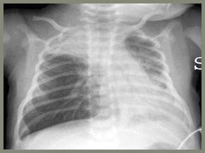
Inappropriate use of chest physiotherapy to treat pediatric bronchiolitis also declined, from 14% of cases in 2007 to 4.2% in 2010, said Dr. Ralston, chief of the division of inpatient pediatrics at the University of Texas Health Science Center, San Antonio.
The investigators had hoped to see declines as well in the inappropriate use of chest x-rays, steroids, and testing for respiratory syncytial virus, but mild declines in these categories did not reach statistical significance.
Bronchiolitis is the most common reason for hospitalizing infants and toddlers, with wide variation in care. Practice guidelines from the American Academy of Pediatrics (AAP) in 2006 recommend treating pediatric bronchiolitis with nothing but supportive care. Bronchodilator therapy and steroid therapy have been shown to have no benefit for these cases in repeated meta-analyses over the past 15 and 12 years, respectively.
"None of the things we all do frequently have any support" in the literature for managing pediatric bronchiolitis, Dr. Ralston, a steering committee member of the Value in Inpatient Pediatrics (VIP) Network.
The VIP Network, formed out of an AAP section on hospital medicine, pulled together physicians who were frustrated with the lack of pediatric resources for quality improvement in smaller academic and community hospitals. The VIP Network chose bronchiolitis as its first target to see if it could start to change management by providing a peer group for local leaders to share resources and benchmark progress within the group.

The VIP Network functions as a virtual community with yearly meetings that adopted group norms for appropriate bronchiolitis care. Its website offers handouts, bibliographies, and other resources, and network members provided peer-to-peer coaching on quality improvement.
Members produced yearly benchmarking reports using hospital billing data for diagnostic codes 466.11 or 466.19 starting in 2008 (with some 2007 data serving as additional baseline data). Dr. Ralston said that 90% of data came from non-freestanding children’s facilities. In all, 70% of participating facilities did not have a guideline for pediatric bronchiolitis management prior to joining the Network, Dr. Ralston said. A total of 30% of facilities were in urban locations.
The results show that an all-volunteer group with no source of specific funding and no new resources can make real-world changes that improve the quality of care, she said.
"The caveat is that it has to be something the physicians really care about," she said. "Clinicians are significantly more motivated for this kind of work than researchers."
Dr. Ralston reported having no financial disclosures. She receives funds from the American Academy of Pediatrics to be editor of the journal Hospital Pediatrics.
SAN DIEGO – Inappropriate use of bronchodilators to treat pediatric bronchiolitis declined from 70% of patients in 2007 to 58% in 2010, thanks to benchmarking and peer-to-peer collaborative efforts of the Value in Inpatient Pediatrics Network at hospitals in 14 states.
Bronchodilator utilization decreased by 45% as measured by doses per patient, from 7.6 doses/patient in 2007 to 4.2 doses/patient in 2010, Dr. Shawn Ralston and her associates reported at the annual meeting of the Society of Hospital Medicine.
Inappropriate use of chest physiotherapy to treat pediatric bronchiolitis also declined, from 14% of cases in 2007 to 4.2% in 2010, said Dr. Ralston, chief of the division of inpatient pediatrics at the University of Texas Health Science Center, San Antonio.
The investigators had hoped to see declines as well in the inappropriate use of chest x-rays, steroids, and testing for respiratory syncytial virus, but mild declines in these categories did not reach statistical significance.
Bronchiolitis is the most common reason for hospitalizing infants and toddlers, with wide variation in care. Practice guidelines from the American Academy of Pediatrics (AAP) in 2006 recommend treating pediatric bronchiolitis with nothing but supportive care. Bronchodilator therapy and steroid therapy have been shown to have no benefit for these cases in repeated meta-analyses over the past 15 and 12 years, respectively.
"None of the things we all do frequently have any support" in the literature for managing pediatric bronchiolitis, Dr. Ralston, a steering committee member of the Value in Inpatient Pediatrics (VIP) Network.
The VIP Network, formed out of an AAP section on hospital medicine, pulled together physicians who were frustrated with the lack of pediatric resources for quality improvement in smaller academic and community hospitals. The VIP Network chose bronchiolitis as its first target to see if it could start to change management by providing a peer group for local leaders to share resources and benchmark progress within the group.
The VIP Network functions as a virtual community with yearly meetings that adopted group norms for appropriate bronchiolitis care. Its website offers handouts, bibliographies, and other resources, and network members provided peer-to-peer coaching on quality improvement.
Members produced yearly benchmarking reports using hospital billing data for diagnostic codes 466.11 or 466.19 starting in 2008 (with some 2007 data serving as additional baseline data). Dr. Ralston said that 90% of data came from non-freestanding children’s facilities. In all, 70% of participating facilities did not have a guideline for pediatric bronchiolitis management prior to joining the Network, Dr. Ralston said. A total of 30% of facilities were in urban locations.
The results show that an all-volunteer group with no source of specific funding and no new resources can make real-world changes that improve the quality of care, she said.
"The caveat is that it has to be something the physicians really care about," she said. "Clinicians are significantly more motivated for this kind of work than researchers."
Dr. Ralston reported having no financial disclosures. She receives funds from the American Academy of Pediatrics to be editor of the journal Hospital Pediatrics.
SAN DIEGO – Inappropriate use of bronchodilators to treat pediatric bronchiolitis declined from 70% of patients in 2007 to 58% in 2010, thanks to benchmarking and peer-to-peer collaborative efforts of the Value in Inpatient Pediatrics Network at hospitals in 14 states.
Bronchodilator utilization decreased by 45% as measured by doses per patient, from 7.6 doses/patient in 2007 to 4.2 doses/patient in 2010, Dr. Shawn Ralston and her associates reported at the annual meeting of the Society of Hospital Medicine.
Inappropriate use of chest physiotherapy to treat pediatric bronchiolitis also declined, from 14% of cases in 2007 to 4.2% in 2010, said Dr. Ralston, chief of the division of inpatient pediatrics at the University of Texas Health Science Center, San Antonio.
The investigators had hoped to see declines as well in the inappropriate use of chest x-rays, steroids, and testing for respiratory syncytial virus, but mild declines in these categories did not reach statistical significance.
Bronchiolitis is the most common reason for hospitalizing infants and toddlers, with wide variation in care. Practice guidelines from the American Academy of Pediatrics (AAP) in 2006 recommend treating pediatric bronchiolitis with nothing but supportive care. Bronchodilator therapy and steroid therapy have been shown to have no benefit for these cases in repeated meta-analyses over the past 15 and 12 years, respectively.
"None of the things we all do frequently have any support" in the literature for managing pediatric bronchiolitis, Dr. Ralston, a steering committee member of the Value in Inpatient Pediatrics (VIP) Network.
The VIP Network, formed out of an AAP section on hospital medicine, pulled together physicians who were frustrated with the lack of pediatric resources for quality improvement in smaller academic and community hospitals. The VIP Network chose bronchiolitis as its first target to see if it could start to change management by providing a peer group for local leaders to share resources and benchmark progress within the group.
The VIP Network functions as a virtual community with yearly meetings that adopted group norms for appropriate bronchiolitis care. Its website offers handouts, bibliographies, and other resources, and network members provided peer-to-peer coaching on quality improvement.
Members produced yearly benchmarking reports using hospital billing data for diagnostic codes 466.11 or 466.19 starting in 2008 (with some 2007 data serving as additional baseline data). Dr. Ralston said that 90% of data came from non-freestanding children’s facilities. In all, 70% of participating facilities did not have a guideline for pediatric bronchiolitis management prior to joining the Network, Dr. Ralston said. A total of 30% of facilities were in urban locations.
The results show that an all-volunteer group with no source of specific funding and no new resources can make real-world changes that improve the quality of care, she said.
"The caveat is that it has to be something the physicians really care about," she said. "Clinicians are significantly more motivated for this kind of work than researchers."
Dr. Ralston reported having no financial disclosures. She receives funds from the American Academy of Pediatrics to be editor of the journal Hospital Pediatrics.
FROM THE ANNUAL MEETING OF THE SOCIETY OF HOSPITAL MEDICINE
Major Finding: Use of bronchodilators for pediatric bronchiolitis declined from 70% of cases to 58%, and bronchodilator doses per patient decreased by 45% from 2007 to 2010.
Data Source: Data came from an analysis of hospital billing data from participating sites in the Value in Inpatient Pediatrics Network.
Disclosures: Dr. Ralston reported having no relevant financial disclosures. She receives funds from the American Academy of Pediatrics to be editor of the journal Hospital Pediatrics.
Red Blood Cell Distribution Width Predicts CAP Outcome
LONDON – An elevated red blood cell distribution width predicted poor outcomes from community-acquired pneumonia in a retrospective analysis of more than 3,000 adult patients.
Red blood cell distribution width (RDW) is routinely measured as a check for anemia, but there have been several previous reports that the marker predicts poor outcome in a variety of conditions, including heart failure and sepsis. "It’s used in hematology, but it proves to be an outstanding marker of adverse outcome in some very important diseases of internal medicine. ... We found it actually works in community-acquired pneumonia [CAP] as well," Dr. Eyal Braun said in an interview at the European Congress of Clinical Microbiology and Infectious Diseases.

Last year, Dr. Braun and his associates at Rambam Health Care Campus, Haifa, Israel, reported that among 637 CAP patients aged 60 years and younger, elevated RDW levels on admission were associated with significantly higher rates of mortality and severe morbidity (Crit. Care 2011;15:R194).
The current study looked at the overall general population of internal medicine wards, comprising a total of 3,815 patients aged 18 years and older seen between Jan. 1, 2005, and Dec. 31, 2010. The patients had a mean age of 69.6 years, and 56% were men.
In the multivariate analysis, factors associated with mortality at 90 days were age greater than 80 years, a high Charlson comorbidity index (above 7), a bloodstream infection (BSI), a blood urea nitrogen (BUN) level greater than 30 mg/dL, an abnormal white blood cell (WBC) count, a systolic blood pressure less than 90 mm Hg, and an elevated RDW greater than 15% (odds ratio, 2.1). Variables associated with complicated hospitalization included a high Charlson comorbidity index, an abnormal WBC count, a BUN greater than 30 mg/dL, a hemoglobin level less than 10 g/dL, a BSI on admission, and an elevated RDW (OR 1.5), Dr. Braun reported in a poster.
The rates of mortality and complicated hospitalization were significantly higher among patients with an increased RDW, regardless of the WBC count or hemoglobin levels, he and his associates found.
In the interview, Dr. Braun said that an elevated RDW might help identify which patients who present with CAP need to be admitted to the hospital. Despite many existing scores aimed at identifying increased risk among CAP patients, "people are still dying from CAP. ... The bottom line is mortality from CAP is still high."
Dr. Braun stated that he has no relevant financial disclosures.
LONDON – An elevated red blood cell distribution width predicted poor outcomes from community-acquired pneumonia in a retrospective analysis of more than 3,000 adult patients.
Red blood cell distribution width (RDW) is routinely measured as a check for anemia, but there have been several previous reports that the marker predicts poor outcome in a variety of conditions, including heart failure and sepsis. "It’s used in hematology, but it proves to be an outstanding marker of adverse outcome in some very important diseases of internal medicine. ... We found it actually works in community-acquired pneumonia [CAP] as well," Dr. Eyal Braun said in an interview at the European Congress of Clinical Microbiology and Infectious Diseases.
Last year, Dr. Braun and his associates at Rambam Health Care Campus, Haifa, Israel, reported that among 637 CAP patients aged 60 years and younger, elevated RDW levels on admission were associated with significantly higher rates of mortality and severe morbidity (Crit. Care 2011;15:R194).
The current study looked at the overall general population of internal medicine wards, comprising a total of 3,815 patients aged 18 years and older seen between Jan. 1, 2005, and Dec. 31, 2010. The patients had a mean age of 69.6 years, and 56% were men.
In the multivariate analysis, factors associated with mortality at 90 days were age greater than 80 years, a high Charlson comorbidity index (above 7), a bloodstream infection (BSI), a blood urea nitrogen (BUN) level greater than 30 mg/dL, an abnormal white blood cell (WBC) count, a systolic blood pressure less than 90 mm Hg, and an elevated RDW greater than 15% (odds ratio, 2.1). Variables associated with complicated hospitalization included a high Charlson comorbidity index, an abnormal WBC count, a BUN greater than 30 mg/dL, a hemoglobin level less than 10 g/dL, a BSI on admission, and an elevated RDW (OR 1.5), Dr. Braun reported in a poster.
The rates of mortality and complicated hospitalization were significantly higher among patients with an increased RDW, regardless of the WBC count or hemoglobin levels, he and his associates found.
In the interview, Dr. Braun said that an elevated RDW might help identify which patients who present with CAP need to be admitted to the hospital. Despite many existing scores aimed at identifying increased risk among CAP patients, "people are still dying from CAP. ... The bottom line is mortality from CAP is still high."
Dr. Braun stated that he has no relevant financial disclosures.
LONDON – An elevated red blood cell distribution width predicted poor outcomes from community-acquired pneumonia in a retrospective analysis of more than 3,000 adult patients.
Red blood cell distribution width (RDW) is routinely measured as a check for anemia, but there have been several previous reports that the marker predicts poor outcome in a variety of conditions, including heart failure and sepsis. "It’s used in hematology, but it proves to be an outstanding marker of adverse outcome in some very important diseases of internal medicine. ... We found it actually works in community-acquired pneumonia [CAP] as well," Dr. Eyal Braun said in an interview at the European Congress of Clinical Microbiology and Infectious Diseases.
Last year, Dr. Braun and his associates at Rambam Health Care Campus, Haifa, Israel, reported that among 637 CAP patients aged 60 years and younger, elevated RDW levels on admission were associated with significantly higher rates of mortality and severe morbidity (Crit. Care 2011;15:R194).
The current study looked at the overall general population of internal medicine wards, comprising a total of 3,815 patients aged 18 years and older seen between Jan. 1, 2005, and Dec. 31, 2010. The patients had a mean age of 69.6 years, and 56% were men.
In the multivariate analysis, factors associated with mortality at 90 days were age greater than 80 years, a high Charlson comorbidity index (above 7), a bloodstream infection (BSI), a blood urea nitrogen (BUN) level greater than 30 mg/dL, an abnormal white blood cell (WBC) count, a systolic blood pressure less than 90 mm Hg, and an elevated RDW greater than 15% (odds ratio, 2.1). Variables associated with complicated hospitalization included a high Charlson comorbidity index, an abnormal WBC count, a BUN greater than 30 mg/dL, a hemoglobin level less than 10 g/dL, a BSI on admission, and an elevated RDW (OR 1.5), Dr. Braun reported in a poster.
The rates of mortality and complicated hospitalization were significantly higher among patients with an increased RDW, regardless of the WBC count or hemoglobin levels, he and his associates found.
In the interview, Dr. Braun said that an elevated RDW might help identify which patients who present with CAP need to be admitted to the hospital. Despite many existing scores aimed at identifying increased risk among CAP patients, "people are still dying from CAP. ... The bottom line is mortality from CAP is still high."
Dr. Braun stated that he has no relevant financial disclosures.
FROM THE EUROPEAN CONGRESS OF CLINICAL MICROBIOLOGY AND INFECTIOUS DISEASES
Major Finding: In multivariate analysis, an elevated RDW greater than 15% (OR, 2.1) was independently associated with 90-day mortality (OR, 2.1).
Data Source: The findings come from a review of 3,815 patients aged 18 and older who were hospitalized with CAP over a 5-year period.
Disclosures: Dr. Braun stated that he has no relevant financial disclosures.