User login
Bringing you the latest news, research and reviews, exclusive interviews, podcasts, quizzes, and more.
Powered by CHEST Physician, Clinician Reviews, MDedge Family Medicine, Internal Medicine News, and The Journal of Clinical Outcomes Management.
The Weight of Sleep Deprivation
Americans are very tired. The National Sleep Foundation recommends that adults sleep 7 to 9 hours per day, yet the percentage of U.S. adults securing at least 8 hours of sleep on weeknights has fallen from 38% to 27% since 2001. Sleep promotion is a recognized national health agenda with a Healthy People 2020 objective to increase the proportion of adults who get sufficient sleep to 71%.
CDC released a report last month on the prevalence of short sleep duration among U.S. workers. Short sleep duration (average ≤ 6 hours per 24-hour period) was reported by 30% of employed U.S. adults (approximately 40.6 million workers). The prevalence of short sleep duration was especially high among night shift workers in the transportation and warehousing (70%) and healthcare and social assistance (52%) industries. Sleep deprivation has been linked to adverse health consequences, such as obesity. But how does sleep deprivation lead to obesity?
Dr. Nathaniel Watson from the University of Washington and colleagues recently published a population-based twin-study evaluating the link between sleep duration and BMI (Sleep. 2012 May 1;35(5):597-603) that begins to unlock the sleep-BMI mystery. Investigators analyzed data from a twin registry involving 1,088 complete twin pairs. Normal sleep duration was considered 7 to 8.9 hours. Findings from this study suggest that restricting sleep provides a permissive environment for the expression of genes promoting obesity. The flipside is that extending sleep suppresses the expression of obesity genes.
The study adds to extant data suggesting that people who sleep six or fewer hours are at greater risk for obesity. Other published data suggest that too much sleep may increase the risk for cardiovascular disease, insulin resistance, and mortality. One may find it helpful to think of the relationship between nightly sleep duration and BMI as a U-shaped curve – both short and long sleepers weigh more than those in the sweet spot around 7 to 9 hours.
This study does not inform us about whether an intervention aimed at increasing sleep will result in clinically significant weight loss. But it does suggest that behavioral measures to lose weight may be more effective when genetic drivers of body weight are ratcheted down through sleep extension.
This knowledge may prove useful to our patients who are trying to get a handle on their body weight and literally feel tired doing so. Evidence is now mounting that a component of obesity treatment may be to tell our patients to get some sleep.
Jon O. Ebbert, M.D. is a professor of medicine and a primary care clinician at the Mayo Clinic in Rochester, MN. The opinions expressed are solely those of the author. Contact him at ebbert.jon@mayo.edu.
Americans are very tired. The National Sleep Foundation recommends that adults sleep 7 to 9 hours per day, yet the percentage of U.S. adults securing at least 8 hours of sleep on weeknights has fallen from 38% to 27% since 2001. Sleep promotion is a recognized national health agenda with a Healthy People 2020 objective to increase the proportion of adults who get sufficient sleep to 71%.
CDC released a report last month on the prevalence of short sleep duration among U.S. workers. Short sleep duration (average ≤ 6 hours per 24-hour period) was reported by 30% of employed U.S. adults (approximately 40.6 million workers). The prevalence of short sleep duration was especially high among night shift workers in the transportation and warehousing (70%) and healthcare and social assistance (52%) industries. Sleep deprivation has been linked to adverse health consequences, such as obesity. But how does sleep deprivation lead to obesity?
Dr. Nathaniel Watson from the University of Washington and colleagues recently published a population-based twin-study evaluating the link between sleep duration and BMI (Sleep. 2012 May 1;35(5):597-603) that begins to unlock the sleep-BMI mystery. Investigators analyzed data from a twin registry involving 1,088 complete twin pairs. Normal sleep duration was considered 7 to 8.9 hours. Findings from this study suggest that restricting sleep provides a permissive environment for the expression of genes promoting obesity. The flipside is that extending sleep suppresses the expression of obesity genes.
The study adds to extant data suggesting that people who sleep six or fewer hours are at greater risk for obesity. Other published data suggest that too much sleep may increase the risk for cardiovascular disease, insulin resistance, and mortality. One may find it helpful to think of the relationship between nightly sleep duration and BMI as a U-shaped curve – both short and long sleepers weigh more than those in the sweet spot around 7 to 9 hours.
This study does not inform us about whether an intervention aimed at increasing sleep will result in clinically significant weight loss. But it does suggest that behavioral measures to lose weight may be more effective when genetic drivers of body weight are ratcheted down through sleep extension.
This knowledge may prove useful to our patients who are trying to get a handle on their body weight and literally feel tired doing so. Evidence is now mounting that a component of obesity treatment may be to tell our patients to get some sleep.
Jon O. Ebbert, M.D. is a professor of medicine and a primary care clinician at the Mayo Clinic in Rochester, MN. The opinions expressed are solely those of the author. Contact him at ebbert.jon@mayo.edu.
Americans are very tired. The National Sleep Foundation recommends that adults sleep 7 to 9 hours per day, yet the percentage of U.S. adults securing at least 8 hours of sleep on weeknights has fallen from 38% to 27% since 2001. Sleep promotion is a recognized national health agenda with a Healthy People 2020 objective to increase the proportion of adults who get sufficient sleep to 71%.
CDC released a report last month on the prevalence of short sleep duration among U.S. workers. Short sleep duration (average ≤ 6 hours per 24-hour period) was reported by 30% of employed U.S. adults (approximately 40.6 million workers). The prevalence of short sleep duration was especially high among night shift workers in the transportation and warehousing (70%) and healthcare and social assistance (52%) industries. Sleep deprivation has been linked to adverse health consequences, such as obesity. But how does sleep deprivation lead to obesity?
Dr. Nathaniel Watson from the University of Washington and colleagues recently published a population-based twin-study evaluating the link between sleep duration and BMI (Sleep. 2012 May 1;35(5):597-603) that begins to unlock the sleep-BMI mystery. Investigators analyzed data from a twin registry involving 1,088 complete twin pairs. Normal sleep duration was considered 7 to 8.9 hours. Findings from this study suggest that restricting sleep provides a permissive environment for the expression of genes promoting obesity. The flipside is that extending sleep suppresses the expression of obesity genes.
The study adds to extant data suggesting that people who sleep six or fewer hours are at greater risk for obesity. Other published data suggest that too much sleep may increase the risk for cardiovascular disease, insulin resistance, and mortality. One may find it helpful to think of the relationship between nightly sleep duration and BMI as a U-shaped curve – both short and long sleepers weigh more than those in the sweet spot around 7 to 9 hours.
This study does not inform us about whether an intervention aimed at increasing sleep will result in clinically significant weight loss. But it does suggest that behavioral measures to lose weight may be more effective when genetic drivers of body weight are ratcheted down through sleep extension.
This knowledge may prove useful to our patients who are trying to get a handle on their body weight and literally feel tired doing so. Evidence is now mounting that a component of obesity treatment may be to tell our patients to get some sleep.
Jon O. Ebbert, M.D. is a professor of medicine and a primary care clinician at the Mayo Clinic in Rochester, MN. The opinions expressed are solely those of the author. Contact him at ebbert.jon@mayo.edu.
Stale Secondhand Smoke Impairs Endothelial Function
Exposure to "aged" secondhand smoke – even to a small amount and even for a brief time – impairs endothelial function, according to a report in the May 22 issue of the Journal of the American College of Cardiology.
"Aged" secondhand smoke refers to smoke that lingers in an indoor area 30 minutes or more after a smoker has finished a cigarette, and it is known to be more toxic to the respiratory epithelium than is fresh secondhand smoke, said Dr. Paul F. Frey of the division of cardiology, San Francisco General Hospital, and his associates.
The investigators performed a study to determine whether stale secondhand smoke also impairs endothelial function at the relatively low exposure levels that people are likely to encounter in the community setting. Endothelial dysfunction is a key mechanism in all stages of cardiovascular disease, they noted.

The typical level of aged secondhand smoke found in smokers’ homes or in restaurants or other public venues that allow smoking is 100 mcg/m3 respirable suspended particles (RSPs), and the typical level found in bars or casinos in which smoke is more concentrated is 400 mcg/m3 RSPs. Dr. Frey and his colleagues assessed the response to 30 minutes of exposure at both of these levels, as well as to filtered smoke-free air, in 33 healthy nonsmoking adults aged 18-40 years.
All the study participants reported no exposure to secondhand smoke during the month preceding the study. None of them had conditions that could adversely affect endothelial function such as diabetes, hypertension, respiratory disease, kidney disease, coronary artery disease, or heart failure.
Endothelial function was assessed using high-resolution ultrasound to measure maximal percent flow-mediated dilation of the brachial artery before and after exposure.
The study participants were exposed to smoke-free air (11 participants), 100 mcg/m3 RSPs (11 participants), or 400 mcg/m3 RSPs (11 participants) in a hooded device attached to a smoking machine. The secondhand smoke was aged for 60 minutes, then routed to the hood for a single 30-minute exposure time. The RSP level was monitored continuously.
Endothelial function was impaired in a dose-dependent fashion at both levels of exposure to aged secondhand smoke. For every 100 mcg/m3 increase in RSP level, maximal percent flow-mediated dilation of the brachial artery decreased by 0.67%, Dr. Frey and his associates said (J. Am. Coll. Cardiol. 2012;59:1908-13).
"Our research strengthens the evidence that secondhand smoke is detrimental to cardiovascular health even at very short exposures and low particulate concentrations," they noted.
The findings highlight the importance of policies that limit the public’s exposure to secondhand smoke, the researchers said.
The study conditions may underestimate the effect of aged secondhand smoke in real-world settings, they added.
The subjects remained at rest throughout their exposure to secondhand smoke and were exposed for only half an hour. In real-world experience, people are exposed for much longer durations and may be physically active during their exposure, which increases minute ventilation. Moreover, "our subjects were healthy and may have been less susceptible to decrements in endothelial function than patients with vascular disease who are at greater risk for secondhand smoke–induced acute cardiovascular events."
This study was supported in part by the Tobacco-Related Disease Research Program and the Flight Attendants Medical Research Institute Bland Lane Center of Excellence on Secondhand Smoke at the University of California, San Francisco. One coauthor reported ties to pharmaceutical companies that develop or market smoking-cessation medications and being a paid expert witness in litigation against tobacco companies.
Exposure to "aged" secondhand smoke – even to a small amount and even for a brief time – impairs endothelial function, according to a report in the May 22 issue of the Journal of the American College of Cardiology.
"Aged" secondhand smoke refers to smoke that lingers in an indoor area 30 minutes or more after a smoker has finished a cigarette, and it is known to be more toxic to the respiratory epithelium than is fresh secondhand smoke, said Dr. Paul F. Frey of the division of cardiology, San Francisco General Hospital, and his associates.
The investigators performed a study to determine whether stale secondhand smoke also impairs endothelial function at the relatively low exposure levels that people are likely to encounter in the community setting. Endothelial dysfunction is a key mechanism in all stages of cardiovascular disease, they noted.
The typical level of aged secondhand smoke found in smokers’ homes or in restaurants or other public venues that allow smoking is 100 mcg/m3 respirable suspended particles (RSPs), and the typical level found in bars or casinos in which smoke is more concentrated is 400 mcg/m3 RSPs. Dr. Frey and his colleagues assessed the response to 30 minutes of exposure at both of these levels, as well as to filtered smoke-free air, in 33 healthy nonsmoking adults aged 18-40 years.
All the study participants reported no exposure to secondhand smoke during the month preceding the study. None of them had conditions that could adversely affect endothelial function such as diabetes, hypertension, respiratory disease, kidney disease, coronary artery disease, or heart failure.
Endothelial function was assessed using high-resolution ultrasound to measure maximal percent flow-mediated dilation of the brachial artery before and after exposure.
The study participants were exposed to smoke-free air (11 participants), 100 mcg/m3 RSPs (11 participants), or 400 mcg/m3 RSPs (11 participants) in a hooded device attached to a smoking machine. The secondhand smoke was aged for 60 minutes, then routed to the hood for a single 30-minute exposure time. The RSP level was monitored continuously.
Endothelial function was impaired in a dose-dependent fashion at both levels of exposure to aged secondhand smoke. For every 100 mcg/m3 increase in RSP level, maximal percent flow-mediated dilation of the brachial artery decreased by 0.67%, Dr. Frey and his associates said (J. Am. Coll. Cardiol. 2012;59:1908-13).
"Our research strengthens the evidence that secondhand smoke is detrimental to cardiovascular health even at very short exposures and low particulate concentrations," they noted.
The findings highlight the importance of policies that limit the public’s exposure to secondhand smoke, the researchers said.
The study conditions may underestimate the effect of aged secondhand smoke in real-world settings, they added.
The subjects remained at rest throughout their exposure to secondhand smoke and were exposed for only half an hour. In real-world experience, people are exposed for much longer durations and may be physically active during their exposure, which increases minute ventilation. Moreover, "our subjects were healthy and may have been less susceptible to decrements in endothelial function than patients with vascular disease who are at greater risk for secondhand smoke–induced acute cardiovascular events."
This study was supported in part by the Tobacco-Related Disease Research Program and the Flight Attendants Medical Research Institute Bland Lane Center of Excellence on Secondhand Smoke at the University of California, San Francisco. One coauthor reported ties to pharmaceutical companies that develop or market smoking-cessation medications and being a paid expert witness in litigation against tobacco companies.
Exposure to "aged" secondhand smoke – even to a small amount and even for a brief time – impairs endothelial function, according to a report in the May 22 issue of the Journal of the American College of Cardiology.
"Aged" secondhand smoke refers to smoke that lingers in an indoor area 30 minutes or more after a smoker has finished a cigarette, and it is known to be more toxic to the respiratory epithelium than is fresh secondhand smoke, said Dr. Paul F. Frey of the division of cardiology, San Francisco General Hospital, and his associates.
The investigators performed a study to determine whether stale secondhand smoke also impairs endothelial function at the relatively low exposure levels that people are likely to encounter in the community setting. Endothelial dysfunction is a key mechanism in all stages of cardiovascular disease, they noted.
The typical level of aged secondhand smoke found in smokers’ homes or in restaurants or other public venues that allow smoking is 100 mcg/m3 respirable suspended particles (RSPs), and the typical level found in bars or casinos in which smoke is more concentrated is 400 mcg/m3 RSPs. Dr. Frey and his colleagues assessed the response to 30 minutes of exposure at both of these levels, as well as to filtered smoke-free air, in 33 healthy nonsmoking adults aged 18-40 years.
All the study participants reported no exposure to secondhand smoke during the month preceding the study. None of them had conditions that could adversely affect endothelial function such as diabetes, hypertension, respiratory disease, kidney disease, coronary artery disease, or heart failure.
Endothelial function was assessed using high-resolution ultrasound to measure maximal percent flow-mediated dilation of the brachial artery before and after exposure.
The study participants were exposed to smoke-free air (11 participants), 100 mcg/m3 RSPs (11 participants), or 400 mcg/m3 RSPs (11 participants) in a hooded device attached to a smoking machine. The secondhand smoke was aged for 60 minutes, then routed to the hood for a single 30-minute exposure time. The RSP level was monitored continuously.
Endothelial function was impaired in a dose-dependent fashion at both levels of exposure to aged secondhand smoke. For every 100 mcg/m3 increase in RSP level, maximal percent flow-mediated dilation of the brachial artery decreased by 0.67%, Dr. Frey and his associates said (J. Am. Coll. Cardiol. 2012;59:1908-13).
"Our research strengthens the evidence that secondhand smoke is detrimental to cardiovascular health even at very short exposures and low particulate concentrations," they noted.
The findings highlight the importance of policies that limit the public’s exposure to secondhand smoke, the researchers said.
The study conditions may underestimate the effect of aged secondhand smoke in real-world settings, they added.
The subjects remained at rest throughout their exposure to secondhand smoke and were exposed for only half an hour. In real-world experience, people are exposed for much longer durations and may be physically active during their exposure, which increases minute ventilation. Moreover, "our subjects were healthy and may have been less susceptible to decrements in endothelial function than patients with vascular disease who are at greater risk for secondhand smoke–induced acute cardiovascular events."
This study was supported in part by the Tobacco-Related Disease Research Program and the Flight Attendants Medical Research Institute Bland Lane Center of Excellence on Secondhand Smoke at the University of California, San Francisco. One coauthor reported ties to pharmaceutical companies that develop or market smoking-cessation medications and being a paid expert witness in litigation against tobacco companies.
FROM THE JOURNAL OF THE AMERICAN COLLEGE OF CARDIOLOGY
Major Finding: Endothelial function was impaired in a dose-dependent fashion after brief exposure to aged secondhand smoke, so that for every 100 mcg/m3 increase in respirable suspended particles, maximal percent flow-mediated dilation of the brachial artery decreased by 0.67%.
Data Source: Data were taken from an analysis of brachial artery flow-mediated dilation in 33 healthy young adults exposed to varying levels of aged secondhand smoke.
Disclosures: This study was supported in part by the Tobacco-Related Disease Research Program and the Flight Attendants Medical Research Institute Bland Lane Center of Excellence on Secondhand Smoke at the University of California, San Francisco. One coauthor reported ties to pharmaceutical companies that develop or market smoking cessation medications and being a paid expert witness in litigation against tobacco companies.
Ex Vivo Perfusion Expands Pool of Lung Transplants
SAN FRANCISCO – Additional research continues to show good outcomes with a new process that allows successful transplant of lungs that might otherwise be deemed unacceptable.
Researchers at the Organ Regeneration Laboratory at the University of Toronto evaluated and repaired 58 donor lungs over 4-6 hours in a process called normothermic ex vivo lung perfusion (EVLP). In all, 50 of these lungs were successfully transplanted into patients, for a final utilization rate of 86%, Dr. Marcelo Cypel reported at the annual meeting of the American Association for Thoracic Surgery.
The current study builds on a previous report on initial experience with EVLP from the same research team (N. Engl. J. Med. 2011;364:1431-40).
"As we all know, one of the major problems in doing lung transplantation is the organ shortage and the low utilization rates. Only 17% of the lungs from brain death donors and 2% of the lungs from cardiac death donors are used currently," Dr. Cypel said. EVLP lungs accounted for 20% of the transplantations at Toronto General Hospital in 2011.
The EVLP lungs came from 32 brain death donors and 26 cardiac death donors. Dr. Cypel and his colleagues compared the outcomes of these EVLP procedures to another 253 conventional lung transplantations performed at their institution from September 2008 to December 2011.
EVLP patients received a significantly higher percentage of lungs from cardiac death donors, which are generally considered less desirable than lungs from brain death donors. They also received a higher percentage of high-risk lungs from brain death donors (PaO2/FIO2 below 300 mm Hg) and more lungs with chest x-ray abnormalities, such as signs of pulmonary edema, compared with the conventional group.
"Donor lungs in the EVLP group were significantly more injured at baseline, however, the outcomes were comparable," said Dr. Cypel, a member of the surgical faculty in the Division of Thoracic Surgery at Toronto General Hospital University Health Network.
For example, posttransplant survival at 1 year was 86% for the EVLP group and 87% for the traditional transplant group in this retrospective study; at 3 years these rates dropped to 70% and 72%. There were no significant differences in survival for patients who received lungs from brain death or cardiac death donors.
Other findings included no significant difference in the rate of primary graft grade 3 dysfunction at 72 hours between groups according to International Society for Heart and Lung Transplantation criteria. The EVLP patients had a trend toward a decreased length of hospital stay, compared with conventional transplant recipients, Dr. Cypel said.
"Again, you and your colleagues have demonstrated the safety and efficacy of using EVLP in the transplantation of lungs that previously would not have been used by your group," said study discussant Dr. R. Duane Davis, director of transplant services at Duke University Health System in Durham, N.C. "Using this technology, we may be able to start applying lung transplant more practically for societal needs."
Dr. Davis asked Dr. Cypel how surgeons at Toronto General Hospital achieved an 86% EVLP utilization rate compared with the 54% rate observed in the U.S. trial and comparable rates in the United Kingdom and elsewhere.
"Our experience with the procedure and extensive laboratory research prior to starting the clinical trial" explain the difference, Dr. Cypel replied. Donor selection criteria also could play a role.
"The important thing is it is taking some of the adventure out of lung transplantation," study coauthor Dr. Shaf Keshavjee said during a separate presentation at the meeting. "Ex vivo lung perfusion is clinically feasible. We can we do a long-term perfusion of lungs outside the body without injuring them. It is possible to keep lung 12 hours outside the body and normothermic."
"We are developing ways to figure out which lungs need a fix and to target treatment to lungs that need treatment," said Dr. Keshavjee, director of the Toronto Lung Transplant program and chair of the Division of Thoracic Surgery at the University of Toronto. Examples include resolution of pulmonary edema and infections through EVLP. Treating infected lungs with lavage and high-dose antibiotics may one day make these organs acceptable for transplantation, even in cases of pneumonia.
The goal is to double or triple the overall number of lung transplants using the EVLP technique in the United States, said Dr. Davis. EVLP comprises 20% of transplants in Toronto, but the overall transplant volume has not increased.
EVLP has allowed the overall number of 100 transplants or so per year in Toronto to remain steady at the same time that organ donation rates have decreased, Dr. Cypel responded.
"The major contribution of EVLP will not be for the large transplant centers like Duke or Toronto, which already use 40% of the organs and for whom a marginal increase is not that large," Dr. Cypel said. "Look at the majority of lung transplant centers that use 10% or less of the offered lungs; that is where we can have a major impact by increasing the number of organs available."
Vitrolife supported the clinical trial. Dr. Cypel and Dr. Keshavjee reported no other relevant disclosures. Dr. Davis said he received research support from Vitrolife for a U.S. study.
SAN FRANCISCO – Additional research continues to show good outcomes with a new process that allows successful transplant of lungs that might otherwise be deemed unacceptable.
Researchers at the Organ Regeneration Laboratory at the University of Toronto evaluated and repaired 58 donor lungs over 4-6 hours in a process called normothermic ex vivo lung perfusion (EVLP). In all, 50 of these lungs were successfully transplanted into patients, for a final utilization rate of 86%, Dr. Marcelo Cypel reported at the annual meeting of the American Association for Thoracic Surgery.
The current study builds on a previous report on initial experience with EVLP from the same research team (N. Engl. J. Med. 2011;364:1431-40).
"As we all know, one of the major problems in doing lung transplantation is the organ shortage and the low utilization rates. Only 17% of the lungs from brain death donors and 2% of the lungs from cardiac death donors are used currently," Dr. Cypel said. EVLP lungs accounted for 20% of the transplantations at Toronto General Hospital in 2011.
The EVLP lungs came from 32 brain death donors and 26 cardiac death donors. Dr. Cypel and his colleagues compared the outcomes of these EVLP procedures to another 253 conventional lung transplantations performed at their institution from September 2008 to December 2011.
EVLP patients received a significantly higher percentage of lungs from cardiac death donors, which are generally considered less desirable than lungs from brain death donors. They also received a higher percentage of high-risk lungs from brain death donors (PaO2/FIO2 below 300 mm Hg) and more lungs with chest x-ray abnormalities, such as signs of pulmonary edema, compared with the conventional group.
"Donor lungs in the EVLP group were significantly more injured at baseline, however, the outcomes were comparable," said Dr. Cypel, a member of the surgical faculty in the Division of Thoracic Surgery at Toronto General Hospital University Health Network.
For example, posttransplant survival at 1 year was 86% for the EVLP group and 87% for the traditional transplant group in this retrospective study; at 3 years these rates dropped to 70% and 72%. There were no significant differences in survival for patients who received lungs from brain death or cardiac death donors.
Other findings included no significant difference in the rate of primary graft grade 3 dysfunction at 72 hours between groups according to International Society for Heart and Lung Transplantation criteria. The EVLP patients had a trend toward a decreased length of hospital stay, compared with conventional transplant recipients, Dr. Cypel said.
"Again, you and your colleagues have demonstrated the safety and efficacy of using EVLP in the transplantation of lungs that previously would not have been used by your group," said study discussant Dr. R. Duane Davis, director of transplant services at Duke University Health System in Durham, N.C. "Using this technology, we may be able to start applying lung transplant more practically for societal needs."
Dr. Davis asked Dr. Cypel how surgeons at Toronto General Hospital achieved an 86% EVLP utilization rate compared with the 54% rate observed in the U.S. trial and comparable rates in the United Kingdom and elsewhere.
"Our experience with the procedure and extensive laboratory research prior to starting the clinical trial" explain the difference, Dr. Cypel replied. Donor selection criteria also could play a role.
"The important thing is it is taking some of the adventure out of lung transplantation," study coauthor Dr. Shaf Keshavjee said during a separate presentation at the meeting. "Ex vivo lung perfusion is clinically feasible. We can we do a long-term perfusion of lungs outside the body without injuring them. It is possible to keep lung 12 hours outside the body and normothermic."
"We are developing ways to figure out which lungs need a fix and to target treatment to lungs that need treatment," said Dr. Keshavjee, director of the Toronto Lung Transplant program and chair of the Division of Thoracic Surgery at the University of Toronto. Examples include resolution of pulmonary edema and infections through EVLP. Treating infected lungs with lavage and high-dose antibiotics may one day make these organs acceptable for transplantation, even in cases of pneumonia.
The goal is to double or triple the overall number of lung transplants using the EVLP technique in the United States, said Dr. Davis. EVLP comprises 20% of transplants in Toronto, but the overall transplant volume has not increased.
EVLP has allowed the overall number of 100 transplants or so per year in Toronto to remain steady at the same time that organ donation rates have decreased, Dr. Cypel responded.
"The major contribution of EVLP will not be for the large transplant centers like Duke or Toronto, which already use 40% of the organs and for whom a marginal increase is not that large," Dr. Cypel said. "Look at the majority of lung transplant centers that use 10% or less of the offered lungs; that is where we can have a major impact by increasing the number of organs available."
Vitrolife supported the clinical trial. Dr. Cypel and Dr. Keshavjee reported no other relevant disclosures. Dr. Davis said he received research support from Vitrolife for a U.S. study.
SAN FRANCISCO – Additional research continues to show good outcomes with a new process that allows successful transplant of lungs that might otherwise be deemed unacceptable.
Researchers at the Organ Regeneration Laboratory at the University of Toronto evaluated and repaired 58 donor lungs over 4-6 hours in a process called normothermic ex vivo lung perfusion (EVLP). In all, 50 of these lungs were successfully transplanted into patients, for a final utilization rate of 86%, Dr. Marcelo Cypel reported at the annual meeting of the American Association for Thoracic Surgery.
The current study builds on a previous report on initial experience with EVLP from the same research team (N. Engl. J. Med. 2011;364:1431-40).
"As we all know, one of the major problems in doing lung transplantation is the organ shortage and the low utilization rates. Only 17% of the lungs from brain death donors and 2% of the lungs from cardiac death donors are used currently," Dr. Cypel said. EVLP lungs accounted for 20% of the transplantations at Toronto General Hospital in 2011.
The EVLP lungs came from 32 brain death donors and 26 cardiac death donors. Dr. Cypel and his colleagues compared the outcomes of these EVLP procedures to another 253 conventional lung transplantations performed at their institution from September 2008 to December 2011.
EVLP patients received a significantly higher percentage of lungs from cardiac death donors, which are generally considered less desirable than lungs from brain death donors. They also received a higher percentage of high-risk lungs from brain death donors (PaO2/FIO2 below 300 mm Hg) and more lungs with chest x-ray abnormalities, such as signs of pulmonary edema, compared with the conventional group.
"Donor lungs in the EVLP group were significantly more injured at baseline, however, the outcomes were comparable," said Dr. Cypel, a member of the surgical faculty in the Division of Thoracic Surgery at Toronto General Hospital University Health Network.
For example, posttransplant survival at 1 year was 86% for the EVLP group and 87% for the traditional transplant group in this retrospective study; at 3 years these rates dropped to 70% and 72%. There were no significant differences in survival for patients who received lungs from brain death or cardiac death donors.
Other findings included no significant difference in the rate of primary graft grade 3 dysfunction at 72 hours between groups according to International Society for Heart and Lung Transplantation criteria. The EVLP patients had a trend toward a decreased length of hospital stay, compared with conventional transplant recipients, Dr. Cypel said.
"Again, you and your colleagues have demonstrated the safety and efficacy of using EVLP in the transplantation of lungs that previously would not have been used by your group," said study discussant Dr. R. Duane Davis, director of transplant services at Duke University Health System in Durham, N.C. "Using this technology, we may be able to start applying lung transplant more practically for societal needs."
Dr. Davis asked Dr. Cypel how surgeons at Toronto General Hospital achieved an 86% EVLP utilization rate compared with the 54% rate observed in the U.S. trial and comparable rates in the United Kingdom and elsewhere.
"Our experience with the procedure and extensive laboratory research prior to starting the clinical trial" explain the difference, Dr. Cypel replied. Donor selection criteria also could play a role.
"The important thing is it is taking some of the adventure out of lung transplantation," study coauthor Dr. Shaf Keshavjee said during a separate presentation at the meeting. "Ex vivo lung perfusion is clinically feasible. We can we do a long-term perfusion of lungs outside the body without injuring them. It is possible to keep lung 12 hours outside the body and normothermic."
"We are developing ways to figure out which lungs need a fix and to target treatment to lungs that need treatment," said Dr. Keshavjee, director of the Toronto Lung Transplant program and chair of the Division of Thoracic Surgery at the University of Toronto. Examples include resolution of pulmonary edema and infections through EVLP. Treating infected lungs with lavage and high-dose antibiotics may one day make these organs acceptable for transplantation, even in cases of pneumonia.
The goal is to double or triple the overall number of lung transplants using the EVLP technique in the United States, said Dr. Davis. EVLP comprises 20% of transplants in Toronto, but the overall transplant volume has not increased.
EVLP has allowed the overall number of 100 transplants or so per year in Toronto to remain steady at the same time that organ donation rates have decreased, Dr. Cypel responded.
"The major contribution of EVLP will not be for the large transplant centers like Duke or Toronto, which already use 40% of the organs and for whom a marginal increase is not that large," Dr. Cypel said. "Look at the majority of lung transplant centers that use 10% or less of the offered lungs; that is where we can have a major impact by increasing the number of organs available."
Vitrolife supported the clinical trial. Dr. Cypel and Dr. Keshavjee reported no other relevant disclosures. Dr. Davis said he received research support from Vitrolife for a U.S. study.
FROM THE ANNUAL MEETING OF THE AMERICAN ASSOCIATION FOR THORACIC SURGERY
Major Finding: One-year survival was 86% in patients who received a lung transplant after optimization with normothermic ex vivo lung perfusion and 87% in those who underwent conventional lung transplantation.
Data Source: A retrospective study was performed of 311 lung transplantations at the University of Toronto from September 2008 to December 2011.
Disclosures: Vitrolife supported the clinical trial. Dr. Cypel and Dr. Keshavjee reported no other relevant disclosures. Dr. Davis said he received research support from Vitrolife.
Hardball, Softball Strategies Manage Night Wakings
BELLEVUE, WASH. – When tired parents complain that their child wakes up frequently at night – and wakes them up – you can offer them a hardball approach or softball approach to dealing with the problem.
The hardball approach is far more effective and more quickly resolves night wakings, but some parents find the idea of it intolerable. For them, there’s the softball approach, Dr. Charles H. Zeanah Jr. said at the annual meeting of the North Pacific Pediatric Society.
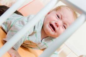
The goal of both strategies is to teach the child to fall asleep on his or her own. Waking during the night is normal at age 3-6 months, and 30% of infants are "signalers" who cry and expect someone to respond. By 8 months of age, 60%-70% of infants self-soothe after waking during the night. Parents report that 20%-30% of toddlers are night wakers, and most of these will continue to have sleep problems as 6-year-olds if the sleep problem is not addressed, he said.
"Somewhere between 6 and 9 months of age, I may start to think of it as a disorder or impairment, not just for the child but [also] for the parent," said Dr. Zeanah, professor of clinical pediatrics and psychiatry and director of child and adolescent psychiatry at Tulane University, New Orleans.
First, take a good history of family structure and routines around bedtime, limit-setting behaviors, distress, what tactics the parents have tried, and whether the parents agree on what needs to be done.
"It’s amazing how many people don’t have a bedtime routine," he said. "The key question is, ‘Is the child put down while awake?’ " Learning how to fall asleep at bedtime on his or her own, without "signaling," allows the child to fall back asleep after waking during the night.
Before talking about the hardball or softball approach to teaching a child to fall asleep on his or her own, Dr. Zeanah plays a little "hard to get" with the parents, to assess their commitment to an intervention. He tells them that there’s no evidence that night wakings cause a mental illness or severely damage a child. "I know it’s annoying, but I’m not sure if you want to do something about this" night-waking problem, he says.
Tired parents usually do want to intervene.
The first steps are to create a set bedtime and establish a consistent bedtime routine (such as reading a story) that ends in the child’s room. Finish the routine before the child falls asleep.
Then he explains the hardball approach: If the child cries when you leave, wait 5 minutes, go back and soothe the child without using words (for instance, by patting or rubbing the child’s back), then leave again. If the child cries again, repeat until the child falls asleep. Over the ensuing days, gradually increase the 5-minute interval to longer stretches between soothings.
"The rigidity of this is the key," Dr. Zeanah said, and it works. In 30 years of practice, he has never seen a child last more than a week before learning to fall asleep on his or her own.

If a parent can’t handle letting the child cry because they fear the child is scared to be alone, he explains the softball approach: When the child cries, go in and sit in a chair next to the child’s bed without interacting with the child, which takes any fear out of the situation. Sit in the chair until the child falls asleep. The next night, move the chair a couple of feet toward the door. Move a little more toward the door each night until the chair is outside the door, but visible to the child when you sit in it. Then pull the chair back so just your knees are visible, and eventually so the parent can’t be seen.
"It’s not as effective" as the hardball approach, but helps in some cases, he said.
With either approach, he asks parents to call him early in the morning after the first night of the intervention to let him know how it went. Check in with parents frequently during the intervention to encourage them and make adjustments, he advised.
With the hardball approach, "by the third night, there’s usually no reason to call," he said.
Dr. Zeanah reported having no financial disclosures.
BELLEVUE, WASH. – When tired parents complain that their child wakes up frequently at night – and wakes them up – you can offer them a hardball approach or softball approach to dealing with the problem.
The hardball approach is far more effective and more quickly resolves night wakings, but some parents find the idea of it intolerable. For them, there’s the softball approach, Dr. Charles H. Zeanah Jr. said at the annual meeting of the North Pacific Pediatric Society.
The goal of both strategies is to teach the child to fall asleep on his or her own. Waking during the night is normal at age 3-6 months, and 30% of infants are "signalers" who cry and expect someone to respond. By 8 months of age, 60%-70% of infants self-soothe after waking during the night. Parents report that 20%-30% of toddlers are night wakers, and most of these will continue to have sleep problems as 6-year-olds if the sleep problem is not addressed, he said.
"Somewhere between 6 and 9 months of age, I may start to think of it as a disorder or impairment, not just for the child but [also] for the parent," said Dr. Zeanah, professor of clinical pediatrics and psychiatry and director of child and adolescent psychiatry at Tulane University, New Orleans.
First, take a good history of family structure and routines around bedtime, limit-setting behaviors, distress, what tactics the parents have tried, and whether the parents agree on what needs to be done.
"It’s amazing how many people don’t have a bedtime routine," he said. "The key question is, ‘Is the child put down while awake?’ " Learning how to fall asleep at bedtime on his or her own, without "signaling," allows the child to fall back asleep after waking during the night.
Before talking about the hardball or softball approach to teaching a child to fall asleep on his or her own, Dr. Zeanah plays a little "hard to get" with the parents, to assess their commitment to an intervention. He tells them that there’s no evidence that night wakings cause a mental illness or severely damage a child. "I know it’s annoying, but I’m not sure if you want to do something about this" night-waking problem, he says.
Tired parents usually do want to intervene.
The first steps are to create a set bedtime and establish a consistent bedtime routine (such as reading a story) that ends in the child’s room. Finish the routine before the child falls asleep.
Then he explains the hardball approach: If the child cries when you leave, wait 5 minutes, go back and soothe the child without using words (for instance, by patting or rubbing the child’s back), then leave again. If the child cries again, repeat until the child falls asleep. Over the ensuing days, gradually increase the 5-minute interval to longer stretches between soothings.
"The rigidity of this is the key," Dr. Zeanah said, and it works. In 30 years of practice, he has never seen a child last more than a week before learning to fall asleep on his or her own.
If a parent can’t handle letting the child cry because they fear the child is scared to be alone, he explains the softball approach: When the child cries, go in and sit in a chair next to the child’s bed without interacting with the child, which takes any fear out of the situation. Sit in the chair until the child falls asleep. The next night, move the chair a couple of feet toward the door. Move a little more toward the door each night until the chair is outside the door, but visible to the child when you sit in it. Then pull the chair back so just your knees are visible, and eventually so the parent can’t be seen.
"It’s not as effective" as the hardball approach, but helps in some cases, he said.
With either approach, he asks parents to call him early in the morning after the first night of the intervention to let him know how it went. Check in with parents frequently during the intervention to encourage them and make adjustments, he advised.
With the hardball approach, "by the third night, there’s usually no reason to call," he said.
Dr. Zeanah reported having no financial disclosures.
BELLEVUE, WASH. – When tired parents complain that their child wakes up frequently at night – and wakes them up – you can offer them a hardball approach or softball approach to dealing with the problem.
The hardball approach is far more effective and more quickly resolves night wakings, but some parents find the idea of it intolerable. For them, there’s the softball approach, Dr. Charles H. Zeanah Jr. said at the annual meeting of the North Pacific Pediatric Society.
The goal of both strategies is to teach the child to fall asleep on his or her own. Waking during the night is normal at age 3-6 months, and 30% of infants are "signalers" who cry and expect someone to respond. By 8 months of age, 60%-70% of infants self-soothe after waking during the night. Parents report that 20%-30% of toddlers are night wakers, and most of these will continue to have sleep problems as 6-year-olds if the sleep problem is not addressed, he said.
"Somewhere between 6 and 9 months of age, I may start to think of it as a disorder or impairment, not just for the child but [also] for the parent," said Dr. Zeanah, professor of clinical pediatrics and psychiatry and director of child and adolescent psychiatry at Tulane University, New Orleans.
First, take a good history of family structure and routines around bedtime, limit-setting behaviors, distress, what tactics the parents have tried, and whether the parents agree on what needs to be done.
"It’s amazing how many people don’t have a bedtime routine," he said. "The key question is, ‘Is the child put down while awake?’ " Learning how to fall asleep at bedtime on his or her own, without "signaling," allows the child to fall back asleep after waking during the night.
Before talking about the hardball or softball approach to teaching a child to fall asleep on his or her own, Dr. Zeanah plays a little "hard to get" with the parents, to assess their commitment to an intervention. He tells them that there’s no evidence that night wakings cause a mental illness or severely damage a child. "I know it’s annoying, but I’m not sure if you want to do something about this" night-waking problem, he says.
Tired parents usually do want to intervene.
The first steps are to create a set bedtime and establish a consistent bedtime routine (such as reading a story) that ends in the child’s room. Finish the routine before the child falls asleep.
Then he explains the hardball approach: If the child cries when you leave, wait 5 minutes, go back and soothe the child without using words (for instance, by patting or rubbing the child’s back), then leave again. If the child cries again, repeat until the child falls asleep. Over the ensuing days, gradually increase the 5-minute interval to longer stretches between soothings.
"The rigidity of this is the key," Dr. Zeanah said, and it works. In 30 years of practice, he has never seen a child last more than a week before learning to fall asleep on his or her own.
If a parent can’t handle letting the child cry because they fear the child is scared to be alone, he explains the softball approach: When the child cries, go in and sit in a chair next to the child’s bed without interacting with the child, which takes any fear out of the situation. Sit in the chair until the child falls asleep. The next night, move the chair a couple of feet toward the door. Move a little more toward the door each night until the chair is outside the door, but visible to the child when you sit in it. Then pull the chair back so just your knees are visible, and eventually so the parent can’t be seen.
"It’s not as effective" as the hardball approach, but helps in some cases, he said.
With either approach, he asks parents to call him early in the morning after the first night of the intervention to let him know how it went. Check in with parents frequently during the intervention to encourage them and make adjustments, he advised.
With the hardball approach, "by the third night, there’s usually no reason to call," he said.
Dr. Zeanah reported having no financial disclosures.
EXPERT ANALYSIS FROM THE ANNUAL MEETING OF THE NORTH PACIFIC PEDIATRIC SOCIETY
Persistent Daytime Sleepiness Common Despite Effective CPAP
NEW ORLEANS – Excessive daytime sleepiness in patients with obstructive sleep apnea remains a common and often debilitating problem, despite optimal use of continuous positive airway pressure treatment, according to Dr. Janine R.E. Vintch.
The two drugs most beneficial as adjunctive therapy for these patients are modafinil (Provigil) and armodafinil (Nuvigil). Both are approved by the Food and Drug Administration for the management of persistent sleepiness and fatigue in patients with sleep apnea who are compliant with their CPAP therapy, Dr. Vintch said at the annual meeting of the American College of Physicians.
In one study, 22% of patients with obstructive sleep apnea using effective CPAP – that is, CPAP for more than 6 hours per night – still had impaired daytime functioning because of excessive daytime sleepiness documented on objective tests, including the Multiple Sleep Latency Test. This persistent daytime somnolence can manifest as cognitive impairment, diminished vigilance, and increased rates of workplace and motor vehicle accidents.
Before turning to drug therapy, however, it’s crucial to rule out other causes of persistent daytime sleepiness. The No. 1 reason is the patient is less adherent to CPAP than claimed. Other possible explanations include an improperly fitting CPAP mask, poor sleep hygiene, depression, and additional sleep disorders, such as restless legs, said Dr. Vintch of the University of California, Los Angeles.
Modafinil is a novel wake-promoting agent approved in 2004 for the management of residual sleepiness in CPAP-compliant patients. In 2006 the American Academy of Sleep Medicine published a practice parameter on the medical therapy of obstructive sleep apnea that endorsed modafinil as a standard recommendation for this indication (Sleep 2006;29:1031-5). It has a low abuse potential. Its mechanism of action remains controversial. Modafinil is cleared by both the liver and kidneys, so lower doses are appropriate in patients with either hepatic or renal dysfunction.
Modafinil has several important interactions with other drugs. These medications include propranolol, diltiazem, phenytoin, carbamazepine, and diazepam; the levels of all these drugs are increased in patients on modafinil. Also, women of childbearing age who are on ethinyl estradiol need to be given an alternative method of contraception, she said.
The most common side effects attributed to modafinil are headache, nausea, and nervousness. The headaches typically disappear after the first 3 days of treatment.
Armodafinil is the R-isomer of modafinil. Its 10- to 15-hour half-life is slightly longer than modafinil’s. It has the same side effects as modafinil.
Long-term studies of both drugs have documented good safety and tolerability along with improved objective and subjective measures of wakefulness and cognition.
Dr. Vintch reported having no relevant financial relationships.
NEW ORLEANS – Excessive daytime sleepiness in patients with obstructive sleep apnea remains a common and often debilitating problem, despite optimal use of continuous positive airway pressure treatment, according to Dr. Janine R.E. Vintch.
The two drugs most beneficial as adjunctive therapy for these patients are modafinil (Provigil) and armodafinil (Nuvigil). Both are approved by the Food and Drug Administration for the management of persistent sleepiness and fatigue in patients with sleep apnea who are compliant with their CPAP therapy, Dr. Vintch said at the annual meeting of the American College of Physicians.
In one study, 22% of patients with obstructive sleep apnea using effective CPAP – that is, CPAP for more than 6 hours per night – still had impaired daytime functioning because of excessive daytime sleepiness documented on objective tests, including the Multiple Sleep Latency Test. This persistent daytime somnolence can manifest as cognitive impairment, diminished vigilance, and increased rates of workplace and motor vehicle accidents.
Before turning to drug therapy, however, it’s crucial to rule out other causes of persistent daytime sleepiness. The No. 1 reason is the patient is less adherent to CPAP than claimed. Other possible explanations include an improperly fitting CPAP mask, poor sleep hygiene, depression, and additional sleep disorders, such as restless legs, said Dr. Vintch of the University of California, Los Angeles.
Modafinil is a novel wake-promoting agent approved in 2004 for the management of residual sleepiness in CPAP-compliant patients. In 2006 the American Academy of Sleep Medicine published a practice parameter on the medical therapy of obstructive sleep apnea that endorsed modafinil as a standard recommendation for this indication (Sleep 2006;29:1031-5). It has a low abuse potential. Its mechanism of action remains controversial. Modafinil is cleared by both the liver and kidneys, so lower doses are appropriate in patients with either hepatic or renal dysfunction.
Modafinil has several important interactions with other drugs. These medications include propranolol, diltiazem, phenytoin, carbamazepine, and diazepam; the levels of all these drugs are increased in patients on modafinil. Also, women of childbearing age who are on ethinyl estradiol need to be given an alternative method of contraception, she said.
The most common side effects attributed to modafinil are headache, nausea, and nervousness. The headaches typically disappear after the first 3 days of treatment.
Armodafinil is the R-isomer of modafinil. Its 10- to 15-hour half-life is slightly longer than modafinil’s. It has the same side effects as modafinil.
Long-term studies of both drugs have documented good safety and tolerability along with improved objective and subjective measures of wakefulness and cognition.
Dr. Vintch reported having no relevant financial relationships.
NEW ORLEANS – Excessive daytime sleepiness in patients with obstructive sleep apnea remains a common and often debilitating problem, despite optimal use of continuous positive airway pressure treatment, according to Dr. Janine R.E. Vintch.
The two drugs most beneficial as adjunctive therapy for these patients are modafinil (Provigil) and armodafinil (Nuvigil). Both are approved by the Food and Drug Administration for the management of persistent sleepiness and fatigue in patients with sleep apnea who are compliant with their CPAP therapy, Dr. Vintch said at the annual meeting of the American College of Physicians.
In one study, 22% of patients with obstructive sleep apnea using effective CPAP – that is, CPAP for more than 6 hours per night – still had impaired daytime functioning because of excessive daytime sleepiness documented on objective tests, including the Multiple Sleep Latency Test. This persistent daytime somnolence can manifest as cognitive impairment, diminished vigilance, and increased rates of workplace and motor vehicle accidents.
Before turning to drug therapy, however, it’s crucial to rule out other causes of persistent daytime sleepiness. The No. 1 reason is the patient is less adherent to CPAP than claimed. Other possible explanations include an improperly fitting CPAP mask, poor sleep hygiene, depression, and additional sleep disorders, such as restless legs, said Dr. Vintch of the University of California, Los Angeles.
Modafinil is a novel wake-promoting agent approved in 2004 for the management of residual sleepiness in CPAP-compliant patients. In 2006 the American Academy of Sleep Medicine published a practice parameter on the medical therapy of obstructive sleep apnea that endorsed modafinil as a standard recommendation for this indication (Sleep 2006;29:1031-5). It has a low abuse potential. Its mechanism of action remains controversial. Modafinil is cleared by both the liver and kidneys, so lower doses are appropriate in patients with either hepatic or renal dysfunction.
Modafinil has several important interactions with other drugs. These medications include propranolol, diltiazem, phenytoin, carbamazepine, and diazepam; the levels of all these drugs are increased in patients on modafinil. Also, women of childbearing age who are on ethinyl estradiol need to be given an alternative method of contraception, she said.
The most common side effects attributed to modafinil are headache, nausea, and nervousness. The headaches typically disappear after the first 3 days of treatment.
Armodafinil is the R-isomer of modafinil. Its 10- to 15-hour half-life is slightly longer than modafinil’s. It has the same side effects as modafinil.
Long-term studies of both drugs have documented good safety and tolerability along with improved objective and subjective measures of wakefulness and cognition.
Dr. Vintch reported having no relevant financial relationships.
EXPERT ANALYSIS FROM THE ANNUAL MEETING OF THE AMERICAN COLLEGE OF PHYSICIANS
For Infants With Bronchiolitis, Nasogastric Feeds Have Advantages
BOSTON – Nasogastric feeding is safe and feasible for infants with viral bronchiolitis.
Infants who received nutrition by NG tube did not experience any exacerbation of their respiratory illness and recovered just as quickly as those who received intravenous fluids, Dr. Amir Kugelman said at the annual meeting of the Pediatric Academic Societies.

Although his randomized controlled study wasn’t designed to evaluate nutritional outcomes, Dr. Kugelman, director of the pediatric pulmonary unit at Bnai Zion Medical Center, Haifa, Israel, said that NG feeding offers some advantages over intravenous fluids.
Intravenous fluids provide limited calories and no lipids or proteins, which can lead to a catabolic state during a time of increased nutritional needs. "Gastric tube feeding is more physiologic and allows mothers to continue breast-feeding" while their infant is unable to tolerate oral intake, said Dr. Kugelman. "There is also the possibility that better nutrition might enhance recovery."
His study included 51 infants (mean age, 2.5 months) who had moderate viral bronchiolitis. They could not sustain oral feeding because of an increased respiratory rate of at least 60 breaths per minute. Most of them (41) were positive for respiratory syncytial virus; 11 had developed pneumonia. There were no significant baseline differences except for length of illness – those randomized to the intravenous group had been ill an average of 4 days, compared with 3 days in the NG group.
The IV group received a continuous infusion of a standard 5% dextrose in normal saline solution. The NG group received a slow drip of breast milk or infant formula. Three infants on NG feeds were switched to IV because of vomiting, and one on an IV was switched to NG feeds because of poor IV access and fluid extravasation.
There were no significant differences in the duration of oxygen needed, the time to full oral feeds, or the length of stay, Dr. Kugelman said. There were no incidences of aspiration. Infants in the IV group had a trend toward a shorter length of stay (100 hours vs. 120 hours), but this is probably because they had a longer duration of illness before hospitalization, he said.
Dr. Kugelman had no financial disclosures.
BOSTON – Nasogastric feeding is safe and feasible for infants with viral bronchiolitis.
Infants who received nutrition by NG tube did not experience any exacerbation of their respiratory illness and recovered just as quickly as those who received intravenous fluids, Dr. Amir Kugelman said at the annual meeting of the Pediatric Academic Societies.
Although his randomized controlled study wasn’t designed to evaluate nutritional outcomes, Dr. Kugelman, director of the pediatric pulmonary unit at Bnai Zion Medical Center, Haifa, Israel, said that NG feeding offers some advantages over intravenous fluids.
Intravenous fluids provide limited calories and no lipids or proteins, which can lead to a catabolic state during a time of increased nutritional needs. "Gastric tube feeding is more physiologic and allows mothers to continue breast-feeding" while their infant is unable to tolerate oral intake, said Dr. Kugelman. "There is also the possibility that better nutrition might enhance recovery."
His study included 51 infants (mean age, 2.5 months) who had moderate viral bronchiolitis. They could not sustain oral feeding because of an increased respiratory rate of at least 60 breaths per minute. Most of them (41) were positive for respiratory syncytial virus; 11 had developed pneumonia. There were no significant baseline differences except for length of illness – those randomized to the intravenous group had been ill an average of 4 days, compared with 3 days in the NG group.
The IV group received a continuous infusion of a standard 5% dextrose in normal saline solution. The NG group received a slow drip of breast milk or infant formula. Three infants on NG feeds were switched to IV because of vomiting, and one on an IV was switched to NG feeds because of poor IV access and fluid extravasation.
There were no significant differences in the duration of oxygen needed, the time to full oral feeds, or the length of stay, Dr. Kugelman said. There were no incidences of aspiration. Infants in the IV group had a trend toward a shorter length of stay (100 hours vs. 120 hours), but this is probably because they had a longer duration of illness before hospitalization, he said.
Dr. Kugelman had no financial disclosures.
BOSTON – Nasogastric feeding is safe and feasible for infants with viral bronchiolitis.
Infants who received nutrition by NG tube did not experience any exacerbation of their respiratory illness and recovered just as quickly as those who received intravenous fluids, Dr. Amir Kugelman said at the annual meeting of the Pediatric Academic Societies.
Although his randomized controlled study wasn’t designed to evaluate nutritional outcomes, Dr. Kugelman, director of the pediatric pulmonary unit at Bnai Zion Medical Center, Haifa, Israel, said that NG feeding offers some advantages over intravenous fluids.
Intravenous fluids provide limited calories and no lipids or proteins, which can lead to a catabolic state during a time of increased nutritional needs. "Gastric tube feeding is more physiologic and allows mothers to continue breast-feeding" while their infant is unable to tolerate oral intake, said Dr. Kugelman. "There is also the possibility that better nutrition might enhance recovery."
His study included 51 infants (mean age, 2.5 months) who had moderate viral bronchiolitis. They could not sustain oral feeding because of an increased respiratory rate of at least 60 breaths per minute. Most of them (41) were positive for respiratory syncytial virus; 11 had developed pneumonia. There were no significant baseline differences except for length of illness – those randomized to the intravenous group had been ill an average of 4 days, compared with 3 days in the NG group.
The IV group received a continuous infusion of a standard 5% dextrose in normal saline solution. The NG group received a slow drip of breast milk or infant formula. Three infants on NG feeds were switched to IV because of vomiting, and one on an IV was switched to NG feeds because of poor IV access and fluid extravasation.
There were no significant differences in the duration of oxygen needed, the time to full oral feeds, or the length of stay, Dr. Kugelman said. There were no incidences of aspiration. Infants in the IV group had a trend toward a shorter length of stay (100 hours vs. 120 hours), but this is probably because they had a longer duration of illness before hospitalization, he said.
Dr. Kugelman had no financial disclosures.
FROM THE ANNUAL MEETING OF THE PEDIATRIC ACADEMIC SOCIETIES
Major Finding: Infants with moderate bronchiolitis who received nasogastric feeding recovered as well as did those with intravenous fluids.
Data Source: This randomized controlled trial included 51 infants.
Disclosures: Dr. Kugelman had no financial disclosures.
Airborne Pollens and Asthma-Related ED Visits Coincide
ORLANDO – Asthma-related visits to emergency departments spike in the spring, when grass and tree pollen levels are at their highest, according to investigators in two separate studies presented here at the annual meeting of the American Academy of Allergy, Asthma, and Immunology.
Over a 9-year period, asthma-related ED visits at two hospitals in the Bronx borough of New York City tended to peak in May, coinciding with high tree pollen levels, reported Dr. Jennifer Toh and colleagues from Albert Einstein College of Medicine, New York, and Southern Methodist University in Dallas.

On the other side of the world in Australia, ED visits for asthma care coincided with high grass pollen levels in November and early December, spring months in the antipodean calendar, reported Dr. Bircan Erbas of La Trobe University in Melbourne and colleagues.
"We have a growth corridor in Melbourne which has been predominantly used for grazing of cattle and sheep, and we’ve seen increases in admissions for asthma and allergies," Dr. Erbas said in an interview.
She noted that following several days of high winds and severe thunderstorms in Melbourne in November 2003, there was a peak in ED attendance of 70 visits per day, "consistent with thunderstorm-associated asthma related to the preceding extreme grass pollen days and strong winds."
In the Bronx, where there is a high prevalence of asthma, tree pollen spikes in the Northern Hemisphere’s spring seem to be related to increases in asthma-related ED visits, Dr. Toh said in an interview.
Dr. Toh and colleagues looked at day-by-day asthma-related emergency department visits, as identified by ICD (International Classification of Disease) codes, at two hospitals in the Bronx. They then matched those data to daily pollen counts collected at the nearest pollen trap, located in Armonk, N.Y., about 27 miles north of New York City.

From 2001 through 2008, the investigators noted distinct peaks of increased ED visits for asthma, one each in January, May, and November.
"For most of the years studied, the spring peak was most prominent and consistently overlapped with high tree pollen levels. In addition, our regression analysis [of excess vs. average daily asthma ED visits] revealed that the largest excess asthma-related emergency department visits consistently overlapped the larger tree pollen levels during the month of May every year," they wrote in a poster presentation.
The investigators did not have data on the type of tree pollen.
"I think that in the entire Northeast, tree pollen is the most relevant allergen in terms of severe seasonal allergies. We don’t have as many problems with grass or ragweed as we do with trees," said Dr. Bernard Silverman, of Mount Sinai Hospital in New York. Dr. Silverman commented on the study in an interview, but was not involved in it.
The height of the pollen traps and their distance from the city may have influenced the results, said another clinician not involved in the study. It is important when studying pollen levels to set pollen traps at the appropriate height for the question the investigators hope to address, said Dr. Karl-Christian Bergmann of Charité Hospital in Berlin. Traps lower to the ground are more useful for measuring the amount of pollen that individuals are likely to inhale at the local level, whereas as traps set at a height of about 15-25 m are more useful for gauging airborne pollen levels over a broader geographic area. Results also can be confounded by higher levels of airborne carbon particulates, such as those found in diesel exhaust, he said.
Dr. Erbas and colleagues noted that "lower levels of pollen may contribute to asthma symptoms, levels that are much lower than the current level of 50 g/m3 which is used as the trigger point for warning asthma patients to avoid nonessential outdoor activities and/or take additional asthma medication."
Both Dr. Toh’s and Dr. Erbas’ studies were supported by the participating institutions. Both clinicians reported having no relevant disclosures.
ORLANDO – Asthma-related visits to emergency departments spike in the spring, when grass and tree pollen levels are at their highest, according to investigators in two separate studies presented here at the annual meeting of the American Academy of Allergy, Asthma, and Immunology.
Over a 9-year period, asthma-related ED visits at two hospitals in the Bronx borough of New York City tended to peak in May, coinciding with high tree pollen levels, reported Dr. Jennifer Toh and colleagues from Albert Einstein College of Medicine, New York, and Southern Methodist University in Dallas.
On the other side of the world in Australia, ED visits for asthma care coincided with high grass pollen levels in November and early December, spring months in the antipodean calendar, reported Dr. Bircan Erbas of La Trobe University in Melbourne and colleagues.
"We have a growth corridor in Melbourne which has been predominantly used for grazing of cattle and sheep, and we’ve seen increases in admissions for asthma and allergies," Dr. Erbas said in an interview.
She noted that following several days of high winds and severe thunderstorms in Melbourne in November 2003, there was a peak in ED attendance of 70 visits per day, "consistent with thunderstorm-associated asthma related to the preceding extreme grass pollen days and strong winds."
In the Bronx, where there is a high prevalence of asthma, tree pollen spikes in the Northern Hemisphere’s spring seem to be related to increases in asthma-related ED visits, Dr. Toh said in an interview.
Dr. Toh and colleagues looked at day-by-day asthma-related emergency department visits, as identified by ICD (International Classification of Disease) codes, at two hospitals in the Bronx. They then matched those data to daily pollen counts collected at the nearest pollen trap, located in Armonk, N.Y., about 27 miles north of New York City.
From 2001 through 2008, the investigators noted distinct peaks of increased ED visits for asthma, one each in January, May, and November.
"For most of the years studied, the spring peak was most prominent and consistently overlapped with high tree pollen levels. In addition, our regression analysis [of excess vs. average daily asthma ED visits] revealed that the largest excess asthma-related emergency department visits consistently overlapped the larger tree pollen levels during the month of May every year," they wrote in a poster presentation.
The investigators did not have data on the type of tree pollen.
"I think that in the entire Northeast, tree pollen is the most relevant allergen in terms of severe seasonal allergies. We don’t have as many problems with grass or ragweed as we do with trees," said Dr. Bernard Silverman, of Mount Sinai Hospital in New York. Dr. Silverman commented on the study in an interview, but was not involved in it.
The height of the pollen traps and their distance from the city may have influenced the results, said another clinician not involved in the study. It is important when studying pollen levels to set pollen traps at the appropriate height for the question the investigators hope to address, said Dr. Karl-Christian Bergmann of Charité Hospital in Berlin. Traps lower to the ground are more useful for measuring the amount of pollen that individuals are likely to inhale at the local level, whereas as traps set at a height of about 15-25 m are more useful for gauging airborne pollen levels over a broader geographic area. Results also can be confounded by higher levels of airborne carbon particulates, such as those found in diesel exhaust, he said.
Dr. Erbas and colleagues noted that "lower levels of pollen may contribute to asthma symptoms, levels that are much lower than the current level of 50 g/m3 which is used as the trigger point for warning asthma patients to avoid nonessential outdoor activities and/or take additional asthma medication."
Both Dr. Toh’s and Dr. Erbas’ studies were supported by the participating institutions. Both clinicians reported having no relevant disclosures.
ORLANDO – Asthma-related visits to emergency departments spike in the spring, when grass and tree pollen levels are at their highest, according to investigators in two separate studies presented here at the annual meeting of the American Academy of Allergy, Asthma, and Immunology.
Over a 9-year period, asthma-related ED visits at two hospitals in the Bronx borough of New York City tended to peak in May, coinciding with high tree pollen levels, reported Dr. Jennifer Toh and colleagues from Albert Einstein College of Medicine, New York, and Southern Methodist University in Dallas.
On the other side of the world in Australia, ED visits for asthma care coincided with high grass pollen levels in November and early December, spring months in the antipodean calendar, reported Dr. Bircan Erbas of La Trobe University in Melbourne and colleagues.
"We have a growth corridor in Melbourne which has been predominantly used for grazing of cattle and sheep, and we’ve seen increases in admissions for asthma and allergies," Dr. Erbas said in an interview.
She noted that following several days of high winds and severe thunderstorms in Melbourne in November 2003, there was a peak in ED attendance of 70 visits per day, "consistent with thunderstorm-associated asthma related to the preceding extreme grass pollen days and strong winds."
In the Bronx, where there is a high prevalence of asthma, tree pollen spikes in the Northern Hemisphere’s spring seem to be related to increases in asthma-related ED visits, Dr. Toh said in an interview.
Dr. Toh and colleagues looked at day-by-day asthma-related emergency department visits, as identified by ICD (International Classification of Disease) codes, at two hospitals in the Bronx. They then matched those data to daily pollen counts collected at the nearest pollen trap, located in Armonk, N.Y., about 27 miles north of New York City.
From 2001 through 2008, the investigators noted distinct peaks of increased ED visits for asthma, one each in January, May, and November.
"For most of the years studied, the spring peak was most prominent and consistently overlapped with high tree pollen levels. In addition, our regression analysis [of excess vs. average daily asthma ED visits] revealed that the largest excess asthma-related emergency department visits consistently overlapped the larger tree pollen levels during the month of May every year," they wrote in a poster presentation.
The investigators did not have data on the type of tree pollen.
"I think that in the entire Northeast, tree pollen is the most relevant allergen in terms of severe seasonal allergies. We don’t have as many problems with grass or ragweed as we do with trees," said Dr. Bernard Silverman, of Mount Sinai Hospital in New York. Dr. Silverman commented on the study in an interview, but was not involved in it.
The height of the pollen traps and their distance from the city may have influenced the results, said another clinician not involved in the study. It is important when studying pollen levels to set pollen traps at the appropriate height for the question the investigators hope to address, said Dr. Karl-Christian Bergmann of Charité Hospital in Berlin. Traps lower to the ground are more useful for measuring the amount of pollen that individuals are likely to inhale at the local level, whereas as traps set at a height of about 15-25 m are more useful for gauging airborne pollen levels over a broader geographic area. Results also can be confounded by higher levels of airborne carbon particulates, such as those found in diesel exhaust, he said.
Dr. Erbas and colleagues noted that "lower levels of pollen may contribute to asthma symptoms, levels that are much lower than the current level of 50 g/m3 which is used as the trigger point for warning asthma patients to avoid nonessential outdoor activities and/or take additional asthma medication."
Both Dr. Toh’s and Dr. Erbas’ studies were supported by the participating institutions. Both clinicians reported having no relevant disclosures.
FROM THE ANNUAL MEETING OF THE AMERICAN ACADEMY OF ALLERGY, ASTHMA, AND IMMUNOLOGY
Major Finding: A regression analysis of excess vs. average daily asthma ED visits to two hospitals revealed that the largest excess number of visits consistently overlapped higher tree pollen levels annually over an 8-year period.
Data Source: Researchers conducted retrospective analyses of data on pollen levels and ED visits in New York City and Melbourne.
Disclosures: Both Dr. Toh’s and Dr. Erbas’ studies were supported by the participating institutions. Both clinicians reported having no relevant disclosures.
In Case You Missed 'Em: Prominent Studies Published in Past Year
MIAMI BEACH – What do the timing of smoking cessation prior to surgery, reliable prediction of postoperative respiratory failure, and biomarkers that point to elevated postoperative myocardial infarction risk have in common? They emerged as some of the most prominent findings published in the past year, according to three experts in hospital medicine.
Dr. Gerald W. Smetana, Dr. Steven L. Cohn, and Dr. Paul J. Grant each selected studies of particular relevance to hospitalists and explained why during a panel presentation at a meeting on perioperative medicine sponsored by the University of Miami:
• Preoperative Smoking Cessation. When a patient stops smoking up to 8 weeks before noncardiac surgery, it does not significantly change the total or pulmonary complication rates, according to a systematic literature review (Arch. Intern. Med. 2011;171:983-9).
"Many patients who quit smoking report they feel worse before they feel better," said Dr. Smetana, an attending in the general medicine division at Beth Israel Deaconess Medical Center, Boston.
Increased cough and sputum production are common right after quitting, he said. "This could be a plausible mechanism to explain why recent quitters might be at increased risk for postoperative pulmonary complications."
However, there was no statistically significant difference in total complications (relative risk of 0.78 for recent quitters, compared with a reference value of 1.0 for current smokers) in the nine eligible studies with 889 participants. Five of the trials assessed pulmonary complications, and found that recent quitters had a nonsignificant increased risk (RR, 1.18) compared with patients who continued to smoke.
Until there is consensus based on large randomized trials, stopping smoking before surgery can be considered safe, said Dr. Smetana, who is also on the medicine faculty at Harvard Medical School. In addition, longer periods of cessation are likely better than shorter ones. He added that despite the lack of a significant finding, it is still worthwhile to counsel patients because any time before surgery is a still a teachable moment for smoking cessation.
• Predicting Postoperative Pulmonary Failure. Type of procedure, emergency surgery, poor dependent functional status, sepsis, and higher American Society of Anesthesiologists classification were the five independent preoperative factors that significantly predicted postoperative respiratory failure in a multicenter database study (Chest 2011;140:1207-15).
The study included 211,410 participants undergoing all types of surgery. Mortality was 26% among those with postoperative respiratory failure, compared with 1% for unaffected patients. "This shows how important pulmonary complications can be," Dr. Smetana said.

The researchers incorporated these five predictors into a surgical risk calculator that is downloadable for free.
Dr. Smetana said, "It is a nice tool that ... helps to stratify patients. It makes a difference in identifying which patients for which you will pull out all the stops."
• Preoperative Peptide Gauges Risk. Although multiple researchers have looked at preoperative B-type natriuretic peptide (BNP) as a predictor of postoperative cardiac events or death, there remain "a lot of unknowns for BNP at this point," said Dr. Cohn, director of the University of Miami Hospital Medical Consultation Service and professor of medicine at the University of Miami.
A recent meta-analysis may shed some light on the predictive value of BNP (J. Am. Coll. Cardiol. 2011;58:522-9). Investigators assessed five BNP studies with 632 patients to determine cutoff values for screening (high sensitivity) and diagnosis (high specificity), and an optimal value that combines both.
The researchers found that patients with BNP above the optimal cutoff point of 116 pg/mL had a significantly elevated risk for the composite outcome of nonfatal myocardial infarction or cardiovascular death (unadjusted odds ratio, 7.36). Compared with use of the Revised Cardiac Risk Index, the optimal BNP cutoff classification improved risk prediction by 58%. This means some patients moved to a more accurate ranking among the low-, intermediate- and high-risk categories, Dr. Cohn said.
• Troponin and Postoperative MI Risk. Myocardial infarction is the most common major, perioperative vascular complication, so investigators continue to search for an accurate way to identify high-risk patients. Some propose elevated serum levels of the protein troponin as a predictor, Dr. Cohn said, but the question remains: Would it change patient management and improve outcomes?
Researchers found that increased troponin after noncardiac surgery did in fact independently predict postoperative MI mortality in a meta-analysis (Anesthesiology 2011;114:796-806).
Elevated levels significantly predicted increased risk in the 14 studies with 3,318 patients overall (OR, 3.4). However, prediction within the first year was higher (OR, 6.7) compared with studies that measured troponin more than 12 months out (OR, 1.8).
These findings could have implications for practice, because the majority of perioperative MIs are asymptomatic, Dr. Cohn said. "Maybe we should, as the authors suggest, recommend routine troponins after high-risk surgery."
More frequent vital-sign monitoring; transfer to a unit with additional monitoring; and/or screening for hypoxia and anemia could be implemented in patients identified as high risk, Dr. Cohn said. Optimization of intravascular volume and initiation of cardiac medications are additional strategies. Also, closely monitored patients who experience an MI potentially could be transferred for cardiac catheterization or revascularization more quickly.
"It is thought provoking that we can change the management," Dr. Cohn said.
• HF, AF, and Cardiovascular Risk. Heart failure and atrial fibrillation should factor more prominently into cardiovascular risk stratification prior to noncardiac surgery, according to a population-based cohort study of more than 38,000 consecutive patients (Circulation 2011:124:289-96).
"There are many cardiovascular risk stratification tools available with quite a bit of variation. Coronary artery disease [CAD] is typically weighted heavily on most models," said Dr. Grant, director of perioperative and consultative medicine and member of the medicine faculty at the University of Michigan in Ann Arbor.
However, researchers found that ischemic and nonischemic heart failure and atrial fibrillation were more commonly associated with 30-day mortality and readmission rates than CAD in this large cohort, Dr. Grant said. The authors concluded that these risk factors are likely underestimated in current prediction models.
• Liberal vs. Conservative Blood Transfusion. "The hemoglobin level at which we decide to transfuse patients after surgery is controversial," Dr. Grant said, "and has not been adequately studied to date. Great variations in practice exist."
A randomized controlled trial of 2,016 hip fracture surgery patients found no significant difference between liberal and conservative postoperative blood transfusion policies in terms of mortality or ability to walk independently at 60 days for patients at high cardiovascular risk (N. Engl. J. Med. 2011;365:2453-62).
The liberal policy allowed transfusions to maintain hemoglobin at 10 g/dL. In contrast, patients randomized to a conservative approach could only be transfused if they had symptoms of anemia or at the physician’s discretion once hemoglobin was below 8 g/dL.
"Implications for practice, from my point of view, include some more evidence that maybe using more of a restrictive transfusion practice may be reasonable, including for higher-risk populations," Dr. Grant said. However, "red-cell transfusion practices still need to be individualized for your perioperative patient."
Dr. Smetana, Dr. Cohn, and Dr. Grant had no relevant financial disclosures.
MIAMI BEACH – What do the timing of smoking cessation prior to surgery, reliable prediction of postoperative respiratory failure, and biomarkers that point to elevated postoperative myocardial infarction risk have in common? They emerged as some of the most prominent findings published in the past year, according to three experts in hospital medicine.
Dr. Gerald W. Smetana, Dr. Steven L. Cohn, and Dr. Paul J. Grant each selected studies of particular relevance to hospitalists and explained why during a panel presentation at a meeting on perioperative medicine sponsored by the University of Miami:
• Preoperative Smoking Cessation. When a patient stops smoking up to 8 weeks before noncardiac surgery, it does not significantly change the total or pulmonary complication rates, according to a systematic literature review (Arch. Intern. Med. 2011;171:983-9).
"Many patients who quit smoking report they feel worse before they feel better," said Dr. Smetana, an attending in the general medicine division at Beth Israel Deaconess Medical Center, Boston.
Increased cough and sputum production are common right after quitting, he said. "This could be a plausible mechanism to explain why recent quitters might be at increased risk for postoperative pulmonary complications."
However, there was no statistically significant difference in total complications (relative risk of 0.78 for recent quitters, compared with a reference value of 1.0 for current smokers) in the nine eligible studies with 889 participants. Five of the trials assessed pulmonary complications, and found that recent quitters had a nonsignificant increased risk (RR, 1.18) compared with patients who continued to smoke.
Until there is consensus based on large randomized trials, stopping smoking before surgery can be considered safe, said Dr. Smetana, who is also on the medicine faculty at Harvard Medical School. In addition, longer periods of cessation are likely better than shorter ones. He added that despite the lack of a significant finding, it is still worthwhile to counsel patients because any time before surgery is a still a teachable moment for smoking cessation.
• Predicting Postoperative Pulmonary Failure. Type of procedure, emergency surgery, poor dependent functional status, sepsis, and higher American Society of Anesthesiologists classification were the five independent preoperative factors that significantly predicted postoperative respiratory failure in a multicenter database study (Chest 2011;140:1207-15).
The study included 211,410 participants undergoing all types of surgery. Mortality was 26% among those with postoperative respiratory failure, compared with 1% for unaffected patients. "This shows how important pulmonary complications can be," Dr. Smetana said.
The researchers incorporated these five predictors into a surgical risk calculator that is downloadable for free.
Dr. Smetana said, "It is a nice tool that ... helps to stratify patients. It makes a difference in identifying which patients for which you will pull out all the stops."
• Preoperative Peptide Gauges Risk. Although multiple researchers have looked at preoperative B-type natriuretic peptide (BNP) as a predictor of postoperative cardiac events or death, there remain "a lot of unknowns for BNP at this point," said Dr. Cohn, director of the University of Miami Hospital Medical Consultation Service and professor of medicine at the University of Miami.
A recent meta-analysis may shed some light on the predictive value of BNP (J. Am. Coll. Cardiol. 2011;58:522-9). Investigators assessed five BNP studies with 632 patients to determine cutoff values for screening (high sensitivity) and diagnosis (high specificity), and an optimal value that combines both.
The researchers found that patients with BNP above the optimal cutoff point of 116 pg/mL had a significantly elevated risk for the composite outcome of nonfatal myocardial infarction or cardiovascular death (unadjusted odds ratio, 7.36). Compared with use of the Revised Cardiac Risk Index, the optimal BNP cutoff classification improved risk prediction by 58%. This means some patients moved to a more accurate ranking among the low-, intermediate- and high-risk categories, Dr. Cohn said.
• Troponin and Postoperative MI Risk. Myocardial infarction is the most common major, perioperative vascular complication, so investigators continue to search for an accurate way to identify high-risk patients. Some propose elevated serum levels of the protein troponin as a predictor, Dr. Cohn said, but the question remains: Would it change patient management and improve outcomes?
Researchers found that increased troponin after noncardiac surgery did in fact independently predict postoperative MI mortality in a meta-analysis (Anesthesiology 2011;114:796-806).
Elevated levels significantly predicted increased risk in the 14 studies with 3,318 patients overall (OR, 3.4). However, prediction within the first year was higher (OR, 6.7) compared with studies that measured troponin more than 12 months out (OR, 1.8).
These findings could have implications for practice, because the majority of perioperative MIs are asymptomatic, Dr. Cohn said. "Maybe we should, as the authors suggest, recommend routine troponins after high-risk surgery."
More frequent vital-sign monitoring; transfer to a unit with additional monitoring; and/or screening for hypoxia and anemia could be implemented in patients identified as high risk, Dr. Cohn said. Optimization of intravascular volume and initiation of cardiac medications are additional strategies. Also, closely monitored patients who experience an MI potentially could be transferred for cardiac catheterization or revascularization more quickly.
"It is thought provoking that we can change the management," Dr. Cohn said.
• HF, AF, and Cardiovascular Risk. Heart failure and atrial fibrillation should factor more prominently into cardiovascular risk stratification prior to noncardiac surgery, according to a population-based cohort study of more than 38,000 consecutive patients (Circulation 2011:124:289-96).
"There are many cardiovascular risk stratification tools available with quite a bit of variation. Coronary artery disease [CAD] is typically weighted heavily on most models," said Dr. Grant, director of perioperative and consultative medicine and member of the medicine faculty at the University of Michigan in Ann Arbor.
However, researchers found that ischemic and nonischemic heart failure and atrial fibrillation were more commonly associated with 30-day mortality and readmission rates than CAD in this large cohort, Dr. Grant said. The authors concluded that these risk factors are likely underestimated in current prediction models.
• Liberal vs. Conservative Blood Transfusion. "The hemoglobin level at which we decide to transfuse patients after surgery is controversial," Dr. Grant said, "and has not been adequately studied to date. Great variations in practice exist."
A randomized controlled trial of 2,016 hip fracture surgery patients found no significant difference between liberal and conservative postoperative blood transfusion policies in terms of mortality or ability to walk independently at 60 days for patients at high cardiovascular risk (N. Engl. J. Med. 2011;365:2453-62).
The liberal policy allowed transfusions to maintain hemoglobin at 10 g/dL. In contrast, patients randomized to a conservative approach could only be transfused if they had symptoms of anemia or at the physician’s discretion once hemoglobin was below 8 g/dL.
"Implications for practice, from my point of view, include some more evidence that maybe using more of a restrictive transfusion practice may be reasonable, including for higher-risk populations," Dr. Grant said. However, "red-cell transfusion practices still need to be individualized for your perioperative patient."
Dr. Smetana, Dr. Cohn, and Dr. Grant had no relevant financial disclosures.
MIAMI BEACH – What do the timing of smoking cessation prior to surgery, reliable prediction of postoperative respiratory failure, and biomarkers that point to elevated postoperative myocardial infarction risk have in common? They emerged as some of the most prominent findings published in the past year, according to three experts in hospital medicine.
Dr. Gerald W. Smetana, Dr. Steven L. Cohn, and Dr. Paul J. Grant each selected studies of particular relevance to hospitalists and explained why during a panel presentation at a meeting on perioperative medicine sponsored by the University of Miami:
• Preoperative Smoking Cessation. When a patient stops smoking up to 8 weeks before noncardiac surgery, it does not significantly change the total or pulmonary complication rates, according to a systematic literature review (Arch. Intern. Med. 2011;171:983-9).
"Many patients who quit smoking report they feel worse before they feel better," said Dr. Smetana, an attending in the general medicine division at Beth Israel Deaconess Medical Center, Boston.
Increased cough and sputum production are common right after quitting, he said. "This could be a plausible mechanism to explain why recent quitters might be at increased risk for postoperative pulmonary complications."
However, there was no statistically significant difference in total complications (relative risk of 0.78 for recent quitters, compared with a reference value of 1.0 for current smokers) in the nine eligible studies with 889 participants. Five of the trials assessed pulmonary complications, and found that recent quitters had a nonsignificant increased risk (RR, 1.18) compared with patients who continued to smoke.
Until there is consensus based on large randomized trials, stopping smoking before surgery can be considered safe, said Dr. Smetana, who is also on the medicine faculty at Harvard Medical School. In addition, longer periods of cessation are likely better than shorter ones. He added that despite the lack of a significant finding, it is still worthwhile to counsel patients because any time before surgery is a still a teachable moment for smoking cessation.
• Predicting Postoperative Pulmonary Failure. Type of procedure, emergency surgery, poor dependent functional status, sepsis, and higher American Society of Anesthesiologists classification were the five independent preoperative factors that significantly predicted postoperative respiratory failure in a multicenter database study (Chest 2011;140:1207-15).
The study included 211,410 participants undergoing all types of surgery. Mortality was 26% among those with postoperative respiratory failure, compared with 1% for unaffected patients. "This shows how important pulmonary complications can be," Dr. Smetana said.
The researchers incorporated these five predictors into a surgical risk calculator that is downloadable for free.
Dr. Smetana said, "It is a nice tool that ... helps to stratify patients. It makes a difference in identifying which patients for which you will pull out all the stops."
• Preoperative Peptide Gauges Risk. Although multiple researchers have looked at preoperative B-type natriuretic peptide (BNP) as a predictor of postoperative cardiac events or death, there remain "a lot of unknowns for BNP at this point," said Dr. Cohn, director of the University of Miami Hospital Medical Consultation Service and professor of medicine at the University of Miami.
A recent meta-analysis may shed some light on the predictive value of BNP (J. Am. Coll. Cardiol. 2011;58:522-9). Investigators assessed five BNP studies with 632 patients to determine cutoff values for screening (high sensitivity) and diagnosis (high specificity), and an optimal value that combines both.
The researchers found that patients with BNP above the optimal cutoff point of 116 pg/mL had a significantly elevated risk for the composite outcome of nonfatal myocardial infarction or cardiovascular death (unadjusted odds ratio, 7.36). Compared with use of the Revised Cardiac Risk Index, the optimal BNP cutoff classification improved risk prediction by 58%. This means some patients moved to a more accurate ranking among the low-, intermediate- and high-risk categories, Dr. Cohn said.
• Troponin and Postoperative MI Risk. Myocardial infarction is the most common major, perioperative vascular complication, so investigators continue to search for an accurate way to identify high-risk patients. Some propose elevated serum levels of the protein troponin as a predictor, Dr. Cohn said, but the question remains: Would it change patient management and improve outcomes?
Researchers found that increased troponin after noncardiac surgery did in fact independently predict postoperative MI mortality in a meta-analysis (Anesthesiology 2011;114:796-806).
Elevated levels significantly predicted increased risk in the 14 studies with 3,318 patients overall (OR, 3.4). However, prediction within the first year was higher (OR, 6.7) compared with studies that measured troponin more than 12 months out (OR, 1.8).
These findings could have implications for practice, because the majority of perioperative MIs are asymptomatic, Dr. Cohn said. "Maybe we should, as the authors suggest, recommend routine troponins after high-risk surgery."
More frequent vital-sign monitoring; transfer to a unit with additional monitoring; and/or screening for hypoxia and anemia could be implemented in patients identified as high risk, Dr. Cohn said. Optimization of intravascular volume and initiation of cardiac medications are additional strategies. Also, closely monitored patients who experience an MI potentially could be transferred for cardiac catheterization or revascularization more quickly.
"It is thought provoking that we can change the management," Dr. Cohn said.
• HF, AF, and Cardiovascular Risk. Heart failure and atrial fibrillation should factor more prominently into cardiovascular risk stratification prior to noncardiac surgery, according to a population-based cohort study of more than 38,000 consecutive patients (Circulation 2011:124:289-96).
"There are many cardiovascular risk stratification tools available with quite a bit of variation. Coronary artery disease [CAD] is typically weighted heavily on most models," said Dr. Grant, director of perioperative and consultative medicine and member of the medicine faculty at the University of Michigan in Ann Arbor.
However, researchers found that ischemic and nonischemic heart failure and atrial fibrillation were more commonly associated with 30-day mortality and readmission rates than CAD in this large cohort, Dr. Grant said. The authors concluded that these risk factors are likely underestimated in current prediction models.
• Liberal vs. Conservative Blood Transfusion. "The hemoglobin level at which we decide to transfuse patients after surgery is controversial," Dr. Grant said, "and has not been adequately studied to date. Great variations in practice exist."
A randomized controlled trial of 2,016 hip fracture surgery patients found no significant difference between liberal and conservative postoperative blood transfusion policies in terms of mortality or ability to walk independently at 60 days for patients at high cardiovascular risk (N. Engl. J. Med. 2011;365:2453-62).
The liberal policy allowed transfusions to maintain hemoglobin at 10 g/dL. In contrast, patients randomized to a conservative approach could only be transfused if they had symptoms of anemia or at the physician’s discretion once hemoglobin was below 8 g/dL.
"Implications for practice, from my point of view, include some more evidence that maybe using more of a restrictive transfusion practice may be reasonable, including for higher-risk populations," Dr. Grant said. However, "red-cell transfusion practices still need to be individualized for your perioperative patient."
Dr. Smetana, Dr. Cohn, and Dr. Grant had no relevant financial disclosures.
EXPERT ANALYSIS FROM A MEETING ON PERIOPERATIVE MEDICINE SPONSORED BY THE UNIVERSITY OF MIAMI
How does smoking in the home affect children with asthma?
CHILDREN WITH ASTHMA who are exposed to smoking in the home are likely to have more severe asthma symptoms, more asthma-related doctor visits (strength of recommendation [SOR]: B, a preponderance of evidence from heterogeneous cohort studies), and a poorer response to asthma therapy (SOR: B, 1 small cohort study) than unexposed children.
Evidence summary
A systematic review from the US Surgeon General’s office of studies addressing the relationship between secondhand smoke exposure and asthma severity in children from 0 to 18 years of age found that children with asthma who were exposed to secondhand smoke had “greater disease severity” than unexposed children.1 The studies—including 8 prospective and retrospective cohort studies (N=6095), one case-control study (N=149), and 11 uncontrolled case series (N=2932)—were performed in the United States, Canada, the United Kingdom, Sweden, Singapore, South Africa, Kenya, and Nigeria.
Investigators found a significant worsening of asthma caused by secondhand smoke in 6 of 11 clinic-based studies and 2 of 9 population-based studies. Children with asthma who were exposed to secondhand smoke had more doctor visits, more frequent flares, and higher disease severity scores than children who weren’t exposed. Heterogeneity among the studies prevented a meta-analysis of data on severity of asthma.
Where there’s smoke, there are worse health outcomes
Three of 4 subsequent cohort studies found poorer health outcomes among children with asthma who were exposed to smoking than children who weren’t. The first study, of 523 children 4 to 16 years of age with physician-diagnosed asthma, correlated smoke exposure, as indicated by serum cotinine levels, with pulmonary function tests and clinical outcomes.2 Children with high serum cotinine levels (>0.63 mg/mL) were more likely to have asthma symptoms monthly or more often, as reported by the family (adjusted odds ratio [OR]=2.7; 95% confidence interval [CI], 1.1-6.5), than children with low cotinine levels (<0.116 ng/mL). High cotinine levels weren’t associated with significant changes in forced expiratory volume in one second, decreased school attendance, or increased physician visits.
Another study of 438 children ages 2 to 12 years with physician-diagnosed asthma and at least one parent who smoked, correlated salivary cotinine levels with the likelihood of contacting a physician for asthma symptoms.3 Children with high salivary cotinine levels (>4.5 ng/mL) had higher asthma-related physician contact rates than children with low cotinine levels (≤2 ng/mL) (incidence rate ratio=1.2; 95% CI, 1.1-1.4).
A third study evaluated asthma treatment response in 167 children from families throughout France who were 6 to 12 years of age and recently diagnosed with mild or moderate persistent asthma.4 Investigators performed pulmonary function tests and collected data on symptoms every 4 months for 3 years. Children who lived with someone who smoked were less likely to have controlled asthma symptoms (OR=0.34; 95% CI, 0.13–0.91).
The fourth study, of 126 urban children ages 6 to 12 years with physician-diagnosed asthma and in-home smoke exposure, correlated urinary cotinine levels and rates of clinical illness. It found no significant differences in parent-reported illness between children with higher urinary cotinine levels and children with lower levels.5
Recommendations
The National Asthma Education and Prevention Program Expert Panel recommends that physicians ask patients about their smoking status and refer adults who have children with asthma to smoking cessation programs.6 The panel further recommends that clinicians advise people with asthma to avoid smoking and limit exposure to environmental tobacco smoke.
1. Respiratory effects in children from exposure to second hand smoke. In: United States Department of Health and Human Services. The Health Consequences of Involuntary Exposure to Tobacco Smoke: A Report of the Surgeon General. Atlanta, Ga: US Department of Health and Human Services, Centers for Disease Control and Prevention, National Center for Chronic Disease Prevention and Health Promotion, Office on Smoking and Health; 2006;355-375.
2. Mannino DM, Homa DM, Redd SC. Involuntary smoking and asthma severity in children: data from the Third National Health and Nutrition Examination Survey. Chest. 2002;122:409-415.
3. Crombie IK, Wright A, Irvine L, et al. Does passive smoking increase the frequency of health service contacts in children with asthma? Thorax. 2001;56:9-12.
4. Soussan D, Liard R, Zureik M, et al. Treatment compliance, passive smoking, and asthma control: a three-year cohort study. Arch Dis Child. 2003;88:229-233.
5. Butz AM, Breysse P, Rand C, et al. Household smoking behavior: effects on indoor air quality and health of urban children with asthma. Matern Child Health J. 2011;15:460-468.
6. Control of environmental factors and comorbid conditions that affect asthma. In: National Asthma Education and Prevention Program (NAEPP). Expert Panel Report 3: Guidelines for the Diagnosis and Management of Asthma. Bethesda, MD: National Heart, Lung, and Blood Institute; 2007;165-212.
CHILDREN WITH ASTHMA who are exposed to smoking in the home are likely to have more severe asthma symptoms, more asthma-related doctor visits (strength of recommendation [SOR]: B, a preponderance of evidence from heterogeneous cohort studies), and a poorer response to asthma therapy (SOR: B, 1 small cohort study) than unexposed children.
Evidence summary
A systematic review from the US Surgeon General’s office of studies addressing the relationship between secondhand smoke exposure and asthma severity in children from 0 to 18 years of age found that children with asthma who were exposed to secondhand smoke had “greater disease severity” than unexposed children.1 The studies—including 8 prospective and retrospective cohort studies (N=6095), one case-control study (N=149), and 11 uncontrolled case series (N=2932)—were performed in the United States, Canada, the United Kingdom, Sweden, Singapore, South Africa, Kenya, and Nigeria.
Investigators found a significant worsening of asthma caused by secondhand smoke in 6 of 11 clinic-based studies and 2 of 9 population-based studies. Children with asthma who were exposed to secondhand smoke had more doctor visits, more frequent flares, and higher disease severity scores than children who weren’t exposed. Heterogeneity among the studies prevented a meta-analysis of data on severity of asthma.
Where there’s smoke, there are worse health outcomes
Three of 4 subsequent cohort studies found poorer health outcomes among children with asthma who were exposed to smoking than children who weren’t. The first study, of 523 children 4 to 16 years of age with physician-diagnosed asthma, correlated smoke exposure, as indicated by serum cotinine levels, with pulmonary function tests and clinical outcomes.2 Children with high serum cotinine levels (>0.63 mg/mL) were more likely to have asthma symptoms monthly or more often, as reported by the family (adjusted odds ratio [OR]=2.7; 95% confidence interval [CI], 1.1-6.5), than children with low cotinine levels (<0.116 ng/mL). High cotinine levels weren’t associated with significant changes in forced expiratory volume in one second, decreased school attendance, or increased physician visits.
Another study of 438 children ages 2 to 12 years with physician-diagnosed asthma and at least one parent who smoked, correlated salivary cotinine levels with the likelihood of contacting a physician for asthma symptoms.3 Children with high salivary cotinine levels (>4.5 ng/mL) had higher asthma-related physician contact rates than children with low cotinine levels (≤2 ng/mL) (incidence rate ratio=1.2; 95% CI, 1.1-1.4).
A third study evaluated asthma treatment response in 167 children from families throughout France who were 6 to 12 years of age and recently diagnosed with mild or moderate persistent asthma.4 Investigators performed pulmonary function tests and collected data on symptoms every 4 months for 3 years. Children who lived with someone who smoked were less likely to have controlled asthma symptoms (OR=0.34; 95% CI, 0.13–0.91).
The fourth study, of 126 urban children ages 6 to 12 years with physician-diagnosed asthma and in-home smoke exposure, correlated urinary cotinine levels and rates of clinical illness. It found no significant differences in parent-reported illness between children with higher urinary cotinine levels and children with lower levels.5
Recommendations
The National Asthma Education and Prevention Program Expert Panel recommends that physicians ask patients about their smoking status and refer adults who have children with asthma to smoking cessation programs.6 The panel further recommends that clinicians advise people with asthma to avoid smoking and limit exposure to environmental tobacco smoke.
CHILDREN WITH ASTHMA who are exposed to smoking in the home are likely to have more severe asthma symptoms, more asthma-related doctor visits (strength of recommendation [SOR]: B, a preponderance of evidence from heterogeneous cohort studies), and a poorer response to asthma therapy (SOR: B, 1 small cohort study) than unexposed children.
Evidence summary
A systematic review from the US Surgeon General’s office of studies addressing the relationship between secondhand smoke exposure and asthma severity in children from 0 to 18 years of age found that children with asthma who were exposed to secondhand smoke had “greater disease severity” than unexposed children.1 The studies—including 8 prospective and retrospective cohort studies (N=6095), one case-control study (N=149), and 11 uncontrolled case series (N=2932)—were performed in the United States, Canada, the United Kingdom, Sweden, Singapore, South Africa, Kenya, and Nigeria.
Investigators found a significant worsening of asthma caused by secondhand smoke in 6 of 11 clinic-based studies and 2 of 9 population-based studies. Children with asthma who were exposed to secondhand smoke had more doctor visits, more frequent flares, and higher disease severity scores than children who weren’t exposed. Heterogeneity among the studies prevented a meta-analysis of data on severity of asthma.
Where there’s smoke, there are worse health outcomes
Three of 4 subsequent cohort studies found poorer health outcomes among children with asthma who were exposed to smoking than children who weren’t. The first study, of 523 children 4 to 16 years of age with physician-diagnosed asthma, correlated smoke exposure, as indicated by serum cotinine levels, with pulmonary function tests and clinical outcomes.2 Children with high serum cotinine levels (>0.63 mg/mL) were more likely to have asthma symptoms monthly or more often, as reported by the family (adjusted odds ratio [OR]=2.7; 95% confidence interval [CI], 1.1-6.5), than children with low cotinine levels (<0.116 ng/mL). High cotinine levels weren’t associated with significant changes in forced expiratory volume in one second, decreased school attendance, or increased physician visits.
Another study of 438 children ages 2 to 12 years with physician-diagnosed asthma and at least one parent who smoked, correlated salivary cotinine levels with the likelihood of contacting a physician for asthma symptoms.3 Children with high salivary cotinine levels (>4.5 ng/mL) had higher asthma-related physician contact rates than children with low cotinine levels (≤2 ng/mL) (incidence rate ratio=1.2; 95% CI, 1.1-1.4).
A third study evaluated asthma treatment response in 167 children from families throughout France who were 6 to 12 years of age and recently diagnosed with mild or moderate persistent asthma.4 Investigators performed pulmonary function tests and collected data on symptoms every 4 months for 3 years. Children who lived with someone who smoked were less likely to have controlled asthma symptoms (OR=0.34; 95% CI, 0.13–0.91).
The fourth study, of 126 urban children ages 6 to 12 years with physician-diagnosed asthma and in-home smoke exposure, correlated urinary cotinine levels and rates of clinical illness. It found no significant differences in parent-reported illness between children with higher urinary cotinine levels and children with lower levels.5
Recommendations
The National Asthma Education and Prevention Program Expert Panel recommends that physicians ask patients about their smoking status and refer adults who have children with asthma to smoking cessation programs.6 The panel further recommends that clinicians advise people with asthma to avoid smoking and limit exposure to environmental tobacco smoke.
1. Respiratory effects in children from exposure to second hand smoke. In: United States Department of Health and Human Services. The Health Consequences of Involuntary Exposure to Tobacco Smoke: A Report of the Surgeon General. Atlanta, Ga: US Department of Health and Human Services, Centers for Disease Control and Prevention, National Center for Chronic Disease Prevention and Health Promotion, Office on Smoking and Health; 2006;355-375.
2. Mannino DM, Homa DM, Redd SC. Involuntary smoking and asthma severity in children: data from the Third National Health and Nutrition Examination Survey. Chest. 2002;122:409-415.
3. Crombie IK, Wright A, Irvine L, et al. Does passive smoking increase the frequency of health service contacts in children with asthma? Thorax. 2001;56:9-12.
4. Soussan D, Liard R, Zureik M, et al. Treatment compliance, passive smoking, and asthma control: a three-year cohort study. Arch Dis Child. 2003;88:229-233.
5. Butz AM, Breysse P, Rand C, et al. Household smoking behavior: effects on indoor air quality and health of urban children with asthma. Matern Child Health J. 2011;15:460-468.
6. Control of environmental factors and comorbid conditions that affect asthma. In: National Asthma Education and Prevention Program (NAEPP). Expert Panel Report 3: Guidelines for the Diagnosis and Management of Asthma. Bethesda, MD: National Heart, Lung, and Blood Institute; 2007;165-212.
1. Respiratory effects in children from exposure to second hand smoke. In: United States Department of Health and Human Services. The Health Consequences of Involuntary Exposure to Tobacco Smoke: A Report of the Surgeon General. Atlanta, Ga: US Department of Health and Human Services, Centers for Disease Control and Prevention, National Center for Chronic Disease Prevention and Health Promotion, Office on Smoking and Health; 2006;355-375.
2. Mannino DM, Homa DM, Redd SC. Involuntary smoking and asthma severity in children: data from the Third National Health and Nutrition Examination Survey. Chest. 2002;122:409-415.
3. Crombie IK, Wright A, Irvine L, et al. Does passive smoking increase the frequency of health service contacts in children with asthma? Thorax. 2001;56:9-12.
4. Soussan D, Liard R, Zureik M, et al. Treatment compliance, passive smoking, and asthma control: a three-year cohort study. Arch Dis Child. 2003;88:229-233.
5. Butz AM, Breysse P, Rand C, et al. Household smoking behavior: effects on indoor air quality and health of urban children with asthma. Matern Child Health J. 2011;15:460-468.
6. Control of environmental factors and comorbid conditions that affect asthma. In: National Asthma Education and Prevention Program (NAEPP). Expert Panel Report 3: Guidelines for the Diagnosis and Management of Asthma. Bethesda, MD: National Heart, Lung, and Blood Institute; 2007;165-212.
Evidence-based answers from the Family Physicians Inquiries Network
Pediatric Asthma Admissions Varied Greatly By Neighborhood
BOSTON – Asthma admissions, like politics, are local.
So suggests the wide variability within a single Ohio county in hospitalization rates for children with acute asthma, Dr. Andrew F. Beck, a fellow in general and community pediatrics at Cincinnati Children’s Hospital Medical Center, said at the annual meeting of the Pediatric Academic Societies.
A neighborhood-by-neighborhood analysis of pediatric asthma admissions in Hamilton County (Cincinnati and environs), showed that some neighborhoods had admission rates as high as 27 per 1,000 children aged 1-16 years while others recorded no pediatric asthma hospitalizations at all, Dr. Beck reported.

"Hamilton County had an admission rate double the national average, with profound in-county variation in admission distribution," he noted. "Given this variation, we expect that neighborhood would be a powerful unit of measure that would be easily translatable to members of the community."
Armed with highly localized data, public health authorities could develop more effective interventions targeted at reducing disparities in asthma care, theoretically reducing admissions and saving millions of health care dollars, he explained.
To characterize variations in asthma admission rates among Hamilton County neighborhoods and assess differences in patient- and neighborhood-level characteristics, the investigators drew data from the population-based, prospective, observational Greater Cincinnati Asthma Risks Study.
They looked at 862 sequential admissions of 757 patients for asthma or wheezing from September 2010 through August 2011 of all children aged 1-16 years with addresses within the county.
All of the admissions were at Cincinnati Children’s Hospital Medical Center, which accounts for about 95% of all county admissions, according to Ohio public health data. To reduce the likelihood of confounding variables, the researchers excluded children with respiratory or cardiovascular comorbidities.
The mean overall admission rate for the county was 5.1 per 1,000 children; that compares with a national average of about 2.5 per 1,000, Dr. Beck noted. Neighborhoods whose residents had the highest third of admission rates averaged 17.0 per 1,000, compared with 7.5 per 1,000 for the middle third and 2.6 per 1,000 for the bottom third.
"If the county rate were reduced to that of the lowest tertile, annual admissions would decrease by more than 50% and $2.1 million could be saved," Dr. Beck said.
The researchers used factors chosen from U.S. Census data to determine differences among the three admission-rate groups. They found that lower household incomes, lower levels of education, greater population density, and lower percentage of home ownership within neighborhoods were all significantly predictive of higher asthma admission rates (P less than .0001 for all factors).
The authors also looked at available survey data for 447 patients, grouped into tertiles with respect to patient factors that are known to affect asthma morbidity. Factors significantly associated with a greater chance of admission included patient-reported "difficulty making ends meet," lack of transportation, cockroach infestation, depressive symptoms, and running out of medications (P less than .01 for all comparisons).
The investigators plan to create multilevel models for assessing the degree to which specific factors may affect asthma morbidity, and they hope to better delineate "geographic hot spots" of potentially modifiable risks, Dr. Beck said.
The study was supported by a National Institutes of Health grant and a National Research Service Award grant. The investigators reported having no relevant financial disclosures.
BOSTON – Asthma admissions, like politics, are local.
So suggests the wide variability within a single Ohio county in hospitalization rates for children with acute asthma, Dr. Andrew F. Beck, a fellow in general and community pediatrics at Cincinnati Children’s Hospital Medical Center, said at the annual meeting of the Pediatric Academic Societies.
A neighborhood-by-neighborhood analysis of pediatric asthma admissions in Hamilton County (Cincinnati and environs), showed that some neighborhoods had admission rates as high as 27 per 1,000 children aged 1-16 years while others recorded no pediatric asthma hospitalizations at all, Dr. Beck reported.
"Hamilton County had an admission rate double the national average, with profound in-county variation in admission distribution," he noted. "Given this variation, we expect that neighborhood would be a powerful unit of measure that would be easily translatable to members of the community."
Armed with highly localized data, public health authorities could develop more effective interventions targeted at reducing disparities in asthma care, theoretically reducing admissions and saving millions of health care dollars, he explained.
To characterize variations in asthma admission rates among Hamilton County neighborhoods and assess differences in patient- and neighborhood-level characteristics, the investigators drew data from the population-based, prospective, observational Greater Cincinnati Asthma Risks Study.
They looked at 862 sequential admissions of 757 patients for asthma or wheezing from September 2010 through August 2011 of all children aged 1-16 years with addresses within the county.
All of the admissions were at Cincinnati Children’s Hospital Medical Center, which accounts for about 95% of all county admissions, according to Ohio public health data. To reduce the likelihood of confounding variables, the researchers excluded children with respiratory or cardiovascular comorbidities.
The mean overall admission rate for the county was 5.1 per 1,000 children; that compares with a national average of about 2.5 per 1,000, Dr. Beck noted. Neighborhoods whose residents had the highest third of admission rates averaged 17.0 per 1,000, compared with 7.5 per 1,000 for the middle third and 2.6 per 1,000 for the bottom third.
"If the county rate were reduced to that of the lowest tertile, annual admissions would decrease by more than 50% and $2.1 million could be saved," Dr. Beck said.
The researchers used factors chosen from U.S. Census data to determine differences among the three admission-rate groups. They found that lower household incomes, lower levels of education, greater population density, and lower percentage of home ownership within neighborhoods were all significantly predictive of higher asthma admission rates (P less than .0001 for all factors).
The authors also looked at available survey data for 447 patients, grouped into tertiles with respect to patient factors that are known to affect asthma morbidity. Factors significantly associated with a greater chance of admission included patient-reported "difficulty making ends meet," lack of transportation, cockroach infestation, depressive symptoms, and running out of medications (P less than .01 for all comparisons).
The investigators plan to create multilevel models for assessing the degree to which specific factors may affect asthma morbidity, and they hope to better delineate "geographic hot spots" of potentially modifiable risks, Dr. Beck said.
The study was supported by a National Institutes of Health grant and a National Research Service Award grant. The investigators reported having no relevant financial disclosures.
BOSTON – Asthma admissions, like politics, are local.
So suggests the wide variability within a single Ohio county in hospitalization rates for children with acute asthma, Dr. Andrew F. Beck, a fellow in general and community pediatrics at Cincinnati Children’s Hospital Medical Center, said at the annual meeting of the Pediatric Academic Societies.
A neighborhood-by-neighborhood analysis of pediatric asthma admissions in Hamilton County (Cincinnati and environs), showed that some neighborhoods had admission rates as high as 27 per 1,000 children aged 1-16 years while others recorded no pediatric asthma hospitalizations at all, Dr. Beck reported.
"Hamilton County had an admission rate double the national average, with profound in-county variation in admission distribution," he noted. "Given this variation, we expect that neighborhood would be a powerful unit of measure that would be easily translatable to members of the community."
Armed with highly localized data, public health authorities could develop more effective interventions targeted at reducing disparities in asthma care, theoretically reducing admissions and saving millions of health care dollars, he explained.
To characterize variations in asthma admission rates among Hamilton County neighborhoods and assess differences in patient- and neighborhood-level characteristics, the investigators drew data from the population-based, prospective, observational Greater Cincinnati Asthma Risks Study.
They looked at 862 sequential admissions of 757 patients for asthma or wheezing from September 2010 through August 2011 of all children aged 1-16 years with addresses within the county.
All of the admissions were at Cincinnati Children’s Hospital Medical Center, which accounts for about 95% of all county admissions, according to Ohio public health data. To reduce the likelihood of confounding variables, the researchers excluded children with respiratory or cardiovascular comorbidities.
The mean overall admission rate for the county was 5.1 per 1,000 children; that compares with a national average of about 2.5 per 1,000, Dr. Beck noted. Neighborhoods whose residents had the highest third of admission rates averaged 17.0 per 1,000, compared with 7.5 per 1,000 for the middle third and 2.6 per 1,000 for the bottom third.
"If the county rate were reduced to that of the lowest tertile, annual admissions would decrease by more than 50% and $2.1 million could be saved," Dr. Beck said.
The researchers used factors chosen from U.S. Census data to determine differences among the three admission-rate groups. They found that lower household incomes, lower levels of education, greater population density, and lower percentage of home ownership within neighborhoods were all significantly predictive of higher asthma admission rates (P less than .0001 for all factors).
The authors also looked at available survey data for 447 patients, grouped into tertiles with respect to patient factors that are known to affect asthma morbidity. Factors significantly associated with a greater chance of admission included patient-reported "difficulty making ends meet," lack of transportation, cockroach infestation, depressive symptoms, and running out of medications (P less than .01 for all comparisons).
The investigators plan to create multilevel models for assessing the degree to which specific factors may affect asthma morbidity, and they hope to better delineate "geographic hot spots" of potentially modifiable risks, Dr. Beck said.
The study was supported by a National Institutes of Health grant and a National Research Service Award grant. The investigators reported having no relevant financial disclosures.
FROM THE ANNUAL MEETING OF THE PEDIATRIC ACADEMIC SOCIETIES
Major Finding: In one Ohio county, asthma admission rates by neighborhood ranged from 2.6 per 1,000 to 17 per 1,000 children over 1 year. The U.S. average is about 2.5 per 1,000.
Data Source: In a prospective, observational study, data on 862 sequential admissions of 757 patients were reviewed.
Disclosures: The study was supported by a National Institutes of Health grant and a National Research Service Award grant. The investigators reported having no relevant financial disclosures.