User login
Nonphysician Providers
The current state of our profession is that the US population is aging rapidly, requiring ever more healthcare, and there is a stagnant number of physicians to care for them. The question of who will care for our aging population has been raised over and over in the past decade but the question is worth repeating. As our country continues to deliver state‐of‐the‐art medical care, it is slow to embrace the notion that in order for it to continue, it will need to incorporate the professions of advanced practice nurses and physician assistants. Without these nonphysician providers our medical community will not be able to reach the patients we have sworn to treat.
The percent of the US population age >65 years is projected to increase from 12.4% in 2000 to 19.6% in 2030. The number of persons age >65 years is expected to increase from approximately 35 million in 2000 to an estimated 71 million in 2030, and the number of persons age >80 years is expected to increase from 9.3 million in 2000 to 19.5 million in 2030.1 Our aging America is also coupled with a growing physician shortage. In its report entitled Physician Workforce Policy Guidelines for the United States, 2000‐2020, the Council on Graduate Medical Education recommended increasing the number of medical school graduates by 3000 per year by the year 2015 to meet the increasing need.2 Given the current trend of decreasing physician reimbursement coupled with the average medical school debt of $139,517,3 it is doubtful that the extra 3000 physicians needed to graduate in 2015 will actually ever do so. Despite this possible additional physician workforce, there still stands to be enormous need for the nonphysician provider with our rapidly expanding senior population.
Our nation's hospitals are by no means spared from our aging population or physician shortage. In fact, they are likely to be the hardest hit. Hospitalists are already feeling the pressure of an overstressed workforce coupled with increasing patient volume.4 There is a growing body of evidence supporting the successful collaboration between hospitalists and nurse practitioners (NPs)/physician assistants (PAs) (collectively, nonphysician providers [NPPs]). No longer are NPPs only working in outpatient practices or in the operating room, but they are actively involved with inpatient medical units improving our Hospital Medicine (HM) specialty. According to Myers et al.,5 the hospitalist NP model improved program finances and increased physician and resident satisfaction. In order for Hospital Medicine to create increasing value for its parent hospital or to the community it serves, NPPs will need increased integration into our care model for improved overall efficiency. We focus herein on the advantages and potential benefits of NPPs relating to their varied roles within HM.
Scope of Practice
The scope of practice of NPPs is regulated by each individual state board of registration. However, differences from state to state are usually minor and general statements on the practice scope of PAs and NPs can be made.
PAs
PAs practice under the supervision of a physician. PAs are trained in programs affiliated with medical schools and according to the medical model of care that emphasizes diagnosis and treatment. Most PAs graduate with a masters of science degree. According to the American Association of Physician Assistants (AAPA), the scope of practice is guided by state law, facility policy, and delegatory decisions made by the supervising physician.6 Prior experience and training should be the framework for scope of practice decisions. All 50 states allow PAs to prescribe with some oversight and restriction of schedule 2 controlled substances or by using a state formulary. The AAPA embraces the concept of the physician as the captain of the healthcare team and sees the PA role as entirely complementary to the care provided by physicians.7 This means that PAs, under an individual supervision agreement, can prescribe medicines, order and interpret tests, diagnose, and treat patients just as a physician would.
Advanced Practice Nurses
Advanced practice nurses (APNs) are trained under the nursing model and generally have some years of nursing experience before they pursue an entry‐level masters of science degree to become an APN. APNs can be divided into two categories: Clinical nurse specialists, who generally focus on patient and institutional education and are considered experts in nursing practice, and NPs, who have a focus on diagnosis and treatment of medical conditions. A clinical nurse specialist does not have prescriptive training or authority. NP training can be general (adult or family) or specific (eg, acute care, geriatric, pediatric, psychiatric). The American Association of Colleges of Nursing (AACN) has recommended that the entry level of all new NPs should be a clinical doctorate of nursing practice. Although controversial, many colleges have embraced this recommendation and are opening clinical doctorate‐level programs.8 Although some states allow NPs to practice independently, most NPs have a practice agreement with a collaborating physician that delineates the degree of supervision. Generally, the NP's scope of practice is identical to PAs and includes the above‐mentioned activities as proscribed by state regulations and facility bylaws. As with PAs, their prior experience and training should be the most important determinant of their scope of practice in a new position.
Potential Benefits of NPPs
Continuity
If a nonacademic hospitalist program has high yearly turnover due to use of recent medical graduates who are planning to do fellowships, NPPs can provide much needed stability and facilitate orientation of new physicians to the hospital. NPPs who work in academic settings can also provide increased continuity for patients and hospital staff. Residents, fellows, and attendings have certain rotational cycles on each medical service. NPPs generally do not rotate and can be the anchor of a medical team for patients and ancillary staff. Utilizing NPPs as liaisons between the hospitalist team and other members of the care team (eg, nurses, case managers, therapists, and administration) provides continuity for these groups and a central person who can help to facilitate change.
Quality Measures
NPPs can play an important role in hospital compliance with internal hospital or insurance provider quality initiatives. Surveillance of patients and charts for compliance with core measures, infection control, and prevention of complications are within the scope of practice of NPPs and can be incorporated into job descriptions. NPs and PAs will have the added responsibility of not only leading these surveillance teams but also in the correction of outliers given their prescriptive abilities. This will become an increasingly important task as reimbursement for preventable complications is curtailed. Additionally, the development and implementation of clinical pathways can be a focus of the NPP role to standardize and enhance quality of care.
Multidisciplinary Team Approach
Multidisciplinary teams that consist of NPPs, physicians, nurses, and therapists have been shown to increase communication and collaboration between participants.9 Mary Naylor, a Professor of Nursing at the University of Pennsylvania, has authored multiple articles and studies which examine the benefit of a multidisciplinary team that includes APNs with hospitalized patients. She has found that involving APNs in patient care, discharges, and routine follow‐up after discharge led to longer time to readmissions and decreased healthcare costs.1012 Furthermore, a nonteaching group consisting of NPPs, fellows, and attendings at the Mayo Clinic found increased physician satisfaction, shorter length of stay (LOS), and increased efficiency for their patients.13 A study done at JFK Medical Center in Florida noted that a collaborative practice which included unit‐based NPs serving in the dual role of NP and clinical nurse specialist increased patient satisfaction and improved patient outcomes.14
Financial Advantages
Efficiency and quality care are the cornerstones of HM. The partnership of NPPs within the specialty is creating even better performance. Models incorporating NPPs in the Hospitalist team approach are continuing to drive efficiency. Cowan et al.15 demonstrated that a multidisciplinary team, including nurse practitioners, decreased LOS from 6.01 to 5.0 and a reduced cost by $1,591 per patient. It is this team approach that will lift our specialty to be the model of care for all future hospital practice.
Another factor in determining the fiscal advantage of NPPs is salary and medical liability comparison. According to the 2007 Society of Hospital Medicine (SHM) Survey, the average hospitalist salary is approaching $190,000, compared to an average NP earning $87,000 and PA earning $84,500.4 Furthermore, the average internal medicine malpractice payment for physicians ranges from $14,237 to $68,867.16 In comparison, the average malpractice insurance premium for NPPs varies from state to state but is approximately $800 to $2000 per year.17, 18 With increasing fiscal scrutiny from hospitals, HM groups (HMGs) will need to include NPPs to be fiscally stable.
Models of Care
There are many models for NPP roles in hospital medicine groups. Some groups use NPPs in the same role as physicians. They perform admissions, rounding, and discharges with varying degrees of oversight by physicians. Other groups use NPPs for a more limited role, such as exclusively performing histories and physicals in the emergency department or handling discharges on the wards. It is important to take into account the preferences and expectations of NPPs when designing job descriptions. While some NPPs may like the fast pace and quick turnover of admissions and discharges, others may prefer to follow patients throughout their hospital stay. The quality of handoffs is crucial if the former model is used, just as it is with physicians in this more truncated role. An NPP who works in a nonacademic model will likely have more autonomy and control over patient care decisions. An NPP role in the teaching service of an academic hospital is likely to be more collaborative and focus more on quality initiatives, patient teaching, and communication. It is crucial to design an NPP model that is sustainable with very strong support of management once the NPP is hired and orientated.
Registered Nurses And Hospital Medicine
Patient handoffs and communication are one of the most challenging aspects of an HMG. There is an increasing movement, throughout the country, to incorporate registered nurses (RNs) into daily workflow. The RN on the HM team can serve to augment the communication and workflow process. A highly motivated and organized registered nurse can help to improve overall provider's workflow efficiency. Communication to primary care physician and collecting ancillary medical information can allow the provider to treat more patients in a given shift and decrease the liability risk from lack of information. As HM organizations and hospitals become more financially bound, HMGs will need to become more efficient at time management and a dedicated RN can help smooth that process.
Potential Unintended Side Effects
Obviously, integration of NPPs can be a disaster for an HMG if not handled properly. Most hospitalists have heard of an integration of NPP into a group that was an unqualified failure. NPPs can feel unsupported, poorly oriented to the job, or thrown into a situation that is over their heads. Before an NPP is hired into an HMG, there needs to be a thorough examination of the rationale behind the decision and assessment of the hospital culture that will be the host of the new NPP. What does the HMG need for support? Are they looking for a short‐term fix for increased volume or a long‐term strategy to build a multidisciplinary team? Does the hospital culture see NPPs as poorly qualified to act as hospitalists or uniquely qualified to address shortcomings of the program? A clear job description should be the first step in determining what the NPP is expected to do. This can then be shared with the hospital leadership in advance to promote buy‐in. The second step is finding an NPP that fits the goals of the program. A new NPP, by virtue of the fact that they have less clinical hours in training than a physician hospitalist, will need more support and a longer orientation. NPPs who have experience in hospital medicine will have a much shorter orientation. A stepwise approach to orientation can be helpful in assessing skill level of new hires. These NPPs can be initially paired with an enthusiastic physician to provide support and assessment of existing skills. A gradual increase in independence can provide assurance that the NPP is qualified to provide care and gives many opportunities for reevaluation of the NPP. Clear expectations and constructive feedback should ultimately lead to a degree of comfort within the HMG, hospital, and the NPPs themselves.
Conclusions
It is clear that our healthcare system will need a very different approach to the economic problems it is facing. Standardization of care, integrated medical records, and expanded and universal resource utilization will drive the next generation of healthcare providers. The model of a private physician working alone under the direction of only his or her own medical knowledge is a thing of the past. Just as the HM specialty has grown from 300 in 1996 to more than 20,000 in 2008, so shall the integration of NPPs grow into our healthcare fabric.
- Centers for Disease Control and Prevention (CDC). Trends in aging—United States and worldwide. MMWR Morb Mortal Wkly Rep. 2003;52(6):101–104, 106.
- Council on Graduate Medical Education. Physician Workforce Policy Guidelines for the U.S. for 2000‐2020. Rockville, MD: U.S. Department of Health and Human Services;2005.
- American Medical Association. Medical Student Section. Advocacy and Policy. Medical Student Debt. Available at: http://www.ama‐assn.org/ama/pub/category/5349.html. Accessed June 2009.
- Society of Hospital Medicine (SHM). 2007‐2008 SHM Survey: State of the Hospital Medicine Movement. Available at: http://www.hospitalmedicine.org/AM/Template.cfm?Section=Surveys2
The current state of our profession is that the US population is aging rapidly, requiring ever more healthcare, and there is a stagnant number of physicians to care for them. The question of who will care for our aging population has been raised over and over in the past decade but the question is worth repeating. As our country continues to deliver state‐of‐the‐art medical care, it is slow to embrace the notion that in order for it to continue, it will need to incorporate the professions of advanced practice nurses and physician assistants. Without these nonphysician providers our medical community will not be able to reach the patients we have sworn to treat.
The percent of the US population age >65 years is projected to increase from 12.4% in 2000 to 19.6% in 2030. The number of persons age >65 years is expected to increase from approximately 35 million in 2000 to an estimated 71 million in 2030, and the number of persons age >80 years is expected to increase from 9.3 million in 2000 to 19.5 million in 2030.1 Our aging America is also coupled with a growing physician shortage. In its report entitled Physician Workforce Policy Guidelines for the United States, 2000‐2020, the Council on Graduate Medical Education recommended increasing the number of medical school graduates by 3000 per year by the year 2015 to meet the increasing need.2 Given the current trend of decreasing physician reimbursement coupled with the average medical school debt of $139,517,3 it is doubtful that the extra 3000 physicians needed to graduate in 2015 will actually ever do so. Despite this possible additional physician workforce, there still stands to be enormous need for the nonphysician provider with our rapidly expanding senior population.
Our nation's hospitals are by no means spared from our aging population or physician shortage. In fact, they are likely to be the hardest hit. Hospitalists are already feeling the pressure of an overstressed workforce coupled with increasing patient volume.4 There is a growing body of evidence supporting the successful collaboration between hospitalists and nurse practitioners (NPs)/physician assistants (PAs) (collectively, nonphysician providers [NPPs]). No longer are NPPs only working in outpatient practices or in the operating room, but they are actively involved with inpatient medical units improving our Hospital Medicine (HM) specialty. According to Myers et al.,5 the hospitalist NP model improved program finances and increased physician and resident satisfaction. In order for Hospital Medicine to create increasing value for its parent hospital or to the community it serves, NPPs will need increased integration into our care model for improved overall efficiency. We focus herein on the advantages and potential benefits of NPPs relating to their varied roles within HM.
Scope of Practice
The scope of practice of NPPs is regulated by each individual state board of registration. However, differences from state to state are usually minor and general statements on the practice scope of PAs and NPs can be made.
PAs
PAs practice under the supervision of a physician. PAs are trained in programs affiliated with medical schools and according to the medical model of care that emphasizes diagnosis and treatment. Most PAs graduate with a masters of science degree. According to the American Association of Physician Assistants (AAPA), the scope of practice is guided by state law, facility policy, and delegatory decisions made by the supervising physician.6 Prior experience and training should be the framework for scope of practice decisions. All 50 states allow PAs to prescribe with some oversight and restriction of schedule 2 controlled substances or by using a state formulary. The AAPA embraces the concept of the physician as the captain of the healthcare team and sees the PA role as entirely complementary to the care provided by physicians.7 This means that PAs, under an individual supervision agreement, can prescribe medicines, order and interpret tests, diagnose, and treat patients just as a physician would.
Advanced Practice Nurses
Advanced practice nurses (APNs) are trained under the nursing model and generally have some years of nursing experience before they pursue an entry‐level masters of science degree to become an APN. APNs can be divided into two categories: Clinical nurse specialists, who generally focus on patient and institutional education and are considered experts in nursing practice, and NPs, who have a focus on diagnosis and treatment of medical conditions. A clinical nurse specialist does not have prescriptive training or authority. NP training can be general (adult or family) or specific (eg, acute care, geriatric, pediatric, psychiatric). The American Association of Colleges of Nursing (AACN) has recommended that the entry level of all new NPs should be a clinical doctorate of nursing practice. Although controversial, many colleges have embraced this recommendation and are opening clinical doctorate‐level programs.8 Although some states allow NPs to practice independently, most NPs have a practice agreement with a collaborating physician that delineates the degree of supervision. Generally, the NP's scope of practice is identical to PAs and includes the above‐mentioned activities as proscribed by state regulations and facility bylaws. As with PAs, their prior experience and training should be the most important determinant of their scope of practice in a new position.
Potential Benefits of NPPs
Continuity
If a nonacademic hospitalist program has high yearly turnover due to use of recent medical graduates who are planning to do fellowships, NPPs can provide much needed stability and facilitate orientation of new physicians to the hospital. NPPs who work in academic settings can also provide increased continuity for patients and hospital staff. Residents, fellows, and attendings have certain rotational cycles on each medical service. NPPs generally do not rotate and can be the anchor of a medical team for patients and ancillary staff. Utilizing NPPs as liaisons between the hospitalist team and other members of the care team (eg, nurses, case managers, therapists, and administration) provides continuity for these groups and a central person who can help to facilitate change.
Quality Measures
NPPs can play an important role in hospital compliance with internal hospital or insurance provider quality initiatives. Surveillance of patients and charts for compliance with core measures, infection control, and prevention of complications are within the scope of practice of NPPs and can be incorporated into job descriptions. NPs and PAs will have the added responsibility of not only leading these surveillance teams but also in the correction of outliers given their prescriptive abilities. This will become an increasingly important task as reimbursement for preventable complications is curtailed. Additionally, the development and implementation of clinical pathways can be a focus of the NPP role to standardize and enhance quality of care.
Multidisciplinary Team Approach
Multidisciplinary teams that consist of NPPs, physicians, nurses, and therapists have been shown to increase communication and collaboration between participants.9 Mary Naylor, a Professor of Nursing at the University of Pennsylvania, has authored multiple articles and studies which examine the benefit of a multidisciplinary team that includes APNs with hospitalized patients. She has found that involving APNs in patient care, discharges, and routine follow‐up after discharge led to longer time to readmissions and decreased healthcare costs.1012 Furthermore, a nonteaching group consisting of NPPs, fellows, and attendings at the Mayo Clinic found increased physician satisfaction, shorter length of stay (LOS), and increased efficiency for their patients.13 A study done at JFK Medical Center in Florida noted that a collaborative practice which included unit‐based NPs serving in the dual role of NP and clinical nurse specialist increased patient satisfaction and improved patient outcomes.14
Financial Advantages
Efficiency and quality care are the cornerstones of HM. The partnership of NPPs within the specialty is creating even better performance. Models incorporating NPPs in the Hospitalist team approach are continuing to drive efficiency. Cowan et al.15 demonstrated that a multidisciplinary team, including nurse practitioners, decreased LOS from 6.01 to 5.0 and a reduced cost by $1,591 per patient. It is this team approach that will lift our specialty to be the model of care for all future hospital practice.
Another factor in determining the fiscal advantage of NPPs is salary and medical liability comparison. According to the 2007 Society of Hospital Medicine (SHM) Survey, the average hospitalist salary is approaching $190,000, compared to an average NP earning $87,000 and PA earning $84,500.4 Furthermore, the average internal medicine malpractice payment for physicians ranges from $14,237 to $68,867.16 In comparison, the average malpractice insurance premium for NPPs varies from state to state but is approximately $800 to $2000 per year.17, 18 With increasing fiscal scrutiny from hospitals, HM groups (HMGs) will need to include NPPs to be fiscally stable.
Models of Care
There are many models for NPP roles in hospital medicine groups. Some groups use NPPs in the same role as physicians. They perform admissions, rounding, and discharges with varying degrees of oversight by physicians. Other groups use NPPs for a more limited role, such as exclusively performing histories and physicals in the emergency department or handling discharges on the wards. It is important to take into account the preferences and expectations of NPPs when designing job descriptions. While some NPPs may like the fast pace and quick turnover of admissions and discharges, others may prefer to follow patients throughout their hospital stay. The quality of handoffs is crucial if the former model is used, just as it is with physicians in this more truncated role. An NPP who works in a nonacademic model will likely have more autonomy and control over patient care decisions. An NPP role in the teaching service of an academic hospital is likely to be more collaborative and focus more on quality initiatives, patient teaching, and communication. It is crucial to design an NPP model that is sustainable with very strong support of management once the NPP is hired and orientated.
Registered Nurses And Hospital Medicine
Patient handoffs and communication are one of the most challenging aspects of an HMG. There is an increasing movement, throughout the country, to incorporate registered nurses (RNs) into daily workflow. The RN on the HM team can serve to augment the communication and workflow process. A highly motivated and organized registered nurse can help to improve overall provider's workflow efficiency. Communication to primary care physician and collecting ancillary medical information can allow the provider to treat more patients in a given shift and decrease the liability risk from lack of information. As HM organizations and hospitals become more financially bound, HMGs will need to become more efficient at time management and a dedicated RN can help smooth that process.
Potential Unintended Side Effects
Obviously, integration of NPPs can be a disaster for an HMG if not handled properly. Most hospitalists have heard of an integration of NPP into a group that was an unqualified failure. NPPs can feel unsupported, poorly oriented to the job, or thrown into a situation that is over their heads. Before an NPP is hired into an HMG, there needs to be a thorough examination of the rationale behind the decision and assessment of the hospital culture that will be the host of the new NPP. What does the HMG need for support? Are they looking for a short‐term fix for increased volume or a long‐term strategy to build a multidisciplinary team? Does the hospital culture see NPPs as poorly qualified to act as hospitalists or uniquely qualified to address shortcomings of the program? A clear job description should be the first step in determining what the NPP is expected to do. This can then be shared with the hospital leadership in advance to promote buy‐in. The second step is finding an NPP that fits the goals of the program. A new NPP, by virtue of the fact that they have less clinical hours in training than a physician hospitalist, will need more support and a longer orientation. NPPs who have experience in hospital medicine will have a much shorter orientation. A stepwise approach to orientation can be helpful in assessing skill level of new hires. These NPPs can be initially paired with an enthusiastic physician to provide support and assessment of existing skills. A gradual increase in independence can provide assurance that the NPP is qualified to provide care and gives many opportunities for reevaluation of the NPP. Clear expectations and constructive feedback should ultimately lead to a degree of comfort within the HMG, hospital, and the NPPs themselves.
Conclusions
It is clear that our healthcare system will need a very different approach to the economic problems it is facing. Standardization of care, integrated medical records, and expanded and universal resource utilization will drive the next generation of healthcare providers. The model of a private physician working alone under the direction of only his or her own medical knowledge is a thing of the past. Just as the HM specialty has grown from 300 in 1996 to more than 20,000 in 2008, so shall the integration of NPPs grow into our healthcare fabric.
- Centers for Disease Control and Prevention (CDC). Trends in aging—United States and worldwide. MMWR Morb Mortal Wkly Rep. 2003;52(6):101–104, 106.
- Council on Graduate Medical Education. Physician Workforce Policy Guidelines for the U.S. for 2000‐2020. Rockville, MD: U.S. Department of Health and Human Services;2005.
- American Medical Association. Medical Student Section. Advocacy and Policy. Medical Student Debt. Available at: http://www.ama‐assn.org/ama/pub/category/5349.html. Accessed June 2009.
- Society of Hospital Medicine (SHM). 2007‐2008 SHM Survey: State of the Hospital Medicine Movement. Available at: http://www.hospitalmedicine.org/AM/Template.cfm?Section=Surveys2
Management of Hypertensive Urgencies
An association between hypertension and operative risk has been reported in small studies since the early 1970s. In two studies, Prys‐Roberts et al.1, 2 found that subjects with uncontrolled hypertension were more likely to have myocardial ischemic changes on electrocardiography with episodes of hypotension during induction of anesthesia. Subjects without hypertension or with hypertension controlled by medication were less likely to have episodes of hypotension, regardless of the type of anesthetic.
Hypertension increases the risk of developing perioperative heart failure (HF), renal failure, myocardial ischemia, or stroke. The level of risk is dependent upon the blood pressure (BP) level. It has been shown that a BP of 180/110 mm Hg without target‐organ damage (TOD) is not an independent risk factor for perioperative cardiovascular (CV) complications, suggesting this level of BP does not need to be reduced rapidly to normal.3, 4
The Joint National Committee defines hypertensive emergency as severe elevations in BP (usually >180/120 mm Hg) that produce evidence of TOD.5 Patients with this level of BP who are asymptomatic and have no signs of TOD are considered to have hypertensive urgency. As patients with this level of BP are at higher risk perioperatively, pharmacotherapy is indicated. When oral medications cannot be administered, hypertensive urgency can be managed with a parenteral medication. The agent should be easily and predictably titrated, safe, and convenient (Table 1). This article reviews the management of perioperative hypertensive urgency with parenteral medications. The management of hypertensive emergencies, aortic dissection, and hypertension of pregnancy is outside the scope of this review.
| Drug | Dose | Onset of Action | Duration | Use With Caution in | Adverse Reactions | Pregnancy Class* | Daily Cost |
|---|---|---|---|---|---|---|---|
| |||||||
| Hydralazine hydrochloride | 1020 mg IV q46h | 1020 minutes | 14 hours | Increased ICP; aortic dissection; myocardial ischemia | Reflex tachycardia; headache, flushing, vomiting | C | 20 mg q4h, $90 |
| Metoprolol | 1.255.0 mg IV q6h | 20 minutes | 58 hours | Heart block; bradycardia; acute heart failure | Bronchospasm | C (first trimester); D (second‐third trimesters) | 5 mg q6h, $10 |
| Enalaprilat | 1.255.0 mg IV q6h | 1530 minutes | 612 hours | Hyperkalemia; acute renal failure; hypovolemia | Hypotension; angioedema | C (first trimester); D (second‐third trimesters) | 5 mg q6h, $60 |
| Labetalol hydrochloride | 2080 mg IV q10min (max 300 mg daily) | 510 minutes | 36 hours | See metoprolol | Bronchospasm; nausea, vomitting; scalp tingling | C (first trimester); D (second‐third trimesters) | 300 mg, $15 |
| Transdermal clonidine | 0.10.3 mg once weekly | 23 days | 7 days | Abrupt withdrawal; elderly | Drowsiness, dizziness; local skin erythema; dry mouth | C | 0.3 mg/24‐hour patch, $10 |
Preoperative Considerations
In normotensive patients the induction of anesthesia can cause an acute elevation in BP (2030 mm Hg) and heart rate (HR) (1520 bpm).6 In patients with preexisting hypertension these changes are often greater, with elevations up to 90 mm Hg and 40 bpm. As anesthesia progresses systolic BP starts to fall (30 mm Hg), as a direct effect of both the anesthetic and the inhibition of the sympathetic nervous system (SNS). Patients with uncontrolled hypertension can have more severe reductions (60 mm Hg).6 This can result in intraoperative hypotension and shock. In a study of over 650 patients, marked intraoperative hypotension (50% of preoperative BP or a 33% reduction for more than 10 minutes) was an independent risk factor for perioperative CV complications (cardiac arrhythmia, ischemia, HF, or renal failure).7
Therefore, when BP is mildly elevated at the time of surgery (180/110 mm Hg), rapid reduction in BP is not necessary, and studies have been unable to demonstrate a benefit to delaying surgery.8 However, when BP is 180/110 mm Hg preoperatively, antihypertensive medications should be administered and intraoperative blood pressure monitored closely. There is a lack of data to support delay of surgery.9
Postoperative Considerations
The postoperative period is also associated with elevations in BP. In the immediate recovery phase from anesthesia, there is a mild elevation in BP within 10 to 15 mm Hg, but there are larger fluctuations in patients with preexisting hypertension.6 Otherwise postoperative hypertension can be seen from a variety of causes such as pain, excitement on emergence from anesthesia, and hypercarbia.10 Less common causes include agitation, hypoxemia, and hypervolemia. These secondary causes should be identified and treated before any antihypertensive medications are administered.
Drug Therapy
When evaluating a patient with a BP of 180/110 mm Hg, the physician must first classify the patient as having a hypertensive emergency or urgency. Hypertensive emergencies require immediate reduction in BP to prevent or limit hypertensive encephalopathy, intracerebral hemorrhage, acute myocardial infarction (MI), HF and aortic dissection.11 This is often accomplished by using continuous infusions of medications such as nitroprusside, nicardipine, or fenoldopam, and requires monitoring in an intensive care unit (ICU) with an intraarterial catheter.
As patients with hypertensive urgency are not at great risk for TOD, continuous infusions of the above medications that require ICU monitoring and intraarterial catheters seem to be unnecessary, and a possible misuse of resources. Treating hypertensive urgency in this manner could also be potentially dangerous.12, 13 Patients with chronic hypertension often have autoregulation of organ perfusion shifted to a higher range of mean arterial pressure, so excessive pressure reductions to normal BP values may induce organ hypoperfusion.14 Therefore, BP in hypertensive urgency can be lowered to 160/100 mm Hg over time.5 When oral medications cannot be used, there are several parenteral agents.
Diltiazem Hydrochloride and Verapamil
Diltiazem hydrochloride and verapamil are non‐dihydropyridine calcium‐channel blockers that produce vasodilation by decreasing calcium entry into vascular smooth muscle. In a study of 18 hypertensive patients, administration of intravenous diltiazem resulted in significant BP reductions within 5 minutes, however a variety of rhythm disturbances and heart block (HB) were observed.15 Verapamil has also been shown to successfully lower BP.16 However, when given at antihypertensive doses, verapamil has been shown to cause prolongation of the PR interval (30%), second‐degree block (0.7%), and complete HB (1.7%).17
Therefore, although oral diltiazem and verapamil may be appropriate for treating hypertension, the intravenous formulations are indicated only for the treatment of atrial fibrillation or flutter, and paroxysmal supraventricular tachycardia.18
Clonidine
Clonidine stimulates alpha2‐adrenoreceptors in the brain stem. This action results in reduced sympathetic outflow from the central nervous system, and decreases in peripheral resistance, renal vascular resistance, HR, and BP. Renal blood flow and glomerular filtration rate remain essentially unchanged. Normal postural reflexes are intact; therefore, orthostatic symptoms are mild and infrequent. Sudden cessation of treatment with clonidine has been associated with dangerous rebound hypertension.
Catapres‐TTS (clonidine) transdermal releases clonidine at a constant rate for 7 days. Therapeutic levels are achieved 2 to 3 days after initial application. After removal, therapeutic levels persist for about 8 hours and decline slowly over several days.19
Perioperatively, beneficial effects of clonidine include decreased anesthetic and opioid requirements, reduced hemodynamic responses to intubation and other stimuli, and improved postoperative renal function.20 Alpha2 agonists have also been shown to have significant antiischemic properties.21, 22
Beta‐adrenoreceptor () Blockers
Beta blockers are of particular interest in the management of perioperative hypertension. Several studies in the 1980s demonstrated that preoperative use of ‐blockers attenuated the severe BP fluctuations in the perioperative period; there was also a reduction in myocardial ischemia.2124 In addition, the preoperative ‐blockers in select at‐risk populations has been shown to decrease the rate of CV events (MI, unstable angina, need for coronary‐artery bypass, HF) and death.25, 26
Given these findings, the American College of Cardiology/American Heart Association (ACC/AHA) guidelines on the perioperative CV evaluation and care for noncardiac surgery recommended ‐blockers in patients receiving ‐blockers for angina, symptomatic arrhythmias, or hypertension; those undergoing vascular surgery with coronary artery disease or a revised cardiac risk index (RCRI) score >1; and those undergoing intermediate risk surgery with a RCRI of >1.27, 28 However, the recently published Perioperative Ischemic Evaluation Study (POISE) trial demonstrated that while ‐blockers reduced the risk of perioperative MI, there was an overall increase in net mortality.29 Given that most of the patients had an RCRI of 1 to 2, the ACC/AHA plans to revise this guideline.
If a ‐blocker is selected to manage perioperative hypertension, there are two available for parenteral use.
Metoprolol Tartrate
Metoprolol is a ‐1 selective adrenoreceptor antagonist available in both oral and intravenous formulations. Acutely, it decreases cardiac output by reducing both HR and contractility, therefore resulting in a decrease in BP. Over the course of a week it antagonizes ‐receptors in the juxtaglomerular complex, suppressing renin release and therefore production of angiotensin II.30 Metoprolol may lower BP by other mechanisms, including alteration of the sympathetic nervous system (SNS) and altered baroreceptor sensitivity.
The oral formulation is most commonly used to treat hypertension, MI, angina, atrial fibrillation, and HF. The intravenous form is only approved for the treatment of acute MI and supraventricular tachycardia. However, intravenous administration does induce its maximal hypotensive response within 20 minutes, generally lasting 3 to 4 hours. In a study investigating metoprolol and perioperative hypertension during extubation, the administration of intravenous metoprolol safely blunted the expected rise in BP.31 Similar findings were demonstrated in neurosurgical patients.32
Even though intravenous metoprolol can effectively lower BP, it does so mainly by reducing cardiac output. Therefore, caution must be taken in patients with a low cardiac index, and it should be avoided in acute HF, bradycardia or greater than first‐degree HB, or bronchospasm.
As metoprolol is a far more commonly used substitute for atenolol, we have deferred its specific discussion.
Labetalol Hydrochloride
Labetalol antagonizes both alpha1‐ and nonselective ‐adrenoreceptors. When given intravenously the onset of action is 5 minutes, but the duration can vary from 20 minutes to 23 hours, with an average of generally 6 hours. An initial dosage of 10 to 20 mg administered over 2 minutes can be followed by repeat doses every 10 minutes until the desired BP goal is achieved (maximum 300 mg daily). It decreases systemic vascular resistance and typically has no significant effect on cardiac index. In a multicenter study, bolus doses produced a rapid, smooth reduction in BP without reflex tachycardia or serious side effects.33 It has been shown to have similar efficacy and safety in cardiac surgery and other surgery requiring anesthesia.34, 35 Furthermore, it does not increase intracranial pressure,36 and is safe in patients with renal insufficiency or pregnancy. Contraindications to labetalol are hypotension, bradycardia, high‐degree HB, and severe asthma or chronic obstructive pulmonary disease.
Hydralazine Hydrochloride
Hydralazine reduces BP by increasing cyclic‐guanosine monophosphate in vascular smooth muscle, therefore leading to direct arterial vasodilation with little effect on venous circulation.37 It causes rapid reductions in BP, sometimes resulting in reflex tachycardia. When given intravenously, it has an onset of action of 5 minutes and duration of 3 to 8 hours, dependent mostly on hepatic clearance. This variability in hepatic acetylation and inactivation leads to some difficulty in drug titration.38 The starting dose is usually 10 mg, and it is administered every 4 to 6 hours. As stated, intravenous administration results in an increase in HR, cardiac output, myocardial contractility, and an overall increase in sympathetic activity.39
Although hydralazine has been used for the management of perioperative hypertension for several decades,40 its overall efficacy and safety have not been adequately defined for this setting. It has proven to be most successful during hypertension in pregnancy41 or hypertensive emergency.42 However, hydralazine is still widely used and is considered by some experts as an acceptable antihypertensive drug in the perioperative setting, as it can be administered in divided doses, routinely at 4 to 6 hour intervals, making it suitable for the treatment of hypertension in subjects unable to take medications by mouth or when a continuous infusion is unnecessary.
Hydralazine should be used with extreme caution in patients with evidence of cardiac ischemia, and it should be avoided in patients with aortic dissection or an increased intracranial pressure. The activation of the SNS and arterial vasodilation could have a potential benefit for patients with renal dysfunction.
Enalaprilat
Enalaprilat is the intravenous preparation of the active form of the angiotensin converting enzyme (ACE) inhibitor enalapril. By ACE inhibition, enalaprilat leads to a reduction in the production of angiotensin II, thereby reducing mean arterial pressure. The usual dose is 1.25 mg, and as much as 5 mg may be given every 6 hours as necessary,43 making it suitable for the treatment of hypertension in subjects unable to take medications by mouth.
Enalaprilat has demonstrated efficacy and safety when used in both CV surgery and neurosurgery. In a study of 14 patients with chronic HF, the administration of enalaprilat resulted in significant reductions in both mean arterial pressure (21%) and pulmonary capillary wedge pressure (33%).44 There was also an increase in the stroke volume index (20%) without a change in coronary blood flow or myocardial oxygen consumption, indicating an improvement in left ventricular function. As ACE inhibitors do not impair cerebral blood flow, enalaprilat may also be used safely in neurosurgery.45 Additionally, enalaprilat has been studied in the treatment of hypertensive urgencies. In a study of patients who had a diastolic BP between 100 and 114 mm Hg, the administration of 1.25 mg of enalaprilat lead to a significant reduction in systolic and diastolic BP within 60 minutes without any major adverse events.46
Even though enalaprilat has demonstrated safety and efficacy in several perioperative trials, its actions may be variable and not always predictable. When investigating the appropriate dose of enalaprilat, Hirschl et al.43 randomized 65 patients to receive different doses of enalaprilat. Response to treatment was defined as a stable reduction in BP to 180/95 mm Hg within 45 minutes. The goal was reached in only 63%, and surprisingly the response rates did not differ across differing dosages: 0.625 mg (67%), 1.25 mg (65%), 2.5 mg (59%), and 5 mg (62%).
Continuing chronic ACE inhibitor therapy within 12 to 24 hours preoperatively has been associated with severe hypotension at or shortly after induction of anesthesia. In a recent meta‐analysis, Rosenman et al.47 assessed the clinical consequences of preoperatively continuing vs. withholding ACE inhibitors or a angiotensin II receptor blocker (ARB) in patients treated chronically with these agents. Patients receiving an immediate preoperative ACE inhibitor or ARB were significantly more likely to develop hypotension requiring vasopressors. Although this observation cannot be directly translated, caution should be advised when selecting intravenous enalaprilat for the acute lowering of BP preoperatively.
Enalaprilat is contraindicated in pregnancy and patients with bilateral renal artery stenosis. It must also be used carefully in patients with hyperkalemia, acute renal failure, or hypovolemia.48 There should also be a dose adjustment when given to patients with severe chronic kidney disease.49 In addition, its use 12 to 24 hours prior to the induction of anesthesia should be discussed with the anesthesiologist.
Discussion
Nitroprusside, nitroglycerin, nicardipine, and fenoldopam are all effective antihypertensive medications. However, their availability only as continuous infusions requires ICU monitoring and an intraarterial catheter, and they are therefore unnecessary in the management of hypertensive urgency. The parenteral medications that do not require a continuous infusion are diltiazem, verapamil, metoprolol, labetalol, enalaprilat, hydralazine, and transdermal clonidine.
As stated, the intravenous formulations of diltiazem and verapamil are indicated only for certain arrhythmias. Because the onset of action of transdermal clonidine is about 2 days and the offset is 8 hours, it has limited usefulness in the treatment of perioperative hypertension. Therefore, only metoprolol, labetalol, enalaprilat, and hydralazine have a major role in the treatment of hypertensive urgency when oral medications cannot be used.
When given intravenously, enalaprilat and hydralazine are safe, effective, widely available, and inexpensive. When deciding between these 2 agents, a few other considerations may be of importance. Even though ACE inhibitors have well‐recognized benefits in the management of HF50 and diabetic nephropathy,51 these characteristics are not relevant in the short‐term use of enalaprilat to treat perioperative hypertension. However, enalaprilat may be preferred over hydralazine when activation of the SNS and reflex tachycardia is to be avoided (cardiac ischemia, aortic dissection, increased intracranial pressure). Hydralazine may be preferred in the setting of hyperkalemia and acute renal failure. It must be preferred in pregnancy or bilateral renal artery stenosis.
Although the weight of the evidence of perioperative ‐blocker use to reduce CV events in noncardiac surgery suggests a benefit, there are significant limitations. Few studies have compared different ‐blockers. Studies to determine the ideal target population, duration of therapy, and route of administration are lacking. Additionally, using perioperative ‐blockers may cause harm in low‐risk patients.52 Care should be taken when using labetalol and metoprolol in combination as they can induce a dangerous reduction in HR. The role of acute administration of intravenous ‐blockers in the setting of myocardial ischemia is debatable, and probably dangerous in the setting of hypotension, bradycardia, HB, pulmonary edema, or bronchospasm.53
Therefore, generalizing the perioperative ‐blocker data to all patients with perioperative hypertension seems unlikely to have significant benefit, and may possibly pose harm.29 However, it seems reasonable to use ‐blockers in those in whom it would be indicated otherwise, and to continue parenteral therapy in those already taking a ‐blocker preoperatively in order to avoid withdrawal.54
When deciding between metoprolol and labetalol, a few considerations may be of importance. First, there is much more evidence documenting the safety and efficacy of labetalol in perioperative hypertension. Second, even though metoprolol has proven benefit in patients with chronic HF, coronary artery disease (CAD), and MI, these long‐term studies investigated oral metoprolol, not the intravenous formulation.55 Most importantly, labetalol is more effective at lowering BP due to its additional blockade of alpha1 adrenoreceptors. Neither drug should be used in acute HF, bradycardia or greater than first‐degree HB, or bronchospasm. In conclusion, intravenous labetalol should be preferred over intravenous metoprolol for the management of perioperative hypertension.
Conclusions
Perioperative hypertension ideally should be evaluated well before the operative time period, when there is adequate time to initiate medications. Secondary causes such as pain, agitation, hypercarbia, hypoxemia, and hypervolemia should be treated directly prior to the administration of antihypertensive medications. It is uncertain whether patients with a BP of 180/110 mm Hg benefit from any specific parenteral medication, as there is little evidence from several studies that this level of BP without TOD leads to an increase in perioperative morbidity or mortality.3, 4, 7, 56 However, patients with hypertensive urgency are at higher risk for perioperative complications; therefore, their BP should be managed gradually to 160/110 mm Hg with the outlined recommended parenteral regimen (Figure 1).

When selecting a parenteral medication, we suggest first to exclude any contraindications, or see if an indication exists for a specific agent. Hydralazine, enalaprilat, metoprolol, or labetalol can be used as first‐line agents. Due to the scarcity of comparative trials looking at clinically significant outcomes (length of hospital stay, morbidity, mortality), decisions for the management of perioperative hypertension should be made based on comorbidity, efficacy, toxicity, and cost (Table 1).
Acknowledgements
The authors Henry R. Black, M.D. for his contribution.
- ,,.Studies of anaesthesia in relation to hypertension. I. Cardiovascular responses of treated and untreated patients.Br J Anaesth.1971;43(2):122–137.
- ,,, et al.Studies of anaesthesia in relation to hypertension. II. Haemodynamic consequences of induction and endotracheal intubation.Br J Anaesth.1971;43(6):531–547.
- ,,, et al.Multifactorial index of cardiac risk in noncardiac surgical procedures.N Engl J Med.1977;297(16):845–850.
- ,,, et al.Cardiac assessment for patients undergoing noncardiac surgery. A multifactorial clinical risk index.Arch Intern Med.1986;146(11):2131–2134.
- ,,, et al.The Seventh Report of the Joint National Committee on Prevention, Detection, Evaluation, and Treatment of High Blood Pressure: the JNC 7 report.JAMA.2003;289(19):2560–2572.
- ,.Management of anesthesia in patients with hypertension or ischemic heart disease.Int Anesthesiol Clin.1980;18(4):181–217.
- ,.Risks of general anesthesia and elective operation in the hypertensive patient.Anesthesiology.1979;50(4):285–292.
- ,,, et al.The dilemma of immediate preoperative hypertension: to treat and operate, or to postpone surgery?J Clin Anesth.2003;15(3):179–183.
- .Is blood pressure control necessary before surgery?Med Clin North Am.1993;77(2):349–363.
- ,.Hypertension in the immediate postoperative period.Br J Anaesth.1975;47(1):70–74.
- .Management of hypertensive emergencies.Lancet. 121994;344(8933):1335–1338.
- ,.Severely increased blood pressure in the emergency department.Ann Emerg Med.2003;41(4):513–529.
- ,,.Rapid reduction of severe asymptomatic hypertension. A prospective, controlled trial.Arch Intern Med.1989;149(10):2186–2189.
- ,.Cerebral blood flow and its pathophysiology in hypertension.Am J Hypertens.1989;2:(6 pt 1):486–492.
- ,,, et al.Efficacy, electrocardiographic and renal effects of intravenous diltiazem for essential hypertension.Am J Cardiol.1987;60(17):78I–84I.
- ,,, et al.Calcium entry blockers for the treatment of severe hypertension and hypertensive crisis.Am J Med.1984;77:(2B):35–45.
- ,.Rapid‐acting parenteral antihypertensive agents.J Clin Pharmacol.1990;30(3):195–209.
- ,.The role of calcium entry blockers in hypertensive emergencies.Circulation.1987;75(6 pt 2):V174–180.
- ,,, et al.Transdermal clonidine therapy in hypertensive patients. Effects on office and ambulatory recorded blood pressure values.JAMA.1985;253(2):233–235.
- ,,.Anesthesia and hypertension: the effect of clonidine on perioperative hemodynamics and isoflurane requirements.Anesthesiology.1987;67(1):3–10.
- ,,, et al.Small, oral dose of clonidine reduces the incidence of intraoperative myocardial ischemia in patients having vascular surgery.Anesthesiology.1996;85(4):706–712.
- ,,, et al.Effect of clonidine on cardiovascular morbidity and mortality after noncardiac surgery.Anesthesiology.2004;101(2):284–293.
- ,,, et al.Beta blockade to decrease silent myocardial ischemia during peripheral vascular surgery.Am J Surg.1989;158(2):113–116.
- ,,, et al.Haemodynamic effects of pretreatment with metoprolol in hypertensive patients undergoing surgery.Br J Anaesth.1986;58(3):251–260.
- ,,, et al.Effect of atenolol on mortality and cardiovascular morbidity after noncardiac surgery. Multicenter Study of Perioperative Ischemia Research Group.N Engl J Med.1996;335(23):1713–1720.
- ,,, et al.The effect of bisoprolol on perioperative mortality and myocardial infarction in high‐risk patients undergoing vascular surgery. Dutch Echocardiographic Cardiac Risk Evaluation Applying Stress Echocardiography Study Group.N Engl J Med.1999;341(24):1789–1794.
- ,,, et al.ACC/AHA 2007 guidelines on perioperative cardiovascular evaluation and care for noncardiac surgery: a report of the American College of Cardiology/American Heart Association Task Force on Practice Guidelines (Writing Committee to Revise the 2002 Guidelines on Perioperative Cardiovascular Evaluation for Noncardiac Surgery) developed in collaboration with the American Society of Echocardiography, American Society of Nuclear Cardiology, Heart Rhythm Society, Society of Cardiovascular Anesthesiologists, Society for Cardiovascular Angiography and Interventions, Society for Vascular Medicine and Biology, and Society for Vascular Surgery.J Am Coll Cardiol.2007;50(17):e159–e241.
- ,,, et al.Derivation and prospective validation of a simple index for prediction of cardiac risk of major noncardiac surgery.Circulation.1999;100(10):1043–1049.
- ,,, et al.Effects of extended‐release metoprolol succinate in patients undergoing non‐cardiac surgery (POISE trial): a randomised controlled trial.Lancet.2008;371(9627):1839–1847.
- ,.Inhibition of angiotensin II potentiation of sympathetic nerve activity by beta‐adrenergic antagonists.Hypertension.1980;2(1):90–96.
- ,,, et al.Comparison of intravenous metoprolol, verapamil and diltiazem on the attenuation of haemodynamic changes associated with tracheal extubation.Eur J Anaesthesiol.1999;16(7):462–467.
- ,,, et al.The effect of metoprolol upon blood pressure, cerebral blood flow and oxygen consumption in patients subjected to craniotomy for cerebral tumours.Acta Anaesthesiol Scand.1994;38(3):271–275.
- ,,, et al.Intravenous labetalol in the treatment of severe hypertension and hypertensive emergencies.Am J Med.1983;75:(4A):95–102.
- ,,.I.V. labetalol in the treatment of hypertension following coronary‐artery surgery.Br J Anaesth.1982;54(11):1191–1196.
- ,,, et al.Intravenous labetalol for treatment of postoperative hypertension.Anesthesiology.1987;67(3):413–416.
- ,,, et al.Labetalol to control blood pressure after cerebrovascular surgery.Crit Care Med.1988;16(8):765–768.
- ,,, et al.Cyclic guanosine 3′,5′ monophosphate concentrations in pre‐eclampsia: effects of hydralazine.Br J Obstet Gynaecol.1996;103(1):33–38.
- ,,, et al.Effect of intravenous dose on hydralazine kinetics after administration.Clin Pharmacol Ther.1983;34(2):148–152.
- ,,.Adverse effects of direct‐acting vasodilators.Drug Saf.1994;11(2):80–85.
- ,,, et al.Incidence and mechanism of post‐carotid endarterectomy hypertension.Arch Surg.1987;122(10):1153–1155.
- ,,, et al.Hydralazine for treatment of severe hypertension in pregnancy: meta‐analysis.Bmj.2003;327(7421):955–960.
- ,.Current diagnosis and management of hypertensive emergency.Semin Dial.2006;19(6):502–512.
- ,,, et al.Clinical evaluation of different doses of intravenous enalaprilat in patients with hypertensive crises.Arch Intern Med.1995;155(20):2217–2223.
- ,,, et al.Enalaprilat, a new parenteral angiotensin‐converting enzyme inhibitor: rapid changes in systemic and coronary hemodynamics and humoral profile in chronic heart failure.J Am Coll Cardiol.1987;9(5):1131–1138.
- ,,, et al.Cerebral blood flow in patients with congestive heart failure treated with captopril.Am J Med.1984;76:(5B):91–95.
- ,,, et al.The effect of intravenous enalaprilat (MK‐422) administration in patients with mild to moderate essential hypertension.J Clin Pharmacol.1987;27(5):415–418.
- ,,, et al.Clinical consequences of withholding versus administering renin‐angiotensin‐aldosterone system antagonists in the preoperative period.J Hosp Med.2008;3(4):319–325.
- ,.Toxic effects of drugs used in the ICU. Nitroprusside, nitroglycerin, and angiotensin‐converting enzyme inhibitors.Crit Care Clin.1991;7(3):555–581.
- ,,, et al.Disposition of enalapril and enalaprilat in renal insufficiency.Kidney Int Suppl.1987;20:S117–122.
- Effects of enalapril on mortality in severe congestive heart failure. Results of the Cooperative North Scandinavian Enalapril Survival Study (CONSENSUS). The CONSENSUS Trial Study Group.N Engl J Med.1987;316(23):1429–1435.
- ,,, et al.Angiotensin‐receptor blockade versus converting‐enzyme inhibition in type 2 diabetes and nephropathy.N Engl J Med.2004;351(19):1952–1961.
- ,,, et al.Perioperative beta‐blocker therapy and mortality after major noncardiac surgery.N Engl J Med. 282005;353(4):349–361.
- ,,, et al.Early intravenous then oral metoprolol in 45,852 patients with acute myocardial infarction: randomised placebo‐controlled trial.Lancet.2005;366(9497):1622–1632.
- ,,, et al.Perioperative beta‐blocker withdrawal and mortality in vascular surgical patients.Am Heart J.2001;141(1):148–153.
- Effect of metoprolol CR/XL in chronic heart failure: Metoprolol CR/XL Randomised Intervention Trial in Congestive Heart Failure (MERIT‐HF).Lancet.1999;353(9169):2001–2007. [No authors listed]
- ,,, et al.Preoperative and long‐term cardiac risk assessment. Predictive value of 23 clinical descriptors, 7 multivariate scoring systems, and quantitative dipyridamole imaging in 360 patients.Ann Surg.1992;216(2):192–204.
An association between hypertension and operative risk has been reported in small studies since the early 1970s. In two studies, Prys‐Roberts et al.1, 2 found that subjects with uncontrolled hypertension were more likely to have myocardial ischemic changes on electrocardiography with episodes of hypotension during induction of anesthesia. Subjects without hypertension or with hypertension controlled by medication were less likely to have episodes of hypotension, regardless of the type of anesthetic.
Hypertension increases the risk of developing perioperative heart failure (HF), renal failure, myocardial ischemia, or stroke. The level of risk is dependent upon the blood pressure (BP) level. It has been shown that a BP of 180/110 mm Hg without target‐organ damage (TOD) is not an independent risk factor for perioperative cardiovascular (CV) complications, suggesting this level of BP does not need to be reduced rapidly to normal.3, 4
The Joint National Committee defines hypertensive emergency as severe elevations in BP (usually >180/120 mm Hg) that produce evidence of TOD.5 Patients with this level of BP who are asymptomatic and have no signs of TOD are considered to have hypertensive urgency. As patients with this level of BP are at higher risk perioperatively, pharmacotherapy is indicated. When oral medications cannot be administered, hypertensive urgency can be managed with a parenteral medication. The agent should be easily and predictably titrated, safe, and convenient (Table 1). This article reviews the management of perioperative hypertensive urgency with parenteral medications. The management of hypertensive emergencies, aortic dissection, and hypertension of pregnancy is outside the scope of this review.
| Drug | Dose | Onset of Action | Duration | Use With Caution in | Adverse Reactions | Pregnancy Class* | Daily Cost |
|---|---|---|---|---|---|---|---|
| |||||||
| Hydralazine hydrochloride | 1020 mg IV q46h | 1020 minutes | 14 hours | Increased ICP; aortic dissection; myocardial ischemia | Reflex tachycardia; headache, flushing, vomiting | C | 20 mg q4h, $90 |
| Metoprolol | 1.255.0 mg IV q6h | 20 minutes | 58 hours | Heart block; bradycardia; acute heart failure | Bronchospasm | C (first trimester); D (second‐third trimesters) | 5 mg q6h, $10 |
| Enalaprilat | 1.255.0 mg IV q6h | 1530 minutes | 612 hours | Hyperkalemia; acute renal failure; hypovolemia | Hypotension; angioedema | C (first trimester); D (second‐third trimesters) | 5 mg q6h, $60 |
| Labetalol hydrochloride | 2080 mg IV q10min (max 300 mg daily) | 510 minutes | 36 hours | See metoprolol | Bronchospasm; nausea, vomitting; scalp tingling | C (first trimester); D (second‐third trimesters) | 300 mg, $15 |
| Transdermal clonidine | 0.10.3 mg once weekly | 23 days | 7 days | Abrupt withdrawal; elderly | Drowsiness, dizziness; local skin erythema; dry mouth | C | 0.3 mg/24‐hour patch, $10 |
Preoperative Considerations
In normotensive patients the induction of anesthesia can cause an acute elevation in BP (2030 mm Hg) and heart rate (HR) (1520 bpm).6 In patients with preexisting hypertension these changes are often greater, with elevations up to 90 mm Hg and 40 bpm. As anesthesia progresses systolic BP starts to fall (30 mm Hg), as a direct effect of both the anesthetic and the inhibition of the sympathetic nervous system (SNS). Patients with uncontrolled hypertension can have more severe reductions (60 mm Hg).6 This can result in intraoperative hypotension and shock. In a study of over 650 patients, marked intraoperative hypotension (50% of preoperative BP or a 33% reduction for more than 10 minutes) was an independent risk factor for perioperative CV complications (cardiac arrhythmia, ischemia, HF, or renal failure).7
Therefore, when BP is mildly elevated at the time of surgery (180/110 mm Hg), rapid reduction in BP is not necessary, and studies have been unable to demonstrate a benefit to delaying surgery.8 However, when BP is 180/110 mm Hg preoperatively, antihypertensive medications should be administered and intraoperative blood pressure monitored closely. There is a lack of data to support delay of surgery.9
Postoperative Considerations
The postoperative period is also associated with elevations in BP. In the immediate recovery phase from anesthesia, there is a mild elevation in BP within 10 to 15 mm Hg, but there are larger fluctuations in patients with preexisting hypertension.6 Otherwise postoperative hypertension can be seen from a variety of causes such as pain, excitement on emergence from anesthesia, and hypercarbia.10 Less common causes include agitation, hypoxemia, and hypervolemia. These secondary causes should be identified and treated before any antihypertensive medications are administered.
Drug Therapy
When evaluating a patient with a BP of 180/110 mm Hg, the physician must first classify the patient as having a hypertensive emergency or urgency. Hypertensive emergencies require immediate reduction in BP to prevent or limit hypertensive encephalopathy, intracerebral hemorrhage, acute myocardial infarction (MI), HF and aortic dissection.11 This is often accomplished by using continuous infusions of medications such as nitroprusside, nicardipine, or fenoldopam, and requires monitoring in an intensive care unit (ICU) with an intraarterial catheter.
As patients with hypertensive urgency are not at great risk for TOD, continuous infusions of the above medications that require ICU monitoring and intraarterial catheters seem to be unnecessary, and a possible misuse of resources. Treating hypertensive urgency in this manner could also be potentially dangerous.12, 13 Patients with chronic hypertension often have autoregulation of organ perfusion shifted to a higher range of mean arterial pressure, so excessive pressure reductions to normal BP values may induce organ hypoperfusion.14 Therefore, BP in hypertensive urgency can be lowered to 160/100 mm Hg over time.5 When oral medications cannot be used, there are several parenteral agents.
Diltiazem Hydrochloride and Verapamil
Diltiazem hydrochloride and verapamil are non‐dihydropyridine calcium‐channel blockers that produce vasodilation by decreasing calcium entry into vascular smooth muscle. In a study of 18 hypertensive patients, administration of intravenous diltiazem resulted in significant BP reductions within 5 minutes, however a variety of rhythm disturbances and heart block (HB) were observed.15 Verapamil has also been shown to successfully lower BP.16 However, when given at antihypertensive doses, verapamil has been shown to cause prolongation of the PR interval (30%), second‐degree block (0.7%), and complete HB (1.7%).17
Therefore, although oral diltiazem and verapamil may be appropriate for treating hypertension, the intravenous formulations are indicated only for the treatment of atrial fibrillation or flutter, and paroxysmal supraventricular tachycardia.18
Clonidine
Clonidine stimulates alpha2‐adrenoreceptors in the brain stem. This action results in reduced sympathetic outflow from the central nervous system, and decreases in peripheral resistance, renal vascular resistance, HR, and BP. Renal blood flow and glomerular filtration rate remain essentially unchanged. Normal postural reflexes are intact; therefore, orthostatic symptoms are mild and infrequent. Sudden cessation of treatment with clonidine has been associated with dangerous rebound hypertension.
Catapres‐TTS (clonidine) transdermal releases clonidine at a constant rate for 7 days. Therapeutic levels are achieved 2 to 3 days after initial application. After removal, therapeutic levels persist for about 8 hours and decline slowly over several days.19
Perioperatively, beneficial effects of clonidine include decreased anesthetic and opioid requirements, reduced hemodynamic responses to intubation and other stimuli, and improved postoperative renal function.20 Alpha2 agonists have also been shown to have significant antiischemic properties.21, 22
Beta‐adrenoreceptor () Blockers
Beta blockers are of particular interest in the management of perioperative hypertension. Several studies in the 1980s demonstrated that preoperative use of ‐blockers attenuated the severe BP fluctuations in the perioperative period; there was also a reduction in myocardial ischemia.2124 In addition, the preoperative ‐blockers in select at‐risk populations has been shown to decrease the rate of CV events (MI, unstable angina, need for coronary‐artery bypass, HF) and death.25, 26
Given these findings, the American College of Cardiology/American Heart Association (ACC/AHA) guidelines on the perioperative CV evaluation and care for noncardiac surgery recommended ‐blockers in patients receiving ‐blockers for angina, symptomatic arrhythmias, or hypertension; those undergoing vascular surgery with coronary artery disease or a revised cardiac risk index (RCRI) score >1; and those undergoing intermediate risk surgery with a RCRI of >1.27, 28 However, the recently published Perioperative Ischemic Evaluation Study (POISE) trial demonstrated that while ‐blockers reduced the risk of perioperative MI, there was an overall increase in net mortality.29 Given that most of the patients had an RCRI of 1 to 2, the ACC/AHA plans to revise this guideline.
If a ‐blocker is selected to manage perioperative hypertension, there are two available for parenteral use.
Metoprolol Tartrate
Metoprolol is a ‐1 selective adrenoreceptor antagonist available in both oral and intravenous formulations. Acutely, it decreases cardiac output by reducing both HR and contractility, therefore resulting in a decrease in BP. Over the course of a week it antagonizes ‐receptors in the juxtaglomerular complex, suppressing renin release and therefore production of angiotensin II.30 Metoprolol may lower BP by other mechanisms, including alteration of the sympathetic nervous system (SNS) and altered baroreceptor sensitivity.
The oral formulation is most commonly used to treat hypertension, MI, angina, atrial fibrillation, and HF. The intravenous form is only approved for the treatment of acute MI and supraventricular tachycardia. However, intravenous administration does induce its maximal hypotensive response within 20 minutes, generally lasting 3 to 4 hours. In a study investigating metoprolol and perioperative hypertension during extubation, the administration of intravenous metoprolol safely blunted the expected rise in BP.31 Similar findings were demonstrated in neurosurgical patients.32
Even though intravenous metoprolol can effectively lower BP, it does so mainly by reducing cardiac output. Therefore, caution must be taken in patients with a low cardiac index, and it should be avoided in acute HF, bradycardia or greater than first‐degree HB, or bronchospasm.
As metoprolol is a far more commonly used substitute for atenolol, we have deferred its specific discussion.
Labetalol Hydrochloride
Labetalol antagonizes both alpha1‐ and nonselective ‐adrenoreceptors. When given intravenously the onset of action is 5 minutes, but the duration can vary from 20 minutes to 23 hours, with an average of generally 6 hours. An initial dosage of 10 to 20 mg administered over 2 minutes can be followed by repeat doses every 10 minutes until the desired BP goal is achieved (maximum 300 mg daily). It decreases systemic vascular resistance and typically has no significant effect on cardiac index. In a multicenter study, bolus doses produced a rapid, smooth reduction in BP without reflex tachycardia or serious side effects.33 It has been shown to have similar efficacy and safety in cardiac surgery and other surgery requiring anesthesia.34, 35 Furthermore, it does not increase intracranial pressure,36 and is safe in patients with renal insufficiency or pregnancy. Contraindications to labetalol are hypotension, bradycardia, high‐degree HB, and severe asthma or chronic obstructive pulmonary disease.
Hydralazine Hydrochloride
Hydralazine reduces BP by increasing cyclic‐guanosine monophosphate in vascular smooth muscle, therefore leading to direct arterial vasodilation with little effect on venous circulation.37 It causes rapid reductions in BP, sometimes resulting in reflex tachycardia. When given intravenously, it has an onset of action of 5 minutes and duration of 3 to 8 hours, dependent mostly on hepatic clearance. This variability in hepatic acetylation and inactivation leads to some difficulty in drug titration.38 The starting dose is usually 10 mg, and it is administered every 4 to 6 hours. As stated, intravenous administration results in an increase in HR, cardiac output, myocardial contractility, and an overall increase in sympathetic activity.39
Although hydralazine has been used for the management of perioperative hypertension for several decades,40 its overall efficacy and safety have not been adequately defined for this setting. It has proven to be most successful during hypertension in pregnancy41 or hypertensive emergency.42 However, hydralazine is still widely used and is considered by some experts as an acceptable antihypertensive drug in the perioperative setting, as it can be administered in divided doses, routinely at 4 to 6 hour intervals, making it suitable for the treatment of hypertension in subjects unable to take medications by mouth or when a continuous infusion is unnecessary.
Hydralazine should be used with extreme caution in patients with evidence of cardiac ischemia, and it should be avoided in patients with aortic dissection or an increased intracranial pressure. The activation of the SNS and arterial vasodilation could have a potential benefit for patients with renal dysfunction.
Enalaprilat
Enalaprilat is the intravenous preparation of the active form of the angiotensin converting enzyme (ACE) inhibitor enalapril. By ACE inhibition, enalaprilat leads to a reduction in the production of angiotensin II, thereby reducing mean arterial pressure. The usual dose is 1.25 mg, and as much as 5 mg may be given every 6 hours as necessary,43 making it suitable for the treatment of hypertension in subjects unable to take medications by mouth.
Enalaprilat has demonstrated efficacy and safety when used in both CV surgery and neurosurgery. In a study of 14 patients with chronic HF, the administration of enalaprilat resulted in significant reductions in both mean arterial pressure (21%) and pulmonary capillary wedge pressure (33%).44 There was also an increase in the stroke volume index (20%) without a change in coronary blood flow or myocardial oxygen consumption, indicating an improvement in left ventricular function. As ACE inhibitors do not impair cerebral blood flow, enalaprilat may also be used safely in neurosurgery.45 Additionally, enalaprilat has been studied in the treatment of hypertensive urgencies. In a study of patients who had a diastolic BP between 100 and 114 mm Hg, the administration of 1.25 mg of enalaprilat lead to a significant reduction in systolic and diastolic BP within 60 minutes without any major adverse events.46
Even though enalaprilat has demonstrated safety and efficacy in several perioperative trials, its actions may be variable and not always predictable. When investigating the appropriate dose of enalaprilat, Hirschl et al.43 randomized 65 patients to receive different doses of enalaprilat. Response to treatment was defined as a stable reduction in BP to 180/95 mm Hg within 45 minutes. The goal was reached in only 63%, and surprisingly the response rates did not differ across differing dosages: 0.625 mg (67%), 1.25 mg (65%), 2.5 mg (59%), and 5 mg (62%).
Continuing chronic ACE inhibitor therapy within 12 to 24 hours preoperatively has been associated with severe hypotension at or shortly after induction of anesthesia. In a recent meta‐analysis, Rosenman et al.47 assessed the clinical consequences of preoperatively continuing vs. withholding ACE inhibitors or a angiotensin II receptor blocker (ARB) in patients treated chronically with these agents. Patients receiving an immediate preoperative ACE inhibitor or ARB were significantly more likely to develop hypotension requiring vasopressors. Although this observation cannot be directly translated, caution should be advised when selecting intravenous enalaprilat for the acute lowering of BP preoperatively.
Enalaprilat is contraindicated in pregnancy and patients with bilateral renal artery stenosis. It must also be used carefully in patients with hyperkalemia, acute renal failure, or hypovolemia.48 There should also be a dose adjustment when given to patients with severe chronic kidney disease.49 In addition, its use 12 to 24 hours prior to the induction of anesthesia should be discussed with the anesthesiologist.
Discussion
Nitroprusside, nitroglycerin, nicardipine, and fenoldopam are all effective antihypertensive medications. However, their availability only as continuous infusions requires ICU monitoring and an intraarterial catheter, and they are therefore unnecessary in the management of hypertensive urgency. The parenteral medications that do not require a continuous infusion are diltiazem, verapamil, metoprolol, labetalol, enalaprilat, hydralazine, and transdermal clonidine.
As stated, the intravenous formulations of diltiazem and verapamil are indicated only for certain arrhythmias. Because the onset of action of transdermal clonidine is about 2 days and the offset is 8 hours, it has limited usefulness in the treatment of perioperative hypertension. Therefore, only metoprolol, labetalol, enalaprilat, and hydralazine have a major role in the treatment of hypertensive urgency when oral medications cannot be used.
When given intravenously, enalaprilat and hydralazine are safe, effective, widely available, and inexpensive. When deciding between these 2 agents, a few other considerations may be of importance. Even though ACE inhibitors have well‐recognized benefits in the management of HF50 and diabetic nephropathy,51 these characteristics are not relevant in the short‐term use of enalaprilat to treat perioperative hypertension. However, enalaprilat may be preferred over hydralazine when activation of the SNS and reflex tachycardia is to be avoided (cardiac ischemia, aortic dissection, increased intracranial pressure). Hydralazine may be preferred in the setting of hyperkalemia and acute renal failure. It must be preferred in pregnancy or bilateral renal artery stenosis.
Although the weight of the evidence of perioperative ‐blocker use to reduce CV events in noncardiac surgery suggests a benefit, there are significant limitations. Few studies have compared different ‐blockers. Studies to determine the ideal target population, duration of therapy, and route of administration are lacking. Additionally, using perioperative ‐blockers may cause harm in low‐risk patients.52 Care should be taken when using labetalol and metoprolol in combination as they can induce a dangerous reduction in HR. The role of acute administration of intravenous ‐blockers in the setting of myocardial ischemia is debatable, and probably dangerous in the setting of hypotension, bradycardia, HB, pulmonary edema, or bronchospasm.53
Therefore, generalizing the perioperative ‐blocker data to all patients with perioperative hypertension seems unlikely to have significant benefit, and may possibly pose harm.29 However, it seems reasonable to use ‐blockers in those in whom it would be indicated otherwise, and to continue parenteral therapy in those already taking a ‐blocker preoperatively in order to avoid withdrawal.54
When deciding between metoprolol and labetalol, a few considerations may be of importance. First, there is much more evidence documenting the safety and efficacy of labetalol in perioperative hypertension. Second, even though metoprolol has proven benefit in patients with chronic HF, coronary artery disease (CAD), and MI, these long‐term studies investigated oral metoprolol, not the intravenous formulation.55 Most importantly, labetalol is more effective at lowering BP due to its additional blockade of alpha1 adrenoreceptors. Neither drug should be used in acute HF, bradycardia or greater than first‐degree HB, or bronchospasm. In conclusion, intravenous labetalol should be preferred over intravenous metoprolol for the management of perioperative hypertension.
Conclusions
Perioperative hypertension ideally should be evaluated well before the operative time period, when there is adequate time to initiate medications. Secondary causes such as pain, agitation, hypercarbia, hypoxemia, and hypervolemia should be treated directly prior to the administration of antihypertensive medications. It is uncertain whether patients with a BP of 180/110 mm Hg benefit from any specific parenteral medication, as there is little evidence from several studies that this level of BP without TOD leads to an increase in perioperative morbidity or mortality.3, 4, 7, 56 However, patients with hypertensive urgency are at higher risk for perioperative complications; therefore, their BP should be managed gradually to 160/110 mm Hg with the outlined recommended parenteral regimen (Figure 1).
When selecting a parenteral medication, we suggest first to exclude any contraindications, or see if an indication exists for a specific agent. Hydralazine, enalaprilat, metoprolol, or labetalol can be used as first‐line agents. Due to the scarcity of comparative trials looking at clinically significant outcomes (length of hospital stay, morbidity, mortality), decisions for the management of perioperative hypertension should be made based on comorbidity, efficacy, toxicity, and cost (Table 1).
Acknowledgements
The authors Henry R. Black, M.D. for his contribution.
- ,,.Studies of anaesthesia in relation to hypertension. I. Cardiovascular responses of treated and untreated patients.Br J Anaesth.1971;43(2):122–137.
- ,,, et al.Studies of anaesthesia in relation to hypertension. II. Haemodynamic consequences of induction and endotracheal intubation.Br J Anaesth.1971;43(6):531–547.
- ,,, et al.Multifactorial index of cardiac risk in noncardiac surgical procedures.N Engl J Med.1977;297(16):845–850.
- ,,, et al.Cardiac assessment for patients undergoing noncardiac surgery. A multifactorial clinical risk index.Arch Intern Med.1986;146(11):2131–2134.
- ,,, et al.The Seventh Report of the Joint National Committee on Prevention, Detection, Evaluation, and Treatment of High Blood Pressure: the JNC 7 report.JAMA.2003;289(19):2560–2572.
- ,.Management of anesthesia in patients with hypertension or ischemic heart disease.Int Anesthesiol Clin.1980;18(4):181–217.
- ,.Risks of general anesthesia and elective operation in the hypertensive patient.Anesthesiology.1979;50(4):285–292.
- ,,, et al.The dilemma of immediate preoperative hypertension: to treat and operate, or to postpone surgery?J Clin Anesth.2003;15(3):179–183.
- .Is blood pressure control necessary before surgery?Med Clin North Am.1993;77(2):349–363.
- ,.Hypertension in the immediate postoperative period.Br J Anaesth.1975;47(1):70–74.
- .Management of hypertensive emergencies.Lancet. 121994;344(8933):1335–1338.
- ,.Severely increased blood pressure in the emergency department.Ann Emerg Med.2003;41(4):513–529.
- ,,.Rapid reduction of severe asymptomatic hypertension. A prospective, controlled trial.Arch Intern Med.1989;149(10):2186–2189.
- ,.Cerebral blood flow and its pathophysiology in hypertension.Am J Hypertens.1989;2:(6 pt 1):486–492.
- ,,, et al.Efficacy, electrocardiographic and renal effects of intravenous diltiazem for essential hypertension.Am J Cardiol.1987;60(17):78I–84I.
- ,,, et al.Calcium entry blockers for the treatment of severe hypertension and hypertensive crisis.Am J Med.1984;77:(2B):35–45.
- ,.Rapid‐acting parenteral antihypertensive agents.J Clin Pharmacol.1990;30(3):195–209.
- ,.The role of calcium entry blockers in hypertensive emergencies.Circulation.1987;75(6 pt 2):V174–180.
- ,,, et al.Transdermal clonidine therapy in hypertensive patients. Effects on office and ambulatory recorded blood pressure values.JAMA.1985;253(2):233–235.
- ,,.Anesthesia and hypertension: the effect of clonidine on perioperative hemodynamics and isoflurane requirements.Anesthesiology.1987;67(1):3–10.
- ,,, et al.Small, oral dose of clonidine reduces the incidence of intraoperative myocardial ischemia in patients having vascular surgery.Anesthesiology.1996;85(4):706–712.
- ,,, et al.Effect of clonidine on cardiovascular morbidity and mortality after noncardiac surgery.Anesthesiology.2004;101(2):284–293.
- ,,, et al.Beta blockade to decrease silent myocardial ischemia during peripheral vascular surgery.Am J Surg.1989;158(2):113–116.
- ,,, et al.Haemodynamic effects of pretreatment with metoprolol in hypertensive patients undergoing surgery.Br J Anaesth.1986;58(3):251–260.
- ,,, et al.Effect of atenolol on mortality and cardiovascular morbidity after noncardiac surgery. Multicenter Study of Perioperative Ischemia Research Group.N Engl J Med.1996;335(23):1713–1720.
- ,,, et al.The effect of bisoprolol on perioperative mortality and myocardial infarction in high‐risk patients undergoing vascular surgery. Dutch Echocardiographic Cardiac Risk Evaluation Applying Stress Echocardiography Study Group.N Engl J Med.1999;341(24):1789–1794.
- ,,, et al.ACC/AHA 2007 guidelines on perioperative cardiovascular evaluation and care for noncardiac surgery: a report of the American College of Cardiology/American Heart Association Task Force on Practice Guidelines (Writing Committee to Revise the 2002 Guidelines on Perioperative Cardiovascular Evaluation for Noncardiac Surgery) developed in collaboration with the American Society of Echocardiography, American Society of Nuclear Cardiology, Heart Rhythm Society, Society of Cardiovascular Anesthesiologists, Society for Cardiovascular Angiography and Interventions, Society for Vascular Medicine and Biology, and Society for Vascular Surgery.J Am Coll Cardiol.2007;50(17):e159–e241.
- ,,, et al.Derivation and prospective validation of a simple index for prediction of cardiac risk of major noncardiac surgery.Circulation.1999;100(10):1043–1049.
- ,,, et al.Effects of extended‐release metoprolol succinate in patients undergoing non‐cardiac surgery (POISE trial): a randomised controlled trial.Lancet.2008;371(9627):1839–1847.
- ,.Inhibition of angiotensin II potentiation of sympathetic nerve activity by beta‐adrenergic antagonists.Hypertension.1980;2(1):90–96.
- ,,, et al.Comparison of intravenous metoprolol, verapamil and diltiazem on the attenuation of haemodynamic changes associated with tracheal extubation.Eur J Anaesthesiol.1999;16(7):462–467.
- ,,, et al.The effect of metoprolol upon blood pressure, cerebral blood flow and oxygen consumption in patients subjected to craniotomy for cerebral tumours.Acta Anaesthesiol Scand.1994;38(3):271–275.
- ,,, et al.Intravenous labetalol in the treatment of severe hypertension and hypertensive emergencies.Am J Med.1983;75:(4A):95–102.
- ,,.I.V. labetalol in the treatment of hypertension following coronary‐artery surgery.Br J Anaesth.1982;54(11):1191–1196.
- ,,, et al.Intravenous labetalol for treatment of postoperative hypertension.Anesthesiology.1987;67(3):413–416.
- ,,, et al.Labetalol to control blood pressure after cerebrovascular surgery.Crit Care Med.1988;16(8):765–768.
- ,,, et al.Cyclic guanosine 3′,5′ monophosphate concentrations in pre‐eclampsia: effects of hydralazine.Br J Obstet Gynaecol.1996;103(1):33–38.
- ,,, et al.Effect of intravenous dose on hydralazine kinetics after administration.Clin Pharmacol Ther.1983;34(2):148–152.
- ,,.Adverse effects of direct‐acting vasodilators.Drug Saf.1994;11(2):80–85.
- ,,, et al.Incidence and mechanism of post‐carotid endarterectomy hypertension.Arch Surg.1987;122(10):1153–1155.
- ,,, et al.Hydralazine for treatment of severe hypertension in pregnancy: meta‐analysis.Bmj.2003;327(7421):955–960.
- ,.Current diagnosis and management of hypertensive emergency.Semin Dial.2006;19(6):502–512.
- ,,, et al.Clinical evaluation of different doses of intravenous enalaprilat in patients with hypertensive crises.Arch Intern Med.1995;155(20):2217–2223.
- ,,, et al.Enalaprilat, a new parenteral angiotensin‐converting enzyme inhibitor: rapid changes in systemic and coronary hemodynamics and humoral profile in chronic heart failure.J Am Coll Cardiol.1987;9(5):1131–1138.
- ,,, et al.Cerebral blood flow in patients with congestive heart failure treated with captopril.Am J Med.1984;76:(5B):91–95.
- ,,, et al.The effect of intravenous enalaprilat (MK‐422) administration in patients with mild to moderate essential hypertension.J Clin Pharmacol.1987;27(5):415–418.
- ,,, et al.Clinical consequences of withholding versus administering renin‐angiotensin‐aldosterone system antagonists in the preoperative period.J Hosp Med.2008;3(4):319–325.
- ,.Toxic effects of drugs used in the ICU. Nitroprusside, nitroglycerin, and angiotensin‐converting enzyme inhibitors.Crit Care Clin.1991;7(3):555–581.
- ,,, et al.Disposition of enalapril and enalaprilat in renal insufficiency.Kidney Int Suppl.1987;20:S117–122.
- Effects of enalapril on mortality in severe congestive heart failure. Results of the Cooperative North Scandinavian Enalapril Survival Study (CONSENSUS). The CONSENSUS Trial Study Group.N Engl J Med.1987;316(23):1429–1435.
- ,,, et al.Angiotensin‐receptor blockade versus converting‐enzyme inhibition in type 2 diabetes and nephropathy.N Engl J Med.2004;351(19):1952–1961.
- ,,, et al.Perioperative beta‐blocker therapy and mortality after major noncardiac surgery.N Engl J Med. 282005;353(4):349–361.
- ,,, et al.Early intravenous then oral metoprolol in 45,852 patients with acute myocardial infarction: randomised placebo‐controlled trial.Lancet.2005;366(9497):1622–1632.
- ,,, et al.Perioperative beta‐blocker withdrawal and mortality in vascular surgical patients.Am Heart J.2001;141(1):148–153.
- Effect of metoprolol CR/XL in chronic heart failure: Metoprolol CR/XL Randomised Intervention Trial in Congestive Heart Failure (MERIT‐HF).Lancet.1999;353(9169):2001–2007. [No authors listed]
- ,,, et al.Preoperative and long‐term cardiac risk assessment. Predictive value of 23 clinical descriptors, 7 multivariate scoring systems, and quantitative dipyridamole imaging in 360 patients.Ann Surg.1992;216(2):192–204.
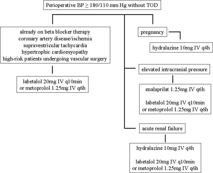
Hospitalist Postgraduate PA Training Program
In recent years, the demand for hospitalists has outstripped the supply, creating a national shortage.1, 2 A recent Society of Hospital Medicine (SHM) survey found that in the last 2 years there has been a 31% mean growth increase in the number of hospitalist groups.3 As hospitalists are becoming more difficult to recruit, many practices are utilizing physician assistants (PAs) and nurse practitioners (NPs), collectively referred to as nonphysician providers (NPPs) to help offset the workload.4 The SHM survey also noted that the number of hospitalist groups utilizing NPPs increased from 29% to 38%.3 The exact number of NPPs working for hospitalist groups is unknown.
Hospitalist NPPs are in demand for reasons other than just physician shortages. NPPs have been utilized to fill the gap in many institutions where the workforce was impacted by the 2002 Accreditation Council for Graduate Medical Education (ACGME) ruling to restrict resident work hours. Several studies have documented NPPs' ability to assist with the compliance of ACCGME resident work‐hour restrictions while maintaining patient continuity of care, improving length of stays, and reducing health care costs on various hospital services.59 Dresselhaus et al.10 found that 56% of medical resident's time on service was delegated to tasks not related to direct patient care. They proposed that these tasks can be delegated to the NPPs, leaving more time for the residents to focus on direct patient care. In a recent study performed at a Pennsylvania hospital, patients presenting to the emergency department with low‐risk chest pain (based upon thrombolysis in myocardial infarction [TIMI] risk score) were admitted to a nonteaching service staffed with NPPs and attending physicians. Simultaneously, a similar group of low‐risk chest pain patients were admitted to a traditional internal medicine resident service. The results demonstrated lower median length of stay and hospital charges on the nonteaching service. This study suggested that NPPs can offset the workload volume for medical residents, allowing them to focus on patients with higher acuity and greater learning value.11
Barriers to Finding Experienced NPPs in Hospital Medicine
Although many hospitalist groups are interested in hiring NPPs, there can be significant obstacles to recruitment. For example, most experienced PAs and NPs have clinical backgrounds in either surgical or medical subspecialties and therefore typically need extensive on‐the‐job training in hospital medicine, which can often take at least 6 to 12 months to acquire the basic skill set.12 Hiring new graduates may require even longer training periods.
The inexperience of new graduates has become an even more pertinent issue due to recent changes in PA education. Traditionally, PA programs attracted older students with prior healthcare experience, who wished to return to school for additional training. However, in 2005 a major shift occurred in PA education: programs began transitioning from graduating trainees with a bachelor's degree to now requiring a master's level degree for completion of the PA program.13 The acquisition of more advanced degrees has changed the demographics of the students matriculating into PA programs, attracting younger students, straight from undergraduate institutions, with less prior healthcare experience.14 As a result, not only are new PA graduates less experienced overall, but they are particularly lacking in exposure to hospital medicine. After PA students complete their first 12 months of PA school in the basic sciences and didactic coursework, they embark on 12 to 15 months of clinical rotations, which are largely rooted in primary care. In fact, many PA programs find it difficult to offer hospital‐based rotations while fulfilling the required rotations in primary care. These factors have resulted in the need for more extensive on‐the‐job training particularly for those new graduates interested in hospital medicine. In light of these challenges, our institution created a 12‐month postgraduate PA fellowship program in Hospital Medicine.
Postgraduate PA Training Programs
Postgraduate PA fellowships, interchangeably called residencies, are voluntary 1‐year training programs that provide both didactic instruction and clinical experience in a medical or surgical subspecialty, thereby lessening the need for on‐the‐job training. These programs are recognized by the Association of Postgraduate Physician Assistant Programs.15 Currently, there are 44 postgraduate training programs in the United States, in a wide range of medical and surgical specialties. At the end of these 1‐year postgraduate PA programs, most graduates receive a certificate of completion. Until now, the only postgraduate education option for PAs interested in Hospital Medicine was a master's completion program only available to PAs who were already employed by a hospitalist group.15 This work reviews the first reported postgraduate hospitalist training program for PAs. Specifically, the program's background, curriculum, anticipated program outcomes, and future plans are discussed.
Background for A Hospitalist Postgraduate PA Fellowship
Mayo Clinic Arizona is a multispecialty private group comprised of both outpatient services and a tertiary care hospital medical center, located in the metropolitan Phoenix, AZ, area. The Mayo Clinic Hospital is a 7‐story facility with 244 licensed beds, 18 operating rooms, and a Level II emergency department. The Mayo Hospitalist group is composed of 15 full time hospitalists and 6 part‐time hospitalists, all of whom are salaried Mayo employees. The group provides 24‐hour in‐house staffing, covering both resident services (teams composed of interns and residents supervised by a staff hospitalist) and nonresident services (staff hospitalists). Over the years there has been steady growth in the number of nonresident services, in part due to resident work‐hour restrictions. To support the physicians working on these nonresident services, the first PA was hired in 2001. Since then, the number of NPPs in our Hospitalist group has increased to 9.35 full‐time equivalents (FTEs), including 1 nurse practitioner. However, one of the greatest challenges in expanding the NPP service was the difficulty finding candidates with experience in hospital internal medicine. This need inspired the creation of a PA fellowship in Hospital Medicine. At the time, there were 2 other postgraduate PA training programs at the Mayo Clinic Arizona in Hepatology and Otolaryngology/Ear, Nose, and Throat (ENT) Surgery.
Program Description
The Mayo Clinic Arizona PA fellowship in Hospital Medicine began in October 2007 and currently accepts 1 fellow per year. Applicants must be graduates of an Accreditation Review Commission in Education for the Physician Assistant (ARC‐PA)‐accredited PA program and be certified through the National Commission on Certification of Physician Assistants (NCCPA). Furthermore, they must be licensed to work as a PA in the state of Arizona. The program is 12 months in duration, and is comprised of both didactic and clinical components. Upon graduation, the fellow earns a certificate of completion from the Mayo Clinic College of Medicine. The program has received recognition with the Association of Postgraduate Physician Assistant Programs (APPAP).
Two physician assistants act as co‐program directors of the PA fellowship in hospital medicine. They are given 0.10 full‐time equivalent (FTE) for management of the program, which includes day‐to‐day operations, curriculum development, and candidate selection. The program also has 2 volunteer physician medical directors, both of whom have previous medical residency experience. The physicians and NPPs in our hospitalist group volunteer their time to serve as faculty for the program, assisting with much of the didactic and clinical education. The program receives a budget of $99,500 per year, which is funded by the organization's foundation through the department of education. This includes the fellow stipend of $44,000 per 12 months and institutional malpractice insurance coverage. The fellow also receives health and dental insurance, 2 weeks of paid vacation, and $500 stipend toward attendance of a continuing medical education (CME) conference.
CURRICULUM
The PA fellowship curriculum is designed in a diverse unique format that strives to accommodate all types of learners. It includes clinical rotations in various medicine/surgical subspecialties, didactic instruction, and teaching modules (Figure 1). The curriculum is based upon the SHM Core Competencies.15
Clinical Rotations
The PA fellow completes 12 to 14 general hospital medicine and medical specialty rotations, each 2 to 4 weeks in duration. The rotation calendar for the current fellow is given in Figure 2. These rotations are all inpatient‐based and are supervised by either the hospitalist or the respective inpatient subspecialists. The PA fellow's specific clinical responsibilities vary from rotation to rotation, and are designed to maximize the fellow's exposure to that particular specialty. Each rotation has specific written objectives created by the program directors and reviewed by the rotation's preceptor(s) (Figure 2). During the clinical rotations, complementary didactic lectures, coursework, and readings are provided to ensure the PA fellow receives a strong foundation. Didactic instruction is designed by the program directors, physician preceptors and staff NPPs, and is coordinated with the clinical rotation specialty. At the end of each rotation the fellow is evaluated by the preceptor and given direct feedback on their performance.
Didactic Instruction
The didactic instruction is organized in a system‐based manner and occurs on a weekly basis during the Hospital Internal Medicine service and Medicine Consults rotations. Hospitalist NPPs and physician faculty are responsible for most of the teaching. This formal didactic instruction is supplemented by journal club presentations given by the PA fellow to faculty in the division of hospital internal medicine. The fellow is also required to attend daily medical resident lunchtime educational lectures, weekly medical grand rounds, and any lectures provided by the medicine subspecialties while the PA is on that particular rotation.
Teaching Modules
One component of the Hospital Medicine PA fellowship curriculum that may be unique is the concept of teaching modules. While receiving regular didactic instruction and completing their clinical rotations, the PA is also expected to complete self‐directed teaching module assignments. These modules serve to educate the PA fellow on the hospital as a systemthe true essence of hospital medicine. The modules cover a variety of topics not directly addressed during their rotations. These topics are outlined in Figure 3. Each teaching module consists of a didactic component, clinical application, and assessment (Figure 4) and has its own specific objectives and goals. Teaching modules are often taught by the local expert in the hospital in that particular area. For example, for the infectious control teaching module, the PA fellow will rotate with the infection control nursing staff learning about the isolation and infection control policies of the institution.
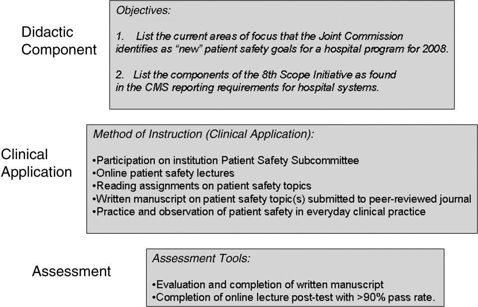
Assessment Tools
There are several tools utilized to assess both the PA fellow and the fellowship program itself (Figure 5). The assessment tools used include both ongoing and summative assessments. To fulfill the ongoing assessment, each rotation and teaching module contains assessment tools provided by the preceptor, which are reviewed by the program directors. Additionally, during the clinical rotations, skills are assessed using competency checklists that require the preceptor to directly observe the PA fellow perform a specific task or skill‐set and sign off on its successful completion (Supplementary Figures 6, 7).
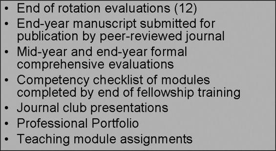
There are 2 forms of summative assessment for the PA fellow. First, to assess the PA fellow's knowledge, comprehensive mid‐year and end‐year examinations are utilized. These multiple‐choice examinations are comprised of questions which align with the didactic lectures/objectives provided by the Hospital Medicine faculty throughout the year. The second form of summative evaluation of the fellow is project‐based and divided into 2 parts. First, the fellow is expected to write a publication‐quality manuscript on a hospital medicine topic by the end of the year. Second, the PA fellow is expected to create a professional portfolio, which is comprised of a collection of all of the rotation/module assessments, the formal program assessments, and documentation of all of the skills obtained by the fellow throughout year (competency checklists). This portfolio can be used by the graduate to demonstrate to future employers what skills they possess and provide documentation of knowledge gained during the fellowship.
The program itself is evaluated by several measures. First, the fellow provides formal feedback during the mid‐year and end‐of‐the‐year assessments, which are used to enhance the experience of future fellows. Second, there is ongoing review by both the division of Hospital Medicine and the institution's Allied Health Education Committee, which ensures that the program maintains the appropriate standards and goals.
Future Goals for the PA Fellowship
The program graduated its first fellow at the end of October 2008 and has enjoyed early success. Integrating the PA fellow onto the hospitalist services augmented the present mid‐level and physician teams. There has been excellent institutional support for the program with extremely positive feedback from the rotation preceptors. There are several futures plans for the program. Our first goal is to seek accreditation from the Accreditation Review Commission for Physician Assistants (ARC‐PA), the organization that accredits entry level PA programs and which began formal, voluntary accreditation of postgraduate programs in early 2008. We plan to begin this process within the next academic year.
Our second long‐term goal for the program is to include NPs in the training program. Because of the desire to seek accreditation, the program directors felt temporarily limiting the fellowship to PAs would aide in the rigorous accreditation process, which can take approximately 1 year to complete. There is an NP on our faculty and the program has received interest from NPs. Once we obtain accreditation, expand the program enrollment, and develop an NP curriculum, we plan to open the fellowship to either PA or NP applicants.
Our third goal is to substantiate our PA Fellowship validity with outcome measures and ultimately publishable data. Thus far, the success of the PA fellowship is qualitative, and with small numbers of graduates it is difficult to quantify. After graduation of many subsequent PA fellows, our goal is to obtain quantifiable data that can be used to improve the quality of the PA fellowship and demonstrate the value of postgraduate training for physician assistants.
Perhaps the most important goal of the program is to eventually accept additional PA/NP fellows per year. While 1 program does not meet the demands of a national shortage of hospitalist providers, it may serve as a model that other institutions can adapt to their own needs. Since the program is based upon the SHM Core Competencies, the curriculum can be applied to a variety of hospitalist programs, and its relatively low operating cost makes it feasible for both academic‐based and community‐based institutions. Importantly, since recruitment and retention of employees is such a challenge for most hospitalist groups, this PA fellowship program may serve as a vehicle for recruitment and long‐term retention of well‐trained employees. This precedent has been set, as our division has hired our first PA fellow, whose transition from PA fellow to PA staff was seamless.
In conclusion, our PA fellowship in Hospital Medicine represents the first reported postgraduate PA program of this kind in the United States offering a certificate of completion. As the need for hospitalists increase so will the need for NPPs, particularly those with additional training in hospital medicine. This program serves as an example of 1 type of training tool for physician assistants looking to work in hospital medicine.
- ,,,.Health care market trends and the evolution of hospitalist use and roles.J Gen Intern Med.2004;20:101–107.
- .Innovations in the management of hospitalized patients. Nurse Pract Spring2006 (suppl):2–3.
- .Hospitalist pay up, productivity steady in SHM's latest survey.Hospitalist.2008;12(5):7,16.
- .Physician assistants: filling the gap in patient care in academic hospitals.Perspect Physician Assist Educ.2003;14(3):158–167.
- ,,, et al.The effect of a multidisciplinary hospitalist/physician and advanced practice nurse collaboration on hospital costs.J Nurs Adm.2006;36(2):79–85.
- ,,, et al.Physician extenders impact trauma systems.J Trauma.2005;58(5):917–920.
- ,,.The role of physician assistants in critical care units.Chest.1991;99:89–91.
- .Alliances: invaluable assistants.Hospitalist.2006;April:32–33.
- ,,.Resource use by physician assistant services versus teaching services.JAAPA.2002;15:33–42.
- ,,,,,.Analyzing the time and value of house staff inpatient work.J Intern Med.1998;13:534–540.
- ,,, et al.Improving resource utilization in a teaching hospital: Development of a nonteaching service for chest pain admissions.Acad Med.2006;81(5):432–435.
- .Midlevels make a rocky entrance into hospital medicine.Todays Hospitalist.2007;5(1):28–32.
- Accreditation Review Commission for Physician Assistant Education.3rd ed. 2005. Available at: http://www.arc‐pa.org/Standards/standards.html. Accessed September2009.
- 22nd Annual Report on Physician Assistant Education in the U.S., 2005–2006. Available at: http://www.paeaonline.org. Accessed September2009.
- Association of Postgraduate Physician Assistant Programs. Available at: http://www.appap.org. Accessed September2009.
- ,,,,.The core competencies in hospital medicine: development and methodology.J Hosp Med.2006;1:48–56.
In recent years, the demand for hospitalists has outstripped the supply, creating a national shortage.1, 2 A recent Society of Hospital Medicine (SHM) survey found that in the last 2 years there has been a 31% mean growth increase in the number of hospitalist groups.3 As hospitalists are becoming more difficult to recruit, many practices are utilizing physician assistants (PAs) and nurse practitioners (NPs), collectively referred to as nonphysician providers (NPPs) to help offset the workload.4 The SHM survey also noted that the number of hospitalist groups utilizing NPPs increased from 29% to 38%.3 The exact number of NPPs working for hospitalist groups is unknown.
Hospitalist NPPs are in demand for reasons other than just physician shortages. NPPs have been utilized to fill the gap in many institutions where the workforce was impacted by the 2002 Accreditation Council for Graduate Medical Education (ACGME) ruling to restrict resident work hours. Several studies have documented NPPs' ability to assist with the compliance of ACCGME resident work‐hour restrictions while maintaining patient continuity of care, improving length of stays, and reducing health care costs on various hospital services.59 Dresselhaus et al.10 found that 56% of medical resident's time on service was delegated to tasks not related to direct patient care. They proposed that these tasks can be delegated to the NPPs, leaving more time for the residents to focus on direct patient care. In a recent study performed at a Pennsylvania hospital, patients presenting to the emergency department with low‐risk chest pain (based upon thrombolysis in myocardial infarction [TIMI] risk score) were admitted to a nonteaching service staffed with NPPs and attending physicians. Simultaneously, a similar group of low‐risk chest pain patients were admitted to a traditional internal medicine resident service. The results demonstrated lower median length of stay and hospital charges on the nonteaching service. This study suggested that NPPs can offset the workload volume for medical residents, allowing them to focus on patients with higher acuity and greater learning value.11
Barriers to Finding Experienced NPPs in Hospital Medicine
Although many hospitalist groups are interested in hiring NPPs, there can be significant obstacles to recruitment. For example, most experienced PAs and NPs have clinical backgrounds in either surgical or medical subspecialties and therefore typically need extensive on‐the‐job training in hospital medicine, which can often take at least 6 to 12 months to acquire the basic skill set.12 Hiring new graduates may require even longer training periods.
The inexperience of new graduates has become an even more pertinent issue due to recent changes in PA education. Traditionally, PA programs attracted older students with prior healthcare experience, who wished to return to school for additional training. However, in 2005 a major shift occurred in PA education: programs began transitioning from graduating trainees with a bachelor's degree to now requiring a master's level degree for completion of the PA program.13 The acquisition of more advanced degrees has changed the demographics of the students matriculating into PA programs, attracting younger students, straight from undergraduate institutions, with less prior healthcare experience.14 As a result, not only are new PA graduates less experienced overall, but they are particularly lacking in exposure to hospital medicine. After PA students complete their first 12 months of PA school in the basic sciences and didactic coursework, they embark on 12 to 15 months of clinical rotations, which are largely rooted in primary care. In fact, many PA programs find it difficult to offer hospital‐based rotations while fulfilling the required rotations in primary care. These factors have resulted in the need for more extensive on‐the‐job training particularly for those new graduates interested in hospital medicine. In light of these challenges, our institution created a 12‐month postgraduate PA fellowship program in Hospital Medicine.
Postgraduate PA Training Programs
Postgraduate PA fellowships, interchangeably called residencies, are voluntary 1‐year training programs that provide both didactic instruction and clinical experience in a medical or surgical subspecialty, thereby lessening the need for on‐the‐job training. These programs are recognized by the Association of Postgraduate Physician Assistant Programs.15 Currently, there are 44 postgraduate training programs in the United States, in a wide range of medical and surgical specialties. At the end of these 1‐year postgraduate PA programs, most graduates receive a certificate of completion. Until now, the only postgraduate education option for PAs interested in Hospital Medicine was a master's completion program only available to PAs who were already employed by a hospitalist group.15 This work reviews the first reported postgraduate hospitalist training program for PAs. Specifically, the program's background, curriculum, anticipated program outcomes, and future plans are discussed.
Background for A Hospitalist Postgraduate PA Fellowship
Mayo Clinic Arizona is a multispecialty private group comprised of both outpatient services and a tertiary care hospital medical center, located in the metropolitan Phoenix, AZ, area. The Mayo Clinic Hospital is a 7‐story facility with 244 licensed beds, 18 operating rooms, and a Level II emergency department. The Mayo Hospitalist group is composed of 15 full time hospitalists and 6 part‐time hospitalists, all of whom are salaried Mayo employees. The group provides 24‐hour in‐house staffing, covering both resident services (teams composed of interns and residents supervised by a staff hospitalist) and nonresident services (staff hospitalists). Over the years there has been steady growth in the number of nonresident services, in part due to resident work‐hour restrictions. To support the physicians working on these nonresident services, the first PA was hired in 2001. Since then, the number of NPPs in our Hospitalist group has increased to 9.35 full‐time equivalents (FTEs), including 1 nurse practitioner. However, one of the greatest challenges in expanding the NPP service was the difficulty finding candidates with experience in hospital internal medicine. This need inspired the creation of a PA fellowship in Hospital Medicine. At the time, there were 2 other postgraduate PA training programs at the Mayo Clinic Arizona in Hepatology and Otolaryngology/Ear, Nose, and Throat (ENT) Surgery.
Program Description
The Mayo Clinic Arizona PA fellowship in Hospital Medicine began in October 2007 and currently accepts 1 fellow per year. Applicants must be graduates of an Accreditation Review Commission in Education for the Physician Assistant (ARC‐PA)‐accredited PA program and be certified through the National Commission on Certification of Physician Assistants (NCCPA). Furthermore, they must be licensed to work as a PA in the state of Arizona. The program is 12 months in duration, and is comprised of both didactic and clinical components. Upon graduation, the fellow earns a certificate of completion from the Mayo Clinic College of Medicine. The program has received recognition with the Association of Postgraduate Physician Assistant Programs (APPAP).
Two physician assistants act as co‐program directors of the PA fellowship in hospital medicine. They are given 0.10 full‐time equivalent (FTE) for management of the program, which includes day‐to‐day operations, curriculum development, and candidate selection. The program also has 2 volunteer physician medical directors, both of whom have previous medical residency experience. The physicians and NPPs in our hospitalist group volunteer their time to serve as faculty for the program, assisting with much of the didactic and clinical education. The program receives a budget of $99,500 per year, which is funded by the organization's foundation through the department of education. This includes the fellow stipend of $44,000 per 12 months and institutional malpractice insurance coverage. The fellow also receives health and dental insurance, 2 weeks of paid vacation, and $500 stipend toward attendance of a continuing medical education (CME) conference.
CURRICULUM
The PA fellowship curriculum is designed in a diverse unique format that strives to accommodate all types of learners. It includes clinical rotations in various medicine/surgical subspecialties, didactic instruction, and teaching modules (Figure 1). The curriculum is based upon the SHM Core Competencies.15
Clinical Rotations
The PA fellow completes 12 to 14 general hospital medicine and medical specialty rotations, each 2 to 4 weeks in duration. The rotation calendar for the current fellow is given in Figure 2. These rotations are all inpatient‐based and are supervised by either the hospitalist or the respective inpatient subspecialists. The PA fellow's specific clinical responsibilities vary from rotation to rotation, and are designed to maximize the fellow's exposure to that particular specialty. Each rotation has specific written objectives created by the program directors and reviewed by the rotation's preceptor(s) (Figure 2). During the clinical rotations, complementary didactic lectures, coursework, and readings are provided to ensure the PA fellow receives a strong foundation. Didactic instruction is designed by the program directors, physician preceptors and staff NPPs, and is coordinated with the clinical rotation specialty. At the end of each rotation the fellow is evaluated by the preceptor and given direct feedback on their performance.
Didactic Instruction
The didactic instruction is organized in a system‐based manner and occurs on a weekly basis during the Hospital Internal Medicine service and Medicine Consults rotations. Hospitalist NPPs and physician faculty are responsible for most of the teaching. This formal didactic instruction is supplemented by journal club presentations given by the PA fellow to faculty in the division of hospital internal medicine. The fellow is also required to attend daily medical resident lunchtime educational lectures, weekly medical grand rounds, and any lectures provided by the medicine subspecialties while the PA is on that particular rotation.
Teaching Modules
One component of the Hospital Medicine PA fellowship curriculum that may be unique is the concept of teaching modules. While receiving regular didactic instruction and completing their clinical rotations, the PA is also expected to complete self‐directed teaching module assignments. These modules serve to educate the PA fellow on the hospital as a systemthe true essence of hospital medicine. The modules cover a variety of topics not directly addressed during their rotations. These topics are outlined in Figure 3. Each teaching module consists of a didactic component, clinical application, and assessment (Figure 4) and has its own specific objectives and goals. Teaching modules are often taught by the local expert in the hospital in that particular area. For example, for the infectious control teaching module, the PA fellow will rotate with the infection control nursing staff learning about the isolation and infection control policies of the institution.
Assessment Tools
There are several tools utilized to assess both the PA fellow and the fellowship program itself (Figure 5). The assessment tools used include both ongoing and summative assessments. To fulfill the ongoing assessment, each rotation and teaching module contains assessment tools provided by the preceptor, which are reviewed by the program directors. Additionally, during the clinical rotations, skills are assessed using competency checklists that require the preceptor to directly observe the PA fellow perform a specific task or skill‐set and sign off on its successful completion (Supplementary Figures 6, 7).
There are 2 forms of summative assessment for the PA fellow. First, to assess the PA fellow's knowledge, comprehensive mid‐year and end‐year examinations are utilized. These multiple‐choice examinations are comprised of questions which align with the didactic lectures/objectives provided by the Hospital Medicine faculty throughout the year. The second form of summative evaluation of the fellow is project‐based and divided into 2 parts. First, the fellow is expected to write a publication‐quality manuscript on a hospital medicine topic by the end of the year. Second, the PA fellow is expected to create a professional portfolio, which is comprised of a collection of all of the rotation/module assessments, the formal program assessments, and documentation of all of the skills obtained by the fellow throughout year (competency checklists). This portfolio can be used by the graduate to demonstrate to future employers what skills they possess and provide documentation of knowledge gained during the fellowship.
The program itself is evaluated by several measures. First, the fellow provides formal feedback during the mid‐year and end‐of‐the‐year assessments, which are used to enhance the experience of future fellows. Second, there is ongoing review by both the division of Hospital Medicine and the institution's Allied Health Education Committee, which ensures that the program maintains the appropriate standards and goals.
Future Goals for the PA Fellowship
The program graduated its first fellow at the end of October 2008 and has enjoyed early success. Integrating the PA fellow onto the hospitalist services augmented the present mid‐level and physician teams. There has been excellent institutional support for the program with extremely positive feedback from the rotation preceptors. There are several futures plans for the program. Our first goal is to seek accreditation from the Accreditation Review Commission for Physician Assistants (ARC‐PA), the organization that accredits entry level PA programs and which began formal, voluntary accreditation of postgraduate programs in early 2008. We plan to begin this process within the next academic year.
Our second long‐term goal for the program is to include NPs in the training program. Because of the desire to seek accreditation, the program directors felt temporarily limiting the fellowship to PAs would aide in the rigorous accreditation process, which can take approximately 1 year to complete. There is an NP on our faculty and the program has received interest from NPs. Once we obtain accreditation, expand the program enrollment, and develop an NP curriculum, we plan to open the fellowship to either PA or NP applicants.
Our third goal is to substantiate our PA Fellowship validity with outcome measures and ultimately publishable data. Thus far, the success of the PA fellowship is qualitative, and with small numbers of graduates it is difficult to quantify. After graduation of many subsequent PA fellows, our goal is to obtain quantifiable data that can be used to improve the quality of the PA fellowship and demonstrate the value of postgraduate training for physician assistants.
Perhaps the most important goal of the program is to eventually accept additional PA/NP fellows per year. While 1 program does not meet the demands of a national shortage of hospitalist providers, it may serve as a model that other institutions can adapt to their own needs. Since the program is based upon the SHM Core Competencies, the curriculum can be applied to a variety of hospitalist programs, and its relatively low operating cost makes it feasible for both academic‐based and community‐based institutions. Importantly, since recruitment and retention of employees is such a challenge for most hospitalist groups, this PA fellowship program may serve as a vehicle for recruitment and long‐term retention of well‐trained employees. This precedent has been set, as our division has hired our first PA fellow, whose transition from PA fellow to PA staff was seamless.
In conclusion, our PA fellowship in Hospital Medicine represents the first reported postgraduate PA program of this kind in the United States offering a certificate of completion. As the need for hospitalists increase so will the need for NPPs, particularly those with additional training in hospital medicine. This program serves as an example of 1 type of training tool for physician assistants looking to work in hospital medicine.
- ,,,.Health care market trends and the evolution of hospitalist use and roles.J Gen Intern Med.2004;20:101–107.
- .Innovations in the management of hospitalized patients. Nurse Pract Spring2006 (suppl):2–3.
- .Hospitalist pay up, productivity steady in SHM's latest survey.Hospitalist.2008;12(5):7,16.
- .Physician assistants: filling the gap in patient care in academic hospitals.Perspect Physician Assist Educ.2003;14(3):158–167.
- ,,, et al.The effect of a multidisciplinary hospitalist/physician and advanced practice nurse collaboration on hospital costs.J Nurs Adm.2006;36(2):79–85.
- ,,, et al.Physician extenders impact trauma systems.J Trauma.2005;58(5):917–920.
- ,,.The role of physician assistants in critical care units.Chest.1991;99:89–91.
- .Alliances: invaluable assistants.Hospitalist.2006;April:32–33.
- ,,.Resource use by physician assistant services versus teaching services.JAAPA.2002;15:33–42.
- ,,,,,.Analyzing the time and value of house staff inpatient work.J Intern Med.1998;13:534–540.
- ,,, et al.Improving resource utilization in a teaching hospital: Development of a nonteaching service for chest pain admissions.Acad Med.2006;81(5):432–435.
- .Midlevels make a rocky entrance into hospital medicine.Todays Hospitalist.2007;5(1):28–32.
- Accreditation Review Commission for Physician Assistant Education.3rd ed. 2005. Available at: http://www.arc‐pa.org/Standards/standards.html. Accessed September2009.
- 22nd Annual Report on Physician Assistant Education in the U.S., 2005–2006. Available at: http://www.paeaonline.org. Accessed September2009.
- Association of Postgraduate Physician Assistant Programs. Available at: http://www.appap.org. Accessed September2009.
- ,,,,.The core competencies in hospital medicine: development and methodology.J Hosp Med.2006;1:48–56.
Copyright © 2010 Society of Hospital Medicine

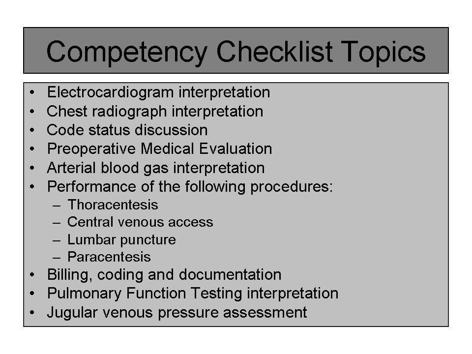

Impact of CI Among Hospitalized Elders
In 2001, approximately 12.6 million individuals age 65 and older were discharged from American hospitals with an average length of stay of 5.8 days1 and up to 66% of them suffered from cognitive impairment (CI).220 CI in hospitalized older adults includes a variety of disorders ranging from mild cognitive deficit, delirium, to full‐blown dementia. Dementia is a syndrome of decline in memory plus at least 1 other cognitive domain, such as language, visuospatial, or executive function sufficient to interfere with social or occupational functioning in an alert person.21 Delirium is a disturbance of consciousness with reduced ability to focus, sustain, or shift attention that occurs over a short period of time and tends to fluctuate over the course of the day.22 Mild CI without dementia is defined as the presence of a cognitive deficit in the absence of delirium that does not affect functional performance.23
Hospitalized older adults with CI are vulnerable to hospital complications, including delirium, physical restraints, urinary catheters, and tethers.2, 3, 2435 The management of their medical or surgical illnesses requires avoiding certain medications with anticholinergic activities that might worsen cognition.36 Furthermore, CI may delay diagnostic and therapeutic procedures, demand more time for informed consentrelated issues, and result in difficulty in adherence to medical recommendations.37, 38 The special needs of hospitalized older adults with delirium and dementia has been shown to increase demands on nursing staff, risk of postdischarge institutionalization, length of stay, and health care costs.310, 27, 3948 We wanted to look specifically at CI because it often goes undetected4951 and can have a great impact on the hospital course of elders.
Screening for CI among hospitalized older adults has been considered to have potential benefit in hospital care of older adults.52 Screening may lead to early detection by uncovering subtle symptoms not yet apparent to families or other caregivers who know the patient well but do not notice small declines or changes in day‐to‐day functioning. Early recognition of CI may lead to early treatment and subsequently may delay progression of cognitive decline and improve health outcomes. Screening may enhance physician prescribing practices and reduce exposure to harmful medications among these vulnerable patients. Finally, delirium is an important prognostic indicator, and screening patients could provide invaluable information toward the overall clinical picture. Despite all of this, the current literature does not provide sufficient information to support the use of routine screening on admission.220, 41, 5254 Most of the published studies were conducted among elders who stayed in the hospital for more than 48 hours, missing data on the crucial first 48 hours of the hospital course.220, 41, 5254 These studies did not evaluate the impact of unrecognized CI on the hospital course and the majority of these studies were not conducted in the urban and lower socioeconomic status populations of elders that are the most vulnerable to bad health outcomes.220, 41, 5254 Finally, few studies evaluated the impact of delirium superimposed on CI on the hospital course and mortality of elders.220, 41, 5254
With these details in mind, we wanted to explore the impact of CI recognition among patients age 65 years and older admitted to the medical services of an urban, public hospital in Indianapolis to determine the prevalence and the impact of recognized and unrecognized CI on the hospital course of these elders. Furthermore, we examined the role of delirium superimposed on these hospitalized elders with CI.
Patients and Methods
The study was approved by the Indiana University Purdue University at Indianapolis Institutional Review Board (IRB).
Study Setting and Population
The study was conducted on the inpatient general medicine service of Wishard Memorial Hospital (WMH). WMH is a 450‐bed, university‐affiliated, urban, public hospital that is staffed by Indiana University School of Medicine faculty and house staff. It serves a population of approximately 750,000 in Marion County.
Inclusion and Exclusion Criteria
Patients were enrolled in the study based on the following criteria: (1) at least 65 years of age; (2) hospitalized on a medical ward; (3) able to speak English; and (4) have CI at the time of hospital admission (see below). Patients were excluded if they had previously enrolled in the study, were enrolled in another clinical study at the time of admission, or were aphasic or unresponsive at the time of screening.
Cognitive Screening
CI was determined by the Short Portable Mental Status Questionnaire (SPMSQ),55, 56 chosen for its accuracy56 and the fact that it is entirely verbal in administration. In most cases, patients were followed and reassessed daily. Patients having 2 or more errors, indicating a score of 8 or less on the SPMSQ after adjusting for race and education were considered to have cognitive impairment. The SPMSQ is a brief 10‐item screening test with a sensitivity of 86% and specificity 99.0% for dementia among medical inpatients.56 At the time of cognitive screening, delirium was assessed by using the Confusion Assessment Method (CAM).22 This was also done daily in most cases. The CAM22 is a structured instrument that evaluates the 10 symptoms of delirium specified in the Diagnostic and Statistical Manual of Mental Disorders (DSM)‐III‐R: acute onset, fluctuating course, inattention, disorganized thinking, altered level of consciousness, disorientation, memory impairment, perceptual disturbances, psychomotor agitation or retardation, and sleep/wake disturbance. The CAM score is determined by examining the patient, investigating the chart and interviewing the nurse and/or a family member for: (1) acute and fluctuating changes in mental status, (2) inattention, (3) disorganized or incoherent thinking, and (4) altered level of consciousness. A CAM score is considered to be positive if the patient displays both (1) and (2) with at least one of (3) or (4). The CAM diagnosis of delirium was validated against the clinical judgment of a psychiatrist and found to have a sensitivity of 97% and a specificity of 92%.22 A research assistant (RA) was trained for a period of 9 months by a physician as a rater to interview the patient and administer both the SPMSQ and the CAM at the time of admission and then every weekday. When feasible, the RA administered both the SPMSQ and the CAM within the first few hours of hospitalization, and then followed up with our patients each day. More than 70% of our initial cognitive screening occurred in the first 48 hours of hospital admission, and was repeated on a daily basis. In addition to cognitive assessment, the RA reported the presence or absence of Foley catheterization, physical restraints, and tethers during the cognitive assessment. Agreement was obtained from the general internal medicine group practice physicians both to participate in the study and to request screening for CI as part of the recognized admission standard of care among their hospitalized patients aged 65 years and older. The study coordinator was notified of all admissions for patients aged 65 or older by the hospital intranet e‐mail and paging system. Admission notifications were sent by page and e‐mail on an hourly basis from Monday through Friday, 8:00 AM through 5:00 PM. Those admissions occurring between the hours of 5:00 PM and 8:00 AM were sent during the next normal batch notification. Pages and e‐mails for admissions occurring on Saturday and Sunday were sent on Monday morning at 8:00 AM.
Regenstrief Medical Record System at WMH
The computerized Regenstrief Medical Record System (RMRS) is the primary instrument for processing data and monitoring patient and physician activity for Wishard Health System.57, 58 The RMRS is a modular system, composed of Registration and Scheduling, Laboratory, and Pharmacy database modules. The Registration and Scheduling module is used to make all outpatient appointments for the office practices associated with Wishard Health System. The Laboratory module handles all data for all inpatient and outpatient laboratories. This module also produces all laboratory reports and data used for billing. In addition to laboratory data, this module stores coded results and full‐text interpretations of all imaging studies and special procedures. The Pharmacy module contains information on medication orders captured by the computerized physician order enter (CPOE). The Database module stores all the above data by date in a fully‐coded form. Thus, these data are readily retrievable for individual patients by healthcare providers using online terminals. Data for large numbers of patients are retrievable using a locally developed English‐like language called CARE. Patients can be identified either by a certain restriction list (eg, the list of subjects in a study) or by clinical criteria. The RMRS also maintains a number of other databases including diagnoses, vital signs, results of laboratory tests and diagnostic tests, full‐text discharge summaries, preventive health maneuvers, and detailed information on all inpatient and outpatient charges. It contains death certificate information from the Indiana State Board of Health for all registered patients who die in, or outside of, Indiana. Therefore, the RMRS collects and monitors a broad array of physician and patient activity, practice patterns, utilization, diagnostic test finding, and offers a wonderful array of outcome measures.
Other Data Collections
Patient demographics such as age, gender, race, and education level were determined by the RMRS and by information obtained during the time of cognitive screening. Length of hospital stay and 30‐day posthospitalization mortality were obtained from the RMRS. Comorbidity level was measured by reviewing the RMRS and determining each patient's Charlson comorbidity index total score.59, 60 This score was determined using International Statistical Classification of Diseases and Related Health Problems, 9th edition (ICD‐9) codes gathered from 1 year prior to admission until the patient was discharged from the hospital. Anticholinergic medications were determined by using the Anticholinergic Cognitive Burden Scale,61 an expert‐based practical index. The scale was developed based on a review of all published studies from 1996 to 2007 that measured the anticholinergic activities of a drug and its association with cognitive function in older adults. The list of drugs reviewed was presented to an expert interdisciplinary panel that included geriatricians, geriatric pharmacists, geriatric psychiatrists, general physicians, geriatric nurses, and aging brain researchers. The panel categorized each medication into a possible or definite anticholinergic category based on the severity of its cognitive anticholinergic effects.61 A patient who received at least 1 order of a possible or definite anticholinergic during their hospitalization was considered to be an anticholinergic user. Prior recognition of CI was determined by searching the RMRS for any ICD‐9 code (see Appendix) indicative of dementia, Alzheimer disease, or delirium reported at hospital admission, discharge, or during an 1‐year period prior to hospitalization for every patient enrolled in the study. Those patients with documented ICD‐9 codes were felt recognized as having some form of cognitive impairment. Those who had a positive screen but no prior documentation according to ICD‐9 coding, were said to have unrecognized CI.
Analysis
Descriptive statistics were calculated, including percentages for binary categorical variables, and means and standard deviations for continuous variables. Comparisons between groups were based upon Fisher's Exact Tests for binary categorical variables and t tests for continuous variables. When controlling for covariates such as age, gender, race, Charlson comorbidity index, and SPMSQ at screening, group comparisons were made by using logistic regression for binary categorical variables and multiple regression for continuous variables. Since the distributions of length of stay and Charlson comorbidity index were skewed, all statistical tests comparing them across groups were actually performed on their log‐transformed values.
Results
The Prevalence and Recognition of CI
Table 1 describes the demographic characteristic of our study population, which is a reflection of the public and urban nature of our target hospital. Our study assessed the cognitive status of 997 older adults usually (>70% of the time) within 48 hours of their admission to the medical ward of this urban hospital between July of 2006 and March 2008 (see Table 1) and found that 43% of these elders had evidence of CI as determined by a SPMSQ score of 8 points or less. However, 61% of the 424 cognitively impaired elders were not documented or recognized by the electronic medical record system to have cognitive deficit.
| Variable | n | %/Mean (SD) |
|---|---|---|
| ||
| Age (years), mean (SD) | 997 | 74.8 (7.5) |
| Age 85 (%) | 997 | 12.6 |
| Female (%) | 997 | 67.8 |
| African American (%) | 997 | 59.4 |
| Education (years), mean (SD) | 910 | 10.3 (2.8) |
| Education <12 years (%) | 910 | 59.1 |
| Screened within 48 hours of admission (%) | 997 | 73.2 |
| SPMSQ score at screening, mean (SD) | 997 | 7.7 (2.8) |
| Cognitive impairment based on the SPMSQ score 8 (%) | 997 | 42.5 |
The Impact of Unrecognized CI on the Hospital Course
As expected, hospitalized elders with documented CI were older (mean age 79.1 years vs. 76.1 years; P < 0.001) and had worse cognitive function upon screening than those with unrecognized CI (mean SPMSQ 3.4 points vs. 6.3; P < 0.001). Furthermore, CI recognition was influenced by the elders' race and comorbidity (Table 2); a higher percentage of elders with documented CI were African American (69% vs. 54%; P = 0.003) and had less comorbidity (mean Charlson index 1.9 vs. 2.3; P = 0.03). After adjusting for age, gender, race, comorbidity, and cognitive function at screening, our study found no differences between elders with previously recognized CI and those with unrecognized CI in regard to the length of hospital stay (6.7 days vs. 7.5 days; P = 0.59), 30‐day posthospital mortality (4.8% vs. 6.6%; P > 0.2), home discharge (32% vs. 45%; P > 0.7), hospital readmission (19.2% vs.18.8%; P > 0.6), delirium incidence (27% vs. 21%; P > 0.9), and physical restraints (1.8% vs. 1.5%; P > 0.4). We also found that elders with undocumented CI were not more likely to receive definite anticholinergics (33.2% vs. 32.7%; P > 0.9).
| CI Documented | CI Undocumented | P Value | P Value* | |
|---|---|---|---|---|
| ||||
| n (%) | 165 (39) | 259 (61) | n/a | |
| Age, mean (SD) | 79.1 (7.9) | 76.1 (8.0) | <0.001 | |
| Female (%) | 68.5 | 64.5 | 0.40 | |
| African American (%) | 68.5 | 53.7 | <0.01 | |
| SPMSQ at screen, mean (SD) | 3.4 (2.7) | 6.3 (2.1) | <0.001 | |
| Charlson comorbidity index, mean (SD) | 1.9 (1.9) | 2.3 (2.1) | 0.03 | |
| Length of hospital stay, mean (SD) | 6.7 (5.1) | 7.5 (7.1) | 0.49 | 0.59 |
| Survived at 30 days postdischarge (%) | 95.2 | 93.4 | 0.53 | 0.25 |
| Discharged home (%) | 31.5 | 45.2 | 0.01 | 0.74 |
| Readmission within 30 days after discharge home (%) | 19.2 | 18.8 | 0.99 | 0.66 |
| Incidence of delirium (%) | 26.7 | 20.6 | 0.52 | 0.99 |
| Observed with Foley catheter (%) | 43.6 | 27.4 | <0.001 | 0.61 |
| Observed with physical restraint (%) | 1.8 | 1.5 | 0.99 | 0.31 |
| Observed with tethers (%) | 81.8 | 73.8 | 0.06 | 0.58 |
| With at least 1 Ach (%) | 83.6 | 90.7 | 0.03 | 0.22 |
| Possible Ach (%) | 81.2 | 88.4 | 0.05 | 0.31 |
| Definite Ach (%) | 32.7 | 33.2 | 0.99 | 0.64 |
The Impact of Delirium on the Hospital Course of Elders with CI
Among the 424 hospitalized elders with CI, 163 (38%) had delirium at least once during their hospital course and 24% had delirium on the day of hospital discharge. In comparison to elders who had CI but not delirium during their hospitalization (Table 3), those with at least 1 day of delirium had a higher 30‐day posthospitalization mortality risk (8.6% vs. 4.2%; P = 0.09), stayed in the hospital 3.3 additional days (9.2 days vs. 5.9 days; P < 0.001), were less likely to be discharged home (25% vs. 49%; P < 0.001), were more likely to receive a Foley catheterization (52% vs. 23%; P < 0.001), more likely to be physically restrained (4% vs. 0%; P < 0.01), and more likely to receive tethers during their care (89% vs. 69%; P < 0.001). There was no statistically significant difference between the 2 groups in terms of 30‐day hospital readmission rates or in their use of definite anticholinergics (Table 3).
| Delirium+* | Delirium | P value | |
|---|---|---|---|
| |||
| n (%) | 163 (38) | 261 (62) | n/a |
| Age, mean (SD) | 78.4 (8.5) | 76.5 (7.8) | 0.02 |
| Female (%) | 60.1 | 69.7 | 0.05 |
| African American (%) | 64.4 | 56.3 | 0.10 |
| Charlson comorbidity index, mean (SD) | 1.8 (1.9) | 2.3 (2.1) | 0.01 |
| Length of hospital stay, mean (SD) | 9.2 (7.9) | 5.9 (4.9) | <0.001 |
| Survived at 30‐day postdischarge (%) | 91.4 | 95.8 | 0.09 |
| Discharged home (%) | 24.5 | 49.4 | <0.001 |
| Readmission within 30 days after discharge home (%) | 22.5 | 17.8 | 0.50 |
| Observed with Foley catheter (%) | 51.5 | 22.6 | <0.001 |
| Observed with physical restraint (%) | 4.3 | 0.0 | <0.01 |
| Observed with tethers (%) | 89.0 | 69.4 | <0.001 |
| With at least 1 anticholinergic (%) | 83.4 | 90.8 | 0.03 |
| Possible anticholinergic (%) | 80.4 | 88.9 | 0.02 |
| Definite anticholinergic (%) | 36.8 | 30.7 | 0.20 |
Discussion
Our study found that in an urban, public hospital, acute or preexisting CI affects more than one‐third of hospitalized elders admitted to general medical services. Unfortunately, our hospital system does not currently recognize the majority of these vulnerable patients. Our study also found that delirium affects more than one‐third of hospitalized elders with CI during their hospital course. Delirium complicates hospital care by prolonging length of stay and decreasing the probability of surviving and getting discharged home. It leads to high use of Foley catheterization, physical restraints, and tethers.
The high prevalence of CI with and without delirium in our cohort is within the rates reported previously in the literature. It is estimated that the prevalence of CI in hospitalized older adults ranges from 14% to 66%, depending on the method used to measure cognition, the definition of CI, and the type of hospital ward (surgical, medical, and geriatric units).220 One particular study that used a similar cognitive assessment method reported higher prevalence rates for both CI and delirium.11 The study randomly evaluated a sample of 201 patients age 65 and over who were hospitalized for a medical illness and found that 56% of the cohort suffered from CI and among those with CI, 47% had delirium.11 The difference between this finding and our study is most likely due to our sampling technique; more than 70% of our cognitive screening occurred in the first 48 hours of hospital admission whereas the Australian study, in similar enrollment criteria to all of the published studies in this area, excluded patients who were discharged within 48 hours of admission. We believe, however, that by including the first 48 hours of admission in our design, our study provides a more generalizable reflection of the actual acute care experience.
The impact of delirium on the course of hospital care found in our study supports some of the findings from previous studies conducted in the past 2 decades.5, 6, 11 Despite 2 decades of clinical research, delirium continues to increase mortality, hospital stays, and posthospital institutionalization.
We were surprised to find that patients suffering from delirium continue to receive at least 1 definite anticholinergic medication. Such medications are considered inappropriate among patients with any form of cognitive impairment.36, 62 Although the impact of anticholinergic medications on hospitalized outcomes is less well‐described, their use has been suspected to negatively impact long‐term outcomes of cognitive impairment.61, 63 Our study found no difference in the use of anticholinergic medications between those with CI who experienced delirium and those who did not; however, the total burden of anticholinergic medication was not assessed in a quantitative manner. It is still unknown if certain anticholinergic medications or a cumulative effect of anticholinergic medications may impact cognitive or health‐related outcomes in a vulnerable older population with CI.
Although our study reported for the first time in a systematic way the rate of undocumented CI among hospitalized elders found to have CI on admission, we found no impact of such underrecognition on the length of hospital stay, mortality, discharge location, and delirium occurrence. Although the use of anticholinergic medications is not recommended for patients with any form of CI, our results indicate that a significant number of patients with cognitive impairment continue to receive inappropriate medications. CI recognition in the elderly was not shown to have a statistically significant affect on length of stay, cost, or mortality.
Our study has some limitations. First of all, we did not determine the underlying types of CI such as Alzheimer disease, vascular dementia, mild cognitive impairment, or reversible etiology other than delirium. Such a categorization requires posthospital assessment, which was not included in our study design. Second, our delirium incidence rate and delirium impact on hospital outcomes might be very conservative and may underestimate its true prevalence and correlation due to our data collection methods. Despite the fluctuating nature of delirium, our study was not designed to assess the presence of delirium every shift and tried to assess cognitive function on a daily basis throughout the patient's hospitalization. Therefore, the severity and duration of delirium could not be accurately assessed. Our reported rates of use of Foley catheterization, physical restraints, and tethers are also very conservative and we could not determine the appropriateness of these procedures. Our study was conducted in 1 public hospital in an urban city with a higher percentage of African Americans. Thus, our sample is not a true representative sample. However, studies with significant representation of minority groups are not common in the research literature, especially in CI research; we hope to fulfill some of the gaps in the literature regarding the most vulnerable older American population. Finally, we were limited in our use of ICD‐9 coding to determine if patients had previously been recognized by other providers as having CI. ICD‐9 coding, while useful, is not perfect in identifying all if a patient's medical problems. Use of coding to determine whether a patient had been recognized as impaired also does not allow us to determine when the diagnosis was made.
In conclusion, our study evaluated cognitive impairment in hospitalized elders and found that in our cohort of 997 patients, 43% were cognitively impaired on admission. Of those with CI, 61% were not documented or recognized as impaired. We found no statistically significant difference between those with documented CI and those with undocumented CI in terms of length of stay, mortality, home discharge, readmission rates, incidence of delirium, or potential to receive anticholinergics or restraints. Among those with CI, 38% had delirium. Those with delirium experienced increased length of stay, decreased discharge to home, and increased use of Foley catheters and restraints.
- ,.National hospital discharge survey: annual summary, 1994.Vital Health Stat 13.1997;(128):i–v;1–50.
- .The dilemma of delirium: clinical and research controversies regarding diagnosis and evaluation of delirium in hospitalized elderly medical patients.Am J Med.1994;97:278–288.
- ,,.Dementia in medical wards.J Clin Epidemiol.1988;41:123–126.
- ,,,,,.Mental and behavioral disturbances in dementia: findings from the Cache County Study on Memory in Aging.Am J Psychiatry.2000;157:708–714.
- ,,, et al.Randomized, placebo‐controlled, double‐blind clinical trial of sertraline in the treatment of depression complicating Alzheimer's disease: initial results from the Depression in Alzheimer's Disease study.Am J Psychiatry.2000;157:1686–1689.
- ,,.Dementia in elderly persons in a general hospital.Am J Psychiatry.2000;157:704–707.
- ,,,,.A prospective study of the impact of psychiatric comorbidity on length of hospital stays of elderly medical‐surgical inpatients.Psychosomatics.1998;39:273–280.
- ,.Psychiatric comorbidity and length of stay in the general hospital. A critical review of outcome studies.Psychosomatics.1994;35:233–252.
- ,,,.Dementia among medical inpatients. Evaluation of 2000 consecutive admissions.Arch Intern Med.1986;146:1923–1926.
- ,,,.The consequences of non‐cognitive symptoms of dementia in medical hospital departments.Int J Psychiatry Med.2003;33:257–271.
- ,,.Cognitive impairment in medical inpatients. I: Screening for dementia—is history better than mental state?Age Ageing.1997;26:31–35.
- ,,, et al.Acute confusional states in elderly patients treated for femoral neck fracture.J Am Geriatr Soc.1988;36:525–530.
- ,.A prospective study of elderly general surgical patients: II. Post‐operative complications.Age Ageing.1989;18:316–326.
- ,,,.Dementia and depression in elderly medical inpatients.Int Psychogeriatr.2000;12:67–75.
- ,,.[Psychiatric disorders in elderly general hospital patients: incidence and long‐term prognosis].Nervenarzt.1993;64:53–61. [German]
- ,.Delirium and dementia in acute medical admissions of elderly patients in Iceland.Acta Psychiatr Scand.1993;87:123–127.
- ,,,,,.[Psychiatric morbidity in elderly patients admitted to a general hospital. A day‐prevalence study].Med Clin (Barc).1991;97:206–210. [Spanish]
- ,,,,,.Detection of psychiatric disorders in elderly medical inpatients.Age Ageing.1994;23:307–311.
- ,,,.Cognitive impairment, emotional disorder and length of stay of elderly patients in a district general hospital.Br J Med Psychol.1987;60(Pt 2):133–139.
- ,,.An investigation of the components of best nursing practice in the care of acutely ill hospitalized older patients with coincidental dementia: a multi‐method design.J Adv Nurs1999;30:1127–1136.
- American Psychiatric Association.Diagnostic and Statistical Manual of Mental Disorders.4th ed.Washington, DC:American Psychiatric Association;1994.
- ,,,,,.Clarifying confusion: the confusion assessment method. A new method for detection of delirium.Ann Intern Med.1990;113:941–948.
- ,,, et al.Prevalence of cognitive impairment: data from the Indianapolis Study of Health and Aging.Neurology.2001;57:1655–1662.
- ,,.Delirium: a symptom of how hospital care is failing older persons and a window to improve quality of hospital care.Am J Med.1999;106:565–573.
- ,,, et al.A multicomponent intervention to prevent delirium in hospitalized older patients.N Engl J Med.1999;340:669–676.
- ,,,.Iatrogenic causes of falls in hospitalised elderly patients: a case‐control study.Postgrad Med J.2002;78:487–489.
- ,,.A prospective study of delirium in hospitalized elderly.JAMA.1990;263:1097–1101.
- ,.The prognostic significance of delirium in older hospital patients.J Am Geriatr Soc.1997;45:174–178.
- ,.Prognosis of delirium in elderly hospital patients.CMAJ.1993;149:41–46.
- ,,,,.The detection of psychiatric morbidity and its effects on outcome in acute elderly medical admissions.Int J Ger Psych1991;6:861–866.
- ,,.Adverse consequences of hospitalization in the elderly.Soc Sci Med.1982;16:1033–1038.
- ,,, et al.Incidence of adverse events and negligence in hospitalized patients. Results of the Harvard Medical Practice Study I.N Engl J Med.1991;324:370–376.
- ,,,.Delirium in elderly patients: an overview of the state of the science.J Gerontol Nurs.2001;27:12–20.
- ,,,,.A predictive model for delirium in hospitalized elderly medical patients based on admission characteristics.Ann Intern Med.1993;119:474–481.
- ,.Precipitating factors for delirium in hospitalized elderly persons. Predictive model and interrelationship with baseline vulnerability.JAMA.1996;275:852–857.
- ,,,,,.Updating the Beers criteria for potentially inappropriate medication use in older adults: results of a US consensus panel of experts.Arch Intern Med.2003;163:2716–2724.
- ,,, et al.A clinical prediction rule for delirium after elective noncardiac surgery.JAMA.1994;271:134–139.
- ,,, et al.The relationship of postoperative delirium with psychoactive medications.JAMA.1994;272:1518–1522.
- ,,, et al.How do delirium and dementia increase length of stay of elderly general medical inpatients?Psychosomatics.2004;45:235–242.
- ,,,,,.The relationship between a dementia diagnosis, chronic illness, Medicare expenditures, and hospital use.J Am Geriatr Soc.2004;52:187–194.
- ,,, et al.Risk factors for delirium in hospitalized elderly.JAMA.1992;267:827–831.
- ,,,,.Psychological comorbidity and length of stay in the general hospital.Am J Psychiatry.1991;148:324–329.
- ,,, et al.Delirium. The occurrence and persistence of symptoms among elderly hospitalized patients.Arch Intern Med.1992;152:334–340.
- ,,.What happens to medical patients with psychiatric disorder?J Psychosom Res1988;32:541–549.
- ,,,,.[Dementia syndromes and length of stay of elderly patients in internal medicine].Ann Med Interne (Paris).1997;148:424–426. [French]
- ,,, et al.[Hospital discharge planning and length of hospital stay in elderly patients admitted through the emergency department].Rev Epidemiol Sante Publique.1995;43:337–347. [French]
- ,,,,.The effect of dementia on acute care in a geriatric medical unit.Int Psychogeriatr.1992;4:231–239.
- ,,,.Cognitive impairment. Can it predict the course of hospitalized patients?J Am Geriatr Soc.1986;34:579–585.
- ,,,,;US Preventive Services Task Force. Screening for dementia in primary care: a summary of the evidence for the U.S. Preventive Services Task Force.Ann Intern Med.2003;138(11):927–937.
- ,,,.Cognitive impairment in the elderly medically ill: how often is it missed?Int J Geriatr Psychiatry.1993;8:929–937.
- .Recognition of cognitive impairment in elderly medical in‐patients.J R Soc Med.1995;88:183–184.
- ,.Quality indicators for dementia in vulnerable community‐dwelling and hospitalized elders.Ann Intern Med.2001;135:668–676.
- ,,,.Reducing delirium after hip fracture: a randomized trial.J Am Geriatr Soc.2001;49:516–522.
- ,.Prevalence of psychotic symptoms in delirium.Psychosomatics.2000;41:519–522.
- .A short portable mental status questionnaire for the assessment of organic brain deficit in elderly patients.J Am Geriatr Soc.1975;23:433–441.
- ,,,.Short Portable Mental Status Questionnaire as a screening test for dementia and delirium among the elderly.J Am Geriatr Soc.1987;35:412–416.
- ,,, et al.The Regenstrief Medical Record System: a quarter century experience.Int J Med Inform.1999;54:225–253.
- ,,,,,.Factors determining the decision to institutionalize dementing individuals: a prospective study.Gerontologist.1993;33:714–720.
- ,,,,,.Resuscitation: how do we decide? A prospective study of physicians' preferences and the clinical course of hospitalized patients.JAMA.1986;255:1316–1322.
- ,,,,,.Assessing illness severity: does clinical judgment work?J Chronic Dis.1986;39:439–452.
- ,,,,.Impact of anticholinergics on the aging brain: a review and practical application.Aging Health.2008;4(3):311–320.
- ,.Delirium in hospitalized older adults. In: Ham R, Sloane P, Warshaw G, eds.Primary Care Geriatrics: A Case‐Based Approach.5th ed.Philadelphia, PA:Mosby Elsevier;2007:210–218.
- ,,, et al.The association between cognition and histamine‐2 receptor antagonists in African Americans.J Am Geriatr Soc.2007;55(8):1248–1253.
In 2001, approximately 12.6 million individuals age 65 and older were discharged from American hospitals with an average length of stay of 5.8 days1 and up to 66% of them suffered from cognitive impairment (CI).220 CI in hospitalized older adults includes a variety of disorders ranging from mild cognitive deficit, delirium, to full‐blown dementia. Dementia is a syndrome of decline in memory plus at least 1 other cognitive domain, such as language, visuospatial, or executive function sufficient to interfere with social or occupational functioning in an alert person.21 Delirium is a disturbance of consciousness with reduced ability to focus, sustain, or shift attention that occurs over a short period of time and tends to fluctuate over the course of the day.22 Mild CI without dementia is defined as the presence of a cognitive deficit in the absence of delirium that does not affect functional performance.23
Hospitalized older adults with CI are vulnerable to hospital complications, including delirium, physical restraints, urinary catheters, and tethers.2, 3, 2435 The management of their medical or surgical illnesses requires avoiding certain medications with anticholinergic activities that might worsen cognition.36 Furthermore, CI may delay diagnostic and therapeutic procedures, demand more time for informed consentrelated issues, and result in difficulty in adherence to medical recommendations.37, 38 The special needs of hospitalized older adults with delirium and dementia has been shown to increase demands on nursing staff, risk of postdischarge institutionalization, length of stay, and health care costs.310, 27, 3948 We wanted to look specifically at CI because it often goes undetected4951 and can have a great impact on the hospital course of elders.
Screening for CI among hospitalized older adults has been considered to have potential benefit in hospital care of older adults.52 Screening may lead to early detection by uncovering subtle symptoms not yet apparent to families or other caregivers who know the patient well but do not notice small declines or changes in day‐to‐day functioning. Early recognition of CI may lead to early treatment and subsequently may delay progression of cognitive decline and improve health outcomes. Screening may enhance physician prescribing practices and reduce exposure to harmful medications among these vulnerable patients. Finally, delirium is an important prognostic indicator, and screening patients could provide invaluable information toward the overall clinical picture. Despite all of this, the current literature does not provide sufficient information to support the use of routine screening on admission.220, 41, 5254 Most of the published studies were conducted among elders who stayed in the hospital for more than 48 hours, missing data on the crucial first 48 hours of the hospital course.220, 41, 5254 These studies did not evaluate the impact of unrecognized CI on the hospital course and the majority of these studies were not conducted in the urban and lower socioeconomic status populations of elders that are the most vulnerable to bad health outcomes.220, 41, 5254 Finally, few studies evaluated the impact of delirium superimposed on CI on the hospital course and mortality of elders.220, 41, 5254
With these details in mind, we wanted to explore the impact of CI recognition among patients age 65 years and older admitted to the medical services of an urban, public hospital in Indianapolis to determine the prevalence and the impact of recognized and unrecognized CI on the hospital course of these elders. Furthermore, we examined the role of delirium superimposed on these hospitalized elders with CI.
Patients and Methods
The study was approved by the Indiana University Purdue University at Indianapolis Institutional Review Board (IRB).
Study Setting and Population
The study was conducted on the inpatient general medicine service of Wishard Memorial Hospital (WMH). WMH is a 450‐bed, university‐affiliated, urban, public hospital that is staffed by Indiana University School of Medicine faculty and house staff. It serves a population of approximately 750,000 in Marion County.
Inclusion and Exclusion Criteria
Patients were enrolled in the study based on the following criteria: (1) at least 65 years of age; (2) hospitalized on a medical ward; (3) able to speak English; and (4) have CI at the time of hospital admission (see below). Patients were excluded if they had previously enrolled in the study, were enrolled in another clinical study at the time of admission, or were aphasic or unresponsive at the time of screening.
Cognitive Screening
CI was determined by the Short Portable Mental Status Questionnaire (SPMSQ),55, 56 chosen for its accuracy56 and the fact that it is entirely verbal in administration. In most cases, patients were followed and reassessed daily. Patients having 2 or more errors, indicating a score of 8 or less on the SPMSQ after adjusting for race and education were considered to have cognitive impairment. The SPMSQ is a brief 10‐item screening test with a sensitivity of 86% and specificity 99.0% for dementia among medical inpatients.56 At the time of cognitive screening, delirium was assessed by using the Confusion Assessment Method (CAM).22 This was also done daily in most cases. The CAM22 is a structured instrument that evaluates the 10 symptoms of delirium specified in the Diagnostic and Statistical Manual of Mental Disorders (DSM)‐III‐R: acute onset, fluctuating course, inattention, disorganized thinking, altered level of consciousness, disorientation, memory impairment, perceptual disturbances, psychomotor agitation or retardation, and sleep/wake disturbance. The CAM score is determined by examining the patient, investigating the chart and interviewing the nurse and/or a family member for: (1) acute and fluctuating changes in mental status, (2) inattention, (3) disorganized or incoherent thinking, and (4) altered level of consciousness. A CAM score is considered to be positive if the patient displays both (1) and (2) with at least one of (3) or (4). The CAM diagnosis of delirium was validated against the clinical judgment of a psychiatrist and found to have a sensitivity of 97% and a specificity of 92%.22 A research assistant (RA) was trained for a period of 9 months by a physician as a rater to interview the patient and administer both the SPMSQ and the CAM at the time of admission and then every weekday. When feasible, the RA administered both the SPMSQ and the CAM within the first few hours of hospitalization, and then followed up with our patients each day. More than 70% of our initial cognitive screening occurred in the first 48 hours of hospital admission, and was repeated on a daily basis. In addition to cognitive assessment, the RA reported the presence or absence of Foley catheterization, physical restraints, and tethers during the cognitive assessment. Agreement was obtained from the general internal medicine group practice physicians both to participate in the study and to request screening for CI as part of the recognized admission standard of care among their hospitalized patients aged 65 years and older. The study coordinator was notified of all admissions for patients aged 65 or older by the hospital intranet e‐mail and paging system. Admission notifications were sent by page and e‐mail on an hourly basis from Monday through Friday, 8:00 AM through 5:00 PM. Those admissions occurring between the hours of 5:00 PM and 8:00 AM were sent during the next normal batch notification. Pages and e‐mails for admissions occurring on Saturday and Sunday were sent on Monday morning at 8:00 AM.
Regenstrief Medical Record System at WMH
The computerized Regenstrief Medical Record System (RMRS) is the primary instrument for processing data and monitoring patient and physician activity for Wishard Health System.57, 58 The RMRS is a modular system, composed of Registration and Scheduling, Laboratory, and Pharmacy database modules. The Registration and Scheduling module is used to make all outpatient appointments for the office practices associated with Wishard Health System. The Laboratory module handles all data for all inpatient and outpatient laboratories. This module also produces all laboratory reports and data used for billing. In addition to laboratory data, this module stores coded results and full‐text interpretations of all imaging studies and special procedures. The Pharmacy module contains information on medication orders captured by the computerized physician order enter (CPOE). The Database module stores all the above data by date in a fully‐coded form. Thus, these data are readily retrievable for individual patients by healthcare providers using online terminals. Data for large numbers of patients are retrievable using a locally developed English‐like language called CARE. Patients can be identified either by a certain restriction list (eg, the list of subjects in a study) or by clinical criteria. The RMRS also maintains a number of other databases including diagnoses, vital signs, results of laboratory tests and diagnostic tests, full‐text discharge summaries, preventive health maneuvers, and detailed information on all inpatient and outpatient charges. It contains death certificate information from the Indiana State Board of Health for all registered patients who die in, or outside of, Indiana. Therefore, the RMRS collects and monitors a broad array of physician and patient activity, practice patterns, utilization, diagnostic test finding, and offers a wonderful array of outcome measures.
Other Data Collections
Patient demographics such as age, gender, race, and education level were determined by the RMRS and by information obtained during the time of cognitive screening. Length of hospital stay and 30‐day posthospitalization mortality were obtained from the RMRS. Comorbidity level was measured by reviewing the RMRS and determining each patient's Charlson comorbidity index total score.59, 60 This score was determined using International Statistical Classification of Diseases and Related Health Problems, 9th edition (ICD‐9) codes gathered from 1 year prior to admission until the patient was discharged from the hospital. Anticholinergic medications were determined by using the Anticholinergic Cognitive Burden Scale,61 an expert‐based practical index. The scale was developed based on a review of all published studies from 1996 to 2007 that measured the anticholinergic activities of a drug and its association with cognitive function in older adults. The list of drugs reviewed was presented to an expert interdisciplinary panel that included geriatricians, geriatric pharmacists, geriatric psychiatrists, general physicians, geriatric nurses, and aging brain researchers. The panel categorized each medication into a possible or definite anticholinergic category based on the severity of its cognitive anticholinergic effects.61 A patient who received at least 1 order of a possible or definite anticholinergic during their hospitalization was considered to be an anticholinergic user. Prior recognition of CI was determined by searching the RMRS for any ICD‐9 code (see Appendix) indicative of dementia, Alzheimer disease, or delirium reported at hospital admission, discharge, or during an 1‐year period prior to hospitalization for every patient enrolled in the study. Those patients with documented ICD‐9 codes were felt recognized as having some form of cognitive impairment. Those who had a positive screen but no prior documentation according to ICD‐9 coding, were said to have unrecognized CI.
Analysis
Descriptive statistics were calculated, including percentages for binary categorical variables, and means and standard deviations for continuous variables. Comparisons between groups were based upon Fisher's Exact Tests for binary categorical variables and t tests for continuous variables. When controlling for covariates such as age, gender, race, Charlson comorbidity index, and SPMSQ at screening, group comparisons were made by using logistic regression for binary categorical variables and multiple regression for continuous variables. Since the distributions of length of stay and Charlson comorbidity index were skewed, all statistical tests comparing them across groups were actually performed on their log‐transformed values.
Results
The Prevalence and Recognition of CI
Table 1 describes the demographic characteristic of our study population, which is a reflection of the public and urban nature of our target hospital. Our study assessed the cognitive status of 997 older adults usually (>70% of the time) within 48 hours of their admission to the medical ward of this urban hospital between July of 2006 and March 2008 (see Table 1) and found that 43% of these elders had evidence of CI as determined by a SPMSQ score of 8 points or less. However, 61% of the 424 cognitively impaired elders were not documented or recognized by the electronic medical record system to have cognitive deficit.
| Variable | n | %/Mean (SD) |
|---|---|---|
| ||
| Age (years), mean (SD) | 997 | 74.8 (7.5) |
| Age 85 (%) | 997 | 12.6 |
| Female (%) | 997 | 67.8 |
| African American (%) | 997 | 59.4 |
| Education (years), mean (SD) | 910 | 10.3 (2.8) |
| Education <12 years (%) | 910 | 59.1 |
| Screened within 48 hours of admission (%) | 997 | 73.2 |
| SPMSQ score at screening, mean (SD) | 997 | 7.7 (2.8) |
| Cognitive impairment based on the SPMSQ score 8 (%) | 997 | 42.5 |
The Impact of Unrecognized CI on the Hospital Course
As expected, hospitalized elders with documented CI were older (mean age 79.1 years vs. 76.1 years; P < 0.001) and had worse cognitive function upon screening than those with unrecognized CI (mean SPMSQ 3.4 points vs. 6.3; P < 0.001). Furthermore, CI recognition was influenced by the elders' race and comorbidity (Table 2); a higher percentage of elders with documented CI were African American (69% vs. 54%; P = 0.003) and had less comorbidity (mean Charlson index 1.9 vs. 2.3; P = 0.03). After adjusting for age, gender, race, comorbidity, and cognitive function at screening, our study found no differences between elders with previously recognized CI and those with unrecognized CI in regard to the length of hospital stay (6.7 days vs. 7.5 days; P = 0.59), 30‐day posthospital mortality (4.8% vs. 6.6%; P > 0.2), home discharge (32% vs. 45%; P > 0.7), hospital readmission (19.2% vs.18.8%; P > 0.6), delirium incidence (27% vs. 21%; P > 0.9), and physical restraints (1.8% vs. 1.5%; P > 0.4). We also found that elders with undocumented CI were not more likely to receive definite anticholinergics (33.2% vs. 32.7%; P > 0.9).
| CI Documented | CI Undocumented | P Value | P Value* | |
|---|---|---|---|---|
| ||||
| n (%) | 165 (39) | 259 (61) | n/a | |
| Age, mean (SD) | 79.1 (7.9) | 76.1 (8.0) | <0.001 | |
| Female (%) | 68.5 | 64.5 | 0.40 | |
| African American (%) | 68.5 | 53.7 | <0.01 | |
| SPMSQ at screen, mean (SD) | 3.4 (2.7) | 6.3 (2.1) | <0.001 | |
| Charlson comorbidity index, mean (SD) | 1.9 (1.9) | 2.3 (2.1) | 0.03 | |
| Length of hospital stay, mean (SD) | 6.7 (5.1) | 7.5 (7.1) | 0.49 | 0.59 |
| Survived at 30 days postdischarge (%) | 95.2 | 93.4 | 0.53 | 0.25 |
| Discharged home (%) | 31.5 | 45.2 | 0.01 | 0.74 |
| Readmission within 30 days after discharge home (%) | 19.2 | 18.8 | 0.99 | 0.66 |
| Incidence of delirium (%) | 26.7 | 20.6 | 0.52 | 0.99 |
| Observed with Foley catheter (%) | 43.6 | 27.4 | <0.001 | 0.61 |
| Observed with physical restraint (%) | 1.8 | 1.5 | 0.99 | 0.31 |
| Observed with tethers (%) | 81.8 | 73.8 | 0.06 | 0.58 |
| With at least 1 Ach (%) | 83.6 | 90.7 | 0.03 | 0.22 |
| Possible Ach (%) | 81.2 | 88.4 | 0.05 | 0.31 |
| Definite Ach (%) | 32.7 | 33.2 | 0.99 | 0.64 |
The Impact of Delirium on the Hospital Course of Elders with CI
Among the 424 hospitalized elders with CI, 163 (38%) had delirium at least once during their hospital course and 24% had delirium on the day of hospital discharge. In comparison to elders who had CI but not delirium during their hospitalization (Table 3), those with at least 1 day of delirium had a higher 30‐day posthospitalization mortality risk (8.6% vs. 4.2%; P = 0.09), stayed in the hospital 3.3 additional days (9.2 days vs. 5.9 days; P < 0.001), were less likely to be discharged home (25% vs. 49%; P < 0.001), were more likely to receive a Foley catheterization (52% vs. 23%; P < 0.001), more likely to be physically restrained (4% vs. 0%; P < 0.01), and more likely to receive tethers during their care (89% vs. 69%; P < 0.001). There was no statistically significant difference between the 2 groups in terms of 30‐day hospital readmission rates or in their use of definite anticholinergics (Table 3).
| Delirium+* | Delirium | P value | |
|---|---|---|---|
| |||
| n (%) | 163 (38) | 261 (62) | n/a |
| Age, mean (SD) | 78.4 (8.5) | 76.5 (7.8) | 0.02 |
| Female (%) | 60.1 | 69.7 | 0.05 |
| African American (%) | 64.4 | 56.3 | 0.10 |
| Charlson comorbidity index, mean (SD) | 1.8 (1.9) | 2.3 (2.1) | 0.01 |
| Length of hospital stay, mean (SD) | 9.2 (7.9) | 5.9 (4.9) | <0.001 |
| Survived at 30‐day postdischarge (%) | 91.4 | 95.8 | 0.09 |
| Discharged home (%) | 24.5 | 49.4 | <0.001 |
| Readmission within 30 days after discharge home (%) | 22.5 | 17.8 | 0.50 |
| Observed with Foley catheter (%) | 51.5 | 22.6 | <0.001 |
| Observed with physical restraint (%) | 4.3 | 0.0 | <0.01 |
| Observed with tethers (%) | 89.0 | 69.4 | <0.001 |
| With at least 1 anticholinergic (%) | 83.4 | 90.8 | 0.03 |
| Possible anticholinergic (%) | 80.4 | 88.9 | 0.02 |
| Definite anticholinergic (%) | 36.8 | 30.7 | 0.20 |
Discussion
Our study found that in an urban, public hospital, acute or preexisting CI affects more than one‐third of hospitalized elders admitted to general medical services. Unfortunately, our hospital system does not currently recognize the majority of these vulnerable patients. Our study also found that delirium affects more than one‐third of hospitalized elders with CI during their hospital course. Delirium complicates hospital care by prolonging length of stay and decreasing the probability of surviving and getting discharged home. It leads to high use of Foley catheterization, physical restraints, and tethers.
The high prevalence of CI with and without delirium in our cohort is within the rates reported previously in the literature. It is estimated that the prevalence of CI in hospitalized older adults ranges from 14% to 66%, depending on the method used to measure cognition, the definition of CI, and the type of hospital ward (surgical, medical, and geriatric units).220 One particular study that used a similar cognitive assessment method reported higher prevalence rates for both CI and delirium.11 The study randomly evaluated a sample of 201 patients age 65 and over who were hospitalized for a medical illness and found that 56% of the cohort suffered from CI and among those with CI, 47% had delirium.11 The difference between this finding and our study is most likely due to our sampling technique; more than 70% of our cognitive screening occurred in the first 48 hours of hospital admission whereas the Australian study, in similar enrollment criteria to all of the published studies in this area, excluded patients who were discharged within 48 hours of admission. We believe, however, that by including the first 48 hours of admission in our design, our study provides a more generalizable reflection of the actual acute care experience.
The impact of delirium on the course of hospital care found in our study supports some of the findings from previous studies conducted in the past 2 decades.5, 6, 11 Despite 2 decades of clinical research, delirium continues to increase mortality, hospital stays, and posthospital institutionalization.
We were surprised to find that patients suffering from delirium continue to receive at least 1 definite anticholinergic medication. Such medications are considered inappropriate among patients with any form of cognitive impairment.36, 62 Although the impact of anticholinergic medications on hospitalized outcomes is less well‐described, their use has been suspected to negatively impact long‐term outcomes of cognitive impairment.61, 63 Our study found no difference in the use of anticholinergic medications between those with CI who experienced delirium and those who did not; however, the total burden of anticholinergic medication was not assessed in a quantitative manner. It is still unknown if certain anticholinergic medications or a cumulative effect of anticholinergic medications may impact cognitive or health‐related outcomes in a vulnerable older population with CI.
Although our study reported for the first time in a systematic way the rate of undocumented CI among hospitalized elders found to have CI on admission, we found no impact of such underrecognition on the length of hospital stay, mortality, discharge location, and delirium occurrence. Although the use of anticholinergic medications is not recommended for patients with any form of CI, our results indicate that a significant number of patients with cognitive impairment continue to receive inappropriate medications. CI recognition in the elderly was not shown to have a statistically significant affect on length of stay, cost, or mortality.
Our study has some limitations. First of all, we did not determine the underlying types of CI such as Alzheimer disease, vascular dementia, mild cognitive impairment, or reversible etiology other than delirium. Such a categorization requires posthospital assessment, which was not included in our study design. Second, our delirium incidence rate and delirium impact on hospital outcomes might be very conservative and may underestimate its true prevalence and correlation due to our data collection methods. Despite the fluctuating nature of delirium, our study was not designed to assess the presence of delirium every shift and tried to assess cognitive function on a daily basis throughout the patient's hospitalization. Therefore, the severity and duration of delirium could not be accurately assessed. Our reported rates of use of Foley catheterization, physical restraints, and tethers are also very conservative and we could not determine the appropriateness of these procedures. Our study was conducted in 1 public hospital in an urban city with a higher percentage of African Americans. Thus, our sample is not a true representative sample. However, studies with significant representation of minority groups are not common in the research literature, especially in CI research; we hope to fulfill some of the gaps in the literature regarding the most vulnerable older American population. Finally, we were limited in our use of ICD‐9 coding to determine if patients had previously been recognized by other providers as having CI. ICD‐9 coding, while useful, is not perfect in identifying all if a patient's medical problems. Use of coding to determine whether a patient had been recognized as impaired also does not allow us to determine when the diagnosis was made.
In conclusion, our study evaluated cognitive impairment in hospitalized elders and found that in our cohort of 997 patients, 43% were cognitively impaired on admission. Of those with CI, 61% were not documented or recognized as impaired. We found no statistically significant difference between those with documented CI and those with undocumented CI in terms of length of stay, mortality, home discharge, readmission rates, incidence of delirium, or potential to receive anticholinergics or restraints. Among those with CI, 38% had delirium. Those with delirium experienced increased length of stay, decreased discharge to home, and increased use of Foley catheters and restraints.
- ,.National hospital discharge survey: annual summary, 1994.Vital Health Stat 13.1997;(128):i–v;1–50.
- .The dilemma of delirium: clinical and research controversies regarding diagnosis and evaluation of delirium in hospitalized elderly medical patients.Am J Med.1994;97:278–288.
- ,,.Dementia in medical wards.J Clin Epidemiol.1988;41:123–126.
- ,,,,,.Mental and behavioral disturbances in dementia: findings from the Cache County Study on Memory in Aging.Am J Psychiatry.2000;157:708–714.
- ,,, et al.Randomized, placebo‐controlled, double‐blind clinical trial of sertraline in the treatment of depression complicating Alzheimer's disease: initial results from the Depression in Alzheimer's Disease study.Am J Psychiatry.2000;157:1686–1689.
- ,,.Dementia in elderly persons in a general hospital.Am J Psychiatry.2000;157:704–707.
- ,,,,.A prospective study of the impact of psychiatric comorbidity on length of hospital stays of elderly medical‐surgical inpatients.Psychosomatics.1998;39:273–280.
- ,.Psychiatric comorbidity and length of stay in the general hospital. A critical review of outcome studies.Psychosomatics.1994;35:233–252.
- ,,,.Dementia among medical inpatients. Evaluation of 2000 consecutive admissions.Arch Intern Med.1986;146:1923–1926.
- ,,,.The consequences of non‐cognitive symptoms of dementia in medical hospital departments.Int J Psychiatry Med.2003;33:257–271.
- ,,.Cognitive impairment in medical inpatients. I: Screening for dementia—is history better than mental state?Age Ageing.1997;26:31–35.
- ,,, et al.Acute confusional states in elderly patients treated for femoral neck fracture.J Am Geriatr Soc.1988;36:525–530.
- ,.A prospective study of elderly general surgical patients: II. Post‐operative complications.Age Ageing.1989;18:316–326.
- ,,,.Dementia and depression in elderly medical inpatients.Int Psychogeriatr.2000;12:67–75.
- ,,.[Psychiatric disorders in elderly general hospital patients: incidence and long‐term prognosis].Nervenarzt.1993;64:53–61. [German]
- ,.Delirium and dementia in acute medical admissions of elderly patients in Iceland.Acta Psychiatr Scand.1993;87:123–127.
- ,,,,,.[Psychiatric morbidity in elderly patients admitted to a general hospital. A day‐prevalence study].Med Clin (Barc).1991;97:206–210. [Spanish]
- ,,,,,.Detection of psychiatric disorders in elderly medical inpatients.Age Ageing.1994;23:307–311.
- ,,,.Cognitive impairment, emotional disorder and length of stay of elderly patients in a district general hospital.Br J Med Psychol.1987;60(Pt 2):133–139.
- ,,.An investigation of the components of best nursing practice in the care of acutely ill hospitalized older patients with coincidental dementia: a multi‐method design.J Adv Nurs1999;30:1127–1136.
- American Psychiatric Association.Diagnostic and Statistical Manual of Mental Disorders.4th ed.Washington, DC:American Psychiatric Association;1994.
- ,,,,,.Clarifying confusion: the confusion assessment method. A new method for detection of delirium.Ann Intern Med.1990;113:941–948.
- ,,, et al.Prevalence of cognitive impairment: data from the Indianapolis Study of Health and Aging.Neurology.2001;57:1655–1662.
- ,,.Delirium: a symptom of how hospital care is failing older persons and a window to improve quality of hospital care.Am J Med.1999;106:565–573.
- ,,, et al.A multicomponent intervention to prevent delirium in hospitalized older patients.N Engl J Med.1999;340:669–676.
- ,,,.Iatrogenic causes of falls in hospitalised elderly patients: a case‐control study.Postgrad Med J.2002;78:487–489.
- ,,.A prospective study of delirium in hospitalized elderly.JAMA.1990;263:1097–1101.
- ,.The prognostic significance of delirium in older hospital patients.J Am Geriatr Soc.1997;45:174–178.
- ,.Prognosis of delirium in elderly hospital patients.CMAJ.1993;149:41–46.
- ,,,,.The detection of psychiatric morbidity and its effects on outcome in acute elderly medical admissions.Int J Ger Psych1991;6:861–866.
- ,,.Adverse consequences of hospitalization in the elderly.Soc Sci Med.1982;16:1033–1038.
- ,,, et al.Incidence of adverse events and negligence in hospitalized patients. Results of the Harvard Medical Practice Study I.N Engl J Med.1991;324:370–376.
- ,,,.Delirium in elderly patients: an overview of the state of the science.J Gerontol Nurs.2001;27:12–20.
- ,,,,.A predictive model for delirium in hospitalized elderly medical patients based on admission characteristics.Ann Intern Med.1993;119:474–481.
- ,.Precipitating factors for delirium in hospitalized elderly persons. Predictive model and interrelationship with baseline vulnerability.JAMA.1996;275:852–857.
- ,,,,,.Updating the Beers criteria for potentially inappropriate medication use in older adults: results of a US consensus panel of experts.Arch Intern Med.2003;163:2716–2724.
- ,,, et al.A clinical prediction rule for delirium after elective noncardiac surgery.JAMA.1994;271:134–139.
- ,,, et al.The relationship of postoperative delirium with psychoactive medications.JAMA.1994;272:1518–1522.
- ,,, et al.How do delirium and dementia increase length of stay of elderly general medical inpatients?Psychosomatics.2004;45:235–242.
- ,,,,,.The relationship between a dementia diagnosis, chronic illness, Medicare expenditures, and hospital use.J Am Geriatr Soc.2004;52:187–194.
- ,,, et al.Risk factors for delirium in hospitalized elderly.JAMA.1992;267:827–831.
- ,,,,.Psychological comorbidity and length of stay in the general hospital.Am J Psychiatry.1991;148:324–329.
- ,,, et al.Delirium. The occurrence and persistence of symptoms among elderly hospitalized patients.Arch Intern Med.1992;152:334–340.
- ,,.What happens to medical patients with psychiatric disorder?J Psychosom Res1988;32:541–549.
- ,,,,.[Dementia syndromes and length of stay of elderly patients in internal medicine].Ann Med Interne (Paris).1997;148:424–426. [French]
- ,,, et al.[Hospital discharge planning and length of hospital stay in elderly patients admitted through the emergency department].Rev Epidemiol Sante Publique.1995;43:337–347. [French]
- ,,,,.The effect of dementia on acute care in a geriatric medical unit.Int Psychogeriatr.1992;4:231–239.
- ,,,.Cognitive impairment. Can it predict the course of hospitalized patients?J Am Geriatr Soc.1986;34:579–585.
- ,,,,;US Preventive Services Task Force. Screening for dementia in primary care: a summary of the evidence for the U.S. Preventive Services Task Force.Ann Intern Med.2003;138(11):927–937.
- ,,,.Cognitive impairment in the elderly medically ill: how often is it missed?Int J Geriatr Psychiatry.1993;8:929–937.
- .Recognition of cognitive impairment in elderly medical in‐patients.J R Soc Med.1995;88:183–184.
- ,.Quality indicators for dementia in vulnerable community‐dwelling and hospitalized elders.Ann Intern Med.2001;135:668–676.
- ,,,.Reducing delirium after hip fracture: a randomized trial.J Am Geriatr Soc.2001;49:516–522.
- ,.Prevalence of psychotic symptoms in delirium.Psychosomatics.2000;41:519–522.
- .A short portable mental status questionnaire for the assessment of organic brain deficit in elderly patients.J Am Geriatr Soc.1975;23:433–441.
- ,,,.Short Portable Mental Status Questionnaire as a screening test for dementia and delirium among the elderly.J Am Geriatr Soc.1987;35:412–416.
- ,,, et al.The Regenstrief Medical Record System: a quarter century experience.Int J Med Inform.1999;54:225–253.
- ,,,,,.Factors determining the decision to institutionalize dementing individuals: a prospective study.Gerontologist.1993;33:714–720.
- ,,,,,.Resuscitation: how do we decide? A prospective study of physicians' preferences and the clinical course of hospitalized patients.JAMA.1986;255:1316–1322.
- ,,,,,.Assessing illness severity: does clinical judgment work?J Chronic Dis.1986;39:439–452.
- ,,,,.Impact of anticholinergics on the aging brain: a review and practical application.Aging Health.2008;4(3):311–320.
- ,.Delirium in hospitalized older adults. In: Ham R, Sloane P, Warshaw G, eds.Primary Care Geriatrics: A Case‐Based Approach.5th ed.Philadelphia, PA:Mosby Elsevier;2007:210–218.
- ,,, et al.The association between cognition and histamine‐2 receptor antagonists in African Americans.J Am Geriatr Soc.2007;55(8):1248–1253.
Copyright © 2010 Society of Hospital Medicine
A Pain in the Bone
A 71‐year‐old man presented to a hospital with a one week history of fatigue, polyuria, and polydipsia. He also reported pain in his back, hips, and ribs, in addition to frequent falls, intermittent confusion, constipation, and a weight loss of 10 pounds over the last 2 weeks. He denied cough, shortness of breath, chest pain, fever, night sweats, headache, and focal weakness.
Polyuria, which is often associated with polydipsia, can be arbitrarily defined as a urine output exceeding 3 L per day. After excluding osmotic diuresis due to uncontrolled diabetes mellitus, the 3 major causes of polyuria are primary polydipsia, central diabetes insipidus, and nephrogenic diabetes insipidus. Approximately 30% to 50% of cases of central diabetes insipidus are idiopathic; however, primary or secondary brain tumors or infiltrative diseases involving the hypothalamic‐pituitary region need to be considered in this 71‐year‐old man. The most common causes of nephrogenic diabetes insipidus in adults are chronic lithium ingestion, hypokalemia, and hypercalcemia. The patient describes symptoms that can result from severe hypercalcemia, including fatigue, confusion, constipation, polyuria, and polydipsia.
The patient's past medical history included long‐standing, insulin‐requiring type 2 diabetes with associated complications including coronary artery disease, transient ischemic attacks, proliferative retinopathy, peripheral diabetic neuropathy, and nephropathy. Seven years prior to presentation, he received a cadaveric renal transplant that was complicated by BK virus (polyomavirus) nephropathy and secondary hyperparathyroidism. Three years after his transplant surgery, he developed squamous cell carcinoma of the skin, which was treated with local surgical resection. Two years after that, he developed stage I laryngeal cancer of the glottis and received laser surgery, and since then he had been considered disease‐free. He also had a history of hypertension, hypercholesterolemia, osteoporosis, and depression. His medications included aspirin, amlodipine, metoprolol succinate, valsartan, furosemide, simvastatin, insulin, prednisone, sirolimus, and sulfamethoxazole/trimethoprim. He was a married psychiatrist. He denied tobacco use and reported occasional alcohol use.
The prolonged immunosuppressive therapy that is required following organ transplantation carries a markedly increased risk of the subsequent development of malignant tumors, including cancers of the lips and skin, lymphoproliferative disorders, and bronchogenic carcinoma. Primary brain lymphoma resulting in central diabetes insipidus would be unlikely in the absence of headache or focal weakness. An increased risk of lung cancer occurs in recipients of heart and lung transplants, and to a much lesser degree, recipients of kidney transplants. However, metastatic lung cancer is less likely in the absence of respiratory symptoms and smoking history (present in approximately 90% of all lung cancers). Nephrogenic diabetes insipidus, in its mild form, is relatively common in elderly patients with acute or chronic renal insufficiency because of a reduction in maximum urinary concentrating ability. On the other hand, this alone does not explain his remaining symptoms. The instinctive diagnosis in this case is tertiary hyperparathyroidism due to progression of untreated secondary hyperparathyroidism. This causes hypercalcemia, nephrogenic diabetes insipidus, and significant bone pain related to renal osteodystrophy.
On physical exam, the patient appeared chronically ill, but was in no acute distress. He weighed 197.6 pounds and his height was 70.5 inches. He was afebrile with a blood pressure of 146/82 mm Hg, a heart rate of 76 beats per minute, a respiratory rate of 12 breaths per minute, and an oxygen saturation of 97% while breathing room air. He had no generalized lymphadenopathy. Thyroid examination was unremarkable. Examination of the lungs, heart, abdomen, and lower extremities was normal. The rectal examination revealed no masses or prostate nodules; a test for fecal occult blood was negative. He had loss of sensation to light touch and vibration in the feet with absent Achilles deep tendon reflexes. He had a poorly healing surgical wound on his forehead at the site of his prior skin cancer, but no rash or other lesions. There was no joint swelling or erythema. There were tender points over the cervical, thoracic, and lumbar spine; on multiple ribs; and on the pelvic rims.
Perhaps of greatest importance is the lack of lymphadenopathy, organomegaly, or other findings suggestive of diffuse lymphoproliferative disease. His multifocal bone tenderness is concerning for renal osteodystrophy, multiple myeloma, or primary or metastatic bone disease. Cancers in men that metastasize to the bone usually originate from the prostate, lung, kidney, or thyroid gland. In any case, his physical examination did not reveal an enlarged, asymmetric, or nodular prostate or thyroid gland. I recommend a chest film to rule out primary lung malignancy and a basic laboratory evaluation to narrow down the differential diagnosis.
A complete blood count showed a normocytic anemia with a hemoglobin of 8.7 g/dL and a hematocrit of 25%. Other laboratory tests revealed the following values: sodium, 139 mmol/L; potassium, 4.1 mmol/L; blood urea nitrogen, 70 mg/dL; creatinine, 3.5 mg/dL (most recent value 2 months ago was 1.9 mg/dL); total calcium, 13.2 mg/dL (normal range, 8.5‐10.5 mg/dL); phosphate, 5.3 mg/dL; magnesium, 2.5 mg/dL; total bilirubin, 0.5 mg/dL; alkaline phosphatase, 130 U/L; aspartate aminotransferase, 28 U/L; alanine aminotransferase, 19 U/L; albumin, 3.5 g/dL; and lactate dehydrogenase (LDH), 1258 IU/L (normal range, 105‐333 IU/L). A chest radiograph was normal.
The most important laboratory findings are severe hypercalcemia, acute on chronic renal failure, and anemia. Hypercalcemia most commonly results from malignancy or hyperparathyroidism. Less frequently, hypercalcemia may result from sarcoidosis, vitamin D intoxication, or hyperthyroidism. The degree of hypercalcemia is useful diagnostically as hyperparathyroidism commonly results in mild hypercalcemia (serum calcium concentration often below 11 mg/dL). Values above 13 mg/dL are unusual in hyperparathyroidism and are most often due to malignancy. Malignancy is often evident clinically by the time it causes hypercalcemia, and patients with hypercalcemia of malignancy are more often symptomatic than those with hyperparathyroidism. Additionally, localized bone pain and weight loss do not result from hypercalcemia itself and their presence also raises concern for malignancy.
Nonmelanoma skin cancer is the most common cancer occurring after transplantation but does not cause hypercalcemia. Squamous cancers of the head and neck can rarely cause hypercalcemia due to secretion of parathyroid hormone‐related peptide; however, his early‐stage laryngeal cancer and the expected high likelihood of cure argue against this possibility. Osteolytic metastases account for approximately 20% of cases of hypercalcemia of malignancy (Table 1). Prostate cancer rarely results in hypercalcemia since bone metastases are predominantly osteoblastic, whereas metastatic non‐small‐cell lung cancer, thyroid cancer, and kidney cancer more commonly cause hypercalcemia due to osteolytic bone lesions. The total alkaline phosphatase has been traditionally used to assess the osteoblastic component of bone remodeling. Its normal level tends to predict a negative bone scan and supports the likelihood of lytic lesions. Posttransplantation lymphoproliferative disorders, which include a wide range of syndromes, can rarely result in hypercalcemia. I am also worried about the possibility of multiple myeloma as he has the classic triad of hypercalcemia, bone pain, and subacute kidney injury.
|
| Osteolytic metastases |
| Breast cancer |
| Multiple myeloma |
| Lymphoma |
| Leukemia |
| Humoral hypercalcemia (PTH‐related protein) |
| Squamous cell carcinomas |
| Renal carcinomas |
| Bladder carcinoma |
| Breast cancer |
| Ovarian carcinoma |
| Leukemia |
| Lymphoma |
| 1,25‐Dihydroxyvitamin D secretion |
| Lymphoma |
| Ovarian dysgerminomas |
| Ectopic PTH secretion (rare) |
| Ovarian carcinoma |
| Lung carcinomas |
| Neuroectodermal tumor |
| Thyroid papillary carcinoma |
| Rhabdomyosarcoma |
| Pancreatic cancer |
The first purpose of the laboratory evaluation is to differentiate parathyroid hormone (PTH)‐mediated hypercalcemia (primary and tertiary hyperparathyroidism) from non‐PTH‐mediated hypercalcemia (primarily malignancy, hyperthyroidism, vitamin D intoxication, and granulomatous disease). The production of vitamin D metabolites, PTH‐related protein, or hypercalcemia from osteolysis in these latter cases results in suppressed PTH levels.
In severe elevations of calcium, the initial goals of treatment are directed toward fluid resuscitation with normal saline and, unless contraindicated, the immediate institution of bisphosphonate therapy. A loop diuretic such as furosemide is often used, but a recent review concluded that there is little evidence to support its use in this setting.
The patient was admitted and treated with intravenous saline and furosemide. Additional laboratory evaluation revealed normal levels of prostate‐specific antigen and thyroid‐stimulating hormone. PTH was 44 pg/mL (the most recent value was 906 pg/mL eight years ago; normal range, 15‐65 pg/mL) and beta‐2 microglobulin (B2M) was 8 mg/L (normal range, 0.8‐2.2 mg/L).
The normal PTH level makes tertiary hyperparathyroidism unlikely and points toward non‐PTH‐related hypercalcemia. An elevated B2M level may occur in patients with chronic graft rejection, renal tubular dysfunction, dialysis‐related amyloidosis, multiple myeloma, or lymphoma. LDH is often elevated in patients with multiple myeloma and lymphoma, but this is not a specific finding. The next laboratory test would be measurement of PTH‐related protein and vitamin D metabolites, as these tests can differentiate between the causes of non‐PTH‐mediated hypercalcemia.
Serum concentrations of the vitamin D metabolites, 25‐hydroxyvitamin D (calcidiol) and 1,25‐dihydroxyvitamin D (calcitriol), were low‐normal. PTH‐related protein was not detected.
The marked elevation of serum LDH and B2M, the relatively suppressed PTH level, combined with undetectable PTH‐related protein suggest multiple myeloma or lymphoma as the likely cause of the patient's clinical presentation. The combination of hypercalcemia and multifocal bone pain makes multiple myeloma the leading diagnosis as hypercalcemia is uncommon in patients with lymphoma, especially at the time of initial clinical presentation.
I would proceed with serum and urine protein electrophoresis (SPEP and UPEP, respectively) and a skeletal survey. If these tests do not confirm the diagnosis of multiple myeloma, I would order a noncontrast computed tomography (CT) of the chest and abdomen and a magnetic resonance imaging (MRI) of the spine. In addition, I would like to monitor his response to the intravenous saline and furosemide.
Forty‐eight hours after presentation, repeat serum calcium and creatinine levels were 11.3 mg/dL and 2.9 mg/dL, respectively. He received salmon calcitonin 4 U/kg every 12 hours. Pamidronate was avoided because of his kidney disease. His confusion resolved. He received intravenous morphine intermittently to alleviate his bone pain.
The SPEP revealed a monoclonal immunoglobulin G (IgG) lambda (light chain) spike representing roughly 3% (200 mg/dL) of total protein. His serum Ig levels were normal. The UPEP was negative for monoclonal immunoglobulin and Bence‐Jones protein. The skeletal survey revealed marked osteopenia, and the bone scan was normal. An MRI of the spine showed multiple round lesions in the cervical, thoracic, and lumbar spine (Figure 1). A CT of the chest showed similar bone lesions in the ribs and pelvis. A CT of the abdomen and chest did not suggest any primary malignancy nor did it show thoracic or abdominal lymphadenopathy.
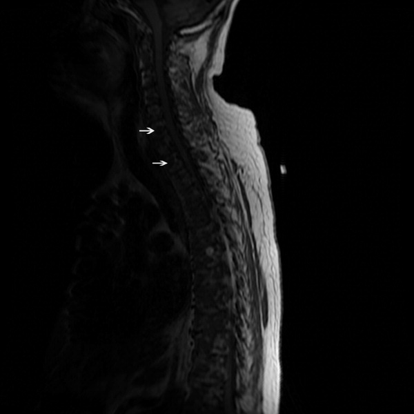
The lack of lymphadenopathy, splenomegaly, or a visceral mass by CT imaging and physical examination, along with the normal PSA level, exclude most common forms of non‐Hodgkin lymphoma and bone metastasis from solid tumors. In multiple myeloma, cytokines secreted by plasma cells suppress osteoblast activity; therefore, while discrete lytic bone lesions are apparent on skeletal survey, the bone scan is typically normal. The absence of lytic lesions, normal serum immunoglobulin levels, and unremarkable UPEP make multiple myeloma or light‐chain deposition disease a less likely diagnosis.
Typically, primary lymphoma of the bone produces increased uptake with bone scanning. However, because primary lymphoma of the bone is one of the least common primary skeletal malignancies and varies widely in appearance on imaging, confident diagnosis based on imaging alone usually is not possible.
Posttransplantation lymphoproliferative disorder (PTLD) refers to a syndrome that ranges from a self‐limited form of lymphoproliferation to an aggressive disseminated disease. Although the patient is at risk for PTLD, isolated bone involvement has only rarely been reported.
Primary lymphoma of the bone and PTLD are my leading diagnoses in this patient. At this point, I recommend a bone marrow biopsy and biopsy of an easily accessible representative bone lesion with special staining for Epstein‐Barr virus (EBV) (EBV‐encoded RNA [EBER] and latent membrane protein 1 [LMP1]). I expect this test to provide a definitive diagnosis. As 95% of PTLD cases are induced by infection with EBV, information regarding pretransplantation EBV status of the patient and the donor, current EBV status of the patient, and type and intensity of immunosuppression at the time of transplantation would be very helpful to determine their likelihood.
Seventy‐two hours after presentation, his serum calcium level normalized and most of his symptoms improved. Calcitonin was discontinued, and he was maintained on oral hydration. On hospital day number 5, he underwent CT‐guided bone biopsy of the L4 vertebral body, which showed large aggregates of atypical lymphoid cells (Figure 2). These cells were predominantly B‐cells interspersed with small reactive T‐cells. The cells did not express EBV LMP1 or EBER (Figure 3). On hospital day 7, he underwent a bone marrow biopsy, which revealed similar large atypical lymphoid cells that comprised the majority of marrow space (Figure 4). By immunohistochemistry, these cells brightly expressed the pan B cell marker, CD20, and coexpressed bcl‐2. EBER and LMP1 were also negative. A flow cytometry of the bone marrow demonstrated a lambda light chain restriction within the B lymphocytes.


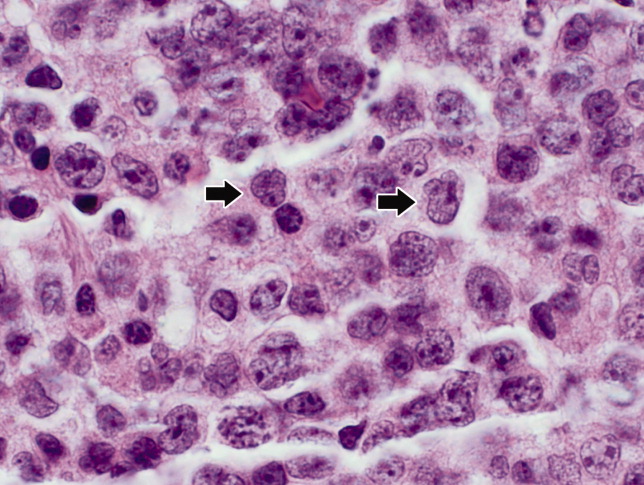
The medical records indicated that the patient had positive pretransplantation EBV serologies. He received a regimen based on sirolimus, mycophenolate mofetil, and prednisone, and did not receive high doses of induction or maintenance immunosuppressive therapy.
The biopsy results establish a diagnosis of diffuse large B‐cell lymphoma of the bone. PTLD is unlikely given his positive pretransplantation EBV status, the late onset of his disease (6 years after transplantation), the isolated bone involvement, and the negative EBER and LMP1 tests.
The patient was discharged and was readmitted 1 week later for induction chemotherapy with etoposide, vincristine, doxorubicin, cyclophosphamide, and prednisone [EPOCH]Rituxan (rituximab). Over the next several months, he received 6 cycles of chemotherapy, his hypercalcemia resolved, and his back pain improved.
Commentary
Hypercalcemia is among the most common causes of nephrogenic diabetes insipidus in adults.1 A urinary concentrating defect usually becomes clinically apparent if the plasma calcium concentration is persistently above 11 mg/dL.1 This defect is generally reversible with correction of the hypercalcemia but may persist in patients in whom interstitial nephritis has induced permanent medullary damage. The mechanism by which the concentrating defect occurs is incompletely understood but may be related to impairments in sodium chloride reabsorption in the thick ascending limb and in the ability of antidiuretic hormone to increase water permeability in the collecting tubules.1
Although hypercalcemia in otherwise healthy outpatients is usually due to primary hyperparathyroidism, malignancy is more often responsible for hypercalcemia in hospitalized patients.2 While the signs and symptoms of hypercalcemia are similar regardless of the cause, several clinical features may help distinguish the etiology of hypercalcemia. For instance, the presence of tachycardia, warm skin, thinning of the hair, stare and lid lag, and widened pulse pressure points toward hypercalcemia related to hyperthyroidism. In addition, risk factors and comorbidities guide the diagnostic process. For example, low‐level hypercalcemia in an asymptomatic postmenopausal woman with a normal physical examination suggests primary hyperparathyroidism. In contrast, hypercalcemia in a transplant patient raises concern of malignancy including PTLDs.3, 4
PTLDs are uncommon causes of hypercalcemia but are among the most serious and potentially fatal complications of chronic immunosuppression in transplant recipients.5 They occur in 1.9% of patients after kidney transplantation. The lymphoproliferative disorders occurring after transplantation have different characteristics from those that occur in the general population. Non‐Hodgkin lymphoma accounts for 65% of lymphomas in the general population, compared to 93% in transplant recipients.5, 6 The pathogenesis of PTLD appears to be related to B cell proliferation induced by infection with EBV in the setting of chronic immunosuppression.6 Therefore, there is an increased frequency of PTLD among transplant recipients who are EBV seronegative at the time of operation. These patients, who have no preoperative immunity to EBV, usually acquire the infection from the donor. The level of immunosuppression (intensity and type) influences PTLD rates as well. The disease typically occurs within 12 months after transplantation and in two‐thirds of cases involves extranodal sites. Among these sites, the gastrointestinal tract is involved in about 26% of cases and central nervous system in about 27%. Isolated bone involvement is exceedingly rare.5, 6
Primary lymphoma of the bone is another rare cause of hypercalcemia and accounts for less than 5% of all primary bone tumors.7 The majority of cases are of the non‐Hodgkin's type, characterized as diffuse large B‐cell lymphomas, with peak occurrence in the sixth to seventh decades of life.8 The classic imaging findings of primary lymphoma of the bone are a solitary metadiaphyseal lesion with a layered periosteal reaction on plain radiographs, and corresponding surrounding soft‐tissue mass on MRI.9 Less commonly, primary lymphoma of the bone can be multifocal with diffuse osseous involvement and variable radiographic appearances, as in this case. Most series have reported that the long bones are affected most frequently (especially the femur), although a large series showed equal numbers of cases presenting in the long bones and the spine.712
In order to diagnose primary lymphoma of the bone, it is necessary to exclude nodal or disseminated disease by physical examination and imaging. As plain films are often normal, bone scan or MRI of clinically affected areas is necessary to establish disease extent.9 Distinguishing primary bone lymphomas (PLB) from other bone tumors is important because PLB has a better response to therapy and a better prognosis.10, 11
Randomized trials addressing treatment options for primary lymphoma of bone are not available. Historically, PLB was treated with radiotherapy alone with good local control. However, the rate of distant relapses was relatively high. Currently, chemotherapy with or without radiation therapy is preferred; 5‐year survival is approximately 70% after combined therapy.10, 11
In this case, symptomatic hypercalcemia, a history of transplantation, marked elevation of both LDH and B2M, and a normal PTH level all pointed toward the correct diagnosis of malignancy. Low or normal levels of vitamin D metabolites and PTH‐related protein occur in 20% of patients with hypercalcemia caused by malignancy.13, 14 Diffuse osteopenia on skeletal survey is a prominent feature of renal osteodystrophy or osteoporosis related to chronic corticosteroid use. However, in a patient with diffuse osteopenia and hypercalcemia, clinicians must consider multiple myeloma and other lymphoproliferative disorders; the absence of osteoblastic or osteolytic lesions and a normal alkaline phosphatase do not rule out these diagnoses. When the results of serum and urine protein electrophoresis exclude multiple myeloma, the next investigation should be a bone biopsy to exclude PLB, an uncommon cause of anemia, hypercalcemia, and osteopenic, painful bones.
Key Points for Hospitalists
-
Normal total alkaline phosphatase does not exclude primary or metastatic bone malignancy. While a normal level tends to predict a negative bone scan, further diagnostic tests are needed to exclude bone malignancy if high clinical suspicion exists.
-
The degree of hypercalcemia is useful diagnostically; values above 13 mg/dL are most often due to malignancy.
-
Hypercalcemia in transplant patients deserves special attention due to an increased risk of malignancy, including squamous cancers of the lips and skin, lymphoproliferative disorders, and bronchogenic carcinoma.
-
While rare, consider primary lymphoma of the bone in patients with hypercalcemia and bone pain, along with the more common diagnoses of multiple myeloma and metastatic bone disease.
The approach to clinical conundrums by an expert clinician is revealed through presentation of an actual patient's case in an approach typical of morning report. Similar to patient care, sequential pieces of information are provided to the clinician who is unfamiliar with the case. The focus is on the thought processes of both the clinical team caring the patient and the discussant.
- ,.Clinical Physiology of Acid‐Base and Electrolyte Disorders.5th ed.New York:McGraw‐Hill;2001:754–758.
- ,.Hypercalcemia: clinical manifestations, pathogenesis, diagnosis, and management. In: Favus MJ, ed.Primer on the Metabolic Bone Diseases and Disorders of Mineral Metabolism.5th ed.Washington, DC:American Society for Bone and Mineral Research;2003:225–230.
- ,,, et al.Malignancy after renal transplantation: analysis of incidence and risk factors in 1700 patients followed during a 25‐year period.Transplant Proc.1997;29:831–833.
- ,.Malignancy‐associated hypercalcemia. In: DeGroot L, Jameson LJ, eds.Endocrinology.4th ed.Philadelphia, PA:Saunders;2001:1093–1100.
- ,.Diagnosis and management of posttransplant lymphoproliferative disorder in solid‐organ transplant recipients.Clin Infect Dis.2001;33(suppl 1):S38–S46.
- ,,, et al.Epstein‐Barr virus‐induced posttransplant lymphoproliferative disorders: ASTS/ASTP EBV‐PTLD Task Force and The Mayo Clinic Organized International Consensus Development Meeting.Transplantation.1999;68:1517–1525.
- ,,, et al.Primary bone lymphoma: a new and detailed characterization of 28 patients in a single‐institution study.Jpn J Clin Oncol.2007;37(3):216–223.
- ,,, et al.Diffuse large B‐cell lymphoma of bone. An analysis of differentiation‐associated antigens with clinical correlation.Am J Surg Pathol.2003;27:1269–1277.
- ,,,,,.Primary bone lymphoma: radiographic‐MR imaging correlation.Radiographics.2003;23:1371–1383.
- ,,, et al.Primary bone lymphoma in 24 patients treated between 1955 and 1999.Clin Orthop.2002;397:271–280.
- ,,, et al.A clinicopathological retrospective study of 131 patients with primary bone lymphoma: a population‐based study of successively treated cohorts from the British Columbia Cancer Agency.Ann Oncol.2007;18:129.
- ,,, et al.Malignant lymphoma of bone.Cancer.1986;58:2646–2655.
- .Hypercalcemia in malignant lymphoma and leukemia.Ann N Y Acad Sci.1974;230:240–246.
- .Incidence and prognostic significance of hypercalcemia in B‐cell non‐Hodgkin's lymphoma. [Letter]J Clin Pathol.2002;55:637–638.
A 71‐year‐old man presented to a hospital with a one week history of fatigue, polyuria, and polydipsia. He also reported pain in his back, hips, and ribs, in addition to frequent falls, intermittent confusion, constipation, and a weight loss of 10 pounds over the last 2 weeks. He denied cough, shortness of breath, chest pain, fever, night sweats, headache, and focal weakness.
Polyuria, which is often associated with polydipsia, can be arbitrarily defined as a urine output exceeding 3 L per day. After excluding osmotic diuresis due to uncontrolled diabetes mellitus, the 3 major causes of polyuria are primary polydipsia, central diabetes insipidus, and nephrogenic diabetes insipidus. Approximately 30% to 50% of cases of central diabetes insipidus are idiopathic; however, primary or secondary brain tumors or infiltrative diseases involving the hypothalamic‐pituitary region need to be considered in this 71‐year‐old man. The most common causes of nephrogenic diabetes insipidus in adults are chronic lithium ingestion, hypokalemia, and hypercalcemia. The patient describes symptoms that can result from severe hypercalcemia, including fatigue, confusion, constipation, polyuria, and polydipsia.
The patient's past medical history included long‐standing, insulin‐requiring type 2 diabetes with associated complications including coronary artery disease, transient ischemic attacks, proliferative retinopathy, peripheral diabetic neuropathy, and nephropathy. Seven years prior to presentation, he received a cadaveric renal transplant that was complicated by BK virus (polyomavirus) nephropathy and secondary hyperparathyroidism. Three years after his transplant surgery, he developed squamous cell carcinoma of the skin, which was treated with local surgical resection. Two years after that, he developed stage I laryngeal cancer of the glottis and received laser surgery, and since then he had been considered disease‐free. He also had a history of hypertension, hypercholesterolemia, osteoporosis, and depression. His medications included aspirin, amlodipine, metoprolol succinate, valsartan, furosemide, simvastatin, insulin, prednisone, sirolimus, and sulfamethoxazole/trimethoprim. He was a married psychiatrist. He denied tobacco use and reported occasional alcohol use.
The prolonged immunosuppressive therapy that is required following organ transplantation carries a markedly increased risk of the subsequent development of malignant tumors, including cancers of the lips and skin, lymphoproliferative disorders, and bronchogenic carcinoma. Primary brain lymphoma resulting in central diabetes insipidus would be unlikely in the absence of headache or focal weakness. An increased risk of lung cancer occurs in recipients of heart and lung transplants, and to a much lesser degree, recipients of kidney transplants. However, metastatic lung cancer is less likely in the absence of respiratory symptoms and smoking history (present in approximately 90% of all lung cancers). Nephrogenic diabetes insipidus, in its mild form, is relatively common in elderly patients with acute or chronic renal insufficiency because of a reduction in maximum urinary concentrating ability. On the other hand, this alone does not explain his remaining symptoms. The instinctive diagnosis in this case is tertiary hyperparathyroidism due to progression of untreated secondary hyperparathyroidism. This causes hypercalcemia, nephrogenic diabetes insipidus, and significant bone pain related to renal osteodystrophy.
On physical exam, the patient appeared chronically ill, but was in no acute distress. He weighed 197.6 pounds and his height was 70.5 inches. He was afebrile with a blood pressure of 146/82 mm Hg, a heart rate of 76 beats per minute, a respiratory rate of 12 breaths per minute, and an oxygen saturation of 97% while breathing room air. He had no generalized lymphadenopathy. Thyroid examination was unremarkable. Examination of the lungs, heart, abdomen, and lower extremities was normal. The rectal examination revealed no masses or prostate nodules; a test for fecal occult blood was negative. He had loss of sensation to light touch and vibration in the feet with absent Achilles deep tendon reflexes. He had a poorly healing surgical wound on his forehead at the site of his prior skin cancer, but no rash or other lesions. There was no joint swelling or erythema. There were tender points over the cervical, thoracic, and lumbar spine; on multiple ribs; and on the pelvic rims.
Perhaps of greatest importance is the lack of lymphadenopathy, organomegaly, or other findings suggestive of diffuse lymphoproliferative disease. His multifocal bone tenderness is concerning for renal osteodystrophy, multiple myeloma, or primary or metastatic bone disease. Cancers in men that metastasize to the bone usually originate from the prostate, lung, kidney, or thyroid gland. In any case, his physical examination did not reveal an enlarged, asymmetric, or nodular prostate or thyroid gland. I recommend a chest film to rule out primary lung malignancy and a basic laboratory evaluation to narrow down the differential diagnosis.
A complete blood count showed a normocytic anemia with a hemoglobin of 8.7 g/dL and a hematocrit of 25%. Other laboratory tests revealed the following values: sodium, 139 mmol/L; potassium, 4.1 mmol/L; blood urea nitrogen, 70 mg/dL; creatinine, 3.5 mg/dL (most recent value 2 months ago was 1.9 mg/dL); total calcium, 13.2 mg/dL (normal range, 8.5‐10.5 mg/dL); phosphate, 5.3 mg/dL; magnesium, 2.5 mg/dL; total bilirubin, 0.5 mg/dL; alkaline phosphatase, 130 U/L; aspartate aminotransferase, 28 U/L; alanine aminotransferase, 19 U/L; albumin, 3.5 g/dL; and lactate dehydrogenase (LDH), 1258 IU/L (normal range, 105‐333 IU/L). A chest radiograph was normal.
The most important laboratory findings are severe hypercalcemia, acute on chronic renal failure, and anemia. Hypercalcemia most commonly results from malignancy or hyperparathyroidism. Less frequently, hypercalcemia may result from sarcoidosis, vitamin D intoxication, or hyperthyroidism. The degree of hypercalcemia is useful diagnostically as hyperparathyroidism commonly results in mild hypercalcemia (serum calcium concentration often below 11 mg/dL). Values above 13 mg/dL are unusual in hyperparathyroidism and are most often due to malignancy. Malignancy is often evident clinically by the time it causes hypercalcemia, and patients with hypercalcemia of malignancy are more often symptomatic than those with hyperparathyroidism. Additionally, localized bone pain and weight loss do not result from hypercalcemia itself and their presence also raises concern for malignancy.
Nonmelanoma skin cancer is the most common cancer occurring after transplantation but does not cause hypercalcemia. Squamous cancers of the head and neck can rarely cause hypercalcemia due to secretion of parathyroid hormone‐related peptide; however, his early‐stage laryngeal cancer and the expected high likelihood of cure argue against this possibility. Osteolytic metastases account for approximately 20% of cases of hypercalcemia of malignancy (Table 1). Prostate cancer rarely results in hypercalcemia since bone metastases are predominantly osteoblastic, whereas metastatic non‐small‐cell lung cancer, thyroid cancer, and kidney cancer more commonly cause hypercalcemia due to osteolytic bone lesions. The total alkaline phosphatase has been traditionally used to assess the osteoblastic component of bone remodeling. Its normal level tends to predict a negative bone scan and supports the likelihood of lytic lesions. Posttransplantation lymphoproliferative disorders, which include a wide range of syndromes, can rarely result in hypercalcemia. I am also worried about the possibility of multiple myeloma as he has the classic triad of hypercalcemia, bone pain, and subacute kidney injury.
|
| Osteolytic metastases |
| Breast cancer |
| Multiple myeloma |
| Lymphoma |
| Leukemia |
| Humoral hypercalcemia (PTH‐related protein) |
| Squamous cell carcinomas |
| Renal carcinomas |
| Bladder carcinoma |
| Breast cancer |
| Ovarian carcinoma |
| Leukemia |
| Lymphoma |
| 1,25‐Dihydroxyvitamin D secretion |
| Lymphoma |
| Ovarian dysgerminomas |
| Ectopic PTH secretion (rare) |
| Ovarian carcinoma |
| Lung carcinomas |
| Neuroectodermal tumor |
| Thyroid papillary carcinoma |
| Rhabdomyosarcoma |
| Pancreatic cancer |
The first purpose of the laboratory evaluation is to differentiate parathyroid hormone (PTH)‐mediated hypercalcemia (primary and tertiary hyperparathyroidism) from non‐PTH‐mediated hypercalcemia (primarily malignancy, hyperthyroidism, vitamin D intoxication, and granulomatous disease). The production of vitamin D metabolites, PTH‐related protein, or hypercalcemia from osteolysis in these latter cases results in suppressed PTH levels.
In severe elevations of calcium, the initial goals of treatment are directed toward fluid resuscitation with normal saline and, unless contraindicated, the immediate institution of bisphosphonate therapy. A loop diuretic such as furosemide is often used, but a recent review concluded that there is little evidence to support its use in this setting.
The patient was admitted and treated with intravenous saline and furosemide. Additional laboratory evaluation revealed normal levels of prostate‐specific antigen and thyroid‐stimulating hormone. PTH was 44 pg/mL (the most recent value was 906 pg/mL eight years ago; normal range, 15‐65 pg/mL) and beta‐2 microglobulin (B2M) was 8 mg/L (normal range, 0.8‐2.2 mg/L).
The normal PTH level makes tertiary hyperparathyroidism unlikely and points toward non‐PTH‐related hypercalcemia. An elevated B2M level may occur in patients with chronic graft rejection, renal tubular dysfunction, dialysis‐related amyloidosis, multiple myeloma, or lymphoma. LDH is often elevated in patients with multiple myeloma and lymphoma, but this is not a specific finding. The next laboratory test would be measurement of PTH‐related protein and vitamin D metabolites, as these tests can differentiate between the causes of non‐PTH‐mediated hypercalcemia.
Serum concentrations of the vitamin D metabolites, 25‐hydroxyvitamin D (calcidiol) and 1,25‐dihydroxyvitamin D (calcitriol), were low‐normal. PTH‐related protein was not detected.
The marked elevation of serum LDH and B2M, the relatively suppressed PTH level, combined with undetectable PTH‐related protein suggest multiple myeloma or lymphoma as the likely cause of the patient's clinical presentation. The combination of hypercalcemia and multifocal bone pain makes multiple myeloma the leading diagnosis as hypercalcemia is uncommon in patients with lymphoma, especially at the time of initial clinical presentation.
I would proceed with serum and urine protein electrophoresis (SPEP and UPEP, respectively) and a skeletal survey. If these tests do not confirm the diagnosis of multiple myeloma, I would order a noncontrast computed tomography (CT) of the chest and abdomen and a magnetic resonance imaging (MRI) of the spine. In addition, I would like to monitor his response to the intravenous saline and furosemide.
Forty‐eight hours after presentation, repeat serum calcium and creatinine levels were 11.3 mg/dL and 2.9 mg/dL, respectively. He received salmon calcitonin 4 U/kg every 12 hours. Pamidronate was avoided because of his kidney disease. His confusion resolved. He received intravenous morphine intermittently to alleviate his bone pain.
The SPEP revealed a monoclonal immunoglobulin G (IgG) lambda (light chain) spike representing roughly 3% (200 mg/dL) of total protein. His serum Ig levels were normal. The UPEP was negative for monoclonal immunoglobulin and Bence‐Jones protein. The skeletal survey revealed marked osteopenia, and the bone scan was normal. An MRI of the spine showed multiple round lesions in the cervical, thoracic, and lumbar spine (Figure 1). A CT of the chest showed similar bone lesions in the ribs and pelvis. A CT of the abdomen and chest did not suggest any primary malignancy nor did it show thoracic or abdominal lymphadenopathy.
The lack of lymphadenopathy, splenomegaly, or a visceral mass by CT imaging and physical examination, along with the normal PSA level, exclude most common forms of non‐Hodgkin lymphoma and bone metastasis from solid tumors. In multiple myeloma, cytokines secreted by plasma cells suppress osteoblast activity; therefore, while discrete lytic bone lesions are apparent on skeletal survey, the bone scan is typically normal. The absence of lytic lesions, normal serum immunoglobulin levels, and unremarkable UPEP make multiple myeloma or light‐chain deposition disease a less likely diagnosis.
Typically, primary lymphoma of the bone produces increased uptake with bone scanning. However, because primary lymphoma of the bone is one of the least common primary skeletal malignancies and varies widely in appearance on imaging, confident diagnosis based on imaging alone usually is not possible.
Posttransplantation lymphoproliferative disorder (PTLD) refers to a syndrome that ranges from a self‐limited form of lymphoproliferation to an aggressive disseminated disease. Although the patient is at risk for PTLD, isolated bone involvement has only rarely been reported.
Primary lymphoma of the bone and PTLD are my leading diagnoses in this patient. At this point, I recommend a bone marrow biopsy and biopsy of an easily accessible representative bone lesion with special staining for Epstein‐Barr virus (EBV) (EBV‐encoded RNA [EBER] and latent membrane protein 1 [LMP1]). I expect this test to provide a definitive diagnosis. As 95% of PTLD cases are induced by infection with EBV, information regarding pretransplantation EBV status of the patient and the donor, current EBV status of the patient, and type and intensity of immunosuppression at the time of transplantation would be very helpful to determine their likelihood.
Seventy‐two hours after presentation, his serum calcium level normalized and most of his symptoms improved. Calcitonin was discontinued, and he was maintained on oral hydration. On hospital day number 5, he underwent CT‐guided bone biopsy of the L4 vertebral body, which showed large aggregates of atypical lymphoid cells (Figure 2). These cells were predominantly B‐cells interspersed with small reactive T‐cells. The cells did not express EBV LMP1 or EBER (Figure 3). On hospital day 7, he underwent a bone marrow biopsy, which revealed similar large atypical lymphoid cells that comprised the majority of marrow space (Figure 4). By immunohistochemistry, these cells brightly expressed the pan B cell marker, CD20, and coexpressed bcl‐2. EBER and LMP1 were also negative. A flow cytometry of the bone marrow demonstrated a lambda light chain restriction within the B lymphocytes.
The medical records indicated that the patient had positive pretransplantation EBV serologies. He received a regimen based on sirolimus, mycophenolate mofetil, and prednisone, and did not receive high doses of induction or maintenance immunosuppressive therapy.
The biopsy results establish a diagnosis of diffuse large B‐cell lymphoma of the bone. PTLD is unlikely given his positive pretransplantation EBV status, the late onset of his disease (6 years after transplantation), the isolated bone involvement, and the negative EBER and LMP1 tests.
The patient was discharged and was readmitted 1 week later for induction chemotherapy with etoposide, vincristine, doxorubicin, cyclophosphamide, and prednisone [EPOCH]Rituxan (rituximab). Over the next several months, he received 6 cycles of chemotherapy, his hypercalcemia resolved, and his back pain improved.
Commentary
Hypercalcemia is among the most common causes of nephrogenic diabetes insipidus in adults.1 A urinary concentrating defect usually becomes clinically apparent if the plasma calcium concentration is persistently above 11 mg/dL.1 This defect is generally reversible with correction of the hypercalcemia but may persist in patients in whom interstitial nephritis has induced permanent medullary damage. The mechanism by which the concentrating defect occurs is incompletely understood but may be related to impairments in sodium chloride reabsorption in the thick ascending limb and in the ability of antidiuretic hormone to increase water permeability in the collecting tubules.1
Although hypercalcemia in otherwise healthy outpatients is usually due to primary hyperparathyroidism, malignancy is more often responsible for hypercalcemia in hospitalized patients.2 While the signs and symptoms of hypercalcemia are similar regardless of the cause, several clinical features may help distinguish the etiology of hypercalcemia. For instance, the presence of tachycardia, warm skin, thinning of the hair, stare and lid lag, and widened pulse pressure points toward hypercalcemia related to hyperthyroidism. In addition, risk factors and comorbidities guide the diagnostic process. For example, low‐level hypercalcemia in an asymptomatic postmenopausal woman with a normal physical examination suggests primary hyperparathyroidism. In contrast, hypercalcemia in a transplant patient raises concern of malignancy including PTLDs.3, 4
PTLDs are uncommon causes of hypercalcemia but are among the most serious and potentially fatal complications of chronic immunosuppression in transplant recipients.5 They occur in 1.9% of patients after kidney transplantation. The lymphoproliferative disorders occurring after transplantation have different characteristics from those that occur in the general population. Non‐Hodgkin lymphoma accounts for 65% of lymphomas in the general population, compared to 93% in transplant recipients.5, 6 The pathogenesis of PTLD appears to be related to B cell proliferation induced by infection with EBV in the setting of chronic immunosuppression.6 Therefore, there is an increased frequency of PTLD among transplant recipients who are EBV seronegative at the time of operation. These patients, who have no preoperative immunity to EBV, usually acquire the infection from the donor. The level of immunosuppression (intensity and type) influences PTLD rates as well. The disease typically occurs within 12 months after transplantation and in two‐thirds of cases involves extranodal sites. Among these sites, the gastrointestinal tract is involved in about 26% of cases and central nervous system in about 27%. Isolated bone involvement is exceedingly rare.5, 6
Primary lymphoma of the bone is another rare cause of hypercalcemia and accounts for less than 5% of all primary bone tumors.7 The majority of cases are of the non‐Hodgkin's type, characterized as diffuse large B‐cell lymphomas, with peak occurrence in the sixth to seventh decades of life.8 The classic imaging findings of primary lymphoma of the bone are a solitary metadiaphyseal lesion with a layered periosteal reaction on plain radiographs, and corresponding surrounding soft‐tissue mass on MRI.9 Less commonly, primary lymphoma of the bone can be multifocal with diffuse osseous involvement and variable radiographic appearances, as in this case. Most series have reported that the long bones are affected most frequently (especially the femur), although a large series showed equal numbers of cases presenting in the long bones and the spine.712
In order to diagnose primary lymphoma of the bone, it is necessary to exclude nodal or disseminated disease by physical examination and imaging. As plain films are often normal, bone scan or MRI of clinically affected areas is necessary to establish disease extent.9 Distinguishing primary bone lymphomas (PLB) from other bone tumors is important because PLB has a better response to therapy and a better prognosis.10, 11
Randomized trials addressing treatment options for primary lymphoma of bone are not available. Historically, PLB was treated with radiotherapy alone with good local control. However, the rate of distant relapses was relatively high. Currently, chemotherapy with or without radiation therapy is preferred; 5‐year survival is approximately 70% after combined therapy.10, 11
In this case, symptomatic hypercalcemia, a history of transplantation, marked elevation of both LDH and B2M, and a normal PTH level all pointed toward the correct diagnosis of malignancy. Low or normal levels of vitamin D metabolites and PTH‐related protein occur in 20% of patients with hypercalcemia caused by malignancy.13, 14 Diffuse osteopenia on skeletal survey is a prominent feature of renal osteodystrophy or osteoporosis related to chronic corticosteroid use. However, in a patient with diffuse osteopenia and hypercalcemia, clinicians must consider multiple myeloma and other lymphoproliferative disorders; the absence of osteoblastic or osteolytic lesions and a normal alkaline phosphatase do not rule out these diagnoses. When the results of serum and urine protein electrophoresis exclude multiple myeloma, the next investigation should be a bone biopsy to exclude PLB, an uncommon cause of anemia, hypercalcemia, and osteopenic, painful bones.
Key Points for Hospitalists
-
Normal total alkaline phosphatase does not exclude primary or metastatic bone malignancy. While a normal level tends to predict a negative bone scan, further diagnostic tests are needed to exclude bone malignancy if high clinical suspicion exists.
-
The degree of hypercalcemia is useful diagnostically; values above 13 mg/dL are most often due to malignancy.
-
Hypercalcemia in transplant patients deserves special attention due to an increased risk of malignancy, including squamous cancers of the lips and skin, lymphoproliferative disorders, and bronchogenic carcinoma.
-
While rare, consider primary lymphoma of the bone in patients with hypercalcemia and bone pain, along with the more common diagnoses of multiple myeloma and metastatic bone disease.
The approach to clinical conundrums by an expert clinician is revealed through presentation of an actual patient's case in an approach typical of morning report. Similar to patient care, sequential pieces of information are provided to the clinician who is unfamiliar with the case. The focus is on the thought processes of both the clinical team caring the patient and the discussant.
- ,.Clinical Physiology of Acid‐Base and Electrolyte Disorders.5th ed.New York:McGraw‐Hill;2001:754–758.
- ,.Hypercalcemia: clinical manifestations, pathogenesis, diagnosis, and management. In: Favus MJ, ed.Primer on the Metabolic Bone Diseases and Disorders of Mineral Metabolism.5th ed.Washington, DC:American Society for Bone and Mineral Research;2003:225–230.
- ,,, et al.Malignancy after renal transplantation: analysis of incidence and risk factors in 1700 patients followed during a 25‐year period.Transplant Proc.1997;29:831–833.
- ,.Malignancy‐associated hypercalcemia. In: DeGroot L, Jameson LJ, eds.Endocrinology.4th ed.Philadelphia, PA:Saunders;2001:1093–1100.
- ,.Diagnosis and management of posttransplant lymphoproliferative disorder in solid‐organ transplant recipients.Clin Infect Dis.2001;33(suppl 1):S38–S46.
- ,,, et al.Epstein‐Barr virus‐induced posttransplant lymphoproliferative disorders: ASTS/ASTP EBV‐PTLD Task Force and The Mayo Clinic Organized International Consensus Development Meeting.Transplantation.1999;68:1517–1525.
- ,,, et al.Primary bone lymphoma: a new and detailed characterization of 28 patients in a single‐institution study.Jpn J Clin Oncol.2007;37(3):216–223.
- ,,, et al.Diffuse large B‐cell lymphoma of bone. An analysis of differentiation‐associated antigens with clinical correlation.Am J Surg Pathol.2003;27:1269–1277.
- ,,,,,.Primary bone lymphoma: radiographic‐MR imaging correlation.Radiographics.2003;23:1371–1383.
- ,,, et al.Primary bone lymphoma in 24 patients treated between 1955 and 1999.Clin Orthop.2002;397:271–280.
- ,,, et al.A clinicopathological retrospective study of 131 patients with primary bone lymphoma: a population‐based study of successively treated cohorts from the British Columbia Cancer Agency.Ann Oncol.2007;18:129.
- ,,, et al.Malignant lymphoma of bone.Cancer.1986;58:2646–2655.
- .Hypercalcemia in malignant lymphoma and leukemia.Ann N Y Acad Sci.1974;230:240–246.
- .Incidence and prognostic significance of hypercalcemia in B‐cell non‐Hodgkin's lymphoma. [Letter]J Clin Pathol.2002;55:637–638.

Small Bowel Obstruction by Gallstone Ileus
A 67‐year‐old female presented with nausea and nonspecific abdominal pain occurring intermittently for 2 weeks. Physical examination revealed no abdominal guarding or rigidity and was significant only for slightly hypoactive bowel sounds. Routine laboratory evaluation including liver panel was unremarkable. X‐ray of the abdomen showed air fluid levels, and a diagnosis of small bowel obstruction/emleus secondary to adhesions from a previous hysterectomy was established. Conservative management with nasogastric suction, intravenous (IV) fluids and nil‐by‐mouth was continued for 3 days but no clinical improvement was seen. Due to continued abdominal pain, an ultrasound of the abdomen was done, which revealed cholelithiasis and chronic cholecystitis. At this point the patient was transferred to our facility and a computerized axial tomography (CAT) scan of the abdomen was done, which revealed a gallstone in the ileum (Figure 1) and the diagnosis of gallstone ileus was made. Air in the biliary tract (pneumobilia) was noted, suggesting the presence of a fistula between the gallbladder and the gastrointestinal (GI) tract. The fistula itself could not be clearly visualized. The patient was immediately taken to surgery, where small bowel exploration was done. A large gallstone completely obstructing the terminal ileum was removed by enterotomy. The gallbladder was adherent to the stomach and a cholecystogastric fistula with a gallstone coming out on the gastric end was noted. This gallstone was removed and the cholecystogastric fistula was repaired. Cholecystectomy was done at the same time. The patient recovered without any complications and has been doing well.

Discussion
Gallstone ileus accounts for approximately 1% to 2% cases of small bowel obstruction, most of which are in the elderly population. It is much more common in females as compared to males.1 Although the morbidity and mortality associated with gallstone ileus has reduced in comparison to the previous decade, probably due to the more effective usage of imaging techniques like ultrasound, CAT scan, magnetic resonance imaging (MRI), and endoscopy in the diagnostic evaluation of abdominal pain, the numbers still range between 12% to 25%.1, 2 Early diagnosis plays a major role in reducing the mortality in these patients. Therefore, it is important to consider gallstone ileus in the differential diagnosis of an elderly patient presenting with bowel obstruction.
Gallstone ileus is usually associated with a biliaryenteric fistula that allows the passage of a gallstone from the gallbladder into the bowel. This gallstone gets impacted in the gastrointestinal lumen and causes mechanical bowel obstruction. The term gallstone ileus is a misnomer as the gallstone causes actual obstruction rather than just ileus. Considering the high prevalence of cholelithiasis, it must be realized that formation of cholecystoenteric fistula is relatively rare (about 2% in patients with cholecystitis). It is proposed that pericholecystic inflammation after an episode of cholecystitis results in the formation of adhesions between the biliary and gastrointestinal tracts. The gallstone causes pressure necrosis of the biliary wall and then erodes through it to form a fistulous communication with the adherent enteric system. Among these fistulous communications, cholecystoduodenal fistulas are the most common (60%) while cholecystocolonic and cholecystogastric fistulas are also seen. After the biliary stone has eroded through the enteric wall, air within the intestinal tract now freely enters the biliary system, leading to an appearance of pneumobilia on imaging studies. Mirizzi syndrome, described as common hepatic duct obstruction caused by an extrinsic compression from an impacted stone in the cystic duct, is often associated with gallstone ileus.3
Once the gallstone enters the enteric tract through the fistula and traverses down the gastrointestinal tract, it causes intermittent abdominal pain, nausea, and vomiting. The symptoms may be spread over multiple days as the gallstone causes transient obstruction with its impaction and disimpaction. These obstructing gallstones that cause luminal obstruction are usually larger than 2 cm. Majority of them will traverse the duodenum, jejunum, and small intestine, and finally get lodged in the terminal ileum (60%), the narrowest part of the small intestine. Other sites where obstruction may occur include jejunum (16%), duodenum, stomach, and colon.1 Bouveret's syndrome is a variant of gallstone ileus wherein the gallstone impacts in the pylorus of the stomach or the duodenum, leading to gastric outlet obstruction.3
The diagnosis of gallstone ileus is not always straightforward and requires a high index of suspicion. It is classically described by the Rigler's triadpneumobilia, partial or complete bowel obstruction, and ectopic gallstone,4 although often all 3 signs are not elicited. Most patients need open enterolithotomy for relief of bowel obstruction. Cholecystectomy may or may not be performed. The literature is controversial regarding the best approach of surgical management. Two surgical approaches are equally accepted: (1) a 1‐stage approach, which includes enterolithotomy, cholecystectomy, and fistula repair at the same time; and (2) the other option is a 2‐stage approach in which enterolithotomy is performed first and biliary surgery is performed later, if indicated. The patient's age, comorbidities, and the associated surgical risks are often used to decide between the 2 surgical approaches.
Gallstone ileus should be considered in the differential for the etiology of small bowel obstruction, especially in an elderly female known to have cholelithiasis.
- ,.Gallstone ileus (a review of 1001 reported cases).Am Surg.1994;60:441–446.
- ,,, et al.Improving the outcome in gallstone ileus.Am J Surg.1986;151:572–576.
- ,,.The relationship of Mirizzi syndrome and cholecystoenteric fistula: validation of a modified classification.World J Surg.2008;32(10):2237–2243.
- ,,.Gallstone obstruction: pathogenesis and roentgen manifestations.JAMA.1941;117:1753–1759.
A 67‐year‐old female presented with nausea and nonspecific abdominal pain occurring intermittently for 2 weeks. Physical examination revealed no abdominal guarding or rigidity and was significant only for slightly hypoactive bowel sounds. Routine laboratory evaluation including liver panel was unremarkable. X‐ray of the abdomen showed air fluid levels, and a diagnosis of small bowel obstruction/emleus secondary to adhesions from a previous hysterectomy was established. Conservative management with nasogastric suction, intravenous (IV) fluids and nil‐by‐mouth was continued for 3 days but no clinical improvement was seen. Due to continued abdominal pain, an ultrasound of the abdomen was done, which revealed cholelithiasis and chronic cholecystitis. At this point the patient was transferred to our facility and a computerized axial tomography (CAT) scan of the abdomen was done, which revealed a gallstone in the ileum (Figure 1) and the diagnosis of gallstone ileus was made. Air in the biliary tract (pneumobilia) was noted, suggesting the presence of a fistula between the gallbladder and the gastrointestinal (GI) tract. The fistula itself could not be clearly visualized. The patient was immediately taken to surgery, where small bowel exploration was done. A large gallstone completely obstructing the terminal ileum was removed by enterotomy. The gallbladder was adherent to the stomach and a cholecystogastric fistula with a gallstone coming out on the gastric end was noted. This gallstone was removed and the cholecystogastric fistula was repaired. Cholecystectomy was done at the same time. The patient recovered without any complications and has been doing well.
Discussion
Gallstone ileus accounts for approximately 1% to 2% cases of small bowel obstruction, most of which are in the elderly population. It is much more common in females as compared to males.1 Although the morbidity and mortality associated with gallstone ileus has reduced in comparison to the previous decade, probably due to the more effective usage of imaging techniques like ultrasound, CAT scan, magnetic resonance imaging (MRI), and endoscopy in the diagnostic evaluation of abdominal pain, the numbers still range between 12% to 25%.1, 2 Early diagnosis plays a major role in reducing the mortality in these patients. Therefore, it is important to consider gallstone ileus in the differential diagnosis of an elderly patient presenting with bowel obstruction.
Gallstone ileus is usually associated with a biliaryenteric fistula that allows the passage of a gallstone from the gallbladder into the bowel. This gallstone gets impacted in the gastrointestinal lumen and causes mechanical bowel obstruction. The term gallstone ileus is a misnomer as the gallstone causes actual obstruction rather than just ileus. Considering the high prevalence of cholelithiasis, it must be realized that formation of cholecystoenteric fistula is relatively rare (about 2% in patients with cholecystitis). It is proposed that pericholecystic inflammation after an episode of cholecystitis results in the formation of adhesions between the biliary and gastrointestinal tracts. The gallstone causes pressure necrosis of the biliary wall and then erodes through it to form a fistulous communication with the adherent enteric system. Among these fistulous communications, cholecystoduodenal fistulas are the most common (60%) while cholecystocolonic and cholecystogastric fistulas are also seen. After the biliary stone has eroded through the enteric wall, air within the intestinal tract now freely enters the biliary system, leading to an appearance of pneumobilia on imaging studies. Mirizzi syndrome, described as common hepatic duct obstruction caused by an extrinsic compression from an impacted stone in the cystic duct, is often associated with gallstone ileus.3
Once the gallstone enters the enteric tract through the fistula and traverses down the gastrointestinal tract, it causes intermittent abdominal pain, nausea, and vomiting. The symptoms may be spread over multiple days as the gallstone causes transient obstruction with its impaction and disimpaction. These obstructing gallstones that cause luminal obstruction are usually larger than 2 cm. Majority of them will traverse the duodenum, jejunum, and small intestine, and finally get lodged in the terminal ileum (60%), the narrowest part of the small intestine. Other sites where obstruction may occur include jejunum (16%), duodenum, stomach, and colon.1 Bouveret's syndrome is a variant of gallstone ileus wherein the gallstone impacts in the pylorus of the stomach or the duodenum, leading to gastric outlet obstruction.3
The diagnosis of gallstone ileus is not always straightforward and requires a high index of suspicion. It is classically described by the Rigler's triadpneumobilia, partial or complete bowel obstruction, and ectopic gallstone,4 although often all 3 signs are not elicited. Most patients need open enterolithotomy for relief of bowel obstruction. Cholecystectomy may or may not be performed. The literature is controversial regarding the best approach of surgical management. Two surgical approaches are equally accepted: (1) a 1‐stage approach, which includes enterolithotomy, cholecystectomy, and fistula repair at the same time; and (2) the other option is a 2‐stage approach in which enterolithotomy is performed first and biliary surgery is performed later, if indicated. The patient's age, comorbidities, and the associated surgical risks are often used to decide between the 2 surgical approaches.
Gallstone ileus should be considered in the differential for the etiology of small bowel obstruction, especially in an elderly female known to have cholelithiasis.
- ,.Gallstone ileus (a review of 1001 reported cases).Am Surg.1994;60:441–446.
- ,,, et al.Improving the outcome in gallstone ileus.Am J Surg.1986;151:572–576.
- ,,.The relationship of Mirizzi syndrome and cholecystoenteric fistula: validation of a modified classification.World J Surg.2008;32(10):2237–2243.
- ,,.Gallstone obstruction: pathogenesis and roentgen manifestations.JAMA.1941;117:1753–1759.
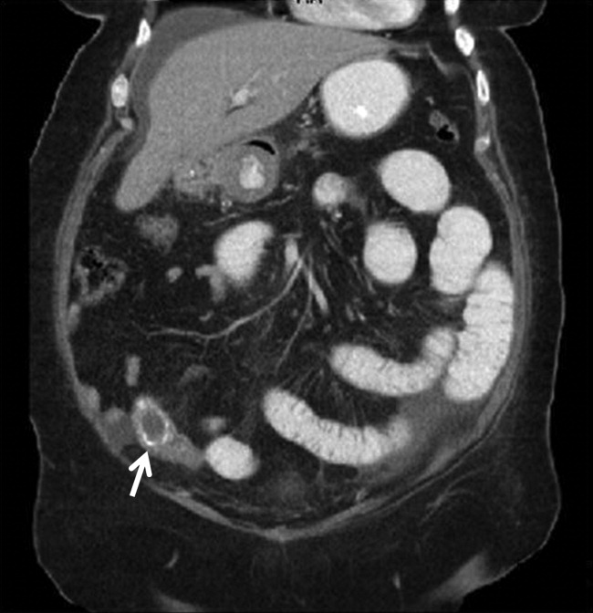
Management of Ischemic Stroke: Part 2
Inpatient stroke management includes many elements of care, at least as important as the initial portion of the patient's stay, as reviewed in part 1 of this article. The extent of further diagnostic evaluation varies widely depending on apparent risk factors on presentation. Likewise, further therapy, both inpatient and secondary prevention is based on identification of stroke mechanism. Hospitalists are uniquely positioned to have a tremendous impact on both stroke care and the prevention of recurrent disease.
Case Presentation
A 76‐year‐old right‐handed male with a history of hyperlipidemia and myocardial infarction was found at 7 AM with right‐sided paralysis and poor responsiveness on the morning of admission. Upon arrival to the emergency department (ED), with symptoms of partial aphasia, right hemiplegia, and left gaze preference, there was a high suspicion for a left middle cerebral artery (MCA) stroke. Unfortunately, he was excluded from receiving intravenous (IV) tissue plasminogen activator (tPA) or any other acute interventions as the last time he was known to be neurologically intact was the prior evening, which is taken to be the time of onset. Antiplatelet therapy was continued, and the patient admitted for further workup.
Inpatient Care
When an acute ischemic stroke patient is admitted to the hospital, he or she should be placed on a standardized acute stroke protocol (also known as (a.k.a.) a care map, order set, clinical pathway)commonly created by a hospitalist/neurologist and a multidisciplinary team and admitted to a stroke unit. A stroke unit can take many forms, either as a physically separate unit in hospitals with sufficient volume or a floor where a lower volume of stroke patients are always admitted. Multidisciplinary care providers in the stroke unit have special training in stroke, and strong evidence from randomized trials shows that patients cared for in these units have significantly decreased mortality with improved functional outcomes.1 Essentials of the stroke protocol or order set include cardiac telemetry, maintaining euthermia and euglycemia, closely following blood pressure and neurologic status, actively avoiding complications, initiation of secondary prevention treatment, early involvement of rehabilitation services, and patient education.
Euthermia may be assisted by administering scheduled Tylenol to the patient for the first 48 hours, but is not strictly evidence‐based.2 Though euthermia and euglycemia have not been shown to improve outcomes in acute stroke, studies have shown that hyperthermia and hyperglycemia are associated with worsened outcomes for patients with acute strokes.35
Blood Pressure Management
Normally, cerebral vascular autoregulation leads to stable cerebral blood flow over a range of systemic blood pressures. In the setting of an acute stroke, the ability to autoregulate is diminished or absent in regions of and surrounding an acute ischemic stroke; as the area becomes ischemic, autoregulation opens the local vasculature maximally in an effort to drawn in as much blood as possible. Maximally dilated arterioles are perfused in direct correlation with systemic blood pressure, thus any drop in the systemic blood pressure leads to direct decreases in blood flow specifically in the area of ischemia; if there is a penumbra of marginally perfused tissue, such systemic blood pressure drops risk extending the area of fatal ischemia (increasing the size of the ischemic stroke).68 Thus in the acute period of an ischemic stroke, the American Heart Association (AHA)/American Stroke Association (ASA) Guidelines for the Early Management of Adults With Ischemic Stroke (referred to herein as the Guidelines)10 suggest avoid treatment unless systolic blood pressures are >220 or diastolic pressures >105, and review the evidence to support this recommendation (p. 16711672). Those patients who receive tPA have a more stringent blood pressure threshold given their risk of intracranial hemorrhage; systolic blood pressures are accepted up to 180 prior to recommending treatment.
Higher‐quality Inpatient Stroke Care and Harmonized Performance Measures
Beginning in January 2008, a set of 10 performance measures (Table 1) for inpatient acute stroke care have been agreed upon (harmonized) by 3 major stakeholders including the Joint Commission, the ASA's Get with the GuidelinesStroke quality improvement program, and the Center for Disease Control and Prevention's (CDC's) Paul Coverdell Acute stroke registries. These performance measures were selected to help avoid complications (deep vein thrombosis [DVT], aspiration pneumonia), encourage appropriately aggressive care (tPA administration), optimize secondary prevention (antithrombotics, cholesterol lowering, smoking cessation, education), and facilitate functional recovery (early rehabilitation). All 10 measures are appropriate for consideration in every ischemic stroke patient, and 5 are appropriate for the hemorrhagic stroke types.
| Performance measure* | Definition* |
|---|---|
| |
| 1. DVT prophylaxis | Patients who are nonambulatory should start receiving DVT prophylaxis by end of hospital day 2 (can be either compression devices or any low‐dose heparin) |
| 2. Discharged on antithrombotic therapy | Antiplatelet agent(s) or warfarin anticoagulation |
| 3. Patients with atrial fibrillation receiving anticoagulation therapy | A proven approach to secondary prevention in such patients; practice at Harborview varies time of warfarin initiation based on infarct size with larger infarcts waiting up to 2 weeks before initiating warfarin (the best randomized trial showed no benefit for full‐dose low‐molecular‐weight heparin over aspirin in the first 2 weeks)50 |
| 4. Thrombolytic therapy administered | In ischemic stroke patients who arrive at the hospital within 120 minutes (2 hours) of time last known well, for whom IV tPA was initiated at this hospital within 180 minutes (3 hours) of time last known well, and who qualify under strict criteria |
| 5. Antithrombotic therapy by end of hospital day 2 | Usually just antiplatelet agents, a minimal standard of care for ischemic stroke patients; should be started as early as possible, usually in ER |
| 6. Discharged on statin medication | If LDL >100, or not measured or if on a statin drug prior to admission; to reduce risk of subsequent ischemic stroke |
| 7. Dysphagia screening | Prior to any PO food, fluids or medications; to reduce the chances of aspiration pneumonia |
| 8. Stroke education | Including for families if patient unable to participate, must include personal risk factors for stroke, warning signs for stroke, activation of emergency medical system, need for follow‐up after discharge, and medications prescribed |
| 9. Smoking cessation/advice/counseling | For any patient who has smoked in the last year |
| 10. Assessed for rehabilitation | Or received therapy services; to facilitate progress to an optimal function outcome |
Further Workup
After the ischemic stroke patient has had their computed tomography (CT) scan, possibly a computed tomography angiography (CTA), been admitted to the stroke unit, started on an antithrombotic medication, and had their blood pressure appropriately treated, attention then turns to defining the pathophysiology related to the stroke and starting an optimal regimen for secondary prevention. Imaging of the cerebral vasculature including both extracranial and intracranial large vessels is a vital first step in understanding the cause of ischemic stroke. There are multiple potential modalities (magnetic resonance angiography [MRA], CTA, and duplex/transcranial Doppler), the choice of which depends on local availability and expertise as well as the specific clinical situation. Magnetic resonance imaging (MRI) of the brain for all ischemic stroke patients is standard of care at most stroke centers; per the Guidelines, MRI is better at distinguishing acute, small cortical, small deep, and posterior fossa infarcts; at distinguishing acute from chronic ischemia; and at identifying subclinical satellite ischemic lesions that provide information on stroke mechanism (p. 1668). New techniques including magnetic resonance (MR) and CT perfusion scanning can show the ischemic region in the acute setting and may one day help select patients for specific therapies, but are not yet widely available nor have they been shown to alter outcomes.
An electrocardiogram is indicated for all stroke patients, as is admission to a cardiac telemetry bed for at least 24 hours to document any arrhythmias, the most common being atrial fibrillation (Guidelines, p. 1666, 1673). An echocardiographic study (ECHO) of the heart with bubble study should be performed in most cases (although which cases may specifically benefit is unclear) to identify a cardioembolic source for the stroke, such as low cardiac ejection fraction, atrial septal aneurysm, patent foramen ovale (PFO), or a cardiac thrombus. The bubble study increases the sensitivity of detecting a PFO, which could serve as a gateway for venous embolization to the cerebral arteries. Assuming a large PFO is discovered, other studies such as lower extremity Doppler may be warranted to investigate other potential sources of thrombi (ie, DVT).
Regarding laboratory testing, fasting lipids should be checked as hyperlipidemia is a common modifiable risk factor for ischemic stroke. The Stroke Prevention by Aggressive Reduction in Cholesterol Levels (SPARCL) trial included ischemic stroke patients that had low‐density lipoprotein (LDL) cholesterol between 100 mg/dL and 190 mg/dL and randomized them to receive atorvastatin 80 mg/day vs. placebo. Results showed a 16% relative risk reduction in recurrent stroke; however, there was a small increased risk of intracranial hemorrhage.9 As shown in Table 1, use of a statin on discharge is now a national performance measure for ischemic stroke.
Dissection is a common cause of stroke in young patients without traditional risk factors. Other serologies, such as hypercoagulable studies, may be warranted in patients with no other risk factors for strokes, paradoxical embolus, or of young age (eg, 45 years and under). The arterial hypercoagulable panel consists of antiphospholipid antibody panel, homocysteine levels, lupus anticoagulant levels, and prothrombin time/partial thromboplastin time (PT/PTT). The venous hypercoagulable panel consists of the laboratory values checked, with the arterial hypercoagulable and activated protein C (APC) resistance, Factor VIII activity, Factor II DNA, Factor V DNA if the APC resistance is positive, antithrombin III activity, and activity of proteins C and S. If a patient is found to have a hypercoagulable state, long‐term therapy often involves careful consideration of the choice of antiplatelet therapy vs. anticoagulation with warfarin.10
Initiating Secondary Prevention
Upon admission, the clinician faces a variety of treatment choices for secondary stroke prevention. The proper choice depends on the results of the workup and the presumptive pathophysiology.
Noncardioembolic/Atherothrombotic/Lacunar
The Antithrombotic Trialists' Collaboration meta‐analysis found that patients with a prior stroke or transient ischemic attack (TIA) had a highly significant decrease in the rate of subsequent vascular events (over about 3 years) on antiplatelet therapy (17.8% vs. 21.4%, P 0.0001) and were unable to find a significant difference between low‐dose and high‐dose aspirin for secondary prevention.11 Thus, it is reasonable to place an acute stroke patient naive to antithrombotic therapy on 81 mg of aspirin or 325 mg for long‐term prevention (325 mg is specifically recommended in the acute setting). Several studies such as the WARSS and ESPRIT trials have shown antiplatelet agents to be at least as effective as anticoagulation in noncardioembolic ischemic strokes.12, 13 Guidelines from Europe, the American College of Chest Physicians, and the AHA/ASA all state it is acceptable to choose either aspirin monotherapy, aspirin/extended release dipyridamole combination therapy, or clopidogrel monotherapy as first‐line agents for long‐term secondary prevention in noncardioembolic ischemic stroke.1416 There is no clear evidence that patients who suffer an ischemic stroke while on aspirin will derive additional benefit from increasing the aspirin dose. The newer guidelines go on to recommend aspirin/extended release dipyridamole (ER‐DP) combination therapy or clopidogrel monotherapy over aspirin monotherapy, the former with a stronger level of recommendation based on the results of 2 randomized trials. These recommendations were all published without knowledge of the results of the Prevention Regimen For Effectively Avoiding Second Strokes (PRoFESS) study, which directly compared aspirin/extended release dipyridamole combination therapy to clopidogrel monotherapy for long‐term secondary prevention. The rate of first recurrent stroke was not significantly different between the 2 therapies (9.0% ER‐DP plus aspirin, 8.8% clopidogrel; hazard ratio [HR], 1.01; 95% confidence interval [CI], 0.921.11). Other outcomes also showed few differences, although there were more major hemorrhagic events in the ER‐DP plus aspirin group (4.1% vs. 3.6%; HR, 1.15; 95% CI, 1.001.32; P = 0.06).17
The ASA Stroke Prevention Guideline from 2006 states, with continued relevance, The selection of an antiplatelet agent should be individualized on the basis of patient risk factor profiles, tolerance, and other clinical characteristics.10 Of note, both the Clopidogrel for High Atherothrombotic Risk and Ischemic Stabilization, Management and Avoidance (CHARISMA) and Management of ATherothrombosis with Clopidogrel in High‐risk patients with recent TIA or ischemic stroke (MATCH) trials found a significant increased risk for hemorrhage complications with long‐term use of the aspirin and clopidogrel combination,18, 19 and the 2008 update to the ASA Stroke prevention guidelines state that the addition of aspirin to clopidogrel increases the risk of hemorrhage. Combination therapy of aspirin and clopidogrel is not routinely recommended for ischemic stroke or TIA patients unless they have a specific indication for this therapy (i.e., coronary stent or acute coronary syndrome).15
Atrial Fibrillation
Though our case patient did not have atrial fibrillation, this condition does deserve mention. About 15% to 20% of ischemic stroke patients have atrial fibrillation. The overall risk for stroke in patients with atrial fibrillation is about 5% per year; however, patients who have a history of stroke increase their risk factors for subsequent strokes to about 12% per year. In most cases, anticoagulation has proven to be the superior agent for primary and secondary stroke prevention with warfarin reducing the risk by 67% compared to aspirin, which only reduces the risk of stroke by 20%. A meta‐analysis from 2002 showed that patients who had a prior stroke or TIA decrease their risk of subsequent strokes to 4%/year on oral anticoagulation therapy, resulting in an 8% absolute risk reduction. Patients on aspirin therapy only decrease their risk to 10%/year, or a 2% reduction in stroke events.20 Unless there is a strong contraindication (eg, bleeding diathesis, history of life threatening gastrointestinal [GI] bleeding, history of fall with subdural hematoma, etc.), virtually all ischemic stroke patients with atrial fibrillation should be anticoagulated for life. Anticoagulation in the setting of atrial fibrillation is seriously underutilized.21 The highest quality study on early anticoagulation for ischemic stroke associated with atrial fibrillation suggested that there was no benefit to starting anticoagulation earlier than 2 weeks after a stroke, and there may actually be a higher complication rate (compared to aspirin).22 Other cardiac indications for anticoagulation include left ventricular thrombus and mechanical valves.
Carotid Stenosis
Significant ipsilateral stenosis of the internal carotid artery in a patient with ischemic stroke is a strong indication for intervention, usually a standard carotid endarterectomy (CEA). Stenosis of 70% to 99% is the strongest indication for CEA, and may be of greatest benefit in men, those 75+ years of age, and if surgery is done 2 weeks after the most recent symptoms.23 In patients with minor stroke or TIA, recent recommendations and our practice is to admit to the hospital and perform endarterectomy as soon as possible (those with major stroke may have a greater risk of complications with early CEA).24 Stenting should only be considered instead of CEA if high risk (for surgical complications) criteria are present. These high risk criteria include patients having significant comorbidities and/or anatomic risk factors (ie, recurrent stenosis and/or previous radical neck dissection), and [who] would be poor candidates for CEA in the opinion of a surgeon.25 For stenoses of 50% to 69%, intervention is not as compelling, and decisions should be individualized based on patient characteristics; in this group, stenting should only be considered in the setting of a clinical trial or if an investigational device exemption (IDE) exists at your institution.26
Dissection of the Carotid or Vertebral Arteries
This is a common cause of stroke in younger adults. It should be suspected in patients without other clear causes of stroke and significant disease of the extracranial arteries. Diagnosis can usually be made with CTA or MRA, though it is suggested that the best modality may be T1‐fat‐saturated MRI images of the neck. Debate exists as to the best approach to treatment of dissections due to the absence of randomized trials. A recent comprehensive review suggested anticoagulation for 3 to 6 months followed by indefinite antiplatelet therapy for symptomatic dissections and antiplatelet therapy alone for asymptomatic dissections.27
PFO‐related Stroke
If the patient is found to have a PFO, its role in comparison to traditional risk factors must be weighed carefully. Epidemiological studies suggest that PFO may be most relevant in younger patients, those with cryptogenic stroke (no obvious cause and lack of traditional risk factors), those with higher risk associations including interatrial septal aneurysm, larger PFOs or history of previous cryptogenic stroke.28, 29 The best medical therapy for seemingly PFO‐related ischemic stroke is also unclear; a reasonable approach might be aspirin if neither high‐risk associations nor a hypercoagulable state is present, and warfarin if either are present. Transcatheter closure of PFO is approved by the U.S. Food and Drug Administration (FDA) only under an IDE for patients who have had a recurrent event on maximally tolerated medical treatment, and requires approval from the human research committee (internal review board [IRB]) at your hospital. It is not known if closure is superior or inferior to best medical therapy, and a practice parameter from the American Academy of Neurology strongly encourages appropriate patients to consider participation in ongoing randomized trials.28 Further information on these trials is available at:
Our patient underwent a CTA of the head and neck in the emergency room to see if he would be a candidate for other interventions; unfortunately, he did not meet the time criteria. CTA showed complete occlusion of the left internal carotid artery at the bifurcation with heterogeneous retrograde filling (Supporting Figure 1). Complete occlusion of the proximal third of the left M1 segment was also seen with relative oligemia in the left MCA distribution, though several small peripheral M3/M4 vessels were opacified in the territory indicating collateralization (Supporting Figure 2). A MRI showed a large area of diffusion‐weighted abnormality (Figure 1). Interestingly, the patient's transthoracic echocardiography (TTE), which did not show evidence of a PFO, did reveal a calcified thrombus in the left ventricle. Though no arrhythmias were captured on telemetry, this thrombus does serve as a potential source of cardioembolic emboli to the cerebral vasculature. It was felt that the most likely source of the patient's acute infarct was from artery‐to‐artery emboli from his internal carotid occlusion given the infarct location and the lack of infarction in other vascular distributions (as one might see from a cardiac embolic source). Therefore, his medical management consisted of an antiplatelet regimen for 2 weeks followed by a transition to warfarin alone 2 weeks after his acute infarct as secondary stroke prevention due to the cardiac thrombus. Given the complete occlusion of the internal carotid artery and M1 segment, there was concern that the penumbra might be at risk of infarction (supporting standard guidelines of permissive hypertension). By the end of his hospitalization, the patient had improved and was transferred to inpatient rehabilitation.
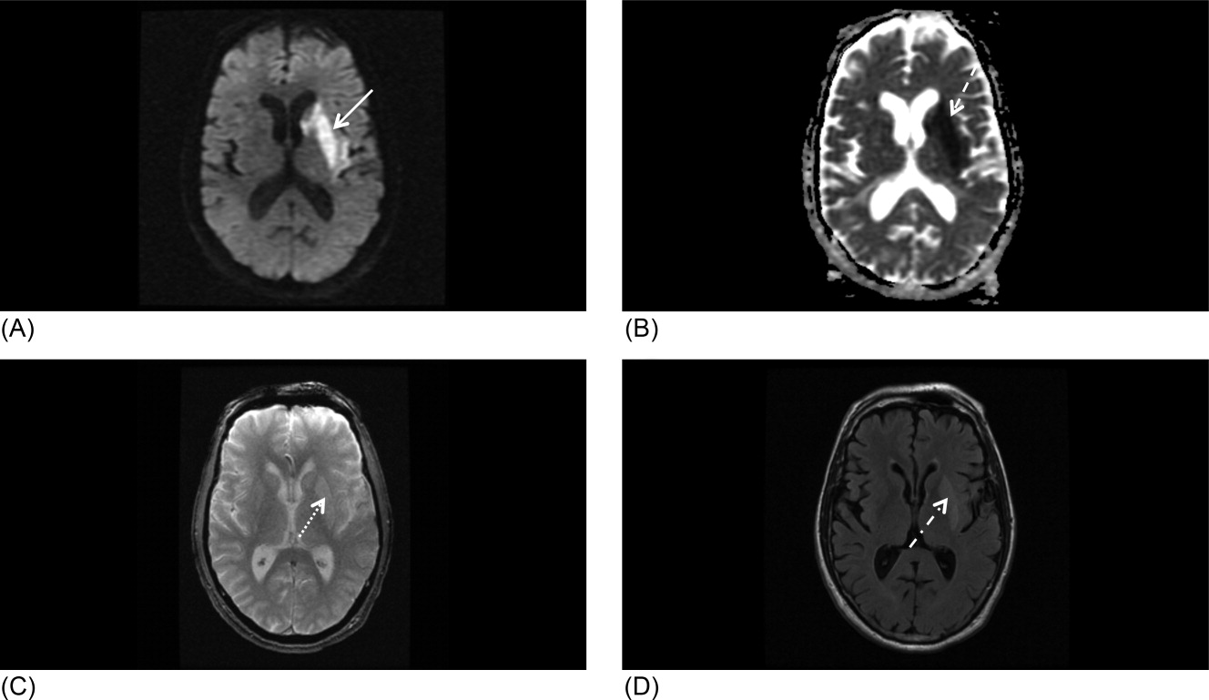
The guidelines for acute stroke management continue to rapidly evolve. Certainly, there are effective treatments for acute ischemic stroke, with variation based on the timing of patient arrival at the hospital, the underlying pathophysiology, and the treatment capabilities of the individual hospital. Secondary stroke prevention is extremely important and has been emphasized during inpatient admissions with the establishment of an appropriate medication regime, given that patients are more likely to stay on treatment that is initiated around the time of a diagnosis.29 Evidence strongly suggests that management of acute stroke is improved by an organized approach to care, including the expertise of a multidisciplinary team in a specialized stroke unit. Hospitals committed to high quality of care for acute stroke patients should strongly consider the Joint Commission certification process or an analogous local certification. Such certification demonstrates a hospital's commitment to providing high‐quality care, what every stroke patient wants and deserves.
- Organised inpatient (stroke unit) care for stroke.Stroke Unit Trialists' Collaboration.Cochrane Database Syst Rev.2000(2):CD000197.
- ,,, et al.Acetaminophen for altering body temperature in acute stroke: a randomized clinical trial.Stroke.2002;33(1):130–134.
- ,,, et al.Fever in acute stroke worsens prognosis. A prospective study.Stroke.1995;26(11):2040–2043.
- ,.Combating hyperthermia in acute stroke: a significant clinical concern.Stroke.1998;29(2):529–534.
- ,,, et al.Body temperature in acute stroke: relation to stroke severity, infarct size, mortality, and outcome.Lancet. 171996;347(8999):422–425.
- ,,.Thresholds in cerebral ischemia—the ischemic penumbra.Stroke.1981;12(6):723–725.
- .Ischaemic brain damage of cerebral perfusion failure type after treatment of severe hypertension.Br Med J. 271975;4(5999):739.
- ,,,,.Imaging of acute stroke.Lancet Neurol.2006;5(9):755–768.
- ,,, et al.High‐dose atorvastatin after stroke or transient ischemic attack.N Engl J Med.2006;355(6):549–559.
- ,,, et al.Guidelines for prevention of stroke in patients with ischemic stroke or transient ischemic attack: a statement for healthcare professionals from the American Heart Association/American Stroke Association Council on Stroke: co‐sponsored by the Council on Cardiovascular Radiology and Intervention: the American Academy of Neurology affirms the value of this guideline.Stroke.2006;37(2):577–617.
- Antithrombotic Trialists' Collaboration.Collaborative meta‐analysis of randomised trials of antiplatelet therapy for prevention of death, myocardial infarction, and stroke in high risk patients.BMJ.2002;324(7329):71–86.
- ,,, et al.A comparison of warfarin and aspirin for the prevention of recurrent ischemic stroke.N Engl J Med.2001;345(20):1444–1451.
- .Warfarin or aspirin for recurrent ischemic stroke.N Engl J Med.2002;346(15):1169–1171.
- ,,, et al.Prevention. European Stroke Initiative.Cerebrovasc Dis.2004;17(suppl 2):15–29.
- ,,, et al.Update to the AHA/ASA recommendations for the prevention of stroke in patients with stroke and transient ischemic attack.Stroke.2008;39(5):1647–1652.
- ,,,,.Antithrombotic and thrombolytic therapy for ischemic stroke: American College of Chest Physicians Evidence‐Based Clinical Practice Guidelines (8th edition).Chest.2008;133(suppl):630S–669S.
- ,,, et al.Aspirin and extended‐release dipyridamole versus clopidogrel for recurrent stroke.N Engl J Med.2008;359(12):1238–1251.
- ,,, et al.Clopidogrel and aspirin versus aspirin alone for the prevention of atherothrombotic events.N Engl J Med.2006;354(16):1706–1717.
- ,,, et al.Aspirin and clopidogrel compared with clopidogrel alone after recent ischaemic stroke or transient ischaemic attack in high‐risk patients (MATCH): randomised, double‐blind, placebo‐controlled trial.Lancet.2004;364(9431):331–337.
- ,,, et al.Oral anticoagulants vs aspirin in nonvalvular atrial fibrillation: an individual patient meta‐analysis.JAMA.2002;288(19):2441–2448.
- .Effective anticoagulation therapy: defining the gap between clinical studies and clinical practice.Am J Manag Care.2004;10(suppl):S297–S306; discussionS312–S297.
- ,,,.Low molecular‐weight heparin versus aspirin in patients with acute ischaemic stroke and atrial fibrillation: a double‐blind randomised study. HAEST Study Group. Heparin in Acute Embolic Stroke Trial.Lancet.2000;355(9211):1205–1210.
- ,,,,.Endarterectomy for symptomatic carotid stenosis in relation to clinical subgroups and timing of surgery.Lancet.2004;363(9413):915–924.
- ,,.The timing of carotid endarterectomy post stroke.Neurol Clin.2006;24(4):669–680.
- Centers for Medicare and Medicaid Services (CMS). Department of Health and Human Services (DHHS). CMS Manual System. Pub 100–03 Medicare National Coverage Determinations. Available at: http://www.cms.hhs.gov/Transmittals/Downloads/R64NCD.pdf. Accessed May2009.
- .Current status of carotid endarterectomy and stenting for symptomatic carotid stenosis.Cerebrovasc Dis.2007;24(suppl 1):116–125.
- ,,, et al.Antiplatelets versus anticoagulation in cervical artery dissection.Stroke.2007;38(9):2605–2611.
- ,,, et al.A comparison of warfarin and aspirin for the prevention of recurrent ischemic stroke.N Engl J Med2001;345(20):1444–1451.
- .Warfarin or aspirin for recurrent ischemic stroke.N Engl J Med2002;346(15):1169–1171.
- ,,, et al.Practice parameter: recurrent stroke with patent foramen ovale and atrial septal aneurysm: report of the Quality Standards Subcommittee of the American Academy of Neurology.Neurology.2004;62(7):1042–1050.
- ,,, et al.In‐hospital initiation of secondary stroke prevention therapies yields high rates of adherence at follow‐up.Stroke.2004;35(12):2879–2883.
Inpatient stroke management includes many elements of care, at least as important as the initial portion of the patient's stay, as reviewed in part 1 of this article. The extent of further diagnostic evaluation varies widely depending on apparent risk factors on presentation. Likewise, further therapy, both inpatient and secondary prevention is based on identification of stroke mechanism. Hospitalists are uniquely positioned to have a tremendous impact on both stroke care and the prevention of recurrent disease.
Case Presentation
A 76‐year‐old right‐handed male with a history of hyperlipidemia and myocardial infarction was found at 7 AM with right‐sided paralysis and poor responsiveness on the morning of admission. Upon arrival to the emergency department (ED), with symptoms of partial aphasia, right hemiplegia, and left gaze preference, there was a high suspicion for a left middle cerebral artery (MCA) stroke. Unfortunately, he was excluded from receiving intravenous (IV) tissue plasminogen activator (tPA) or any other acute interventions as the last time he was known to be neurologically intact was the prior evening, which is taken to be the time of onset. Antiplatelet therapy was continued, and the patient admitted for further workup.
Inpatient Care
When an acute ischemic stroke patient is admitted to the hospital, he or she should be placed on a standardized acute stroke protocol (also known as (a.k.a.) a care map, order set, clinical pathway)commonly created by a hospitalist/neurologist and a multidisciplinary team and admitted to a stroke unit. A stroke unit can take many forms, either as a physically separate unit in hospitals with sufficient volume or a floor where a lower volume of stroke patients are always admitted. Multidisciplinary care providers in the stroke unit have special training in stroke, and strong evidence from randomized trials shows that patients cared for in these units have significantly decreased mortality with improved functional outcomes.1 Essentials of the stroke protocol or order set include cardiac telemetry, maintaining euthermia and euglycemia, closely following blood pressure and neurologic status, actively avoiding complications, initiation of secondary prevention treatment, early involvement of rehabilitation services, and patient education.
Euthermia may be assisted by administering scheduled Tylenol to the patient for the first 48 hours, but is not strictly evidence‐based.2 Though euthermia and euglycemia have not been shown to improve outcomes in acute stroke, studies have shown that hyperthermia and hyperglycemia are associated with worsened outcomes for patients with acute strokes.35
Blood Pressure Management
Normally, cerebral vascular autoregulation leads to stable cerebral blood flow over a range of systemic blood pressures. In the setting of an acute stroke, the ability to autoregulate is diminished or absent in regions of and surrounding an acute ischemic stroke; as the area becomes ischemic, autoregulation opens the local vasculature maximally in an effort to drawn in as much blood as possible. Maximally dilated arterioles are perfused in direct correlation with systemic blood pressure, thus any drop in the systemic blood pressure leads to direct decreases in blood flow specifically in the area of ischemia; if there is a penumbra of marginally perfused tissue, such systemic blood pressure drops risk extending the area of fatal ischemia (increasing the size of the ischemic stroke).68 Thus in the acute period of an ischemic stroke, the American Heart Association (AHA)/American Stroke Association (ASA) Guidelines for the Early Management of Adults With Ischemic Stroke (referred to herein as the Guidelines)10 suggest avoid treatment unless systolic blood pressures are >220 or diastolic pressures >105, and review the evidence to support this recommendation (p. 16711672). Those patients who receive tPA have a more stringent blood pressure threshold given their risk of intracranial hemorrhage; systolic blood pressures are accepted up to 180 prior to recommending treatment.
Higher‐quality Inpatient Stroke Care and Harmonized Performance Measures
Beginning in January 2008, a set of 10 performance measures (Table 1) for inpatient acute stroke care have been agreed upon (harmonized) by 3 major stakeholders including the Joint Commission, the ASA's Get with the GuidelinesStroke quality improvement program, and the Center for Disease Control and Prevention's (CDC's) Paul Coverdell Acute stroke registries. These performance measures were selected to help avoid complications (deep vein thrombosis [DVT], aspiration pneumonia), encourage appropriately aggressive care (tPA administration), optimize secondary prevention (antithrombotics, cholesterol lowering, smoking cessation, education), and facilitate functional recovery (early rehabilitation). All 10 measures are appropriate for consideration in every ischemic stroke patient, and 5 are appropriate for the hemorrhagic stroke types.
| Performance measure* | Definition* |
|---|---|
| |
| 1. DVT prophylaxis | Patients who are nonambulatory should start receiving DVT prophylaxis by end of hospital day 2 (can be either compression devices or any low‐dose heparin) |
| 2. Discharged on antithrombotic therapy | Antiplatelet agent(s) or warfarin anticoagulation |
| 3. Patients with atrial fibrillation receiving anticoagulation therapy | A proven approach to secondary prevention in such patients; practice at Harborview varies time of warfarin initiation based on infarct size with larger infarcts waiting up to 2 weeks before initiating warfarin (the best randomized trial showed no benefit for full‐dose low‐molecular‐weight heparin over aspirin in the first 2 weeks)50 |
| 4. Thrombolytic therapy administered | In ischemic stroke patients who arrive at the hospital within 120 minutes (2 hours) of time last known well, for whom IV tPA was initiated at this hospital within 180 minutes (3 hours) of time last known well, and who qualify under strict criteria |
| 5. Antithrombotic therapy by end of hospital day 2 | Usually just antiplatelet agents, a minimal standard of care for ischemic stroke patients; should be started as early as possible, usually in ER |
| 6. Discharged on statin medication | If LDL >100, or not measured or if on a statin drug prior to admission; to reduce risk of subsequent ischemic stroke |
| 7. Dysphagia screening | Prior to any PO food, fluids or medications; to reduce the chances of aspiration pneumonia |
| 8. Stroke education | Including for families if patient unable to participate, must include personal risk factors for stroke, warning signs for stroke, activation of emergency medical system, need for follow‐up after discharge, and medications prescribed |
| 9. Smoking cessation/advice/counseling | For any patient who has smoked in the last year |
| 10. Assessed for rehabilitation | Or received therapy services; to facilitate progress to an optimal function outcome |
Further Workup
After the ischemic stroke patient has had their computed tomography (CT) scan, possibly a computed tomography angiography (CTA), been admitted to the stroke unit, started on an antithrombotic medication, and had their blood pressure appropriately treated, attention then turns to defining the pathophysiology related to the stroke and starting an optimal regimen for secondary prevention. Imaging of the cerebral vasculature including both extracranial and intracranial large vessels is a vital first step in understanding the cause of ischemic stroke. There are multiple potential modalities (magnetic resonance angiography [MRA], CTA, and duplex/transcranial Doppler), the choice of which depends on local availability and expertise as well as the specific clinical situation. Magnetic resonance imaging (MRI) of the brain for all ischemic stroke patients is standard of care at most stroke centers; per the Guidelines, MRI is better at distinguishing acute, small cortical, small deep, and posterior fossa infarcts; at distinguishing acute from chronic ischemia; and at identifying subclinical satellite ischemic lesions that provide information on stroke mechanism (p. 1668). New techniques including magnetic resonance (MR) and CT perfusion scanning can show the ischemic region in the acute setting and may one day help select patients for specific therapies, but are not yet widely available nor have they been shown to alter outcomes.
An electrocardiogram is indicated for all stroke patients, as is admission to a cardiac telemetry bed for at least 24 hours to document any arrhythmias, the most common being atrial fibrillation (Guidelines, p. 1666, 1673). An echocardiographic study (ECHO) of the heart with bubble study should be performed in most cases (although which cases may specifically benefit is unclear) to identify a cardioembolic source for the stroke, such as low cardiac ejection fraction, atrial septal aneurysm, patent foramen ovale (PFO), or a cardiac thrombus. The bubble study increases the sensitivity of detecting a PFO, which could serve as a gateway for venous embolization to the cerebral arteries. Assuming a large PFO is discovered, other studies such as lower extremity Doppler may be warranted to investigate other potential sources of thrombi (ie, DVT).
Regarding laboratory testing, fasting lipids should be checked as hyperlipidemia is a common modifiable risk factor for ischemic stroke. The Stroke Prevention by Aggressive Reduction in Cholesterol Levels (SPARCL) trial included ischemic stroke patients that had low‐density lipoprotein (LDL) cholesterol between 100 mg/dL and 190 mg/dL and randomized them to receive atorvastatin 80 mg/day vs. placebo. Results showed a 16% relative risk reduction in recurrent stroke; however, there was a small increased risk of intracranial hemorrhage.9 As shown in Table 1, use of a statin on discharge is now a national performance measure for ischemic stroke.
Dissection is a common cause of stroke in young patients without traditional risk factors. Other serologies, such as hypercoagulable studies, may be warranted in patients with no other risk factors for strokes, paradoxical embolus, or of young age (eg, 45 years and under). The arterial hypercoagulable panel consists of antiphospholipid antibody panel, homocysteine levels, lupus anticoagulant levels, and prothrombin time/partial thromboplastin time (PT/PTT). The venous hypercoagulable panel consists of the laboratory values checked, with the arterial hypercoagulable and activated protein C (APC) resistance, Factor VIII activity, Factor II DNA, Factor V DNA if the APC resistance is positive, antithrombin III activity, and activity of proteins C and S. If a patient is found to have a hypercoagulable state, long‐term therapy often involves careful consideration of the choice of antiplatelet therapy vs. anticoagulation with warfarin.10
Initiating Secondary Prevention
Upon admission, the clinician faces a variety of treatment choices for secondary stroke prevention. The proper choice depends on the results of the workup and the presumptive pathophysiology.
Noncardioembolic/Atherothrombotic/Lacunar
The Antithrombotic Trialists' Collaboration meta‐analysis found that patients with a prior stroke or transient ischemic attack (TIA) had a highly significant decrease in the rate of subsequent vascular events (over about 3 years) on antiplatelet therapy (17.8% vs. 21.4%, P 0.0001) and were unable to find a significant difference between low‐dose and high‐dose aspirin for secondary prevention.11 Thus, it is reasonable to place an acute stroke patient naive to antithrombotic therapy on 81 mg of aspirin or 325 mg for long‐term prevention (325 mg is specifically recommended in the acute setting). Several studies such as the WARSS and ESPRIT trials have shown antiplatelet agents to be at least as effective as anticoagulation in noncardioembolic ischemic strokes.12, 13 Guidelines from Europe, the American College of Chest Physicians, and the AHA/ASA all state it is acceptable to choose either aspirin monotherapy, aspirin/extended release dipyridamole combination therapy, or clopidogrel monotherapy as first‐line agents for long‐term secondary prevention in noncardioembolic ischemic stroke.1416 There is no clear evidence that patients who suffer an ischemic stroke while on aspirin will derive additional benefit from increasing the aspirin dose. The newer guidelines go on to recommend aspirin/extended release dipyridamole (ER‐DP) combination therapy or clopidogrel monotherapy over aspirin monotherapy, the former with a stronger level of recommendation based on the results of 2 randomized trials. These recommendations were all published without knowledge of the results of the Prevention Regimen For Effectively Avoiding Second Strokes (PRoFESS) study, which directly compared aspirin/extended release dipyridamole combination therapy to clopidogrel monotherapy for long‐term secondary prevention. The rate of first recurrent stroke was not significantly different between the 2 therapies (9.0% ER‐DP plus aspirin, 8.8% clopidogrel; hazard ratio [HR], 1.01; 95% confidence interval [CI], 0.921.11). Other outcomes also showed few differences, although there were more major hemorrhagic events in the ER‐DP plus aspirin group (4.1% vs. 3.6%; HR, 1.15; 95% CI, 1.001.32; P = 0.06).17
The ASA Stroke Prevention Guideline from 2006 states, with continued relevance, The selection of an antiplatelet agent should be individualized on the basis of patient risk factor profiles, tolerance, and other clinical characteristics.10 Of note, both the Clopidogrel for High Atherothrombotic Risk and Ischemic Stabilization, Management and Avoidance (CHARISMA) and Management of ATherothrombosis with Clopidogrel in High‐risk patients with recent TIA or ischemic stroke (MATCH) trials found a significant increased risk for hemorrhage complications with long‐term use of the aspirin and clopidogrel combination,18, 19 and the 2008 update to the ASA Stroke prevention guidelines state that the addition of aspirin to clopidogrel increases the risk of hemorrhage. Combination therapy of aspirin and clopidogrel is not routinely recommended for ischemic stroke or TIA patients unless they have a specific indication for this therapy (i.e., coronary stent or acute coronary syndrome).15
Atrial Fibrillation
Though our case patient did not have atrial fibrillation, this condition does deserve mention. About 15% to 20% of ischemic stroke patients have atrial fibrillation. The overall risk for stroke in patients with atrial fibrillation is about 5% per year; however, patients who have a history of stroke increase their risk factors for subsequent strokes to about 12% per year. In most cases, anticoagulation has proven to be the superior agent for primary and secondary stroke prevention with warfarin reducing the risk by 67% compared to aspirin, which only reduces the risk of stroke by 20%. A meta‐analysis from 2002 showed that patients who had a prior stroke or TIA decrease their risk of subsequent strokes to 4%/year on oral anticoagulation therapy, resulting in an 8% absolute risk reduction. Patients on aspirin therapy only decrease their risk to 10%/year, or a 2% reduction in stroke events.20 Unless there is a strong contraindication (eg, bleeding diathesis, history of life threatening gastrointestinal [GI] bleeding, history of fall with subdural hematoma, etc.), virtually all ischemic stroke patients with atrial fibrillation should be anticoagulated for life. Anticoagulation in the setting of atrial fibrillation is seriously underutilized.21 The highest quality study on early anticoagulation for ischemic stroke associated with atrial fibrillation suggested that there was no benefit to starting anticoagulation earlier than 2 weeks after a stroke, and there may actually be a higher complication rate (compared to aspirin).22 Other cardiac indications for anticoagulation include left ventricular thrombus and mechanical valves.
Carotid Stenosis
Significant ipsilateral stenosis of the internal carotid artery in a patient with ischemic stroke is a strong indication for intervention, usually a standard carotid endarterectomy (CEA). Stenosis of 70% to 99% is the strongest indication for CEA, and may be of greatest benefit in men, those 75+ years of age, and if surgery is done 2 weeks after the most recent symptoms.23 In patients with minor stroke or TIA, recent recommendations and our practice is to admit to the hospital and perform endarterectomy as soon as possible (those with major stroke may have a greater risk of complications with early CEA).24 Stenting should only be considered instead of CEA if high risk (for surgical complications) criteria are present. These high risk criteria include patients having significant comorbidities and/or anatomic risk factors (ie, recurrent stenosis and/or previous radical neck dissection), and [who] would be poor candidates for CEA in the opinion of a surgeon.25 For stenoses of 50% to 69%, intervention is not as compelling, and decisions should be individualized based on patient characteristics; in this group, stenting should only be considered in the setting of a clinical trial or if an investigational device exemption (IDE) exists at your institution.26
Dissection of the Carotid or Vertebral Arteries
This is a common cause of stroke in younger adults. It should be suspected in patients without other clear causes of stroke and significant disease of the extracranial arteries. Diagnosis can usually be made with CTA or MRA, though it is suggested that the best modality may be T1‐fat‐saturated MRI images of the neck. Debate exists as to the best approach to treatment of dissections due to the absence of randomized trials. A recent comprehensive review suggested anticoagulation for 3 to 6 months followed by indefinite antiplatelet therapy for symptomatic dissections and antiplatelet therapy alone for asymptomatic dissections.27
PFO‐related Stroke
If the patient is found to have a PFO, its role in comparison to traditional risk factors must be weighed carefully. Epidemiological studies suggest that PFO may be most relevant in younger patients, those with cryptogenic stroke (no obvious cause and lack of traditional risk factors), those with higher risk associations including interatrial septal aneurysm, larger PFOs or history of previous cryptogenic stroke.28, 29 The best medical therapy for seemingly PFO‐related ischemic stroke is also unclear; a reasonable approach might be aspirin if neither high‐risk associations nor a hypercoagulable state is present, and warfarin if either are present. Transcatheter closure of PFO is approved by the U.S. Food and Drug Administration (FDA) only under an IDE for patients who have had a recurrent event on maximally tolerated medical treatment, and requires approval from the human research committee (internal review board [IRB]) at your hospital. It is not known if closure is superior or inferior to best medical therapy, and a practice parameter from the American Academy of Neurology strongly encourages appropriate patients to consider participation in ongoing randomized trials.28 Further information on these trials is available at:
Our patient underwent a CTA of the head and neck in the emergency room to see if he would be a candidate for other interventions; unfortunately, he did not meet the time criteria. CTA showed complete occlusion of the left internal carotid artery at the bifurcation with heterogeneous retrograde filling (Supporting Figure 1). Complete occlusion of the proximal third of the left M1 segment was also seen with relative oligemia in the left MCA distribution, though several small peripheral M3/M4 vessels were opacified in the territory indicating collateralization (Supporting Figure 2). A MRI showed a large area of diffusion‐weighted abnormality (Figure 1). Interestingly, the patient's transthoracic echocardiography (TTE), which did not show evidence of a PFO, did reveal a calcified thrombus in the left ventricle. Though no arrhythmias were captured on telemetry, this thrombus does serve as a potential source of cardioembolic emboli to the cerebral vasculature. It was felt that the most likely source of the patient's acute infarct was from artery‐to‐artery emboli from his internal carotid occlusion given the infarct location and the lack of infarction in other vascular distributions (as one might see from a cardiac embolic source). Therefore, his medical management consisted of an antiplatelet regimen for 2 weeks followed by a transition to warfarin alone 2 weeks after his acute infarct as secondary stroke prevention due to the cardiac thrombus. Given the complete occlusion of the internal carotid artery and M1 segment, there was concern that the penumbra might be at risk of infarction (supporting standard guidelines of permissive hypertension). By the end of his hospitalization, the patient had improved and was transferred to inpatient rehabilitation.
The guidelines for acute stroke management continue to rapidly evolve. Certainly, there are effective treatments for acute ischemic stroke, with variation based on the timing of patient arrival at the hospital, the underlying pathophysiology, and the treatment capabilities of the individual hospital. Secondary stroke prevention is extremely important and has been emphasized during inpatient admissions with the establishment of an appropriate medication regime, given that patients are more likely to stay on treatment that is initiated around the time of a diagnosis.29 Evidence strongly suggests that management of acute stroke is improved by an organized approach to care, including the expertise of a multidisciplinary team in a specialized stroke unit. Hospitals committed to high quality of care for acute stroke patients should strongly consider the Joint Commission certification process or an analogous local certification. Such certification demonstrates a hospital's commitment to providing high‐quality care, what every stroke patient wants and deserves.
- Organised inpatient (stroke unit) care for stroke.Stroke Unit Trialists' Collaboration.Cochrane Database Syst Rev.2000(2):CD000197.
- ,,, et al.Acetaminophen for altering body temperature in acute stroke: a randomized clinical trial.Stroke.2002;33(1):130–134.
- ,,, et al.Fever in acute stroke worsens prognosis. A prospective study.Stroke.1995;26(11):2040–2043.
- ,.Combating hyperthermia in acute stroke: a significant clinical concern.Stroke.1998;29(2):529–534.
- ,,, et al.Body temperature in acute stroke: relation to stroke severity, infarct size, mortality, and outcome.Lancet. 171996;347(8999):422–425.
- ,,.Thresholds in cerebral ischemia—the ischemic penumbra.Stroke.1981;12(6):723–725.
- .Ischaemic brain damage of cerebral perfusion failure type after treatment of severe hypertension.Br Med J. 271975;4(5999):739.
- ,,,,.Imaging of acute stroke.Lancet Neurol.2006;5(9):755–768.
- ,,, et al.High‐dose atorvastatin after stroke or transient ischemic attack.N Engl J Med.2006;355(6):549–559.
- ,,, et al.Guidelines for prevention of stroke in patients with ischemic stroke or transient ischemic attack: a statement for healthcare professionals from the American Heart Association/American Stroke Association Council on Stroke: co‐sponsored by the Council on Cardiovascular Radiology and Intervention: the American Academy of Neurology affirms the value of this guideline.Stroke.2006;37(2):577–617.
- Antithrombotic Trialists' Collaboration.Collaborative meta‐analysis of randomised trials of antiplatelet therapy for prevention of death, myocardial infarction, and stroke in high risk patients.BMJ.2002;324(7329):71–86.
- ,,, et al.A comparison of warfarin and aspirin for the prevention of recurrent ischemic stroke.N Engl J Med.2001;345(20):1444–1451.
- .Warfarin or aspirin for recurrent ischemic stroke.N Engl J Med.2002;346(15):1169–1171.
- ,,, et al.Prevention. European Stroke Initiative.Cerebrovasc Dis.2004;17(suppl 2):15–29.
- ,,, et al.Update to the AHA/ASA recommendations for the prevention of stroke in patients with stroke and transient ischemic attack.Stroke.2008;39(5):1647–1652.
- ,,,,.Antithrombotic and thrombolytic therapy for ischemic stroke: American College of Chest Physicians Evidence‐Based Clinical Practice Guidelines (8th edition).Chest.2008;133(suppl):630S–669S.
- ,,, et al.Aspirin and extended‐release dipyridamole versus clopidogrel for recurrent stroke.N Engl J Med.2008;359(12):1238–1251.
- ,,, et al.Clopidogrel and aspirin versus aspirin alone for the prevention of atherothrombotic events.N Engl J Med.2006;354(16):1706–1717.
- ,,, et al.Aspirin and clopidogrel compared with clopidogrel alone after recent ischaemic stroke or transient ischaemic attack in high‐risk patients (MATCH): randomised, double‐blind, placebo‐controlled trial.Lancet.2004;364(9431):331–337.
- ,,, et al.Oral anticoagulants vs aspirin in nonvalvular atrial fibrillation: an individual patient meta‐analysis.JAMA.2002;288(19):2441–2448.
- .Effective anticoagulation therapy: defining the gap between clinical studies and clinical practice.Am J Manag Care.2004;10(suppl):S297–S306; discussionS312–S297.
- ,,,.Low molecular‐weight heparin versus aspirin in patients with acute ischaemic stroke and atrial fibrillation: a double‐blind randomised study. HAEST Study Group. Heparin in Acute Embolic Stroke Trial.Lancet.2000;355(9211):1205–1210.
- ,,,,.Endarterectomy for symptomatic carotid stenosis in relation to clinical subgroups and timing of surgery.Lancet.2004;363(9413):915–924.
- ,,.The timing of carotid endarterectomy post stroke.Neurol Clin.2006;24(4):669–680.
- Centers for Medicare and Medicaid Services (CMS). Department of Health and Human Services (DHHS). CMS Manual System. Pub 100–03 Medicare National Coverage Determinations. Available at: http://www.cms.hhs.gov/Transmittals/Downloads/R64NCD.pdf. Accessed May2009.
- .Current status of carotid endarterectomy and stenting for symptomatic carotid stenosis.Cerebrovasc Dis.2007;24(suppl 1):116–125.
- ,,, et al.Antiplatelets versus anticoagulation in cervical artery dissection.Stroke.2007;38(9):2605–2611.
- ,,, et al.A comparison of warfarin and aspirin for the prevention of recurrent ischemic stroke.N Engl J Med2001;345(20):1444–1451.
- .Warfarin or aspirin for recurrent ischemic stroke.N Engl J Med2002;346(15):1169–1171.
- ,,, et al.Practice parameter: recurrent stroke with patent foramen ovale and atrial septal aneurysm: report of the Quality Standards Subcommittee of the American Academy of Neurology.Neurology.2004;62(7):1042–1050.
- ,,, et al.In‐hospital initiation of secondary stroke prevention therapies yields high rates of adherence at follow‐up.Stroke.2004;35(12):2879–2883.

Gastric Involvement in NSG
Necrotizing sarcoid granulomatosis (NSG) is an immune system disorder characterized by necrotizing granulomas, as opposed to noncaseating granulomas in classical sarcoidosis. Over the past 3 decades there have been over 120 reported cases of NSG with pulmonary and extrapulmonary involvement.1 We present a patient found to have histological evidence of necrotizing granuloma in her gastric antrum, and we believe this is the first Case Report of NSG involving the stomach.
Case Report
A 21‐year‐old African‐American female first presented to an outside hospital with fever, epigastric pain, shortness of breath, and headache. Two days later she complained of nonbloody, nonbilious vomiting, and was found to have leukocytosis (17,770 cells/m3), elevated lipase (224 U/L), elevated C‐reactive protein (14.7 mg/L), and an inflamed pancreas on computed tomography (CT). She was treated conservatively for pancreatitis and started on ampicillin/sulbactam. After 2 contrast CT scans on consecutive days, she developed acute renal failure (creatinine 2.0 mg/dL compared to baseline of 1.0 mg/dL), and was transferred to our hospital for further evaluation and management.
Upon transfer, the patient's temperature was 37.0C, pulse was 102 beats/minute, blood pressure was 141/84 mm Hg, and oxygen saturation was 94%. On examination, she was tender to palpation in her epigastrium and right upper quadrant, but the remainder of the physical exam was unremarkable. She was started on moxifloxacin and managed with intravenous (IV) fluid hydration and pain control. Within 3 days, CT showed resolving pancreatitis, magnetic resonance cholangiopancreatography (MRCP) was negative, and her creatinine began normalizing (1.3 mg/dL). Nonetheless, she continued to complain of abdominal pain, shortness of breath, and intermittent low‐grade fevers. She then also developed bilateral panuveitis requiring high‐dose steroid eye drops.
Chest x‐ray showed subtle bilateral nodular and bronchiolitic infiltrates with no evidence of enlarged hilar nodes, and subsequent bronchoscopy showed no abnormalities. Additional workup included negative blood and urine cultures, purified protein derivative (PPD), and Clostridium difficile assay; as well as negative human immunodeficiency virus (HIV), cryptococcus, Helicobacter pylori, Borellia burgdorferi, syphilis (fluorescent treponemal antibody), aspergillus, histoplasma, and rheumatological serologies. Her white blood count (20,900 cells/m3), C‐reactive protein (6.6 mg/L), and erythrocyte sedimentation rate (100 mm/hour) remained elevated.
The patient continued to complain of epigastric pain. Repeat abdominal CT scan showed large retroperitoneal and mesenteric lymph nodes, and esophagogastroduodenoscopy (EGD) showed gastritis with an antral nodule (Figure 1). Biopsy of the nodule revealed a necrotizing granuloma with mixed cellular infiltrate. Biopsy stains were negative for bacteria, borellia, treponemes, acid‐fast bacilli, and fungi. The patient was diagnosed with necrotizing sarcoid granulomatosis and started on an oral prednisone taper. She responded to steroid treatment with prompt resolution of her uveitis, shortness of breath, abdominal pain, and fevers. She was discharged following treatment, has continued to do well, and is seen regularly at the sarcoid clinic for follow‐up.
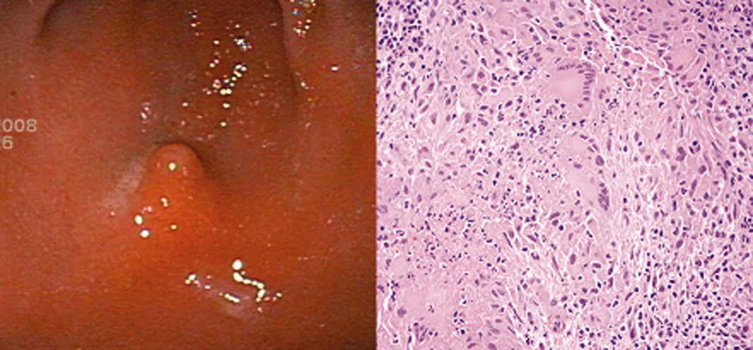
Discussion
NSG was first characterized as a distinct variation from sarcoidosis by Liebow2 in 1973, and was noted to have 3 characteristic differences: (1) histological evidence of sarcoid‐appearing granuloma and necrosis, (2) pulmonary nodules without hilar lymphadenopathy on imaging, and (3) a clinically benign course. Over the past 3 decades, there have been more than 120 cases of reported NSG involving the lungs, gastrointestinal tract, kidney, skin, and central nervous system.1 Since Liebow's2 description, newly reported NSG cases have generally been found to be consistent with the aforementioned criteria, although hilar lymphadenopathy may be particularly more common than previously thought. One review noted a range of 8% to 79% prevalence of hilar lymphadenopathy in reported NSG series.3 Therefore, while hilar lymphadenopathy still currently appears to be less common in NSG than in typical sarcoidosis, its presence should not rule out the diagnosis.
Our patient's history is consistent with Liebow's2 criteria since she had histological evidence of necrotizing granuloma and pulmonary involvement without hilar lymphadenopathy, and responded promptly to steroid treatment. To our knowledge, this is the first case of NSG reported in the stomach.
While less than 1% of sarcoid patients are reported to have gastrointestinal involvement, all of these cases have either been noncaseating granulomas (classical sarcoidosis) or were found outside the stomach.1, 4, 5 Most of the data regarding sarcoid symptomatology and treatment are derived from reports on classical sarcoidosis. In classical sarcoidosis, there is gastric antral involvement in approximately 10% of patients with systemic disease.4 These patients may present with nausea, vomiting, and weight loss, and are often effectively treated with a single dose of prednisone 30 to 40 mg followed by a maintenance dose of 10 to 15 mg daily over 6 months.5 Less data are available regarding necrotizing sarcoid presentation and treatment, especially with regard to gastrointestinal involvement.
We hope to raise awareness regarding: (1) the variation in noncaseating versus necrotizing sarcoid‐type disorders, (2) the benefit of steroid treatment once infectious etiologies are ruled out, and (3) the potential for further extrapulmonary involvement in previously unreported organ systems.
- ,,.Necrotizing sarcoid granulomatosis mimicking an intracranial neoplasm: clinicopathologic features and review of the literature.Mod Pathol.2000;13(8)909–913.
- .Pulmonary angiitis and granulomatosis.Am J Respir Dis.1973;108:1–18.
- ,,,.Pulmonary angiitis and granulomatosis: radiologic‐pathologic correlation.Radiographics.1998;18(3):687–710.
- ,,, et al.Multiple antral ulcers in gastric sarcoid.J Clin Gastroenterol.1997;24(2):97–99.
- ,,.Gastric sarcoidosis: a case report and review of the literature.South Med J.2007;100(3):301–303.
Necrotizing sarcoid granulomatosis (NSG) is an immune system disorder characterized by necrotizing granulomas, as opposed to noncaseating granulomas in classical sarcoidosis. Over the past 3 decades there have been over 120 reported cases of NSG with pulmonary and extrapulmonary involvement.1 We present a patient found to have histological evidence of necrotizing granuloma in her gastric antrum, and we believe this is the first Case Report of NSG involving the stomach.
Case Report
A 21‐year‐old African‐American female first presented to an outside hospital with fever, epigastric pain, shortness of breath, and headache. Two days later she complained of nonbloody, nonbilious vomiting, and was found to have leukocytosis (17,770 cells/m3), elevated lipase (224 U/L), elevated C‐reactive protein (14.7 mg/L), and an inflamed pancreas on computed tomography (CT). She was treated conservatively for pancreatitis and started on ampicillin/sulbactam. After 2 contrast CT scans on consecutive days, she developed acute renal failure (creatinine 2.0 mg/dL compared to baseline of 1.0 mg/dL), and was transferred to our hospital for further evaluation and management.
Upon transfer, the patient's temperature was 37.0C, pulse was 102 beats/minute, blood pressure was 141/84 mm Hg, and oxygen saturation was 94%. On examination, she was tender to palpation in her epigastrium and right upper quadrant, but the remainder of the physical exam was unremarkable. She was started on moxifloxacin and managed with intravenous (IV) fluid hydration and pain control. Within 3 days, CT showed resolving pancreatitis, magnetic resonance cholangiopancreatography (MRCP) was negative, and her creatinine began normalizing (1.3 mg/dL). Nonetheless, she continued to complain of abdominal pain, shortness of breath, and intermittent low‐grade fevers. She then also developed bilateral panuveitis requiring high‐dose steroid eye drops.
Chest x‐ray showed subtle bilateral nodular and bronchiolitic infiltrates with no evidence of enlarged hilar nodes, and subsequent bronchoscopy showed no abnormalities. Additional workup included negative blood and urine cultures, purified protein derivative (PPD), and Clostridium difficile assay; as well as negative human immunodeficiency virus (HIV), cryptococcus, Helicobacter pylori, Borellia burgdorferi, syphilis (fluorescent treponemal antibody), aspergillus, histoplasma, and rheumatological serologies. Her white blood count (20,900 cells/m3), C‐reactive protein (6.6 mg/L), and erythrocyte sedimentation rate (100 mm/hour) remained elevated.
The patient continued to complain of epigastric pain. Repeat abdominal CT scan showed large retroperitoneal and mesenteric lymph nodes, and esophagogastroduodenoscopy (EGD) showed gastritis with an antral nodule (Figure 1). Biopsy of the nodule revealed a necrotizing granuloma with mixed cellular infiltrate. Biopsy stains were negative for bacteria, borellia, treponemes, acid‐fast bacilli, and fungi. The patient was diagnosed with necrotizing sarcoid granulomatosis and started on an oral prednisone taper. She responded to steroid treatment with prompt resolution of her uveitis, shortness of breath, abdominal pain, and fevers. She was discharged following treatment, has continued to do well, and is seen regularly at the sarcoid clinic for follow‐up.
Discussion
NSG was first characterized as a distinct variation from sarcoidosis by Liebow2 in 1973, and was noted to have 3 characteristic differences: (1) histological evidence of sarcoid‐appearing granuloma and necrosis, (2) pulmonary nodules without hilar lymphadenopathy on imaging, and (3) a clinically benign course. Over the past 3 decades, there have been more than 120 cases of reported NSG involving the lungs, gastrointestinal tract, kidney, skin, and central nervous system.1 Since Liebow's2 description, newly reported NSG cases have generally been found to be consistent with the aforementioned criteria, although hilar lymphadenopathy may be particularly more common than previously thought. One review noted a range of 8% to 79% prevalence of hilar lymphadenopathy in reported NSG series.3 Therefore, while hilar lymphadenopathy still currently appears to be less common in NSG than in typical sarcoidosis, its presence should not rule out the diagnosis.
Our patient's history is consistent with Liebow's2 criteria since she had histological evidence of necrotizing granuloma and pulmonary involvement without hilar lymphadenopathy, and responded promptly to steroid treatment. To our knowledge, this is the first case of NSG reported in the stomach.
While less than 1% of sarcoid patients are reported to have gastrointestinal involvement, all of these cases have either been noncaseating granulomas (classical sarcoidosis) or were found outside the stomach.1, 4, 5 Most of the data regarding sarcoid symptomatology and treatment are derived from reports on classical sarcoidosis. In classical sarcoidosis, there is gastric antral involvement in approximately 10% of patients with systemic disease.4 These patients may present with nausea, vomiting, and weight loss, and are often effectively treated with a single dose of prednisone 30 to 40 mg followed by a maintenance dose of 10 to 15 mg daily over 6 months.5 Less data are available regarding necrotizing sarcoid presentation and treatment, especially with regard to gastrointestinal involvement.
We hope to raise awareness regarding: (1) the variation in noncaseating versus necrotizing sarcoid‐type disorders, (2) the benefit of steroid treatment once infectious etiologies are ruled out, and (3) the potential for further extrapulmonary involvement in previously unreported organ systems.
- ,,.Necrotizing sarcoid granulomatosis mimicking an intracranial neoplasm: clinicopathologic features and review of the literature.Mod Pathol.2000;13(8)909–913.
- .Pulmonary angiitis and granulomatosis.Am J Respir Dis.1973;108:1–18.
- ,,,.Pulmonary angiitis and granulomatosis: radiologic‐pathologic correlation.Radiographics.1998;18(3):687–710.
- ,,, et al.Multiple antral ulcers in gastric sarcoid.J Clin Gastroenterol.1997;24(2):97–99.
- ,,.Gastric sarcoidosis: a case report and review of the literature.South Med J.2007;100(3):301–303.

Nonphysicians in Hospital Medicine
Ford and Britting's1 editorial in this month's Journal of Hospital Medicine raises important questions concerning the use of nonphysician providers in hospital medicine. They focus primarily on the use of mid‐level providers (MLPs), namely physician‐assistants (PAs) and nurse practitioners (NPs), as a potential solution to the current physician workforce shortages in our field. While we acknowledge the challenges of meeting workforce needs, we also believe that much is unknown about the optimal use of MLPs on inpatient general medicine services and it is premature to tout MLPs as the solution to hospital medicine staffing problems. This is especially true in those hospitals where hospitalists care for complex, general medical patients with a wide variety of medical conditions, a trend that is especially common in academic medical centers.2
This article discusses the current literature, our own experiences with MLPs, and suggests some future initiatives that might help better integrate MLPs into hospital medicine.
The Literature on MLPs in Inpatient Venues
The existing literature on the use of MLPs in inpatient venues is quite limited, and a recent review, while suggesting that the existing literature does describe benefits of MLPs in the inpatient setting, also states that the overall quality of the evidence is quite poor and that many studies suffer from significant limitations, including small populations, limited patient mixes, use of selected settings, and short durations of outcome assessment.3
Ford and Britting,1 in their article, cite several studies46 as evidence that a MLP model of care either improved outcomes or provided cost benefits. Each of these studies has important limitations that are worth examining.
The study by Myers et al.4 described the use of MLPs in a chest pain unit. NPs partnered with hospitalists to care for a low‐acuity chest pain population. In addition, 5 NPs only staffed the unit during daytime weekday hours. Off‐hour and weekend staffing was accomplished through the use of resident physicians. Notably, the work suggests the service only admitted 113 low‐risk patients over 10 months. The service was staffed by 3 full‐time equivalent (FTE) NPs in addition to involving hospitalists during the day. It is not surprising, given the extremely low volume of patients coupled with a daytime‐only focus, that this service showed efficiency gains. In addition, given the service was only staffed by NPs 40 hours a week and by resident physicians on nights and weekends, the true cost of such an intervention needs to take into account the full cost of 24/7 coverage. In addition, the model of using residents to cover nonteaching patients is no longer permitted by the current Accreditation Council for Graduate Medical Education (ACGME) Internal Medicine Residency Requirements7 and thus implementation of a model such as this in 2009 would require alternative means of nighttime coverage.
The study by Nishimura et al.,5 also describing the use of MLPs in cardiovascular care, has important caveats that make full assessment of the model impossible. The model describes the implementation of a care team consisting of an attending, a fellow, and MLPs to replace a traditional teaching team of an attending, senior resident, and 2 interns. The study states that the model resulted in a lower length of stay (LOS) and lower costs per case. Importantly, the new MLP‐based team only admitted during the hours of 7 AM to 2 PM. The study does not fully describe the number of MLPs required nor does it fully describe the role of cardiovascular fellows in the model. The study does state that the cost savings offset the cost of the MLPs but it is not clear if this cost analysis took into account the cost of the fellow's daytime involvement or if it measured attending time required before and after the implementation of the new model. In addition, this model presumes the availability of other services to admit patients during afternoon and nighttime hours and so may not be generalizable to other settings.
The final study by Cowan et al.6 describes the addition of a NP, a hospitalist medical director, and daily multidisciplinary rounds to a traditional teaching service model. Importantly, the NP was not involved in the admission process nor were they the primary providers for day‐to‐day medical care but rather they focused on implementation of care protocols, multidisciplinary coordination of care and discharge planning, and postdischarge follow‐up. In addition, the NP worked only weekdays for about 40 hours a week. It is not surprising that adding multiple additional resources to existing care models might provide benefits but this does not address any issues in terms of the workforce since the care in this model required a higher total input of providers than the usual care model being studied. Cost savings from such a model may make it cost‐effective but it does not represent a workforce solution.
There have been other studies examining the use of MLPs in the inpatient setting in internal medicine. Some of these studies have suggested that MLP‐based models result in equivalent outcomes and efficiency810 to traditional teaching or nonteaching physician‐only models. There are 2 important caveats, however, that must be considered. The total resources required for such models may be quite high, especially taking into account the costs of 24/7 coverage and physician backup of the MLPs, and most importantly there is almost no literature that robustly examines ultimate clinical outcomes in these models. We do note that a recent study11 did show a lower inpatient mortality rate over a 2‐year period of time after substituting a PA‐hospitalist model for a traditional academic medicine residency model in a community hospital. Importantly, however, the new model also added 24/7 hospitalist physicians and night and weekend intensivists that were not present in the prior residency‐based model. Thus, the lower mortality rate could be attributed to the addition of hospitalists or the more robust in‐house physician coverage during off‐hours rather than the use of MLPs.
Notably, while the evidence base in internal medicine is not robust, many studies have described successful use of MLPs in non‐internal medicine inpatient settings.1214 The reasons for this success is debatable, but it may be that MLPs are more successful in settings where the care is either more protocol‐driven or where there is less diagnostic and therapeutic complexity.
Recent Experiences with MLPs in Academic Hospital Medicine
Given the paucity of data, it is clear that further research is needed on the role of MLPs in hospital medicine. While waiting for such evidence to appear, it may be worthwhile to reflect on the recent experience of 3 major medical centers. A recent article described 5 hospitalist models at major academic medical centers across the country. Two of the institutions described at the time (University of Michigan Health System, Ann Arbor, MI; and Brigham and Women's Hospital, Boston, MA) utilized MLPs as a major element of their staffing of nonresident hospitalist services while another (University of California, San Francisco [UCSF] Medical Center at Mt. Zion, San Francisco, CA) had previously used MLPs as part of its model but phased them out about 1 year prior to publication of the article.2 The model used by the Brigham and Women's Hospital was later described in more detail in a subsequent publication.8 Recently 1 of these institutions (Michigan) has chosen to phase out MLPs. At Michigan, a 4‐year experience with PAs on a general‐medicine focused hospitalist service eventually led to the conclusion that continued use of PAs was not cost‐effective. Significant barriers to success included a steep learning curve and the significant time required before PAs developed sufficient autonomy and efficiency in caring for a highly complex heterogeneous patient population. In the Michigan experience, PAs took up to 2 years to attain a significant level of autonomy and efficiency and even then some PAs still required a significant amount of physician oversight. Similar concerns at UCSF Mt. Zion led to the elimination of their MLP program as well. At Brigham and Women's, the MLP service continues but has required additional hospitalist staffing due to difficulties recruiting qualified MLPs with appropriate inpatient experience. In all cases, the models were challenged by high costs and the difficulty of developing MLPs to attain the level of autonomy and efficiency needed to justify their continued use. A key point is that in each institution, MLPs continue to play an important role in some specialty inpatient areas such as Hematology/Oncology and Bone Marrow Transplant, which is where MLPs have traditionally found their niche in inpatient Internal Medicine. These focus shops allow MLPs to develop a niche and expertise in a specialized area, where they may become more autonomous and efficient than house staff. Thus these settings may be more appropriate for MLPs than a heterogeneous general medicine inpatient setting.
Reviewing the Financial Case
In their article, Ford and Britting1 cite potential financial advantages for the use of MLPs in hospital medicine by comparing the relative salaries of MLPs to Hospitalists. What was missing in their analysis was the relative productivity of the 2 types of providers. We do have some limited data from the Society of Hospital Medicine (SHM) annual survey that looks at MLPs in hospital medicine but, again, the number of respondents for most data elements is less than 70, making generalizability difficult. Nonetheless, the data suggest that MLPs in hospital medicine average about 60% to 75% of the productivity of a physician when measured by encounters, although there is wide variability depending on the employment model (academic vs. multispecialty group).15 Importantly, the existing data do not provide any measure of how much physician input is provided to these MLPs but we suspect that in most models there is some physician time and input. If we presume that the MLPs bill independently and collect 85% of the physician fee schedule for a Medicare population, then collections would be about 50% to 65% of a typical physician. Given that median total compensation including benefits from the SHM survey was $120,000 for MLPs and $216,000 for physiciansabout a 55% ratiothis would argue for potential financial neutrality when substituting MLPs for physicians in a 2:1 ratio but only if we presume they require no physician supervision, which in our own experience is not likely in a general medicine population. In an alternative model, in which the physician sees every patient with the MLP and the physician bills, one would need to see roughly 50% more patients to achieve a financially neutral situation. In our experience at our own institutions, this level of increased productivity was not achievable. It is important to note that our figures are median compensation and benefit cost figures and local markets vary widely. We know that in major east and west coast cities MLPs may command far higher salaries while early career hospitalist physicians may be paid somewhat less than the reported medians. Recent market changes have significantly pressured MLP salaries,15, 16 further impacting the financial equation and perhaps tilting it farther against a financial benefit for MLPs. Furthermore, night coverage for MLP services should always be considered in a financial analysis and is not captured in this simple analysis.
Next Steps
Given the current shortage of physicians, we imagine that many hospitalist groups will consider the use of MLPs as a solution to the current workforce issues. However, data on how best to utilize MLPs and the true impact on both the cost and quality of such models is lacking. In addition to urging increased publication and dissemination of existing experiences with NP and PAs, we strongly suggest that groups considering starting a MLP model do so in a way which would facilitate robust analysis and comparison of the model with alternatives. We also suggest that SHM consider the following: modifying its biennial survey to better capture the nuances of MLP productivity (such as assessing the amount of physician input and supervision required); targeting MLPs so as to increase the number of respondents; and doing an additional survey to capture demographics and basic data on existing MLP models given the lack of published literature.
In addition to gathering more data on effective models, a critical gap that we have identified is the development of models for the training and development of MLPs interested in hospital medicine. It would be a mistake to believe that MLPs could function in a manner similar to residency‐trained physicians if they do not undergo similar training. NP/PA programs generally do not have a significant inpatient internal medicine focus and so newly minted graduates often lack the skills needed to succeed in hospital medicine.17 Some hospitalist programs train their MLPs on the job, but many programs cannot afford the amount of time and effort required to do this on their own. There are a small number of advanced training options for MLPs in hospital medicine18 but it is not likely such models will proliferate given the inherent opportunity costs that exist for extended training in the current competitive job market for MLPs. Instead we think that very motivated hospital medicine groups may develop training relationships with PA and NP schools in an effort to train their own. In addition, national initiatives such as the Hospital Medicine Boot Camp for NPs and PAs, which is cosponsored by SHM, the American Association of Physician Assistants (AAPA), and the American Academy of Nurse Practitioners (AANP),19 can help fill the educational needs for MLPs who are already in practice.
Conclusions
While some literature exists that suggests that MLPs can successfully be used in the inpatient internal medicine setting, it is important to note that the evidence is quite limited and cannot be generalized across all care settings and patient populations. There is an urgent need to gather more data and share our collective experiences to better inform our decision‐making before we state that MLPs are the solution to workforce shortages in hospital medicine. In addition, existing data and experience suggest that MLPs may not be a cost‐effective workforce solution for complex general medical patients who require significant physician input. We believe that redesigning the clinical training of MLPs to focus on inpatient skills may hold promise and encourage interested parties to consider developing partnerships with MLP training programs and hospital medicine groups, as a way to build a more robust and successful hospital medicine MLP workforce.
- ,.Nonphysician providers in the hospitalist model: a prescription for change and a warning about unintended side effects.J Hosp Med.2010;5:99–102.
- ,,,,.Non‐housestaff medicine services in academic medical centers: models and challenges.J Hosp Med.2008;3:247–255.
- ,,.Nurse practitioners and physician assistants in the intensive care unit: an evidence‐based review.Crit Care Med.2008;36:2888–2897.
- ,,.Improving resource utilization in a teaching hospital: development of a nonteaching service for chest pain admissions.Acad Med.2006;81:432–435.
- ,,,,,.A nonresident cardiovascular inpatient service improves residents' experiences in an academic medical center: a new model to meet the challenges of the new millennium.Acad Med.2004;79;426–431.
- .The effect of a multidisciplinary hospitalist/physician and advance practice nurse collaboration on hospital care.J Nurs Adm.2006;36:79–85.
- Accreditation Council for Graduate Medical Education. ACGME Program Requirements for Residency Education in Internal Medicine. Available at: http://www.acgme.org/acWebsite/downloads/RRC_progReq/140_internal_ medicine_07012009.pdf. Accessed July2009.
- ,,, et al.Implementation of a physician assistant/hospitalist service in an academic medical center: impact on efficiency and patient outcomes.J Hosp Med.2008;3:361–368.
- ,,.Resource use by physician assistant services versus teaching services.JAAPA.2002;15:33–38.
- ,,,, et al.Outcomes‐based trial of an inpatient nurse practitioner service for general medical patients.J Eval Clin Pract.2001;7:21–33.
- ,.Replacing an academic internal medicine residency program with a physician assistant‐hospitalist model: a comparative analysis study.Am J Med Qual.2009;2:132–139.
- ,,,,.Integrating midlevel practitioners into a teaching service.Am J Surg.2006;1:119–124.
- ,,, et al.Physician extenders impact trauma systems.J Trauma.2005;58(5):917–920.
- ,.Physician assistants in cardiothoracic surgery: a 30‐year experience in a university center.Ann Thorac Surg.2006;1:195–199.
- 2007–2008 Society of Hospital Medicine Bi‐Annual Survey: the Authoritative Source on the State of the Hospital Medicine Movement.Philadelphia:Society of Hospital Medicine;2008.
- American Association of Physician Assistants. Physician Assistant Income. Available at: http://www.aapa.org/images/stories/iu08incchange. pdf. Accessed July2009.
- Accreditation Review Commission on Education for the Physician Assistant. Accreditation Standards for Physician Assistant Education, 3rd ed. Available at: http://www.arcpa.org/Standards/3rdeditionwithPDchangesandregionals4.24.08a.pdf. Accessed July2009.
- Association of Postgraduate PA Programs. Postgraduate PA Program Listing by State. Available at: http://www.appap.org/index1.html. Accessed July2009.
- American Association of Physician Assistants. Adult Hospitalist Physician Assistant and Nurse Practitioner Boot Camp. Available at: http://www. aapa.org/component/content/article/23‐‐general‐/673‐adult‐hospitalist‐physician‐assistant‐and‐nurse‐practitioner‐boot‐camp. Accessed July2009.
Ford and Britting's1 editorial in this month's Journal of Hospital Medicine raises important questions concerning the use of nonphysician providers in hospital medicine. They focus primarily on the use of mid‐level providers (MLPs), namely physician‐assistants (PAs) and nurse practitioners (NPs), as a potential solution to the current physician workforce shortages in our field. While we acknowledge the challenges of meeting workforce needs, we also believe that much is unknown about the optimal use of MLPs on inpatient general medicine services and it is premature to tout MLPs as the solution to hospital medicine staffing problems. This is especially true in those hospitals where hospitalists care for complex, general medical patients with a wide variety of medical conditions, a trend that is especially common in academic medical centers.2
This article discusses the current literature, our own experiences with MLPs, and suggests some future initiatives that might help better integrate MLPs into hospital medicine.
The Literature on MLPs in Inpatient Venues
The existing literature on the use of MLPs in inpatient venues is quite limited, and a recent review, while suggesting that the existing literature does describe benefits of MLPs in the inpatient setting, also states that the overall quality of the evidence is quite poor and that many studies suffer from significant limitations, including small populations, limited patient mixes, use of selected settings, and short durations of outcome assessment.3
Ford and Britting,1 in their article, cite several studies46 as evidence that a MLP model of care either improved outcomes or provided cost benefits. Each of these studies has important limitations that are worth examining.
The study by Myers et al.4 described the use of MLPs in a chest pain unit. NPs partnered with hospitalists to care for a low‐acuity chest pain population. In addition, 5 NPs only staffed the unit during daytime weekday hours. Off‐hour and weekend staffing was accomplished through the use of resident physicians. Notably, the work suggests the service only admitted 113 low‐risk patients over 10 months. The service was staffed by 3 full‐time equivalent (FTE) NPs in addition to involving hospitalists during the day. It is not surprising, given the extremely low volume of patients coupled with a daytime‐only focus, that this service showed efficiency gains. In addition, given the service was only staffed by NPs 40 hours a week and by resident physicians on nights and weekends, the true cost of such an intervention needs to take into account the full cost of 24/7 coverage. In addition, the model of using residents to cover nonteaching patients is no longer permitted by the current Accreditation Council for Graduate Medical Education (ACGME) Internal Medicine Residency Requirements7 and thus implementation of a model such as this in 2009 would require alternative means of nighttime coverage.
The study by Nishimura et al.,5 also describing the use of MLPs in cardiovascular care, has important caveats that make full assessment of the model impossible. The model describes the implementation of a care team consisting of an attending, a fellow, and MLPs to replace a traditional teaching team of an attending, senior resident, and 2 interns. The study states that the model resulted in a lower length of stay (LOS) and lower costs per case. Importantly, the new MLP‐based team only admitted during the hours of 7 AM to 2 PM. The study does not fully describe the number of MLPs required nor does it fully describe the role of cardiovascular fellows in the model. The study does state that the cost savings offset the cost of the MLPs but it is not clear if this cost analysis took into account the cost of the fellow's daytime involvement or if it measured attending time required before and after the implementation of the new model. In addition, this model presumes the availability of other services to admit patients during afternoon and nighttime hours and so may not be generalizable to other settings.
The final study by Cowan et al.6 describes the addition of a NP, a hospitalist medical director, and daily multidisciplinary rounds to a traditional teaching service model. Importantly, the NP was not involved in the admission process nor were they the primary providers for day‐to‐day medical care but rather they focused on implementation of care protocols, multidisciplinary coordination of care and discharge planning, and postdischarge follow‐up. In addition, the NP worked only weekdays for about 40 hours a week. It is not surprising that adding multiple additional resources to existing care models might provide benefits but this does not address any issues in terms of the workforce since the care in this model required a higher total input of providers than the usual care model being studied. Cost savings from such a model may make it cost‐effective but it does not represent a workforce solution.
There have been other studies examining the use of MLPs in the inpatient setting in internal medicine. Some of these studies have suggested that MLP‐based models result in equivalent outcomes and efficiency810 to traditional teaching or nonteaching physician‐only models. There are 2 important caveats, however, that must be considered. The total resources required for such models may be quite high, especially taking into account the costs of 24/7 coverage and physician backup of the MLPs, and most importantly there is almost no literature that robustly examines ultimate clinical outcomes in these models. We do note that a recent study11 did show a lower inpatient mortality rate over a 2‐year period of time after substituting a PA‐hospitalist model for a traditional academic medicine residency model in a community hospital. Importantly, however, the new model also added 24/7 hospitalist physicians and night and weekend intensivists that were not present in the prior residency‐based model. Thus, the lower mortality rate could be attributed to the addition of hospitalists or the more robust in‐house physician coverage during off‐hours rather than the use of MLPs.
Notably, while the evidence base in internal medicine is not robust, many studies have described successful use of MLPs in non‐internal medicine inpatient settings.1214 The reasons for this success is debatable, but it may be that MLPs are more successful in settings where the care is either more protocol‐driven or where there is less diagnostic and therapeutic complexity.
Recent Experiences with MLPs in Academic Hospital Medicine
Given the paucity of data, it is clear that further research is needed on the role of MLPs in hospital medicine. While waiting for such evidence to appear, it may be worthwhile to reflect on the recent experience of 3 major medical centers. A recent article described 5 hospitalist models at major academic medical centers across the country. Two of the institutions described at the time (University of Michigan Health System, Ann Arbor, MI; and Brigham and Women's Hospital, Boston, MA) utilized MLPs as a major element of their staffing of nonresident hospitalist services while another (University of California, San Francisco [UCSF] Medical Center at Mt. Zion, San Francisco, CA) had previously used MLPs as part of its model but phased them out about 1 year prior to publication of the article.2 The model used by the Brigham and Women's Hospital was later described in more detail in a subsequent publication.8 Recently 1 of these institutions (Michigan) has chosen to phase out MLPs. At Michigan, a 4‐year experience with PAs on a general‐medicine focused hospitalist service eventually led to the conclusion that continued use of PAs was not cost‐effective. Significant barriers to success included a steep learning curve and the significant time required before PAs developed sufficient autonomy and efficiency in caring for a highly complex heterogeneous patient population. In the Michigan experience, PAs took up to 2 years to attain a significant level of autonomy and efficiency and even then some PAs still required a significant amount of physician oversight. Similar concerns at UCSF Mt. Zion led to the elimination of their MLP program as well. At Brigham and Women's, the MLP service continues but has required additional hospitalist staffing due to difficulties recruiting qualified MLPs with appropriate inpatient experience. In all cases, the models were challenged by high costs and the difficulty of developing MLPs to attain the level of autonomy and efficiency needed to justify their continued use. A key point is that in each institution, MLPs continue to play an important role in some specialty inpatient areas such as Hematology/Oncology and Bone Marrow Transplant, which is where MLPs have traditionally found their niche in inpatient Internal Medicine. These focus shops allow MLPs to develop a niche and expertise in a specialized area, where they may become more autonomous and efficient than house staff. Thus these settings may be more appropriate for MLPs than a heterogeneous general medicine inpatient setting.
Reviewing the Financial Case
In their article, Ford and Britting1 cite potential financial advantages for the use of MLPs in hospital medicine by comparing the relative salaries of MLPs to Hospitalists. What was missing in their analysis was the relative productivity of the 2 types of providers. We do have some limited data from the Society of Hospital Medicine (SHM) annual survey that looks at MLPs in hospital medicine but, again, the number of respondents for most data elements is less than 70, making generalizability difficult. Nonetheless, the data suggest that MLPs in hospital medicine average about 60% to 75% of the productivity of a physician when measured by encounters, although there is wide variability depending on the employment model (academic vs. multispecialty group).15 Importantly, the existing data do not provide any measure of how much physician input is provided to these MLPs but we suspect that in most models there is some physician time and input. If we presume that the MLPs bill independently and collect 85% of the physician fee schedule for a Medicare population, then collections would be about 50% to 65% of a typical physician. Given that median total compensation including benefits from the SHM survey was $120,000 for MLPs and $216,000 for physiciansabout a 55% ratiothis would argue for potential financial neutrality when substituting MLPs for physicians in a 2:1 ratio but only if we presume they require no physician supervision, which in our own experience is not likely in a general medicine population. In an alternative model, in which the physician sees every patient with the MLP and the physician bills, one would need to see roughly 50% more patients to achieve a financially neutral situation. In our experience at our own institutions, this level of increased productivity was not achievable. It is important to note that our figures are median compensation and benefit cost figures and local markets vary widely. We know that in major east and west coast cities MLPs may command far higher salaries while early career hospitalist physicians may be paid somewhat less than the reported medians. Recent market changes have significantly pressured MLP salaries,15, 16 further impacting the financial equation and perhaps tilting it farther against a financial benefit for MLPs. Furthermore, night coverage for MLP services should always be considered in a financial analysis and is not captured in this simple analysis.
Next Steps
Given the current shortage of physicians, we imagine that many hospitalist groups will consider the use of MLPs as a solution to the current workforce issues. However, data on how best to utilize MLPs and the true impact on both the cost and quality of such models is lacking. In addition to urging increased publication and dissemination of existing experiences with NP and PAs, we strongly suggest that groups considering starting a MLP model do so in a way which would facilitate robust analysis and comparison of the model with alternatives. We also suggest that SHM consider the following: modifying its biennial survey to better capture the nuances of MLP productivity (such as assessing the amount of physician input and supervision required); targeting MLPs so as to increase the number of respondents; and doing an additional survey to capture demographics and basic data on existing MLP models given the lack of published literature.
In addition to gathering more data on effective models, a critical gap that we have identified is the development of models for the training and development of MLPs interested in hospital medicine. It would be a mistake to believe that MLPs could function in a manner similar to residency‐trained physicians if they do not undergo similar training. NP/PA programs generally do not have a significant inpatient internal medicine focus and so newly minted graduates often lack the skills needed to succeed in hospital medicine.17 Some hospitalist programs train their MLPs on the job, but many programs cannot afford the amount of time and effort required to do this on their own. There are a small number of advanced training options for MLPs in hospital medicine18 but it is not likely such models will proliferate given the inherent opportunity costs that exist for extended training in the current competitive job market for MLPs. Instead we think that very motivated hospital medicine groups may develop training relationships with PA and NP schools in an effort to train their own. In addition, national initiatives such as the Hospital Medicine Boot Camp for NPs and PAs, which is cosponsored by SHM, the American Association of Physician Assistants (AAPA), and the American Academy of Nurse Practitioners (AANP),19 can help fill the educational needs for MLPs who are already in practice.
Conclusions
While some literature exists that suggests that MLPs can successfully be used in the inpatient internal medicine setting, it is important to note that the evidence is quite limited and cannot be generalized across all care settings and patient populations. There is an urgent need to gather more data and share our collective experiences to better inform our decision‐making before we state that MLPs are the solution to workforce shortages in hospital medicine. In addition, existing data and experience suggest that MLPs may not be a cost‐effective workforce solution for complex general medical patients who require significant physician input. We believe that redesigning the clinical training of MLPs to focus on inpatient skills may hold promise and encourage interested parties to consider developing partnerships with MLP training programs and hospital medicine groups, as a way to build a more robust and successful hospital medicine MLP workforce.
- ,.Nonphysician providers in the hospitalist model: a prescription for change and a warning about unintended side effects.J Hosp Med.2010;5:99–102.
- ,,,,.Non‐housestaff medicine services in academic medical centers: models and challenges.J Hosp Med.2008;3:247–255.
- ,,.Nurse practitioners and physician assistants in the intensive care unit: an evidence‐based review.Crit Care Med.2008;36:2888–2897.
- ,,.Improving resource utilization in a teaching hospital: development of a nonteaching service for chest pain admissions.Acad Med.2006;81:432–435.
- ,,,,,.A nonresident cardiovascular inpatient service improves residents' experiences in an academic medical center: a new model to meet the challenges of the new millennium.Acad Med.2004;79;426–431.
- .The effect of a multidisciplinary hospitalist/physician and advance practice nurse collaboration on hospital care.J Nurs Adm.2006;36:79–85.
- Accreditation Council for Graduate Medical Education. ACGME Program Requirements for Residency Education in Internal Medicine. Available at: http://www.acgme.org/acWebsite/downloads/RRC_progReq/140_internal_ medicine_07012009.pdf. Accessed July2009.
- ,,, et al.Implementation of a physician assistant/hospitalist service in an academic medical center: impact on efficiency and patient outcomes.J Hosp Med.2008;3:361–368.
- ,,.Resource use by physician assistant services versus teaching services.JAAPA.2002;15:33–38.
- ,,,, et al.Outcomes‐based trial of an inpatient nurse practitioner service for general medical patients.J Eval Clin Pract.2001;7:21–33.
- ,.Replacing an academic internal medicine residency program with a physician assistant‐hospitalist model: a comparative analysis study.Am J Med Qual.2009;2:132–139.
- ,,,,.Integrating midlevel practitioners into a teaching service.Am J Surg.2006;1:119–124.
- ,,, et al.Physician extenders impact trauma systems.J Trauma.2005;58(5):917–920.
- ,.Physician assistants in cardiothoracic surgery: a 30‐year experience in a university center.Ann Thorac Surg.2006;1:195–199.
- 2007–2008 Society of Hospital Medicine Bi‐Annual Survey: the Authoritative Source on the State of the Hospital Medicine Movement.Philadelphia:Society of Hospital Medicine;2008.
- American Association of Physician Assistants. Physician Assistant Income. Available at: http://www.aapa.org/images/stories/iu08incchange. pdf. Accessed July2009.
- Accreditation Review Commission on Education for the Physician Assistant. Accreditation Standards for Physician Assistant Education, 3rd ed. Available at: http://www.arcpa.org/Standards/3rdeditionwithPDchangesandregionals4.24.08a.pdf. Accessed July2009.
- Association of Postgraduate PA Programs. Postgraduate PA Program Listing by State. Available at: http://www.appap.org/index1.html. Accessed July2009.
- American Association of Physician Assistants. Adult Hospitalist Physician Assistant and Nurse Practitioner Boot Camp. Available at: http://www. aapa.org/component/content/article/23‐‐general‐/673‐adult‐hospitalist‐physician‐assistant‐and‐nurse‐practitioner‐boot‐camp. Accessed July2009.
Acute Pancreatitis with Eruptive Xanthomas
A 54‐year‐old man presented with a 1‐day history of epigastric abdominal pain. He also described 1 month of a nonpruritic but tender rash, on his right elbow, abdomen, buttocks, posterior thighs, and knees. He was obese with a past medical history remarkable for uncontrolled type 2 diabetes mellitus and hemoglobin A1C of 12.7. His abdomen was tender to palpation in the epigastric area with no rebound or guarding. Skin examination demonstrated multiple yellow waxy papules on the extensor surfaces of his arms, abdomen, thighs, knees, and buttocks, consistent with eruptive xanthomas (Figure 1). Laboratory values were significant for lipase at 852 g/L (normal, 8‐78 g/L) and triglycerides at 6200 mg/dL. An abdominal computed tomography (CT) scan without contrast demonstrated extensive inflammatory changes surrounding the head of the pancreas, consistent with acute pancreatitis (Figure 2). The diagnosis of acute pancreatitis secondary to hypertriglyceridemia was made. Dietary and pharmacologic interventions helped decrease the triglyceride level and his rash and abdominal pain were improved at outpatient follow‐up 2 weeks later.

Hypertriglyceridemia increases the risk of acute pancreatitis.1 Eruptive xanthomas can be associated with primary and secondary hypertriglyceridemia, particularly in the setting of poorly controlled diabetes.2 The risk of acute pancreatitis and eruptive xanthomas increases when the level of serum triglyceride reaches the thousands.
Rapid recognition of eruptive xanthomas can assist in identifying the etiology of acute pancreatitis.
- .Hyperlipidemic pancreatitis. [Review].Gastroenterol Clin North Am.1990;19(4):783–791.
- .Xanthomas and hyperlipidemias.J Am Acad Dermatol.1985;13(1):1–30.
A 54‐year‐old man presented with a 1‐day history of epigastric abdominal pain. He also described 1 month of a nonpruritic but tender rash, on his right elbow, abdomen, buttocks, posterior thighs, and knees. He was obese with a past medical history remarkable for uncontrolled type 2 diabetes mellitus and hemoglobin A1C of 12.7. His abdomen was tender to palpation in the epigastric area with no rebound or guarding. Skin examination demonstrated multiple yellow waxy papules on the extensor surfaces of his arms, abdomen, thighs, knees, and buttocks, consistent with eruptive xanthomas (Figure 1). Laboratory values were significant for lipase at 852 g/L (normal, 8‐78 g/L) and triglycerides at 6200 mg/dL. An abdominal computed tomography (CT) scan without contrast demonstrated extensive inflammatory changes surrounding the head of the pancreas, consistent with acute pancreatitis (Figure 2). The diagnosis of acute pancreatitis secondary to hypertriglyceridemia was made. Dietary and pharmacologic interventions helped decrease the triglyceride level and his rash and abdominal pain were improved at outpatient follow‐up 2 weeks later.
Hypertriglyceridemia increases the risk of acute pancreatitis.1 Eruptive xanthomas can be associated with primary and secondary hypertriglyceridemia, particularly in the setting of poorly controlled diabetes.2 The risk of acute pancreatitis and eruptive xanthomas increases when the level of serum triglyceride reaches the thousands.
Rapid recognition of eruptive xanthomas can assist in identifying the etiology of acute pancreatitis.
- .Hyperlipidemic pancreatitis. [Review].Gastroenterol Clin North Am.1990;19(4):783–791.
- .Xanthomas and hyperlipidemias.J Am Acad Dermatol.1985;13(1):1–30.
- .Hyperlipidemic pancreatitis. [Review].Gastroenterol Clin North Am.1990;19(4):783–791.
- .Xanthomas and hyperlipidemias.J Am Acad Dermatol.1985;13(1):1–30.
