User login
Drug gets orphan designation for WM
The US Food and Drug Administration (FDA) has granted orphan drug designation for IMO-8400, an antagonist of the endosomal Toll-like receptors (TLRs) 7, 8 and 9, for the treatment of Waldenström’s macroglobulinemia (WM).
The designation provides the drug’s maker, Idera Pharmaceuticals, with certain incentives, including eligibility for federal grants, research and development tax credits, and 7 years of marketing exclusivity if the product is approved.
Preclinical studies have shown that, in WM and other B‐cell lymphomas characterized by the MYD88 L265P oncogenic mutation, TLR signaling is overactivated. And this enables tumor cell survival and proliferation.
About 90% of WM patients are reported to harbor the MYD88 L265P mutation.
In research presented at the 2014 AACR Annual Meeting, investigators showed that IMO-8400 decreased the viability of mutated WM cells and diffuse large B-cell lymphoma (DLBCL) cells in vitro. The drug also decreased tumor growth and prolonged survival in mice with MYD88 L265P-positive DLBCL.
Now, Idera is conducting a phase 1/2 trial (NCT02092909) of IMO-8400 in patients with WM who have a history of relapse or failure to respond to one or more prior therapies. The protocol includes 3 dose-escalation cohorts of IMO-8400 administered subcutaneously.
The trial’s independent data review committee has completed its review of 4-week safety data from the second dose cohort (1.2 mg/kg/week) and has determined that Idera may open enrollment in the third dose cohort (2.4 mg/kg/week).
Final 24-week safety and clinical activity data are anticipated in the second half of 2015.
Aside from WM, Idera is pursuing clinical development of IMO-8400 in DLBCL patients harboring the MYD88 L265P mutation and in rare autoimmune diseases, including dermatomyositis. ![]()
A night in the tropicals
In a recent column, I considered the different meanings some words we use every day can have when patients use them. The word I discussed was “biopsy.” There are, of course, many other words our patients use, or at least pronounce, differently than we do.
Many middle-aged men, for instance, have troubles with their prostrate.
Patients of both genders may be quite outgoing in general, but the cells in their skin cancers are squeamish.
And lots of people ask me to take a look at their molds. Or remove them. Or they write as a reason for “Why are you seeing the doctor today?” the answer “Check molds.”
Or sometimes patients tell me that the medicine I prescribed for their eczema not only hadn’t helped, but had exasperated things. (This works both ways. The other day a friend complained that his kids were really exacerbating him. As a parent, I can relate.)
And then there was Jim, who came in last month. “Dr. Skirball sent me over to have you look at this rash,” he said. “He wants you to do an autopsy.”
Well, Dr. Skirball was just going to have to wait, wasn’t he?
But then I saw Emma, who presented me with a linguistic insight I never heard before. Even after many years, patients can surprise you.
Emma is 17. She has acne. One glance showed that after 2 months of treatment, Emma wasn’t getting any better.
“Is the cream irritating you at all?” I asked.
“No,” she said. “I’m not using it, Doctor.”
OK, I thought. That happens often enough. I needed to find out why, though. Maybe I could convince her to try it after all.
“How come you didn’t use it?” I asked.
“I read the instructions that came with it,” Emma said, brightly. “And I followed them!”
“That’s great,” I said. “What do you mean?”
“Well, I read the small print at the end, and I saw that there was a warning: ‘Only for tropical use.’ ”
“What?”
“It said it was just for tropical use. And just around then it got kind of chilly, so I decided not to take a chance.”
I’ve seen plenty of people who read a label warning that says, “Avoid excessive sun exposure,” (whatever that means) and think they should stop the medicine every time the sun comes out. In fact, I always tell patients up front to ignore that warning, to follow routine sun precautions when relevant, and take the medicine.
And I’ve also heard plenty of people pronounce topical treatment, “tropical treatment.” Or refer to the branded version of desoximetasone as “Tropicort.”
But never, ever, had I met someone who not only mispronounced “topical” as “tropical,” but understood it as “of or pertaining to the tropics.” And then didn’t use the product, because they live in the temperate zone.
Besides, it’s late fall in Boston. What was Emma planning to do? Wait till next spring? Move to the Cayman Islands?
While we’re at it, why don’t many patients bother calling to tell us that the reason they’ve decided to stop using something we prescribed? But that’s another story.
“Emma,” I explained. “It’s not ‘tropical use.’ It’s ‘topical use.’ That just means you use it externally. On top of the skin.”
“Oh, I get it,” Emma said.
As I said, patients never cease to amaze. The weather’s gotten even chillier around here, but now that Emma will use the cream, we’ll see how she does. If she goes to Mexico for winter break, she’ll do even better.
Where is global warming when you need it?
Dr. Rockoff practices dermatology in Brookline, Mass., and is a longtime contributor to Dermatology News. He serves on the clinical faculty at Tufts University, Boston, and has taught senior medical students and other trainees for 30 years.

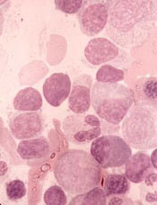
Certain cancers primarily result from ‘bad luck’

in the bone marrow
Scientists have created a statistical model that measures the proportion of cancer incidence, across many tissue types, caused mainly by random mutations that occur when stem cells divide.
By their measure, two-thirds of adult cancers—including certain leukemias—can be explained primarily by “bad luck,” when these random mutations occur in genes that can drive cancer growth.
The remaining third are due to environmental factors and inherited genes.
“All cancers are caused by a combination of bad luck, the environment, and heredity, and we’ve created a model that may help quantify how much of these three factors contribute to cancer development,” said Bert Vogelstein, MD, of the Johns Hopkins University School of Medicine.
Dr Vogelstein and Cristian Tomasetti, PhD, also of the Johns Hopkins University School of Medicine, detailed these findings in Science.
The pair came to their conclusions by searching the scientific literature for information on the cumulative number of stem cell divisions in 31 tissue types during an average individual’s lifetime.
The researchers knew that cancer arises when tissue-specific stem cells make random mistakes, or mutations. But the actual contribution of these random mistakes to cancer incidence, in comparison to the contribution of hereditary or environmental factors, was unclear.
To sort out the role of random mutations in cancer risk, the team charted the number of stem cell divisions in 31 tissues and compared these rates with the lifetime risks of cancer in the same tissues among Americans.
From this data scatterplot, Drs Tomasetti and Vogelstein determined the correlation between the total number of stem cell divisions and cancer risk to be 0.804. Mathematically, the closer this value is to 1, the more stem cell divisions and cancer risk are correlated.
“Our study shows, in general, that a change in the number of stem cell divisions in a tissue type is highly correlated with a change in the incidence of cancer in that same tissue,” Dr Vogelstein said.
One example is in colon tissue, which undergoes 4 times more stem cell divisions than small intestine tissue in humans. Likewise, colon cancer is much more prevalent than small intestinal cancer.
“You could argue that the colon is exposed to more environmental factors than the small intestine, which increases the potential rate of acquired mutations,” Dr Tomasetti said.
However, the scientists observed the opposite in mouse colons, which had a lower number of stem cell divisions than in their small intestines. In mice, cancer incidence is lower in the colon than in the small intestine. The researchers believe this supports the role of the total number of stem cell divisions in the development of cancer.
Using statistical theory, the pair calculated how much of the variation in cancer risk can be explained by the number of stem cell divisions, which is 0.804 squared, or, in percentage form, approximately 65%.
Finally, the scientists classified the types of cancers they studied into two groups. They calculated which cancer types had an incidence predicted by the number of stem cell divisions and which had higher incidence.
They found that 22 cancer types—including acute myeloid leukemia and chronic lymphocytic leukemia—could be largely explained by the “bad luck” factor of random DNA mutations during cell division.
The other 9 cancer types had incidences higher than predicted by “bad luck” and were presumably due to a combination of bad luck plus environmental or inherited factors.
“We found that the types of cancer that had higher risk than predicted by the number of stem cell divisions were precisely the ones you’d expect, including lung cancer, which is linked to smoking; skin cancer, linked to sun exposure; and forms of cancers associated with hereditary syndromes,” Dr Vogelstein said.
“This study shows that you can add to your risk of getting cancers by smoking or other poor lifestyle factors. However, many forms of cancer are due largely to the bad luck of acquiring a mutation in a cancer driver gene regardless of lifestyle and heredity factors. The best way to eradicate these cancers will be through early detection, when they are still curable by surgery.”
The researchers noted that some cancers, such as breast and prostate cancer, were not included in the report because the team was unable to find reliable stem cell division rates in the scientific literature.
They hope other scientists will help refine their statistical model by finding more precise stem cell division rates. ![]()
Insomnia and e-books
Seems that none of my patients sleeps, or at least not very well. Indeed, population-based studies suggest that almost one-third of adults report difficulty initiating or maintaining sleep, waking up too early, and/or nonrestorative or poor quality of sleep.
Light-emitting electronic “e-readers” or “e-books” have exploded as a favorite medium for reading. Many of my patients tell me that when they are unable to sleep, they read. Much of this reading occurs on e-books. But artificial light can produce alerting effects and suppress melatonin. This might be causing or worsening the national insomnia epidemic.
Dr. Anne-Marie Chang and colleagues conducted a randomized, crossover study evaluating the impact of an e-book and a traditional book on sleep (PNAS 2014 Dec. 22 [doi:10.1073/pnas.1418490112]). In the study, 12 healthy young adults were randomized to two conditions: 1) reading an e-book for 4 hours before bedtime; or 2) reading a printed book for 4 hours before bedtime on 5 consecutive evenings. Participants then switched conditions.
The e-book suppressed evening levels of melatonin by 55%, whereas the printed book showed no suppression, and the e-book shifted the melatonin onset to more than 1.5 hours later. Compared with the printed book, the e-book also significantly increased sleep latency (10 minutes longer), decreased REM sleep by11 minutes, decreased evening sleepiness, and increased morning sleepiness.
But not all e-book readers are created equal. Screens of devices used in the current study emit blue light (wavelength 452 nm), the type of light most implicated in melatonin suppression. Newer “e-ink” readers do not emit this light and are “front lit,” with light cast inward rather than outward.
People struggling with insomnia should be encouraged to explore the e-ink options. The table included in the study can provide some guidance as to the type of e-book that may be most beneficial to our sleepy patients.
Dr. Ebbert is professor of medicine, a general internist at the Mayo Clinic in Rochester, Minn., and a diplomate of the American Board of Addiction Medicine. The opinions expressed are those of the author. The opinions expressed in this article should not be used to diagnose or treat any medical condition nor should they be used as a substitute for medical advice from a qualified, board-certified practicing clinician.
Qualifying as an expert
Question: A patient alleges that an ophthalmologist was negligent in performing blepharoplasty. The surgical site became infected and left the patient with a disfigured eye. An infectious disease specialist was to testify as plaintiff’s expert, but the defense, relying on a state statute, objected that the expert-to-be had no specialized training in ophthalmology.
Correct statements about expert medical testimony include the following, except:
A. The federal Rules of Evidence require that an expert possess the relevant knowledge, skill, education, training, or experience.
B. Only a few states have enacted statutes specifying that an expert must be in the same or similar specialty as the defendant.
C. Advocates contend that such statutes reduce frivolous lawsuits by limiting expert shopping and the use of “hired guns.”
D. It is the sitting judge, not the jury, who interprets the statute and decides whether a witness qualifies as an expert.
E. In some cases, a nondoctor such as a nurse or pharmacist may be allowed to offer expert testimony against a doctor.
Answer: B. Lay testimony is usually insufficient to define the standard of care in a claim of medical malpractice, and “the question of negligence must be decided by reference to relevant medical standards of care for which the plaintiff carries the burden of proving through expert medical testimony” (Craft v. Peebles, 893 P.2d 138 [Haw.1995]). Court rules of evidence dictate that the expert must possess the knowledge, skill, experience, training, or education for establishing that standard.
In coming to an opinion, the expert may rely on external evidence in the form of a book, treatise, or article; and although these sources represent hearsay evidence, they are admissible to enable the expert witness to form his/her opinion.
The facts of the multiple-choice question above are taken from a recent case (Edwards v. Sunrise Ophthalmology ASC, LLC, 134 So. 3d 1056 [Fla. 4th DCA 2013]), whose appeal is yet to be heard by the Florida Supreme Court. The plaintiff alleged that the surgical site was infected with nocardia during a lower-lid blepharoplasty, which caused her to undergo additional surgery with resulting disfigurement of the eye.
A lower court disqualified the plaintiff’s expert, an infectious disease specialist, based on a Florida statute stipulating that expert medical opinion can be offered only by one in the “same or similar specialty” (Section 766.102, Florida Statutes (2009)). In the words of the court, “Simply put, the infectious disease doctor is not an eye surgeon, nor is the ophthalmologist an infectious disease doctor.”
More than half of all the states have a medical-expert law, many with language comparable to the Florida statute. The idea behind such a statute, frequently enacted as part of a state’s tort reform, is to limit expert shopping and the use of hired guns and “junk science.”
Unsurprisingly, litigation abounds over the statutory language.
For example, a Maryland court ruled that a vascular surgeon was qualified to set the standard of care when an orthopedic surgeon’s alleged negligence caused a patient to lose a leg following knee surgery. The court found the two specialties to be “related,” because the orthopedic complication was vascular in origin.
In some jurisdictions without strict statutory requirements, doctors are more likely to be allowed to testify outside their specialty.
Instances of professionals of unlike specialties qualifying as experts include a nephrologist testifying against a urologist, an infectious disease specialist offering an expert opinion in a stroke case, a pharmacist testifying on the issue of a medication side effect, and a nurse on bedsores. Georgia requires only that an expert show significant familiarity with the area of practice in which the expert opinion is to be given. Still, in a sleep apnea case (Nathans v. Diamond, 654 S.E.2d 121 [Ga. 2007]), the court held that a pulmonologist was not qualified to testify on the standard of surgical care provided by an otolaryngologist.
Besides arguing over the statutory language, litigants have also raised questions of constitutionality. For example, Arizona’s statute ARS §12-2604 (A) requires a medical expert to be a specialist who is actively practicing or teaching in that area of medicine. The state court of appeals held that this violated the separation of powers doctrine (conflicting with Arizona Rule of Evidence 702), but the Supreme Court of Arizona subsequently reversed and reinstated the law (Seisinger v. Siebel, 203 P.3d 483 [Ariz. 2009]).
More recently, the same court upheld the constitutionality of the requirement that an expert share “the same specialty” as the treating physician, and disqualified an adult hematologist from serving as an expert because the defendant was a pediatric hematologist, not an adult hematologist (Baker v. University Physicians Healthcare, 296 P.3d 42 [Ariz. 2013]).
Another issue deals with the locality rule. In an Illinois case, the court accepted an out-of-state plaintiff expert based on his qualifications, competency, and familiarity with standards in the defendant’s community. The case dealt with the development of a rectovaginal fistula that complicated an episiotomy during delivery (Purtill v. Hess, 489 N.E.2d 867 [Ill. 1986]).
The defense attempted to exclude the expert, alleging the lack of familiarity with the standards in the community (Rantoul, Ill.). However, the expert stated that he was familiar with the minimum standards of medical practice in relation to the diagnosis and treatment of rectovaginal fistulae, and those minimum standards were uniform throughout the country.
It is not necessary that the expert witness has the highest possible qualifications to testify about a particular matter. Still, in Domingo v. T.K. (289 F.3d 600 [9th Cir. 2002]), a federal court excluded the testimony of the plaintiff’s expert witness, because it lacked reliability. The plaintiff developed brain damage from fat embolism following hip surgery, and alleged that prolonged malleting of a hip prosthesis was the cause of the fat embolism syndrome (FES).
The court found that “there was no evidence of widespread acceptance of Dr. Harrington’s theory linking extended malleting to FES; indeed, no theory linking extensive malleting to FES had ever been published.” It also noted the lack of any objective source, peer review, clinical tests, establishment of an error rate or other evidence to show that Dr. Harrington followed a valid, scientific method in developing his theory.
Being disqualified as an expert is one thing, but a recent case goes further. In addition to dismissing an expert’s testimony, a state judge barred the expert from ever testifying in his courtroom after it was determined that the testimony was untruthful.
Dr. Tan is professor emeritus of medicine and former adjunct professor of law at the University of Hawaii, and currently directs the St. Francis International Center for Healthcare Ethics in Honolulu. This article is meant to be educational and does not constitute medical, ethical, or legal advice. Some of the articles in this series are adapted from the author’s 2006 book, “Medical Malpractice: Understanding the Law, Managing the Risk,” and his 2012 Halsbury treatise, “Medical Negligence and Professional Misconduct.” For additional information, readers may contact the author at siang@hawaii.edu

Umbilical cord clamping
Embryogenesis is an incredible phenomenon. It begins with fertilization and implantation of the blastocyst into the uterine wall, followed by development of the extraembryonic membrane which gives rise to the placenta, which is, literally, the lifeblood of the fetus. Although physicians often focus on the health of the placenta during pregnancy, the umbilical cord is an equally important organ.
The umbilical cord acts as a conduit through which metabolic products and biproducts, such as nutrients, antibodies, iron, and blood, pass bidirectionally between a mother and her baby. Many problems can arise if the cord becomes altered. For example, true umbilical cord knot is associated with small-for-gestational-age fetuses, premature birth, neonatal intensive care unit admissions, and fetal death (Int. J. Gynaecol. Obstet. 2013;122:18-21). In addition, short or long cord length may lead to fetal heart rate anomalies and higher risk for birth asphyxia (J. Obstet. Gynaecol. India. 2012;62:520-5).
While the health of the umbilical cord during pregnancy closely correlates to successful outcomes, the cord’s functions conclude at birth. Physicians clamp the umbilical cord at parturition as a routine part of the delivery process. Many ob.gyns. may not even stop to think about cord clamping. To borrow the phrase from the old Nike slogan, they “just do it.”
However, exactly when physicians should clamp the umbilical cord remains a topic for debate. Should ob.gyns. clamp the cord immediately or shortly after birth? Proponents of immediate clamping might argue that waiting too long could cause an influx of placental* blood in the neonate, leading to risk for jaundice. Those on the side of delayed cord clamping might say that waiting to terminate the cord prevents the baby’s oxygen supply from being prematurely cut off. Also, how long should the delay last: One minute? Two minutes? More? Less?
Clearly, the issue of cord clamping requires discussion. Therefore, we have devoted the first Master Class of 2015 to this important topic. We have invited Dr. George A. Macones, the Mitchell and Elaine Yanow Professor and Chair, and director of the division of maternal-fetal medicine and ultrasound in the department of obstetrics and gynecology at Washington University, St. Louis, to explore the debate. As the recent vice chair for the Committee on Practice Bulletins – Obstetrics for the American College of Obstetricians and Gynecologists, Dr. Macones has a unique perspective on the arguments for and against immediate or delayed cord clamping, as well as significant experience as a practicing ob.gyn. and leader at a vibrant academic medical center.
Dr. Reece, who specializes in maternal-fetal medicine, is vice president for medical affairs at the University of Maryland, Baltimore, as well as the John Z. and Akiko K. Bowers Distinguished Professor and dean of the school of medicine. Dr. Reece said he had no relevant financial disclosures. He is the medical editor of this column. Contact him at obnews@frontlinemedcom.com.
Correction, 4/14/15: An earlier version of this article misstated the potential consequences of waiting too long to clamp the umbilical cord.

5 ways digital health technologies are patient advocacy tools
When technology is mentioned in the context of health care, it is often received as an impersonal, profit- or regulatory-driven interface between a provider and patient. If designed – hopefully with a clinician involved – with the purpose of actually solving a problem, digital technology will ultimately gain favor. Examples of such tools include links and apps which provide reference information. Epocrates and doximity on the provider side and WebMD on the consumer/patient side are prime examples. There are increasingly more digital tools for patients and caregivers to help them improve self-participation in their health care as well as to navigate the system. The challenge in the health care technology space is to make people (both providers and patients) aware of them, to facilitate use, and to incorporate relevant and actionable data seamlessly into the patient’s electronic record. Technology needs to be designed in a way in which it conforms to the clinical work flow between the patient and provider. I will give examples of available tools that can improve a patient’s daunting journey. I do not have any financial or other affiliation with any companies mentioned.
1. They can help prepare for the office visit.
I don’t know why, but patients have evolved a belief that they need to present a self-diagnosed condition at the office visit. They often feel guilty not providing the diagnosis. I believe firmly (and tell patients) that their responsibility is to know when something isn’t right and to call the provider. Notwithstanding this, I encourage patients to do online research into their symptoms. Tools found at FamilyDoctor.org, the Mayo Clinic Symptom Checker, or iTriage can help frame thoughts or prompt a discussion with a caregiver prior to a visit, which can then serve as a foundation for the office encounter.
2. Patient education content.
The term “patient engagement” is used commonly today. It implies the active participation of the patient in health care and disease management. Many believe that patient engagement should be focused on medication adherence. While this is critical, it remains a reflection of a patient’s understanding of diagnosis; long-term treatment goals (which need be personalized per a discussion about them); and the components of the treatment itself, which include lifestyle changes and nontraditional pharmacologic therapies as well. Seeing disease through a patient’s eyes (empathy) is the key to good relationships that in turn promote engagement. Excellent digital patient education tools are now available for download and review by patients and caregivers. They explain diagnoses, tests, procedures, and medications. Some are proprietary and made by pharmaceutical and medical device companies, while others are produced by third-party companies that allow the provider to white label the product or even customize the content. One excellent example is Liberate Health. (Ed. note: This publication’s parent company has a relationship with Liberate Health.)
3. Social media.
This is where the patients and caregivers are. It follows then that social media is where providers should be. There are some excellent online patient communities that contain disease-specific groups. Examples are Smart Patients and Treatment Diaries. Social media is a big part of motivating patients and giving support to them and to caregivers. It allows for information exchange in a convenient, relaxing, and nonthreatening setting. While skeptics might question the validity of medical information and advice on these sites, I would say that encouraging patients to participate shows empathy. If a disclaimer is offered stating that this is not a substitute for a health care provider, it can be a significant source of support.
4. Connections to caregivers.
Caregivers are left out of many digital health tools. A good working definition of a caregiver is “an unpaid individual (a spouse, partner, family member, friend, or neighbor) involved in assisting others with activities of daily living and/or medical tasks.” About 29% of the U.S. adult population (65.7 million) provides care to someone who is ill, disabled, or aged. Other statistics about caregivers are more impressive. Health and medical apps are promising tools that can be offered to patients. The rubber has yet to fully meet the road in this arena for a few reasons, many of which are tied to the reputation, usability, and priorities of present electronic health record vendors who represent the face of digital health technology to most physicians and other health care providers. However, there is little denial that they (and other mobile health tools) will play an important role in health care’s future. Both patients and caregivers have expressed what is desired in a mobile app. As aging at home becomes a necessary goal of health care from social, financial, and societal standpoints, caregivers will assume an even greater portion of care.
5. Provide for better continuity of care.
Lack of continuity of care leading to medical errors is not a new topic of discussion. This is relevant in both the inpatient and the outpatient setting. Mobile digital technologies can reduce errors by improving communication to both providers and patients as well as among providers themselves. Use of digital tablets at the bedside by patients can improve provider-patient communication and decrease errors. Handoff of patients among providers is another opportunity for mobile health tools to decrease errors. One such app is Smart Sign Out. Ultimately, any tool that decreases errors is a patient advocate tool.
While some physicians believe that patient advocacy is distinct from patient care, I submit that patient advocacy is something any good physician does every day with every patient, including conveying empathy, providing easy to understand explanations of conditions, and offering advice to be considered in a shared decision-making process. We all enter the field of medicine because we want to contribute to the well-being of others. Let’s not lose sight of that, and let’s look to available and emerging technologies to assist us in this mission.
Dr. Scher is an electrophysiologist with the Heart Group of Lancaster (Pa.) General Health. He is also director of DLS Healthcare Consulting, Harrisburg, Pa., and clinical associate professor of medicine at the Pennsylvania State University, Hershey.

Nerve monitoring linked to higher risk of vocal cord paralysis
SAN FRANCISCO – Use of intraoperative nerve monitoring during thyroidectomy to avoid injuring the recurrent laryngeal nerve is counterintuitively associated with a higher risk of vocal cord paralysis, in a cohort study of data from the Nationwide Inpatient Sample.
“We do caution against perhaps the broad adoption of nerve monitoring until we can really study this further,” said Dr. Thomas K. Chung, a research fellow in the department of surgery, division of otolaryngology, at the University of Alabama at Birmingham, and the study’s lead investigator.
He and his colleagues compared outcomes between 12,742 patients who had nerve monitoring and 230,433 patients who did not (the conventional practice) while undergoing thyroidectomy between 2008 and 2011.
The proportion of patients who developed vocal cord paralysis was significantly higher with monitoring than without it (1.9% vs. 1.4%), he reported at the annual clinical congress of the American College of Surgeons. The findings were essentially the same in propensity-adjusted analyses that took into account differences between groups in preoperative factors (1.8% vs. 1.3%).
There was no evidence that the difference was related to differences in the use of laryngoscopy to check for paralysis, in hospitals’ coding and billing for monitoring, or in payers’ coverage of this surgical adjunct.
Stratified analyses looking at the extent of surgery showed total thyroidectomy with neck dissection to be the exception, as patients monitored during these more complex operations were significantly less likely to develop vocal cord paralysis than were nonmonitored counterparts (2.8% vs. 4.5%).
The more often hospitals used nerve monitoring as indicated by the volume of thyroidectomy cases, the lower the rate of vocal cord paralysis – with the exception of cases of partial thyroidectomy, in which more frequent use was associated with a counterintuitive increase in the rate of this complication, according to Dr. Chung, who disclosed that he had no relevant conflicts of interest.
“Nerve monitoring demonstrates a significant benefit particularly in complex cases such as total thyroidectomy with neck dissection,” he said. “Low nerve monitoring and utilization with partial thyroidectomy appears to be associated with higher vocal cord paralysis; with respect to the partial thyroidectomies, this may be due to the fact that the burden of complication is already so low, with vocal cord paralysis rates of about 0.8%, that additional use of nerve monitoring may not confer any benefit.”
Dr. Chung offered several possible reasons as to why monitoring may be associated with a higher risk of vocal cord paralysis, including presence of a learning curve, substitution of monitoring for direct visualization of the nerve, and false-negatives whereby a lack of signal from the monitor may lead to more aggressive ablation when the nerve is in fact nearby.
The study had its limitations, he acknowledged. “Nerve monitoring may not be coded all the time,” he said. Information about prior neck radiation and surgery, which increase the risk of vocal cord paralysis, was unavailable. “In the group with thyroidectomy with neck dissection, there is no code for central neck dissection. And even if it is a partial thyroidectomy with neck dissection, the central neck dissection would put both nerves at risk and therefore certainly increase the risk of vocal cord paralysis,” he noted.
Invited discussant Julie Ann Sosa, chief of endocrine surgery at the Duke Cancer Institute in Durham, N.C., said, “I would like to congratulate you and your whole group for tackling what is perhaps one of the most highly contested and contentious issues within endocrine surgery and otolaryngology. It’s also I think a very important area for study because there is a relative paucity of data demonstrating for or against the use of this technology as an adjunct. Current guidelines basically say it’s a wash: We can’t say one way or the other whether folks should be using it. And the anticipated guidelines, those coming out from the American Thyroid Association, similarly will say that more data are needed. So I think you are filling a clear vacuum.”
Dr. Sosa questioned the generalizability of the findings, noting that nearly two-thirds of thyroid procedures are now done in the ambulatory setting. “You used the Nationwide Inpatient Sample, so I think you are looking at a minority of cases and highly complex cases, with a length of stay on the order of 2-3 days, which is really exceptional. Most of us send home patients the same day. So how generalizable do you think your conclusions are, and have you thought about potentially using some of the ambulatory surgery databases to try to ask similar questions?” she queried.
The investigators plan to repeat analyses using the Nationwide Inpatient Sample’s ambulatory data set next, according to Dr. Chung. “Being able to see whether or not this still holds true in an outpatient setting is definitely worthwhile,” he agreed.
Dr. Sosa further wondered about the roles of bilateral versus unilateral monitoring, and continuous versus intermittent monitoring, saying, “I think the approach an individual surgeon takes could definitely result in different outcomes. So I wonder, were you able to address at a more granular level the specific technologies employed?”
The Nationwide Inpatient Sample unfortunately does not capture information on these aspects of monitoring, said Dr. Chung. However, “with respect to continuous versus intermittent, we do think that that’s actually an important variable. It is possible that those who are using nerve monitoring are doing this in a continuous fashion, so when they perhaps injure the first side of the vocal cords, they may stop. So what was originally planned to be a total thyroidectomy from the get-go that had an intraoperative nerve injury may be aborted so as to not create any disastrous airway complications. That may also artificially increase the partial thyroidectomy results, thereby increasing vocal cord paralysis complications in the partial thyroidectomy group.”
Recurrent laryngeal nerve injury resulting in vocal cord paralysis, voice, and swallowing dysfunction is a dreaded complication of thyroid surgery.
Fortunately, this is a relatively rare event, occurring anywhere from 0.5% to 2%, in experienced hands. The utilization of nerve monitoring has been proposed to potentially help decrease nerve injury, however, its real strength may be in helping to identify recurrent nerve injury intraoperatively. Knowing the recurrent laryngeal nerve is injured on one side may lead the surgeon to perform a partial thyroidectomy to avoid major airway issues.
The surgeon may choose to wait for nerve function to return before operating on the contralateral side. Utilization of nerve monitoring allows the surgeon to alter the course of the operation potentially leading to better patient outcomes.
Dr. Chung and colleagues have done an excellent job in adding valuable data to the controversial area of nerve monitoring during thyroid surgery. As they point out, their study is subject to the inherent limitations of large national database reviews. Since most thyroid surgeries are performed in the outpatient setting, it is likely that the Nationwide Inpatient Sample does not capture majority of the cases and reflects the more complicated cases, which required a longer length of stay. Recent data also suggest that the percentage of surgeons using nerve monitoring has increased significantly and that more than 70% of all fellows in training programs use nerve monitoring. This technology is not a substitute for an experienced surgeon but in the proper setting can be useful in making critical operative decisions.
Dr. Kepal N. Patel is an ACS Fellow; chief, Division of Endocrine Surgery; associate professor of surgery, biochemistry and otolaryngology; and director, Thyroid Cancer Interdisciplinary Program, New York University Langone Medical Center.
SAN FRANCISCO – Use of intraoperative nerve monitoring during thyroidectomy to avoid injuring the recurrent laryngeal nerve is counterintuitively associated with a higher risk of vocal cord paralysis, in a cohort study of data from the Nationwide Inpatient Sample.
“We do caution against perhaps the broad adoption of nerve monitoring until we can really study this further,” said Dr. Thomas K. Chung, a research fellow in the department of surgery, division of otolaryngology, at the University of Alabama at Birmingham, and the study’s lead investigator.
He and his colleagues compared outcomes between 12,742 patients who had nerve monitoring and 230,433 patients who did not (the conventional practice) while undergoing thyroidectomy between 2008 and 2011.
The proportion of patients who developed vocal cord paralysis was significantly higher with monitoring than without it (1.9% vs. 1.4%), he reported at the annual clinical congress of the American College of Surgeons. The findings were essentially the same in propensity-adjusted analyses that took into account differences between groups in preoperative factors (1.8% vs. 1.3%).
There was no evidence that the difference was related to differences in the use of laryngoscopy to check for paralysis, in hospitals’ coding and billing for monitoring, or in payers’ coverage of this surgical adjunct.
Stratified analyses looking at the extent of surgery showed total thyroidectomy with neck dissection to be the exception, as patients monitored during these more complex operations were significantly less likely to develop vocal cord paralysis than were nonmonitored counterparts (2.8% vs. 4.5%).
The more often hospitals used nerve monitoring as indicated by the volume of thyroidectomy cases, the lower the rate of vocal cord paralysis – with the exception of cases of partial thyroidectomy, in which more frequent use was associated with a counterintuitive increase in the rate of this complication, according to Dr. Chung, who disclosed that he had no relevant conflicts of interest.
“Nerve monitoring demonstrates a significant benefit particularly in complex cases such as total thyroidectomy with neck dissection,” he said. “Low nerve monitoring and utilization with partial thyroidectomy appears to be associated with higher vocal cord paralysis; with respect to the partial thyroidectomies, this may be due to the fact that the burden of complication is already so low, with vocal cord paralysis rates of about 0.8%, that additional use of nerve monitoring may not confer any benefit.”
Dr. Chung offered several possible reasons as to why monitoring may be associated with a higher risk of vocal cord paralysis, including presence of a learning curve, substitution of monitoring for direct visualization of the nerve, and false-negatives whereby a lack of signal from the monitor may lead to more aggressive ablation when the nerve is in fact nearby.
The study had its limitations, he acknowledged. “Nerve monitoring may not be coded all the time,” he said. Information about prior neck radiation and surgery, which increase the risk of vocal cord paralysis, was unavailable. “In the group with thyroidectomy with neck dissection, there is no code for central neck dissection. And even if it is a partial thyroidectomy with neck dissection, the central neck dissection would put both nerves at risk and therefore certainly increase the risk of vocal cord paralysis,” he noted.
Invited discussant Julie Ann Sosa, chief of endocrine surgery at the Duke Cancer Institute in Durham, N.C., said, “I would like to congratulate you and your whole group for tackling what is perhaps one of the most highly contested and contentious issues within endocrine surgery and otolaryngology. It’s also I think a very important area for study because there is a relative paucity of data demonstrating for or against the use of this technology as an adjunct. Current guidelines basically say it’s a wash: We can’t say one way or the other whether folks should be using it. And the anticipated guidelines, those coming out from the American Thyroid Association, similarly will say that more data are needed. So I think you are filling a clear vacuum.”
Dr. Sosa questioned the generalizability of the findings, noting that nearly two-thirds of thyroid procedures are now done in the ambulatory setting. “You used the Nationwide Inpatient Sample, so I think you are looking at a minority of cases and highly complex cases, with a length of stay on the order of 2-3 days, which is really exceptional. Most of us send home patients the same day. So how generalizable do you think your conclusions are, and have you thought about potentially using some of the ambulatory surgery databases to try to ask similar questions?” she queried.
The investigators plan to repeat analyses using the Nationwide Inpatient Sample’s ambulatory data set next, according to Dr. Chung. “Being able to see whether or not this still holds true in an outpatient setting is definitely worthwhile,” he agreed.
Dr. Sosa further wondered about the roles of bilateral versus unilateral monitoring, and continuous versus intermittent monitoring, saying, “I think the approach an individual surgeon takes could definitely result in different outcomes. So I wonder, were you able to address at a more granular level the specific technologies employed?”
The Nationwide Inpatient Sample unfortunately does not capture information on these aspects of monitoring, said Dr. Chung. However, “with respect to continuous versus intermittent, we do think that that’s actually an important variable. It is possible that those who are using nerve monitoring are doing this in a continuous fashion, so when they perhaps injure the first side of the vocal cords, they may stop. So what was originally planned to be a total thyroidectomy from the get-go that had an intraoperative nerve injury may be aborted so as to not create any disastrous airway complications. That may also artificially increase the partial thyroidectomy results, thereby increasing vocal cord paralysis complications in the partial thyroidectomy group.”
AT THE ACS CLINICAL CONGRESS
Key clinical point: Patients who had intraoperative nerve monitoring were more likely to develop vocal cord paralysis.
Major finding: The propensity-adjusted rate of vocal cord paralysis was 1.3% without monitoring and 1.8% with monitoring.
Data source: A retrospective cohort study of 243,175 patients undergoing thyroidectomy.
Disclosures: Dr. Chung disclosed that he had no relevant conflicts of interest.

CARs come in different makes and models

Credit: NIAID
SAN FRANCISCO—CTL019, a chimeric antigen receptor (CAR) T cell targeting CD19, is not the only CAR in the production line.
Investigators at the National Cancer Institute in Bethesda, Maryland, and Memorial Sloan Kettering Cancer Center (MSKCC) in New York City are also pursuing CAR T-cell therapy.
These groups are using a retroviral platform to transduce the T cells rather than a lentiviral one, as is the case with CTL019.
Investigators reported progress to date on these makes of CARs at the 2014 ASH Annual Meeting.
Daniel W. Lee III, MD, of the National Cancer Institute, reported on a phase 1 study of CD19 CAR T cells in children and young adults with CD19+ acute lymphoblastic leukemia or non-Hodgkin lymphoma.
And Jae H. Park, MD, of MSKCC, presented data from a trial of JCAR015—autologous T cells genetically modified to express a 19-28z CAR targeting CD19—in patients with B-cell acute lymphoblastic leukemia.
The study is sponsored by MSKCC, but JCAR015 is a product of Juno Therapeutics. ![]()
CAR is feasible in majority of ALL patients, team says

Credit: Bill Branson
SAN FRANCISCO—A chimeric antigen receptor (CAR) T-cell therapy is feasible in 90% of heavily pretreated or transplanted patients with acute lymphoblastic leukemia (ALL) and can serve as a bridge to transplant, according to investigators.
Daniel W. Lee III, MD, of the National Cancer Institute in Bethesda, Maryland, reported on a phase 1 study of this CD19 CAR T-cell therapy in children and young adults with CD19+ ALL or non-Hodgkin lymphoma at the 2014 ASH Annual Meeting (abstract 381*).
Twenty-one patients were enrolled on the trial. They had a preparative regimen of fludarabine and cyclophosphamide and were infused with CAR T cells 11 days after the peripheral blood mononuclear cells were collected.
Dose levels were 1 x 10⁶ CAR+ T cells/kg, 3 x 10⁶ CAR+ T cells/kg, or the maximum number of cells generated if below either one of these levels. Two patients received less than the dose assigned and were not evaluated for toxicity.
Patients were a median age of 13 years (range, 5 to 27). Fourteen were male, 20 had ALL, and 1 had diffuse large B-cell lymphoma.
All had detectable disease, and 2 were CNS2 at the time of T-cell infusion. Six had primary refractory disease, 8 had at least 1 prior stem cell transplant, and 4 had prior immunotherapy.
The investigators determined that the maximally tolerated dose was 1 x 10⁶ CAR+ T cells/kg. The dose-limiting toxicities were related to cytokine release syndrome (CRS), which was reversible if managed appropriately with tocilizumab, with or without steroids.
Grade 3 adverse events possibly related to therapy included fever (47%), febrile neutropenia (37%), electrolyte disturbance (29%), CRS (16%), hypotension (11%), transaminitis (16%), and 5% each for hypertension, prolonged QTc, dysphasia, LV systolic dysfunction, multiorgan failure, hypoxia, and pulmonary edema.
Grade 4 events possibly related to treatment included electrolyte disturbance (5%), CRS (16%), hypotension (11%), cardiac arrest (5%), and hypoxia (5%). There was no evidence of graft-vs-host disease.
The complete response (CR) rate was 67% in the intent-to-treat population and 70% in patients with ALL.
“Those patients who responded tended to have some degree of cytokine release syndrome, whereas those patients who did not respond or had stable disease did not have any CRS,” Dr Lee said. “But, also, it’s important to note that you don’t have to have severe grade 3 or grade 4 CRS in order to have significant response.”
Dr Lee also pointed out that in vivo CAR T-cell expansion significantly correlated with response (P=0.0028). And CRS severity correlated with IL-6 (P=0.0002), INF-γ (P=0.0002), C-reactive protein (P=0.0015), and CAR (P=0.0011).
At a median follow-up of 10 months, minimal residual disease-negative patients had a 79% leukemia-free survival. Overall survival was 52% for all patients enrolled. Two patients had CD19-negative relapses.
The investigators also found that CAR T cells can eliminate CNS leukemia, with 11 of 17 patients (65%) having CAR T cells detectable in their cerebrospinal fluid.
The team concluded that this therapy is feasible in 90% of heavily pretreated or transplanted ALL patients and can serve as a bridge to transplant. ![]()
*Information in the abstract differs from that presented at the meeting.