User login
Join us for CHEST Annual Meeting 2020
Registration for CHEST Annual Meeting 2020 has opened! It is important now, more than ever, to stay up to date in clinical chest education. CHEST Annual Meeting is prepared to equip attendees with the latest education and original research in the field that can be taken back home and implemented into practices.
While CHEST is excited to bring the premier event in clinical chest medicine to their Second City Home of Chicago, Illinois, this October 17-21, it is understood that now may not be the best time to be planning for a conference that is 6 months down the road. Currently, your full attention is likely on your patients, your families, your health, and your safety, and it should be! Here at CHEST, the hope is to create a “light at the end of the tunnel” to give you and your colleagues something to look forward to – an opportunity to relax, learn, explore, and reconnect with your peers in the chest medicine field.
This year’s annual meeting will be filled with both new and returning educational opportunities, including CHEST Games; virtual patient tours; hands-on simulation courses; problem-based learning; and the return of FISH Bowl, an innovation competition. Along with the advanced education, there will be countless opportunities to network at after-hour events, such as the CHEST Challenge final competition, the Young Professionals Reception, and the CHEST Foundation Casino Night. Our hope is that you will be able to look ahead to October and be excited about the chance to experience everything that will be offered at CHEST 2020.
Before the meeting in October, don’t forget to submit your abstracts and case reports for consideration to be presented at CHEST 2020. CHEST is excited to give you and your colleagues the opportunity to present new and original research at this year’s meeting, which is why the deadline for submissions has been extended to June 1, 2020.
CHEST acknowledges that your workload is becoming increasingly heavier each day, and we are also making the safety of attendees the top priority.
That is why CHEST will be granting full refunds to any registrant who finds that they can no longer attend CHEST 2020 as the meeting approaches. Any hotel reservation that is made through CHEST’s official housing site, onPeak, will be able to be changed or canceled up to 24 hours in advance of the reservation date. Visit chestmeeting.chestnet.org/hotel-accommodations for more information.
CHEST 2020 meeting chair, Victor Test, MD, FCCP, hopes to leave CHEST learners with a beacon of hope, saying, “Signing up to come to the meeting and participating may seem impossible to think about right now. We are working hard to provide a high-quality experience and are encouraging everyone to look forward to the future, which will be a lot brighter.”
For all of the latest information on CHEST 2020, visit chestmeeting.chestnet.org.
Registration for CHEST Annual Meeting 2020 has opened! It is important now, more than ever, to stay up to date in clinical chest education. CHEST Annual Meeting is prepared to equip attendees with the latest education and original research in the field that can be taken back home and implemented into practices.
While CHEST is excited to bring the premier event in clinical chest medicine to their Second City Home of Chicago, Illinois, this October 17-21, it is understood that now may not be the best time to be planning for a conference that is 6 months down the road. Currently, your full attention is likely on your patients, your families, your health, and your safety, and it should be! Here at CHEST, the hope is to create a “light at the end of the tunnel” to give you and your colleagues something to look forward to – an opportunity to relax, learn, explore, and reconnect with your peers in the chest medicine field.
This year’s annual meeting will be filled with both new and returning educational opportunities, including CHEST Games; virtual patient tours; hands-on simulation courses; problem-based learning; and the return of FISH Bowl, an innovation competition. Along with the advanced education, there will be countless opportunities to network at after-hour events, such as the CHEST Challenge final competition, the Young Professionals Reception, and the CHEST Foundation Casino Night. Our hope is that you will be able to look ahead to October and be excited about the chance to experience everything that will be offered at CHEST 2020.
Before the meeting in October, don’t forget to submit your abstracts and case reports for consideration to be presented at CHEST 2020. CHEST is excited to give you and your colleagues the opportunity to present new and original research at this year’s meeting, which is why the deadline for submissions has been extended to June 1, 2020.
CHEST acknowledges that your workload is becoming increasingly heavier each day, and we are also making the safety of attendees the top priority.
That is why CHEST will be granting full refunds to any registrant who finds that they can no longer attend CHEST 2020 as the meeting approaches. Any hotel reservation that is made through CHEST’s official housing site, onPeak, will be able to be changed or canceled up to 24 hours in advance of the reservation date. Visit chestmeeting.chestnet.org/hotel-accommodations for more information.
CHEST 2020 meeting chair, Victor Test, MD, FCCP, hopes to leave CHEST learners with a beacon of hope, saying, “Signing up to come to the meeting and participating may seem impossible to think about right now. We are working hard to provide a high-quality experience and are encouraging everyone to look forward to the future, which will be a lot brighter.”
For all of the latest information on CHEST 2020, visit chestmeeting.chestnet.org.
Registration for CHEST Annual Meeting 2020 has opened! It is important now, more than ever, to stay up to date in clinical chest education. CHEST Annual Meeting is prepared to equip attendees with the latest education and original research in the field that can be taken back home and implemented into practices.
While CHEST is excited to bring the premier event in clinical chest medicine to their Second City Home of Chicago, Illinois, this October 17-21, it is understood that now may not be the best time to be planning for a conference that is 6 months down the road. Currently, your full attention is likely on your patients, your families, your health, and your safety, and it should be! Here at CHEST, the hope is to create a “light at the end of the tunnel” to give you and your colleagues something to look forward to – an opportunity to relax, learn, explore, and reconnect with your peers in the chest medicine field.
This year’s annual meeting will be filled with both new and returning educational opportunities, including CHEST Games; virtual patient tours; hands-on simulation courses; problem-based learning; and the return of FISH Bowl, an innovation competition. Along with the advanced education, there will be countless opportunities to network at after-hour events, such as the CHEST Challenge final competition, the Young Professionals Reception, and the CHEST Foundation Casino Night. Our hope is that you will be able to look ahead to October and be excited about the chance to experience everything that will be offered at CHEST 2020.
Before the meeting in October, don’t forget to submit your abstracts and case reports for consideration to be presented at CHEST 2020. CHEST is excited to give you and your colleagues the opportunity to present new and original research at this year’s meeting, which is why the deadline for submissions has been extended to June 1, 2020.
CHEST acknowledges that your workload is becoming increasingly heavier each day, and we are also making the safety of attendees the top priority.
That is why CHEST will be granting full refunds to any registrant who finds that they can no longer attend CHEST 2020 as the meeting approaches. Any hotel reservation that is made through CHEST’s official housing site, onPeak, will be able to be changed or canceled up to 24 hours in advance of the reservation date. Visit chestmeeting.chestnet.org/hotel-accommodations for more information.
CHEST 2020 meeting chair, Victor Test, MD, FCCP, hopes to leave CHEST learners with a beacon of hope, saying, “Signing up to come to the meeting and participating may seem impossible to think about right now. We are working hard to provide a high-quality experience and are encouraging everyone to look forward to the future, which will be a lot brighter.”
For all of the latest information on CHEST 2020, visit chestmeeting.chestnet.org.
This month in the journal CHEST®
Editor’s Picks
Fighting the novel coronavirus together with you. By Dr. J. Li.
Comparative Safety and Effectiveness of Inhaled Corticosteroid and Long-Acting Beta2-AgonistCombinations in Patients With COPD.By Dr. T-U Chang, et al.
The Evolving Landscape of e-Cigarettes: A Systematic Review of Recent Evidence. By Dr. J. Bozier, et al.
Editor’s Picks
Editor’s Picks
Fighting the novel coronavirus together with you. By Dr. J. Li.
Comparative Safety and Effectiveness of Inhaled Corticosteroid and Long-Acting Beta2-AgonistCombinations in Patients With COPD.By Dr. T-U Chang, et al.
The Evolving Landscape of e-Cigarettes: A Systematic Review of Recent Evidence. By Dr. J. Bozier, et al.
Fighting the novel coronavirus together with you. By Dr. J. Li.
Comparative Safety and Effectiveness of Inhaled Corticosteroid and Long-Acting Beta2-AgonistCombinations in Patients With COPD.By Dr. T-U Chang, et al.
The Evolving Landscape of e-Cigarettes: A Systematic Review of Recent Evidence. By Dr. J. Bozier, et al.
3D printing and pulmonology. COVID-19. Lung volume measurements. Pulmonary hypertension.
Interventional and Chest Diagnostic Procedures
3D printing and pulmonology
Recent advances in 3D printing has enabled physicians to apply this technology in medical education, procedural planning, tissue modeling, and implantable device manufacturing. This is especially true in the field of pulmonology. Advancements in 3D printing have made personalized airway stents a reality, both by 3D printing-assisted injection molding or direct 3D printing.
Airway stents have significantly evolved over the last half century. With use of silicone, bare metallic, and hybrid stents, pulmonologists have an ever-expanding option to address airway stenosis due to both benign and malignancy etiologies. Personalized airway stents hold the potential for advance customization, minimizing pressure points, and improving airflow dynamics to increase mucus clearance. In January 2020, the US Food and Drug Administration (FDA) cleared patient-specific airway stents developed by Dr. Thomas Gildea of Cleveland Clinic. The patient-specific silicone stents are created using CT scans and 3D visualization software to generate a 3D-printed mold that was subsequently used to inject with medical-grade silicone. Two years earlier, a Duke University startup known as restor3D created the first direct 3D printed airway stent using a compressible biocompatible material with properties similar to that of silicone. Both of these stents have been used in patients with promising response.
As we look into the future, the field of pulmonology will experience significant changes with more adoption of 3D printing (ie, additive manufacturing). We may soon be able to create personalized airway prosthesis of any type (stents, spigots, valves, tracheostomies, t-tubes) for the benefit of our patients.

Disclosure: Dr. George Cheng is a cofounder of restor3D.
George Cheng, MD, PhD, FCCP
Steering Committee Member
Pediatric Chest Medicine
COVID-19: Pediatric story of a new pandemic
In December 2019, an outbreak of pneumonia identified to be caused by 2019 novel coronavirus (2019-nCoV) emerged in Wuhan, China, possibly originating from the local wet market selling many species of live animals. A novel member of enveloped RNA coronavirus was identified in samples of BAL fluid from a patient in Wuhan.
It has since rapidly spread globally to countries across six continents. As of early April, 1,286,409 cases have been reported worldwide with 337,933 cases (9,600 deaths) in the US (https://coronavirus.jhu.edu/map.html) with more cases and deaths every day. Most of these initial reports of COVID-19 (COronaVIrusDisease) in children are from China. Fever (60%) and cough (65%) were the most common symptoms. Procalcitonin elevation (80% and co-infection (80%) were prominent clinical findings. Consolidation with surrounding halo sign (50%) and ground-glass opacities (60%) on CT scan were typical radiologic findings. Almost all children recovered without needing intensive care support.
Increased IgM COVID-19 antibody levels observed in three neonates raise questions of potential in-utero transmission (Kimberlin et al. JAMA 2020 Mar 26. doi: 10.1001/jama.2020.4868). One study provided evidence for persistent fecal shedding and possibility of fecal-oral transmission (Xu et al. Nat Med 2020 Mar 13. doi: 10.1038/s41591-020-0817-4).

Initial reports show that children appear to be at similar risk of infection as adults, though less likely to have severe symptoms. Young children, particularly infants, are more vulnerable to infection (Dong et al. Pediatrics. 2020 Apr. doi: 10.1542/peds.2020-0702); (Bi et al. medRxiv 2020 Mar 27. doi: 10.1101/2020.03.03.20028423v3). Thus far, few deaths have been reported in the pediatric age group. Trials are being conducted on a war footing to find a cure and a vaccine.
Harish Rao, MD, MBBS
Steering Committee Member
Pulmonary Physiology, Function, and Rehabilitation
Controversies and the clinical value of lung volume measurements
Lung volumes are often measured by body plethysmography or gas dilution. Their clinic importance in decision making is unclear. Though measured differently, predicted sets obtained by plethysmography from Caucasian populations are often used for gas dilution measurements (Ruppel GL. Respir Care. 2012 Jan;57[1]:26). Recently the GLI felt lung volume data were insufficient to develop universal reference equations (Cooper B, et al. Breathe (Sheff). 2017 Sep;13[3]:e56-e64). ERS/ATS guidelines recommend adjusting Caucasian predicted values depending on race, without advising how to adjust the confidence limits. Their algorithms show if the VC is normal, lung volumes are unnecessary, though it is not unusual to see a normal VC with reduced TLC. Does this suggest the VC is more important than the TLC, even if lacking predicted volume equations for non-Caucasians? Because combined obstructive and restrictive abnormalities occur simultaneously, recommendations state severity of impairment be determined by the FEV1 percent of predicted rather than TLC (Pellegrino R, et al. Eur Respir J. 2005;26:948). The value of quantifying other volumes such as FRC and ERV in conditions such as obesity and musculoskeletal defects is also not clear. In obstruction, volumes can indicate air trapping or hyperinflation measuring RV and RV/TLC. Though cutoffs of <80% and >120% of predicted are often used, guidelines discourage this practice, recommending using predicted equations based on age, race, height, and sex, with statistical limits of normal (Ruppel GL. Respir Care. 2012 Jan;57(1):26).

Further research is needed to define comprehensive racially appropriate predicted equations for lung volumes to support their clinical applicability in decision making, as well as if predicted values by plethysmography are applicable to values obtained from gas dilution.

Said A. Chaaban, MD
Steering Committee Member
Zachary Q. Morris, MD
NetWork Member
Pulmonary Vascular Disease
Pulmonary hypertension associated with atrial septal defect in adults: closing time?
Up to 10% of adults with atrial septal defects (ASDs) can develop pulmonary arterial hypertension (PAH) according to European Guidelines on pulmonary hypertension (PH) (Galie, et al. Eur Heart J. 2016;37[1]:67). If ASD closure is considered, they propose a pulmonary vascular resistance index (PVRi) <4 Wood units (WU) m² as a safe cutoff. Higher PVRi carries a higher operative risk, warranting evaluation in specialized PH centers.
American guidelines (Stout, et al. Circulation. 2019 Apr 2;139[14]:e698) recommend closure in symptomatic patients with a net shunt (Qp/Qs) of >1.5:1. Closure appears safe if pulmonary artery (PA) systolic pressure is <1/2 systemic blood pressure, and PVR / systemic vascular resistance is <0.3. They recommend specialized evaluation for higher pressures and to avoid closure once a net right to left shunt is present (Qp/Qs <1.0).
However, in severe cases, experienced centers have reported some success with a “treat-and-close” approach if post-therapy PVR reaches <6.5 WU (Bradley, et al. Int J Cardiol. 2019;291:127).

Finally, consider the following when evaluating ASD-associated PAH: 1. A thermodilution cardiac output method should not be used to calculate PVR/PVRi because of confounding recirculation from the intracardiac shunt (Kwan, et al. Clin Cardiol. 2019;42[3]:334). Qp is used instead and is calculated using Fick equation, requiring accurate oxygen saturation measurements. 2. Mixed venous saturation (MvO2) is needed to determine Qs, and PA saturation cannot be used as MvO2 surrogate. MvO2 must be calculated using superior and inferior vena cava saturations. 3. Some patients with idiopathic PAH may have a small coexisting ASD that is not responsible for the abnormal hemodynamics. Closing the ASD in those cases would be contraindicated. 4. Patients may have more than one type of coexistent congenital heart defect.
Francisco J. Soto, MD, MS, FCCP
Steering Committee Member
Interventional and Chest Diagnostic Procedures
3D printing and pulmonology
Recent advances in 3D printing has enabled physicians to apply this technology in medical education, procedural planning, tissue modeling, and implantable device manufacturing. This is especially true in the field of pulmonology. Advancements in 3D printing have made personalized airway stents a reality, both by 3D printing-assisted injection molding or direct 3D printing.
Airway stents have significantly evolved over the last half century. With use of silicone, bare metallic, and hybrid stents, pulmonologists have an ever-expanding option to address airway stenosis due to both benign and malignancy etiologies. Personalized airway stents hold the potential for advance customization, minimizing pressure points, and improving airflow dynamics to increase mucus clearance. In January 2020, the US Food and Drug Administration (FDA) cleared patient-specific airway stents developed by Dr. Thomas Gildea of Cleveland Clinic. The patient-specific silicone stents are created using CT scans and 3D visualization software to generate a 3D-printed mold that was subsequently used to inject with medical-grade silicone. Two years earlier, a Duke University startup known as restor3D created the first direct 3D printed airway stent using a compressible biocompatible material with properties similar to that of silicone. Both of these stents have been used in patients with promising response.
As we look into the future, the field of pulmonology will experience significant changes with more adoption of 3D printing (ie, additive manufacturing). We may soon be able to create personalized airway prosthesis of any type (stents, spigots, valves, tracheostomies, t-tubes) for the benefit of our patients.
Disclosure: Dr. George Cheng is a cofounder of restor3D.
George Cheng, MD, PhD, FCCP
Steering Committee Member
Pediatric Chest Medicine
COVID-19: Pediatric story of a new pandemic
In December 2019, an outbreak of pneumonia identified to be caused by 2019 novel coronavirus (2019-nCoV) emerged in Wuhan, China, possibly originating from the local wet market selling many species of live animals. A novel member of enveloped RNA coronavirus was identified in samples of BAL fluid from a patient in Wuhan.
It has since rapidly spread globally to countries across six continents. As of early April, 1,286,409 cases have been reported worldwide with 337,933 cases (9,600 deaths) in the US (https://coronavirus.jhu.edu/map.html) with more cases and deaths every day. Most of these initial reports of COVID-19 (COronaVIrusDisease) in children are from China. Fever (60%) and cough (65%) were the most common symptoms. Procalcitonin elevation (80% and co-infection (80%) were prominent clinical findings. Consolidation with surrounding halo sign (50%) and ground-glass opacities (60%) on CT scan were typical radiologic findings. Almost all children recovered without needing intensive care support.
Increased IgM COVID-19 antibody levels observed in three neonates raise questions of potential in-utero transmission (Kimberlin et al. JAMA 2020 Mar 26. doi: 10.1001/jama.2020.4868). One study provided evidence for persistent fecal shedding and possibility of fecal-oral transmission (Xu et al. Nat Med 2020 Mar 13. doi: 10.1038/s41591-020-0817-4).
Initial reports show that children appear to be at similar risk of infection as adults, though less likely to have severe symptoms. Young children, particularly infants, are more vulnerable to infection (Dong et al. Pediatrics. 2020 Apr. doi: 10.1542/peds.2020-0702); (Bi et al. medRxiv 2020 Mar 27. doi: 10.1101/2020.03.03.20028423v3). Thus far, few deaths have been reported in the pediatric age group. Trials are being conducted on a war footing to find a cure and a vaccine.
Harish Rao, MD, MBBS
Steering Committee Member
Pulmonary Physiology, Function, and Rehabilitation
Controversies and the clinical value of lung volume measurements
Lung volumes are often measured by body plethysmography or gas dilution. Their clinic importance in decision making is unclear. Though measured differently, predicted sets obtained by plethysmography from Caucasian populations are often used for gas dilution measurements (Ruppel GL. Respir Care. 2012 Jan;57[1]:26). Recently the GLI felt lung volume data were insufficient to develop universal reference equations (Cooper B, et al. Breathe (Sheff). 2017 Sep;13[3]:e56-e64). ERS/ATS guidelines recommend adjusting Caucasian predicted values depending on race, without advising how to adjust the confidence limits. Their algorithms show if the VC is normal, lung volumes are unnecessary, though it is not unusual to see a normal VC with reduced TLC. Does this suggest the VC is more important than the TLC, even if lacking predicted volume equations for non-Caucasians? Because combined obstructive and restrictive abnormalities occur simultaneously, recommendations state severity of impairment be determined by the FEV1 percent of predicted rather than TLC (Pellegrino R, et al. Eur Respir J. 2005;26:948). The value of quantifying other volumes such as FRC and ERV in conditions such as obesity and musculoskeletal defects is also not clear. In obstruction, volumes can indicate air trapping or hyperinflation measuring RV and RV/TLC. Though cutoffs of <80% and >120% of predicted are often used, guidelines discourage this practice, recommending using predicted equations based on age, race, height, and sex, with statistical limits of normal (Ruppel GL. Respir Care. 2012 Jan;57(1):26).
Further research is needed to define comprehensive racially appropriate predicted equations for lung volumes to support their clinical applicability in decision making, as well as if predicted values by plethysmography are applicable to values obtained from gas dilution.
Said A. Chaaban, MD
Steering Committee Member
Zachary Q. Morris, MD
NetWork Member
Pulmonary Vascular Disease
Pulmonary hypertension associated with atrial septal defect in adults: closing time?
Up to 10% of adults with atrial septal defects (ASDs) can develop pulmonary arterial hypertension (PAH) according to European Guidelines on pulmonary hypertension (PH) (Galie, et al. Eur Heart J. 2016;37[1]:67). If ASD closure is considered, they propose a pulmonary vascular resistance index (PVRi) <4 Wood units (WU) m² as a safe cutoff. Higher PVRi carries a higher operative risk, warranting evaluation in specialized PH centers.
American guidelines (Stout, et al. Circulation. 2019 Apr 2;139[14]:e698) recommend closure in symptomatic patients with a net shunt (Qp/Qs) of >1.5:1. Closure appears safe if pulmonary artery (PA) systolic pressure is <1/2 systemic blood pressure, and PVR / systemic vascular resistance is <0.3. They recommend specialized evaluation for higher pressures and to avoid closure once a net right to left shunt is present (Qp/Qs <1.0).
However, in severe cases, experienced centers have reported some success with a “treat-and-close” approach if post-therapy PVR reaches <6.5 WU (Bradley, et al. Int J Cardiol. 2019;291:127).
Finally, consider the following when evaluating ASD-associated PAH: 1. A thermodilution cardiac output method should not be used to calculate PVR/PVRi because of confounding recirculation from the intracardiac shunt (Kwan, et al. Clin Cardiol. 2019;42[3]:334). Qp is used instead and is calculated using Fick equation, requiring accurate oxygen saturation measurements. 2. Mixed venous saturation (MvO2) is needed to determine Qs, and PA saturation cannot be used as MvO2 surrogate. MvO2 must be calculated using superior and inferior vena cava saturations. 3. Some patients with idiopathic PAH may have a small coexisting ASD that is not responsible for the abnormal hemodynamics. Closing the ASD in those cases would be contraindicated. 4. Patients may have more than one type of coexistent congenital heart defect.
Francisco J. Soto, MD, MS, FCCP
Steering Committee Member
Interventional and Chest Diagnostic Procedures
3D printing and pulmonology
Recent advances in 3D printing has enabled physicians to apply this technology in medical education, procedural planning, tissue modeling, and implantable device manufacturing. This is especially true in the field of pulmonology. Advancements in 3D printing have made personalized airway stents a reality, both by 3D printing-assisted injection molding or direct 3D printing.
Airway stents have significantly evolved over the last half century. With use of silicone, bare metallic, and hybrid stents, pulmonologists have an ever-expanding option to address airway stenosis due to both benign and malignancy etiologies. Personalized airway stents hold the potential for advance customization, minimizing pressure points, and improving airflow dynamics to increase mucus clearance. In January 2020, the US Food and Drug Administration (FDA) cleared patient-specific airway stents developed by Dr. Thomas Gildea of Cleveland Clinic. The patient-specific silicone stents are created using CT scans and 3D visualization software to generate a 3D-printed mold that was subsequently used to inject with medical-grade silicone. Two years earlier, a Duke University startup known as restor3D created the first direct 3D printed airway stent using a compressible biocompatible material with properties similar to that of silicone. Both of these stents have been used in patients with promising response.
As we look into the future, the field of pulmonology will experience significant changes with more adoption of 3D printing (ie, additive manufacturing). We may soon be able to create personalized airway prosthesis of any type (stents, spigots, valves, tracheostomies, t-tubes) for the benefit of our patients.
Disclosure: Dr. George Cheng is a cofounder of restor3D.
George Cheng, MD, PhD, FCCP
Steering Committee Member
Pediatric Chest Medicine
COVID-19: Pediatric story of a new pandemic
In December 2019, an outbreak of pneumonia identified to be caused by 2019 novel coronavirus (2019-nCoV) emerged in Wuhan, China, possibly originating from the local wet market selling many species of live animals. A novel member of enveloped RNA coronavirus was identified in samples of BAL fluid from a patient in Wuhan.
It has since rapidly spread globally to countries across six continents. As of early April, 1,286,409 cases have been reported worldwide with 337,933 cases (9,600 deaths) in the US (https://coronavirus.jhu.edu/map.html) with more cases and deaths every day. Most of these initial reports of COVID-19 (COronaVIrusDisease) in children are from China. Fever (60%) and cough (65%) were the most common symptoms. Procalcitonin elevation (80% and co-infection (80%) were prominent clinical findings. Consolidation with surrounding halo sign (50%) and ground-glass opacities (60%) on CT scan were typical radiologic findings. Almost all children recovered without needing intensive care support.
Increased IgM COVID-19 antibody levels observed in three neonates raise questions of potential in-utero transmission (Kimberlin et al. JAMA 2020 Mar 26. doi: 10.1001/jama.2020.4868). One study provided evidence for persistent fecal shedding and possibility of fecal-oral transmission (Xu et al. Nat Med 2020 Mar 13. doi: 10.1038/s41591-020-0817-4).
Initial reports show that children appear to be at similar risk of infection as adults, though less likely to have severe symptoms. Young children, particularly infants, are more vulnerable to infection (Dong et al. Pediatrics. 2020 Apr. doi: 10.1542/peds.2020-0702); (Bi et al. medRxiv 2020 Mar 27. doi: 10.1101/2020.03.03.20028423v3). Thus far, few deaths have been reported in the pediatric age group. Trials are being conducted on a war footing to find a cure and a vaccine.
Harish Rao, MD, MBBS
Steering Committee Member
Pulmonary Physiology, Function, and Rehabilitation
Controversies and the clinical value of lung volume measurements
Lung volumes are often measured by body plethysmography or gas dilution. Their clinic importance in decision making is unclear. Though measured differently, predicted sets obtained by plethysmography from Caucasian populations are often used for gas dilution measurements (Ruppel GL. Respir Care. 2012 Jan;57[1]:26). Recently the GLI felt lung volume data were insufficient to develop universal reference equations (Cooper B, et al. Breathe (Sheff). 2017 Sep;13[3]:e56-e64). ERS/ATS guidelines recommend adjusting Caucasian predicted values depending on race, without advising how to adjust the confidence limits. Their algorithms show if the VC is normal, lung volumes are unnecessary, though it is not unusual to see a normal VC with reduced TLC. Does this suggest the VC is more important than the TLC, even if lacking predicted volume equations for non-Caucasians? Because combined obstructive and restrictive abnormalities occur simultaneously, recommendations state severity of impairment be determined by the FEV1 percent of predicted rather than TLC (Pellegrino R, et al. Eur Respir J. 2005;26:948). The value of quantifying other volumes such as FRC and ERV in conditions such as obesity and musculoskeletal defects is also not clear. In obstruction, volumes can indicate air trapping or hyperinflation measuring RV and RV/TLC. Though cutoffs of <80% and >120% of predicted are often used, guidelines discourage this practice, recommending using predicted equations based on age, race, height, and sex, with statistical limits of normal (Ruppel GL. Respir Care. 2012 Jan;57(1):26).
Further research is needed to define comprehensive racially appropriate predicted equations for lung volumes to support their clinical applicability in decision making, as well as if predicted values by plethysmography are applicable to values obtained from gas dilution.
Said A. Chaaban, MD
Steering Committee Member
Zachary Q. Morris, MD
NetWork Member
Pulmonary Vascular Disease
Pulmonary hypertension associated with atrial septal defect in adults: closing time?
Up to 10% of adults with atrial septal defects (ASDs) can develop pulmonary arterial hypertension (PAH) according to European Guidelines on pulmonary hypertension (PH) (Galie, et al. Eur Heart J. 2016;37[1]:67). If ASD closure is considered, they propose a pulmonary vascular resistance index (PVRi) <4 Wood units (WU) m² as a safe cutoff. Higher PVRi carries a higher operative risk, warranting evaluation in specialized PH centers.
American guidelines (Stout, et al. Circulation. 2019 Apr 2;139[14]:e698) recommend closure in symptomatic patients with a net shunt (Qp/Qs) of >1.5:1. Closure appears safe if pulmonary artery (PA) systolic pressure is <1/2 systemic blood pressure, and PVR / systemic vascular resistance is <0.3. They recommend specialized evaluation for higher pressures and to avoid closure once a net right to left shunt is present (Qp/Qs <1.0).
However, in severe cases, experienced centers have reported some success with a “treat-and-close” approach if post-therapy PVR reaches <6.5 WU (Bradley, et al. Int J Cardiol. 2019;291:127).
Finally, consider the following when evaluating ASD-associated PAH: 1. A thermodilution cardiac output method should not be used to calculate PVR/PVRi because of confounding recirculation from the intracardiac shunt (Kwan, et al. Clin Cardiol. 2019;42[3]:334). Qp is used instead and is calculated using Fick equation, requiring accurate oxygen saturation measurements. 2. Mixed venous saturation (MvO2) is needed to determine Qs, and PA saturation cannot be used as MvO2 surrogate. MvO2 must be calculated using superior and inferior vena cava saturations. 3. Some patients with idiopathic PAH may have a small coexisting ASD that is not responsible for the abnormal hemodynamics. Closing the ASD in those cases would be contraindicated. 4. Patients may have more than one type of coexistent congenital heart defect.
Francisco J. Soto, MD, MS, FCCP
Steering Committee Member
Meet the FISH Bowl finalists
CHEST 2019 marked the inaugural FISH Bowl competition for attendees. Inspired by Shark Tank, our kinder, gentler, yet still competitive and cutting-edge FISH Bowl (Furthering Innovation and Science for Health) featured CHEST members disrupting our beliefs about how clinical care and education are performed. As health-care providers, they presented innovative ideas pertaining to education and clinical disease for pulmonary, critical care, and sleep medicine. Six finalists were chosen from dozens of submissions, and three emerged winners. In this new Meet the FISH Bowl Finalists series, CHEST introduces you to many of them – including the People’s Choice Award winning team that includes Dr. Russ Acevedo, Wendy Fascia, and Jennifer Pedley.
Names: Russ Acevedo, MD, FCCP; Wendy Fascia MA, RRT; Jennifer Pedley, RRT
Institutional Affiliation: Crouse Health
Title: Crouse Lung PaRTners
Brief Summary of Submission: The goal of our program is to improve the quality of life for patients with COPD by establishing a primary life-long relationship with a respiratory therapist who ensures that they and their caretakers have a thorough understanding of the disease process, as well as the ability to carry out prescribed therapy, obtain resources, and reach out for help once they leave the hospital.
Once enrolled in the Lung Partners Program, patients receive an in-depth initial assessment and daily assessments by a team of specially trained, primary respiratory therapists who will screen them for health literacy, physical functionality, anxiety, depression, sleep disorders, nutrition, and medication management.
Clinical protocols are in place to allow for optimal treatment plans in an efficient timeframe and to assist in timely referral of patients to specialists for further assessment and follow-up.
1. What inspired your innovation? By maximizing the Respiratory Care department efficiency, this allowed for the ability of a primary respiratory care inpatient disease management program. This allows us to use our respiratory therapists to the full extent of their licensure.
2. Who do you think can benefit most from it, and why? We feel this will most benefit the patients, the respiratory therapists, and our physician partners. In the end, the major benefit is to decrease health-care fractionation.
3. What do you see as challenges to your innovation gaining widespread acceptance? How can they be overcome? To be successful, there needs to be very strong direction from the medical director. We do a poor job in training our fellows to be strong medical directors. Increasing attention to training our fellows in the science of respiratory care will help to overcome this challenge.
Getting the word out is also a challenge that can be overcome by increased exposure of our program like we are receiving from the Fish Bowl Competition and presentations at national meetings.
4. What impact has winning Fish Bowl 2019 had on your vision for the innovation? The positive feedback and networking from our winning has confirmed the value of our program. We have received many requests for our Lung Partner Handbook.
5. How do you think your success at Fish Bowl 2019 will continue to impact your career overall in the months and years to come? We would like to grow our involvement in state and national leadership. In all that we have learned in the development and implementation of Lung Partners, we can help support other local and national COPD initiatives.
CHEST 2019 marked the inaugural FISH Bowl competition for attendees. Inspired by Shark Tank, our kinder, gentler, yet still competitive and cutting-edge FISH Bowl (Furthering Innovation and Science for Health) featured CHEST members disrupting our beliefs about how clinical care and education are performed. As health-care providers, they presented innovative ideas pertaining to education and clinical disease for pulmonary, critical care, and sleep medicine. Six finalists were chosen from dozens of submissions, and three emerged winners. In this new Meet the FISH Bowl Finalists series, CHEST introduces you to many of them – including the People’s Choice Award winning team that includes Dr. Russ Acevedo, Wendy Fascia, and Jennifer Pedley.
Names: Russ Acevedo, MD, FCCP; Wendy Fascia MA, RRT; Jennifer Pedley, RRT
Institutional Affiliation: Crouse Health
Title: Crouse Lung PaRTners
Brief Summary of Submission: The goal of our program is to improve the quality of life for patients with COPD by establishing a primary life-long relationship with a respiratory therapist who ensures that they and their caretakers have a thorough understanding of the disease process, as well as the ability to carry out prescribed therapy, obtain resources, and reach out for help once they leave the hospital.
Once enrolled in the Lung Partners Program, patients receive an in-depth initial assessment and daily assessments by a team of specially trained, primary respiratory therapists who will screen them for health literacy, physical functionality, anxiety, depression, sleep disorders, nutrition, and medication management.
Clinical protocols are in place to allow for optimal treatment plans in an efficient timeframe and to assist in timely referral of patients to specialists for further assessment and follow-up.
1. What inspired your innovation? By maximizing the Respiratory Care department efficiency, this allowed for the ability of a primary respiratory care inpatient disease management program. This allows us to use our respiratory therapists to the full extent of their licensure.
2. Who do you think can benefit most from it, and why? We feel this will most benefit the patients, the respiratory therapists, and our physician partners. In the end, the major benefit is to decrease health-care fractionation.
3. What do you see as challenges to your innovation gaining widespread acceptance? How can they be overcome? To be successful, there needs to be very strong direction from the medical director. We do a poor job in training our fellows to be strong medical directors. Increasing attention to training our fellows in the science of respiratory care will help to overcome this challenge.
Getting the word out is also a challenge that can be overcome by increased exposure of our program like we are receiving from the Fish Bowl Competition and presentations at national meetings.
4. What impact has winning Fish Bowl 2019 had on your vision for the innovation? The positive feedback and networking from our winning has confirmed the value of our program. We have received many requests for our Lung Partner Handbook.
5. How do you think your success at Fish Bowl 2019 will continue to impact your career overall in the months and years to come? We would like to grow our involvement in state and national leadership. In all that we have learned in the development and implementation of Lung Partners, we can help support other local and national COPD initiatives.
CHEST 2019 marked the inaugural FISH Bowl competition for attendees. Inspired by Shark Tank, our kinder, gentler, yet still competitive and cutting-edge FISH Bowl (Furthering Innovation and Science for Health) featured CHEST members disrupting our beliefs about how clinical care and education are performed. As health-care providers, they presented innovative ideas pertaining to education and clinical disease for pulmonary, critical care, and sleep medicine. Six finalists were chosen from dozens of submissions, and three emerged winners. In this new Meet the FISH Bowl Finalists series, CHEST introduces you to many of them – including the People’s Choice Award winning team that includes Dr. Russ Acevedo, Wendy Fascia, and Jennifer Pedley.
Names: Russ Acevedo, MD, FCCP; Wendy Fascia MA, RRT; Jennifer Pedley, RRT
Institutional Affiliation: Crouse Health
Title: Crouse Lung PaRTners
Brief Summary of Submission: The goal of our program is to improve the quality of life for patients with COPD by establishing a primary life-long relationship with a respiratory therapist who ensures that they and their caretakers have a thorough understanding of the disease process, as well as the ability to carry out prescribed therapy, obtain resources, and reach out for help once they leave the hospital.
Once enrolled in the Lung Partners Program, patients receive an in-depth initial assessment and daily assessments by a team of specially trained, primary respiratory therapists who will screen them for health literacy, physical functionality, anxiety, depression, sleep disorders, nutrition, and medication management.
Clinical protocols are in place to allow for optimal treatment plans in an efficient timeframe and to assist in timely referral of patients to specialists for further assessment and follow-up.
1. What inspired your innovation? By maximizing the Respiratory Care department efficiency, this allowed for the ability of a primary respiratory care inpatient disease management program. This allows us to use our respiratory therapists to the full extent of their licensure.
2. Who do you think can benefit most from it, and why? We feel this will most benefit the patients, the respiratory therapists, and our physician partners. In the end, the major benefit is to decrease health-care fractionation.
3. What do you see as challenges to your innovation gaining widespread acceptance? How can they be overcome? To be successful, there needs to be very strong direction from the medical director. We do a poor job in training our fellows to be strong medical directors. Increasing attention to training our fellows in the science of respiratory care will help to overcome this challenge.
Getting the word out is also a challenge that can be overcome by increased exposure of our program like we are receiving from the Fish Bowl Competition and presentations at national meetings.
4. What impact has winning Fish Bowl 2019 had on your vision for the innovation? The positive feedback and networking from our winning has confirmed the value of our program. We have received many requests for our Lung Partner Handbook.
5. How do you think your success at Fish Bowl 2019 will continue to impact your career overall in the months and years to come? We would like to grow our involvement in state and national leadership. In all that we have learned in the development and implementation of Lung Partners, we can help support other local and national COPD initiatives.
Sharing your philanthropic dollars
Amid the COVID-19 pandemic, we are filled with gratitude because of the support you have provided the CHEST Foundation. Along with our sincere thanks, we wanted to share how your philanthropic dollars are being put to use fulfilling the urgent needs of our community during this crisis. Specifically, the CHEST Foundation is:
1. Continuing to provide reliable educational materials and resources that support our clinicians, their patients, and caregivers;
2. Actively working with manufacturers and vendors from around the globe to secure life-saving equipment for US hospitals; and
3. Partnering with other leading health-care organizations to increase our impact in vulnerable and at-risk communities.
These are just some of the ways the CHEST Foundation and CHEST are rallying to support the fight against COVID-19. To see more of what we are doing, and to keep an eye out for future resources, please visit us here: CHEST COVID-19 Website. We will continue to identify new ways in which we can support the efforts of our health-care providers and serve as a leading resource for patients, caregivers, and those we consider “at-risk, noninfected” populations.
Additionally, the CHEST Foundation’s redesigned website will be launching May 1! Be sure to visit us at chestfoundation.org to view and share our clinician-authored patient education guides with anyone who needs them.
Thank you for providing your generous support, which has allowed us to develop these much-needed resources. We would not be able to do it without you.
Amid the COVID-19 pandemic, we are filled with gratitude because of the support you have provided the CHEST Foundation. Along with our sincere thanks, we wanted to share how your philanthropic dollars are being put to use fulfilling the urgent needs of our community during this crisis. Specifically, the CHEST Foundation is:
1. Continuing to provide reliable educational materials and resources that support our clinicians, their patients, and caregivers;
2. Actively working with manufacturers and vendors from around the globe to secure life-saving equipment for US hospitals; and
3. Partnering with other leading health-care organizations to increase our impact in vulnerable and at-risk communities.
These are just some of the ways the CHEST Foundation and CHEST are rallying to support the fight against COVID-19. To see more of what we are doing, and to keep an eye out for future resources, please visit us here: CHEST COVID-19 Website. We will continue to identify new ways in which we can support the efforts of our health-care providers and serve as a leading resource for patients, caregivers, and those we consider “at-risk, noninfected” populations.
Additionally, the CHEST Foundation’s redesigned website will be launching May 1! Be sure to visit us at chestfoundation.org to view and share our clinician-authored patient education guides with anyone who needs them.
Thank you for providing your generous support, which has allowed us to develop these much-needed resources. We would not be able to do it without you.
Amid the COVID-19 pandemic, we are filled with gratitude because of the support you have provided the CHEST Foundation. Along with our sincere thanks, we wanted to share how your philanthropic dollars are being put to use fulfilling the urgent needs of our community during this crisis. Specifically, the CHEST Foundation is:
1. Continuing to provide reliable educational materials and resources that support our clinicians, their patients, and caregivers;
2. Actively working with manufacturers and vendors from around the globe to secure life-saving equipment for US hospitals; and
3. Partnering with other leading health-care organizations to increase our impact in vulnerable and at-risk communities.
These are just some of the ways the CHEST Foundation and CHEST are rallying to support the fight against COVID-19. To see more of what we are doing, and to keep an eye out for future resources, please visit us here: CHEST COVID-19 Website. We will continue to identify new ways in which we can support the efforts of our health-care providers and serve as a leading resource for patients, caregivers, and those we consider “at-risk, noninfected” populations.
Additionally, the CHEST Foundation’s redesigned website will be launching May 1! Be sure to visit us at chestfoundation.org to view and share our clinician-authored patient education guides with anyone who needs them.
Thank you for providing your generous support, which has allowed us to develop these much-needed resources. We would not be able to do it without you.
Coffee drinking linked with fewer arrhythmias
Moderate, daily coffee consumption had no apparent adverse effect for triggering incident heart arrhythmias, and even linked with a small but statistically significant drop in arrhythmias in an analysis of prospectively collected data from nearly 300,000 U.K. residents.

“In this large, population-based, prospective study, moderate habitual coffee drinking was associated with a lower risk of arrhythmia,” EunJeong Kim, MD, said at the annual scientific sessions of the Heart Rhythm Society, held online because of COVID-19.
Her analysis found that on average each additional daily cup of coffee that people said they drank reduced the incidence of arrhythmic episodes by a statistically significant 3%, compared with those who drank fewer daily cups. The relationship held for people who reported drinking as many as five or six cups of coffee daily.
“The main message of our study is that it does not appear to be deleterious to continue with moderate amounts of habitual coffee intake regarding a risk of overall arrhythmia,” said Dr. Kim, a cardiac electrophysiologist at the University of California, San Francisco.
Evidence builds for coffee’s safety
The finding adds to a substantial existing evidence base documenting the safety of moderate, habitual coffee drinking when it comes to heart rhythms. For example, a recent report from the Physicians Health Study of nearly 19,000 American men showed a statistically significant decrease in the incidence of atrial fibrillation during an average follow-up of 9 years among men who reported drinking one to three cups of coffee daily (J Am Heart Assoc. 2019 Aug 6;8[15]:e011346). In addition, a recent review of several reports found that “mild-to-moderate habitual consumption of caffeinated beverages, particularly a daily intake of 2-3 cups of coffee or tea, appears to be safe across a broad range of cardiovascular conditions, and may even be beneficial with respect to diabetes mellitus, atherosclerosis, heart failure, arrhythmia and total mortality,” but also concluded that “acute consumption of high doses of caffeine, particularly in the form of energy drinks, is best avoided”(Trends Cardiovasc Med. 2019 Aug;29[6]:345-50). Specifically about cardiac arrhythmias, the review said “while caffeine is commonly considered a trigger for arrhythmias by physicians and patients alike there is minimal evidence to support this misconception. Rather caffeine is associated with a mild reduction in the incidence of atrial fibrillation in observational studies.”

“There has been a lot of public interest about a possible association of caffeine and arrhythmias,” but an adverse effect from daily consumption of a moderate amount of coffee “is more legend and anecdote than fact based,” commented Andrew D. Krahn, MD, an electrophysiologist, professor of medicine, and head of cardiology at the University of British Columbia and St. Paul’s Hospital in Vancouver. “Increasingly we’re finding that there really is nothing here” when the proarrhythmic effects of moderate coffee undergo detailed assessment, he said in an interview.
What the study did
The study run by Dr. Kim and her associates used prospectively collected data from 296,227 participants in the UK Biobank during 2006-2016 who had complete data on their coffee intake and for the other covariables used in the analysis. During an average 5.25 years of follow-up, these people had more than 13,000 incident arrhythmic events, including 4,748 episodes of atrial fibrillation or flutter and 798 supraventricular tachycardia events, as well as fewer numbers of ventricular arrhythmias and many episodes of less clinically relevant events like skipped beats.
The multivariate analysis the researchers ran controlled for more than 20 demographic, lifestyle, and clinical variables, including adjustment for tea intake but not for consumption of other caffeine-containing drinks.
The adjusted analysis showed an average, statistically significant 3% incremental drop in both all incident arrhythmias and in incident atrial fibrillation episodes for each additional cup of coffee drunk a day, for up to 6 daily cups.
A strength of this study is that it included a large number of people, Dr. Krahn noted, and “the UK Biobank includes a very diverse, community-based sample” of people, said Dr. Kim. The analysis excluded people with prevalent arrhythmia at baseline, so the study couldn’t address the impact of coffee consumption in this setting. A limitation of the study is that participants in the UK Biobank are all volunteers, which could result in a selection bias, Dr. Krahn said.
What it tells us
While the main message from the results is that moderate daily coffee drinking is not arrhythmogenic, “it is also possible that coffee is beneficial” based on the small but statistically significant decline in new-onset events, Dr. Kim added. “Multiple studies revealed that caffeine and potentially other constituents in coffee have antioxidant and anti-inflammatory properties. Multiple studies have reported the potential benefit of coffee in multiple chronic medical conditions such as cardiovascular disease, diabetes, and certain types of cancers, as well as for all-cause mortality.”
“It’s plausible that a moderate amount of coffee intake a day will not cause big physiologic changes, and moderate coffee intake may link with other characteristics” of moderate behavior that result in average or better than average outcomes, Dr. Krahn commented. “These results add to the existing data in a different and large population,” which strengthens the case that moderate coffee intake isn’t harmful, he said.
The study received no commercial funding. Dr. Kim and Dr. Krahn had no disclosures. The senior author on Dr. Kim’s study, Gregory M. Marcus, MD, has been a consultant to Johnson & Johnson and Incardia, has an equity interest in Incardia, and has received research funding from Baylis, Eight Sleep, and Medtronic.
SOURCE: Kim EJ et al. Heart Rhythm 2020, abstract D-PO01-032.
Moderate, daily coffee consumption had no apparent adverse effect for triggering incident heart arrhythmias, and even linked with a small but statistically significant drop in arrhythmias in an analysis of prospectively collected data from nearly 300,000 U.K. residents.
“In this large, population-based, prospective study, moderate habitual coffee drinking was associated with a lower risk of arrhythmia,” EunJeong Kim, MD, said at the annual scientific sessions of the Heart Rhythm Society, held online because of COVID-19.
Her analysis found that on average each additional daily cup of coffee that people said they drank reduced the incidence of arrhythmic episodes by a statistically significant 3%, compared with those who drank fewer daily cups. The relationship held for people who reported drinking as many as five or six cups of coffee daily.
“The main message of our study is that it does not appear to be deleterious to continue with moderate amounts of habitual coffee intake regarding a risk of overall arrhythmia,” said Dr. Kim, a cardiac electrophysiologist at the University of California, San Francisco.
Evidence builds for coffee’s safety
The finding adds to a substantial existing evidence base documenting the safety of moderate, habitual coffee drinking when it comes to heart rhythms. For example, a recent report from the Physicians Health Study of nearly 19,000 American men showed a statistically significant decrease in the incidence of atrial fibrillation during an average follow-up of 9 years among men who reported drinking one to three cups of coffee daily (J Am Heart Assoc. 2019 Aug 6;8[15]:e011346). In addition, a recent review of several reports found that “mild-to-moderate habitual consumption of caffeinated beverages, particularly a daily intake of 2-3 cups of coffee or tea, appears to be safe across a broad range of cardiovascular conditions, and may even be beneficial with respect to diabetes mellitus, atherosclerosis, heart failure, arrhythmia and total mortality,” but also concluded that “acute consumption of high doses of caffeine, particularly in the form of energy drinks, is best avoided”(Trends Cardiovasc Med. 2019 Aug;29[6]:345-50). Specifically about cardiac arrhythmias, the review said “while caffeine is commonly considered a trigger for arrhythmias by physicians and patients alike there is minimal evidence to support this misconception. Rather caffeine is associated with a mild reduction in the incidence of atrial fibrillation in observational studies.”
“There has been a lot of public interest about a possible association of caffeine and arrhythmias,” but an adverse effect from daily consumption of a moderate amount of coffee “is more legend and anecdote than fact based,” commented Andrew D. Krahn, MD, an electrophysiologist, professor of medicine, and head of cardiology at the University of British Columbia and St. Paul’s Hospital in Vancouver. “Increasingly we’re finding that there really is nothing here” when the proarrhythmic effects of moderate coffee undergo detailed assessment, he said in an interview.
What the study did
The study run by Dr. Kim and her associates used prospectively collected data from 296,227 participants in the UK Biobank during 2006-2016 who had complete data on their coffee intake and for the other covariables used in the analysis. During an average 5.25 years of follow-up, these people had more than 13,000 incident arrhythmic events, including 4,748 episodes of atrial fibrillation or flutter and 798 supraventricular tachycardia events, as well as fewer numbers of ventricular arrhythmias and many episodes of less clinically relevant events like skipped beats.
The multivariate analysis the researchers ran controlled for more than 20 demographic, lifestyle, and clinical variables, including adjustment for tea intake but not for consumption of other caffeine-containing drinks.
The adjusted analysis showed an average, statistically significant 3% incremental drop in both all incident arrhythmias and in incident atrial fibrillation episodes for each additional cup of coffee drunk a day, for up to 6 daily cups.
A strength of this study is that it included a large number of people, Dr. Krahn noted, and “the UK Biobank includes a very diverse, community-based sample” of people, said Dr. Kim. The analysis excluded people with prevalent arrhythmia at baseline, so the study couldn’t address the impact of coffee consumption in this setting. A limitation of the study is that participants in the UK Biobank are all volunteers, which could result in a selection bias, Dr. Krahn said.
What it tells us
While the main message from the results is that moderate daily coffee drinking is not arrhythmogenic, “it is also possible that coffee is beneficial” based on the small but statistically significant decline in new-onset events, Dr. Kim added. “Multiple studies revealed that caffeine and potentially other constituents in coffee have antioxidant and anti-inflammatory properties. Multiple studies have reported the potential benefit of coffee in multiple chronic medical conditions such as cardiovascular disease, diabetes, and certain types of cancers, as well as for all-cause mortality.”
“It’s plausible that a moderate amount of coffee intake a day will not cause big physiologic changes, and moderate coffee intake may link with other characteristics” of moderate behavior that result in average or better than average outcomes, Dr. Krahn commented. “These results add to the existing data in a different and large population,” which strengthens the case that moderate coffee intake isn’t harmful, he said.
The study received no commercial funding. Dr. Kim and Dr. Krahn had no disclosures. The senior author on Dr. Kim’s study, Gregory M. Marcus, MD, has been a consultant to Johnson & Johnson and Incardia, has an equity interest in Incardia, and has received research funding from Baylis, Eight Sleep, and Medtronic.
SOURCE: Kim EJ et al. Heart Rhythm 2020, abstract D-PO01-032.
Moderate, daily coffee consumption had no apparent adverse effect for triggering incident heart arrhythmias, and even linked with a small but statistically significant drop in arrhythmias in an analysis of prospectively collected data from nearly 300,000 U.K. residents.
“In this large, population-based, prospective study, moderate habitual coffee drinking was associated with a lower risk of arrhythmia,” EunJeong Kim, MD, said at the annual scientific sessions of the Heart Rhythm Society, held online because of COVID-19.
Her analysis found that on average each additional daily cup of coffee that people said they drank reduced the incidence of arrhythmic episodes by a statistically significant 3%, compared with those who drank fewer daily cups. The relationship held for people who reported drinking as many as five or six cups of coffee daily.
“The main message of our study is that it does not appear to be deleterious to continue with moderate amounts of habitual coffee intake regarding a risk of overall arrhythmia,” said Dr. Kim, a cardiac electrophysiologist at the University of California, San Francisco.
Evidence builds for coffee’s safety
The finding adds to a substantial existing evidence base documenting the safety of moderate, habitual coffee drinking when it comes to heart rhythms. For example, a recent report from the Physicians Health Study of nearly 19,000 American men showed a statistically significant decrease in the incidence of atrial fibrillation during an average follow-up of 9 years among men who reported drinking one to three cups of coffee daily (J Am Heart Assoc. 2019 Aug 6;8[15]:e011346). In addition, a recent review of several reports found that “mild-to-moderate habitual consumption of caffeinated beverages, particularly a daily intake of 2-3 cups of coffee or tea, appears to be safe across a broad range of cardiovascular conditions, and may even be beneficial with respect to diabetes mellitus, atherosclerosis, heart failure, arrhythmia and total mortality,” but also concluded that “acute consumption of high doses of caffeine, particularly in the form of energy drinks, is best avoided”(Trends Cardiovasc Med. 2019 Aug;29[6]:345-50). Specifically about cardiac arrhythmias, the review said “while caffeine is commonly considered a trigger for arrhythmias by physicians and patients alike there is minimal evidence to support this misconception. Rather caffeine is associated with a mild reduction in the incidence of atrial fibrillation in observational studies.”
“There has been a lot of public interest about a possible association of caffeine and arrhythmias,” but an adverse effect from daily consumption of a moderate amount of coffee “is more legend and anecdote than fact based,” commented Andrew D. Krahn, MD, an electrophysiologist, professor of medicine, and head of cardiology at the University of British Columbia and St. Paul’s Hospital in Vancouver. “Increasingly we’re finding that there really is nothing here” when the proarrhythmic effects of moderate coffee undergo detailed assessment, he said in an interview.
What the study did
The study run by Dr. Kim and her associates used prospectively collected data from 296,227 participants in the UK Biobank during 2006-2016 who had complete data on their coffee intake and for the other covariables used in the analysis. During an average 5.25 years of follow-up, these people had more than 13,000 incident arrhythmic events, including 4,748 episodes of atrial fibrillation or flutter and 798 supraventricular tachycardia events, as well as fewer numbers of ventricular arrhythmias and many episodes of less clinically relevant events like skipped beats.
The multivariate analysis the researchers ran controlled for more than 20 demographic, lifestyle, and clinical variables, including adjustment for tea intake but not for consumption of other caffeine-containing drinks.
The adjusted analysis showed an average, statistically significant 3% incremental drop in both all incident arrhythmias and in incident atrial fibrillation episodes for each additional cup of coffee drunk a day, for up to 6 daily cups.
A strength of this study is that it included a large number of people, Dr. Krahn noted, and “the UK Biobank includes a very diverse, community-based sample” of people, said Dr. Kim. The analysis excluded people with prevalent arrhythmia at baseline, so the study couldn’t address the impact of coffee consumption in this setting. A limitation of the study is that participants in the UK Biobank are all volunteers, which could result in a selection bias, Dr. Krahn said.
What it tells us
While the main message from the results is that moderate daily coffee drinking is not arrhythmogenic, “it is also possible that coffee is beneficial” based on the small but statistically significant decline in new-onset events, Dr. Kim added. “Multiple studies revealed that caffeine and potentially other constituents in coffee have antioxidant and anti-inflammatory properties. Multiple studies have reported the potential benefit of coffee in multiple chronic medical conditions such as cardiovascular disease, diabetes, and certain types of cancers, as well as for all-cause mortality.”
“It’s plausible that a moderate amount of coffee intake a day will not cause big physiologic changes, and moderate coffee intake may link with other characteristics” of moderate behavior that result in average or better than average outcomes, Dr. Krahn commented. “These results add to the existing data in a different and large population,” which strengthens the case that moderate coffee intake isn’t harmful, he said.
The study received no commercial funding. Dr. Kim and Dr. Krahn had no disclosures. The senior author on Dr. Kim’s study, Gregory M. Marcus, MD, has been a consultant to Johnson & Johnson and Incardia, has an equity interest in Incardia, and has received research funding from Baylis, Eight Sleep, and Medtronic.
SOURCE: Kim EJ et al. Heart Rhythm 2020, abstract D-PO01-032.
FROM HEART RHYTHM 2020
Solitary Papule on the Shoulder
The Diagnosis: Dermatofibroma With Sebaceous Induction
The biopsy of the lesion revealed a fibrohistiocytic dermal pattern with overlying benign epidermal and sebaceous hyperplasia with a proliferation of fibroblasts in the dermis. Other sections revealed hyperplastic sebaceous glands of the superficial and mid dermis. These findings were suggestive of a dermatofibroma (DF) that had induced epidermal and sebaceous hyperplasia.
Dermatofibromas are common benign fibrous soft tissue growths that account for approximately 3% of dermatopathology specimens.1 The etiology of DFs is unknown; however, they are thought to arise from sites of prior trauma or arthropod bites. Multiple or eruptive DFs have been reported in patients with lupus and atopic dermatitis.2 They commonly appear as round firm nodules measuring less than 1 cm in diameter on the extremities of young adults. Eruptive dermatofibromas also have been reported in human immunodeficiency virus-positive and immunosuppressed patients.3,4 On physical examination, gently pinching the lesion causes a downward movement known as the "dimple sign." If left undisturbed, DFs persist but may undergo partial regression, especially in the center; they also may be excised if symptomatic.
The clinical differential for this papule included a scar and sebaceous hyperplasia. The lack of history of skin cancer or prior procedure made a scar less likely. Sebaceous glands are less prominent on the shoulders, making sebaceous hyperplasia less likely, though dermoscopy showed pale yellow lobules. Sebaceous adenomas most commonly are seen on the head or neck and present as a flesh-colored papule. Sebaceous induction by DFs is rare but has been reported in the literature.5,6
The histology of DFs is described as a nodular proliferation of spindle-shaped fibroblasts and myofibroblasts with short intersecting fascicles. A predilection for sebaceous induction from an underlying DF on the shoulder has been reported.5 Sebaceous differentiation has been reported in 16% to 31.6% of DFs.5,6 Seborrheic keratosis-like epidermal hyperplasia frequently has been seen in DFs with sebaceous induction in comparison to DFs without sebaceous induction.5 Immunohistochemical stains are important to help differentiate DF from dermatofibrosarcoma protuberans, especially when approaching the subcutis. Dermatofibromas stain positive for factor XIIIa and negative for CD34, whereas dermatofibrosarcoma protuberans stain negative for factor XIIIa and positive for CD34.7 Dermatofibromas also demonstrate positive immunostaining for vimentin, stromelysin 3,8 muscle-specific actin, and CD68.
- Rahbari H, Mehregan AH. Adnexal displacement and regression in association with histiocytoma (dermatofibroma). J Cutan Pathol. 1985;12:94-102.
- Yazici AC, Baz K, Ikizoglu G, et al. Familial eruptive dermatofibromas in atopic dermatitis. J Eur Acad Dermatol Venereol. 2006;20:90-92.
- Kanitakis J, Carbonnel E, Delmonte S, et al. Multiple eruptive dermatofibromas in a patient with HIV infection: case report and literature review. J Cutan Pathol. 2000;27:54-56.
- Zaccaria E, Rebora A, Rongioletti F. Multiple eruptive dermatofibromas and immunosuppression: report of two cases and review of the literature. Int J Dermatol. 2008;47:723-727.
- Zeidi M, North JP. Sebaceous induction in dermatofibroma: a common feature of dermatofibromas on the shoulder. J Cutan Pathol. 2015;42:400-405.
- Shuweiter M, Böer A. Spectrum of follicular and sebaceous differentiation induced by dermatofibroma. Am J Dermatopathol. 2009;31:778.
- Abenoza P, Lillemoe T. CD34 and factor XIIIa in the differential diagnosis of dermatofibroma and dermatofibrosarcoma protuberans. Am J Dermatopathol. 1993;15:429-434.
- Kim HJ, Lee JY, Kim SH, et al. Stromelysin-3 expression in the differential diagnosis of dermatofibroma and dermatofibrosarcoma protuberans: comparison with factor XIIIa and CD34. Br J Dermatol. 2007;157:319-324.
The Diagnosis: Dermatofibroma With Sebaceous Induction
The biopsy of the lesion revealed a fibrohistiocytic dermal pattern with overlying benign epidermal and sebaceous hyperplasia with a proliferation of fibroblasts in the dermis. Other sections revealed hyperplastic sebaceous glands of the superficial and mid dermis. These findings were suggestive of a dermatofibroma (DF) that had induced epidermal and sebaceous hyperplasia.
Dermatofibromas are common benign fibrous soft tissue growths that account for approximately 3% of dermatopathology specimens.1 The etiology of DFs is unknown; however, they are thought to arise from sites of prior trauma or arthropod bites. Multiple or eruptive DFs have been reported in patients with lupus and atopic dermatitis.2 They commonly appear as round firm nodules measuring less than 1 cm in diameter on the extremities of young adults. Eruptive dermatofibromas also have been reported in human immunodeficiency virus-positive and immunosuppressed patients.3,4 On physical examination, gently pinching the lesion causes a downward movement known as the "dimple sign." If left undisturbed, DFs persist but may undergo partial regression, especially in the center; they also may be excised if symptomatic.
The clinical differential for this papule included a scar and sebaceous hyperplasia. The lack of history of skin cancer or prior procedure made a scar less likely. Sebaceous glands are less prominent on the shoulders, making sebaceous hyperplasia less likely, though dermoscopy showed pale yellow lobules. Sebaceous adenomas most commonly are seen on the head or neck and present as a flesh-colored papule. Sebaceous induction by DFs is rare but has been reported in the literature.5,6
The histology of DFs is described as a nodular proliferation of spindle-shaped fibroblasts and myofibroblasts with short intersecting fascicles. A predilection for sebaceous induction from an underlying DF on the shoulder has been reported.5 Sebaceous differentiation has been reported in 16% to 31.6% of DFs.5,6 Seborrheic keratosis-like epidermal hyperplasia frequently has been seen in DFs with sebaceous induction in comparison to DFs without sebaceous induction.5 Immunohistochemical stains are important to help differentiate DF from dermatofibrosarcoma protuberans, especially when approaching the subcutis. Dermatofibromas stain positive for factor XIIIa and negative for CD34, whereas dermatofibrosarcoma protuberans stain negative for factor XIIIa and positive for CD34.7 Dermatofibromas also demonstrate positive immunostaining for vimentin, stromelysin 3,8 muscle-specific actin, and CD68.
The Diagnosis: Dermatofibroma With Sebaceous Induction
The biopsy of the lesion revealed a fibrohistiocytic dermal pattern with overlying benign epidermal and sebaceous hyperplasia with a proliferation of fibroblasts in the dermis. Other sections revealed hyperplastic sebaceous glands of the superficial and mid dermis. These findings were suggestive of a dermatofibroma (DF) that had induced epidermal and sebaceous hyperplasia.
Dermatofibromas are common benign fibrous soft tissue growths that account for approximately 3% of dermatopathology specimens.1 The etiology of DFs is unknown; however, they are thought to arise from sites of prior trauma or arthropod bites. Multiple or eruptive DFs have been reported in patients with lupus and atopic dermatitis.2 They commonly appear as round firm nodules measuring less than 1 cm in diameter on the extremities of young adults. Eruptive dermatofibromas also have been reported in human immunodeficiency virus-positive and immunosuppressed patients.3,4 On physical examination, gently pinching the lesion causes a downward movement known as the "dimple sign." If left undisturbed, DFs persist but may undergo partial regression, especially in the center; they also may be excised if symptomatic.
The clinical differential for this papule included a scar and sebaceous hyperplasia. The lack of history of skin cancer or prior procedure made a scar less likely. Sebaceous glands are less prominent on the shoulders, making sebaceous hyperplasia less likely, though dermoscopy showed pale yellow lobules. Sebaceous adenomas most commonly are seen on the head or neck and present as a flesh-colored papule. Sebaceous induction by DFs is rare but has been reported in the literature.5,6
The histology of DFs is described as a nodular proliferation of spindle-shaped fibroblasts and myofibroblasts with short intersecting fascicles. A predilection for sebaceous induction from an underlying DF on the shoulder has been reported.5 Sebaceous differentiation has been reported in 16% to 31.6% of DFs.5,6 Seborrheic keratosis-like epidermal hyperplasia frequently has been seen in DFs with sebaceous induction in comparison to DFs without sebaceous induction.5 Immunohistochemical stains are important to help differentiate DF from dermatofibrosarcoma protuberans, especially when approaching the subcutis. Dermatofibromas stain positive for factor XIIIa and negative for CD34, whereas dermatofibrosarcoma protuberans stain negative for factor XIIIa and positive for CD34.7 Dermatofibromas also demonstrate positive immunostaining for vimentin, stromelysin 3,8 muscle-specific actin, and CD68.
- Rahbari H, Mehregan AH. Adnexal displacement and regression in association with histiocytoma (dermatofibroma). J Cutan Pathol. 1985;12:94-102.
- Yazici AC, Baz K, Ikizoglu G, et al. Familial eruptive dermatofibromas in atopic dermatitis. J Eur Acad Dermatol Venereol. 2006;20:90-92.
- Kanitakis J, Carbonnel E, Delmonte S, et al. Multiple eruptive dermatofibromas in a patient with HIV infection: case report and literature review. J Cutan Pathol. 2000;27:54-56.
- Zaccaria E, Rebora A, Rongioletti F. Multiple eruptive dermatofibromas and immunosuppression: report of two cases and review of the literature. Int J Dermatol. 2008;47:723-727.
- Zeidi M, North JP. Sebaceous induction in dermatofibroma: a common feature of dermatofibromas on the shoulder. J Cutan Pathol. 2015;42:400-405.
- Shuweiter M, Böer A. Spectrum of follicular and sebaceous differentiation induced by dermatofibroma. Am J Dermatopathol. 2009;31:778.
- Abenoza P, Lillemoe T. CD34 and factor XIIIa in the differential diagnosis of dermatofibroma and dermatofibrosarcoma protuberans. Am J Dermatopathol. 1993;15:429-434.
- Kim HJ, Lee JY, Kim SH, et al. Stromelysin-3 expression in the differential diagnosis of dermatofibroma and dermatofibrosarcoma protuberans: comparison with factor XIIIa and CD34. Br J Dermatol. 2007;157:319-324.
- Rahbari H, Mehregan AH. Adnexal displacement and regression in association with histiocytoma (dermatofibroma). J Cutan Pathol. 1985;12:94-102.
- Yazici AC, Baz K, Ikizoglu G, et al. Familial eruptive dermatofibromas in atopic dermatitis. J Eur Acad Dermatol Venereol. 2006;20:90-92.
- Kanitakis J, Carbonnel E, Delmonte S, et al. Multiple eruptive dermatofibromas in a patient with HIV infection: case report and literature review. J Cutan Pathol. 2000;27:54-56.
- Zaccaria E, Rebora A, Rongioletti F. Multiple eruptive dermatofibromas and immunosuppression: report of two cases and review of the literature. Int J Dermatol. 2008;47:723-727.
- Zeidi M, North JP. Sebaceous induction in dermatofibroma: a common feature of dermatofibromas on the shoulder. J Cutan Pathol. 2015;42:400-405.
- Shuweiter M, Böer A. Spectrum of follicular and sebaceous differentiation induced by dermatofibroma. Am J Dermatopathol. 2009;31:778.
- Abenoza P, Lillemoe T. CD34 and factor XIIIa in the differential diagnosis of dermatofibroma and dermatofibrosarcoma protuberans. Am J Dermatopathol. 1993;15:429-434.
- Kim HJ, Lee JY, Kim SH, et al. Stromelysin-3 expression in the differential diagnosis of dermatofibroma and dermatofibrosarcoma protuberans: comparison with factor XIIIa and CD34. Br J Dermatol. 2007;157:319-324.
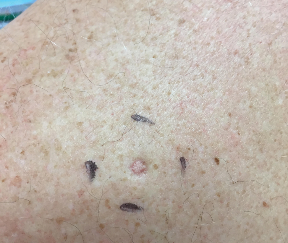

A 64-year-old man presented to dermatology for a full-body skin examination. He had no history of skin cancer. Physical examination revealed an asymptomatic, 4-mm, yellowish pink papule on the left posterior shoulder (top). Dermoscopy revealed yellow globules (bottom). The patient was unsure of the duration of the lesion and denied any prior trauma or medical procedure to the area. Subsequently, a shave biopsy was performed.

Pandemic-related stress rising among ICU clinicians
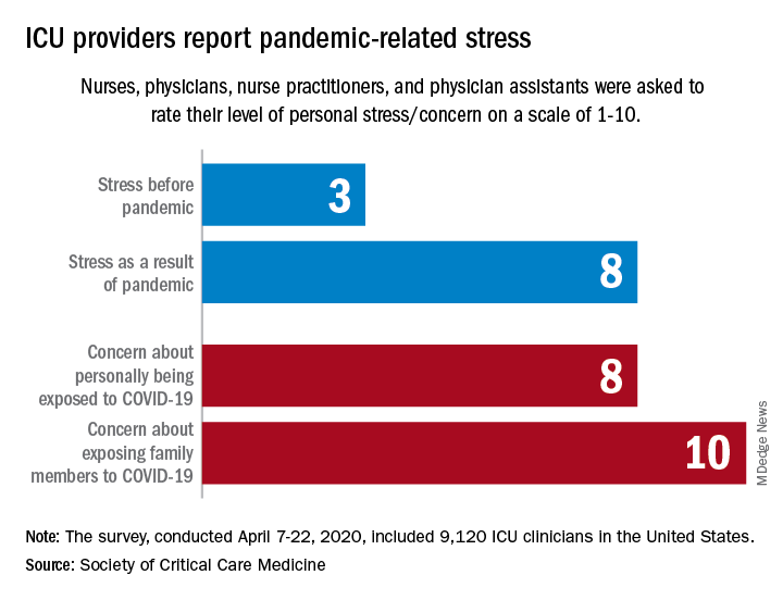
They are worried about getting infected, and they are even more worried about infecting family members, according to the Society for Critical Care Medicine, which surveyed members of four professional organizations – the American Association of Critical-Care Nurses, American College of Chest Physicians, American Thoracic Society, and the SCCM – April 7-22, 2020.
Four items in the survey assessed respondents’ level of stress or concern on a scale of 1-10:
- Personal stress before the COVID-19 pandemic.
- Personal stress as a result of COVID-19 pandemic.
- Concern about personally being exposed to COVID-19.
- Concern about exposing family members to COVID-19.
Personal stress rose from a median of 3 before the pandemic to a current 8, a level that was equaled by personal concerns about being exposed and surpassed (10) by concerns about exposing family members, the SCCM reported in a blog post.
Most of the respondents “are taking special measures to limit the potential spread of the virus to their loved ones, including implementing a decontamination routine before interacting with families,” the SCCM wrote.
The most common strategy, employed by 72% of ICU clinicians, is changing clothes before/after work. Showering before joining family was mentioned by 64% of providers, followed by limiting contact until decontamination (57%) and using hand sanitizer before entering home (51%), the SCCM said.
More extreme measures included self-isolating within their homes (16%) and staying in alternative housing away from their families (12%), the SCCM said, based on data for 9,120 clinicians in the United States.
Most of the respondents (88%) reported having cared for a patient with confirmed or presumed COVID-19. Nurses made up the majority (91%) of the sample, which also included nurse practitioners and physician assistants (4.5%) and physicians (2.9%), as well as smaller numbers of respiratory therapists, pharmacists, and emergency medicine flight personnel.
The results of the survey “underline the personal sacrifices of critical care clinicians during the COVID-19 response and suggest the need to help them proactively manage stress,” the SCCM wrote.
They are worried about getting infected, and they are even more worried about infecting family members, according to the Society for Critical Care Medicine, which surveyed members of four professional organizations – the American Association of Critical-Care Nurses, American College of Chest Physicians, American Thoracic Society, and the SCCM – April 7-22, 2020.
Four items in the survey assessed respondents’ level of stress or concern on a scale of 1-10:
- Personal stress before the COVID-19 pandemic.
- Personal stress as a result of COVID-19 pandemic.
- Concern about personally being exposed to COVID-19.
- Concern about exposing family members to COVID-19.
Personal stress rose from a median of 3 before the pandemic to a current 8, a level that was equaled by personal concerns about being exposed and surpassed (10) by concerns about exposing family members, the SCCM reported in a blog post.
Most of the respondents “are taking special measures to limit the potential spread of the virus to their loved ones, including implementing a decontamination routine before interacting with families,” the SCCM wrote.
The most common strategy, employed by 72% of ICU clinicians, is changing clothes before/after work. Showering before joining family was mentioned by 64% of providers, followed by limiting contact until decontamination (57%) and using hand sanitizer before entering home (51%), the SCCM said.
More extreme measures included self-isolating within their homes (16%) and staying in alternative housing away from their families (12%), the SCCM said, based on data for 9,120 clinicians in the United States.
Most of the respondents (88%) reported having cared for a patient with confirmed or presumed COVID-19. Nurses made up the majority (91%) of the sample, which also included nurse practitioners and physician assistants (4.5%) and physicians (2.9%), as well as smaller numbers of respiratory therapists, pharmacists, and emergency medicine flight personnel.
The results of the survey “underline the personal sacrifices of critical care clinicians during the COVID-19 response and suggest the need to help them proactively manage stress,” the SCCM wrote.
They are worried about getting infected, and they are even more worried about infecting family members, according to the Society for Critical Care Medicine, which surveyed members of four professional organizations – the American Association of Critical-Care Nurses, American College of Chest Physicians, American Thoracic Society, and the SCCM – April 7-22, 2020.
Four items in the survey assessed respondents’ level of stress or concern on a scale of 1-10:
- Personal stress before the COVID-19 pandemic.
- Personal stress as a result of COVID-19 pandemic.
- Concern about personally being exposed to COVID-19.
- Concern about exposing family members to COVID-19.
Personal stress rose from a median of 3 before the pandemic to a current 8, a level that was equaled by personal concerns about being exposed and surpassed (10) by concerns about exposing family members, the SCCM reported in a blog post.
Most of the respondents “are taking special measures to limit the potential spread of the virus to their loved ones, including implementing a decontamination routine before interacting with families,” the SCCM wrote.
The most common strategy, employed by 72% of ICU clinicians, is changing clothes before/after work. Showering before joining family was mentioned by 64% of providers, followed by limiting contact until decontamination (57%) and using hand sanitizer before entering home (51%), the SCCM said.
More extreme measures included self-isolating within their homes (16%) and staying in alternative housing away from their families (12%), the SCCM said, based on data for 9,120 clinicians in the United States.
Most of the respondents (88%) reported having cared for a patient with confirmed or presumed COVID-19. Nurses made up the majority (91%) of the sample, which also included nurse practitioners and physician assistants (4.5%) and physicians (2.9%), as well as smaller numbers of respiratory therapists, pharmacists, and emergency medicine flight personnel.
The results of the survey “underline the personal sacrifices of critical care clinicians during the COVID-19 response and suggest the need to help them proactively manage stress,” the SCCM wrote.
Cardiovascular health among US pregnant women


Keratotic Papule on the Abdomen
The Diagnosis: Hypergranulotic Dyscornification
Hypergranulotic dyscornification (HD) is a rarely reported reaction pattern present in benign solitary keratoses with only few reports to date. It may be an underrecognized reaction pattern based on the paucity of reported cases as well as the histologic similarities to other entities. It has been hypothesized that this pattern reflects an underlying keratin mutation or disorder of keratinization.1
Clinically, HD most commonly presents as a waxy, tan-colored, solitary keratosis generally found on the lower limbs, trunk, or back in individuals aged 20 to 60 years.1,2 Histopathology shows marked hyperkeratosis, papillomatosis, and clumped basophilic keratohyalin granules within the corneocytes with digitated epidermal hyperplasia. There is abnormal cornification across the entire lesion with papillomatosis and marked hypergranulosis.3 There often are homogeneous orthokeratotic mounds of large, dull, eosinophilic-staining anucleate keratinocytes that are sharply demarcated from the thickened granular layer.1,2 Within the spinous, granular, and corneal layers, there is a pale, gray-staining, basophilic, cytoplasmic substance intercellularly.1
Histopathologically, HD may be mistaken for several other entities both benign and malignant.1 Epidermolytic hyperkeratosis can be a genetic disorder, an incidental finding in a variety of skin conditions, or an isolated lesion.4 The genetic syndrome, caused by mutation in keratins 1 or 10, clinically presents with hyperkeratosis, erosions, blisters, and thickening of the epidermis, often with a corrugated appearance. Epidermal nevi findings often are seen in conjunction with histologic changes of epidermolytic hyperkeratosis caused by mutation. Solitary lesions also can resemble seborrheic keratosis or verruca. In all examples of epidermolytic hyperkeratosis, the histopathologic findings are identical.4 The granular layer is thickened, and coarse keratohyalin granules aggregate in the suprabasal cells.5 There is acantholysis with perinuclear vacuolization in the spinous and granular layers with characteristic pale cytoplasmic areas devoid of keratin filaments (Figure 1). The basal layer may be hyperproliferative.5
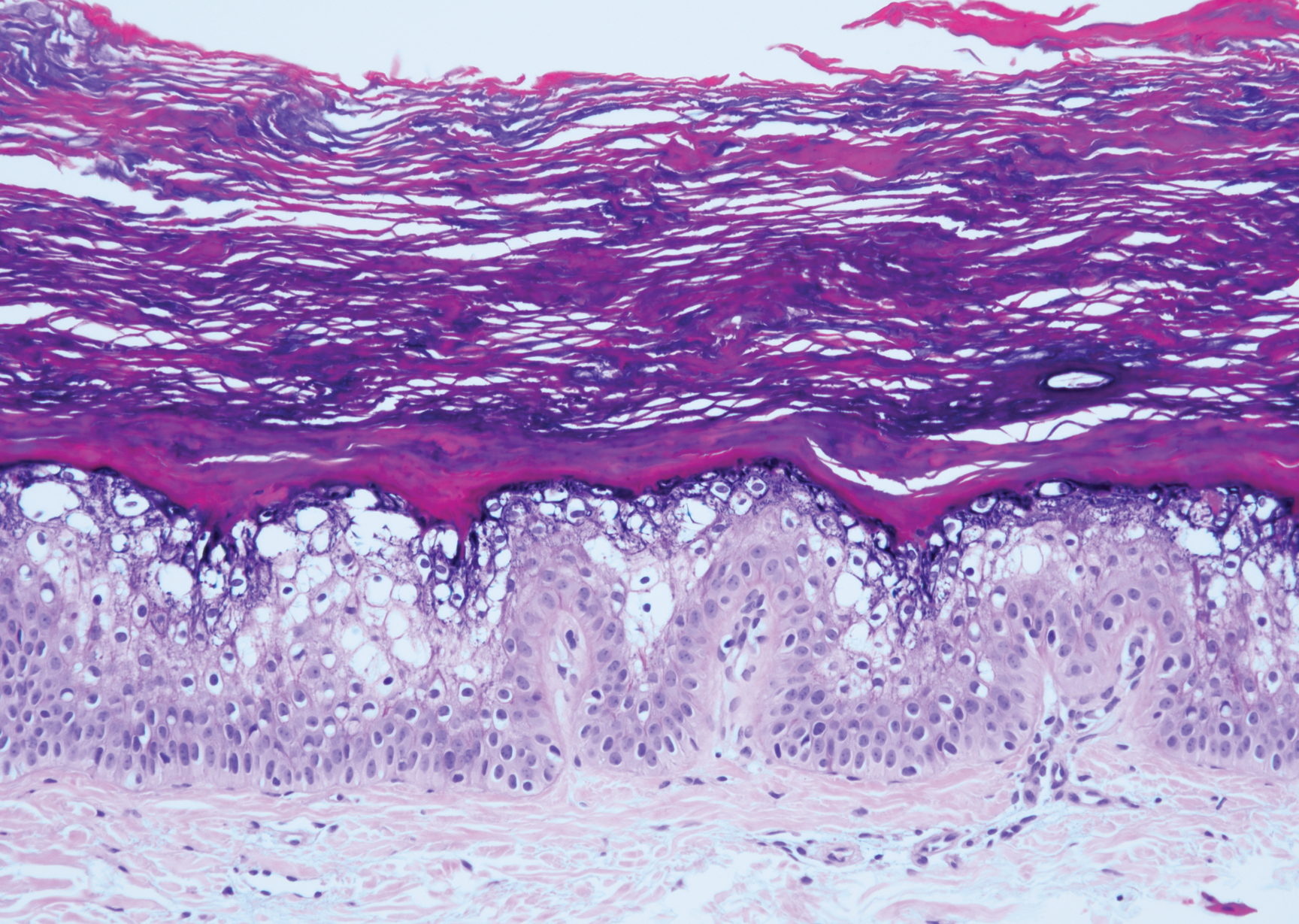
Irritated seborrheic keratosis presents as an exophytic, waxy, dark, sharply demarcated plaque with a stuck-on appearance.6 There is visible keratinization with comedolike openings, fissures and ridges, and scale; it also can contain milialike cysts. Histopathologically there is papillomatosis with prominent rete ridges, often including keratin pseudohorn cysts and squamous eddies. Enlarged capillaries can be seen in the dermal papillae. There is normal cytology with benign sheets of basaloid cells (Figure 2).7 Activating mutation in fibroblast growth factor receptor 3 leads to the growth and thickness of the epidermis that has been identified in these benign lesions.8
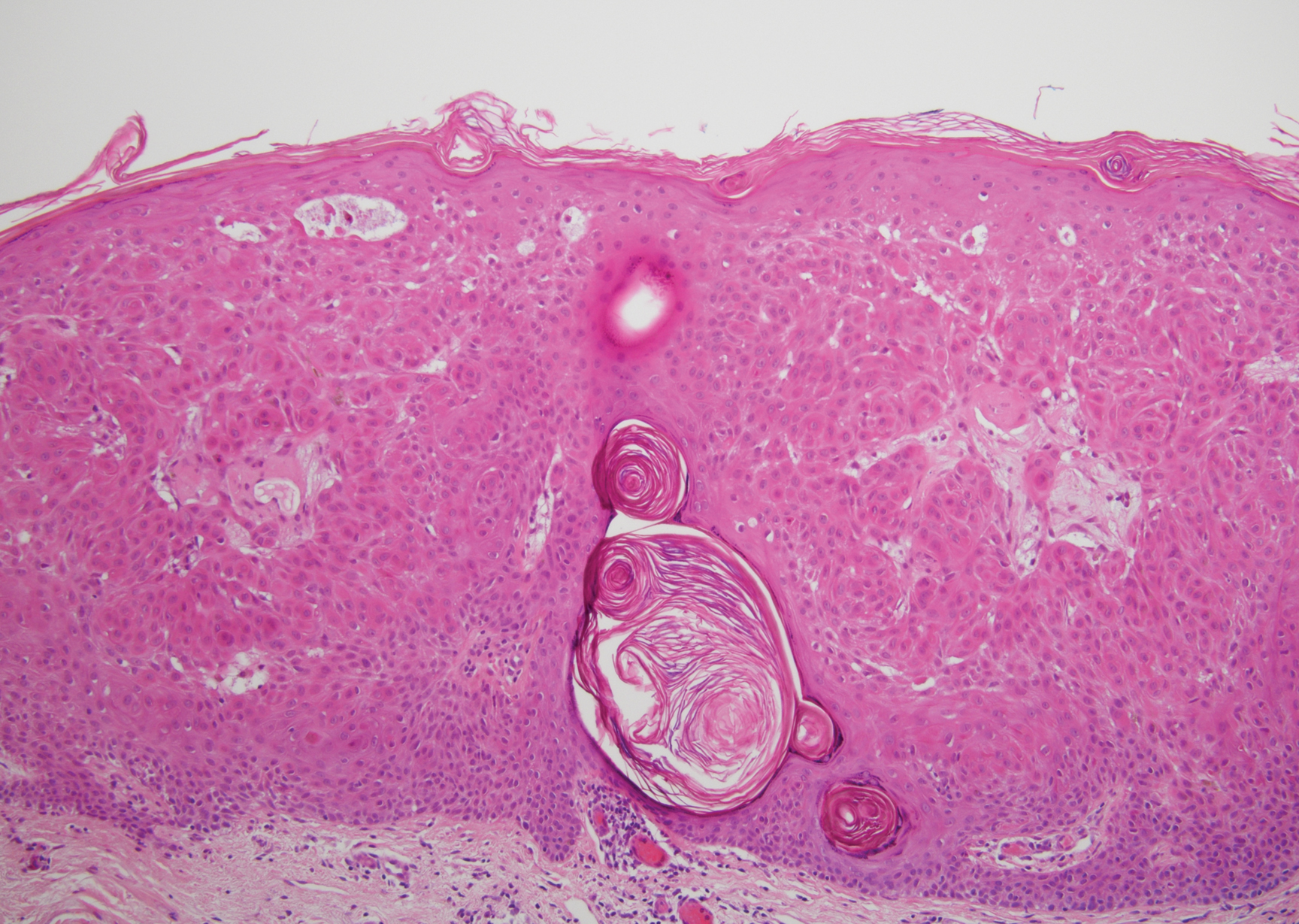
Verruca plana appears as a flesh-colored or reddish, warty, flat-topped papule that often forms clusters. Histopathologically it shows prominent hypergranulosis, thickened stratum spinosum, and vacuolized keratinocytes.9 The nuclei demonstrate a characteristic cytopathic effect of the virion, blurring the nuclear chromatin due to viral particle accumulation, known as koilocytes (Figure 3). The cause is the double-stranded DNA human papillomavirus types 2, 3, and 10.10
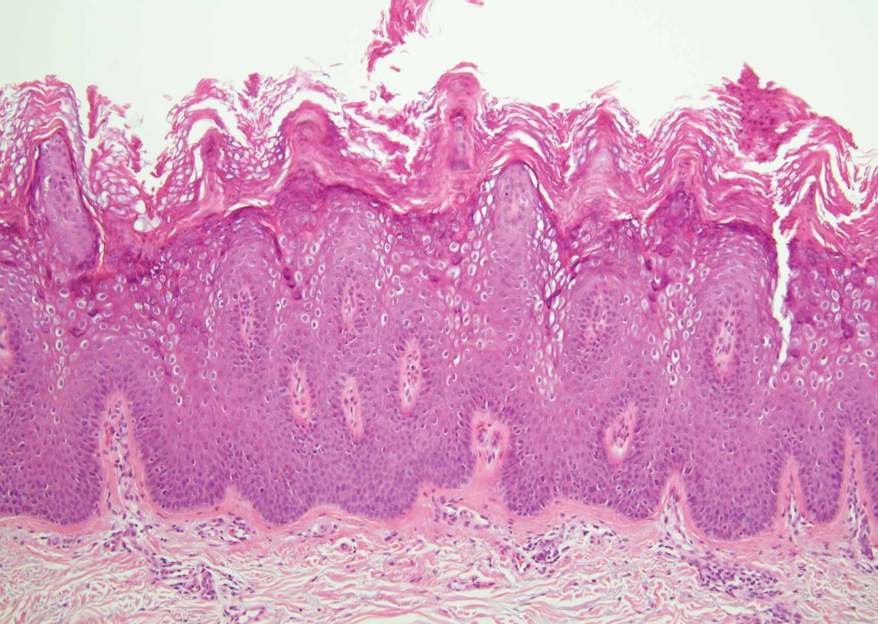
Bowen disease is a form of squamous cell carcinoma in situ characterized by an enlarging, well-demarcated, erythematous plaque with an irregular border and crusting or scaling. Histopathology reveals pleomorphic epidermal keratinization that becomes incorporated in the stratum corneum as parakeratotic nuclei. There is acanthosis, elongation of the rete ridges, and disorganized keratinocytes with atypia.11 The granular and spinous layers show an atypical honeycomb pattern with atypical cellular morphology (Figure 4).12 Bowen disease is a malignant lesion commonly found in older adults on sun-exposed skin that can evolve into invasive squamous cell carcinoma.
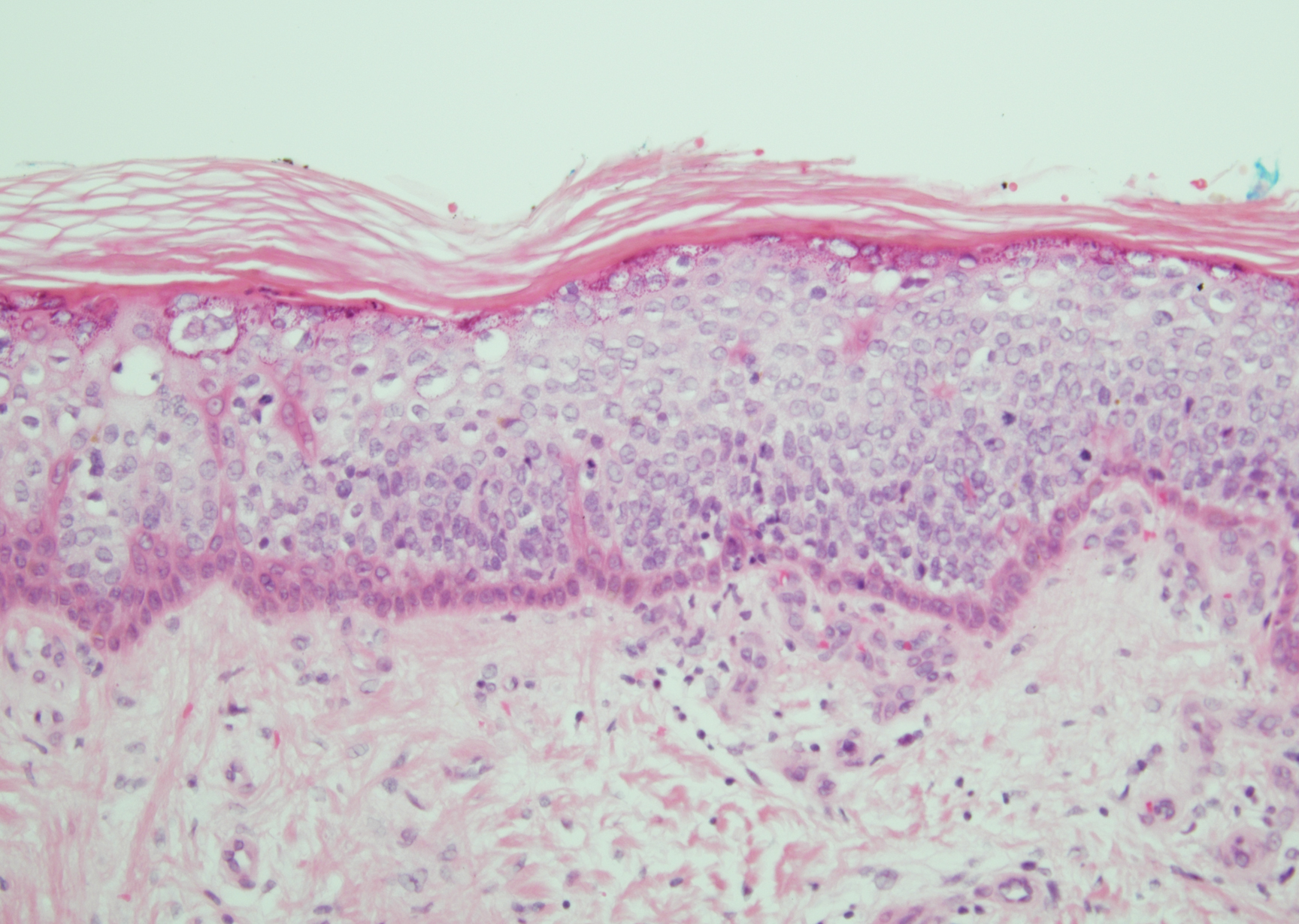
- Roy SF, Ko CJ, Moeckel GW, et al. Hypergranulotic dyscornification: 30 cases of a striking epithelial reaction pattern. J Cutan Pathol. 2019;46:742-747.
- Dohse L, Elston D, Lountzis N, et al. Benign hypergranulotic keratosis with dyscornification. J Am Acad Dermatol. 2010;62:AB52.
- Reichel M. Hypergranulotic dyscornification. Am J Dermatopathol. 1999;21:21-24.
- Kumar P, Kumar R, Kumar Mandal RK, et al. Systematized linear epidermolytic hyperkeratosis. Dermatol Online J. 2014;20:21248.
- Peter Rout D, Nair A, Gupta A, et al. Epidermolytic hyperkeratosis: clinical update. Clin Cosmet Investig Dermatol. 2019;12:333-344.
- Ingraffea A. Benign skin neoplasms. Facial Plast Surg Clin North Am. 2013;21:21-32.
- Braun R. Dermoscopy of pigmented seborrheic keratosis. Arch Dermatol. 2002;138:1556.
- Duperret EK, Oh SJ, McNeal A, et al. Activating FGFR3 mutations cause mild hyperplasia in human skin, but are insufficient to drive benign or malignant skin tumors. Cell Cycle. 2014;13:1551-1559.
- Liu H, Chen S, Zhang F, et al. Seborrheic keratosis or verruca plana? a pilot study with confocal laser scanning microscopy. Skin Res Technol. 2010;16:408-412.
- Prieto-Granada CN, Lobo AZC, Mihm MC. Skin infections. In: Kradin RL, ed. Diagnostic Pathology of Infectious Disease. Philadelphia, PA: Saunders Elsevier; 2010:519-616.
- DeCoste R, Moss P, Boutilier R, et al. Bowen disease with invasive mucin-secreting sweat gland differentiation: report of a case and review of the literature. J Cutan Pathol. 2019;46:425-430.
- Ulrich M, Kanitakis J, González S, et al. Evaluation of Bowen disease by in vivo reflectance confocal microscopy. Br J Dermatol. 2011;166:451-453.
The Diagnosis: Hypergranulotic Dyscornification
Hypergranulotic dyscornification (HD) is a rarely reported reaction pattern present in benign solitary keratoses with only few reports to date. It may be an underrecognized reaction pattern based on the paucity of reported cases as well as the histologic similarities to other entities. It has been hypothesized that this pattern reflects an underlying keratin mutation or disorder of keratinization.1
Clinically, HD most commonly presents as a waxy, tan-colored, solitary keratosis generally found on the lower limbs, trunk, or back in individuals aged 20 to 60 years.1,2 Histopathology shows marked hyperkeratosis, papillomatosis, and clumped basophilic keratohyalin granules within the corneocytes with digitated epidermal hyperplasia. There is abnormal cornification across the entire lesion with papillomatosis and marked hypergranulosis.3 There often are homogeneous orthokeratotic mounds of large, dull, eosinophilic-staining anucleate keratinocytes that are sharply demarcated from the thickened granular layer.1,2 Within the spinous, granular, and corneal layers, there is a pale, gray-staining, basophilic, cytoplasmic substance intercellularly.1
Histopathologically, HD may be mistaken for several other entities both benign and malignant.1 Epidermolytic hyperkeratosis can be a genetic disorder, an incidental finding in a variety of skin conditions, or an isolated lesion.4 The genetic syndrome, caused by mutation in keratins 1 or 10, clinically presents with hyperkeratosis, erosions, blisters, and thickening of the epidermis, often with a corrugated appearance. Epidermal nevi findings often are seen in conjunction with histologic changes of epidermolytic hyperkeratosis caused by mutation. Solitary lesions also can resemble seborrheic keratosis or verruca. In all examples of epidermolytic hyperkeratosis, the histopathologic findings are identical.4 The granular layer is thickened, and coarse keratohyalin granules aggregate in the suprabasal cells.5 There is acantholysis with perinuclear vacuolization in the spinous and granular layers with characteristic pale cytoplasmic areas devoid of keratin filaments (Figure 1). The basal layer may be hyperproliferative.5
Irritated seborrheic keratosis presents as an exophytic, waxy, dark, sharply demarcated plaque with a stuck-on appearance.6 There is visible keratinization with comedolike openings, fissures and ridges, and scale; it also can contain milialike cysts. Histopathologically there is papillomatosis with prominent rete ridges, often including keratin pseudohorn cysts and squamous eddies. Enlarged capillaries can be seen in the dermal papillae. There is normal cytology with benign sheets of basaloid cells (Figure 2).7 Activating mutation in fibroblast growth factor receptor 3 leads to the growth and thickness of the epidermis that has been identified in these benign lesions.8
Verruca plana appears as a flesh-colored or reddish, warty, flat-topped papule that often forms clusters. Histopathologically it shows prominent hypergranulosis, thickened stratum spinosum, and vacuolized keratinocytes.9 The nuclei demonstrate a characteristic cytopathic effect of the virion, blurring the nuclear chromatin due to viral particle accumulation, known as koilocytes (Figure 3). The cause is the double-stranded DNA human papillomavirus types 2, 3, and 10.10
Bowen disease is a form of squamous cell carcinoma in situ characterized by an enlarging, well-demarcated, erythematous plaque with an irregular border and crusting or scaling. Histopathology reveals pleomorphic epidermal keratinization that becomes incorporated in the stratum corneum as parakeratotic nuclei. There is acanthosis, elongation of the rete ridges, and disorganized keratinocytes with atypia.11 The granular and spinous layers show an atypical honeycomb pattern with atypical cellular morphology (Figure 4).12 Bowen disease is a malignant lesion commonly found in older adults on sun-exposed skin that can evolve into invasive squamous cell carcinoma.
The Diagnosis: Hypergranulotic Dyscornification
Hypergranulotic dyscornification (HD) is a rarely reported reaction pattern present in benign solitary keratoses with only few reports to date. It may be an underrecognized reaction pattern based on the paucity of reported cases as well as the histologic similarities to other entities. It has been hypothesized that this pattern reflects an underlying keratin mutation or disorder of keratinization.1
Clinically, HD most commonly presents as a waxy, tan-colored, solitary keratosis generally found on the lower limbs, trunk, or back in individuals aged 20 to 60 years.1,2 Histopathology shows marked hyperkeratosis, papillomatosis, and clumped basophilic keratohyalin granules within the corneocytes with digitated epidermal hyperplasia. There is abnormal cornification across the entire lesion with papillomatosis and marked hypergranulosis.3 There often are homogeneous orthokeratotic mounds of large, dull, eosinophilic-staining anucleate keratinocytes that are sharply demarcated from the thickened granular layer.1,2 Within the spinous, granular, and corneal layers, there is a pale, gray-staining, basophilic, cytoplasmic substance intercellularly.1
Histopathologically, HD may be mistaken for several other entities both benign and malignant.1 Epidermolytic hyperkeratosis can be a genetic disorder, an incidental finding in a variety of skin conditions, or an isolated lesion.4 The genetic syndrome, caused by mutation in keratins 1 or 10, clinically presents with hyperkeratosis, erosions, blisters, and thickening of the epidermis, often with a corrugated appearance. Epidermal nevi findings often are seen in conjunction with histologic changes of epidermolytic hyperkeratosis caused by mutation. Solitary lesions also can resemble seborrheic keratosis or verruca. In all examples of epidermolytic hyperkeratosis, the histopathologic findings are identical.4 The granular layer is thickened, and coarse keratohyalin granules aggregate in the suprabasal cells.5 There is acantholysis with perinuclear vacuolization in the spinous and granular layers with characteristic pale cytoplasmic areas devoid of keratin filaments (Figure 1). The basal layer may be hyperproliferative.5
Irritated seborrheic keratosis presents as an exophytic, waxy, dark, sharply demarcated plaque with a stuck-on appearance.6 There is visible keratinization with comedolike openings, fissures and ridges, and scale; it also can contain milialike cysts. Histopathologically there is papillomatosis with prominent rete ridges, often including keratin pseudohorn cysts and squamous eddies. Enlarged capillaries can be seen in the dermal papillae. There is normal cytology with benign sheets of basaloid cells (Figure 2).7 Activating mutation in fibroblast growth factor receptor 3 leads to the growth and thickness of the epidermis that has been identified in these benign lesions.8
Verruca plana appears as a flesh-colored or reddish, warty, flat-topped papule that often forms clusters. Histopathologically it shows prominent hypergranulosis, thickened stratum spinosum, and vacuolized keratinocytes.9 The nuclei demonstrate a characteristic cytopathic effect of the virion, blurring the nuclear chromatin due to viral particle accumulation, known as koilocytes (Figure 3). The cause is the double-stranded DNA human papillomavirus types 2, 3, and 10.10
Bowen disease is a form of squamous cell carcinoma in situ characterized by an enlarging, well-demarcated, erythematous plaque with an irregular border and crusting or scaling. Histopathology reveals pleomorphic epidermal keratinization that becomes incorporated in the stratum corneum as parakeratotic nuclei. There is acanthosis, elongation of the rete ridges, and disorganized keratinocytes with atypia.11 The granular and spinous layers show an atypical honeycomb pattern with atypical cellular morphology (Figure 4).12 Bowen disease is a malignant lesion commonly found in older adults on sun-exposed skin that can evolve into invasive squamous cell carcinoma.
- Roy SF, Ko CJ, Moeckel GW, et al. Hypergranulotic dyscornification: 30 cases of a striking epithelial reaction pattern. J Cutan Pathol. 2019;46:742-747.
- Dohse L, Elston D, Lountzis N, et al. Benign hypergranulotic keratosis with dyscornification. J Am Acad Dermatol. 2010;62:AB52.
- Reichel M. Hypergranulotic dyscornification. Am J Dermatopathol. 1999;21:21-24.
- Kumar P, Kumar R, Kumar Mandal RK, et al. Systematized linear epidermolytic hyperkeratosis. Dermatol Online J. 2014;20:21248.
- Peter Rout D, Nair A, Gupta A, et al. Epidermolytic hyperkeratosis: clinical update. Clin Cosmet Investig Dermatol. 2019;12:333-344.
- Ingraffea A. Benign skin neoplasms. Facial Plast Surg Clin North Am. 2013;21:21-32.
- Braun R. Dermoscopy of pigmented seborrheic keratosis. Arch Dermatol. 2002;138:1556.
- Duperret EK, Oh SJ, McNeal A, et al. Activating FGFR3 mutations cause mild hyperplasia in human skin, but are insufficient to drive benign or malignant skin tumors. Cell Cycle. 2014;13:1551-1559.
- Liu H, Chen S, Zhang F, et al. Seborrheic keratosis or verruca plana? a pilot study with confocal laser scanning microscopy. Skin Res Technol. 2010;16:408-412.
- Prieto-Granada CN, Lobo AZC, Mihm MC. Skin infections. In: Kradin RL, ed. Diagnostic Pathology of Infectious Disease. Philadelphia, PA: Saunders Elsevier; 2010:519-616.
- DeCoste R, Moss P, Boutilier R, et al. Bowen disease with invasive mucin-secreting sweat gland differentiation: report of a case and review of the literature. J Cutan Pathol. 2019;46:425-430.
- Ulrich M, Kanitakis J, González S, et al. Evaluation of Bowen disease by in vivo reflectance confocal microscopy. Br J Dermatol. 2011;166:451-453.
- Roy SF, Ko CJ, Moeckel GW, et al. Hypergranulotic dyscornification: 30 cases of a striking epithelial reaction pattern. J Cutan Pathol. 2019;46:742-747.
- Dohse L, Elston D, Lountzis N, et al. Benign hypergranulotic keratosis with dyscornification. J Am Acad Dermatol. 2010;62:AB52.
- Reichel M. Hypergranulotic dyscornification. Am J Dermatopathol. 1999;21:21-24.
- Kumar P, Kumar R, Kumar Mandal RK, et al. Systematized linear epidermolytic hyperkeratosis. Dermatol Online J. 2014;20:21248.
- Peter Rout D, Nair A, Gupta A, et al. Epidermolytic hyperkeratosis: clinical update. Clin Cosmet Investig Dermatol. 2019;12:333-344.
- Ingraffea A. Benign skin neoplasms. Facial Plast Surg Clin North Am. 2013;21:21-32.
- Braun R. Dermoscopy of pigmented seborrheic keratosis. Arch Dermatol. 2002;138:1556.
- Duperret EK, Oh SJ, McNeal A, et al. Activating FGFR3 mutations cause mild hyperplasia in human skin, but are insufficient to drive benign or malignant skin tumors. Cell Cycle. 2014;13:1551-1559.
- Liu H, Chen S, Zhang F, et al. Seborrheic keratosis or verruca plana? a pilot study with confocal laser scanning microscopy. Skin Res Technol. 2010;16:408-412.
- Prieto-Granada CN, Lobo AZC, Mihm MC. Skin infections. In: Kradin RL, ed. Diagnostic Pathology of Infectious Disease. Philadelphia, PA: Saunders Elsevier; 2010:519-616.
- DeCoste R, Moss P, Boutilier R, et al. Bowen disease with invasive mucin-secreting sweat gland differentiation: report of a case and review of the literature. J Cutan Pathol. 2019;46:425-430.
- Ulrich M, Kanitakis J, González S, et al. Evaluation of Bowen disease by in vivo reflectance confocal microscopy. Br J Dermatol. 2011;166:451-453.
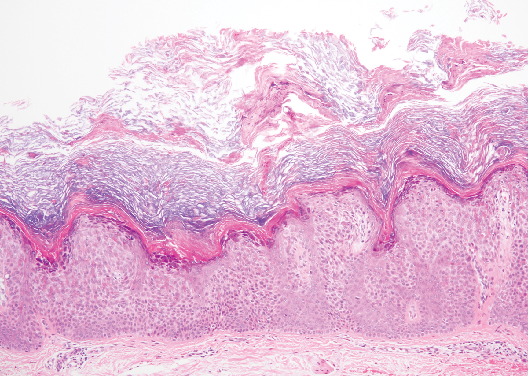
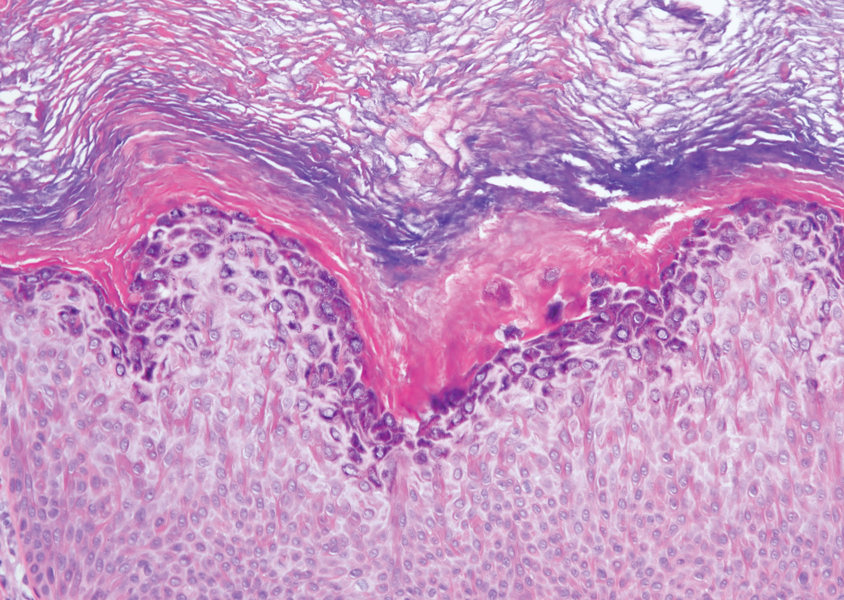
A 59-year-old woman with a history of basal cell carcinoma, uterine and ovarian cancer, and verrucae presented with an asymptomatic 3-mm lesion on the left side of the lower abdomen. Physical examination revealed a waxy, tan-colored, solitary keratosis. A shave biopsy was performed. Histopathology showed hyperkeratosis, focal parakeratosis, papillomatosis, and marked hypergranulosis with pale gray cytoplasm of the spinous-layer keratinocytes.