User login
The Journal of Clinical Outcomes Management® is an independent, peer-reviewed journal offering evidence-based, practical information for improving the quality, safety, and value of health care.
div[contains(@class, 'header__large-screen')]
div[contains(@class, 'read-next-article')]
div[contains(@class, 'nav-primary')]
nav[contains(@class, 'nav-primary')]
section[contains(@class, 'footer-nav-section-wrapper')]
footer[@id='footer']
div[contains(@class, 'main-prefix')]
section[contains(@class, 'nav-hidden')]
div[contains(@class, 'ce-card-content')]
nav[contains(@class, 'nav-ce-stack')]
Diet high in plant omega-3s tied to better HF prognosis
Heart failure (HF) patients with high serum levels of alpha-linolenic acid (ALA) had a better prognosis than those with the lowest levels, in an observational study.
ALA is an omega-3 fatty acid that is found mainly in plants, including flaxseed, chia, walnuts, or canola oil.

“The most striking finding to us is the clear difference between patients at the bottom quartile compared to the other 75%, pointing to a threshold on the putative effect of ALA, reinforcing the notion that ‘one size does not fill all,’ ” Aleix Sala-Vila, PharmD, PhD, of the Hospital del Mar Medical Research Institute, Barcelona, told this news organization.The analysis, which was published online in the Journal of the American College of Cardiology, showed statistically significant reductions in all-cause death, cardiovascular (CV) death, and first HF hospitalization among those in the three upper quartiles of serum ALA levels, compared with those in the lowest quartile.
The team’s earlier finding that higher levels of serum phosphatidylcholine eicosapentaenoic acid (PC EPA) and ALA were associated with a lower risk of adverse events in patients with ST-segment elevation myocardial infarction prompted the current study, Dr. Sala-Vila said.
Although their findings are hypothesis-generating at this point, he added, “inclusion of some ALA-rich foods, such as walnuts, in the diet of any individual, whether they have HF or not, might translate into CV benefits, besides the putative effect on HF. There is no evidence of any deleterious effect of one daily serving of walnuts, not even on weight gain.”
Plant power
Dr. Sala-Vila and colleagues analyzed data and samples from 905 patients (mean age, 67; 32% women) with HF of different etiologies. ALA was assessed by gas chromatography in serum phospholipids, which reflect long-term dietary ALA intake and metabolism.
The primary outcome was a composite of all-cause death or first HF hospitalization. The secondary outcome was the composite of CV death or HF hospitalization.
After a median follow-up of 2.4 years, 140 all-cause deaths, 85 CV deaths, and 141 first HF hospitalizations occurred (composite of all-cause death and first HF hospitalization, 238; composite of CV death and HF hospitalization, 184).
Compared with patients at the lowest quartile of ALA in serum phospholipids, those at the three upper quartiles showed a 39% reduction in the risk of the primary endpoint (hazard ratio, 0.61).
Statistically significant reductions also were observed for all-cause death (HR, 0.58), CV death (HR, 0.51), first HF hospitalization (HR, 0.58), and the composite of CV death and HF hospitalization (HR, 0.58).
By contrast, nonstatistically significant associations were seen for fish-derived EPA, DHA, and the sum of EPA + DHA.
Limitations of the study include its observational nature; a relatively young cohort with reduced or mid-range ejection fraction and stage 2 chronic kidney disease; and no dietary data except for those regarding fatty acids.
“Controversial results from landmark recent trials on omega-3 might have translated into confusion/negative impact on the reputation of these fatty acids,” Dr. Sala-Vila noted. “Many factors affect how each participant responds to a certain intervention (precision nutrition), such as genetics, the microbiome, and the environment. In this regard, nutritional status – omega-3 background – is emerging as a key determinant.”
Randomized trials needed
JoAnn E. Manson, MD, MPH, DrPH, chief of the Division of Preventive Medicine at Brigham and Women’s Hospital, Boston, said the findings “are promising in the context of earlier research on omega-3s.”
Those studies include the landmark GISSI-HF trial, a randomized, controlled trial (RCT) that showed a small benefit of n-3 polyunsaturated fatty acids regarding hospital admissions and mortality among patients with chronic HF, and her team’s VITAL-HF study, which showed a significant reduction in recurrent HF hospitalization with marine omega-3 supplementation versus placebo.
“This may not be a causal association, and the authors acknowledge that they don’t have information on other dietary factors,” Dr. Manson said. “It may be that the foods that are leading to this higher blood level of ALA comprise the type of plant-based diet that’s been linked to lower risk of CVD, such as the Mediterranean diet. The findings also could be the result of other factors that aren’t fully controlled for in the analysis, or the participants could be more compliant with their medications.”
Nevertheless, she said, “it’s reasonable to recommend that people with a history of HF or who are at high risk of HF increase their intake of ALA-enriched foods, including canola oil, flaxseed oils, soybeans and soybean oils, and walnuts.”
“I think the evidence is promising enough that an RCT of ALA in people with heart failure also would be reasonable,” she added.
Similarly, Abdallah Al-Mohammad, MD, of Northern General Hospital, Sheffield, England, writes in a related editorial that while a potential role for ALA in improving morbidity and mortality in HF patients cannot be substantiated yet, the findings “open the field to more questions” for which “the judge and jury ... shall be prospective randomized controlled trials.”
No commercial funding or relevant conflicts of interest were declared.
A version of this article first appeared on Medscape.com.
Heart failure (HF) patients with high serum levels of alpha-linolenic acid (ALA) had a better prognosis than those with the lowest levels, in an observational study.
ALA is an omega-3 fatty acid that is found mainly in plants, including flaxseed, chia, walnuts, or canola oil.
“The most striking finding to us is the clear difference between patients at the bottom quartile compared to the other 75%, pointing to a threshold on the putative effect of ALA, reinforcing the notion that ‘one size does not fill all,’ ” Aleix Sala-Vila, PharmD, PhD, of the Hospital del Mar Medical Research Institute, Barcelona, told this news organization.The analysis, which was published online in the Journal of the American College of Cardiology, showed statistically significant reductions in all-cause death, cardiovascular (CV) death, and first HF hospitalization among those in the three upper quartiles of serum ALA levels, compared with those in the lowest quartile.
The team’s earlier finding that higher levels of serum phosphatidylcholine eicosapentaenoic acid (PC EPA) and ALA were associated with a lower risk of adverse events in patients with ST-segment elevation myocardial infarction prompted the current study, Dr. Sala-Vila said.
Although their findings are hypothesis-generating at this point, he added, “inclusion of some ALA-rich foods, such as walnuts, in the diet of any individual, whether they have HF or not, might translate into CV benefits, besides the putative effect on HF. There is no evidence of any deleterious effect of one daily serving of walnuts, not even on weight gain.”
Plant power
Dr. Sala-Vila and colleagues analyzed data and samples from 905 patients (mean age, 67; 32% women) with HF of different etiologies. ALA was assessed by gas chromatography in serum phospholipids, which reflect long-term dietary ALA intake and metabolism.
The primary outcome was a composite of all-cause death or first HF hospitalization. The secondary outcome was the composite of CV death or HF hospitalization.
After a median follow-up of 2.4 years, 140 all-cause deaths, 85 CV deaths, and 141 first HF hospitalizations occurred (composite of all-cause death and first HF hospitalization, 238; composite of CV death and HF hospitalization, 184).
Compared with patients at the lowest quartile of ALA in serum phospholipids, those at the three upper quartiles showed a 39% reduction in the risk of the primary endpoint (hazard ratio, 0.61).
Statistically significant reductions also were observed for all-cause death (HR, 0.58), CV death (HR, 0.51), first HF hospitalization (HR, 0.58), and the composite of CV death and HF hospitalization (HR, 0.58).
By contrast, nonstatistically significant associations were seen for fish-derived EPA, DHA, and the sum of EPA + DHA.
Limitations of the study include its observational nature; a relatively young cohort with reduced or mid-range ejection fraction and stage 2 chronic kidney disease; and no dietary data except for those regarding fatty acids.
“Controversial results from landmark recent trials on omega-3 might have translated into confusion/negative impact on the reputation of these fatty acids,” Dr. Sala-Vila noted. “Many factors affect how each participant responds to a certain intervention (precision nutrition), such as genetics, the microbiome, and the environment. In this regard, nutritional status – omega-3 background – is emerging as a key determinant.”
Randomized trials needed
JoAnn E. Manson, MD, MPH, DrPH, chief of the Division of Preventive Medicine at Brigham and Women’s Hospital, Boston, said the findings “are promising in the context of earlier research on omega-3s.”
Those studies include the landmark GISSI-HF trial, a randomized, controlled trial (RCT) that showed a small benefit of n-3 polyunsaturated fatty acids regarding hospital admissions and mortality among patients with chronic HF, and her team’s VITAL-HF study, which showed a significant reduction in recurrent HF hospitalization with marine omega-3 supplementation versus placebo.
“This may not be a causal association, and the authors acknowledge that they don’t have information on other dietary factors,” Dr. Manson said. “It may be that the foods that are leading to this higher blood level of ALA comprise the type of plant-based diet that’s been linked to lower risk of CVD, such as the Mediterranean diet. The findings also could be the result of other factors that aren’t fully controlled for in the analysis, or the participants could be more compliant with their medications.”
Nevertheless, she said, “it’s reasonable to recommend that people with a history of HF or who are at high risk of HF increase their intake of ALA-enriched foods, including canola oil, flaxseed oils, soybeans and soybean oils, and walnuts.”
“I think the evidence is promising enough that an RCT of ALA in people with heart failure also would be reasonable,” she added.
Similarly, Abdallah Al-Mohammad, MD, of Northern General Hospital, Sheffield, England, writes in a related editorial that while a potential role for ALA in improving morbidity and mortality in HF patients cannot be substantiated yet, the findings “open the field to more questions” for which “the judge and jury ... shall be prospective randomized controlled trials.”
No commercial funding or relevant conflicts of interest were declared.
A version of this article first appeared on Medscape.com.
Younger doctors call for more attention to patients with disabilities
As an undergraduate student at Northeastern University in Boston, Meghan Chin spent her summers working for a day program in Rhode Island. Her charges were adults with various forms of intellectual and developmental disabilities (IDD).
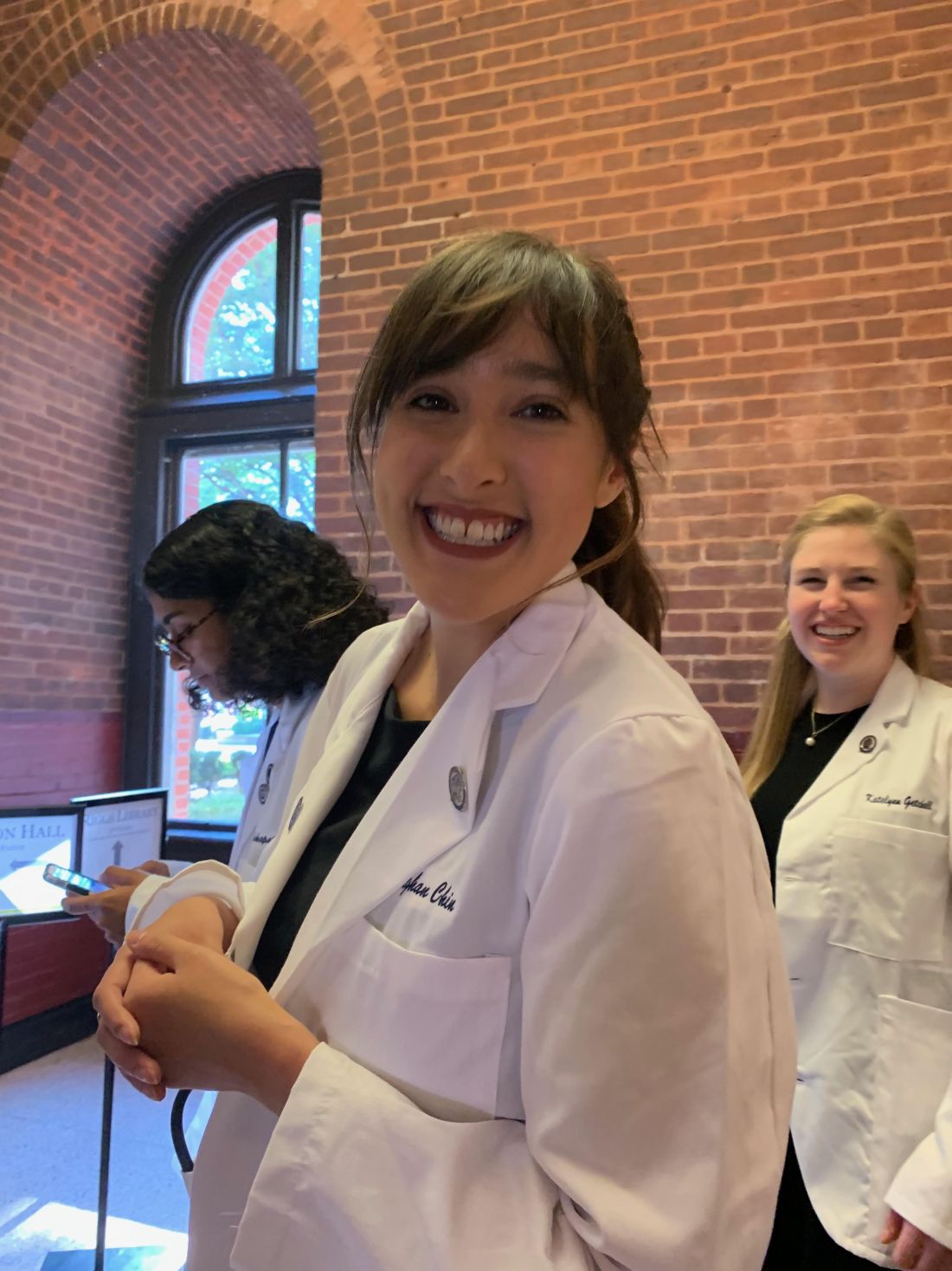
“I was very much a caretaker,” Ms. Chin, now 29, said. “It was everything from helping them get dressed in the morning to getting them to medical appointments.”
During one such visit Ms. Chin got a lesson about how health care looks from the viewpoint of someone with an IDD.
The patient was a woman in her 60s and she was having gastrointestinal issues; symptoms she could have articulated, if asked. “She was perfectly capable of telling a clinician where it hurt, how long she had experienced the problem, and what she had done or not done to alleviate it,” Ms. Chin said.
And of comprehending a response. But she was not given the opportunity.
“She would explain what was going on to the clinician,” Ms. Chin recalled. “And the clinician would turn to me and answer. It was this weird three-way conversation – as if she wasn’t even there in the room with us.”
Ms. Chin was incensed at the rude and disrespectful way the patient had been treated. But her charge didn’t seem upset or surprised. Just resigned. “Sadly, she had become used to this,” Ms. Chin said.
For the young aide, however, the experience was searing. “It didn’t seem right to me,” Ms. Chin said. “That’s why, when I went to medical school, I knew I wanted to do better for this population.”

Serendipity led her to Georgetown University, Washington, where she met Kim Bullock, MD, one of the country’s leading advocates for improved health care delivery to those with IDDs.
Dr. Bullock, an associate professor of family medicine, seeks to create better training and educational opportunities for medical students who will likely encounter patients with these disabilities in their practices.
When Dr. Bullock heard Ms. Chin’s story about the patient being ignored, she was not surprised.
“This is not an unusual or unique situation,” said Dr. Bullock, who is also director of Georgetown’s community health division and a faculty member of the university’s Center for Excellence for Developmental Disabilities. “In fact, it’s quite common and is part of what spurred my own interest in educating pre-med and medical students about effective communication techniques, particularly when addressing neurodiverse patients.”
More than 13% of Americans, or roughly 44 million people, have some form of disability, according to the National Institute on Disability at the University of New Hampshire, a figure that does not include those who are institutionalized. The Centers for Disease Control and Prevention estimates that 17% of children aged 3-17 years have a developmental disability.
Even so, many physicians feel ill-prepared to care for disabled patients. A survey of physicians, published in the journal Health Affairs, found that some lacked the resources and training to properly care for patients with disabilities, or that they struggled to coordinate care for such individuals. Some said they did not know which types of accessible equipment, like adjustable tables and chair scales, were needed or how to use them. And some said they actively try to avoid treating patients with disabilities.
Don’t assume
The first step at correcting the problem, Dr. Bullock said, is to not assume that all IDD patients are incapable of communicating. By talking not to the patient but to their caregiver or spouse or child, as the clinician did with Ms. Chin years ago, “we are taking away their agency, their autonomy to speak for and about themselves.”
Change involves altering physicians’ attitudes and assumptions toward this population, through education. But how?
“The medical school curriculum is tight as it is,” Dr. Bullock acknowledged. “There’s a lot of things students have to learn. People wonder: where we will add this?”
Her suggestion: Incorporate IDD all along the way, through programs or experiences that will enable medical students to see such patients “not as something separate, but as people that have special needs just as other populations have.”
Case in point: Operation House Call, a program in Massachusetts designed to support young health care professionals, by building “confidence, interest, and sensitivity” toward individuals with IDD.
Eight medical and allied health schools, including those at Harvard Medical School and Yale School of Nursing, participate in the program, the centerpiece of which is time spent by teams of medical students in the homes of families with neurodiverse members. “It’s transformational,” said Susan Feeney, DNP, NP-C, director of adult gerontology and family nurse practitioner programs at the graduate school of nursing at the University of Massachusetts, Worcester. “They spend a few hours at the homes of these families, have this interaction with them, and journal about their experiences.”
Dr. Feeney described as “transformational” the experience of the students after getting to know these families. “They all come back profoundly changed,” she told this news organization. “As a medical or health care professional, you meet people in an artificial environment of the clinic and hospital. Here, they become human, like you. It takes the stigma away.”
One area of medicine in which this is an exception is pediatrics, where interaction with children with IDD and their families is common – and close. “They’re going to be much more attuned to this,” Dr. Feeney said. “The problem is primary care or internal medicine. Once these children get into their mid and later 20s, and they need a practitioner to talk to about adult concerns.”
And with adulthood come other medical needs, as the physical demands of age fall no less heavily on individuals with IDDs than those without. For example: “Neurodiverse people get pregnant,” Dr. Bullock said. They also can get heart disease as they age; or require the care of a rheumatologist, a neurologist, an orthopedic surgeon, or any other medical specialty.
Generation gap
Fortunately, the next generation of physicians may be more open to this more inclusionary approach toward a widely misunderstood population.
Like Ms. Chin, Sarah Bdeir had experience with this population prior to beginning her training in medicine. She had volunteered at a school for people with IDD.
“It was one of the best experiences I’ve ever had,” Ms. Bdeir, now 23 and a first-year medical student at Wayne State University, Detroit, said. She found that the neurodiverse individuals she worked with had as many abilities as disabilities. “They are capable of learning, but they do it differently,” she said. “You have to adjust to the way they learn. And you have to step out of your own box.”
Ms. Bdeir also heard about Dr. Bullock’s work and is assisting her in a research project on how to better improve nutritional education for people with IDDs. And although she said it may take time for curriculum boards at medical schools to integrate this kind of training into their programs, she believes they will, in part because the rising cohort of medical students today have an eagerness to engage with and learn more about IDD patients.
As does Ms. Chin.
“When I talk to my peers about this, they’re very receptive,” Ms. Chin said. “They want to learn how to better support the IDD population. And they will learn. I believe in my generation of future doctors.”
A version of this article first appeared on Medscape.com.
As an undergraduate student at Northeastern University in Boston, Meghan Chin spent her summers working for a day program in Rhode Island. Her charges were adults with various forms of intellectual and developmental disabilities (IDD).
“I was very much a caretaker,” Ms. Chin, now 29, said. “It was everything from helping them get dressed in the morning to getting them to medical appointments.”
During one such visit Ms. Chin got a lesson about how health care looks from the viewpoint of someone with an IDD.
The patient was a woman in her 60s and she was having gastrointestinal issues; symptoms she could have articulated, if asked. “She was perfectly capable of telling a clinician where it hurt, how long she had experienced the problem, and what she had done or not done to alleviate it,” Ms. Chin said.
And of comprehending a response. But she was not given the opportunity.
“She would explain what was going on to the clinician,” Ms. Chin recalled. “And the clinician would turn to me and answer. It was this weird three-way conversation – as if she wasn’t even there in the room with us.”
Ms. Chin was incensed at the rude and disrespectful way the patient had been treated. But her charge didn’t seem upset or surprised. Just resigned. “Sadly, she had become used to this,” Ms. Chin said.
For the young aide, however, the experience was searing. “It didn’t seem right to me,” Ms. Chin said. “That’s why, when I went to medical school, I knew I wanted to do better for this population.”
Serendipity led her to Georgetown University, Washington, where she met Kim Bullock, MD, one of the country’s leading advocates for improved health care delivery to those with IDDs.
Dr. Bullock, an associate professor of family medicine, seeks to create better training and educational opportunities for medical students who will likely encounter patients with these disabilities in their practices.
When Dr. Bullock heard Ms. Chin’s story about the patient being ignored, she was not surprised.
“This is not an unusual or unique situation,” said Dr. Bullock, who is also director of Georgetown’s community health division and a faculty member of the university’s Center for Excellence for Developmental Disabilities. “In fact, it’s quite common and is part of what spurred my own interest in educating pre-med and medical students about effective communication techniques, particularly when addressing neurodiverse patients.”
More than 13% of Americans, or roughly 44 million people, have some form of disability, according to the National Institute on Disability at the University of New Hampshire, a figure that does not include those who are institutionalized. The Centers for Disease Control and Prevention estimates that 17% of children aged 3-17 years have a developmental disability.
Even so, many physicians feel ill-prepared to care for disabled patients. A survey of physicians, published in the journal Health Affairs, found that some lacked the resources and training to properly care for patients with disabilities, or that they struggled to coordinate care for such individuals. Some said they did not know which types of accessible equipment, like adjustable tables and chair scales, were needed or how to use them. And some said they actively try to avoid treating patients with disabilities.
Don’t assume
The first step at correcting the problem, Dr. Bullock said, is to not assume that all IDD patients are incapable of communicating. By talking not to the patient but to their caregiver or spouse or child, as the clinician did with Ms. Chin years ago, “we are taking away their agency, their autonomy to speak for and about themselves.”
Change involves altering physicians’ attitudes and assumptions toward this population, through education. But how?
“The medical school curriculum is tight as it is,” Dr. Bullock acknowledged. “There’s a lot of things students have to learn. People wonder: where we will add this?”
Her suggestion: Incorporate IDD all along the way, through programs or experiences that will enable medical students to see such patients “not as something separate, but as people that have special needs just as other populations have.”
Case in point: Operation House Call, a program in Massachusetts designed to support young health care professionals, by building “confidence, interest, and sensitivity” toward individuals with IDD.
Eight medical and allied health schools, including those at Harvard Medical School and Yale School of Nursing, participate in the program, the centerpiece of which is time spent by teams of medical students in the homes of families with neurodiverse members. “It’s transformational,” said Susan Feeney, DNP, NP-C, director of adult gerontology and family nurse practitioner programs at the graduate school of nursing at the University of Massachusetts, Worcester. “They spend a few hours at the homes of these families, have this interaction with them, and journal about their experiences.”
Dr. Feeney described as “transformational” the experience of the students after getting to know these families. “They all come back profoundly changed,” she told this news organization. “As a medical or health care professional, you meet people in an artificial environment of the clinic and hospital. Here, they become human, like you. It takes the stigma away.”
One area of medicine in which this is an exception is pediatrics, where interaction with children with IDD and their families is common – and close. “They’re going to be much more attuned to this,” Dr. Feeney said. “The problem is primary care or internal medicine. Once these children get into their mid and later 20s, and they need a practitioner to talk to about adult concerns.”
And with adulthood come other medical needs, as the physical demands of age fall no less heavily on individuals with IDDs than those without. For example: “Neurodiverse people get pregnant,” Dr. Bullock said. They also can get heart disease as they age; or require the care of a rheumatologist, a neurologist, an orthopedic surgeon, or any other medical specialty.
Generation gap
Fortunately, the next generation of physicians may be more open to this more inclusionary approach toward a widely misunderstood population.
Like Ms. Chin, Sarah Bdeir had experience with this population prior to beginning her training in medicine. She had volunteered at a school for people with IDD.
“It was one of the best experiences I’ve ever had,” Ms. Bdeir, now 23 and a first-year medical student at Wayne State University, Detroit, said. She found that the neurodiverse individuals she worked with had as many abilities as disabilities. “They are capable of learning, but they do it differently,” she said. “You have to adjust to the way they learn. And you have to step out of your own box.”
Ms. Bdeir also heard about Dr. Bullock’s work and is assisting her in a research project on how to better improve nutritional education for people with IDDs. And although she said it may take time for curriculum boards at medical schools to integrate this kind of training into their programs, she believes they will, in part because the rising cohort of medical students today have an eagerness to engage with and learn more about IDD patients.
As does Ms. Chin.
“When I talk to my peers about this, they’re very receptive,” Ms. Chin said. “They want to learn how to better support the IDD population. And they will learn. I believe in my generation of future doctors.”
A version of this article first appeared on Medscape.com.
IV potassium and magnesium an acute treatment for AFib?
Compared with no treatment, potassium and magnesium administration was associated with a 10% higher rate of SVC.
The finding suggests that giving intravenous potassium and magnesium might lessen the need for antiarrhythmic therapy and the associated potential adverse effects in patients with nonpermanent atrial fibrillation (AFib), the study authors say.
Still, they add, “The results of our study have no direct implications for clinical practice in the management of care for patients with AF [atrial fibrillation] or AFL [atrial flutter] in the ED. The findings are purely exploratory and hypothesis-generating but could potentially provide a rationale for an appropriate prospective trial.”
The study was published online in JAMA Network Open.
“Atrial fibrillation is becoming an increasing burden for health care systems worldwide owing to population aging,” write Filippo Cacioppo, MD, and colleagues from Medical University of Vienna (Austria).
“Pharmacologic and electrical conversion are common therapies in emergency departments, especially for highly symptomatic patients. Each intervention has specific risks, and neither is considered cost-effective owing to frequent recurrence of AF. In addition, AF often terminates spontaneously,” Dr. Cacioppo and colleagues write.
They add that evidence suggests hypokalemia and hypomagnesemia contribute to AFib development, and so the administration of potassium and magnesium could be a reasonable strategy to improve SCV rates.
To test their hypothesis, Dr. Cacioppo and associates conducted a registry-based cohort study in all patients with AFib or AFL presenting to their center’s ED between Feb. 6, 2009, and Feb. 16, 2020.
During this time, they observed a total of 2,546 episodes of nonpermanent AFib. The median patient age was 68 years (interquartile range, 58-75 years). Most were men (n = 1,411 patients, 55.4%).
In addition, there were 573 episodes of nonpermanent AFL. The median patient age was 68 years (IQR, 58-75 years), and 332 patients (57.9%) were men.
Intravenous potassium and magnesium were administered in just over half (n = 1,763; 56.5%) of the episodes.
The median amount of potassium and magnesium was delivered via one 250-mL infusion bag, which consisted of 24 mEq potassium and 145.8 mg magnesium combined with 500 mL of balanced crystalloid fluid containing 2.5 mEq potassium and 18.2 mg magnesium, administered for 90 minutes, the authors write.
If patients experienced pain at the injection site, the infusion rate was reduced until the pain subsided.
Conversion to sinus rhythm was considered spontaneous if no attempt at pharmacologic rhythm control was made until conversion occurred; if SCV occurred after an unsuccessful attempt at electrical cardioversion; or following rate control with beta-blockers, nondihydropyridine calcium channel blockers, or digitalis glycosides, the authors state.
IV treatment increased odds of SCV
The median duration of stay in the ED was 6.4 hours (IQR, 3.9-11.6 hours) for patients with AFib and 6.1 hours (IQR, 3.9-11.8 hours) for patients with AFL.
During the stay in the ED, SCV occurred in 15.4% (n = 393) of AFib episodes and 12.7% (n = 73) of AFL episodes.
Intravenous potassium and magnesium increased the possibility of SCV compared with no IV potassium and magnesium in AFib, but not in AFL.
In episodes of AFib, administration of intravenous potassium and magnesium was associated with 19.2% increased odds of SCV, compared with 10.4% with no administration (odds ratio, 1.98; 95% CI, 1.53-2.57).
In contrast, for AFL, no association was observed for the probability of SCV with potassium and magnesium administration when compared with no administration (13.0% vs. 12.5%; OR, 1.05; 95% CI, 0.65-1.69).
Not in the guidelines
“To date, it is unclear whether potassium and magnesium administration might be reasonable in the acute treatment of AF and AFL, and although this intervention may be common practice in some EDs, it is not part of the treatment recommendations in current guidelines,” Dr. Cacioppo and colleagues write.
“Our findings suggest that intravenous potassium and magnesium administration may increase the chance of SCV in patients with AF with either hypokalemia or with plasma potassium levels in the range of 3.50 to 3.99 mEq/L. In patients with AFL, however, potassium and magnesium administration may not be associated with SCV probability,” they write.
Dr. Cacioppo and associates add that in their study IV administration of potassium and magnesium was associated with SCV only in patients with symptom onset of less than 48 hours, suggesting a time-dependent outcome. However, they caution, “because only a limited number of patients with SCV had symptom onset greater than or equal to 48 hours, this finding warrants further investigation.”
A Band-Aid approach
“I’m a little skeptical about this study,” Georgios Syros, MD, director of arrhythmia services at Mount Sinai Queens and Mount Sinai Brooklyn, New York, said in an interview.
“Atrial fibrillation is a chronic disease. The natural history of this disease is that it is paroxysmal in the beginning, and at some point the episodes become more frequent and longer in duration. For some people, at some point, it becomes permanent,” Dr. Syros said.
“Suppose I cut my finger while slicing bread. I put a Band-Aid on the cut. That doesn’t mean I have fixed it, it means I’ve helped it temporarily. Atrial fibrillation in this paper is very analogous,” he said. “The patient may have episodes, goes to the emergency room, you give them medication, and temporarily alleviate the situation so that the patient does not have to be admitted. It’s simple, inexpensive, you make the heart rate go back to normal, not permanently, with few side effects, except perhaps for some pain at the injection site, but that doesn’t mean you have fixed the AFib permanently. But for someone who has had a first incidence, or doesn’t want to stay in the hospital because it’s the weekend, yes, you can use this as a Band-Aid,” he said.
Intravenous potassium and magnesium, as proposed in the current study, is similar to a medication currently in use in Europe, called vernakalant, Dr. Syros said.
“Vernakalant is not FDA approved in the U.S. It is not meant to treat atrial fibrillation permanently, so we have to inform the public about the limitations of what we are doing,” he said. “Vernakalant is similar to IV potassium and magnesium, as given in this study, but it is more expensive. It temporarily allows people to go back to sinus rhythm, but it’s not going to be there forever and you may go back to permanent AFib, so this is not magic, unfortunately.”
Dr. Syros emphasized that the current study results apply only to cases of paroxysmal atrial fibrillation of less than 48 hours duration. “This is a very important distinction,” he said.
“For example, a patient who drank a lot and the day after is in AFib, with what we call holiday heart, would be a good candidate for the treatment in this study. He’s young, without any heart damage, no diabetes, no hypertension, no prior stroke, so sure, help him out with potassium and magnesium, provided that he can prove to us that this started within 48 hours,” Dr. Syros said.
Dr. Cacioppo and colleagues and Dr. Syros report no relevant financial relationships. Study corresponding author Jan Niederdoeckl, MD, PhD, obtained funding for the study.
A version of this article first appeared on Medscape.com.
FROM JAMA NETWORK OPEN
A better way to predict fall risk in patients with MS?
Compared with patients with MS who didn’t fall, those who did fall had worse neuromuscular function as evidenced by a reduced rate of force development.
“Our study suggests that instead of looking at reduced maximum muscle strength, perhaps we should start looking at reduced rate of force development when trying to identify potential fallers,” said Laurits Taul-Madsen, PhD student, Aarhus University, Denmark.
The study was presented at the annual meeting of the European Committee for Treatment and Research in Multiple Sclerosis (ECTRIMS).
Explosive strength
In contrast to maximal muscle strength, the rate of force development is a measure of explosive strength, or simply the amount of force that an individual can produce over a given time period. When a patient is about to fall, what’s most important is not how strong the person is, but how quickly they can produce enough force to counteract the balance perturbation, thus avoid falling, said Dr. Taul-Madsen.
“If a person is very slow to produce this force, [that person] will have fallen before he or she has produced enough force to counteract the balance perturbation that the person is experiencing,” he added.
Research has shown a reduced rate of force development (RFD) in patients with MS, compared with healthy controls. However, little is known about the impact of RFD on falls in those with MS.
To investigate, researchers studied 53 adults with MS: Twenty-four had no fall history in the prior year, 16 had one to two prior falls, and 13 had three or more falls. The two groups of fallers were both slightly older and had a slightly higher Expanded Disability Status Scale (EDSS) scores, “which may not be so surprising,” Dr. Taul-Madsen said.
Knee extensor neuromuscular function, including maximum muscle strength and RFD at 50 and 200 milliseconds, was assessed via isokinetic dynamometry.
A high RFD is “good and the non-fallers had the highest RFD at 50 ms.” On this measure, “we saw quite a big difference between the non-fallers and the two groups of fallers,” Dr. Taul-Madsen reported.
At 200 ms, the RFD was again highest in the group of non-fallers but the difference was somewhat smaller. Non-fallers also had greater maximum muscle strength than that of the fallers.
There was “good” correlation between these neuromuscular measurements and falls, Dr. Taul-Madsen said.
He noted that RFD, which can be improved with resistance training, “seems like a specialized and difficult measurement, but it doesn’t have to be. It can be measured with just a linear encoder and a chair to perform the sit-to-stand test, so in clinical practice, it’s quite easily measured.”
‘Highly promising’ approach
“There are some data on predictors of falls in persons with MS, but not yet on neuromuscular function, as has been done in other populations,” said Brian Sandroff, PhD, senior research scientist, Kessler Foundation, West Orange, N.J.
This study is “interesting in that recurrent fallers were distinguished based on having worse neuromuscular function,” said Dr. Sandroff, who was not part of the research team.
“Although this relationship is somewhat intuitive,” RFD provides a “potentially sensitive measure that can be addressed via specific resistance exercise programs as a highly promising approach for reducing fall risk and falls themselves in persons with MS,” Dr. Sandroff said.
More generally, he said this study provides “more evidence on the multisystemic benefits of exercise training and having better physical fitness in persons with MS.
“The evidence seems to be converging more and more on this, as research groups across countries and continents are reporting on similar themes,” said Dr. Sandroff.
The study had no specific funding. Dr. Taul-Madsen and Dr. Sandroff report no relevant financial relationships.
A version of this article first appeared on Medscape.com.
FROM ECTRIMS 2022
Listen up: Birdsong may calm anxiety, paranoia
Investigators found that people who listened to recordings of birds singing experienced a significant reduction in anxiety and paranoia. In contrast, the researchers also found that recordings of traffic noises, including car engines, sirens, and construction, increased depressive states.
“The results suggest that it may be worthwhile to investigate the targeted use of natural sounds such as birdsong in a clinical setting – for example, in hospital waiting rooms or in psychiatric settings,” study investigator Emil Stobbe, MSc, a predoctoral fellow at the Max Planck Institute for Human Development, Berlin, said in an interview.
“If someone is seeking an easily accessible intervention to lower distress, listening to an audio clip of birds singing might be a great option,” he added.
The study was published online in Scientific Reports.
Nature’s calming effect
The aim of the research was “to investigate how the physical environment impact brain and mental health,” Mr. Stobbe said.
Mr. Stobbe said that there is significantly more research examining visual properties of the physical environment but that the auditory domain is not as well researched, although, he added, that the beneficial effects of interactions with nature are “well studied.”
He noted that anxiety and paranoia can be experienced by many individuals even though they may be unaware that they are experiencing these states.
“We wanted to investigate if the beneficial effects of nature can also exert their impact on these states. In theory, birds can be representational for natural and vital environment, which, in turn, transfer the positive effects of nature on birdsong listeners,” he said.
A previous study compared nature versus city soundscape conditions and showed that the nature soundscape improved participants’ cognitive performance but did not improve mood. The present study added diversity to the soundscapes and focused not only on cognition and general mood but also on state paranoia, “which can be measured in a change-sensitive manner” and “has been shown to increase in response to traffic noise.”
The researchers hypothesized that birdsong would have a greater beneficial effect on mood and paranoia and on cognitive performance compared with traffic noise. They also investigated whether greater versus lower diversity of bird species or noise sources within the soundscapes “would be a relevant factor modulating the effects.”
The researchers recruited participants (n = 295) from a crowdsourcing platform. Participants’ mean age was late 20s (standard deviations ranged from 6.30 to 7.72), with a greater proportion of male versus female participants.
To be included, participants were required to have no history of mental illness, hearing difficulties, substance/drug intake, or suicidal thoughts/tendencies.
The outcomes of interest (mood, paranoia, cognitive performance) were measured before and after soundscape exposure and each soundscape had a low- versus high-diversity version. This resulted in several analyses that compared two types of sounds (birdsongs vs. traffic noise) x two levels of diversity (low vs. high diversity) and two time points (pre- vs. post exposure).
The exposure to sounds lasted for 6 minutes, after which they were asked to report (on a 0-100 visual scale) how diverse/monotone, beautiful, and pleasant they perceived the soundscape to be.
Reduction in depressive symptoms
Participants were divided into four groups: low-diversity traffic noise soundscape (n = 83), high-diversity traffic noise soundscape (n = 60), low-diversity birdsong soundscape (n = 63), and high-diversity birdsong soundscape (n = 80)
In addition to listening to the sounds, participants completed questionnaires measuring mood (depression and anxiety) and paranoia as well as a test of digit span cognitive performance (both the forward and the backward versions).
The type, diversity, and type x diversity all revealed significant effect sizes (F[3, 276] = 78.6; P < .001; eta-squared = 0.461; F[3, 276] = 3.16; P = .025; eta-squared = 0.033; and F[3, 276] = 2.66; P = .028, respectively), “suggesting that all of these factors, as well as their interaction, had a significant impact on the perception of soundscapes (that is, ratings on monotony/diversity, beauty, and pleasantness).”
A post hoc examination showed that depressive symptoms significantly increased within the low- and high-diversity urban soundscapes but decreased significantly in the high-diversity birdsong soundscapes (T[1, 60] = –2.57; P = .012; d = –0.29).
For anxiety, the post hoc within-group analyses found no effects within low- and high-diversity traffic noise conditions (T[1, 82] = –1.37; P = .174; d = –0.15 and T[1, 68] = 0.49; P = .629; d = 0.06, respectively). By contrast, there were significant declines in both birdsong conditions (low diversity: T[1, 62] = –6.13; P < .001; d = –0.77; high diversity: T[1, 60] = –6.32; P < .001; d = –0.70).
Similarly, there were no changes in participants with paranoia when they listened to either low- or high-diversity traffic noises (T[1, 82] = –0.55; P = .583; d = –0.06 and T[1, 68] = 0.67; P = .507; d = 0.08, respectively). On the other hand, both birdsong conditions yielded reductions in paranoia (low diversity: T[1, 62] = –5.90; P < .001; d = –0.74; high diversity: T[1, 60] = –4.11; P < .001; d = –0.46).
None of the soundscapes had any effect on cognition.
“In theory, birds can be representational for natural and vital environments which, in turn, transfer the positive effects of nature on birdsong listeners,” said Mr. Stobbe.
“Taken together, the findings of the current study provide another facet of why interactions with nature can be beneficial for our mental health, and it is highly important to preserve nature,” he added.
Mr. Stobbe said that future research should focus on investigating mixed soundscapes including examining whether the presence of natural sounds in urban settings lower stressors such as traffic noise.
An understudied area
Commenting for this article, Ken Duckworth, MD, chief medical officer of the National Alliance on Mental Illness called the study “interesting but limited.”
Dr. Duckworth, who was not involved in the research said that the “benefits of nature are understudied” and agreed with the investigators that it is potentially important to study the use of birdsongs in psychiatric facilities. “Future studies could also correlate the role of birdsong with the mental health benefits/aspects of ‘being in nature,’ which has been found to have some effect.”
Open Access funding was enabled and organized by Projekt DEAL. The authors and Dr. Duckworth declared no competing interests.
A version of this article first appeared on Medscape.com.
FROM SCIENTIFIC REPORTS
I’m a physician battling long COVID. I can assure you it’s real
One in 5. It almost seems unimaginable that this is the real number of people who are struggling with long COVID, especially considering how many people in the United States have had COVID-19 at this point (more than 96 million).
Even more unimaginable at this time is that it’s happening to me. I’ve experienced not only the disabling effects of long COVID, but I’ve also seen, firsthand, the frustration of navigating diagnosis and treatment. It’s given me a taste of what millions of other patients are going through.
Vaxxed, masked, and (too) relaxed
I caught COVID-19 (probably Omicron BA.5) that presented as sniffles, making me think it was probably just allergies. However, my resting heart rate was up on my Garmin watch, so of course I got tested and was positive.
With my symptoms virtually nonexistent, it seemed, at the time, merely an inconvenience, because I was forced to isolate away from family and friends, who all stayed negative.
But 2 weeks later, I began to have urticaria – hives – after physical exertion. Did that mean my mast cells were angry? There’s some evidence these immune cells become overactivated in some patients with COVID. Next, I began to experience lightheadedness and the rapid heartbeat of tachycardia. The tachycardia was especially bad any time I physically exerted myself, including on a walk. Imagine me – a lover of all bargain shopping – cutting short a trip to the outlet mall on a particularly bad day when my heart rate was 140 after taking just a few steps. This was orthostatic intolerance.
Then came the severe worsening of my migraines – which are often vestibular, making me nauseated and dizzy on top of the throbbing.
I was of course familiar with these symptoms, as professor and chair of the department of rehabilitation medicine at the Joe R. and Teresa Lozano Long School of Medicine at University of Texas Health Science Center, San Antonio. I developed a post-COVID recovery clinic to help patients.
So I knew about postexertional malaise (PEM) and postexertional symptom exacerbation (PESE), but I was now experiencing these distressing symptoms firsthand.
Clinicians really need to look for this cardinal sign of long COVID as well as evidence of myalgic encephalomyelitis or chronic fatigue syndrome (ME/CFS). ME/CFS is marked by exacerbation of fatigue or symptoms after an activity that could previously be done without these aftereffects. In my case, as an All-American Masters miler with several marathons under my belt, running 5 miles is a walk in the park. But now, I pay for those 5 miles for the rest of the day on the couch or with palpitations, dizziness, and fatigue the following day. Busy clinic day full of procedures? I would have to be sitting by the end of it. Bed by 9 PM was not always early enough.
Becoming a statistic
Here I am, one of the leading experts in the country on caring for people with long COVID, featured in the national news and having testified in front of Congress, and now I am part of that lived experience. Me – a healthy athlete, with no comorbidities, a normal BMI, vaccinated and boosted, and after an almost asymptomatic bout of COVID-19, a victim to long COVID.
You just never know how your body is going to react. Neuroinflammation occurred in studies with mice with mild respiratory COVID and could be happening to me. I did not want a chronic immune-mediated vasculopathy.
So, I did what any other hyperaware physician-researcher would do. I enrolled in the RECOVER trial – a study my own institution is taking part in and one that I recommend to my own patients.
I also decided that I need to access care and not just ignore my symptoms or try to treat them myself.
That’s when things got difficult. There was a wait of at least a month to see my primary care provider – but I was able to use my privileged position as a physician to get in sooner.
My provider said that she had limited knowledge of long COVID, and she hesitated to order some of the tests and treatments that I recommended because they were not yet considered standard of care. I can understand the hesitation. It is engrained in medical education to follow evidence based on the highest-quality research studies. We are slowly learning more about long COVID, but acknowledging the learning curve offers little to patients who need help now.
This has made me realize that we cannot wait on an evidence-based approach – which can take decades to develop – while people are suffering. And it’s important that everyone on the front line learn about some of the manifestations and disease management of long COVID.
I left this first physician visit feeling more defeated than anything and decided to try to push through. That, I quickly realized, was not the right thing to do.
So again, after a couple of significant crashes and days of severe migraines, I phoned a friend: Ratna Bhavaraju-Sanka, MD, the amazing neurologist who treats patients with long COVID alongside me. She squeezed me in on a non-clinic day. Again, I had the privilege to see a specialist most people wait half a year to see. I was diagnosed with both autonomic dysfunction and intractable migraine.
She ordered some intravenous fluids and IV magnesium that would probably help both. But then another obstacle arose. My institution’s infusion center is focused on patients with cancer, and I was unable to schedule treatments there.
Luckily, I knew about the concierge mobile IV hydration therapy companies that come to your house – mostly offering a hangover treatment service. And I am thankful that I had the health literacy and financial ability to pay for some fluids at home.
On another particularly bad day, I phoned other friends – higher-ups at the hospital – who expedited a slot at the hospital infusion center and approval for the IV magnesium.
Thanks to my access, knowledge, and other privileges, I got fairly quick if imperfect care, enrolled in a research trial, and received medications. I knew to pace myself. The vast majority of others with long COVID lack these advantages.
The patient with long COVID
Things I have learned that others can learn, too:
- Acknowledge and recognize that long COVID is a disease that is affecting 1 in 5 Americans who catch COVID. Many look completely “normal on the outside.” Please listen to your patients.
- Autonomic dysfunction is a common manifestation of long COVID. A 10-minute stand test goes a long way in diagnosing this condition, from the American Academy of Physical Medicine and Rehabilitation. It is not just anxiety.
- “That’s only in research” is dismissive and harmful. Think outside the box. Follow guidelines. Consider encouraging patients to sign up for trials.
- Screen for PEM/PESE and teach your patients to pace themselves, because pushing through it or doing graded exercises will be harmful.
- We need to train more physicians to treat postacute sequelae of SARS-CoV-2 infection () and other postinfectious conditions, such as ME/CFS.
If long COVID is hard for physicians to understand and deal with, imagine how difficult it is for patients with no expertise in this area.
It is exponentially harder for those with fewer resources, time, and health literacy. My lived experience with long COVID has shown me that being a patient is never easy. You put your body and fate into the hands of trusted professionals and expect validation and assistance, not gaslighting or gatekeeping.
Along with millions of others, I am tired of waiting.
Dr. Gutierrez is Professor and Distinguished Chair, department of rehabilitation medicine, University of Texas Health Science Center at San Antonio. She reported receiving honoraria for lecturing on long COVID and receiving a research grant from Co-PI for the NIH RECOVER trial.
A version of this article first appeared on Medscape.com.
Terlipressin decreases need for renal replacement therapy in liver transplant recipients
In a subgroup of patients with hepatorenal syndrome type 1 (HRS) who received a liver transplant, terlipressin treatment appears to reduce the need for renal replacement therapy (RRT) through 12 months of follow-up, according to a study presented at the annual meeting of the American College of Gastroenterology.
Among transplant recipients, overall 12-month survival was 11% higher for those treated with terlipressin compared with placebo, said K. Rajender Reddy, MD, director of hepatology and medical director of liver transplantation at the University of Pennsylvania, Philadelphia.
“Hepatorenal syndrome type 1 is a potentially reversible form of acute kidney injury that occurs in the setting of end-stage liver disease,” he said.
Liver transplantation, which eliminates end-stage liver disease, is the only definitive treatment for HRS. However, renal replacement therapy is common and associated with poor clinical outcomes and low patient survival rates in both the pretransplant and posttransplant settings, he noted.
Terlipressin, an injectable synthetic vasopressin analogue, restores renal blood flow and reverses HRS in 20%-40% of patients, Dr. Reddy said. In September, the U.S. Food and Drug Administration approved terlipressin (Terlivaz) for patients with HRS type 1. The label has a boxed warning for serious or fatal respiratory failure.
The safety and efficacy were assessed in the phase 3 CONFIRM trial, which Dr. Reddy and colleagues previously published. The randomized, placebo-controlled study demonstrated that terlipressin reversed HRS and reduced the need for RRT through day 30. The reversal of HRS with terlipressin did not improve 90-day survival as compared with placebo, which researchers attributed to a higher death rate within 90 days after the first dose despite improved kidney function.
A closer look at the liver transplant patients
In the subgroup analysis of the CONFIRM study, Dr. Reddy and colleagues analyzed the clinical outcomes through 12 months of follow-up in patients with HRS who received a liver transplant. They looked at the incidence of verified HRS reversal, HRS reversal, need for RRT, and overall survival.
Verified HRS reversal was defined as two consecutive serum creatinine measurements of 1.5 mg/dL or less at least 2 hours apart up to day 14 and survival without RRT for at least 10 days. HRS reversal was defined as a serum creatinine level of 1.5 mg/dL or less while on treatment. In addition, the need for RRT and overall survival were assessed at days 30, 60, 90, 180, and 365.
RRT was defined as any procedure that replaced nonendocrine kidney function, including continuous hemofiltration and hemodialysis, intermittent hemodialysis, peritoneal dialysis, ultrafiltration, or other dialysis and filtration techniques.
In the CONFIRM study, 199 patients with HRS were treated with terlipressin plus albumin, and 101 patients were treated with placebo plus albumin for up to 14 days. In the terlipressin group, 46 patients received liver transplants within the first 2 months of the study, as did 29 in the placebo group. Two patients in the terlipressin group and one in the placebo group received a simultaneous liver-kidney transplant.
Meaningful clinical outcomes
In the 12-month follow-up subgroup analysis, verified HRS reversal was statistically comparable between the groups, with a 30% decrease in the terlipressin group and 17% decrease in the placebo group, Dr. Reddy reported.
HRS reversal was higher in the terlipressin group, at 37%, as compared with 14% in the placebo group.
The pretransplant need for RRT was lower in the terlipressin group, at 30%, as compared with 62% in the placebo group. The posttransplant need for RRT remained numerically lower in the terlipressin group at all time points and was significantly lower at day 180 and day 365.
Overall survival for transplant recipients in the terlipressin group was 94%, as compared with 83% in the placebo group. Posttreatment adverse events and severe adverse events were similar between the groups.
“Collectively, these data indicate that terlipressin treatment in patients with HRS led to better long-term clinical outcomes in those who received a liver transplant,” Dr. Reddy said.
The study was funded by Mallinckrodt Pharmaceuticals, which manufactures terlipressin. One author is an employee of Mallinckrodt, and the other authors have served in an advisory role or received grant support from Mallinckrodt. The authors also disclosed consultant roles and research support from several other pharmaceutical companies.
FROM ACG 2022
High-quality index colonoscopies pay off down the road for low-risk patients
CHARLOTTE, N.C.– Performing high-quality index colonoscopies may pay off later in your patients’ reduced risk for advanced neoplasia, investigators report.
A study of registry data on more than 2,200 patients who had an index colonoscopy showing no evidence of neoplasia found that, on repeat colonoscopy 10 years later, the absolute risk for advanced neoplasia outcomes was lower for those with a high-quality index exam, compared with those who had a lesser-quality index colonoscopy.
The adjusted odds ratio for patients who underwent high-quality index exams was 0.59%, reported Joseph Anderson, MD, from the Geisel School of Medicine at Dartmouth, Hanover, N.H.
“These data demonstrate that high-quality index colonoscopy provides better protection from interval lesions than low-quality exams with no polyps detected at that index exam,” he said in an oral abstract presentation at the annual meeting of the American College of Gastroenterology.
“These data support the importance of high-quality index exams in the prevention of interval colorectal cancer, and support the 10-year interval for normal exams,” Dr. Anderson added.
He recommended that endoscopists focus on the quality of their exams by using adequate scope withdrawal time – 8-10 minutes – to ensure optimal adenoma detection, and by ensuring the use of optimal bowel preparation in their practices.
Registry study
Dr. Anderson and colleagues studied how the quality of index colonoscopies could affect the risk of advanced outcomes in low-risk patients at the 10-year or later follow-up. They used records from the New Hampshire Colonoscopy Registry, which includes data from 2004 to the present on more than 250,000 exams performed by more than 150 endoscopists in more than 30 Granite State practices.
The investigators also looked at data on patients with less than 5 years of follow-up, and those with follow-up from 5 to less than 10 years.
The study sample included patients with no adenoma or significant serrated polyps on their index exams who had at least one follow-up exam 12 months or more after the index exams. Patients with inflammatory bowel disease or familial colon cancer syndromes were excluded.
They defined a high-quality colonoscopy as an exam complete to cecum, with adequate bowel preparation, and performed by an endoscopist with an adenoma detection rate of 25 or higher.
The adenoma detection rate is calculated as the number of screening colonoscopies with at least one adenoma divided by the total number of screening colonoscopies.
The definition of advanced outcomes included advanced adenomas, colorectal cancer, and/or large serrated polyps (1 cm or greater).
Of the 14,011 patients in the sample, 2,283 had a follow-up exam at 10 years. The absolute risk for advanced outcomes among patients who had a high quality index exam was 4.0% vs. 6.7% for those with lower quality exams.
Among patients with low-quality index exams – but not patients with high quality exams – there was a statistically significant increase in the absolute risk for advanced outcomes at all time periods, from 5.1% in the less than 5-year follow-up group, to 6.7% in the 10-years or more follow-up group.
Patients with initial high-quality exams also had a lower risk for postcolonoscopy colorectal cancer, compared with patients who had low-quality index exams: 0.4% vs. 0.8%. This difference translated into an adjusted hazard ratio for colorectal cancer after a high-quality exam of 0.53.
It’s getting better all the time
In an interview, Daniel J. Pambianco, MD, FACG from Charlottesville (Va.) Gastroenterology Associates, who was not involved in the study, commented that Dr. Anderson and colleagues highlighted the importance of the quality of the bowel prep and the quality of the examination itself.
He noted that the use of devices such as colonoscopy caps can help further improve adenoma detection rates and pointed to up-and-coming developments such as the use of artificial intelligence algorithms to aid human endoscopists.
Dr. Pambianco comoderated the session where the data were presented.
The investigators did not report a study funding source. Dr. Anderson and Dr. Pambianco reported having no relevant financial disclosures.
AT ACG 2022
Has the pandemic affected babies’ brain development?
There’s some good overall news in a large analysis that looked at whether a mother’s COVID-19 infection or birth during the pandemic could affect a baby’s brain development.
Researchers studied 21,419 infants who had neurodevelopmental screening during the pandemic (from January 2020 to January 2021) and compared them with babies born before the pandemic (2015-2019).
They found in an analysis of eight studies that, generally, brain development in infants ages 6-12 months old was not changed by COVID-19.
Communication skill scores lower than prepandemic
However, one area did see a significant difference when they looked at answers to the Ages and Stages Questionnaire, 3rd edition (ASQ-3): Scores were lower in communication skills.
Compared with the prepandemic babies, the pandemic group of babies was more likely to have communication impairment (odds were 1.7 times higher).
Additionally, mothers’ SARS-CoV-2 infection was not associated with significant differences in any neurodevelopment sector in offspring, with one exception: Odds were 3.5 times higher for fine motor impairment in the pandemic baby group.
The babies in this study were either exposed in the womb to the SARS-CoV-2 infection or screened during the pandemic regardless of whether they were exposed to the virus.
The study, led by Kamran Hessami, MD, with the Maternal Fetal Care Center at Boston Children’s Hospital and Harvard Medical School in Boston, was published in JAMA Network Open.
Potential reasons for lower communication skills
The study points to some factors of the pandemic that may be tied to impaired communication skills.
“Higher levels of COVID-19–related stress were reported for both mothers and fathers of infants aged 0-6 months and were associated with insensitive parenting practices, including decreased emotional responsiveness in only mothers, which could lessen the reciprocal exchanges that support language development in early childhood,” they write. “Additionally, opportunities to promote language and social development through new experiences outside the home, including visits with extended family and friends or attendance at a child care center, were lessened for many during the pandemic.”
Viviana M. Fajardo Martinez, MD, with neonatal/perinatal medicine at University of California, Los Angeles, Health, told this publication her team is also studying child development before and after the pandemic over a 3-year period, and delayed communication skills is something she is seeing in clinic there.
She says some parents have been concerned, saying their babies aren’t talking enough or are behind in vocabulary.
Babies can catch up after 12 months
One thing she tells parents is that babies who are a bit delayed at 12 months can catch up.
Up to 18 months, they can catch up, she said, adding that they can be reevaluated then for improvement. If, at that point, the baby is not catching up, “that’s when we refer for early intervention,” she said.
Dr. Martinez also tells parents concerned about their infant’s communication skills that it’s important to talk, read, and sing to their child. She said amid pandemic stress, corners may have been cut in asking children to use language skills.
For instance, if a child points to an apple, a stressed parent may just give the child the apple instead of asking the child to request it by name and repeat the word several times.
She also said a limitation of this study is the use of the ASQ-3 questionnaire, which is filled out by parents. Answers are subjective, she notes, and sometimes differ between one child’s two parents. The questionnaire was commonly used during the pandemic because a more objective, professional evaluation has been more difficult.
However, a measure like the Bayley Scales of Infant and Toddler Development Screening Test adds objectivity and will likely give a better picture as research progresses, Dr. Martinez said.
Some information missing
Andréane Lavallée, PhD, and Dani Dumitriu, MD, PhD, both with the department of pediatrics at Columbia University, New York, write in an invited commentary that the overall positive message of the study “should not make researchers complacent” and results should be viewed with caution.
They point out that the precise effects of this novel virus are still unclear and the age group and variables studied may not tell the whole story.
“It should be noted that this systematic review did not consider timing of exposure during pregnancy, maternal infection severity, or exposure to various SARS-CoV-2 variants – all factors that could eventually be proven to contribute to subtle adverse neurodevelopmental outcomes,” they write.
Additionally, past pandemics “such as the 1918 Spanish flu, 1964 rubella, and 2009 H1N1” have taught researchers to watch for increases in diagnoses such as autism spectrum disorder (ASD) and schizophrenia in subsequent years.
“ASD is generally diagnosed at age 3-5 years (and often not until early teens), while schizophrenia is generally diagnosed in mid-to-late 20s,” the editorialists point out. The authors agree and emphasize the need for long-term studies.
Authors report no relevant financial relationships. Editorialist Dr. Dumitriu reports grants from National Institute of Mental Health, the U.S. Centers for Disease Control and Prevention, and the W. K. Kellogg Foundation; and has received gift funds from Einhorn Collaborative during the conduct of the study to the Nurture Science Program, for which Dr Dumitriu serves as director. Dr. Dumitriu received personal fees from Medela outside the submitted work; and is the corresponding author for one of the studies (Shuffrey et al., 2022) included in the systematic review conducted by Dr. Hessami et al. Dr. Lavallée reports grants from the Canadian Institutes of Health Research. Dr. Martinez reports no relevant financial relationships.
FROM JAMA NETWORK OPEN
Would a national provider directory save docs’ time, help patients?
When a consumer uses a health plan provider directory to look up a physician, there’s a high probability that the entry for that doctor is incomplete or inaccurate. The Centers for Medicare & Medicaid Services would like to change that by creating a National Directory of Healthcare Providers and Services, which the agency believes would be more valuable to consumers.
In asking for public comments on whether and how it should establish the directory, CMS argues that this data repository would help patients locate physicians and could help with care coordination, health information exchange, and public health data reporting.
However, it’s not clear that such a directory would be any better than current insurance company listings or that people would use it. But a national directory could benefit physician practices by reducing their administrative work, according to observers.
In requesting public comment on the proposed national directory, CMS explains that provider organizations face “redundant and burdensome reporting requirements to multiple databases.” The directory could greatly reduce this challenge by requiring health care organizations to report provider information to a single database. Currently, physician practices have to submit these data to an average of 20 payers each, according to CMS.
“Right now, [physicians are] inundated with requests, and it takes a lot of time to update this stuff,” said David Zetter, a practice management consultant in Mechanicsburg, Pa.. “If there were one national repository of this information, that would be a good move.”
CMS envisions the National Directory as a central hub from which payers could obtain the latest provider data, which would be updated through a standardized application programming interface (API). Consequently, the insurers would no longer need to have providers submit this information to them separately.
CMS is soliciting input on what should be included in the directory. It notes that in addition to contact information, insurer directories also include a physicians’ specialties, health plan affiliations, and whether they accept new patients.
CMS’ 60-day public comment period ends Dec. 6. After that, the agency will decide what steps to take if it is decided that CMS has the legal authority to create the directory.
Terrible track record
In its annual reviews of health plan directories, CMS found that, from 2017 to 2022, only 47% of provider entries were complete. Only 73% of the providers could be matched to published directories. And only 28% of the provider names, addresses, and specialties in the directories matched those in the National Provider Identifier (NPI) registry.
Many of the mistakes in provider directories stem from errors made by practice staff, who have many other duties besides updating directory data. Yet an astonishing amount of time and effort is devoted to this task. A 2019 survey found that physician practices spend $2.76 billion annually on directory maintenance, or nearly $1000 per month per practice, on average.
The Council for Affordable Quality Healthcare, which conducted the survey, estimated that placing all directory data collection on a single platform could save the average practice $4,746 per year. For all practices in the United States, that works out to about $1.1 billion annually, CAQH said.
Pros and cons of national directory
For all the money spent on maintaining provider directories, consumers don’t use them very much. According to a 2021 Press Ganey survey, fewer than 5% of consumers seeking a primary care doctor get their information from an insurer or a benefits manager. About half search the internet first, and 24% seek a referral from a physician.
A national provider directory would be useful only if it were done right, Mr. Zetter said. Citing the inaccuracy and incompleteness of health plan directories, he said it was likely that a national directory would have similar problems. Data entered by practice staff would have to be automatically validated, perhaps through use of some kind of AI algorithm.
Effect on coordination of care
Mr. Zetter doubts the directory could improve care coordination, because primary care doctors usually refer patients to specialists they already know.
But Julia Adler-Milstein, PhD, professor of medicine and director of the Center for Clinical Informatics at the University of California, San Francisco, said that a national directory could improve communications among providers when patients select specialists outside of their primary care physician’s referral network.
“Especially if it’s not an established referral relationship, that’s where a national directory would be helpful, not only to locate the physicians but also to understand their preferences in how they’d like to receive information,” she said in an interview.
Dr. Adler-Milstein worries less than Mr. Zetter does about the challenge of ensuring the accuracy of data in the directory. She pointed out that the National Plan and Provider Enumeration System, which includes the NPI registry, has done a good job of validating provider name, address, and specialty information.
Dr. Adler-Milstein is more concerned about whether the proposed directory would address physician preferences as to how they wish to receive information. For example, while some physicians may prefer to be contacted directly, others may prefer or are required to communicate through their practices or health systems.
Efficiency in data exchange
The API used by the proposed directory would be based on the Fast Health Interoperability Resources standard that all electronic health record vendors must now include in their products. That raises the question of whether communications using contact information from the directory would be sent through a secure email system or through integrated EHR systems, Dr. Adler-Milstein said.
“I’m not sure whether the directory could support that [integration],” she said. “If it focuses on the concept of secure email exchange, that’s a relatively inefficient way of doing it,” because providers want clinical messages to pop up in their EHR workflow rather than their inboxes.
Nevertheless, Dr. Milstein-Adler added, the directory “would clearly take a lot of today’s manual work out of the system. I think organizations like UCSF would be very motivated to support the directory, knowing that people were going to a single source to find the updated information, including preferences in how we’d like people to communicate with us. There would be a lot of efficiency reasons for organizations to use this national directory.”
A version of this article first appeared on Medscape.com.