User login
Investigational Vaccine Prevents Some HSV-1 Genital Disease
An investigational vaccine had some efficacy in preventing genital herpes simplex virus type 1 infection and disease, but none in preventing HSV-2 infection or disease in an industry-sponsored field trial published in the Jan. 5 issue of the New England Journal of Medicine.
"Although development of a vaccine that provides protection against HSV-1 genital disease is a substantial step forward, additional progress is needed before a herpes vaccine is likely to be approved for general use," said Dr. Robert B. Belshe of the division of infectious diseases, allergy, and immunology at St. Louis University, and his associates.
They assessed the performance of the HSV-2 glycoprotein D–based subunit (gD-2) vaccine in a double-blind, randomized field study involving 8,323 women aged 18-30 years who were seronegative for HSV-1 and HSV-2. The study subjects were vaccinated and followed at 40 sites in the United States and 10 in Canada for 20 months.
The women were randomly assigned to receive three intramuscular injections of either the gD-2 vaccine (4,577 subjects) or a control injection of inactivated hepatitis A vaccine (3,746 subjects) at baseline, 1 month, and 6 months. They were followed for symptoms and adverse effects by monthly telephone or computer contact. They also provided serum samples for surveillance of asymptomatic infection and responded to questionnaires on sexual-risk behavior at regular intervals.
The primary end point of the study was the prevention of genital herpes disease caused by HSV-1, HSV-2, or both. The vaccine did not achieve this end point, with an overall efficacy of only 20%, the investigators said (New Engl. J. Med. 2012;366:34-43).
The vaccine was somewhat effective at preventing HSV-1 genital disease, with an efficacy of 58%, but did not prevent HSV-2 genital disease.
The results were similar in an analysis restricted to only subjects who received all three doses of the assigned injections. The gD-2 vaccine showed 77% efficacy against genital HSV-1 disease but did not prevent HSV-2 disease in this subpopulation.
Similarly, the gD-2 vaccine showed 82% efficacy against HSV-1 but none against HSV-2 in the subgroup restricted to women who had culture-positive cases.
Regarding asymptomatic infection, the gD-2 vaccine showed 22% overall efficacy against HSV-1 or HSV-2 genital infection. When the data were broken down by viral type, the vaccine showed 35% efficacy against HSV-1 infection but no efficacy against HSV-2 infection.
There was a small but significant difference between the two study groups in adverse events, with women who received the gD-2 vaccine reporting more systemic symptoms, including fatigue, fever, headache, and malaise, than did women who received the control injections.
The finding of efficacy against HSV-1 but not HSV-2 is "puzzling," given that previous pilot studies showed efficacy against both viral types. The discrepancy is likely to be due to some difference between the study populations, Dr. Belshe and his colleagues said.
Nevertheless, efficacy against only HSV-1 is important. In this study, 60% of the cases of genital disease and 66% of the cases of infection in the control group were caused by HSV-1. In addition, other studies have suggested that sexual transmission of HSV-1 is increasing, that HSV-1 is now the cause of most cases of genital herpes among college students and young heterosexual women, and that HSV-1 now rivals HSV-2 as a cause of neonatal herpes disease, they noted.
This study was funded by the U.S. National Institute of Allergy and Infectious Diseases and GlaxoSmithKline, maker of the gD-2 vaccine. Dr. Belshe reported ties to GSK, Vivaldi Biosciences, MedImmune, and Merck, and his associates reported ties to numerous industry sources.
FROM THE NEW ENGLAND JOURNAL OF MEDICINE
Major Finding: The herpes simplex vaccine had 58% efficacy against HSV-1 genital disease and 35% efficacy against HSV-1 infection, but no efficacy against HSV-2 genital disease or infection.
Data Source: A randomized, controlled, double-blind field study of the HSV gD-2 vaccine in 8,323 young women in the United States and Canada who were followed for 20 months.
Disclosures: This study was funded by the U.S. National Institute of Allergy and Infectious Diseases and GlaxoSmithKline, maker of the gD-2 vaccine. Dr. Belshe reported ties to GSK, Vivaldi Biosciences, MedImmune, and Merck, and his associates reported ties to numerous industry sources.
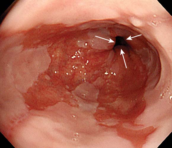
Alter Surveillance for Barrett's Esophagus?
The incidence of esophageal adenocarcinoma among patients with Barrett’s esophagus was only 1.2 cases per 1,000 person-years in a study of the entire population of Denmark reported in the New England Journal of Medicine.
That rate is four to five times lower than rates reported previously, said Dr. Frederik Hvid-Jensen of the department of surgical gastroenterology at Aarhus (Denmark) University and his associates.
"Our study provides solid evidence that esophageal adenocarcinoma will develop in very few patients with Barrett’s esophagus. Together with another recent study, as well as studies of cost-effectiveness and patients’ quality of life, the results of our study suggest that the risk of esophageal adenocarcinoma among patients with Barrett’s esophagus is so minor that in the absence of dysplasia, routine surveillance of such patients is of doubtful value," the investigators said.
The relevance of such surveillance programs has been questioned before because they have never been shown to improve survival and because an estimated 95% of patients with a new diagnosis of esophageal adenocarcinoma do not have a previous diagnosis of Barrett’s esophagus, they noted.
Dr. Hvid-Jensen and his colleagues used data from Denmark’s nationwide pathology and cancer registries to calculate the incidence of adenocarcinoma among patients with Barrett’s esophagus and compare it with the expected incidence in the general population of 5.4 million people.
A total of 11,028 patients underwent endoscopic biopsy and received a diagnosis of Barrett’s esophagus during 1992-2009. The median age at baseline was 63 years, and patients were followed for a median of 5.2 years.
During that time, 197 of these patients with Barrett’s esophagus developed new esophageal adenocarcinomas, which comprised 7.6% of all the 2,602 incident esophageal adenocarcinomas diagnosed in the general Danish population during 1992-2009.
After excluding cancer cases that developed in the first year after a diagnosis of Barrett’s esophagus, the incidence of esophageal adenocarcinoma among patients with Barrett’s esophagus was found to be 1.2 cases per 1,000 person-years, the investigators said (N. Engl. J. Med. 2011;365:1375-83).
The annual risk of developing the malignancy was only 0.12%, or one case of adenocarcinoma per 860 patient-years.
In contrast, there were four reviews of the literature published in the past decade that pooled the results of numerous small studies conducted throughout the United States and Europe. These studies calculated the esophageal adenocarcinoma incidence as ranging from 5.2 to 7.0 cases per 1,000 person-years. In addition, two previous registry studies calculated similar incidences of 4.0 and 5.0 cases of esophogeal cardinoma per 1,000 person-years.
Current surveillance guidelines are based upon these earlier studies, which appear to have overstated the risks, Dr. Hvid-Jensen and his associates stated.
Their population-based, nationwide study is one of the largest studies of the issue; it included patients of all ages and both sexes and had almost no loss to follow-up. Because of Denmark’s universal health care plan, this study also had no referral bias or diagnostic bias.
"The generalizability of our results is therefore high," they noted.
Moreover, a recent population-based study in Northern Ireland found remarkably similar results: an incidence of 1.3 cases of esophageal adenocarcinoma per 1,000 patient-years among people with Barrett’s esophagus.
And another recent study "in which Markov models were used to evaluate available data on the incidence of adenocarcinoma supports our findings ... [and suggests] that surveillance is not beneficial," the researchers added.
This study was supported by the University of Aarhus Clinical Institute. No financial conflicts of interest were reported.☐
The "elegant" epidemiologic study by Dr. Hvid-Jensen and associates clearly shows that the relative risk of esophageal adenocarcinoma for a person with Barrett’s esophagus, as compared with the general population, is 11.3, "a substantial drop from the increase by a factor of 30 or 40 estimated in early reports," said Dr. Peter J. Kahrilas.
"As our knowledge of the biologic characteristics of Barrett’s esophagus has matured, the significance of the lesion has dwindled. In fact, patients with Barrett’s esophagus have the same life expectancy as does the general population, and esophageal cancer proves to be an uncommon cause of death in patients with Barrett’s esophagus regardless of surveillance," he noted.
Dr. Kahrilas is with the department of medicine at Northwestern University, Chicago. He reported ties to numerous industry sources. These remarks were taken from his editorial accompanying Dr. Hvid-Jensen’s report (N. Engl. J. Med. 2011;365:1437-8).
The incidence of esophageal adenocarcinoma among patients with Barrett’s esophagus was only 1.2 cases per 1,000 person-years in a study of the entire population of Denmark reported in the New England Journal of Medicine.
That rate is four to five times lower than rates reported previously, said Dr. Frederik Hvid-Jensen of the department of surgical gastroenterology at Aarhus (Denmark) University and his associates.
"Our study provides solid evidence that esophageal adenocarcinoma will develop in very few patients with Barrett’s esophagus. Together with another recent study, as well as studies of cost-effectiveness and patients’ quality of life, the results of our study suggest that the risk of esophageal adenocarcinoma among patients with Barrett’s esophagus is so minor that in the absence of dysplasia, routine surveillance of such patients is of doubtful value," the investigators said.
The relevance of such surveillance programs has been questioned before because they have never been shown to improve survival and because an estimated 95% of patients with a new diagnosis of esophageal adenocarcinoma do not have a previous diagnosis of Barrett’s esophagus, they noted.
Dr. Hvid-Jensen and his colleagues used data from Denmark’s nationwide pathology and cancer registries to calculate the incidence of adenocarcinoma among patients with Barrett’s esophagus and compare it with the expected incidence in the general population of 5.4 million people.
A total of 11,028 patients underwent endoscopic biopsy and received a diagnosis of Barrett’s esophagus during 1992-2009. The median age at baseline was 63 years, and patients were followed for a median of 5.2 years.
During that time, 197 of these patients with Barrett’s esophagus developed new esophageal adenocarcinomas, which comprised 7.6% of all the 2,602 incident esophageal adenocarcinomas diagnosed in the general Danish population during 1992-2009.
After excluding cancer cases that developed in the first year after a diagnosis of Barrett’s esophagus, the incidence of esophageal adenocarcinoma among patients with Barrett’s esophagus was found to be 1.2 cases per 1,000 person-years, the investigators said (N. Engl. J. Med. 2011;365:1375-83).
The annual risk of developing the malignancy was only 0.12%, or one case of adenocarcinoma per 860 patient-years.
In contrast, there were four reviews of the literature published in the past decade that pooled the results of numerous small studies conducted throughout the United States and Europe. These studies calculated the esophageal adenocarcinoma incidence as ranging from 5.2 to 7.0 cases per 1,000 person-years. In addition, two previous registry studies calculated similar incidences of 4.0 and 5.0 cases of esophogeal cardinoma per 1,000 person-years.
Current surveillance guidelines are based upon these earlier studies, which appear to have overstated the risks, Dr. Hvid-Jensen and his associates stated.
Their population-based, nationwide study is one of the largest studies of the issue; it included patients of all ages and both sexes and had almost no loss to follow-up. Because of Denmark’s universal health care plan, this study also had no referral bias or diagnostic bias.
"The generalizability of our results is therefore high," they noted.
Moreover, a recent population-based study in Northern Ireland found remarkably similar results: an incidence of 1.3 cases of esophageal adenocarcinoma per 1,000 patient-years among people with Barrett’s esophagus.
And another recent study "in which Markov models were used to evaluate available data on the incidence of adenocarcinoma supports our findings ... [and suggests] that surveillance is not beneficial," the researchers added.
This study was supported by the University of Aarhus Clinical Institute. No financial conflicts of interest were reported.☐
Major Finding: The incidence of esophageal adenocarcinoma among all patients in Denmark with Barrett’s esophagus was 1.2 cases per 1,000 person-years, which is four to five times lower than estimated in previous, smaller studies.
Data Source: An epidemiologic cohort study of esophageal adenocarcinoma among the 5.4 million residents of Denmark, including 11,028 with Barrett’s esophagus followed for a median of 5.2 years.
Disclosures: This study was supported by the University of Aarhus Clinical Institute. No financial conflicts of interest were reported.
Overeating? Less Protein Means More Fat
Overeating on a diet low in protein caused less weight gain than overeating on a normal- or high-protein diet – but most of the body mass gained was fat, rather than the leaner muscle mass created by high-protein overeating, according to a report published in the Jan. 4 issue of JAMA.
The distinction is important, because accumulation of excess fat is associated with obesity-related medical conditions, while accumulation of muscle mass is beneficial, noted Dr. George A. Bray of Pennington Biomedical Research Center, Baton Rouge, La., and his associates (JAMA 2012;307:47-55).

Dr. Bray and his colleagues examined whether the level of dietary protein affected weight gain and body composition. Their randomized trial included 25 healthy, weight-stable adults (BMI 19-30 kg/m2) who lived under tightly controlled conditions in a metabolic unit for 10-12 weeks. During the first 2-4 weeks of the study – a weight stabilization period – the 16 men and 9 women (aged 18-35 years) consumed an isocaloric diet with 15% of energy from protein, 25% from fat, and 60% from carbohydrates.
During the final 8 weeks of the trial, subjects were deliberately overfed 40% more energy than was needed for weight maintenance, an excess of approximately 1,000 calories each day. During that overeating period, they were randomly assigned to diets containing 5% of energy from protein (the low-protein diet), 15% of energy from protein (the normal-protein diet), or 25% of energy from protein (the high-protein diet).
Body composition was measured by dual x-ray absorptiometry. The study subjects included 7 non-Hispanic whites, 16 blacks, and 2 Asians.
All the study subjects gained weight during the overeating period, and there were no significant differences in weight gain by subjects’ race or sex. That suggests that the data are generalizable to all races and both sexes.
The weight gain in the low-protein group was 3.16 kg, significantly less than the weight gain in the other two groups – 6.05 kg in the normal-protein group and 6.51 kg in the high-protein group.
In the low-protein group, more than 90% of the extra energy was stored as fat. In contrast, in the normal- and high-protein groups, only about 50% of the extra energy was stored as fat, while the remaining 50% was lean body mass.
The findings imply that a low-protein diet is metabolically different from a normal- or high-protein diet, the investigators added.
The study was supported in part by the U.S. Department of Agriculture and Louisiana State University, New Orleans. Dr. Bray reported ties to Abbott Laboratories, Takeda Global Research Institute, Medifast, Herbalife, and Global Direction in Medicine. One of his associates reported ties to Bristol-Myers Squibb, Eli Lilly, Elcelyx, Merck, Philips, the International Life Sciences Institute, Catapult Health, and Domain & Associates.
The findings of Bray and colleagues "suggest that overeating low-protein diets may increase fat deposition, leading to loss of lean body mass, despite lesser increases in body weight," said Dr. Zhaoping Li and Dr. David Heber.
"Clinicians should consider assessing a patient’s overall fatness rather than simply measuring body weight or body mass index, and concentrate on the potential complications of excess fat accumulation. The goals for obesity treatment should involve fat reduction rather than simply weight loss, along with a better understanding of nutrition science," they noted.
Dr. Li and Dr. Heber are at the Center for Human Nutrition, University of California, Los Angeles. Dr. Heber reported ties to POM Wonderful, Herbalife, McCormick Spices, and the Obesity Society for Clinical Research. Dr. Li reported no financial conflicts of interest. These remarks were taken from their editorial accompanying Dr. Bray’s report (JAMA 2012;307:86-7).
Overeating on a diet low in protein caused less weight gain than overeating on a normal- or high-protein diet – but most of the body mass gained was fat, rather than the leaner muscle mass created by high-protein overeating, according to a report published in the Jan. 4 issue of JAMA.
The distinction is important, because accumulation of excess fat is associated with obesity-related medical conditions, while accumulation of muscle mass is beneficial, noted Dr. George A. Bray of Pennington Biomedical Research Center, Baton Rouge, La., and his associates (JAMA 2012;307:47-55).
Dr. Bray and his colleagues examined whether the level of dietary protein affected weight gain and body composition. Their randomized trial included 25 healthy, weight-stable adults (BMI 19-30 kg/m2) who lived under tightly controlled conditions in a metabolic unit for 10-12 weeks. During the first 2-4 weeks of the study – a weight stabilization period – the 16 men and 9 women (aged 18-35 years) consumed an isocaloric diet with 15% of energy from protein, 25% from fat, and 60% from carbohydrates.
During the final 8 weeks of the trial, subjects were deliberately overfed 40% more energy than was needed for weight maintenance, an excess of approximately 1,000 calories each day. During that overeating period, they were randomly assigned to diets containing 5% of energy from protein (the low-protein diet), 15% of energy from protein (the normal-protein diet), or 25% of energy from protein (the high-protein diet).
Body composition was measured by dual x-ray absorptiometry. The study subjects included 7 non-Hispanic whites, 16 blacks, and 2 Asians.
All the study subjects gained weight during the overeating period, and there were no significant differences in weight gain by subjects’ race or sex. That suggests that the data are generalizable to all races and both sexes.
The weight gain in the low-protein group was 3.16 kg, significantly less than the weight gain in the other two groups – 6.05 kg in the normal-protein group and 6.51 kg in the high-protein group.
In the low-protein group, more than 90% of the extra energy was stored as fat. In contrast, in the normal- and high-protein groups, only about 50% of the extra energy was stored as fat, while the remaining 50% was lean body mass.
The findings imply that a low-protein diet is metabolically different from a normal- or high-protein diet, the investigators added.
The study was supported in part by the U.S. Department of Agriculture and Louisiana State University, New Orleans. Dr. Bray reported ties to Abbott Laboratories, Takeda Global Research Institute, Medifast, Herbalife, and Global Direction in Medicine. One of his associates reported ties to Bristol-Myers Squibb, Eli Lilly, Elcelyx, Merck, Philips, the International Life Sciences Institute, Catapult Health, and Domain & Associates.
FROM JAMA
Major Finding: Weight gain in subjects deliberately overfed a low-protein diet was only 3 kg, half of the 6-kg weight gain in subjects deliberately overfed a normal-protein or a high-protein diet.
Data Source: A randomized clinical trial involving 25 healthy adults who consumed closely supervised diets with varying protein contents for 8 weeks.
Disclosures: This study was supported in part by the U.S. Department of Agriculture and Louisiana State University. Dr. Bray reported ties to Abbott Laboratories, Takeda Global Research Institute, Medifast, Herbalife, and Global Direction in Medicine. One of his associates reported ties to Bristol-Myers Squibb, Eli Lilly, Elcelyx, Merck, Philips, the International Life Sciences Institute, Catapult Health, and Domain & Associates.
Readmission After STEMI in U.S. Higher Than in 16 Other Countries
The United States had by far the highest rate of 30-day readmission after a discharge diagnosis of ST-elevation myocardial infarction in an international study of 17 Western nations, published in the Jan. 4 issue of JAMA.
Even though the readmission rate varied greatly from one country to the next, America’s 14.5% rate of readmission within 30 days was at least one-third higher than that of the other 16 countries, said Dr. Robb D. Kociol of Duke Clinical Research Institute, Durham, N.C., and his associates.
This discrepancy appeared to be related to patients’ hospital length of stay, which was significantly shorter in the United States than in all the other countries, they noted.
Dr. Kociol and his colleagues studied 30-day readmission rates because "high" rates are increasingly used as an indicator of both poor quality of care and unnecessarily high health care costs. Researchers are trying to identify predictors of readmission in hopes of lowering these rates.
The investigators performed a post hoc analysis of data from an international clinical trial of acute STEMI – Assessment of Pexelizumab in Acute Myocardial Infarction – that had been conducted at 296 sites between 2004 and 2006. There were 5,571 study subjects who survived to hospital discharge, and 631 (11.3%) of them were readmitted within 30 days.
The rate of early readmission was 14.5% within the United States, compared with 9.9% outside the United States. This represents a 68% increase in the risk of readmission for U.S. patients, Dr. Kociol and his associates said (JAMA 2011;307:66-74).
"Higher readmission rates in the U.S. may be an adverse effect of the short length-of-stay practice."
When subjects who were readmitted for elective revascularization procedures were excluded from the analysis, the rate of readmission was 10.5% in the United States, compared with 7.7% outside this country.
Several countries had significantly lower odds of 30-day readmission than the United States, including Italy, Germany, Canada, Portugal, the Netherlands, and the Czech Republic. The other countries participating in the MI study were Australia, Austria, Belgium, Switzerland, Denmark, Spain, France, New Zealand, Poland, and Sweden.
In a further analysis of the data, increased disease severity (as measured by the number of involved vessels), a high baseline heart rate, and high-risk changes on ECG were all important predictors of early readmission. However, residence in the United States was the strongest predictor of 30-day readmission for any cause, they added.
Several predictive factors were comparable between the United States and the other countries, including patient age, sex, and comorbidities. Rates of appropriate discharge prescriptions for medications such as aspirin, ticlopidine, clopidogrel, beta-blockers, and statins also were similar.
One factor that stood out as different was hospital length of stay. "Perhaps the most intriguing finding" of this study was that length of stay showed an inverse correlation with risk of readmission. Each 1-day increase in length of stay was associated with a 17% reduction in risk of readmission.
The United States had the shortest hospitalizations. Length of stay was 3 days or fewer for two-thirds of U.S. patients but for only 16% of non-U.S. patients. And only 16.6% of U.S. patients were hospitalized for 6 days or longer, compared with 54% of patients outside the U.S.
"These data raise the possibility that higher readmission rates in the U.S. may be an adverse effect of the short length-of-stay practice," the investigators noted.
Another system-related difference between the United States and the other countries in this study is that most of them have near-universal health care coverage for all citizens, whether through a state-run system, private compulsory health insurance, or a combination of the two. This may allow people in the other countries to obtain faster and easier access to post-discharge follow-up with a primary caregiver, heading off readmission, Dr. Kociol and his colleagues added.
This study was supported by the Duke Clinical Research Institute. The original MI study was funded by Procter & Gamble, Alexion, and the American Heart Association Pharmaceutical Roundtable.
FROM JAMA
Major Finding: The 30-day readmission rate after STEMI was 14.5% in the United States, compared with 9.9% outside the country, representing a 68% higher risk of readmission for U.S. patients.
Data Source: A post hoc analysis of data from an international clinical trial of acute STEMI involving 5,571 study subjects treated in 2004-2006 at 296 sites in 17 countries.
Disclosures: This study was supported by the Duke Clinical Research Institute. The original STEMI study was funded by Procter & Gamble, Alexion, and the American Heart Association Pharmaceutical Roundtable.
Low-Protein Diet May Cause Less Weight Gain, More Fat
Overeating on a diet low in protein caused less weight gain than overeating on a normal- or high-protein diet – but most of the body mass gained was fat, rather than the leaner muscle mass created by high-protein overeating, according to a recent report.
The distinction is important, because the accumulation of excess fat is associated with obesity-related medical conditions, while accumulation of muscle mass is beneficial, noted Dr. George A. Bray of the Pennington Biomedical Research Center, Baton Rouge, La., and his associates (JAMA 2012;307:47-55).
Dr. Bray and his colleagues examined whether the level of dietary protein affected weight gain and body composition. Their randomized trial included 25 healthy, weight-stable adults (BMI 19-30 kg/m
During the final 8 weeks of the trial, subjects were deliberately overfed 40% more energy than was needed for weight maintenance, an excess of approximately 1,000 calories each day. During that overeating period, they were randomly assigned to diets containing 5% of energy from protein (the low-protein diet), 15% of energy from protein (the normal-protein diet), or 25% of energy from protein (the high-protein diet).
Body composition was measured by dual-energy x-ray absorptiometry. The study subjects included 7 non-Hispanic whites, 16 blacks, and 2 Asians.
All the study subjects gained weight during the overeating period, and there were no significant differences in weight gain by subjects' race or sex. That suggests that the data are generalizable to all races and both sexes.
Weight gain in the low-protein group was 3.16 kg, significantly less than the weight gain in the other two groups – 6.05 kg in the normal-protein group and 6.51 kg in the high-protein group.
In the low-protein group, more than 90% of the extra energy was stored as fat. In contrast, in the normal- and high-protein groups, only about 50% of the extra energy was stored as fat, while the remaining 50% was lean body mass.
The findings imply that a low-protein diet is metabolically different from a normal- or high-protein diet, the investigators added.
The study was supported in part by the U.S. Department of Agriculture and Louisiana State University, New Orleans. Dr. Bray reported ties to Abbott Laboratories, Takeda Global Research Institute, Medifast, Herbalife, and Global Direction in Medicine. One of his associates reported ties to Bristol-Myers Squibb, Eli Lilly, Elcelyx, Merck, Philips, the International Life Sciences Institute, Catapult Health, and Domain & Associates.
In subjects overeating on a low-protein diet, more than 90% of the extra energy was stored as fat.
Source ©imagedepotpro/iStockphoto.com
View on the News
Focus on Fatness, Not Weight Gain
The findings of Bray and colleagues “suggest that overeating low-protein diets may increase fat deposition, leading to loss of lean body mass, despite lesser increases in body weight,” said Dr. Zhaoping Li and Dr. David Heber.
“Clinicians should consider assessing a patient's overall fatness rather than simply measuring body weight or body mass index, and concentrate on the potential complications of excess fat accumulation. The goals for obesity treatment should involve fat reduction rather than simply weight loss, along with a better understanding of nutrition science,” they noted.
DR. LI and DR. HEBER are at the Center for Human Nutrition, University of California, Los Angeles. Dr. Heber reported ties to POM Wonderful, Herbalife, McCormick Spices, and the Obesity Society for Clinical Research. Dr. Li reported no financial conflicts of interest. These remarks were taken from their editorial accompanying Dr. Bray's report (JAMA 2012;307:86-7).
From JAMA
CPAP Aids Metabolic Syndrome in Apnea Patients
Major Finding: The metabolic syndrome resolved in 11 of 86 patients after CPAP therapy, compared with 1 of those same patients after sham therapy. The treatment also significantly improved systolic and diastolic BP; total, LDL, and non-HDL cholesterol; triglycerides; glycated hemoglobin; weight; and visceral and subcutaneous fat.
Data Source: A double-blind, randomized trial involving 86 patients with moderate to severe obstructive sleep apnea and components of the metabolic syndrome who received 3 months of real and 3 months of sham CPAP therapy.
Disclosures: This study was funded by Pfizer. All investigators reported having no financial conflicts of interest. The investigators received technical support from ResMed Corp. in designing a sham CPAP machine.
Continuous positive airway pressure therapy improved several components of the metabolic syndrome along with obstructive sleep apnea in patients who had both disorders, according to researchers.
In most cases, only one component of the metabolic syndrome improved significantly after continuous positive airway pressure (CPAP), but that improvement was significant enough to “reverse” the syndrome, reported Dr. Surendra K. Sharma of All India Institute of Medical Sciences, New Delhi, and his associates.
No particular component stood out as being the most responsive to CPAP; statistically significant improvements were seen in systolic BP, diastolic BP, total cholesterol, non-HDL cholesterol, LDL cholesterol, triglycerides, glycated hemoglobin, weight, and visceral and subcutaneous fat.
“These results suggest a significant clinical benefit that will lead to a reduction in cardiovascular risk,” the investigators noted.
To examine the effect of CPAP on components of the metabolic syndrome, Dr. Sharma and his coinvestigators recruited 86 patients aged 30-65 years from the sleep laboratory at the institute who had obstructive sleep apnea that was moderate or worse in severity. All of the subjects reported excessive daytime somnolence.
A total of 75 study subjects (87%) had the metabolic syndrome, and the remainder had some of the components of the metabolic syndrome.
These patients were randomly assigned to undergo either CPAP or sham CPAP for 3 months, followed by a washout period of 1 month. They then crossed over to receive the other intervention for 3 months.
The sham CPAP was not discernible to the study subjects or the investigators.
The metabolic syndrome resolved in 14 (20%) of the study subjects after CPAP. This was attributed to decreased blood pressure in five; decreased fasting blood glucose in two; decreased triglycerides in two; increased HDL cholesterol in three; improved triglycerides plus HDL cholesterol in one; and improved triglycerides, HDL cholesterol, and fasting blood glucose in one, Dr. Sharma and his colleagues reported. Symptoms of the syndrome developed in three patients who did not have metabolic syndrome at the start of the study.
Overall, CPAP was associated with a mean decrease in systolic BP of 3.9 mm Hg, a mean decrease in diastolic BP of 2.5 mm Hg, a mean decrease in total cholesterol of 13.3 mg/dL, and a mean decrease in triglycerides of 18.7 mg/dL.
CT scans revealed a significant decrease in both visceral and subcutaneous fat, which was accompanied by a decrease in BMI, with CPAP therapy. “These findings could be secondary to a decrease in daytime somnolence and a consequent increase in physical activity after CPAP use at night,” the researchers noted.
In addition, “we speculate that CPAP has a favorable effect on leptin levels, which have been shown to be elevated in patients with obstructive sleep apnea and to normalize with CPAP therapy,” the investigators said (N. Engl. J. Med. 2011;365:2277-86).
In a subgroup analysis involving only the 51 subjects who were most compliant with CPAP, with a mean use of at least 5 hours every night, the improvements in components of the metabolic syndrome were even greater. In particular, systolic BP decreased by 5.6 mm Hg and diastolic BP decreased by 3.3 mm Hg.
This subgroup of patients also showed significant improvement in carotid intima-media thickness, “suggesting a potential role for CPAP therapy in reversing endothelial damage due to obstructive sleep apnea and the metabolic syndrome,” Dr. Sharma and his associates said.
Two patients could not tolerate CPAP and one could not tolerate sham CPAP within the first month of treatment, and they withdrew from the study. “Other adverse events reported included skin irritation (in 51% of all patients), nasal bridge discomfort (in 44%), nasal congestion (in 28%), headache (in 26%), and mask leaks (in 30%).”
No particular component of the metabolic syndrome stood out as being the most responsive to CPAP.
Source ©David Cannings-Bushell/iStockphoto
From the New England Journal of Medicine
Breast Ca Risk Not Worse for Noncarriers in BRCA Families
Women who don't carry their family's BRCA1 or BRCA2 mutation showed no increase in breast cancer risk in a study of 3,047 population-based families reported .
“These results support the standard clinical practice of advising noncarriers that they do not have any increase in breast cancer risk attributable to the family-specific BRCA mutation and, in the absence of other strong risk factors, should follow general population guidelines for breast cancer screening,” said Dr. Allison W. Kurian of Stanford (Calif.) University and her associates.
Some recent studies have suggested that noncarriers of a family-specific mutation may have a two- to fivefold increase in risk of developing breast cancer, compared with the general population. While lower than the 5- to 20-fold increase in risk for carriers of the mutation, this rate would still be high enough to warrant breast cancer surveillance. Other studies have found no increase in risk among noncarriers.
To clarify the issue, Dr. Kurian and her colleagues assessed breast cancer risk using population-based cancer registries in the United States, Australia, and Canada. They identified 3,047 families in which one woman (the proband) was diagnosed as having breast cancer at a relatively young age, in most cases during 1996-2000, and she and her female first-degree relatives underwent genetic testing for BRCA1 and BRCA2 mutations.
Overall, 160 families had BRCA1 mutations and 132 had BRCA2 mutations, the investigators reported (J. Clin. Oncol. 2011 [doi:10.1200.JCO.2010.34.4440]).
Among noncarriers of family-specific mutations, the risk of developing breast cancer was not significantly higher than the risk among women in 2,755 families without any BRCA1 or BRCA2 mutations. This relative risk was 0.39.
It is possible that previous studies reporting an increased risk in noncarriers have overestimated this risk, because they compared study subjects with women in the general population rather than women whose relatives have breast cancer. The latter group undergoes more frequent screening and consequently has more frequent diagnoses of breast cancer than women in the general population, Dr. Kurian and her associates noted.
This study was supported by the National Cancer Institute, the National Institutes of Health, Cancer Care Ontario, Cancer Prevention Institute of California, and the University of Melbourne. No financial conflicts of interest were reported.
From the Journal of Clinical Oncology
Bevacizumab Effective With First-Line Treatment for Ovarian Cancer
Adding bevacizumab to standard first-line carboplatin and paclitaxel chemotherapy therapy against ovarian cancer prolonged progression-free survival in two separate, complementary, phase III clinical trials reported in the Dec. 29 issue of the New England Journal of Medicine.
Bevacizumab (Avastin) did not interfere with chemotherapy. Although it raised the rate of toxic effects, this did not impair patients’ quality of life in either study. Both trials received industry support.
The GOG Study
In the Gynecologic Oncology Group (GOG) study, 1,873 women were treated at 336 medical centers in the United States, Canada, South Korea, and Japan. All had previously untreated, incompletely resectable, stage III or stage IV epithelial ovarian, primary peritoneal, or fallopian-tube cancer, for which they had undergone debulking surgery, said Dr. Robert A. Burger of the Fox Chase Cancer Center, Philadelphia, and his associates.

The cohort as a whole had a relatively poor prognosis, and factors that could influence treatment outcome were evenly distributed among the treatment groups. Patients were randomly assigned in a double-blind fashion to one of three regimens:
• 15 mg/kg bevacizumab added to chemotherapy cycles 2-22.
• 15 mg/kg bevacizumab added only to cycles 2-6, plus placebo added to cycles 7-22.
• Placebo added to chemotherapy cycles 2-22 (the control group).
A monoclonal antibody, bevacizumab targets the vascular endothelial growth factor (VEGF), which is known to promote angiogenesis and progression of ovarian cancer.
At the primary data analysis after a median of 17.4 months of follow-up, median progression-free survival, the primary end point, was 14.1 months when bevacizumab was given throughout the chemotherapy course. This was longer than the 11.2-month median when bevacizumab was given only at the beginning of the chemotherapy course and significantly longer than the 10.3-month median when it wasn’t given at all.
Thus, the addition of bevacizumab throughout the entire chemotherapy course prolonged progression-free survival by a median of 4 months, compared with chemotherapy alone, Dr. Burger and his colleagues said (New Engl. J. Med. 2011;365:2473-83).
In a further analysis of progression-free survival "in which data for patients with increased CA-125 levels were censored, as required by regulatory agencies, the median progression-free survival was 12.0 months in the control group but 18.0 months in the bevacizumab-throughout group," they added.
This benefit in progression-free survival was consistent across all subgroups of patients, regardless of prognostic factors such as cancer stage, lesion size, the tumor’s histologic type, the patient’s performance status, or the patient’s age, the researchers noted.
There was no significant benefit in overall survival, but the ability to detect such a benefit was limited by the lack of control for subsequent treatments, including crossover to bevacizumab or other anti-VEGF agents, they noted.
Regarding adverse effects, there were no significant differences among the three groups in quality-of-life scores on the Functional Assessment of Cancer Therapy–Ovary survey.
The frequency of hypertension was significantly higher with the addition of bevacizumab, which was not unexpected. But it was easily controlled with medication and led to discontinuation of the drug in only 2.4% of affected patients.
There were no significant differences among the three treatment groups in rates of other adverse events, including some that have been reported with previous monoclonal antibody therapy (gastrointestinal perforation or fistula; proteinuria; neutropenia, especially febrile neutropenia; venous or arterial thrombosis; and wound disruption).
"Rates of gastrointestinal perforation and fistula in the two bevacizumab groups were almost twice those in the control group but were still less than 3%, consistent with rates seen in metastatic nongynecologic tumors," the investigators said.
Fatal adverse events occurred in 1% of the control group, in 1.6% of those who took bevacizumab only at the beginning of chemotherapy, and in 2.3% in those who took bevacizumab throughout the entire course of chemotherapy.
The ICON7 Study
The other phase III trial, the ICON7 (International Collaboration on Ovarian Neoplasms) study led by the U.K. Medical Research Council clinical trials unit, involved women with advanced-stage ovarian cancer but no visible residual disease, as well as some women with high-risk early-stage disease.
This trial examined the efficacy and safety of a lower dose of bevacizumab (7.5 mg/kg) for a shorter interval (5 or 6 cycles with chemotherapy, and up to 12 cycles afterward), said Dr. Timothy J. Perren of the Institute of Oncology at St. James’s University Hospital in Leeds, England, and his associates
The 1,528 study subjects were treated at 263 medical centers in the United Kingdom, Germany, France, Canada, Australia, New Zealand, Denmark, Finland, Norway, Sweden, and Spain. They were randomly assigned to receive either standard carboplatin-plus-paclitaxel chemotherapy every 3 weeks for 6 cycles (764 patients), or the same chemotherapy plus concurrent 7.5 mg/kg bevacizumab, with the monoclonal antibody alone extended for 12 additional cycles (764 patients).
At the primary data analysis after a median follow-up of 19.4 months, the median progression-free survival was 19.0 months with the addition of bevacizumab, which was significantly longer than the 17.3-month progression-free survival with standard chemotherapy alone.
Thus, the addition of bevacizumab prolonged progression-free survival by approximately 2 months. In addition, it increased the overall rate of treatment response by 19%, with a complete or partial remission rate of 67%, compared with 48% in the control group, Dr. Perren and his colleagues said (N. Engl. J. Med. 2011;365:2484-96).
The benefit of bevacizumab treatment was greatest in patients at the highest risk for cancer progression. Median progression-free survival was 15.9 months in this subgroup of patients who received bevacizumab, compared with only 10.5 months for those who did not.
Overall survival cannot yet be determined in this study, and is expected to be calculated and reported in 2013. "Some will argue that final overall survival data are needed before the results can be fully interpreted," the investigators noted.
Adverse events of grade 3 or higher were reported in 66% of the women who received bevacizumab and in 56% of the control group. The monoclonal antibody appeared to be associated with increased rates of bleeding, hypertension (18% of patients receiving bevacizumab vs. 2% of the controls), thromboembolic events (7% vs. 3%), and gastrointestinal perforations (10 patients vs. 3 patients).
However, both study groups showed improved global quality of life on two EORTC (European Organisation for Research and Treatment of Cancer) QoL questionnaires, and differences between the two groups were not clinically significant.
The GOG study was funded by the U.S. National Cancer Institute and Genentech. The ICON7 study was funded by Roche (parent company of Genentech) and the U.K. National Institute for Health Research, through the U.K. National Cancer Research Network. Dr. Burger, Dr. Perren, and their associates reported numerous ties to industry sources.
Adding bevacizumab to standard first-line carboplatin and paclitaxel chemotherapy therapy against ovarian cancer prolonged progression-free survival in two separate, complementary, phase III clinical trials reported in the Dec. 29 issue of the New England Journal of Medicine.
Bevacizumab (Avastin) did not interfere with chemotherapy. Although it raised the rate of toxic effects, this did not impair patients’ quality of life in either study. Both trials received industry support.
The GOG Study
In the Gynecologic Oncology Group (GOG) study, 1,873 women were treated at 336 medical centers in the United States, Canada, South Korea, and Japan. All had previously untreated, incompletely resectable, stage III or stage IV epithelial ovarian, primary peritoneal, or fallopian-tube cancer, for which they had undergone debulking surgery, said Dr. Robert A. Burger of the Fox Chase Cancer Center, Philadelphia, and his associates.
The cohort as a whole had a relatively poor prognosis, and factors that could influence treatment outcome were evenly distributed among the treatment groups. Patients were randomly assigned in a double-blind fashion to one of three regimens:
• 15 mg/kg bevacizumab added to chemotherapy cycles 2-22.
• 15 mg/kg bevacizumab added only to cycles 2-6, plus placebo added to cycles 7-22.
• Placebo added to chemotherapy cycles 2-22 (the control group).
A monoclonal antibody, bevacizumab targets the vascular endothelial growth factor (VEGF), which is known to promote angiogenesis and progression of ovarian cancer.
At the primary data analysis after a median of 17.4 months of follow-up, median progression-free survival, the primary end point, was 14.1 months when bevacizumab was given throughout the chemotherapy course. This was longer than the 11.2-month median when bevacizumab was given only at the beginning of the chemotherapy course and significantly longer than the 10.3-month median when it wasn’t given at all.
Thus, the addition of bevacizumab throughout the entire chemotherapy course prolonged progression-free survival by a median of 4 months, compared with chemotherapy alone, Dr. Burger and his colleagues said (New Engl. J. Med. 2011;365:2473-83).
In a further analysis of progression-free survival "in which data for patients with increased CA-125 levels were censored, as required by regulatory agencies, the median progression-free survival was 12.0 months in the control group but 18.0 months in the bevacizumab-throughout group," they added.
This benefit in progression-free survival was consistent across all subgroups of patients, regardless of prognostic factors such as cancer stage, lesion size, the tumor’s histologic type, the patient’s performance status, or the patient’s age, the researchers noted.
There was no significant benefit in overall survival, but the ability to detect such a benefit was limited by the lack of control for subsequent treatments, including crossover to bevacizumab or other anti-VEGF agents, they noted.
Regarding adverse effects, there were no significant differences among the three groups in quality-of-life scores on the Functional Assessment of Cancer Therapy–Ovary survey.
The frequency of hypertension was significantly higher with the addition of bevacizumab, which was not unexpected. But it was easily controlled with medication and led to discontinuation of the drug in only 2.4% of affected patients.
There were no significant differences among the three treatment groups in rates of other adverse events, including some that have been reported with previous monoclonal antibody therapy (gastrointestinal perforation or fistula; proteinuria; neutropenia, especially febrile neutropenia; venous or arterial thrombosis; and wound disruption).
"Rates of gastrointestinal perforation and fistula in the two bevacizumab groups were almost twice those in the control group but were still less than 3%, consistent with rates seen in metastatic nongynecologic tumors," the investigators said.
Fatal adverse events occurred in 1% of the control group, in 1.6% of those who took bevacizumab only at the beginning of chemotherapy, and in 2.3% in those who took bevacizumab throughout the entire course of chemotherapy.
The ICON7 Study
The other phase III trial, the ICON7 (International Collaboration on Ovarian Neoplasms) study led by the U.K. Medical Research Council clinical trials unit, involved women with advanced-stage ovarian cancer but no visible residual disease, as well as some women with high-risk early-stage disease.
This trial examined the efficacy and safety of a lower dose of bevacizumab (7.5 mg/kg) for a shorter interval (5 or 6 cycles with chemotherapy, and up to 12 cycles afterward), said Dr. Timothy J. Perren of the Institute of Oncology at St. James’s University Hospital in Leeds, England, and his associates
The 1,528 study subjects were treated at 263 medical centers in the United Kingdom, Germany, France, Canada, Australia, New Zealand, Denmark, Finland, Norway, Sweden, and Spain. They were randomly assigned to receive either standard carboplatin-plus-paclitaxel chemotherapy every 3 weeks for 6 cycles (764 patients), or the same chemotherapy plus concurrent 7.5 mg/kg bevacizumab, with the monoclonal antibody alone extended for 12 additional cycles (764 patients).
At the primary data analysis after a median follow-up of 19.4 months, the median progression-free survival was 19.0 months with the addition of bevacizumab, which was significantly longer than the 17.3-month progression-free survival with standard chemotherapy alone.
Thus, the addition of bevacizumab prolonged progression-free survival by approximately 2 months. In addition, it increased the overall rate of treatment response by 19%, with a complete or partial remission rate of 67%, compared with 48% in the control group, Dr. Perren and his colleagues said (N. Engl. J. Med. 2011;365:2484-96).
The benefit of bevacizumab treatment was greatest in patients at the highest risk for cancer progression. Median progression-free survival was 15.9 months in this subgroup of patients who received bevacizumab, compared with only 10.5 months for those who did not.
Overall survival cannot yet be determined in this study, and is expected to be calculated and reported in 2013. "Some will argue that final overall survival data are needed before the results can be fully interpreted," the investigators noted.
Adverse events of grade 3 or higher were reported in 66% of the women who received bevacizumab and in 56% of the control group. The monoclonal antibody appeared to be associated with increased rates of bleeding, hypertension (18% of patients receiving bevacizumab vs. 2% of the controls), thromboembolic events (7% vs. 3%), and gastrointestinal perforations (10 patients vs. 3 patients).
However, both study groups showed improved global quality of life on two EORTC (European Organisation for Research and Treatment of Cancer) QoL questionnaires, and differences between the two groups were not clinically significant.
The GOG study was funded by the U.S. National Cancer Institute and Genentech. The ICON7 study was funded by Roche (parent company of Genentech) and the U.K. National Institute for Health Research, through the U.K. National Cancer Research Network. Dr. Burger, Dr. Perren, and their associates reported numerous ties to industry sources.
FROM THE NEW ENGLAND JOURNAL OF MEDICINE
Major Finding: The addition of 15 mg/kg bevacizumab to standard chemotherapy prolonged progression-free survival by about 4 months in women with advanced ovarian cancer, and the addition of 7.5 mg/kg bevacizumab prolonged progression-free survival by 2 months in women with advanced-stage and high-risk early-stage ovarian cancer.
Data Source: Two international, phase III, randomized, double-blind clinical trials assessing the addition of bevacizumab to standard chemotherapy as first-line treatment for advanced ovarian cancer in a combined total of 3,401 women.
Disclosures: The GOG study was funded by the U.S. National Cancer Institute and Genentech. The ICON7 study was funded by Roche (parent company of Genentech) and the U.K. National Institute for Health Research, through the U.K. National Cancer Research Network. Dr. Burger, Dr. Perren, and their associates reported numerous ties to industry sources.
Super Sensitive Troponin Assay Aids MI Diagnosis
A new, highly-sensitive troponin I assay may prove to be more effective than a conventional troponin I assay at ruling in a diagnosis of myocardial infarction within 3 hours after admission to the emergency department, according to a report published in the Dec. 28 issue of JAMA.
Changes in the results of the highly-sensitive assay between admission and 3 hours ruled in the diagnosis of MI with a positive predictive value of 83% compared with the 58% PPV of the conventional troponin assay, said Dr. Till Keller of the University Heart Center Hamburg (Germany) and associates (JAMA 2011;306:2684-93).
The researchers noted that the results need to be independently validated.
The study compared the performance of the new, highly-sensitive assay (ArchitectSTATHigh Sensitive Troponin, Abbott Diagnostics) with that of one currently in clinical use (Architect STAT troponin-I assay, Abbott Diagnostics) in a study of 1,818 adults who presented consecutively to three German chest pain units with suspected acute coronary syndromes during a 2-year period. A total of 413 of these subjects (23%) received a final discharge diagnosis of MI.
Blood for the troponin I assays and assays for 10 other biomarkers was drawn at admission, and 3 hours later, and ECGs were performed at the same intervals.
For ruling out MI, the highly sensitive troponin I assay taken at admission (with the diagnostic cutoff value at the 99th percentile of 30 pg/mL) had a sensitivity of 82% and a negative predictive value of 95%. At 3 hours after admission, this assay had a sensitivity of 98% and a negative predictive value of 99%. The results for the conventional troponin I assay were comparable at admission and at 3 hours, the investigators said.
For ruling in MI, changes in the results of the highly sensitive troponin I assay were instructive. There were 951 patients with levels that were well below the 99th percentile at admission that increased above this cutoff point within 3 hours. This change, combined with a positive troponin level after 3 hours, had a positive predictive value of 83% – considerably greater than the 58% PPV for similar results on the conventional troponin assay.
A further analysis showed that the optimized relative change for the highly sensitive assay was a cutoff of 266%. "Using such a change of 266% in combination with the 99th percentile cutoff on admission, the specificity and the positive predictive value of [the highly sensitive assay] increased to over 99% and nearly 96%, respectively," Dr. Keller and colleagues said.
The assay was even more accurate at both ruling out and ruling in MI in the subgroup of 1,176 patients who had ST-elevation or clearly elevated troponin levels at admission. The highest negative predictive value to rule out MI, over 99%, was attained in these patients, as was the highest PPV, over 96%, to rule in MI.
This study was funded by the Johannes Gutenberg-University of Mainz, Brahms AG and Abbott Diagnostics. Dr. Keller and several of his associates reported grant support from Brahms AG and Abbott Diagnostics.
FROM JAMA
Major Finding: Changes in the results of the highly-sensitive troponin I assay between admission and 3 hours ruled in the diagnosis of MI with a positive predictive value of 83% compared with the 58% PPV of the conventional troponin assay.
Data Source: A study of 1,818 adults who presented consecutively to three German chest pain units with suspected acute coronary syndromes during a 2-year period.
Disclosures: This study was funded by the Johannes Gutenberg-University of Mainz, Brahms AG and Abbott Diagnostics. Dr. Keller and several of his associates reported grant support from Brahms AG and Abbott Diagnostics.