User login
Doug Brunk is a San Diego-based award-winning reporter who began covering health care in 1991. Before joining the company, he wrote for the health sciences division of Columbia University and was an associate editor at Contemporary Long Term Care magazine when it won a Jesse H. Neal Award. His work has been syndicated by the Los Angeles Times and he is the author of two books related to the University of Kentucky Wildcats men's basketball program. Doug has a master’s degree in magazine journalism from the S.I. Newhouse School of Public Communications at Syracuse University. Follow him on Twitter @dougbrunk.
ASLMS: SmoothShapes Reduced Thigh Circumference in Controlled Study
PHOENIX - A dual-wavelength laser system with mechanical manipulation effectively reduced thigh circumference in a multicenter, 83-patient study.
The SmoothShapes system (Elemé Medical Inc.) combines a 915-nm laser with a 650-nm LED light, which is believed to stimulate collagen regeneration and to improve and restore blood and lymphatic circulation.
The system is cleared by the Food and Drug Administration for the temporary reduction in the appearance of cellulite and features contoured rollers that massage tissue and move interstitial fluid toward the lymphatic system. A vacuum enables consistent light penetration to tissue.
|
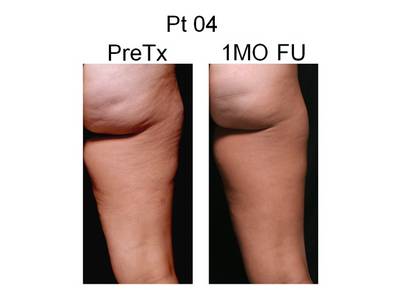
A study patient is shown before, and 1 month after, treatment with the SmoothShapes system for reduction of thigh circumference. Photo courtesy Dr. Robert A. Weiss |
At the annual meeting of the American Society for Laser Medicine and Surgery, Dr. Robert A. Weiss, director of the Maryland Laser, Skin, and Vein Institute in Hunt Valley, Md., presented results from a single-blinded clinical study in which he and his associates randomly assigned women to have one thigh treated using the SmoothShapes system. The other thigh served as the control.
Each patient had circumferential measurements of the upper, mid, and lower thigh taken pre-treatment, after 4 treatments, immediately after 8 treatments, and at 1 month follow-up after completion of treatments.
Location for the measurements was determined with a laser-based device and recorded for accuracy and reproducibility. Treatment consisted of 8 twice weekly, 30-minute treatments of the entire thigh.
Dr. Weiss, also of the department of dermatology at Johns Hopkins University in Baltimore, reported results from 83 patients treated at one of six centers. All subjects completed at least 1 month of follow-up and had no data points missing throughout the study.
The mean differences in circumferential measures on the treated and untreated thighs were consistently statistically significant at all three thigh locations (upper, mid, and lower) and at all 3 time points (after 4 treatments, immediately after 8 treatments, and at 1 month after the last treatment).
The average overall reduction among all three thigh areas treated at 1 month follow-up was 1.9 cm; the minimum loss was 0.2 cm; and the maximum loss was 8.7 cm. "We generally saw the greatest results in the upper thigh," Dr. Weiss said.
Preliminary results were similar for 47 patients who completed 3 months of follow-up, Dr. Weiss said. The average overall loss in circumference for all three thigh areas was 1.0 cm; the minimum loss was 0.1 cm; and the maximum loss was 8.7 cm.
He concluded that the SmoothShapes system "was highly effective in producing thigh circumferential reduction at each thigh location and at each evaluation time-point in the study with 1- and 3-month follow-up."
Dr. Weiss disclosed that he received equipment and consulting fees from Elemé Medical Inc. as part of the study.
PHOENIX - A dual-wavelength laser system with mechanical manipulation effectively reduced thigh circumference in a multicenter, 83-patient study.
The SmoothShapes system (Elemé Medical Inc.) combines a 915-nm laser with a 650-nm LED light, which is believed to stimulate collagen regeneration and to improve and restore blood and lymphatic circulation.
The system is cleared by the Food and Drug Administration for the temporary reduction in the appearance of cellulite and features contoured rollers that massage tissue and move interstitial fluid toward the lymphatic system. A vacuum enables consistent light penetration to tissue.
|
|
A study patient is shown before, and 1 month after, treatment with the SmoothShapes system for reduction of thigh circumference. Photo courtesy Dr. Robert A. Weiss |
At the annual meeting of the American Society for Laser Medicine and Surgery, Dr. Robert A. Weiss, director of the Maryland Laser, Skin, and Vein Institute in Hunt Valley, Md., presented results from a single-blinded clinical study in which he and his associates randomly assigned women to have one thigh treated using the SmoothShapes system. The other thigh served as the control.
Each patient had circumferential measurements of the upper, mid, and lower thigh taken pre-treatment, after 4 treatments, immediately after 8 treatments, and at 1 month follow-up after completion of treatments.
Location for the measurements was determined with a laser-based device and recorded for accuracy and reproducibility. Treatment consisted of 8 twice weekly, 30-minute treatments of the entire thigh.
Dr. Weiss, also of the department of dermatology at Johns Hopkins University in Baltimore, reported results from 83 patients treated at one of six centers. All subjects completed at least 1 month of follow-up and had no data points missing throughout the study.
The mean differences in circumferential measures on the treated and untreated thighs were consistently statistically significant at all three thigh locations (upper, mid, and lower) and at all 3 time points (after 4 treatments, immediately after 8 treatments, and at 1 month after the last treatment).
The average overall reduction among all three thigh areas treated at 1 month follow-up was 1.9 cm; the minimum loss was 0.2 cm; and the maximum loss was 8.7 cm. "We generally saw the greatest results in the upper thigh," Dr. Weiss said.
Preliminary results were similar for 47 patients who completed 3 months of follow-up, Dr. Weiss said. The average overall loss in circumference for all three thigh areas was 1.0 cm; the minimum loss was 0.1 cm; and the maximum loss was 8.7 cm.
He concluded that the SmoothShapes system "was highly effective in producing thigh circumferential reduction at each thigh location and at each evaluation time-point in the study with 1- and 3-month follow-up."
Dr. Weiss disclosed that he received equipment and consulting fees from Elemé Medical Inc. as part of the study.
ASLMS: Expert Reframes Skin of Color to "Skin of Cultures"
PHOENIX - Using laser technology to treat patients requires not only paying attention to the color of their skin, but also to the patient's ethnicity, according to Dr. Eliot F. Battle Jr.
"In my practice we don't just talk about skin of color anymore," Dr. Battle said at the annual meeting of the American Society for Laser Medicine and Surgery. "We talk about skin of cultures. It's not just their skin color but their ethnicity also makes a difference."
He explained that African Americans "have different combinations of black, white, Latin, Spanish, and Indian. Is my ethnicity a mixture of Nigerian, Italian, and Cherokee Indian, or am I a mixture of Cameroonian, Spanish, and Creek Indian? The future of this field, to improve the way we treat people, is going to appreciate more ethnicity as it relates to skin of color."
Brown skin "competes for the light of the laser, because melanin is absorbed across most of the photobiological spectrum, and we are a diverse population of people," said Dr. Battle, who said that he is a mix of Saharan African, Indo-European, and Asian. "To safely treat skin of color we have to use lower wavelengths, lower fluences, lower pulse durations, and aggressive epidermal cooling."
When he treats skin of color, Dr. Battle, a cosmetic dermatologist and laser

surgeon who is director of Cultura Cosmetic Medical Spa in Washington, normally favors an integrated approach and often combines laser treatments, medical skin care, and facial injectables.
For safe laser-hair removal in patients with Fitzpatrick skin types V and VI, Dr. Battle recommends using a long-pulsed diode laser and the long-pulsed Nd:YAG laser.
Safe lasers for complexion blending and skin rejuvenation to improve texture include the microsecond Nd:YAG laser, fractional therapy, the excimer laser, the Q-switched Nd:YAG laser, and the potassium-titanyl-phosphate (KTP) laser, while safe lasers for skin tightening include infrared lasers and radiofrequency technology.
"There are many lasers we can use for skin of color," he said. "The key is to find a laser that you're comfortable with. Become an expert with that laser and go forward. I don't treat skin of color with vascular lasers, resurfacing lasers, or with intense pulsed light. I don't trust most parameters supplied by the manufacturers. I find my own, safe parameters. I treat more conservatively, and I do not cause erythema or edema."
Aggressive skin cooling is key, he said, and can include ice packs, cold gel, cold liquid, cold air flow, a copper plate, and sapphire windows. "We all get side effects when our skin temperature goes past 45° C," said Dr. Battle, who is also on the dermatology faculty at Howard University, Washington. "By using longer pulse durations, and thus pouring the energy in slower, I'm able to remove heat from the skin more effectively and more efficiently. Longer pulse durations are by far the safest way to treat skin of color."
More than two-thirds of Dr. Battle's patients have darker skin. Between 2002 and 2009, the two most popular nonsurgical procedures in his practice were laser hair removal and laser complexion blending, followed by Botox injections, laser skin rejuvenation, fillers, and laser skin tightening.
Lasers "have come a long way in the last decade," he said.
If side effects occur "show compassion and empathy," Dr. Battle noted. "Allow time to help, and always be available to your patients, even if it means giving them your cell phone number."
Dr. Battle disclosed that he has received equipment, discounts, and honoraria from a number of medical device companies.
PHOENIX - Using laser technology to treat patients requires not only paying attention to the color of their skin, but also to the patient's ethnicity, according to Dr. Eliot F. Battle Jr.
"In my practice we don't just talk about skin of color anymore," Dr. Battle said at the annual meeting of the American Society for Laser Medicine and Surgery. "We talk about skin of cultures. It's not just their skin color but their ethnicity also makes a difference."
He explained that African Americans "have different combinations of black, white, Latin, Spanish, and Indian. Is my ethnicity a mixture of Nigerian, Italian, and Cherokee Indian, or am I a mixture of Cameroonian, Spanish, and Creek Indian? The future of this field, to improve the way we treat people, is going to appreciate more ethnicity as it relates to skin of color."
Brown skin "competes for the light of the laser, because melanin is absorbed across most of the photobiological spectrum, and we are a diverse population of people," said Dr. Battle, who said that he is a mix of Saharan African, Indo-European, and Asian. "To safely treat skin of color we have to use lower wavelengths, lower fluences, lower pulse durations, and aggressive epidermal cooling."
When he treats skin of color, Dr. Battle, a cosmetic dermatologist and laser
surgeon who is director of Cultura Cosmetic Medical Spa in Washington, normally favors an integrated approach and often combines laser treatments, medical skin care, and facial injectables.
For safe laser-hair removal in patients with Fitzpatrick skin types V and VI, Dr. Battle recommends using a long-pulsed diode laser and the long-pulsed Nd:YAG laser.
Safe lasers for complexion blending and skin rejuvenation to improve texture include the microsecond Nd:YAG laser, fractional therapy, the excimer laser, the Q-switched Nd:YAG laser, and the potassium-titanyl-phosphate (KTP) laser, while safe lasers for skin tightening include infrared lasers and radiofrequency technology.
"There are many lasers we can use for skin of color," he said. "The key is to find a laser that you're comfortable with. Become an expert with that laser and go forward. I don't treat skin of color with vascular lasers, resurfacing lasers, or with intense pulsed light. I don't trust most parameters supplied by the manufacturers. I find my own, safe parameters. I treat more conservatively, and I do not cause erythema or edema."
Aggressive skin cooling is key, he said, and can include ice packs, cold gel, cold liquid, cold air flow, a copper plate, and sapphire windows. "We all get side effects when our skin temperature goes past 45° C," said Dr. Battle, who is also on the dermatology faculty at Howard University, Washington. "By using longer pulse durations, and thus pouring the energy in slower, I'm able to remove heat from the skin more effectively and more efficiently. Longer pulse durations are by far the safest way to treat skin of color."
More than two-thirds of Dr. Battle's patients have darker skin. Between 2002 and 2009, the two most popular nonsurgical procedures in his practice were laser hair removal and laser complexion blending, followed by Botox injections, laser skin rejuvenation, fillers, and laser skin tightening.
Lasers "have come a long way in the last decade," he said.
If side effects occur "show compassion and empathy," Dr. Battle noted. "Allow time to help, and always be available to your patients, even if it means giving them your cell phone number."
Dr. Battle disclosed that he has received equipment, discounts, and honoraria from a number of medical device companies.
ASLMS: New Device Shrinks Upper Arms in Small Study
PHOENIX - Treatment with a radiofrequency-assisted liposuction device resulted in improved reduction of upper arm circumference up to 12 weeks post treatment, results from a small single-center study showed.
| |
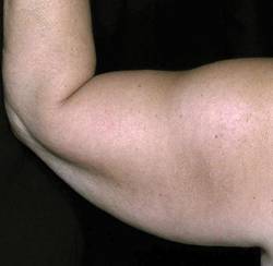
A patient was treated with 20 W to 30 W of power, with a total energy of 31.5 kj per arm. |
"Traditional liposuction of the upper arms has been a challenge for patients, in particular for those with great volumes or a great amount of laxity," Dr. Lori Brightman said at the annual meeting of the American Society for Laser Medicine and Surgery. "This is a concern … for patients who want reduction of their upper arms or abdomen, because we're concerned about the loose skin that can result after removing the volume."
Dr. Brightman, of the Laser and Skin Surgery Center of New York, and her associates enrolled seven female patients with Fitzpatrick skin types II-IV to undergo treatment with BodyTite, a novel, bipolar, radiofrequency-assisted liposuction device with coagulation and simultaneous aspiration.
Manufactured by Invasix, the investigational device delivers radiofrequency energy from an internal electrode to an external electrode, which results in coagulation of adipose, vascular, and fibrous tissue, as well as heating of the soft-tissue matrix.
BodyTite features online monitoring of skin temperature, as well as monitoring of power and tissue impedance. It is powered by a 50- and 75-watt bipolar radiofrequency output system.
|
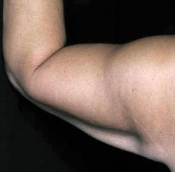
At baseline, the arm circumference was 39 cm. It reduced to 34 cm at week 12. Images courtesy Dr. Lori Brightman, Laser and Skin Surgery Center of New York |
The patients were 21-58 years old. They received one treatment on each upper arm and were assessed at 1, 3, 6, and 12 weeks. Assessments included the patient's body weight, standardized circumference tape measurements, two- and three-dimensional photography, caliper measurements, and biomechanical tissue characterization (BTC), which is an assessment of skin elasticity.
After standard tumescent anesthesia, the investigators treated the deeper planes of the upper arm skin, followed by the superficial planes; they treated to the loss of resistance at each level. The target epidermal temperature ranged from 38° C to 40° C, the radiofrequency energy ranged from 20 W to 35 W, and the total energy per zone ranged from 20.7 kJ to 30.4 kJ.
Dr. Brightman reported that the mean circumferential reduction among six of the seven patients who achieved a 6-week follow-up was 2.7 cm. Of the five patients who achieved a 12-week follow-up, the mean circumferential reduction was 4.3 cm.
The reduction in skinfold thickness (as measured by calipers) increased over time - from 7.5 mm between baseline and 3 weeks to 13.5 mm between baseline and 12 weeks. Mean skin laxity as measured by BTC went from 68% at 3 weeks to 58% at 12 weeks. Reduction in upper arm circumference was also evident on two- and three-dimensional photographs, Dr. Brightman said.
"You can improve and reduce the size of these larger arms with greater laxity with this bipolar radiofrequency device," she concluded.
Dr. Brightman disclosed that she is a clinical investigator for a number of device companies, including Invasix. She also holds stock options in the company.
PHOENIX - Treatment with a radiofrequency-assisted liposuction device resulted in improved reduction of upper arm circumference up to 12 weeks post treatment, results from a small single-center study showed.
|
| |
A patient was treated with 20 W to 30 W of power, with a total energy of 31.5 kj per arm. |
"Traditional liposuction of the upper arms has been a challenge for patients, in particular for those with great volumes or a great amount of laxity," Dr. Lori Brightman said at the annual meeting of the American Society for Laser Medicine and Surgery. "This is a concern … for patients who want reduction of their upper arms or abdomen, because we're concerned about the loose skin that can result after removing the volume."
Dr. Brightman, of the Laser and Skin Surgery Center of New York, and her associates enrolled seven female patients with Fitzpatrick skin types II-IV to undergo treatment with BodyTite, a novel, bipolar, radiofrequency-assisted liposuction device with coagulation and simultaneous aspiration.
Manufactured by Invasix, the investigational device delivers radiofrequency energy from an internal electrode to an external electrode, which results in coagulation of adipose, vascular, and fibrous tissue, as well as heating of the soft-tissue matrix.
BodyTite features online monitoring of skin temperature, as well as monitoring of power and tissue impedance. It is powered by a 50- and 75-watt bipolar radiofrequency output system.
|
|
At baseline, the arm circumference was 39 cm. It reduced to 34 cm at week 12. Images courtesy Dr. Lori Brightman, Laser and Skin Surgery Center of New York |
The patients were 21-58 years old. They received one treatment on each upper arm and were assessed at 1, 3, 6, and 12 weeks. Assessments included the patient's body weight, standardized circumference tape measurements, two- and three-dimensional photography, caliper measurements, and biomechanical tissue characterization (BTC), which is an assessment of skin elasticity.
After standard tumescent anesthesia, the investigators treated the deeper planes of the upper arm skin, followed by the superficial planes; they treated to the loss of resistance at each level. The target epidermal temperature ranged from 38° C to 40° C, the radiofrequency energy ranged from 20 W to 35 W, and the total energy per zone ranged from 20.7 kJ to 30.4 kJ.
Dr. Brightman reported that the mean circumferential reduction among six of the seven patients who achieved a 6-week follow-up was 2.7 cm. Of the five patients who achieved a 12-week follow-up, the mean circumferential reduction was 4.3 cm.
The reduction in skinfold thickness (as measured by calipers) increased over time - from 7.5 mm between baseline and 3 weeks to 13.5 mm between baseline and 12 weeks. Mean skin laxity as measured by BTC went from 68% at 3 weeks to 58% at 12 weeks. Reduction in upper arm circumference was also evident on two- and three-dimensional photographs, Dr. Brightman said.
"You can improve and reduce the size of these larger arms with greater laxity with this bipolar radiofrequency device," she concluded.
Dr. Brightman disclosed that she is a clinical investigator for a number of device companies, including Invasix. She also holds stock options in the company.
Attorney: Proper Pathology Slide Storage Helps Mitigate Lawsuits
SAN DIEGO - Reliably tracking and storing pathology slides is key to avoiding the possibility of major liability, Robert A. Cosgrove said at a melanoma conference sponsored by the Scripps Clinic.
To illustrate his point, Dr. Cosgrove presented the personal injury case of a woman with advanced melanoma. About 1 year after she presented to the physician-defendant for evaluation of a lesion on her toe, she required lymph node resection and amputation of the toe.
The plaintiff claimed that the physician had negligently lost biopsy results, resulting in a delay in proper treatment.
The defendant claimed that the plaintiff's lesion had disintegrated at the time it was originally removed and left nothing for further testing.
The jury awarded the plaintiff a settlement in excess of $8 million.
"I've had this come up in a number of cases," said Mr. Cosgrove, a health care attorney based in Encinitas, Calif. "The problem with losing or misplacing pathology slides [is that] it raises questions about whether that was done intentionally or not. Plus, it puts you in a difficult position of being able to prove or disprove what your diagnosis is or your interpretation of the slides."
Another case he discussed underscored the importance of solid chart documentation. The case was brought to trial by the heirs of a 36-year-old woman who died of melanoma while under the defendant dermatologist's care. The heirs contended that the defendant failed to diagnose the decedent's shoulder mole, which was cancerous, in a timely manner.
The defendant denied liability and contended that the decedent was comparatively negligent and failed to make her appointments for yearly full skin exams.
"It is imperative to have documentation in your charts - photo documentation, and in this situation, documentation of yearly examinations. … You can rely on custom and practice, but it's better evidence for the jury when you present the case at trial to have documentation in the chart to that effect," Mr. Cosgrove said.
The defendant also contended that the cancerous mole was not the same as the one that she complained about, and that the decedent would have survived at the time of diagnosis because the lesion was thin.
"Many of these cases are presented at time of trial 2 and 3 years downstream," Mr. Cosgrove noted. "So it comes down to chart documentation. Was it adequately documented in terms of the location of where the lesion was when you evaluated the patient?"
In this case the jury ruled in favor of the defense.
During a question and answer session, a meeting attendee asked Mr. Cosgrove how long physicians should keep medical records on file. "I would find out what the legal policy is in your state on this and follow it accordingly," he advised. "It's important to keep good records and to not do anything after the fact to change your records. By changing the record, by altering the records, by falsifying information, or by destroying records you expose yourself to tremendous personal liability."
Mr. Cosgrove said he had no relevant financial conflicts to disclose.
ASLMS: Complexion Blending Tops Hair Removal in Skin of Color Practice
PHOENIX - In the last 2 years, complexion blending has been the most sought-after procedure in Dr. Eliot F. Battle Jr.'s practice.

One of his favorite devices for treating dark spots, melasma, postinflammatory hyperpigmentation, and scarring is the microsecond Nd:YAG laser. With this device, "we're driving heat down deep and trying to break up pigment … that's been encapsulated by membranes," Dr. Battle said. "For a dark spot to last for months or years, we know there is a pigment problem in the dermis. Fractional technology has also been used, but we treat conservatively. I don't ever use the manufacturer's parameters. I don't cause erythema or edema. I'm always under the threshold of redness and swelling."
Speaking during the annual meeting of the American Society for Laser Medicine and Surgery on laser treatment for skin of color patients, Dr. Battle's take-home advice was to reduce conventional parameters and fluences to avoid erythema and edema.
"There are many lasers to use on Caucasians for complexion blending," he said.
"For skin of color, we're limited to the microsecond Nd:YAG, Q-switched Nd:YAG, KTP [potassium-titanyl-phosphate], and infrared lasers."
Prior to treatment he asks patients about their ethnic background because ethnicity plays as much a role in how their skin reacts as their skin color, he said.
He also gets a sun protection history to find out whether he will be treating brown skin that is sun protected, as is the case with some people of Pacific-Asian ancestry, or whether he will be treating brown skin that has been suntanned, as is the case with some people of African-American ancestry.
"Don't make assumptions," he said. "Don't think that the person in front of you, who has the same skin color as the person you just treated, has the same response to lasers, because the ethnicity of the patient dictates how the skin reacts as much as their skin color."
He also recommends investing in an imaging system, such as the VISIA Complexion Analysis system, "that allows us to see what we're treating."
To maximize results of complexion blending, consider concomitant nonlaser "old-school" treatments, such as aesthetic peels, hydroquinone, and a hyfrecator, he said.
"The true art of cosmetic therapy is integrating spa treatments, cosmeceuticals,

prescription medications, and lasers all together," remarked Dr. Battle, who is also on the dermatology faculty at Howard University, Washington. "Lasers don't speak for themselves, particularly when treating pigmented lesions such as melasma and dark spots. We really have to take advantage of the product side of this, also. We have most of our patients on alpha hydroxy acids, Retin A, vitamin C, and we use hydroquinone bleaching cream up to 12%. I'm not bashful with hydroquinone. I monitor them very closely, but I think our patients can benefit from a higher strength."
Dr. Battle disclosed that he has received equipment, discounts, and honoraria from numerous medical device companies, including Canfield.
PHOENIX - In the last 2 years, complexion blending has been the most sought-after procedure in Dr. Eliot F. Battle Jr.'s practice.
One of his favorite devices for treating dark spots, melasma, postinflammatory hyperpigmentation, and scarring is the microsecond Nd:YAG laser. With this device, "we're driving heat down deep and trying to break up pigment … that's been encapsulated by membranes," Dr. Battle said. "For a dark spot to last for months or years, we know there is a pigment problem in the dermis. Fractional technology has also been used, but we treat conservatively. I don't ever use the manufacturer's parameters. I don't cause erythema or edema. I'm always under the threshold of redness and swelling."
Speaking during the annual meeting of the American Society for Laser Medicine and Surgery on laser treatment for skin of color patients, Dr. Battle's take-home advice was to reduce conventional parameters and fluences to avoid erythema and edema.
"There are many lasers to use on Caucasians for complexion blending," he said.
"For skin of color, we're limited to the microsecond Nd:YAG, Q-switched Nd:YAG, KTP [potassium-titanyl-phosphate], and infrared lasers."
Prior to treatment he asks patients about their ethnic background because ethnicity plays as much a role in how their skin reacts as their skin color, he said.
He also gets a sun protection history to find out whether he will be treating brown skin that is sun protected, as is the case with some people of Pacific-Asian ancestry, or whether he will be treating brown skin that has been suntanned, as is the case with some people of African-American ancestry.
"Don't make assumptions," he said. "Don't think that the person in front of you, who has the same skin color as the person you just treated, has the same response to lasers, because the ethnicity of the patient dictates how the skin reacts as much as their skin color."
He also recommends investing in an imaging system, such as the VISIA Complexion Analysis system, "that allows us to see what we're treating."
To maximize results of complexion blending, consider concomitant nonlaser "old-school" treatments, such as aesthetic peels, hydroquinone, and a hyfrecator, he said.
"The true art of cosmetic therapy is integrating spa treatments, cosmeceuticals,
prescription medications, and lasers all together," remarked Dr. Battle, who is also on the dermatology faculty at Howard University, Washington. "Lasers don't speak for themselves, particularly when treating pigmented lesions such as melasma and dark spots. We really have to take advantage of the product side of this, also. We have most of our patients on alpha hydroxy acids, Retin A, vitamin C, and we use hydroquinone bleaching cream up to 12%. I'm not bashful with hydroquinone. I monitor them very closely, but I think our patients can benefit from a higher strength."
Dr. Battle disclosed that he has received equipment, discounts, and honoraria from numerous medical device companies, including Canfield.
Non–Sentinel Node Metastasis Important Prognostic Factor in Melanoma
ST. LOUIS - Non–sentinel node status is an independent prognostic factor for disease-free survival and overall survival in patients with melanoma, results from a large study with long-term follow-up demonstrated.
“This effect is not simply a result of greater numbers of positive nodes,” Dr. Russell E. Brown said at a symposium sponsored by the Society of Surgical Oncology.
The findings support the notion that the sentinel lymph node (SLN) acts as the first line of defense against metastasis in melanoma and that metastasis past the SLN predicts a poorer prognosis.
“Status of sentinel lymph nodes is the most powerful predictor of outcomes for early-stage melanoma,” said Dr. Brown, a surgical oncology fellow at the University of Louisville, Kentucky. “Non–sentinel nodes (NSN) are evaluated at completion of lymph node dissection and are found in 8%-33% of complete lymph node dissection specimens.”
Multiple factors are used to predict NSN metastases, he said, including include age, tumor thickness, number of sentinel lymph nodes, SLN tumor burden, ulceration, male gender, perinodal intralymphatic tumor, and extranodal extension. While single-institution studies have shown a survival disadvantage for NSN-positive patients, Dr. Brown and his associates hypothesized that metastasis beyond the SLN to the non–sentinel nodes is an important predictor of survival.
To test their hypothesis, the researchers performed a post hoc analysis of data from 2,335 patients in the Sunbelt Melanoma Trial, a prospective, randomized study of patients aged 18-70 years at 79 centers in the United States and Canada. The Sunbelt Melanoma Trial was the world’s largest prospective study of melanoma, led by Dr. Kelly McMasters, chair of the department of surgery at the University of Louisville. Patients were treated between 1997 and 2003 and were clinically node negative, with primary tumors at least 1 mm thick. All subjects underwent SLN biopsy and were followed for a median of 68 months.
The researchers used Kaplan-Meier analysis and univariate and multivariate analysis to compare disease-free survival and overall survival in three groups of patients: 1,988 who were SLN negative (group 1), 296 who were SLN-only positive (group 2), and 51 who were SLN and NSN positive (group 3). Dr. Brown reported that the 5-year disease-free survival rates for groups 1, 2, and 3 were 86%, 65%, and 43%, respectively, while the 5-year overall survival rates were 86%, 65%, and 49%, respectively.
Univariate analysis revealed the following significant independent predictors of decreased disease-free survival: SLN positivity (hazard ratio of 2.4), NSN positivity (HR 4.57), increased total number of positive lymph nodes (HR 1.18), increased ratio of positive lymph node count to total lymph node count (HR 1.03), increased Breslow thickness (HR 3.28), presence of ulceration (HR 2.76), Clark level of 4 or greater (HR 1.65), axial primary (HR 1.45), and male gender (1.31).
When the total number of positive lymph nodes and NSN status were evaluated in a multivariate Cox model with other univariate predictors of decreased disease-free survival, NSN status remained statistically significant (HR 3.95), while the total number of positive lymph nodes and lymph node ratio did not (both were an HR of 1.01).
Univariate analysis also revealed the following significant predictors of decreased overall survival: SLN positivity (HR 2.29), NSN positivity (HR 3.6), increased total number of positive lymph nodes (HR 1.16), increased ratio of positive lymph node count to total lymph node count (HR 1.03), increased Breslow thickness (HR 2.96), presence of ulceration (HR 2.92), Clark level of 4 or greater (HR 1.41), axial primary (HR 1.71), male gender (1.68), and increased age (HR 1.24).
“The results of Dr. Brown’s study are important and suggest that there is a biological significance to the sentinel nodes as the first line of defense against metastasis in melanoma,” said Dr. McMasters, a melanoma specialist.
When the total number of positive lymph nodes and NSN status were evaluated in a multivariate Cox model with other univariate predictors of decreased overall survival, NSN status remained statistically significant (HR 2.81) while the total number of positive lymph nodes and lymph node ratio did not (HR of 1.03 and 1.11, respectively).
Dr. Brown noted that lymphovascular invasion, the number of involved nodal basins, the presence of a vertical growth phase, and evidence of regression were not significant predictors of overall survival.
Dr. McMasters has received grant support for the study from Schering Oncology/Biotech and has been on the company’s speakers bureau.
Dr. Brown had no relevant financial disclosures.
Presentation of Lupus Differs Before and After Age 50
SAN DIEGO — Although an estimated 80% of patients with systemic lupus erythematosus acquire the disease before age 50 years, beware of ruling out the potential for diagnosing new cases in older patients.
“It certainly can happen,” Dr. Bevra H. Hahn said at the annual meeting of the North American Menopause Society. “It's not rare, so it's okay to let it cross your mind.”
The clinical presentation of SLE that develops before the age of 50 differs from that of disease that occurs later in life, said Dr. Hahn, chief of rheumatology at the University of California, Los Angeles, Ronald Reagan Medical Center. SLE that develops before age 50 is marked by development of nephritis, anti-DNA antibodies, malar rash, and/or discoid lupus. This relatively early-onset form of SLE causes less organ damage in general. Mortality in this patient population “is primarily from active lupus or from infections that relate to being sick and having immunosuppressive therapies,” Dr. Hahn said.
SLE that develops after age 50 is marked by cardiac and pulmonary problems. “I see a lot of patients who present with heart failure or with pericarditis, or arrhythmias, and they have a strongly positive antinuclear antibody, so it's fine to screen for ANA in that situation,” she said.
Compared with their younger counterparts, patients who develop SLE after age 50 are also more likely to have arthritis, Sjögren's syndrome, and a high damage index. “Their mortality is more from coronary artery disease or stroke, some from infection, and less of it from antilupus medications,” said Dr. Hahn, who is also a professor of medicine at UCLA. “For this group, preventive care is very important for the coronary artery disease.”
Several clinical studies have shown that the use of the antimalarial drug hydroxychloroquine reduces damage over time, whether the lupus is mild or severe. “We think most people should be on [hydroxychloroquine] if there is not a contraindication,” she said. Hydroxychloroquine use has a slight risk for associated retinal damage, Dr. Hahn added.
Dr. Hahn's approach to treatment involves first determining whether the disease threatens life or organs. If the disease is mild, she will consider a number of different agents to lessen pain, fatigue, and rash, including NSAIDs; topical agents, such as steroids or tacrolimus; sunscreen with SPF 50; antimalarials, such as hydroxychloroquine at a dose of 200-400 mg/day or quinacrine dosed at 100 mg/day; DHEA (dehydroepiandrosterone) dosed at 100-200 mg/day; or low-dose prednisone.
She cautioned that the use of NSAIDs “can bump creatinine levels and cause aseptic meningitis in patients with lupus. It's not common, but it happens.”
“The three phases of treatment for severe SLE are induce improvement, maintain improvement, and prevent damage,” she said. For these patients, high-dose prednisone “saves lives. It causes cataracts, osteoporosis, and diabetes, but it really is life saving.”
Other options to consider for patients with severe lupus include antimalarials and the cytotoxic agents mycophenolate mofetil (CellCept), azathioprine (Imuran), and cyclophosphamide (Cytoxan). Other agents showing promise include rituximab, which is a monoclonal antibody against the protein CD20, and belimumab, an investigational human monoclonal antibody.
One certainty of the disease is that infections are common. Dr. Hahn said that 60% of the time, fever that occurs in SLE is the result of infection; the other 40% result from flare. “If you think these patients have an infection, treat it as early as possible,” she advised.
Survival rates in SLE patients have improved in recent decades. Currently, Dr. Hahn said, the 10-year survival for SLE patients without nephritis stands at about 95%, whereas the 10-year survival for SLE patients with nephritis stands at about 88%.
“This is so different from 20-30 years ago,” she remarked. “We've had tremendous improvement in our therapies and our diagnoses.”
Disclosures: Dr. Hahn had no relevant financial disclosures.
About 5% of Melanoma Patients Have Subsequent Melanoma
SAN DIEGO — The chances of a patient's developing multiple primary melanomas over a lifetime is a real phenomenon, with an incidence ranging from 2% to 8% among patients who have had a first melanoma, or an average of about 5%.
“That's significantly higher than if we apply the risk of melanoma to all fair-skinned people in the country,” Dr. William M. Burrows observed at a melanoma update sponsored by the Scripps Clinic.
Of patients who develop additional melanomas, about 80% develop one in addition to the original, 15% develop two, and the remainder develop three or more.
“In my practice I have about four people I follow who have had five or six primary melanomas,” said Dr. Burrows, who has practiced dermatology for nearly 40 years and is currently with the division of dermatology at Scripps Clinic Rancho Bernardo in San Diego.
He went on to note that the risk of multiple primary melanomas is twofold higher among men, and that the majority of subsequent primary melanomas (70%) occur on a different anatomical site, while 30% occur on the same site. “They have the same distribution as melanomas in general,” he said.
The majority of subsequent primary melanomas occurs after 2 months, while 30% occur within 2 months or less.
Depth of invasion is similar to national statistics for all primary melanomas. “But the second primary melanoma tends to be thinner than the first one, which makes sense,” Dr. Burrows commented. “After the first primary melanoma we raise our index of suspicion on lesions that are irregular. In addition, the patient has a significant level of worry.”
Recognized risk factors for multiple melanomas include presence of atypical/dysplastic nevi, family history, and early age of onset.
According to a retrospective review of 1,258 melanoma patients treated at the Scripps Clinic between 1990 and 2000, 149 (12%) developed multiple primary lesions, which is more than double the national incidence. “This could be due to one of two things,” Dr. Burrows said. “One is the criteria that are used in making the diagnosis of melanoma in situ. I wonder if we [at Scripps] diagnose melanoma in situ more often, as opposed to others who might sign it out as atypical melanocytic hyperplasia or another worrisome diagnosis.”
The other possibility is that incidence of melanoma is rising. “We know that we are making the diagnosis of primary melanomas at a younger age than we did 20 years ago,” Dr. Burrows said.
In the Scripps series, 75% of patients had two primary melanomas, 15% had three, and the remainder had four or more. The average age at initial primary melanoma diagnosis was 64 among men and 56 among women.
Nearly half of the patients (49%) developed subsequent melanomas less than 3 years after their initial primary melanoma diagnoses.
When managing patients with multiple melanomas, a full skin exam during initial work-up and follow-up intervals is essential, he said. “Follow-up should be lifelong.”
Disclosures: Dr. Burrows said he had no financial conflicts.
'I have about four people I follow who have had five or six primary melanomas.'
Source DR. BURROWS
Salt Tax Could Save $22.4 Billion in Medical Costs
If the United States government were to establish a tax on sodium consumption or—preferably—to collaborate with food manufacturers to reduce sodium in processed foods, the cardiovascular benefits and cost savings to the nation would be significant, a novel analysis demonstrated.
Either strategy would likely avert acute strokes and myocardial infarctions, increase quality-adjusted life-years, and save billions of dollars in medical costs, investigators led by Dr. Crystal M. Smith-Spangler of Stanford (Calif.) University reported onlineMar the Archives of Internal Medicine. But collaboration with industry “is likely to be more effective than a sodium tax and appears to be an appropriate first step [toward] reducing population sodium intake and the burden of cardiovascular disease,” the researchers concluded.
Using a computer-simulated Markov model and evidence from other populations, the researchers assessed dietary, health, and cost effects of the two strategies. Government collaboration with food manufacturers to cut sodium in processed foods would achieve a 9.5% reduction in sodium intake among adults aged 40-85 years in the United States, the team estimated. A sodium tax—similar to the cigarette tax—would decrease sodium intake among adults by 6%.
To assess the economic and cardiovascular impacts of those reductions, the researchers drew data from sources including the Medical Panel Expenditure Survey, the Framingham Heart Study, and the DASH (Dietary Approaches to Stop Hypertension) trial. The impacts of the two strategies were measured in 2008 U.S. dollars, quality-adjusted life-years, and the number of MIs and strokes averted.
The researchers projected that the 9.5% sodium-intake reduction from government-industry collaboration would result in a 1.25–mm Hg decrease in mean systolic blood pressure of adults aged 40-85 years. In turn, this blood pressure reduction “would avert 513,885 strokes and 480,358 MIs and increase life-years lived by over 1.3 million over the lifetime of U.S. adults [aged] 40-85 years alive today, saving $32.1 billion in direct medical costs,” the team concluded. More than half of the direct medical cost savings (56%) would come from fewer persons needing chronic care after MIs or strokes.
In comparison, the 6% decrease in sodium intake from a tax “would avert 327,892 strokes and 306,137 MIs and increase life-years lived by 840,000, saving $22.4 billion over the lifetime of adults [aged 40-85 years who are] alive today,” the researchers said.
Dr. Smith-Spangler and her associates said that their model found greater benefits from the strategy of collaborating with food manufacturers “because we have assumed that manufacturers do not reformulate their products in response to a tax and currently demand for salty foods is relatively unresponsive to prices.” A tax's effect would increase if there were more acceptable alternatives to salty foods available, decreasing demand for the latter with higher prices, they said.
The team acknowledged that the reduction of sodium intake from either strategy “might lead people to consume more fats and sugars or simply more calories, leading to other health risks. It will be important to monitor the unintended consequences, both positive and negative, of any strategy for decreasing sodium intake.”
Disclosures: The study was supported by grants from the VA Palo Alto (Calif.) Health Care System, the Stanford University Management Science and Engineering Advisory Board Fellowship Fund, the National Defense Science and Engineering Graduate Fellowship, the National Science Foundation Graduate Fellowship, and the Department of Veterans Affairs.
Overhaul of TBI Classification Is Explored
SAN DIEGO – The way Dr. Geoffrey T. Manley sees it, the classification of traumatic brain injury needs an extreme makeover.
For the past 35 years, clinicians have relied on symptomatology from the Glasgow Coma Scale (GCS) to classify traumatic brain injuries (TBIs) as mild, moderate, or severe, but such emphasis on symptoms “misses the point,” Dr. Manley, chief of neurotrauma and vice chairman of the department of neurosurgery at the University of California, San Francisco, said at the annual meeting of the American Association for the Advancement of Science.
“The brain is not like the heart, where if you lose a certain percentage of your heart muscle then you'll have an unexpected reduction in cardiac function. The brain is a unique organ in that it's an organ of functional connectivity. You can have very small lesions in discrete pathways, which can have a phenomenal impact on outcome. Many of these lesions can only be seen with MRI, which is not routinely used for TBI.”
He went on to note that the GCS was developed “before the advent of CT scans, so this is a very old system that we're using.”
In 2007, Dr. Manley and a working group of TBI experts–including Prof. Sir Graham Teasdale, who developed the GCS–convened to explore the potential for improving TBI classification (J. Neurotrauma 2008;25:719-38). It became clear to the group, Dr. Manley said, “that if we were going to try to change the field, we were going to have to start defining a common set of data elements and technical standards so that we could be able to collect the same information on patients from site to site and to make sure that assessment tools are applied in the same way.”
Common data elements are needed in TBI research “because accurate collection of structured data is essential, especially if you want to do meta-analyses and if you want to share data,” he added. “It reduces time, cost, and effort of initiating clinical trials and provides opportunities for lessons learned and best practices, even if a trial isn't considered successful.”
The group's recommendations call for the following:
▸ Broaden TBI trials. They should include less severely injured patients.
▸ Improve CT imaging classification. “The systems that we use now are different from hospital to hospital and radiologist to radiologist,” Dr. Manley said. “There is no standardization.”
▸ Increase use of early MRI. “Many of us have seen a lot of value in using MRIs,” he said. “We will get an MRI on a stroke patient in a moment, but we almost never get an MRI in a TBI patient. This is a cultural change that needs to happen in this field.”
▸ Examine phase II trials and surrogate end points more closely. TBI patients “have such a long recovery: an injury, an acute hospitalization, rehabilitation, and then you look at an outcome at 6 months or a year,” he said. “Lots of things happen during that time period.”
▸ Develop more complex statistical and bioinformatics tools. TBI studies “aren't like cancer studies,” he said. “You can't phenotype these patients as well as you can in studies of other diseases. We need some novel statistical methods to deal with the realities of studying these patients with life-threatening diseases.”
In March 2009, Dr. Manley and about 160 other representatives from 49 agencies and institutes, including the Department of Defense, the Department of Education, and the National Institute of Neurological Disorders and Stroke convened in Washington to begin an unprecedented effort to develop standards for TBI data collection and to better define and classify TBI.
The experts were divided into numerous work groups charged with assembling white papers on specific areas of TBI research, including demographics and acute clinical assessment, biospecimens and biomarkers, neuroimaging, posttraumatic stress disorder, and outcome measures. White papers from the various work groups will be published later in 2010, and the TBI Common Data Elements will be available online at www.nindscommondataelements.org
The next step in this multidisciplinary effort is to establish a prospective, multivariate TBI database to validate common data elements, including a contemporary snapshot of TBI and treatment and a cross-sectional overview of patients. “So rather than saying we're looking at patients with severe, moderate, or mild injury, we're going to be agnostic to [the label of] mild, moderate, and severe, and we'll look across the entire spectrum of injury,” Dr. Manley explained.
The database “will also allow the researchers to validate prognostic models, establish process indicators, and develop improved TBI classification,” he said.
Dr. Manley and his colleagues were recently awarded a National Institutes of Health Grand Opportunities Challenge Grant to pilot this effort. The global goal is to develop, test, and refine standards for data collection in TBI studies in a multicenter observational study of 1,000 patients at high-volume TBI centers, including UCSF; the University of Pittsburgh; Mount Sinai School of Medicine, New York; and Seton Hospital in Austin, Tex. Dr. Andrew Maas, of University Hospital Antwerp, Brussels, is leading a European group of TBI investigators that also will be contributing to this effort.
“If we really want to transform TBI research, we're going to have to work in multidisciplinary teams,” Dr. Manley concluded. “We need this infrastructure. We need the appropriate collaboration and tools.”
Dr. Manley had no relevant financial conflicts of interest to disclose.
The Glasgow Coma Scale was developed 'before the advent of CT scans, so this is a very old system that we're using.'
Source DR. MANLEY