User login
Doug Brunk is a San Diego-based award-winning reporter who began covering health care in 1991. Before joining the company, he wrote for the health sciences division of Columbia University and was an associate editor at Contemporary Long Term Care magazine when it won a Jesse H. Neal Award. His work has been syndicated by the Los Angeles Times and he is the author of two books related to the University of Kentucky Wildcats men's basketball program. Doug has a master’s degree in magazine journalism from the S.I. Newhouse School of Public Communications at Syracuse University. Follow him on Twitter @dougbrunk.
Novel Device Uses Ultrasound to Treat Acne
PHOENIX – An investigational intense-therapy ultrasound device safely and effectively treated mild to moderate acne in a preliminary study.
The device, manufactured by Xthetix Inc., was used to treat 5-15 active acne lesions on one side of the face for 5 consecutive days in 18 women and 7 men with a mean age of 26 years. Of the patients, about 84% showed significant improvement after five treatment sessions.
According to a poster presented during the annual meeting of the American Society for Laser Medicine and Surgery, energy generated by the device is delivered to the level of the sebaceous gland, "raising the temperature 5-15 degrees Celsius above ambient skin temperature. It is designed not to cause thermal coagulation, but to increase the tissue temperature for a period of time sufficient to produce a therapeutic effect."
In an interview, lead investigator Dr. Bill Halmi said that although there are a few devices currently on the market that use heat to reduce the duration of an inflammatory acne papule, the machine uses ultrasound to heat the targeted area.
"Putting the energy intradermally into the pilosebaceous unit is the unique aspect of the device," he said. "It changes the inner workings of the gland, kills bacteria, [and] peaks the inflammatory process, among other things."
Each treatment session lasted about 15 minutes. The contralateral side of the face did not receive any ultrasound treatment and served as the control.
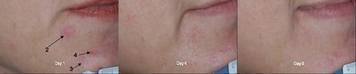
The researchers took photographs on all visit days and a masked reviewer conducted posttreatment and facial acne assessments at every visit. Each patient completed daily pain and acne assessments.
Dr. Halmi, who practices dermatology in Phoenix, reported that the greatest percentage of lesion clearance (30%) occurred on day 4 of treatment. "This differs from 'blue light' devices that expect to see improvements after at least 1 week of treatment," he said. "Going into the study we really weren't certain that we would see an immediate effect from the device. We were certainly pleased to discover that this ultrasound device measurably reduced the duration of an acne lesion."
A majority of patients (84%) reported moderate to significant improvement of their treated sides during the course of treatment, while 40% experienced mild transient erythema that lasted less than 30 minutes. The average pain score was 3 on a scale of 1-10, with 10 being the most severe.
The findings suggest that "there is a role for heat in reducing the duration of an acne lesion," Dr. Halmi said. "The advantage ultrasound has is that it can target that heat to the depth desired."
He cautioned that the study is preliminary and that, while encouraging, "repeated studies using more patients will help confirm our findings. Additionally, the role of ultrasound in the prophylactic treatment of acne remains to be revealed."
Dr. Halmi disclosed that he is a paid consultant for Xthetix.
PHOENIX – An investigational intense-therapy ultrasound device safely and effectively treated mild to moderate acne in a preliminary study.
The device, manufactured by Xthetix Inc., was used to treat 5-15 active acne lesions on one side of the face for 5 consecutive days in 18 women and 7 men with a mean age of 26 years. Of the patients, about 84% showed significant improvement after five treatment sessions.
According to a poster presented during the annual meeting of the American Society for Laser Medicine and Surgery, energy generated by the device is delivered to the level of the sebaceous gland, "raising the temperature 5-15 degrees Celsius above ambient skin temperature. It is designed not to cause thermal coagulation, but to increase the tissue temperature for a period of time sufficient to produce a therapeutic effect."
In an interview, lead investigator Dr. Bill Halmi said that although there are a few devices currently on the market that use heat to reduce the duration of an inflammatory acne papule, the machine uses ultrasound to heat the targeted area.
"Putting the energy intradermally into the pilosebaceous unit is the unique aspect of the device," he said. "It changes the inner workings of the gland, kills bacteria, [and] peaks the inflammatory process, among other things."
Each treatment session lasted about 15 minutes. The contralateral side of the face did not receive any ultrasound treatment and served as the control.
The researchers took photographs on all visit days and a masked reviewer conducted posttreatment and facial acne assessments at every visit. Each patient completed daily pain and acne assessments.
Dr. Halmi, who practices dermatology in Phoenix, reported that the greatest percentage of lesion clearance (30%) occurred on day 4 of treatment. "This differs from 'blue light' devices that expect to see improvements after at least 1 week of treatment," he said. "Going into the study we really weren't certain that we would see an immediate effect from the device. We were certainly pleased to discover that this ultrasound device measurably reduced the duration of an acne lesion."
A majority of patients (84%) reported moderate to significant improvement of their treated sides during the course of treatment, while 40% experienced mild transient erythema that lasted less than 30 minutes. The average pain score was 3 on a scale of 1-10, with 10 being the most severe.
The findings suggest that "there is a role for heat in reducing the duration of an acne lesion," Dr. Halmi said. "The advantage ultrasound has is that it can target that heat to the depth desired."
He cautioned that the study is preliminary and that, while encouraging, "repeated studies using more patients will help confirm our findings. Additionally, the role of ultrasound in the prophylactic treatment of acne remains to be revealed."
Dr. Halmi disclosed that he is a paid consultant for Xthetix.
Fascinated by Medicine's Past
One of Dr. Robert E. Greenspan's favorite pastimes as a child was a trip with his parents from his home in Montgomery County, Md., to the National Museum of Health and Medicine in Washington. There, he would gaze at the medical instruments, artifacts, and specimens on display.
“I liked medicine, liked seeing the anatomy and seeing the specimens,” recalled Dr. Greenspan, a nephrologist based in Northern Virginia. “I always had an interest and pursued that.”
Fascinated by the illustrations and photographs of old medical instruments and descriptions of how they were used, he began to collect medically themed books from second-hand bookstores while he was in medical school at the University of Maryland, Baltimore. In 1974, he acquired a first edition of the 1892 book by Sir William Osler, “The Principles and Practice of Medicine.”
Before long, he began to assemble his own collection of books and medical artifacts. In 2006, he self-published a 12-chapter, 596-page book entitled, “Medicine: Perspectives in History and Art” (Alexandria, Va.: Ponteverde Press, 2006), which contains stories of medical procedures, dentistry, pharmacy, and quackery revealed through paintings, photographs, poetry, anecdotes, and instrumentation (www.medicalhistoryandart.com
“No one had ever written a medical textbook that was personal,” he said. “Most of the books you see are about events and discoveries. The thing that interests me is not really names, dates, and who discovered what. I'm more interested in learning from the past directly through letters, documents, and instruments.”
The book's chapter on general surgery contains excerpts from a letter written by Fanny Burney, a patient who underwent a mastectomy in 1811 without anesthesia at the hand of Napoleon's surgeon, Dominique-Jean Larrey. After the surgical knife cut into her breast, “I began a scream that lasted unintermittently during the whole time of the incision—and I almost marvel that it rings not in my ears still!” she wrote. “So excruciating was the agony. When the wound was made, and the instrument was withdrawn, the pain seemed diminished, for the air that suddenly rushed into those delicate parts felt like a mass of minute but sharp and forked poniards, that were tearing the edges of the wound.”
Another remarkable letter acquired by Dr. Greenspan appears in the book's chapter on trauma. Written by 18-year-old Union soldier Merari Bunajah Stevens to a friend back home, the letter describes the 1864 Battle of Cold Harbor, Va., one of the Civil War's bloodiest battles. Mr. Stevens' father became a casualty of that conflict.
“Men were struck down as if by a great scythe—like grass in haying time—and it was here that Father was struck,” Mr. Stevens wrote. “He was at my side during the charge when I suddenly heard him groan. Just then I looked over and saw Father fall to the ground, his uniform soaked with blood. He was shot but a few feet from me. The bullet went in a little back and a little above the left groin and exited near the right hip. On the battlefield, I took off his belts and clothes and then with my Bowie, took the ball out on the field as it was located near the surface under his cartridge box. I have it now in my pocket.”
Two years ago, Dr. Greenspan received an e-mail from Mr. Stevens' great-great-granddaughter. “She had the bullet,” Dr. Greenspan said. “It turns out that the soldier had become a physician in the Midwest. She sent me pictures of him when he was a soldier, and his two sons who became surgeons. So I have a picture of him, the bullet, and his family, which all came out of this letter. That is fascinating to me.”
Dr. Greenspan finds such letters in books or as stand-alone items on eBay or by browsing the inventory of online booksellers. “I fish around and I find medical history books that no one's ever heard of,” he said. “I'll buy it for $4 or $5 and I'll scan it and there might be a great story or a great quote or it will lead me into another area.”
The book also contains images of artwork depicting the practice of medicine dating back to the age of Hippocrates, as well as photographs of medical instruments in his collection. These images include ceramic leech jars and homeopathic sets from the 1800s, a bottle of prescription whiskey from the early 1900s, and the Abrams “dynamizer” and “reflexophone,” a quack machine from the early 1900s purported to help clinicians diagnose diseases ranging from syphilis to cancer.
His books and artifacts are on display at home, where he often hosts interested curators, collectors, writers, and artists. “That's how I learn, and that's one of the reasons for the book: not to put it in a corner and in a locked room, but to share those instruments,” he said. “A lot of the instruments in the book no one will ever see unless they see them pictured.”
According to his Web site, Dr. Greenspan's “wish list” of items to acquire includes a tobacco enema set, a von Helmholtz ophthalmoscope, and a Kolff rotating drum dialysis machine from around 1943.
Dr. Greenspan considers his avocation more of an obligation than an amusement. “As a collector, putting this book together in a personal way was a challenge,” he said. “History is not interesting unless it involves people: why they did things and how they did things. From my standpoint, that has been missing in medical books. If I don't take the time to pursue these antiques and related stories, it won't be done.”
'I'm more interested in learning from the past directly through letters, documents, and instruments.'
Source DR. GREENSPAN
Dr. Robert E. Greenspan's collection includes a Dieulafoy's aspirator, manufactured around 1880.
This all-metal general operating set, circa 1900, also has found its way into Dr. Greenspan's collection.
Source Photos courtesy Dr. Robert E. Greenspan
A photo of Union soldier Merari Bunajah Stevens is displayed with the bullet that hit his father.
Cryolipolysis Appears Safe for Fat Reduction
PHOENIX - Using noninvasive cryolipolysis for fat-layer reduction in the flanks and back fat pads caused no serious adverse events and no reports of skin damage or pigment changes up to 6 months after treatment, results from a large multicenter study showed.
Side effects included "mostly erythema and edema, numbness/tingling, and a little blanching," Dr. A. Jay Burns said at the annual meeting of the American Society for Laser Medicine and Surgery. "Occasionally there was bruising on the treatment site as well, but all side effects resolved by 6 months."
Invented by Dr. Dieter Manstein and Dr. R. Rox Anderson at Massachusetts General Hospital and Harvard Medical School in Boston, cryolipolysis is noninvasive way to cool fat cells to induce lipolysis without damage to other tissue. Pleasanton, Calif.-based Zeltiq currently owns exclusive rights to the technology and markets a cryolipolysis device known as the Zeltiq system.
The Zeltiq system is cleared in the European Union and Canada for noninvasive fat-layer reduction through cold-assisted lipolysis. It is also cleared by the Food and Drug Administration for various applications related to skin cooling during dermatologic treatments, with a pending application for noninvasive fat-layer reduction. The system is available for sale on a limited basis to physicians in the European Union, Canada, and the United States and other selected international markets.
At the meeting, Dr. Burns reported side effect data from two prospective, controlled clinical studies in which researchers at 16 centers used the Zeltiq system to treat 341 patients on their flanks and back fat pads. Efficacy was evaluated by ultrasound, comparison of pre- and post-treatment photographs, and physician assessment. Final follow-up occurred at 6 months or less.
"There were no reports of serious adverse effects, and all side effects were transient," said Dr. Burns, a plastic surgeon in Dallas who is also with the University of Texas Southwestern Medical Center. The most common post-treatment side effect was erythema, followed by hypoesthesia/tingling, edema, bruising, temperature sensation, blanching, and pressure at the treatment site.
ASLMS: Tips for Effective Laser Hair Removal in Darker Skin
PHOENIX - When performing laser hair removal in patients with Fitzpatrick skin types IV-VI, keep in mind that darker skin contains more melanin in the epidermis, which acts as a competing chromophore, "so the risk for epidermal injury is higher," Dr. Andrew F. Alexis said.
Other tips to remember when treating this patient population include the melanocytes' tendency "to be labile in response to injury and inflammation, so there is an increased risk of dyschromia," Dr. Alexis said at the annual meeting of the American Society for Laser Medicine and Surgery. "This can manifest as hyperpigmentation or hypopigmentation following laser hair removal procedures."
He went on to note that fibroblasts in the dermis "are also more reactive to injury, so there is an increased risk of hypertrophic scars and keloids when performing surgical or some cosmetic procedures. In addition, having curved follicles [which are primarily seen in people of African descent] is associated with an increased prevalence of a number of follicular disorders."
His guidelines for performing laser hair removal safely on skin of color include longer wavelengths, lower fluences, longer pulse durations, and increased epidermal cooling.
"You want longer wavelengths because we're trying to maximize the ratio of follicular bulb temperature to epidermal temperature," explained Dr. Alexis, director of the Skin of Color Center at St. Luke's & Roosevelt Hospitals, New York, and assistant professor of dermatology at Columbia University.
"Using wavelengths in the near infrared range, we are at a lower point on the melanin absorption curve and therefore are compromising some efficacy, but this is the range that is considered safest for darker skin types," he noted.
A review found that the long-pulsed 1064-nm laser had the lowest incidence of adverse events in dark-skinned patients, followed by the long-pulsed 800-nm or 810-nm diode laser (J. Drugs Dermatol. 2007;6:40-6).
"For patients with type IV or V skin, the diode laser is appropriate, as long as you are using long pulse durations," he said, "but for the darker skin types, particularly type VI, the 1064-nm Nd:YAG laser is the best choice for safety reasons."
In one study, researchers used a 1064-nm laser with contact cooling to treat pseudofolliculitis barbae in 37 patients with skin types IV, V, and VI. They found that the highest fluences tolerated by the epidermis were 50 J/cm2 for skin type VI and 100 J/cm2 for skin types IV and V (J. Am. Acad. Dermatol. 2002;47:263-70).
In another study, researchers used a 1064-nm laser with contact cooling for hair removal in 36 patients with skin types I-VI (Dermatol. Surg. 2004;30:13-7). Patients underwent three consecutive treatments at 4- to 6-week intervals. For skin types V-VI, investigators used a 30-millisecond pulse duration and a fluence of 30-45 J/cm2 on the face and 35-50 J/cm2 on nonfacial sites. Six months post treatment, the mean facial hair reduction ranged from 41% to 46%, while the mean hair reduction on the body ranged from 48% to 53%.
Based on the results of these and other studies, and from his own clinical experience, Dr. Alexis recommends a pulse duration of 100 milliseconds or 400 milliseconds when using a 810-nm diode laser and a duration of 20-30 milliseconds when using a 1064-nm Nd:YAG laser with contact cooling.
One recent study used a 1064-nm Nd:YAG laser with lower than traditional fluences for treating 22 patients with skin types IV-VI who had pseudofolliculitis barbae (Dermatol. Surg. 2009;35:98-107). Patients underwent five weekly treatments on the neck with a fluence of 12 J/cm2 and a pulse duration of 20 milliseconds and a spot size of 10 mm. At 4 weeks follow-up, the papule count had been reduced by a mean of 91% and dyspigmentation by a mean of 60%.
In another recent development, researchers studying a novel diode laser with a fluence of 5-10 J/cm2 used at a repetition rate of 10 Hz found that it resulted in less pain, faster treatment, and fewer adverse events compared with one pass of a high-fluence diode laser at 25-40 J/cm2 in Fitzpatrick skin types I-V (J. Drugs Dermatol. 2009;8:s14-7).
Options for epidermal cooling include cold gel, contact cryogen or forced air, and post treatment with ice packs for 10-15 minutes. Devices for contact cooling feature either a sapphire tip or chilled copper plate. Regardless of which type of contact cooling chosen, Dr. Alexis suggested "using a slower treatment speed in order to ensure adequate cooling before delivering pulse."
Dr. Alexis said that he had no relevant financial conflicts.
Aspirin Use May Boost Survival After Breast Cancer
Major Finding: Aspirin use after the diagnosis of stage I-III breast cancer was associated with a multivariate adjusted relative risk of breast cancer death of 1.07 among those who used aspirin 1 day per week, 0.29 for those who used aspirin 2-5 days per week, and 0.36 for those who used aspirin 6-7 days per week.
Data Source: Responses from 4,164 female RNs in the Nurses' Health Study who were diagnosed with stage I-III breast cancer between 1976 and 2002. The women were observed until June 2006 or until they died, whichever came first.
Disclosures: Supported by a grant from the National Institutes of Health. The researchers indicated they had no conflicts of interest.
Aspirin use after the diagnosis of stage I-III breast cancer was associated with a decreased risk of breast cancer death and distant recurrence, results from the ongoing Nurses' Health Study demonstrated.
The study is believed to be the first to report a survival advantage among women with breast cancer who take aspirin.
“If confirmed, our results may broaden the scope of interventions available to reduce breast cancer–related death and mortality,” researchers led by Dr. Michelle D. Holmes of at Harvard Medical School and Harvard School of Public Health, Boston, reported.
They emphasized that the results “may be generalizable only to longer term breast cancer survivors,” described as those who have lived long enough after diagnosis to report aspirin use after diagnosis (about 4 years). “Fortunately, almost 90% of women diagnosed with breast cancer live at least 5 years. Thus, our findings have considerable clinical importance.”
For the study, the researchers drew from questionnaires to evaluate aspirin use among 4,164 female registered nurses in the Nurses' Health Study who were diagnosed with stage I, II, or III breast cancer between 1976 and 2002, and who were observed until they died or until June 2006, whichever came first (J. Clin. Oncol. 2010 Feb. 16 [doi:10.1200/JCO.2009.22.7918
Dr. Holmes and her associates reported that 314 deaths attributed to breast cancer and 400 distant recurrences occurred during the study period. Compared with women who never used aspirin, the multivariate adjusted relative risk of breast cancer death was 1.07 among those who used aspirin 1 day per week, 0.29 for those who used aspirin 2-5 days per week, and 0.36 for those who used aspirin 6-7 days per week. “Results did not differ appreciably when stratified by stage, [body mass index], menopausal status, or [estrogen receptor] status.”
Aspirin use had a similar impact on distant recurrence of cancer. Compared with women who never used aspirin, the multivariate adjusted relative risk of distant recurrence was 0.91 among those who used aspirin 1 day per week, 0.40 for those who used aspirin 2-5 days per week, and 0.57 for those who used aspirin 6-7 days per week.
Suspect Ventriculomegaly? Try Fetal MRI
Major Finding: The diagnosis of ventriculomegaly was confirmed on fetal MRI in 21 cases (84%), while the fetal brain appeared normal on fetal MRI in the remaining 4 cases (16%).
Data Source: A single-center retrospective study of 25 cases of isolated lateral ventriculomegaly.
Disclosures: None was reported.
SAN DIEGO — Fetal magnetic resonance imaging can be considered when isolated lateral ventriculomegaly is suspected on ultrasound, results from a single-center study demonstrated.
At the annual meeting of the American Institute of Ultrasound in Medicine, Dr. Fadi Mirza presented findings from a retrospective study of sonographically suspected cases of isolated ventriculomegaly seen at Columbia University Medical Center, New York, between 2004 and 2008 that subsequently underwent fetal brain MRI. The researchers compared the results of the fetal MRI and the last prenatal ultrasound before it.
Fetal ventriculomegaly was defined as dilated lateral ventricles with a mean transverse diameter of 10 mm or greater. The condition can be unilateral or bilateral and is often referred to as severe when the diameter is greater than 15 mm, said Dr. Mirza of the division of maternal-fetal medicine in the department of obstetrics and gynecology at the medical center.
It's regarded as isolated when the fetus has no other anomalies.
Isolated ventriculomegaly has been associated with multiple etiologies, “yet it's most often secondary to congenital aqueductal stenosis,” he said.
“Narrowing may be developmental or acquired, such as by infection, intraventricular hemorrhage, or a mass.
Diagnosis is generally based on prenatal ultrasound, but in recent years there has been a growing role for fetal MRI.”
In this small, retrospective study, there were 25 suspected cases of isolated ventriculomegaly on prenatal ultrasound that subsequently underwent fetal MRI during the 4-year study period. The mean gestational age at the time of the index ultrasound was 28.1 weeks while the mean gestational age at the time of the fetal MRI was 29.5 weeks.
Of the 25 cases, Dr. Mirza reported that 16 (64%) were unilateral and 22 (88%) were mild. The diagnosis of ventriculomegaly was confirmed on fetal MRI in 21 (84%) cases, while the fetal brain appeared normal on fetal MRI in the remaining 4 cases (16%).
Additional intracranial abnormalities that were not seen on prenatal ultrasound were identified in 5 of the 21 (24%) abnormal cases. These included three cases of agenesis of the corpus callosum, one case of interventricular hemorrhage, and one case of focal encephalomalacia.
Dr. Mirza acknowledged limitations of the study, including its retrospective design and small sample size, making it difficult to generalize its results and make management recommendations.
When asked by a meeting attendee what the clinical implications of the findings are, Dr. Mirza said that “the jury's still out on this question.
“In our small series, we did identify some cases where MRI did show other abnormalities, although I think it's still hard to appreciate what this practice would change in terms of our management.
“With the growing role of fetal MRI, we should have a better idea in the future.”
He said more prospective studies with a systematic approach are needed to better evaluate and refine the role of fetal MRI in cases of isolated ventriculomegaly.
This MRI shows dilatation of the occipital horns of the lateral ventricles.
Source Courtesy Dr. Fadi Mirza
Postmastectomy Radiation May Not Be Needed
Major Finding: When the researchers examined the overall locoregional recurrence related to pathologic lymph node status, patients with one to three positive nodes had a 10-year locoregional recurrence rate of 4.3%, compared with 2.1% in patients with negative nodes, a significant difference.
Data Source: A study of 1,019 women treated at M.D. Anderson Cancer Center between 1997 and 2002.
Disclosures: None was reported.
ST. LOUIS — Locoregional recurrence rates in patients with T1 and T2 breast cancer treated with surgery and adjuvant systemic therapies stand at less than 3%, a rate far lower than the rates of more than 15% reported some decades ago, according to results of a single-center retrospective study.
The findings call into question the routine use of postmastectomy radiotherapy in this population, a practice based on landmark studies completed more than 20 years ago, Dr. Ranjna Sharma said at a symposium sponsored by the Society of Surgical Oncology. I
In turn, those studies influenced 2007 guidelines from the National Comprehensive Cancer Network, which “suggested strong consideration of the use of postmastectomy radiation in patients with one to three positive axillary nodes,” said Dr. Sharma, a fellow in surgical oncology at M.D. Anderson Cancer Center, Houston.
“This has resulted in a shift in clinical practice, with more patients receiving radiotherapy and many centers beginning to deny immediate breast reconstruction in patients with early-stage disease,” she said.
Previously reported high rates of locoregional recurrence after mastectomy “may have been due to patients presenting with much more advanced disease, without routine margin control, without adequate axillary surgery, and without modern-day systemic therapies. Therefore, our hypothesis is that patients treated in the present era will have much lower rates of local-regional recurrence,” Dr. Sharma said.
She and her associates performed a retrospective study to determine the risk of locoregional recurrence (LRR) in 1,019 patients with T1 and T2 tumors with 0 and 1-3 positive lymph nodes who had a mastectomy at M.D. Anderson between 1997 and 2002. None of the patients received preoperative chemotherapy or postoperative radiotherapy.
The study's lead author was Dr. Henry M. Kuerer, a professor in the surgical oncology department at M.D. Anderson. The researchers used the Kaplan-Meier method and the Cox proportional hazards regression model for analysis. The primary outcome was LRR, calculated from the date of initial surgery.
Dr. Sharma reported findings from a median follow-up of 7.5 years. The median age of study participants was 54 years, and the median number of lymph nodes removed during axillary lymph node dissection was 16. About one-third of patients (32%) received a sentinel lymph node biopsy as their initial nodal staging procedure, and 77% received chemotherapy and/or hormonal therapy.
“Patients in the series had very early breast cancer, with 79% having T1 tumors,” Dr. Sharma said.
More than one-quarter of patients (26%) had nodal metastasis, “with the majority having only one node with metastases. Only 2% of the patients in the series had three nodes with metastases,” she reported.
The median time to LRR was 3.8 years.
“There were a total of only 23 local-regional recurrences in this series: 48% were isolated and 13% occurred before distant metastases development,” she said.
About 40% of LRRs were seen concurrently or after the development of distant metastases.
Slightly more than half (52%) involved the chest wall, and 48% involved the regional nodes alone.
Univariate analysis revealed that LRR was associated with age younger than 40 years, tumor size, positive nodes, higher-stage estrogen receptor–negative tumors, and use of systemic adjuvant therapy.
In a multivariate analysis, the only independent predictor of LRR in this series was age younger than 40 years. The 10-year rate of LRR was 11.1% in patients younger than 40 years and less than 3% for patients older than age 40.
When the researchers examined the overall LRR rate related to pathologic lymph node status, they found that patients with one to three positive nodes had a 10-year LRR rate of 4.3%, compared with 2.1% in patients with negative nodes, a significant difference. However, Dr. Sharma noted, “this rate of locoregional recurrence is still substantially lower than the previously reported rates in series of patients who were treated several decades ago.”
The researchers then examined the risk of LRR in patients with one positive node.
“Interestingly, the 10-year rate of locoregional recurrence of 3.3% was not significantly different from the 2.1% risk of locoregional recurrence in patients with node-negative disease,” Dr. Sharma said. For patients with two positive nodes, the 10-year LRR rate was 7.9%, which was significantly higher than the 2.1% rate of patients with node-negative disease.
Dr. Sharma said that no conclusions could be drawn about the risk of LRR in patients with three positive nodes because “this was a very rare event,” with only 21 patients meeting the criteria.
The 10-year LRR rate among patients with T2 tumors and lymph node metastases was 9.7%, which was significantly higher than the rate of less than 3% for their counterparts with T1 tumors with and without nodal metastases and those with T2 tumors and no nodal metastases.
“Although the 9.7% rate of locoregional recurrence is significantly higher compared with the other groups of patients, it is important to note that at this level of risk, experts have estimated that approximately 100 women would need to be treated to potentially obtain a survival benefit in one or two patients,” Dr. Sharma said.
She also reported that 25 contralateral breast cancers developed during the study period.
Although the median time to occurrence of contralateral breast cancer was longer than the time to development of LRR (a median of 7.2 years vs. 3.8 years, respectively), there was no significant difference between the rates of a locoregional or a contralateral event between the two groups (5.5% vs. 2.7%, respectively).
This was true for node-negative patients and for patients with one to three positive lymph nodes.
Dr. Sharma acknowledged the potential for bias in the study, including the fact that patients who received neoadjuvant chemotherapy were excluded.
“Those patients likely presented with more advanced disease. That might have altered results if those patients were included.”
Dr. Ranjna Sharma (left) and Dr. Henry M. Kuerer found low locoregional recurrence rates in T1, T2 breast cancer patients treated with surgery, adjuvants.
Source Courtesy Sandra Soule
My Take
Prospective Trial Is Needed
The findings reported at the Society of Surgical Oncology are provocative, and may cause a shift away from a trend to recommend postmastectomy radiation to all women with lymph node–positive breast cancer—even those with one to three involved axillary nodes. Although the report is based on a large number of subjects, it is limited by its retrospective nature and the few local and regional recurrences that did develop.
Even with those limitations, the study finds that certain subsets of patients appear to have a risk of local recurrence (in the absence of postoperative radiation therapy or primary chemotherapy) that is not unlike the risk in patients with T1 and axillary node–negative disease. This suggests that the use of postmastectomy radiation therapy may be better directed toward patients with larger primary tumors, those with more than one involved axillary node, and those younger than 40 years at diagnosis. To confirm these findings, a prospective trial of postmastectomy radiation therapy, or not, will be required.
DR. WILLIAM J. GRADISHAR is a professor of medicine at Northwestern University and director of Breast Medical Oncology at the Robert H. Lurie Comprehensive Cancer Center in Chicago.
Study Looks at US Predictors of Adenomyosis
Major Finding: When transvaginal imaging of the uterus reveals a heterogeneous myometrium, subendometrial myometrial cysts, striations, and increased vascularity in the area of interest, adenomyosis can be diagnosed with a sensitivity greater than 90%, a specificity greater than 66%, a positive predictive value greater than 86%, and an accuracy of greater than 84%.
Data Source: A single-center review of 85 patients diagnosed with adenomyosis by ultrasound.
Disclosures: None was reported.
SAN DIEGO — The best clinical predictors of adenomyosis on ultrasound are a heterogeneous myometrium, myometrial cysts, striations, and increased vascularity in the area of interest.
Those are the key findings from a single-center study that set out to determine which findings suggestive of adenomyosis increase the accuracy and diagnostic capabilities of transvaginal ultrasound and to evaluate whether the presence of fibroids impacts the correct diagnosis of the condition.
Defined as the presence of ectopic endometrial glands and stroma in the myometrium, the incidence of adenomyosis is believed to range from 1% to 70%, “depending on how meticulous the pathologic analysis is,” Dr. Francisco Cruz-Pachano said at the annual meeting of the American Institute of Ultrasound in Medicine.
“Symptoms that accompany adenomyosis are dysmenorrhea, pelvic pain, and menorrhagia, but most of the time it is asymptomatic. Up to two-thirds of cases coexist with other pathology, most of the time with fibroids,” he said.
Dr. Cruz-Pachano of the ultrasound division of the department of obstetrics and gynecology at the University of Miami and his associates reviewed the pathology reports and images of 85 patients diagnosed with adenomyosis by ultrasound at Jackson Memorial Hospital in Miami from August 2007 to August 2009. They made the diagnosis of adenomyosis on the basis of seven different ultrasound findings: an enlarged and globular uterus, a heterogeneous myometrium, myometrial cysts, a difference in the anteroposterior myometrial thickness, endomyometrial striations, loss of the endomyometrial junction, and myometrial hypervascularity. Each finding was analyzed to assess the diagnostic accuracy by itself and as part of the group.
Among the patients (mean age, 46), 54% were Hispanic; 38%, black; and 8%, white. The researchers initially divided patients into two groups: 59 with adenomyosis and 26 without the condition. They further divided the adenomyosis group into 25 who had adenomyosis and 34 who had adenomyosis plus fibroids, to control for potential confounders.
Dr. Cruz-Pachano said that the four most significant clinical findings of adenomyosis on ultrasound were increased vascularity (100%), the presence of myometrial cysts (86%), striations (82%), and a heterogeneous appearance (78%).
He noted that when transvaginal imaging of the uterus reveals these four findings, adenomyosis can be diagnosed with a sensitivity greater than 90%, a specificity greater than 66%, a positive predictive value greater than 86%, and an accuracy of more than 84%.
In the subset of patients with adenomyosis and fibroids, the only significant clinical findings were increased vascularity (64%) and striations (55%). “When fibroids are present, the enlarged/globular and loss of endomyometrial junction characteristics can't be used to diagnose adenomyosis,” Dr. Cruz-Pachano said. “When fibroids are present, the visualization of striations or increased vascularity has an increased sensitivity to help diagnose adenomyosis.”
Echogenic linear striations (above) are one of four ultrasound findings shown to predict adenomyosis.
Source Courtesy Dr. Francisco Cruz-Pachano
Fascinated by Medicine's Past
One of Dr. Robert E. Greenspan's favorite pastimes as a child was a trip with his parents from his home in Montgomery County, Md., to the National Museum of Health and Medicine in Washington. There, he would gaze at the array of medical instruments, artifacts, and specimens on display.
“I liked medicine, liked seeing the anatomy and seeing the specimens,” recalled Dr. Greenspan, a nephrologist based in Northern Virginia. “I always had an interest and pursued that.”
Fascinated by the illustrations and photographs of old medical instruments and descriptions of how they were used, Dr. Greenspan began to collect medically themed books from secondhand bookstores while he was in medical school at the University of Maryland, Baltimore. In 1974, he acquired a first edition of the 1892 book by Sir William Osler, “The Principles and Practice of Medicine.”
Before long, he began to assemble his own collection of books and medical artifacts. In 2006, he self-published a 596-page book entitled “Medicine: Perspectives in History and Art” (Alexandria, Va.: Ponteverde Press), which contains stories of medical procedures, dentistry, pharmacy, and quackery revealed through paintings, photographs, poetry, and anecdotes (www.medicalhistoryandart.com
“To me, no one had ever written a medical textbook that was personal,” he said. “Most of the books you see are about events and discoveries. The thing that interests me is not really names, dates, and who discovered what. I'm more interested in learning from the past directly through letters, documents, and instruments.”
The book's chapter on general surgery contains excerpts from a letter written by Fanny Burney, a patient who underwent a mastectomy in 1811 without anesthesia at the hand of Napoleon's surgeon, Dominique-Jean Larrey. After the surgical knife cut into her breast, “I began a scream that lasted unintermittently during the whole time of the incision—and I almost marvel that it rings not in my ears still!” she wrote. “So excruciating was the agony. When the wound was made, and the instrument was withdrawn, the pain seemed diminished, for the air that suddenly rushed into those delicate parts felt like a mass of minute but sharp and forked poniards, that were tearing the edges of the wound.”
Another remarkable letter acquired by Dr. Greenspan appears in the book's chapter on trauma. Written by 18-year-old Union soldier Merari Bunajah Stevens to a friend, the letter describes the 1864 Battle of Cold Harbor, Va., one of the Civil War's bloodiest battles. Mr. Stevens' father became a casualty of that conflict.
“Men were struck down as if by a great scythe—like grass in haying time—and it was here that Father was struck,” Mr. Stevens wrote. “He was at my side during the charge when I suddenly heard him groan. Just then I looked over and saw Father fall to the ground, his uniform soaked with blood. He was shot but a few feet from me. The bullet went in a little back and a little above the left groin and exited near the right hip. On the battlefield, I took off his belts and clothes and then with my Bowie, took the ball out on the field as it was located near the surface under his cartridge box. I have it now in my pocket.”
Two years ago, Dr. Greenspan received an e-mail from Mr. Stevens' great-great-granddaughter. “She had the bullet,” he said. “It turns out that the soldier had become a physician in the Midwest. She sent me pictures of him when he was a soldier, and his two sons who became surgeons. So I have a picture of him, the bullet, and his family, which all came out of this letter. That is fascinating to me.”
Dr. Greenspan finds such letters in books or as stand-alone items on eBay, or by browsing online booksellers. “I fish around and I find medical history books that no one's ever heard of,” he said. “I'll buy it for $4 or $5 and I'll scan it and there might be a great story or a great quote or it will lead me into another area.”
The book also contains images of artwork depicting the practice of medicine dating back to the age of Hippocrates, as well as photographs of medical instruments in his collection. These images include ceramic leech jars and homeopathic sets from the 1800s, a bottle of prescription whiskey from the early 1900s, and the Abrams “dynamizer” and “reflexophone,” a quack machine from the early 1900s purported to help clinicians diagnose diseases ranging from syphilis to cancer.
He keeps his medical books and artifacts on display at home, where he often hosts interested curators, collectors, writers, and artists. “That's how I learn, and that's one of the reasons for the book: not to put it in a corner and in a locked room, but to share those instruments,” he said. “A lot of the instruments in the book no one will ever see unless they see them pictured.”
Dr. Greenspan's “wish list” of items to acquire includes a tobacco enema set, a von Helmholtz ophthalmoscope, and a Kolff rotating drum dialysis machine from around 1943.
Dr. Greenspan sees his avocation as a kind of obligation. “As a collector, putting this book together in a personal way was a challenge,” he said. “History is not interesting unless it involves people: why they did things and how they did things. From my standpoint, that has been missing in medical books. If I don't take the time to pursue these antiques and related stories, it won't be done.”
A photo of Union soldier Merari Stevens and the bullet he removed from his father.
Source Photos courtesy Dr. Robert E. Greenspan
'I'm more interested in learning from the past directly through letters, documents, and instruments.'
Source DR. GREENSPAN
Dr. Robert E. Greenspan collected this general operating set, circa 1900.
This Dieulafoy's aspirator from circa 1880 is part of the collection.
ASLMS: SlimLipo Gets High Marks from Both Patients and Physicians
PHOENIX - Cosmetic results following treatment with a 924-nm laser-assisted lipolysis device achieved high patient and physician satisfaction 18 months after treatment, a single-center study demonstrated.
At the annual meeting of the American Society for Laser Medicine and Surgery, Dr. Robert A. Weiss reported treatment results with the SlimLipo laser system (Palomar Medical Technologies Inc.), which he began using in April 2008.

|
This patient is shown before, and 3 months after, treatment with laser-assisted lipolysis. Photo courtesy Dr. Robert A Weiss |
The procedure involves up to three repetitions of a laser pass to melt fat and heat connective tissue followed by suction, said Dr. Weiss, director of the Maryland Laser, Skin, and Vein Institute in Hunt Valley, Md.
Data were obtained from 88 patients during follow-up visits at 1, 5, 6, 12, and 18 months. Areas treated included the abdomen (27), flanks/waist (31), thighs (14), arms (10), the submental area (12), and the knees (7). Some patients had more than one area treated.
The average fluence delivered was 28.6 kJ during the first pass, except for the submental area, which averaged 10 kJ, while the abdominal area averaged 35 kJ.
"Overall patient satisfaction was high, with 98% reporting that they were very satisfied with the results," said Dr. Weiss, also of the department of dermatology at Johns Hopkins University in Baltimore. "Investigator assessment correlated with patient ratings, as 88% showed marked improvement at 6 and 12 months. Side effects included mild bruising in 39% of patients, which lasted for an average of 4 days." No skin burns were observed.
Dr. Weiss noted that with standard tumescent liposuction, drainage of tumescent lasts between 72 and 96 hours. With the SlimLipo, he said, duration of drainage was 12-24 hours, with a median of 14 hours. "We believe that is due to the laser channels that we make in the fat layer," he said.
Postoperative pain was rated as mild, with only five patients (6%) requiring more than nonsteroidal anti-inflammatory medications for pain control.
"No contour irregularities were noted and curved surfaces remained smooth without depressions," Dr. Weiss said. "Results achieved at 6 months were sustained for the longest follow-up at 2 years."
Dr. Weiss disclosed that he serves as a paid consultant for several medical device companies, including Palomar. He has also received speaker honoraria and research grants from the company.
PHOENIX - Cosmetic results following treatment with a 924-nm laser-assisted lipolysis device achieved high patient and physician satisfaction 18 months after treatment, a single-center study demonstrated.
At the annual meeting of the American Society for Laser Medicine and Surgery, Dr. Robert A. Weiss reported treatment results with the SlimLipo laser system (Palomar Medical Technologies Inc.), which he began using in April 2008.
|
|
This patient is shown before, and 3 months after, treatment with laser-assisted lipolysis. Photo courtesy Dr. Robert A Weiss |
The procedure involves up to three repetitions of a laser pass to melt fat and heat connective tissue followed by suction, said Dr. Weiss, director of the Maryland Laser, Skin, and Vein Institute in Hunt Valley, Md.
Data were obtained from 88 patients during follow-up visits at 1, 5, 6, 12, and 18 months. Areas treated included the abdomen (27), flanks/waist (31), thighs (14), arms (10), the submental area (12), and the knees (7). Some patients had more than one area treated.
The average fluence delivered was 28.6 kJ during the first pass, except for the submental area, which averaged 10 kJ, while the abdominal area averaged 35 kJ.
"Overall patient satisfaction was high, with 98% reporting that they were very satisfied with the results," said Dr. Weiss, also of the department of dermatology at Johns Hopkins University in Baltimore. "Investigator assessment correlated with patient ratings, as 88% showed marked improvement at 6 and 12 months. Side effects included mild bruising in 39% of patients, which lasted for an average of 4 days." No skin burns were observed.
Dr. Weiss noted that with standard tumescent liposuction, drainage of tumescent lasts between 72 and 96 hours. With the SlimLipo, he said, duration of drainage was 12-24 hours, with a median of 14 hours. "We believe that is due to the laser channels that we make in the fat layer," he said.
Postoperative pain was rated as mild, with only five patients (6%) requiring more than nonsteroidal anti-inflammatory medications for pain control.
"No contour irregularities were noted and curved surfaces remained smooth without depressions," Dr. Weiss said. "Results achieved at 6 months were sustained for the longest follow-up at 2 years."
Dr. Weiss disclosed that he serves as a paid consultant for several medical device companies, including Palomar. He has also received speaker honoraria and research grants from the company.