User login
Doug Brunk is a San Diego-based award-winning reporter who began covering health care in 1991. Before joining the company, he wrote for the health sciences division of Columbia University and was an associate editor at Contemporary Long Term Care magazine when it won a Jesse H. Neal Award. His work has been syndicated by the Los Angeles Times and he is the author of two books related to the University of Kentucky Wildcats men's basketball program. Doug has a master’s degree in magazine journalism from the S.I. Newhouse School of Public Communications at Syracuse University. Follow him on Twitter @dougbrunk.
Gene Therapy Trial Yields Promising Outcomes
Major Finding: Patients with NYHA stage III or IV heart failure who received gene therapy with MYDICAR had cardiovascular-related hospital stays that averaged 2 fewer days than those who received placebo.
Data Source: A phase II study of 39 patients enrolled in the CUPID trial.
Disclosures: Celladon Corp. funded the trial. Dr. Greenberg said that he had no relevant financial disclosures.
SAN DIEGO – In a phase II study of patients with advanced heart failure, gene therapy with MYDICAR was found to be safe and was associated with benefit in clinical outcomes, symptoms, functional status, and cardiac structure.
Deficiency of the protein SERCA2a is central to the progression of heart failure, resulting in abnormal calcium transfer and impairing myocardial relaxation and contraction, Dr. Barry H. Greenberg said
MYDICAR, manufactured by Celladon Corp., is an enzyme replacement therapy that is designed to restore levels of SERCA2a. A viral vectoridelivers the SERCA2a gene.
The objectives of the study, known as CUPID (Calcium Up-Regulation by Percutaneous Administration of Gene Therapy in Cardiac Disease), were to evaluate safety and feasibility and to explore the activity and efficacy of MYDICAR in patients with advanced heart failure, said Dr. Greenberg, professor of medicine and director of the Advanced Heart Failure Treatment Program at the University of California, San Diego.
To be eligible for the trial, patients had to be 18–75 years old, have New York State Heart Association class III or IV heart failure caused by an ischemic or nonischemic etiology, have a maximal oxygen consumption of 20 mL/kg per minute or less, have a left ventricular fraction of 35% or less, and be on a stable, optimized heart failure regimen for 30 days.
Dr. Greenberg and his colleagues randomized 39 patients to one of three MYDICAR doses or to placebo, and all were treated via single intracoronary infusion. All patients were observed for 12 months, with primary analysis after 6 months of therapy.
CUPID's primary efficacy end point was defined as evidence of success in one of four areas: group-level analysis, individual analysis, time-to-event analysis, and duration of cardiovascular-related hospitalization analysis. All were deemed significant (P less than .2).
CUPID met the primary efficacy end point for high-dose MYDICAR treatment group vs. placebo in three of the four areas. In the group-level analysis, significant improvements were seen in the treatment group, compared with the placebo group, in 6-minute walk tests and end systolic volume, with no clinically significant worsening in any end point and numerical superiority to placebo in all other end points.
In the individual analysis, the mean efficacy “score” for the treatment was significantly greater than that of the placebo group. In the time-to-death analysis, the treatment group scored numerically better than the placebo group, but theidifferencedias not significantce.
In the duration of cardiovascular-related hospitalization analysis, the duration of stay was significantly shorter for the treatment group than for the placebo group (mean, 2 fewer days).
Improvements were seen in the treatment group, vs. placebo, in 6-minute walk tests and end systolic volume.
Source DR. GREENBERG
Serotonin Flags Progression of Heart Failure
SAN DIEGO – Plasma levels of serotonin were significantly elevated in patients with decompensated systolic heart failure, compared with patients in the compensated state and with normal controls, according to a single-center study.
The finding suggests that serotonin has an active role in the progression of heart failure, researchers led by Dr. Ahmed M. Selim wrote in a poster at the meeting.
“More studies should be done to test the sensitivity, specificity, and prognostic value of serotonin as a marker for congestive heart failure and also to investigate the therapeutic benefits of the medications affecting this pathway,” noted the researchers of the cardiology department at Albert Einstein College of Medicine, New York.
Dr. Selim and his associates collected plasma serotonin levels from 29 patients who were admitted with decompensated heart failure, 61 patients with stable heart failure, and 22 normal controls. They excluded patients on medications affecting serotonin receptors and those with pulmonary hypertension. All heart failure patients were on stable doses ofheedications and had left-ventricular ejection fractions of 40% or less;, wntrols had a mean ejection fraction of 59%. Ppatients' mean age was 55 years;, a% were male.
The mean serotonin level in controls was 2.4 ng/mL, vs. 4.1 ng/mL in the compensated group and 11.8 ng/mL in the decompensated group,. ndependent of age, race, renal function, diabetes mend hypertension. “All results were highly significant,” the researchers wrote.
Shortage of Providers in Rural Areas May Affect Suicide Rate
Major Finding: Alaska, the state with the lowest population density (1.2 people/square mile), has the highest suicide rate (23.1 deaths/100,000 population. Conversely, the District of Columbia, which has the highest population density (9,316 people/square mile), has the lowest suicide rate (5.3 deaths/100,000 population).
Data Source: National Center for Health Statistics and Bureau of Census Data from 2004.
Disclosures: Dr. D'Mello disclosed that he is a member of the speakers' bureau for Pfizer, AstraZeneca, and Schering.
Suicides in the United States tend to occur more often in rural compared with urban locations, but findings from a new study suggest that the relative scarcity of mental health care providers in rural areas may factor in to the association.
“More than 600 counties in the United States don't have any health care provider, and 1,600 counties don't have a mental health care provider,” Dr. Dale D'Mello of the department of psychiatry at Michigan State University, Lansing, said during a press briefing held at the annual meeting of the American Psychiatric Association.
“One recent study found that the distribution of health care providers is imbalanced in rural compared with urban areas.
“We also know that the prevalence of conditions like depression is equal in urban and rural areas.”
To examine the association between population density, the availability of mental health providers, and suicide rates in each state, Dr. D'Mello and his associates evaluated National Center for Health Statistics and Bureau of Census Data information from the year 2004. Suicide rates were defined as deaths per 100,000 population.
They correlated these data with the population density (persons per square mile) and the number of mental health providers per 100,000 population.
Dr. D'Mello reported that a significant powerful negative correlation was observed between the rate of suicide and population density.
For example, Alaska, the state with the lowest population density (1.2 people/square mile), also has the highest suicide rate (23.1 deaths/100,000 population). On the other hand, the District of Columbia, which has the highest population density (9,316 people/square mile), has the lowest suicide rate (5.3/100,000 population).
“The other states start off between these two extremes,” Dr. D'Mello said. “I work in Lansing, Michigan, but I do telepsychiatry in a county 200 miles away that's rural. I'm interested in this area, because the suicide rate in [that] population is twice that of people I see face-to-face in Lansing.”
Similar and striking significant negative correlations were observed between the distribution of psychiatrists, psychologists, and social workers and suicide rates.
Certain economic and cultural factors also may contribute to the higher rate of suicides in rural population, he said. “Economic causes include the recent change in the rural economy from agriculture to manufacturing, resulting in a disproportionate burden of rural poverty in most countries around the globe,” he said.
As for cultural factors, Dr. D'Mello continued, “there are a large number of books and papers written about the rural culture as being different from urban culture, focusing on the male farmer – the importance based on self-reliance and independence in forming the identity of the male farmer, and the fact that health is correlated with productivity. When productivity declines, a crisis occurs.”
One possible solution to improving mental health care for people in rural settings, he concluded, is to deliver mental health care in primary settings, “integrating the delivery of mental health and medical health. This has occurred in many cases across the country,” Dr. D'Mello said.
pH Impacts in Vitro Activity of Antifungals for Vulvovaginal Candidiasis
SANTA FE, N.M. – Of seven antifungal agents studied for the treatment of vulvovaginal Candida glabrata and Candida albicans infections, caspofungin and flucytosine remained stable as pH decreased to physiological levels, results from a novel in vitro study demonstrated.
In particular, caspofungin’s stability at a lower pH may "make it a promising candidate for topical treatment of complicated vulvovaginal candidiasis," Dr. Claire S. Danby said at the annual meeting of the Infectious Diseases Society for Obstetrics and Gynecology.
"The diagnosis of Candida glabrata can be challenging," said Dr. Danby, a third-year resident in the department of obstetrics and gynecology at Maine Medical Center, Portland. "Subjectively, the symptoms are more subtle and can be more mild. Objectively, patients with the condition can present with scant discharge or mild erythema. Eradication is quite difficult. We know that the infection demonstrates variable intrinsic resistance to azole drugs. And despite the fact that a lot of the time it can be successfully treated with boric acid, this is not curative in all patients. Also, Candida glabrata demonstrates a varied response to treatment with topical flucytosine, oral itraconazole, and vaginal suppositories."
Considering the fact that the vaginal pH of patients with vulvovaginal candidiasis remains unchanged at 4-4.5, Dr. Danby worked with Dr. Jack D. Sobel of the division of infectious diseases at Wayne State University, Detroit, to study the effect of pH on the in vitro activity of seven antifungal agents against 40 C. glabrata and 6 C. albicans clinical isolates from the microbiology lab at the university’s vaginitis clinic. They used CLSI (Clinical and Laboratory Standards Institute) broth guidelines to determine in vitro susceptibility to flucytosine, fluconazole, voriconazole, posaconazole, ciclopirox olamine, amphotericin B, and caspofungin. Solutions were buffered to a pH of 4, 5, 6, and 7, and minimum inhibitory concentrations (MICs) were recorded after 48 hours of incubation at 35??C.
Dr. Danby reported the MIC90 for a change in pH from 7 to 4. C. glabrata MICs for azoles increased from 32 to more than 64 mcg/mL for fluconazole, 0.5 to greater than 16 mcg/mL for voriconazole, and 0.5 to greater than 16 mcg/mL for posaconazole. MICs also increased for ciclopirox olamine (from 0.5 to 2 mcg/mL) and amphotericin B (from 0.25 to 4 mcg/mL), but remained constant for flucytosine (0.125 mcg/mL) and caspofungin (0.5 mcg/mL).
MICs of C. albicans remained stable at 0.03 mcg/mL for voriconazole and posaconazole, and increased for fluconazole (from 0.25 to 1 mcg/mL), ciclopirox olamine (from 0.5 to 2 mcg/mL) and amphotericin B (from 0.03 to 4 mcg/mL). MICs for flucytosine changed from 0.5 to 0.25 mcg/mL, and from 0.125 to 0.25 mcg/mL for caspofungin, with a decrease in pH from 7 to 4.
"All azoles showed dramatic class effect," Dr. Danby commented. "The effect of pH was most noticeable with C. glabrata. This may explain the frequent failure of fluconazole therapy."
Although caspofungin’s stability at the lower pH makes it a promising candidate for the topical treatment of complicated vulvovaginal candidiasis, the product "is not currently available in topical form," she said. "It’s only available in IV form."
Dr. Danby said that she had no financial conflicts to disclose.
Major Finding: Minimum inhibitory concentrations of azoles increased from 32 to more than 64 mcg/mL for fluconazole, but remained constant for flucytosine (0.125 mcg/mL) and caspofungin (0.5 mcg/mL).
Data Source: A study of the effect of pH on the in vitro activity of seven antifungal agents against 40 C. glabrata and 6 C. albicans clinical isolates from microbiology lab at the Wayne State University vaginitis clinic.
Disclosures: Dr. Danby said that she had no relevant financial disclosures.
Fat Transfer Boosts HIV Patients' Quality of Life
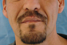
SANTA BARBARA, Calif. - Fat transfer for lipoatrophy is safe and cost effective, and provides excellent long-term results, lasting over a year in some cases, said Dr. Ilya Reyter.
Dr. Reyter, along with Dr. David Sawcer, launched a lipodystrophy clinic in the department of dermatology at the University of Southern California's Keck School of Medicine in early 2007. The clinic is dedicated to treating HIV-associated lipodystrophy and lipoatrophy using tumescent liposuction and fat transfer.

Dr. Reyter, an assistant clinical professor of dermatology at the university, also has extensive experience with fat transfer and liposuction for non-HIV cosmetic improvement, and performs the procedure in his private practice in Beverly Hills, Calif.
Lipodystrophy is marked by central fat deposition on the abdomen, dorsocervical area, salivary glands, breasts, face, and neck. "Before we started this clinic, a lot of our patients were being sent to plastic surgeons, especially for excision of lipodystrophy, particularly in the dorsocervical area," Dr. Reyter said. The dystrophic fat accumulations can be removed more simply using local anesthesia with tumescent liposuction, thereby avoiding the risks of surgery with general anesthesia, he said.
Patients are referred to the HIV lipodystrophy clinic by the HIV dermatology clinic and by other physicians working with HIV patients at the medical school. Dr. Reyter has treated numerous patients for non-HIV cosmetic fat transfer, "but in the HIV clinic, we have treated a few dozen patients," he said. "We select the patients carefully, to make sure that they are appropriate candidates. Not everyone with facial atrophy is a candidate for fat transfer, or fillers, for that matter."
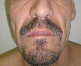
He went on to describe his methods for treating lipoatrophy using fat transfer. Lipoatrophy, also know as fat wasting, particularly affects the face, and is marked by prominent zygomata, reduction of Bichat’s fat pads, sunken eyeballs, and a cachectic appearance.
HIV-associated lipoatrophy was recognized shortly after the first protease inhibitors were approved in 1995. "You can see some of these features in patients who are HIV positive but not on any medication, but it's a really small subset, only about 3% of them," said Dr. Reyter.
He said that lipoatrophy has replaced Kaposi’s sarcoma as the "scarlet letter" of HIV disease, "and it can lead to noncompliance with therapy. It has been shown that correction of the problem can lead to physical and psychological improvement. Lipoatrophy is increasing in prevalence, because HIV is becoming a chronic disease. The longer patients are on medications for their disease, the more of this we will see. The patients have nowhere else to go. As dermatologists we are the experts of skin and subcutaneous fat. If we don't treat it, who will?"
The exact causes of lipoatrophy and lipodystrophy remain unclear, but they are most likely related to multiple factors, he said, including a decrease in retinoic acid receptors, a decrease in triglyceride uptake, inhibition of mitochondrial DNA, inhibition of lipid metabolism, and prevention of adenocyte development.
For small volume areas that need treatment, Dr. Reyter prefers hyaluronic acid fillers such as Restylane and Juvéderm, but for large volume areas such as the cheeks, fillers "don't last a very long time, and they cost a lot. To me, those are prohibitive features for being widely used in HIV. It was not a cost-effective model to be doing this about every 6 months, using 10-14 syringes per side on a patient's face. It just made no sense," he said.
Other filler options include poly-L-lactic acid (Sculptra) and calcium hydroxylapatite (Radiesse), but Dr. Reyter said that he has seen patients with and without HIV develop complications including granulomas after being treated with these agents. "I don't feel as comfortable offering these particular fillers," he said, even though they are Food and Drug Administration approved. "I think it's up to physicians to evaluate whether or not a filler is something they would want to use on their own patients."
Fat transfer wins out for larger volumes, he said, if donor fat is available. "Fat is very cheap to harvest; most patients have adequate fat reserves, and it can be done safely."
In one study of 38 HIV patients who were treated with fat transfer, researchers graded the results at 1 year on a scale from 1 to 4, with 4 being excellent. The mean score reported by the patients was 3.7 and the mean score reported by the surgeon who performed the procedure was 3.2 (Plast. Reconstr. Surg. 2004;114:551-5). No serious adverse effects were observed.
In a separate study of 33 HIV patients who were treated with fat transfer and surveyed 1 year later, 93% of patients reported being satisfied (14 reported being very satisfied and 17 reported being partly satisfied). Furthermore, 81% (27 patients) reported an improved quality of life.
Three independent evaluators reported a 52% improvement in the area treated at 1 year (Arch. Dermatol. 2005;141:1220-4). No significant complications were observed.
In Dr. Reyter's clinical experience, some patients might need another fat transfer at 1 year, "but even so, a year is a significant improvement, especially for a procedure that didn't entail a lot of risk, didn’t have a lot of cost of consumables, and resulted in a benefit for a person."
In another study, researchers used computed tomography analysis and volume calculating software to evaluate the effects of fat transfer in 26 patients (18 men and 8 women aged 34-59 years) with HIV (Aesthetic Surg. J. 2008;28:380-6). The investigators observed increasing volumes of fat after 1 year, leading some to speculate that fat transfer may involve the transfer of stem cells.
"Are stem cells somehow influencing this result?" Dr. Reyter asked, adding that the data on stem cells in fat transfer are inconclusive. "Stem cells have been shown to be present in fat, and they have been shown to be transferred."
According to Dr. Reyter, his pretreatment protocol is the same as for tumescent liposuction: a CBC test; CD4 measurement; a viral load test; confirmation serum transaminases levels are not elevated; and clearance from the primary care physician.
"An important step is marking," he added. "I like to view the sites of deficit as triangles on the face. I make a topographical overlay. After we numb up the area, a lot of that numbing will distort the facial architecture. So if youdidn’t do a good marking job beforehand, your landmarks will be distorted."
For the fat transfer procedure Dr. Reyter uses small, blunt microcannulas. To harvest donor fat, he uses a straight Coleman harvesting cannula connected to a 10-cc syringe.
"I harvest on manual pressure, and then I let the fat sit to allow it to separate" he said. "There's a lot of debate as to whether or not you should spin the fat. I prefer to do nothing that would introduce trauma to the fat or to expose the fat to contamination."
He uses a 1-cc syringe attached to a blunt-tipped 18-gauge cannula to re-inject the fat, injecting 0.1-0.2 cc per pass on withdrawal.
The donor harvesting of fat takes about 20-30 minutes, allowing the fat to sit takes about 10-12 minutes, and reinjecting the fat takes about 30 minutes. "The whole procedure can take an hour to an hour and 15 minutes to do 20-30 ccs of fat per side, which I think is pretty efficient," he remarked.
Typical filling volumes are 10-20 cc for each cheek, 5-8 ccs for the temple, and 5-10 ccs for nasolabial folds. "Edema during and after fat transfer is common," said Dr. Reyter. "Because of this you have to overfill by 25%-50%. That’s where the skill comes in."
The procedures appear to change the overlying skin texture, "producing a global rejuvenation effect," he said. "It brings people back to speculating what the role of stem cells is."
To date there have been no serious complications since the lipodystrophy clinic opened its doors, said Dr. Reyter, who estimated that 25%-50% of his current clinical work involves fat transfer.
"There is a high degree of patient satisfaction at 6-12 months, and very few touch-ups are necessary," he said.
The responses from patients who have gone through the fat transfer procedure "have been overwhelmingly positive," he added. "So many patients tell us that their lives have been transformed."
For example, one patient with severe facial atrophy and longstanding unemployment was able to finally find employment after the fat transfer – "because he no longer looked so ill," Dr. Reyter said. "Another patient just wrote 'thank you for my new face. ... I look so healthy!' The doctors in the clinic regularly receive thank you notes and tokens of appreciation from the patients – in my experience, much more than we typically get in the course of providing any other medical care."
Dr. Reyter said that he had no relevant financial disclosures to make.
SANTA BARBARA, Calif. - Fat transfer for lipoatrophy is safe and cost effective, and provides excellent long-term results, lasting over a year in some cases, said Dr. Ilya Reyter.
Dr. Reyter, along with Dr. David Sawcer, launched a lipodystrophy clinic in the department of dermatology at the University of Southern California's Keck School of Medicine in early 2007. The clinic is dedicated to treating HIV-associated lipodystrophy and lipoatrophy using tumescent liposuction and fat transfer.
Dr. Reyter, an assistant clinical professor of dermatology at the university, also has extensive experience with fat transfer and liposuction for non-HIV cosmetic improvement, and performs the procedure in his private practice in Beverly Hills, Calif.
Lipodystrophy is marked by central fat deposition on the abdomen, dorsocervical area, salivary glands, breasts, face, and neck. "Before we started this clinic, a lot of our patients were being sent to plastic surgeons, especially for excision of lipodystrophy, particularly in the dorsocervical area," Dr. Reyter said. The dystrophic fat accumulations can be removed more simply using local anesthesia with tumescent liposuction, thereby avoiding the risks of surgery with general anesthesia, he said.
Patients are referred to the HIV lipodystrophy clinic by the HIV dermatology clinic and by other physicians working with HIV patients at the medical school. Dr. Reyter has treated numerous patients for non-HIV cosmetic fat transfer, "but in the HIV clinic, we have treated a few dozen patients," he said. "We select the patients carefully, to make sure that they are appropriate candidates. Not everyone with facial atrophy is a candidate for fat transfer, or fillers, for that matter."
He went on to describe his methods for treating lipoatrophy using fat transfer. Lipoatrophy, also know as fat wasting, particularly affects the face, and is marked by prominent zygomata, reduction of Bichat’s fat pads, sunken eyeballs, and a cachectic appearance.
HIV-associated lipoatrophy was recognized shortly after the first protease inhibitors were approved in 1995. "You can see some of these features in patients who are HIV positive but not on any medication, but it's a really small subset, only about 3% of them," said Dr. Reyter.
He said that lipoatrophy has replaced Kaposi’s sarcoma as the "scarlet letter" of HIV disease, "and it can lead to noncompliance with therapy. It has been shown that correction of the problem can lead to physical and psychological improvement. Lipoatrophy is increasing in prevalence, because HIV is becoming a chronic disease. The longer patients are on medications for their disease, the more of this we will see. The patients have nowhere else to go. As dermatologists we are the experts of skin and subcutaneous fat. If we don't treat it, who will?"
The exact causes of lipoatrophy and lipodystrophy remain unclear, but they are most likely related to multiple factors, he said, including a decrease in retinoic acid receptors, a decrease in triglyceride uptake, inhibition of mitochondrial DNA, inhibition of lipid metabolism, and prevention of adenocyte development.
For small volume areas that need treatment, Dr. Reyter prefers hyaluronic acid fillers such as Restylane and Juvéderm, but for large volume areas such as the cheeks, fillers "don't last a very long time, and they cost a lot. To me, those are prohibitive features for being widely used in HIV. It was not a cost-effective model to be doing this about every 6 months, using 10-14 syringes per side on a patient's face. It just made no sense," he said.
Other filler options include poly-L-lactic acid (Sculptra) and calcium hydroxylapatite (Radiesse), but Dr. Reyter said that he has seen patients with and without HIV develop complications including granulomas after being treated with these agents. "I don't feel as comfortable offering these particular fillers," he said, even though they are Food and Drug Administration approved. "I think it's up to physicians to evaluate whether or not a filler is something they would want to use on their own patients."
Fat transfer wins out for larger volumes, he said, if donor fat is available. "Fat is very cheap to harvest; most patients have adequate fat reserves, and it can be done safely."
In one study of 38 HIV patients who were treated with fat transfer, researchers graded the results at 1 year on a scale from 1 to 4, with 4 being excellent. The mean score reported by the patients was 3.7 and the mean score reported by the surgeon who performed the procedure was 3.2 (Plast. Reconstr. Surg. 2004;114:551-5). No serious adverse effects were observed.
In a separate study of 33 HIV patients who were treated with fat transfer and surveyed 1 year later, 93% of patients reported being satisfied (14 reported being very satisfied and 17 reported being partly satisfied). Furthermore, 81% (27 patients) reported an improved quality of life.
Three independent evaluators reported a 52% improvement in the area treated at 1 year (Arch. Dermatol. 2005;141:1220-4). No significant complications were observed.
In Dr. Reyter's clinical experience, some patients might need another fat transfer at 1 year, "but even so, a year is a significant improvement, especially for a procedure that didn't entail a lot of risk, didn’t have a lot of cost of consumables, and resulted in a benefit for a person."
In another study, researchers used computed tomography analysis and volume calculating software to evaluate the effects of fat transfer in 26 patients (18 men and 8 women aged 34-59 years) with HIV (Aesthetic Surg. J. 2008;28:380-6). The investigators observed increasing volumes of fat after 1 year, leading some to speculate that fat transfer may involve the transfer of stem cells.
"Are stem cells somehow influencing this result?" Dr. Reyter asked, adding that the data on stem cells in fat transfer are inconclusive. "Stem cells have been shown to be present in fat, and they have been shown to be transferred."
According to Dr. Reyter, his pretreatment protocol is the same as for tumescent liposuction: a CBC test; CD4 measurement; a viral load test; confirmation serum transaminases levels are not elevated; and clearance from the primary care physician.
"An important step is marking," he added. "I like to view the sites of deficit as triangles on the face. I make a topographical overlay. After we numb up the area, a lot of that numbing will distort the facial architecture. So if youdidn’t do a good marking job beforehand, your landmarks will be distorted."
For the fat transfer procedure Dr. Reyter uses small, blunt microcannulas. To harvest donor fat, he uses a straight Coleman harvesting cannula connected to a 10-cc syringe.
"I harvest on manual pressure, and then I let the fat sit to allow it to separate" he said. "There's a lot of debate as to whether or not you should spin the fat. I prefer to do nothing that would introduce trauma to the fat or to expose the fat to contamination."
He uses a 1-cc syringe attached to a blunt-tipped 18-gauge cannula to re-inject the fat, injecting 0.1-0.2 cc per pass on withdrawal.
The donor harvesting of fat takes about 20-30 minutes, allowing the fat to sit takes about 10-12 minutes, and reinjecting the fat takes about 30 minutes. "The whole procedure can take an hour to an hour and 15 minutes to do 20-30 ccs of fat per side, which I think is pretty efficient," he remarked.
Typical filling volumes are 10-20 cc for each cheek, 5-8 ccs for the temple, and 5-10 ccs for nasolabial folds. "Edema during and after fat transfer is common," said Dr. Reyter. "Because of this you have to overfill by 25%-50%. That’s where the skill comes in."
The procedures appear to change the overlying skin texture, "producing a global rejuvenation effect," he said. "It brings people back to speculating what the role of stem cells is."
To date there have been no serious complications since the lipodystrophy clinic opened its doors, said Dr. Reyter, who estimated that 25%-50% of his current clinical work involves fat transfer.
"There is a high degree of patient satisfaction at 6-12 months, and very few touch-ups are necessary," he said.
The responses from patients who have gone through the fat transfer procedure "have been overwhelmingly positive," he added. "So many patients tell us that their lives have been transformed."
For example, one patient with severe facial atrophy and longstanding unemployment was able to finally find employment after the fat transfer – "because he no longer looked so ill," Dr. Reyter said. "Another patient just wrote 'thank you for my new face. ... I look so healthy!' The doctors in the clinic regularly receive thank you notes and tokens of appreciation from the patients – in my experience, much more than we typically get in the course of providing any other medical care."
Dr. Reyter said that he had no relevant financial disclosures to make.
EXPERT ANALYSIS FROM THE ANNUAL MEETING OF THE CALIFORNIA SOCIETY OF DERMATOLOGY AND DERMATOLOGIC SURGERY
Melanonychia Striata Warrants Biopsy in Most Cases
SANTA BARBARA, Calif. - The only way to definitively rule out melanoma in a case of melanonychia striata is to perform a biopsy of the nail matrix, according to Dr. Richard K. Scher.
"The source of pigmentation is in the nail matrix, so the biopsy specimen must be taken from there," Dr. Scher said at the annual meeting of the California Society of Dermatology and Dermatologic Surgery.
"It's of no value to do a biopsy of the nail bed. There are almost never melanocytes there, but it's amazing to me how many dermatologists are still doing nail bed biopsies," Dr. Scher, professor of dermatology at the University of North Carolina, Chapel Hill.
While some clinicians prefer to use dermoscopy to inspect cases of melanonychia striata, it is not as accurate or as reliable as microscopic examination.
"At most, dermoscopy should be an aide," he commented.
There are probably too many biopsies being performed, but "I really don't know what the alternative is," Dr. Scher noted. "We need reliable clinical criteria to determine melanoma probability, but we don't have it yet. ... We also need reliable histologic criteria to determine melanoma probability, but we don't always have that, either."
Dr. Scher estimated that 90% of melanocytic bands arise from the distal matrix, which contains more melanocytes than the proximal matrix does. Melanonychia melanoma most commonly occurs in the thumb and the big toe, followed by the index finger. "About 20% of nail melanomas are amelanotic," he said. "That's a number that keeps me awake at night."

Two risk factors for developing melanonychia include older age and being African American. "In general, African Americans do not get melanomas to a great extent, but they do have a lot of benign melanonychia," Dr. Scher noted. "However, acral melanoma is more common in African Americans, compared with other populations."
Other causes of melanonychia include trauma to the nail, infection, and certain medications including antibiotics and chemotherapeutics agents.
Wider, darker nail lesions are more likely to be melanoma, compared with thinner, lighter lesions. "That's true, but I've seen very narrow bands that were melanomas, and I’ve seen very wide bands that were not," Dr. Scher said. "Uniformity of pigment is relatively more reliable than some of the other criteria. So if you see lack of uniformity, think of things like atypical nevi, dysplastic nevi, or melanoma."
In addition, if a patient presents with pigmented nails in four or five digits, "it's not likely that patient will have four or five melanomas," he said. "But if you see a patient particularly a dark-skinned individual who has normal ethnic melanonychia in many digits, but one of them is very different, then you have to become suspicious."
Evolution of the lesion is another worrisome sign. "If the band is changing if it’s getting wider or darker or more streaky then you have to think that you’re dealing with a melanoma."
He emphasized the importance of retrieving the nail plate during the nail biopsy procedure and sending it to the pathologist with the nail matrix specimen, "because sometimes it's hard for the pathologist to find the pigment," he explained. "If the pathologist sends you back a report that there’s no pigment here, the implication is that you missed the lesion. But if you have nail plate there, which has pigment in it, you didn’t miss the lesion. The pathologist did."
Some recent articles in the medical literature suggest that if cases of melanonychia striata in childhood are stable and not atypical in appearance, observation may be an option. However, Dr. Scher urged caution with this approach, saying that "every case must be evaluated individually."
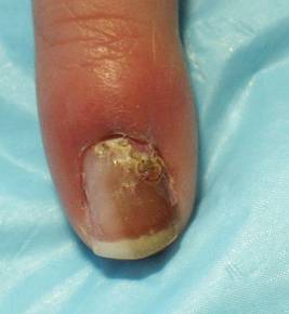
To illustrate his point he discussed the case of a 4-year-old healthy girl who presented to his office with an 8-month history of melanonychia and a 3-month history of possible nail fold pigmentation (see image above). Family history revealed a maternal second cousin with acral melanoma. Dermoscopy was consistent with a nevus diagnosis.
The parents agreed to proceed with a nail biopsy, Dr. Scher said. The pathology report read (in part): "atypical junctional melanocytic proliferation with increased numbers of single melanocytes ...; although this lesion is most probably a nevus, it is recommended that it be completely but conservatively excised."
The girl's parents sought the opinion of seven other pathologists and clinicians at a melanoma conference, and all agreed with complete excision. "In this case, there was not much alternative as to what to do," Dr. Scher said.
The entire nail unit was removed during surgery and the lesion healed with "a terrific cosmetic result," he said.
Dr. Scher said that he had no relevant conflicts to disclose.
SANTA BARBARA, Calif. - The only way to definitively rule out melanoma in a case of melanonychia striata is to perform a biopsy of the nail matrix, according to Dr. Richard K. Scher.
"The source of pigmentation is in the nail matrix, so the biopsy specimen must be taken from there," Dr. Scher said at the annual meeting of the California Society of Dermatology and Dermatologic Surgery.
"It's of no value to do a biopsy of the nail bed. There are almost never melanocytes there, but it's amazing to me how many dermatologists are still doing nail bed biopsies," Dr. Scher, professor of dermatology at the University of North Carolina, Chapel Hill.
While some clinicians prefer to use dermoscopy to inspect cases of melanonychia striata, it is not as accurate or as reliable as microscopic examination.
"At most, dermoscopy should be an aide," he commented.
There are probably too many biopsies being performed, but "I really don't know what the alternative is," Dr. Scher noted. "We need reliable clinical criteria to determine melanoma probability, but we don't have it yet. ... We also need reliable histologic criteria to determine melanoma probability, but we don't always have that, either."
Dr. Scher estimated that 90% of melanocytic bands arise from the distal matrix, which contains more melanocytes than the proximal matrix does. Melanonychia melanoma most commonly occurs in the thumb and the big toe, followed by the index finger. "About 20% of nail melanomas are amelanotic," he said. "That's a number that keeps me awake at night."
Two risk factors for developing melanonychia include older age and being African American. "In general, African Americans do not get melanomas to a great extent, but they do have a lot of benign melanonychia," Dr. Scher noted. "However, acral melanoma is more common in African Americans, compared with other populations."
Other causes of melanonychia include trauma to the nail, infection, and certain medications including antibiotics and chemotherapeutics agents.
Wider, darker nail lesions are more likely to be melanoma, compared with thinner, lighter lesions. "That's true, but I've seen very narrow bands that were melanomas, and I’ve seen very wide bands that were not," Dr. Scher said. "Uniformity of pigment is relatively more reliable than some of the other criteria. So if you see lack of uniformity, think of things like atypical nevi, dysplastic nevi, or melanoma."
In addition, if a patient presents with pigmented nails in four or five digits, "it's not likely that patient will have four or five melanomas," he said. "But if you see a patient particularly a dark-skinned individual who has normal ethnic melanonychia in many digits, but one of them is very different, then you have to become suspicious."
Evolution of the lesion is another worrisome sign. "If the band is changing if it’s getting wider or darker or more streaky then you have to think that you’re dealing with a melanoma."
He emphasized the importance of retrieving the nail plate during the nail biopsy procedure and sending it to the pathologist with the nail matrix specimen, "because sometimes it's hard for the pathologist to find the pigment," he explained. "If the pathologist sends you back a report that there’s no pigment here, the implication is that you missed the lesion. But if you have nail plate there, which has pigment in it, you didn’t miss the lesion. The pathologist did."
Some recent articles in the medical literature suggest that if cases of melanonychia striata in childhood are stable and not atypical in appearance, observation may be an option. However, Dr. Scher urged caution with this approach, saying that "every case must be evaluated individually."
To illustrate his point he discussed the case of a 4-year-old healthy girl who presented to his office with an 8-month history of melanonychia and a 3-month history of possible nail fold pigmentation (see image above). Family history revealed a maternal second cousin with acral melanoma. Dermoscopy was consistent with a nevus diagnosis.
The parents agreed to proceed with a nail biopsy, Dr. Scher said. The pathology report read (in part): "atypical junctional melanocytic proliferation with increased numbers of single melanocytes ...; although this lesion is most probably a nevus, it is recommended that it be completely but conservatively excised."
The girl's parents sought the opinion of seven other pathologists and clinicians at a melanoma conference, and all agreed with complete excision. "In this case, there was not much alternative as to what to do," Dr. Scher said.
The entire nail unit was removed during surgery and the lesion healed with "a terrific cosmetic result," he said.
Dr. Scher said that he had no relevant conflicts to disclose.
annual meeting of the California Society of Dermatology and Dermatologic Surgery
Melanonychia Striata Warrants Biopsy in Most Cases
SANTA BARBARA, Calif. – The only way to definitively rule out melanoma in a case of melanonychia striata is to perform a biopsy of the nail matrix, according to Dr. Richard K. Scher.
“The source of pigmentation is in the nail matrix, so the biopsy specimen must be taken from there,” Dr. Scher said at the annual meeting of the California Society of Dermatology and Dermatologic Surgery.
“It’s of no value to do a biopsy of the nail bed. There are almost never melanocytes there, but it’s amazing to me how many dermatologists are still doing nail bed biopsies,” Dr. Scher, professor of dermatology at the University of North Carolina, Chapel Hill.
While some clinicians prefer to use dermoscopy to inspect cases of melanonychia striata, it is not as accurate or as reliable as microscopic examination.
“At most, dermoscopy should be an aide,” he commented.
There are probably too many biopsies being performed, but “I really don’t know what the alternative is,” Dr. Scher noted. “We need reliable clinical criteria to determine melanoma probability, but we don’t have it yet. ... We also need reliable histologic criteria to determine melanoma probability, but we don’t always have that, either.”
Dr. Scher estimated that 90% of melanocytic bands arise from the distal matrix, which contains more melanocytes than the proximal matrix does. Melanonychia melanoma most commonly occurs in the thumb and the big toe, followed by the index finger. “About 20% of nail melanomas are amelanotic,” he said. “That’s a number that keeps me awake at night.”
Two risk factors for developing melanonychia include older age and being African American. “In general, African Americans do not get melanomas to a great extent, but they do have a lot of benign melanonychia,” Dr. Scher noted. “However, acral melanoma is more common in African Americans, compared with other populations.”
Other causes of melanonychia include trauma to the nail, infection, and certain medications including antibiotics and chemotherapeutics agents.
Wider, darker nail lesions are more likely to be melanoma, compared with thinner, lighter lesions. “That’s true, but I’ve seen very narrow bands that were melanomas, and I’ve seen very wide bands that were not,” Dr. Scher said. “Uniformity of pigment is relatively more reliable than some of the other criteria. So if you see lack of uniformity, think of things like atypical nevi, dysplastic nevi, or melanoma.”
In addition, if a patient presents with pigmented nails in four or five digits, “it’s not likely that patient will have four or five melanomas,” he said. “But if you see a patient particularly a dark-skinned individual who has normal ethnic melanonychia in many digits, but one of them is very different, then you have to become suspicious.”
Evolution of the lesion is another worrisome sign. “If the band is changing if it’s getting wider or darker or more streaky then you have to think that you’re dealing with a melanoma.”
He emphasized the importance of retrieving the nail plate during the nail biopsy procedure and sending it to the pathologist with the nail matrix specimen, “because sometimes it’s hard for the pathologist to find the pigment,” he explained. “If the pathologist sends you back a report that there’s no pigment here, the implication is that you missed the lesion. But if you have nail plate there, which has pigment in it, you didn’t miss the lesion. The pathologist did.”
Some recent articles in the medical literature suggest that if cases of melanonychia striata in childhood are stable and not atypical in appearance, observation may be an option. However, Dr. Scher urged caution with this approach, saying that “every case must be evaluated individually.”
To illustrate his point he discussed the case of a 4-year-old healthy girl who presented to his office with an 8-month history of melanonychia and a 3-month history of possible nail fold pigmentation. Family history revealed a maternal second cousin with acral melanoma. Dermoscopy was consistent with a nevus diagnosis.
The parents agreed to proceed with a nail biopsy, Dr. Scher said. The pathology report read (in part): “atypical junctional melanocytic proliferation with increased numbers of single melanocytes ...; although this lesion is most probably a nevus, it is recommended that it be completely but conservatively excised.”
The girl’s parents sought the opinion of seven other pathologists and clinicians at a melanoma conference, and all agreed with complete excision. “In this case, there was not much alternative as to what to do,” Dr. Scher said.
The entire nail unit was removed during surgery and the lesion healed with “a terrific cosmetic result,” he said.
Dr. Scher said that he had no relevant conflicts to disclose.
Trial of HeartNet Stopped Due to Lack of Change in Peak VO2
SAN DIEGO – Patients with stage C heart failure who received HeartNet, an investigational implantable device made of nitinol that is designed to wrap around the walls of the heart for support, experienced no statistically significant improvements in peak oxygen consumption, compared with a control group who received standard medical care alone, results from a prospective randomized trial showed.
However, the trial – known as PEERLESS-HF (Prospective Evaluation of Elastic Restraint to Lessen the Effects of Heart Failure) – showed evidence of reverse remodeling as well as a significant reduction in serious heart failure events at 6 months in the treatment group, Dr. William T. Abraham reported at the annual meeting of the Heart Failure Society of America.

When 50% of the patients reached 6 months’ follow-up, an independent data safety-monitoring board reviewed the data and recommended that the trial be stopped “for futility, particularly because there was no demonstrable difference between the treatment and control group for change in peak VO2,” explained Dr. Abraham, who directs the division of cardiovascular medicine at Ohio State University in Columbus. “This was due in large part to a much-better-than-expected improvement of peak VO2 in the control group. There was a 0.7 mL/kg per minute average improvement in peak VO2 in the control patients, something that we haven’t seen in other trials that have used peak VO2 as a primary end point.”
Manufactured by Sunnyvale, Calif.–based Paracor Medical Inc., HeartNet is delivered using a minimally invasive approach through a minithoracotomy. The device is self-anchoring and self-tensioning, and is presized based on echo measurements.
“Based on preclinical or animal data, we believe that it exerts just the right amount of cardiac support that is below the level at which myocardial constriction might occur,” Dr. Abraham said of the device. “The goal is to provide some external support without creating constriction.”

The purpose of PEERLESS-HF was to evaluate the safety and efficacy of the HeartNet ventricular elastic support system plus optimal HF therapy (treatment group) compared with optimal therapy alone (control group) in 244 patients. “We purposely enriched enrollment of patients with cardiac resynchronization therapy [because] there is preclinical animal mechanistic data [that suggest] possible synergy between cardiac support and CRT,” he said. “Our own preliminary data from the feasibility trial of this device also suggested the potential for a better response in a CRT population.”
To be eligible for the trial, patients had to be aged 18-75 years with stage C symptomatic ischemic or nonischemic heart failure for 6 months; be on stable, evidence-based heart failure therapy for at least 3 months; have a left ventricular ejection fraction of 35% or less; and have a 6-minute walk distance of 150-450 meters. Patients on current cardiac resynchronization therapy were accepted into the trial, but the relevant device must have been placed at least 3 months prior to randomization.
The study’s three coprimary end points, assessed at 6 months, were peak VO2, with the responder threshold set at 1 mL/kg per minute; 6-minute walk test, with the responder threshold set at 45 meters; and quality of life as measured by the Minnesota Living With Heart Failure Questionnaire (MLHFQ), with the responder threshold set at 7 units.
The primary safety end point, assessed at 1 year, was all-cause mortality by noninferiority analysis.
In all, 217 patients were enrolled in the study, with 114 in the control group and 103 in the treatment group. Ultimately, 92 patients were actually treated because of premature termination of the trial.
The mean age of patients was 55 years, 70% were male, 48% had NYHA class III heart failure, 47% had systemic hypertension, and 34% had diabetes mellitus.
“At baseline, this was a moderately sick group of patients,” Dr. Abraham said.
The average peak VO2 was 15.5 mL/kg per minute, the average 6-minute walk distance was about 350 meters, and the mean MLHFQ score was 52.
At 6 months, there were no statistically significant differences between the treatment and control groups on any of the coprimary end points. There were no safety concerns in the trial, and the rate of survival at 12 months was similar between groups (93% in the control group and 92% in the treatment group).
“The HeartNet is safe, demonstrating no increase in all-cause mortality,” Dr. Abraham said. “Even though terminated early, this trial did hit its prespecified safety end point of all-cause mortality at 12 months.”
He noted that there was nonsignificant evidence of reverse remodeling at 6 months that favored the treatment group. There was also a significant reduction of serious heart failure events in the treatment group, compared with the control group. “In a subgroup of patients with CRT at baseline, there appeared to be a better reduction in the serious heart failure events,” he added. “We believe that these findings support undertaking a clinical trial that targets this patient population.”
The trial was sponsored by Paracor Medical Inc. Dr. Abraham disclosed that he is a paid consultant for the company.
SAN DIEGO – Patients with stage C heart failure who received HeartNet, an investigational implantable device made of nitinol that is designed to wrap around the walls of the heart for support, experienced no statistically significant improvements in peak oxygen consumption, compared with a control group who received standard medical care alone, results from a prospective randomized trial showed.
However, the trial – known as PEERLESS-HF (Prospective Evaluation of Elastic Restraint to Lessen the Effects of Heart Failure) – showed evidence of reverse remodeling as well as a significant reduction in serious heart failure events at 6 months in the treatment group, Dr. William T. Abraham reported at the annual meeting of the Heart Failure Society of America.
When 50% of the patients reached 6 months’ follow-up, an independent data safety-monitoring board reviewed the data and recommended that the trial be stopped “for futility, particularly because there was no demonstrable difference between the treatment and control group for change in peak VO2,” explained Dr. Abraham, who directs the division of cardiovascular medicine at Ohio State University in Columbus. “This was due in large part to a much-better-than-expected improvement of peak VO2 in the control group. There was a 0.7 mL/kg per minute average improvement in peak VO2 in the control patients, something that we haven’t seen in other trials that have used peak VO2 as a primary end point.”
Manufactured by Sunnyvale, Calif.–based Paracor Medical Inc., HeartNet is delivered using a minimally invasive approach through a minithoracotomy. The device is self-anchoring and self-tensioning, and is presized based on echo measurements.
“Based on preclinical or animal data, we believe that it exerts just the right amount of cardiac support that is below the level at which myocardial constriction might occur,” Dr. Abraham said of the device. “The goal is to provide some external support without creating constriction.”
The purpose of PEERLESS-HF was to evaluate the safety and efficacy of the HeartNet ventricular elastic support system plus optimal HF therapy (treatment group) compared with optimal therapy alone (control group) in 244 patients. “We purposely enriched enrollment of patients with cardiac resynchronization therapy [because] there is preclinical animal mechanistic data [that suggest] possible synergy between cardiac support and CRT,” he said. “Our own preliminary data from the feasibility trial of this device also suggested the potential for a better response in a CRT population.”
To be eligible for the trial, patients had to be aged 18-75 years with stage C symptomatic ischemic or nonischemic heart failure for 6 months; be on stable, evidence-based heart failure therapy for at least 3 months; have a left ventricular ejection fraction of 35% or less; and have a 6-minute walk distance of 150-450 meters. Patients on current cardiac resynchronization therapy were accepted into the trial, but the relevant device must have been placed at least 3 months prior to randomization.
The study’s three coprimary end points, assessed at 6 months, were peak VO2, with the responder threshold set at 1 mL/kg per minute; 6-minute walk test, with the responder threshold set at 45 meters; and quality of life as measured by the Minnesota Living With Heart Failure Questionnaire (MLHFQ), with the responder threshold set at 7 units.
The primary safety end point, assessed at 1 year, was all-cause mortality by noninferiority analysis.
In all, 217 patients were enrolled in the study, with 114 in the control group and 103 in the treatment group. Ultimately, 92 patients were actually treated because of premature termination of the trial.
The mean age of patients was 55 years, 70% were male, 48% had NYHA class III heart failure, 47% had systemic hypertension, and 34% had diabetes mellitus.
“At baseline, this was a moderately sick group of patients,” Dr. Abraham said.
The average peak VO2 was 15.5 mL/kg per minute, the average 6-minute walk distance was about 350 meters, and the mean MLHFQ score was 52.
At 6 months, there were no statistically significant differences between the treatment and control groups on any of the coprimary end points. There were no safety concerns in the trial, and the rate of survival at 12 months was similar between groups (93% in the control group and 92% in the treatment group).
“The HeartNet is safe, demonstrating no increase in all-cause mortality,” Dr. Abraham said. “Even though terminated early, this trial did hit its prespecified safety end point of all-cause mortality at 12 months.”
He noted that there was nonsignificant evidence of reverse remodeling at 6 months that favored the treatment group. There was also a significant reduction of serious heart failure events in the treatment group, compared with the control group. “In a subgroup of patients with CRT at baseline, there appeared to be a better reduction in the serious heart failure events,” he added. “We believe that these findings support undertaking a clinical trial that targets this patient population.”
The trial was sponsored by Paracor Medical Inc. Dr. Abraham disclosed that he is a paid consultant for the company.
Trial of HeartNet Stopped Due to Lack of Change in Peak VO2
SAN DIEGO – Patients with stage C heart failure who received HeartNet, an investigational implantable device made of nitinol that is designed to wrap around the walls of the heart for support, experienced no statistically significant improvements in peak oxygen consumption, compared with a control group who received standard medical care alone, results from a prospective randomized trial showed.
However, the trial – known as PEERLESS-HF (Prospective Evaluation of Elastic Restraint to Lessen the Effects of Heart Failure) – showed evidence of reverse remodeling as well as a significant reduction in serious heart failure events at 6 months in the treatment group, Dr. William T. Abraham reported at the annual meeting of the Heart Failure Society of America.
When 50% of the patients reached 6 months’ follow-up, an independent data safety-monitoring board reviewed the data and recommended that the trial be stopped “for futility, particularly because there was no demonstrable difference between the treatment and control group for change in peak VO2,” explained Dr. Abraham, who directs the division of cardiovascular medicine at Ohio State University in Columbus. “This was due in large part to a much-better-than-expected improvement of peak VO2 in the control group. There was a 0.7 mL/kg per minute average improvement in peak VO2 in the control patients, something that we haven’t seen in other trials that have used peak VO2 as a primary end point.”
Manufactured by Sunnyvale, Calif.–based Paracor Medical Inc., HeartNet is delivered using a minimally invasive approach through a minithoracotomy. The device is self-anchoring and self-tensioning, and is presized based on echo measurements.
“Based on preclinical or animal data, we believe that it exerts just the right amount of cardiac support that is below the level at which myocardial constriction might occur,” Dr. Abraham said of the device. “The goal is to provide some external support without creating constriction.”

The purpose of PEERLESS-HF was to evaluate the safety and efficacy of the HeartNet ventricular elastic support system plus optimal HF therapy (treatment group) compared with optimal therapy alone (control group) in 244 patients. “We purposely enriched enrollment of patients with cardiac resynchronization therapy [because] there is preclinical animal mechanistic data [that suggest] possible synergy between cardiac support and CRT,” he said. “Our own preliminary data from the feasibility trial of this device also suggested the potential for a better response in a CRT population.”
To be eligible for the trial, patients had to be aged 18-75 years with stage C symptomatic ischemic or nonischemic heart failure for 6 months; be on stable, evidence-based heart failure therapy for at least 3 months; have a left ventricular ejection fraction of 35% or less; and have a 6-minute walk distance of 150-450 meters. Patients on current cardiac resynchronization therapy were accepted into the trial, but the relevant device must have been placed at least 3 months prior to randomization.
The study’s three coprimary end points, assessed at 6 months, were peak VO2, with the responder threshold set at 1 mL/kg per minute; 6-minute walk test, with the responder threshold set at 45 meters; and quality of life as measured by the Minnesota Living With Heart Failure Questionnaire (MLHFQ), with the responder threshold set at 7 units.
The primary safety end point, assessed at 1 year, was all-cause mortality by noninferiority analysis.
In all, 217 patients were enrolled in the study, with 114 in the control group and 103 in the treatment group. Ultimately, 92 patients were actually treated because of premature termination of the trial.
The mean age of patients was 55 years, 70% were male, 48% had NYHA class III heart failure, 47% had systemic hypertension, and 34% had diabetes mellitus.
“At baseline, this was a moderately sick group of patients,” Dr. Abraham said.
The average peak VO2 was 15.5 mL/kg per minute, the average 6-minute walk distance was about 350 meters, and the mean MLHFQ score was 52.
At 6 months, there were no statistically significant differences between the treatment and control groups on any of the coprimary end points. There were no safety concerns in the trial, and the rate of survival at 12 months was similar between groups (93% in the control group and 92% in the treatment group).
“The HeartNet is safe, demonstrating no increase in all-cause mortality,” Dr. Abraham said. “Even though terminated early, this trial did hit its prespecified safety end point of all-cause mortality at 12 months.”
He noted that there was nonsignificant evidence of reverse remodeling at 6 months that favored the treatment group. There was also a significant reduction of serious heart failure events in the treatment group, compared with the control group. “In a subgroup of patients with CRT at baseline, there appeared to be a better reduction in the serious heart failure events,” he added. “We believe that these findings support undertaking a clinical trial that targets this patient population.”
The trial was sponsored by Paracor Medical Inc. Dr. Abraham disclosed that he is a paid consultant for the company.
SAN DIEGO – Patients with stage C heart failure who received HeartNet, an investigational implantable device made of nitinol that is designed to wrap around the walls of the heart for support, experienced no statistically significant improvements in peak oxygen consumption, compared with a control group who received standard medical care alone, results from a prospective randomized trial showed.
However, the trial – known as PEERLESS-HF (Prospective Evaluation of Elastic Restraint to Lessen the Effects of Heart Failure) – showed evidence of reverse remodeling as well as a significant reduction in serious heart failure events at 6 months in the treatment group, Dr. William T. Abraham reported at the annual meeting of the Heart Failure Society of America.
When 50% of the patients reached 6 months’ follow-up, an independent data safety-monitoring board reviewed the data and recommended that the trial be stopped “for futility, particularly because there was no demonstrable difference between the treatment and control group for change in peak VO2,” explained Dr. Abraham, who directs the division of cardiovascular medicine at Ohio State University in Columbus. “This was due in large part to a much-better-than-expected improvement of peak VO2 in the control group. There was a 0.7 mL/kg per minute average improvement in peak VO2 in the control patients, something that we haven’t seen in other trials that have used peak VO2 as a primary end point.”
Manufactured by Sunnyvale, Calif.–based Paracor Medical Inc., HeartNet is delivered using a minimally invasive approach through a minithoracotomy. The device is self-anchoring and self-tensioning, and is presized based on echo measurements.
“Based on preclinical or animal data, we believe that it exerts just the right amount of cardiac support that is below the level at which myocardial constriction might occur,” Dr. Abraham said of the device. “The goal is to provide some external support without creating constriction.”
The purpose of PEERLESS-HF was to evaluate the safety and efficacy of the HeartNet ventricular elastic support system plus optimal HF therapy (treatment group) compared with optimal therapy alone (control group) in 244 patients. “We purposely enriched enrollment of patients with cardiac resynchronization therapy [because] there is preclinical animal mechanistic data [that suggest] possible synergy between cardiac support and CRT,” he said. “Our own preliminary data from the feasibility trial of this device also suggested the potential for a better response in a CRT population.”
To be eligible for the trial, patients had to be aged 18-75 years with stage C symptomatic ischemic or nonischemic heart failure for 6 months; be on stable, evidence-based heart failure therapy for at least 3 months; have a left ventricular ejection fraction of 35% or less; and have a 6-minute walk distance of 150-450 meters. Patients on current cardiac resynchronization therapy were accepted into the trial, but the relevant device must have been placed at least 3 months prior to randomization.
The study’s three coprimary end points, assessed at 6 months, were peak VO2, with the responder threshold set at 1 mL/kg per minute; 6-minute walk test, with the responder threshold set at 45 meters; and quality of life as measured by the Minnesota Living With Heart Failure Questionnaire (MLHFQ), with the responder threshold set at 7 units.
The primary safety end point, assessed at 1 year, was all-cause mortality by noninferiority analysis.
In all, 217 patients were enrolled in the study, with 114 in the control group and 103 in the treatment group. Ultimately, 92 patients were actually treated because of premature termination of the trial.
The mean age of patients was 55 years, 70% were male, 48% had NYHA class III heart failure, 47% had systemic hypertension, and 34% had diabetes mellitus.
“At baseline, this was a moderately sick group of patients,” Dr. Abraham said.
The average peak VO2 was 15.5 mL/kg per minute, the average 6-minute walk distance was about 350 meters, and the mean MLHFQ score was 52.
At 6 months, there were no statistically significant differences between the treatment and control groups on any of the coprimary end points. There were no safety concerns in the trial, and the rate of survival at 12 months was similar between groups (93% in the control group and 92% in the treatment group).
“The HeartNet is safe, demonstrating no increase in all-cause mortality,” Dr. Abraham said. “Even though terminated early, this trial did hit its prespecified safety end point of all-cause mortality at 12 months.”
He noted that there was nonsignificant evidence of reverse remodeling at 6 months that favored the treatment group. There was also a significant reduction of serious heart failure events in the treatment group, compared with the control group. “In a subgroup of patients with CRT at baseline, there appeared to be a better reduction in the serious heart failure events,” he added. “We believe that these findings support undertaking a clinical trial that targets this patient population.”
The trial was sponsored by Paracor Medical Inc. Dr. Abraham disclosed that he is a paid consultant for the company.
Major Finding: At 6 months, patients with stage C heart failure who were treated with the HeartNet device experienced a significant reduction of serious heart failure events, compared with patients who received standard medical care alone (HR, 1.9).
Data Source: A study of 217 patients enrolled in the PEERLESS-HF trial.
Disclosures: The study was funded by Paracor Medical Inc. Dr. Abraham disclosed that he is a paid consultant for the company.
Mitotic Rate a Game Changer in Melanoma Tx
SANTA BARBARA, CALIF. – Current American Joint Committee on Cancer melanoma staging criteria incorporate a mitotic rate of 1/mm
This change, which went into effect in January, “is going to have a profound impact in whom we consider eligible for staging with sentinel lymph node biopsy,” Dr. Susan Swetter predicted at the meeting. “The reason is that only about 6%–8% of these T1 tumors (1.0 mm) are ulcerated, but it is estimated that up to 30%–40% will have an elevated mitotic rate of 1.0 or more per square millimeter. This leaves us with a conundrum: Are all of these patients going to be eligible for sentinel lymph node biopsy?”
AJCC melanoma staging criteria are based on data analysis from the 2008 AJCC collaborative melanoma staging database from 14 cancer centers and cooperative oncology organizations worldwide, said Dr. Swetter, professor of dermatology and director of the pigmented lesion and melanoma program at Stanford University Medical Center/VA Palo Alto Health Care System in California.
The criteria are based on prognostic factor analysis of nearly 60,000 patients to validate staging criteria and groupings for the 7th edition of the AJCC Cancer Staging Manual (Springer, New York), which was published in the fall of 2009 and became active in January 2010.
“The concept is that each stage grouping has a uniform risk for survival, and there are a wealth of patients per tumor node metastasis (TNM) categories, with more than 27,000 with stage I and II disease, more than 3,400 with stage III disease, and more than 7,600 with stage IV disease,” Dr. Swetter said.
The newest revision of the AJCC staging for melanoma involves no major changes for TNM and stage grouping criteria, with the exception of mitotic rate.
Other changes involve more advanced disease. For example, “immunohistochemical detection of nodal metastases is now acceptable, whereas only routine histology was used previously,” Dr. Swetter said. “Also, there is no longer a lower limit to designate node-positive disease. The size of the isolated tumor cells is no longer used, although that is quite controversial.”
Thickness of tumors is potentially a marker of duration of growth, with increasing tumor thickness correlating adversely with survival. According to the 2008 AJCC Melanoma Database, patients with tumor thickness of 0.01–0.55 mm have a 10-year survival rate of 95%, while those with a tumor thickness of 4.01–6.0 mm have a 10-year survival rate of 54%.
The most relevant correlate of a mitotic rate increase and its effect on prognosis appears to be in thin tumors, Dr. Swetter said. “This is why the new AJCC guidelines incorporate mitotic rate in tumors that are less than or equal to 1.0 mm thick.”
Survival data from the 2008 AJCC Melanoma Database suggest there is little to no value in promoting sentinel lymph node biopsy in patients who have tumors up to 0.50 mm in depth, regardless of mitotic rate, because the survival rate in these patients is excellent. Currently, the T1b designation is used for staging in terms of survival. “It is not a criterion in itself to perform sentinel lymph node biopsy,” Dr. Swetter emphasized. “There is some evolving data suggesting that mitotic rate as a continuous variable may be predictive of occult regional nodal disease.”
One published study suggests that sentinel lymph node biopsy is appropriate for patients with T1b melanomas, including those defined by mitotic rate (J. Natl. Compr. Canc. Netw. 2009;7:308-17).
“We are now awaiting publication of a larger analysis of patients with thin melanoma,” Dr. Swetter said. “Both the National Comprehensive Cancer Network and the AAD [American Academy of Dermatology] melanoma panels are weighing in to establish an appropriate threshold in these T1b patients.”
Dr. Swetter, who serves on both of the panels, noted that there is currently “very little enthusiasm from a surgical perspective to be pursuing sentinel lymph node biopsy on every patient who is T1b by virtue of mitotic rate.”
The National Comprehensive Cancer Network recommends that clinicians “discuss and offer” sentinel lymph node biopsy for patients with stage IB and stage II cutaneous melanoma, “recognizing that the sentinel node biopsy is an important staging tool, but its impact on overall survival is unclear,” Dr. Swetter said.
The procedure should also be considered for stage IA melanomas with adverse features including positive deep margins, lymphovascular invasion, thickness of 0.75 mm or greater, or younger age.
The decision not to perform sentinel lymph node biopsy can be based on significant patient comorbidities, patient preference, or other factors.
An unresolved question is whether or not sentinel lymph node biopsy is a valid staging technique in older patients. “Older age is associated with worse prognosis, but lower rates of sentinel lymph node positivity,” Dr. Swetter said. “The question is, why? One issue is whether there is different biology of melanoma in the elderly, or whether host immune factors come into play. Another [factor] could be delayed lymphatic spread, or it might be that tumors are more likely to disseminate hematogenously in older patients, compared with younger patients.”
In the future, Dr. Swetter predicted the emergence of individualized prognoses based on novel weighted mathematical equations using AJCC staging and other factors. A Web-based predictive tool for prognosis developed by the AJCC Melanoma Database is currently available at www.melanomaprognosis.org
Dr. Swetter stated having no relevant financial conflicts to disclose.
'The new AJCC guidelines incorporate mitotic rate in tumors that are less than or equal to 1.0 mm thick.'
Source DR. SWETTER