User login
Doug Brunk is a San Diego-based award-winning reporter who began covering health care in 1991. Before joining the company, he wrote for the health sciences division of Columbia University and was an associate editor at Contemporary Long Term Care magazine when it won a Jesse H. Neal Award. His work has been syndicated by the Los Angeles Times and he is the author of two books related to the University of Kentucky Wildcats men's basketball program. Doug has a master’s degree in magazine journalism from the S.I. Newhouse School of Public Communications at Syracuse University. Follow him on Twitter @dougbrunk.
Internet-Based Weight Maintenance Program Yields Mixed Results
SAN DIEGO – There were no significant differences in the amount of weight lost at 18-month follow-up among adults randomized to a weight management program delivered online, compared with those randomized to the same program delivered in person.
However, a significantly greater proportion of self-monitoring records were submitted by adults assigned to the online group, compared with those assigned to the in-person group.

"We didn’t know whether or not the delivery channel impacts weight loss maintenance, but there are reasons to think it might be different online," Delia Smith West, Ph.D., said at the annual meeting of the Obesity Society. "We know that the maintenance phase is characterized by a decrease in session attendance. It can be a fairly marked decrease between the initial weekly sessions of weight loss reduction and weight maintenance. We also know that self-monitoring falls off. The ability to attend your session online might be associated with a decreased burden and therefore greater adherence."
For the study, known as iREACH, Dr. West and her associates evaluated weight maintenance and treatment adherence among 481 adults who received the identical group weight-loss program at clinics in Arkansas and Vermont. The treatment goals were to help participants modify eating and exercise habits, with modest calorie restriction, 25% or fewer calories from fat, exercise up to 200 minutes per week, and daily self-monitoring of dietary intake and physical activity. For this component, the participants who were randomized to the in-person group tracked their progress in a paper-based journal while the online group tracked their progress in a computer-based journal. Behavioral strategies covered during the sessions included self-monitoring, problem solving, goal setting, stimulus control, relapse prevention, assertiveness training, social support, and homework.
Of the 481 patients, 161 were randomized to online delivery of the program during weekly sessions for 6 months, 159 were randomized to in-person group delivery of the program during weekly in-person sessions for 6 months, and 161 were randomized to receive a mix of both weekly online and in-person delivery sessions for 6 months. Maintenance continued for 12 months, said Dr. West of the Fay W. Boozman College of Public Health at the University of Arkansas for Medical Sciences, Little Rock. The in-person group met face to face for monthly sessions while the online group "met" for monthly synchronous online chats in real time, moderated by a facilitator. The hybrid group alternated between the two delivery methods.
The mean age of patients was 47 years, 28% were black, and 93% were female. Their mean weight was 97 kg, their mean body mass index was 36 kg/m2, and 65% reported being a college graduate.
Dr. West reported that at 6 months, the amount of weight loss significantly favored the in-person group (a mean of 18 lbs., compared with a mean of 14 lbs. for the hybrid group and a mean of 12 lbs for the online group). However, there were no statistically significant differences in total weight loss between the groups at 18 months (a mean of 12 lbs. for the in-person group, a mean of 9 lbs. for the hybrid group, and a mean of 6 lbs for the online group).
The overall rate of weight regain at 18 months was similar between the groups (a mean of 6 lbs. for the in-person group, a mean of 4 lbs. for the hybrid group, and a mean of 5 lbs. for the online group).
The proportion of study participants who were able to attend all scheduled sessions over the 12 months of the maintenance program did not differ between the groups (37% for the in-person group, 33% for the hybrid group, and 41% for the online group), but a significantly greater proportion of self-monitoring records were submitted by the Internet group (28%, compared with 14% by the in-person group and 20% by the hybrid group).
"One of the questions that come to mind is, given the very strong association between continued self-monitoring and long-term weight maintenance success, what would have happened if we followed these folks for more than 18 months?" Dr. West asked. "You might assume that a longer-term horizon of follow-up would eventually confer some advantages for the Internet group, but that remains to be answered."
The study was funded by the National Institute of Diabetes and Digestive and Kidney Diseases.
Dr. West said that she had no relevant financial disclosures to make.
SAN DIEGO – There were no significant differences in the amount of weight lost at 18-month follow-up among adults randomized to a weight management program delivered online, compared with those randomized to the same program delivered in person.
However, a significantly greater proportion of self-monitoring records were submitted by adults assigned to the online group, compared with those assigned to the in-person group.
"We didn’t know whether or not the delivery channel impacts weight loss maintenance, but there are reasons to think it might be different online," Delia Smith West, Ph.D., said at the annual meeting of the Obesity Society. "We know that the maintenance phase is characterized by a decrease in session attendance. It can be a fairly marked decrease between the initial weekly sessions of weight loss reduction and weight maintenance. We also know that self-monitoring falls off. The ability to attend your session online might be associated with a decreased burden and therefore greater adherence."
For the study, known as iREACH, Dr. West and her associates evaluated weight maintenance and treatment adherence among 481 adults who received the identical group weight-loss program at clinics in Arkansas and Vermont. The treatment goals were to help participants modify eating and exercise habits, with modest calorie restriction, 25% or fewer calories from fat, exercise up to 200 minutes per week, and daily self-monitoring of dietary intake and physical activity. For this component, the participants who were randomized to the in-person group tracked their progress in a paper-based journal while the online group tracked their progress in a computer-based journal. Behavioral strategies covered during the sessions included self-monitoring, problem solving, goal setting, stimulus control, relapse prevention, assertiveness training, social support, and homework.
Of the 481 patients, 161 were randomized to online delivery of the program during weekly sessions for 6 months, 159 were randomized to in-person group delivery of the program during weekly in-person sessions for 6 months, and 161 were randomized to receive a mix of both weekly online and in-person delivery sessions for 6 months. Maintenance continued for 12 months, said Dr. West of the Fay W. Boozman College of Public Health at the University of Arkansas for Medical Sciences, Little Rock. The in-person group met face to face for monthly sessions while the online group "met" for monthly synchronous online chats in real time, moderated by a facilitator. The hybrid group alternated between the two delivery methods.
The mean age of patients was 47 years, 28% were black, and 93% were female. Their mean weight was 97 kg, their mean body mass index was 36 kg/m2, and 65% reported being a college graduate.
Dr. West reported that at 6 months, the amount of weight loss significantly favored the in-person group (a mean of 18 lbs., compared with a mean of 14 lbs. for the hybrid group and a mean of 12 lbs for the online group). However, there were no statistically significant differences in total weight loss between the groups at 18 months (a mean of 12 lbs. for the in-person group, a mean of 9 lbs. for the hybrid group, and a mean of 6 lbs for the online group).
The overall rate of weight regain at 18 months was similar between the groups (a mean of 6 lbs. for the in-person group, a mean of 4 lbs. for the hybrid group, and a mean of 5 lbs. for the online group).
The proportion of study participants who were able to attend all scheduled sessions over the 12 months of the maintenance program did not differ between the groups (37% for the in-person group, 33% for the hybrid group, and 41% for the online group), but a significantly greater proportion of self-monitoring records were submitted by the Internet group (28%, compared with 14% by the in-person group and 20% by the hybrid group).
"One of the questions that come to mind is, given the very strong association between continued self-monitoring and long-term weight maintenance success, what would have happened if we followed these folks for more than 18 months?" Dr. West asked. "You might assume that a longer-term horizon of follow-up would eventually confer some advantages for the Internet group, but that remains to be answered."
The study was funded by the National Institute of Diabetes and Digestive and Kidney Diseases.
Dr. West said that she had no relevant financial disclosures to make.
FROM THE ANNUAL MEETING OF THE OBESITY SOCIETY
Major Finding: Adults randomized to a weight-loss program delivered online lost a statistically similar amount of weight at 18 months, compared with those who received the program information in person (a mean of 6 lbs. vs. 12 lbs., respectively). However, adults in the online group submitted a significantly greater proportion of self-monitoring records, compared with their counterparts in the in-person group (28% vs. 14%).
Data Source: A study of 481 patients at clinics in Arkansas and Vermont who were randomized to different ways of delivering weight loss program material.
Disclosures: The study was funded by the National Institute of Diabetes and Digestive and Kidney Diseases. Dr. West said that she had no relevant financial disclosures to make.
Radiation Therapy After Lumpectomy Positively Impacts Recurrence and Survival
SAN DIEGO – Adding radiation therapy after lumpectomy reduced the risk of recurrent breast cancer within 10 years by nearly 15% and reduced the overall chance of dying from the disease within 15 years by nearly 4%, results from a large long-term analysis demonstrated.
During a Nov. 1 press briefing at the annual meeting of the American Society for Radiation Oncology, Sarah C. Darby, Ph.D., presented the findings on behalf of the Early Breast Cancer Trialists’ Collaborative Group (EBCTCG) that reviewed the medical records of 10,906 women who participated in 17 randomized trials of radiation therapy after breast-conserving surgery with a median follow-up of 9.5 years.
Every 5 years since 1985, the 400-plus members of the EBCTCG worldwide have shared their data, said Dr. Darby, professor of medical statistics at Oxford University’s Clinical Trial Service Unit and Epidemiological Studies Unit. The combination of moderate gains in treatment and screening has led to a halving of breast cancer mortality among women aged 35-69 years in the United States and in the United Kingdom since the late 1980s, she said.
Among all 10,906 women evaluated in the current analysis, Dr. Darby reported that adding radiation therapy reduced the 10-year risk of breast cancer recurrence by 14.6% (from 37.3% to 22.7%) and the 15-year risk of death from the disease by 3.7% (from 25.4% to 21.7%).
Of the 7,334 women with pathology-confirmed node-negative disease, adding radiation therapy reduced the 10-year risk of isolated locoregional recurrence by 15.4% (from 22.5% to 7.1%) and the 10-year risk of any recurrence by 14.5% (from 33.4% to 18.9%).
Certain factors associated with node-negative disease were associated with a greater benefit from radiation therapy. For example, among women with estrogen receptor–positive tumors in trials where tamoxifen use was planned, radiation therapy reduced the 10-year risk of recurrence for women aged 40 years and younger by 35% (from 55% to 20%). At the same time, radiation therapy reduced the 10-year risk of recurrence for women aged 70 and older with low-grade tumors by only 5% (from 11% to 6%).
Of the 1,108 women studied who had pathology-confirmed node-positive disease, radiation therapy reduced the 10-year risk of recurrence by 17.7% (from 64.5 to 46.8%) and reduced their 15-year risk of death by 7.8% (from 51.2% to 43.4%).
Dr. Darby said that she had no relevant financial conflicts to disclose.
FROM the ANNUAL MEETING OF THE AMERICAN sOCIETY FOR RADIATION ONCOLOGY
Major Finding: Among the 7,334 women with pathology-confirmed node-negative disease, adding radiation therapy reduced the 10-year risk of isolated locoregional recurrence by 15.4% (from 22.5% to 7.1%) and the 10-year risk of any recurrence by 14.5% (from 33.4% to 18.9%).
Data Source: An analysis of 10,906 women who participated in 17 randomized trials of radiation therapy after breast-conserving surgery with a median follow-up of 9.5 years.
Disclosures: Dr. Darby said that she had no relevant financial conflicts of interest.
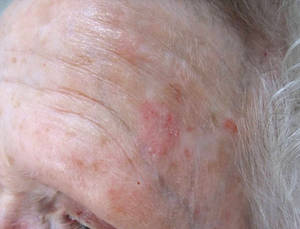
Imiquimod Effective for Many Skin Cancers, Expert Says
SANTA BARBARA, CALIF. - Imiquimod should be considered a possible treatment option for most skin cancer patients, according to Dr. Craig Kraffert.
Currently, imiquimod is approved only for the treatment actinic keratoses and basal cell carcinoma (BCC), but its greatest therapeutic benefit may occur with off-label use, Dr. Kraffert said at the annual meeting of the California Society of Dermatology and Dermatologic Surgery.
"It’s reasonable to consider for all basal cell carcinoma subtypes, but it’s not for aggressive or large squamous cell carcinoma, and it’s certainly not for invasive melanoma," said Dr. Kraffert, who practices dermatology in Redding, Calif.
Advantages to using imiquimod, he said, include the fact it doesn’t require surgery, it has a good cure rate, noncures are identifiable, and it causes minimal scarring "Disadvantages are that it’s time consuming, there’s transient morbidity, and it requires a coherent, compliant patient as well as provider oversight," he said. "And it’s a bit expensive."
In his practice, he uses imiquimod for patients with all types of BCC, for those with in situ and superficially invasive squamous cell carcinoma (SCC), and for those with melanoma in situ. "It’s also good for premalignant epithelial targets such as actinic keratoses, actinic cheilitis, vulvar intraepithelial neoplasia, vaginal intraepithelial neoplasia, and oral leukoplakia," he said. "Imiquimod therapeutic considerations for malignant and premalignant processes can be considered essentially equivalent."
He presented clinical data from patients in his practice who have been treated with imiquimod to date. Of 50 patients with SCC in situ, 70% had success with combination therapy, 28% had success with monotherapy, and 2% discontinued therapy.
Of 328 patients with BCC, 53% had success with monotherapy, 34% had success with combination therapy, and 13% discontinued therapy.

Of 25 patients with melanoma in situ, 20% had success with monotherapy, 56% had success with combination therapy, and 24% discontinued therapy.
The drug’s mechanism of action is not entirely understood, but it appears to activate both innate immunity with a cytokine cascade, as well as adaptive immunity with Langerhans cells. "It also causes a direct apoptotic and antineoplastic effect," he said.

In his opinion, high-utility niches include any patient seeking minimized scarring and who doesn’t want surgery, electrodesiccation and curettage (ED & C) failure, partially infiltrative or partially superficial BCC, multifocal temporal BCC, and large and/or ill-defined facial SCC in situ.
He finds that using imiquimod after ED & C in challenging tumors or in tumors with an infiltrative component increases the cure rate and decreases scarring. "I contend that imiquimod after ED & C generally gives you a better scar than ED & C alone," he said. "I don’t know why, but it does. Brisk reaction can be challenging, and close monitoring is required."
Using imiquimod prior to excision "may shrink tumors, leading to a smaller defect, better cosmesis, possible lower recurrence rate, and potential for complete nonsurgical response," he added.
He also finds the drug helpful for infiltrative BCCs in cosmetically vulnerable areas and for edge persistence after Mohs surgery. "Part of this is because imiquimod seems to work better on infiltrative basal cell carcinomas than it does on nodular basal cell carcinomas," he explained. "I think that’s because of access of the immune system to the thin threads of tumor. It’s also useful for melanoma in situ, particularly in cosmetically vulnerable areas. With melanoma in situ, melanin is a visible marker that serves as a clinical treatment site monitor. I don’t know of any reports of amelanotic transformation from imiquimod. Scarring is often minimal or absent, and there is tumor margin auto detection. It does have curative potential for melanoma in situ."
He emphasized that imiquimod monotherapy in patients with melanoma in situ is difficult, with varied response. "That’s where 5-FU [fluorouracil] in addition to imiquimod is often extremely useful," he said.
Dr. Kraffert described imiquimod as "a thinking practitioner’s medication" that requires thought and oversight. "It may provide excellent outcomes when least expected, so it’s important to keep an open mind regarding the clinical utility of this medication with skin cancer," he said.
Patients must be able to follow instructions, comply with treatment, return for monitoring and surveillance, and call or return to the clinic when appropriate. "Believe it or not, these limited requirements exclude many potential candidates," he remarked.
Patients typically use the drug for 6 weeks, beginning with twice daily dosing, "but it’s variable," he said. "For optimal success treatment must be strong enough for long enough."
At the initial consultation Dr. Kraffert educates his patient about the concept of delayed gratification, the reasons why the medication was suggested, and reaffirms that he will be there to oversee treatment.
He instructs them on how to use the medication, explaining they need to rub it into the lesion twice a day very well. "A pinhead amount is all that’s needed," he said, only a small portion of the 0.25 g included in each packet. His patients typically get a few to several applications from each packet.
He also educates patients about possible side effects from imiquimod, including redness, crusting, oozing, and some tenderness. "Nausea, malaise, pain, and secondary infection are rare," he said. "Patients are advised to call the office with any significant treatment related concerns and are generally offered the option of a same day add-on appointment."
At the 2-week follow-up visit he gauges the response as very brisk, brisk, moderate, mild, or none. If the response is very brisk, he halts therapy for 3-7 days, re-initiates treatment once daily for 4 weeks, and schedules a follow-up visit after 2 more weeks of treatment.
If the response is brisk, he halts therapy for 1-3 days, and then continues therapy once daily for 4 weeks.
If the response is moderate he continues therapy once daily for 4 weeks, and if the response is mild he continues therapy twice daily for 4 weeks.
"If there is no response, consider further imiquimod treatment with 2-4-week re-evaluation, or consider application of a small (less than a pea-sized) amount of 5% 5-FU after imiquimod twice daily," Dr. Kraffert said. "Or, you could consider nonimiquimod management of the problem. Sometimes imiquimod just doesn’t work."
After the 2-week follow-up, further visits are discretionary, he said, but some patients require closer follow-up, including nonresponders and patients with severe reactions, systemic complaints, and large or challenging tumors.
"Scheduling a follow-up visit for 3-6 months is best because the reaction takes a month or 2 to diminish," he said. "Taking photos pretherapy and at each visit thereafter improves surveillance."
"There are many approaches to skin cancer," Dr. Kraffert concluded. "Excision or destruction is often the best, but having more options is a plus. Imiquimod doesn’t replace what we have, it just adds to it."
Dr. Kraffert said he had no relevant financial conflicts.
SANTA BARBARA, CALIF. - Imiquimod should be considered a possible treatment option for most skin cancer patients, according to Dr. Craig Kraffert.
Currently, imiquimod is approved only for the treatment actinic keratoses and basal cell carcinoma (BCC), but its greatest therapeutic benefit may occur with off-label use, Dr. Kraffert said at the annual meeting of the California Society of Dermatology and Dermatologic Surgery.
"It’s reasonable to consider for all basal cell carcinoma subtypes, but it’s not for aggressive or large squamous cell carcinoma, and it’s certainly not for invasive melanoma," said Dr. Kraffert, who practices dermatology in Redding, Calif.
Advantages to using imiquimod, he said, include the fact it doesn’t require surgery, it has a good cure rate, noncures are identifiable, and it causes minimal scarring "Disadvantages are that it’s time consuming, there’s transient morbidity, and it requires a coherent, compliant patient as well as provider oversight," he said. "And it’s a bit expensive."
In his practice, he uses imiquimod for patients with all types of BCC, for those with in situ and superficially invasive squamous cell carcinoma (SCC), and for those with melanoma in situ. "It’s also good for premalignant epithelial targets such as actinic keratoses, actinic cheilitis, vulvar intraepithelial neoplasia, vaginal intraepithelial neoplasia, and oral leukoplakia," he said. "Imiquimod therapeutic considerations for malignant and premalignant processes can be considered essentially equivalent."
He presented clinical data from patients in his practice who have been treated with imiquimod to date. Of 50 patients with SCC in situ, 70% had success with combination therapy, 28% had success with monotherapy, and 2% discontinued therapy.
Of 328 patients with BCC, 53% had success with monotherapy, 34% had success with combination therapy, and 13% discontinued therapy.
Of 25 patients with melanoma in situ, 20% had success with monotherapy, 56% had success with combination therapy, and 24% discontinued therapy.
The drug’s mechanism of action is not entirely understood, but it appears to activate both innate immunity with a cytokine cascade, as well as adaptive immunity with Langerhans cells. "It also causes a direct apoptotic and antineoplastic effect," he said.
In his opinion, high-utility niches include any patient seeking minimized scarring and who doesn’t want surgery, electrodesiccation and curettage (ED & C) failure, partially infiltrative or partially superficial BCC, multifocal temporal BCC, and large and/or ill-defined facial SCC in situ.
He finds that using imiquimod after ED & C in challenging tumors or in tumors with an infiltrative component increases the cure rate and decreases scarring. "I contend that imiquimod after ED & C generally gives you a better scar than ED & C alone," he said. "I don’t know why, but it does. Brisk reaction can be challenging, and close monitoring is required."
Using imiquimod prior to excision "may shrink tumors, leading to a smaller defect, better cosmesis, possible lower recurrence rate, and potential for complete nonsurgical response," he added.
He also finds the drug helpful for infiltrative BCCs in cosmetically vulnerable areas and for edge persistence after Mohs surgery. "Part of this is because imiquimod seems to work better on infiltrative basal cell carcinomas than it does on nodular basal cell carcinomas," he explained. "I think that’s because of access of the immune system to the thin threads of tumor. It’s also useful for melanoma in situ, particularly in cosmetically vulnerable areas. With melanoma in situ, melanin is a visible marker that serves as a clinical treatment site monitor. I don’t know of any reports of amelanotic transformation from imiquimod. Scarring is often minimal or absent, and there is tumor margin auto detection. It does have curative potential for melanoma in situ."
He emphasized that imiquimod monotherapy in patients with melanoma in situ is difficult, with varied response. "That’s where 5-FU [fluorouracil] in addition to imiquimod is often extremely useful," he said.
Dr. Kraffert described imiquimod as "a thinking practitioner’s medication" that requires thought and oversight. "It may provide excellent outcomes when least expected, so it’s important to keep an open mind regarding the clinical utility of this medication with skin cancer," he said.
Patients must be able to follow instructions, comply with treatment, return for monitoring and surveillance, and call or return to the clinic when appropriate. "Believe it or not, these limited requirements exclude many potential candidates," he remarked.
Patients typically use the drug for 6 weeks, beginning with twice daily dosing, "but it’s variable," he said. "For optimal success treatment must be strong enough for long enough."
At the initial consultation Dr. Kraffert educates his patient about the concept of delayed gratification, the reasons why the medication was suggested, and reaffirms that he will be there to oversee treatment.
He instructs them on how to use the medication, explaining they need to rub it into the lesion twice a day very well. "A pinhead amount is all that’s needed," he said, only a small portion of the 0.25 g included in each packet. His patients typically get a few to several applications from each packet.
He also educates patients about possible side effects from imiquimod, including redness, crusting, oozing, and some tenderness. "Nausea, malaise, pain, and secondary infection are rare," he said. "Patients are advised to call the office with any significant treatment related concerns and are generally offered the option of a same day add-on appointment."
At the 2-week follow-up visit he gauges the response as very brisk, brisk, moderate, mild, or none. If the response is very brisk, he halts therapy for 3-7 days, re-initiates treatment once daily for 4 weeks, and schedules a follow-up visit after 2 more weeks of treatment.
If the response is brisk, he halts therapy for 1-3 days, and then continues therapy once daily for 4 weeks.
If the response is moderate he continues therapy once daily for 4 weeks, and if the response is mild he continues therapy twice daily for 4 weeks.
"If there is no response, consider further imiquimod treatment with 2-4-week re-evaluation, or consider application of a small (less than a pea-sized) amount of 5% 5-FU after imiquimod twice daily," Dr. Kraffert said. "Or, you could consider nonimiquimod management of the problem. Sometimes imiquimod just doesn’t work."
After the 2-week follow-up, further visits are discretionary, he said, but some patients require closer follow-up, including nonresponders and patients with severe reactions, systemic complaints, and large or challenging tumors.
"Scheduling a follow-up visit for 3-6 months is best because the reaction takes a month or 2 to diminish," he said. "Taking photos pretherapy and at each visit thereafter improves surveillance."
"There are many approaches to skin cancer," Dr. Kraffert concluded. "Excision or destruction is often the best, but having more options is a plus. Imiquimod doesn’t replace what we have, it just adds to it."
Dr. Kraffert said he had no relevant financial conflicts.
EXPERT ANALYSIS FROM THE ANNUAL MEETING OF THE CALIFORNIA SOCIETY OF DERMATOLOGY AND DERMATOLOGIC SURGERY
Imiquimod Effective for Many Skin Cancers, Expert Says
SANTA BARBARA, CALIF. – Imiquimod should be considered a possible treatment option for most skin cancer patients, according to Dr. Craig Kraffert.
Currently, imiquimod is approved only for the treatment actinic keratoses and basal cell carcinoma (BCC), but its greatest therapeutic benefit may occur with off-label use, Dr. Kraffert said at the annual meeting of the California Society of Dermatology and Dermatologic Surgery.
"It’s reasonable to consider for all basal cell carcinoma subtypes, but it’s not for aggressive or large squamous cell carcinoma, and it’s certainly not for invasive melanoma," said Dr. Kraffert, who practices dermatology in Redding, Calif.
Advantages to using imiquimod, he said, include the fact it doesn’t require surgery, it has a good cure rate, noncures are identifiable, and it causes minimal scarring "Disadvantages are that it’s time consuming, there’s transient morbidity, and it requires a coherent, compliant patient as well as provider oversight," he said. "And it’s a bit expensive."
In his practice, he uses imiquimod for patients with all types of BCC, for those with in situ and superficially invasive squamous cell carcinoma (SCC), and for those with melanoma in situ. "It’s also good for premalignant epithelial targets such as actinic keratoses, actinic cheilitis, vulvar intraepithelial neoplasia, vaginal intraepithelial neoplasia, and oral leukoplakia," he said. "Imiquimod therapeutic considerations for malignant and premalignant processes can be considered essentially equivalent."
He presented clinical data from patients in his practice who have been treated with imiquimod to date. Of 50 patients with SCC in situ, 70% had success with combination therapy, 28% had success with monotherapy, and 2% discontinued therapy.
Of 328 patients with BCC, 53% had success with monotherapy, 34% had success with combination therapy, and 13% discontinued therapy.
Of 25 patients with melanoma in situ, 20% had success with monotherapy, 56% had success with combination therapy, and 24% discontinued therapy.
The drug’s mechanism of action is not entirely understood, but it appears to activate both innate immunity with a cytokine cascade, as well as adaptive immunity with Langerhans cells. "It also causes a direct apoptotic and antineoplastic effect," he said.
In his opinion, high-utility niches include any patient seeking minimized scarring and who doesn’t want surgery, electrodesiccation and curettage (ED & C) failure, partially infiltrative/partially superficial BCC, multifocal temporal BCC, and large and/or ill-defined facial SCC in situ.
He finds that using imiquimod after ED & C in challenging tumors or in tumors with an infiltrative component increases the cure rate and decreases scarring. "I contend that imiquimod after ED & C generally gives you a better scar than ED & C alone," he said. "I don’t know why, but it does. Brisk reaction can be challenging, and close monitoring is required."
Using imiquimod prior to excision "may shrink tumors, leading to a smaller defect, better cosmesis, possible lower recurrence rate, and potential for complete nonsurgical response," he added.
He also finds the drug helpful for infiltrative BCCs in cosmetically vulnerable areas and for edge persistence after Mohs surgery. "Part of this is because imiquimod seems to work better on infiltrative basal cell carcinomas than it does on nodular basal cell carcinomas," he explained. "I think that’s because of access of the immune system to the thin threads of tumor. It’s also useful for melanoma in situ, particularly in cosmetically vulnerable areas. With melanoma in situ, melanin is a visible marker that serves as a clinical treatment site monitor. I don’t know of any reports of amelanotic transformation from imiquimod. Scarring is often minimal or absent, and there is tumor margin auto detection. It does have curative potential for melanoma in situ."
He emphasized that imiquimod monotherapy in patients with melanoma in situ is difficult, with varied response. "That’s where 5-FU [fluorouracil] in addition to imiquimod is often extremely useful," he said.
Dr. Kraffert described imiquimod as "a thinking practitioner’s medication" that requires thought and oversight. "It may provide excellent outcomes when least expected, so it’s important to keep an open mind regarding the clinical utility of this medication with skin cancer," he said.
Patients must be able to follow instructions, comply with treatment, return for monitoring and surveillance, and call or return to the clinic when appropriate. "Believe it or not, these limited requirements exclude many potential candidates," he remarked.
Patients typically use the drug for 6 weeks, beginning with twice daily dosing, "but it’s variable," he said. "For optimal success treatment must be strong enough for long enough."
At the initial consultation Dr. Kraffert educates his patient about the concept of delayed gratification, the reasons why the medication was suggested, and reaffirms that he will be there to oversee treatment.
He instructs them on how to use the medication, explaining they need to rub it into the lesion twice a day very well. "A pinhead amount is all that’s needed," he said, only a small portion of the 0.25 g included in each packet. His patients typically get a few to several applications from each packet.
He also educates patients about possible side effects from imiquimod, including redness, crusting, oozing, and some tenderness. "Nausea, malaise, pain, and secondary infection are rare," he said. "Patients are advised to call the office with any significant treatment related concerns and are generally offered the option of a same day add-on appointment."
At the 2-week follow-up visit he gauges the response as very brisk, brisk, moderate, mild, or none. If the response is very brisk, he halts therapy for 3-7 days, re-initiates treatment once daily for 4 weeks, and schedules a follow-up visit after 2 more weeks of treatment.
If the response is brisk, he halts therapy for 1-3 days, and then continues therapy once daily for 4 weeks.
If the response is moderate he continues therapy once daily for 4 weeks, and if the response is mild he continues therapy twice daily for 4 weeks.
"If there is no response, consider further imiquimod treatment with 2-4-week re-evaluation, or consider application of a small (less than a pea-sized) amount of 5% 5-FU after imiquimod twice daily," Dr. Kraffert said. "Or, you could consider nonimiquimod management of the problem. Sometimes imiquimod just doesn’t work."
After the 2-week follow-up, further visits are discretionary, he said, but some patients require closer follow-up, including nonresponders and patients with severe reactions, systemic complaints, and large or challenging tumors.
"Scheduling a follow-up visit for 3-6 months is best because the reaction takes a month or 2 to diminish," he said. "Taking photos pretherapy and at each visit thereafter improves surveillance."
"There are many approaches to skin cancer," Dr. Kraffert concluded. "Excision or destruction is often the best, but having more options is a plus. Imiquimod doesn’t replace what we have, it just adds to it."
Dr. Kraffert said he had no relevant financial conflicts.
EXPERT ANALYSIS FROM THE ANNUAL MEETING OF THE CALIFORNIA SOCIETY OF DERMATOLOGY AND DERMATOLOGIC SURGERY
MRI Spotted Additional Breast Cancer in Women With Early Disease
SAN DIEGO – Magnetic resonance imaging identified additional breast cancer in 11% of women with newly diagnosed disease who were otherwise eligible for partial breast irradiation, preliminary results from an ongoing study demonstrated.
Treatment of these patients with limited radiation fields would result in undertreatment of the tumor and could potentially compromise disease control, Dr. Paige L. Dorn said at the annual meeting of the American Society for Radiation Oncology.
Treatment for early-stage breast cancer with partial breast irradiation is currently under investigation in a multi-institutional, randomized controlled trial, NSABP B-39. Retrospective data from the University of Chicago Hospitals and Clinics and other institutions "have shown that MRI is able to find additional disease in 5%-10% of otherwise partial breast irradiation–eligible candidates, based on mammogram and ultrasound alone," said Dr. Dorn of the university’s department of radiation oncology. "The purpose of this study is to evaluate the utility of MRI in detecting clinically occult foci of disease in a prospectively identified cohort of partial breast irradiation candidates uniformly undergoing MRI in addition to mammogram and ultrasound."
Since June of 2009, all imaging and surgical pathology have been reviewed in a multidisciplinary setting by radiologists, surgeons, pathologists, and radiation oncologists at the university to determine candidacy for partial breast irradiation. In patients eligible for partial breast irradiation, Dr. Dorn and her associates prospectively documented whether MRI identified additional lesions in the same quadrant (multifocal), a different quadrant (multicentric), or the contralateral breast. They biopsied suspicious MRI findings to confirm pathology and then prospectively recorded whether MRI findings prompted a change in the eligibility for partial breast irradiation according to the entry criteria outlined in NSABP B-39. Prospectively collected data was verified by retrospective evaluation of all patient records.
Of 486 patients screened by the researchers between June 2009 and October 2010, Dr. Dorn reported that 91 (18.7%) were deemed eligible for partial breast irradiation based on mammogram, ultrasound, and pathology alone. Their median age was 56 years.
Of the 91 patients, 66 had invasive ductal or lobular carcinoma, 18 patients had ductal carcinoma in situ, and 7 had invasive lobular disease. MRI identified additional disease in 10 patients. Multifocal disease was seen in 9 of these patients, while contralateral disease was confirmed in 1.
The researchers also found that MRI was more likely to identify occult disease in patients younger than age 50, in those who were premenopausal, and in those who had tumor sizes of 2 cm or greater.
Dr. Dorn said the study had certain limitations, including the fact that the potential impact of MRI on disease outcomes after partial breast irradiation remains unknown. "Whether these multifocal lesions would have been surgically excised is unknown," she added.
Based on the results of this and other studies, she concluded that MRI "should be increasingly considered as part of the work-up for partial breast irradiation candidates. MRI may help refine criteria for patient selection, especially in those with higher-risk features. We plan to continue this prospective study to further define the role of MRI in this population."
Dr. Dorn said she had no relevant financial conflicts.
FROM THE ANNUAL MEETING OF THE AMERICAN SOCIETY FOR RADIATION ONCOLOGY
Major Finding: Magnetic resonance imaging identified additional breast cancer in 10 of 91 patients with newly diagnosed disease who were otherwise eligible for partial breast irradiation.
Data Source: A study of 486 women with a median age of 56 years who were treated by a multidisciplinary team of investigators at the University of Chicago Hospitals and Clinics.
Disclosures: Dr. Dorn said she had no relevant financial conflicts.
Online Weight Management Yields Mixed Results
SAN DIEGO – There were no significant differences in the amount of weight lost at 18-month follow-up among adults randomized to a weight management program delivered online, compared with those randomized to the same program delivered in person.
However, a significantly greater proportion of self-monitoring records were submitted by adults assigned to the online group, compared with those assigned to the in-person group.
“We didn't know whether or not the delivery channel impacts weight-loss maintenance, but there are reasons to think it might be different online,” Delia Smith West, Ph.D., said at the annual meeting of the Obesity Society.
“We know that the maintenance phase is characterized by a decrease in session attendance. It can be a fairly marked decrease between the initial weekly sessions of weight reduction and weight maintenance. We also know that self-monitoring falls off,” she added.
For the study, known as iREACH, Dr. West and her associates evaluated weight maintenance and treatment adherence among 481 adults who received the identical group weight-loss program at clinics in Arkansas and Vermont. The treatment goals were to help participants modify eating and exercise habits, with modest calorie restriction, 25% or fewer calories from fat, exercise up to 200 minutes per week, and daily self-monitoring of dietary intake and physical activity. For this component, the participants who were randomized to the in-person group tracked their progress in a paper-based journal while the online group tracked their progress in a computer-based journal. Behavioral strategies included self-monitoring, problem solving, goal setting, stimulus control, relapse prevention, assertiveness training, social support, and homework.
Of the 481 patients, 161 were randomized to online delivery of the program during weekly sessions for 6 months, 159 were randomized to in-person group delivery of the program during weekly in-person sessions for 6 months, and 161 were randomized to receive a mix of both weekly online and in-person delivery sessions for 6 months.
Maintenance continued for 12 months, said Dr. West of the Fay W. Boozman College of Public Health at the University of Arkansas for Medical Sciences, Little Rock.
The mean age of patients was 47 years, 28% were black, and 93% were female. Their mean weight was 97 kg, their mean body mass index was 36 kg/m
Dr. West reported that at 6 months, the mean amount of weight loss significantly favored the in-person group (18 lbs, vs. 14 lbs for the hybrid group and 12 lbs for the online group). At 18 months, however, total mean weight loss did not differ significantly between the groups (12 lbs, 9 lbs, 6 lbs, respectively). The overall mean weight regain at 18 months was similar between the groups (6 lbs, 4 lbs, and 5 lbs).
The proportion of participants who attended all scheduled sessions over the 12 months of the maintenance program did not differ between the groups (37% for the in-person group, 33% for the hybrid group, and 41% for the online group), but a significantly greater proportion of self-monitoring records were submitted by the Internet group (28%, compared with 14% by the in-person group and 20% by the hybrid group).
The study was funded by the National Institute of Diabetes and Digestive and Kidney Diseases. Dr. West said that she had no relevant financial disclosures.
The weight-loss advantage in patients attending in-person sessions at 6 months faded a year later.
Source DR. WEST
From the Annual Meeting of the Obesity Society
Low Vitamin C Levels Linked to Obesity in Children
Major Finding: The odds of being obese were 0.42 lower among children with serum vitamin C levels of 76.1-195 micromol/L, compared with children who had serum vitamin C levels of 9-76 micromol/L.
Data Source: A study of 1,133 healthy weight or obese children aged 6-11 years who were included in the NHANES data sets from 2003-2004 and 2005-2006.
Disclosures: Ms. Ewing had no relevant financial conflicts.
SAN DIEGO — Low serum levels of vitamin C are associated with obesity in children aged 6-11 years, results from an analysis of data from the National Health and Nutrition Examination Survey showed.
“Childhood nutrition and obesity are significant public health concerns that may result in abnormal growth and development,” researchers led by Kelly Ewing wrote in a poster presented at the meeting “Although numerous studies have characterized vitamin C intake and serum levels, and have demonstrated an inverse association with obesity in adults, as far as we are aware, no studies to date have looked at this relationship in a pediatric population.”
She and her associates performed a cross-sectional, retrospective study of 1,133 healthy weight or obese children aged 6-11 years who were included in the NHANES data sets from 2003-2004 and 2005-2006, for whom relevant medical, socioeconomic, physical activity, anthropometric, and serum vitamin C data were complete.
The researchers divided the children into two groups based on their serum vitamin C concentrations: a lower 50% group (defined as having a vitamin C level of 9-76 micromol/L), and an upper 50% group (vitamin C level of 76.1-195 micromol/L).
Of the 1,133 children, 876 (77%) had a healthy weight and 257 (23%) were obese, said Ms. Ewing, who worked on the study while completing her master's degree in the department of clinical research at the Campbell University College of Pharmacy and Health Sciences, Buies Creek, N.C.
The researchers then conducted a bivariate analysis to identify significant associations between weight status and vitamin C concentration, race, sex, physical activity, and socioeconomic status. They found that that the odds of being obese were 0.42 lower in the upper 50% vitamin C level group, compared with the lower 50% group, when socioeconomic status was also considered.
Hours of physical activity and sex were not significant factors in the link between serum vitamin C levels and obesity, but race and income were. A higher proportion of nonwhite children were obese, compared with white children (26% vs. 16%, respectively), and a higher proportion of obese children were from households with an income of $24,999 or less per year (29% vs. 20%).
“I was surprised that socioeconomic status didn't kill the relationship,” Ms. Ewing said in an interview. “I figured that would have a bigger effect than just the vitamin C itself, because it's very established in the medical literature that obesity and economic status are inversely related.”
She acknowledged certain limitations of the study, including the fact that there was no way to know if serum was drawn while the children were fasting. “Our biggest limitation is that we were not sure what the kids were eating,” she added. “There was a food frequency questionnaire that we looked at. However, it was a yearlong recall that did not account for serving size.”
From the Annual Meeting of the Obesity Society
Behavioral Intervention Benefits Severely Obese
Major Finding: After 1 year of participation in an intensive lifestyle intervention program, patients with class III obesity lost a mean of 11 kg, compared with 9 kg and 6 kg in patients with class II and class I obesity, respectively.
Data Source: An analysis of 2,503 patients in the multicenter, randomized Look AHEAD clinical trial.
Disclosures: The study was funded by the National Institute of Diabetes and Digestive and Kidney Diseases. Dr. Unick said she had no conflicts of interest.
SAN DIEGO — Obese patients with a body mass index of 40 kg/m
The findings suggest that behavioral weight-loss programs are a viable treatment option for class III obese patients, Jessica L. Unick, Ph.D., said at the meeting.
“Severe obesity is the fastest-growing segment of the overweight population, increasing at a rate of two to three times faster than obesity alone,” said Dr. Unick of the centers for behavioral and preventive medicine at Miriam Hospital, Brown University, Providence, R.I. “Given these alarming statistics as well as the known health consequences associated with this magnitude of obesity, it's critical that we begin to identify successful treatment options for this population.”
She pointed out that currently, behavioral weight-loss programs are typically recommended for individuals with a BMI of less than 40. As a result, “severely obese individuals are often excluded from clinical weight-loss trials, usually due to other obesity-related comorbidities or upper BMI cutoff inclusionary criteria for the study. Therefore, we have limited data regarding the effectiveness of behavioral weight loss programs in this population.”
For the study, Dr. Unick and her associates examined whether severely obese patients randomized to the intensive lifestyle intervention arm of the Look AHEAD trial achieved similar weight losses, experienced comparable changes in cardiovascular disease risk factors, and had been equally adherent to program recommendations at 1 year of treatment, compared with those who had lesser degrees of obesity. The Look AHEAD trial is an ongoing, multicenter, randomized clinical trial examining the long-term effects of an intensive lifestyle intervention program on cardiovascular morbidity and mortality in 5,145 overweight or obese patients with type 2 diabetes from 16 centers in the United States. The individuals will be followed for up to 13.5 years.
To be eligible for the Look AHEAD trial, patients had to have type 2 diabetes, be 45-76 years of age, have a BMI of at least 25 (or at least 27 if taking insulin), have a hemoglobin A1c level of 11% or lower, and meet certain cardiovascular disease cutoff values, including triglycerides below 600 mg/dL and a blood pressure of less than 160/100 mm Hg. The patients were randomized to an intensive lifestyle intervention or to diabetes support and education. The study conducted by Dr. Unick and her associates evaluated only the 2,503 patients randomized to the intensive lifestyle intervention.
The researchers grouped the study participants into one of four categories based on BMI: overweight (25-29), class I obese (30-34), class II obese (35-39), and class III obese (40 or greater). Their mean age at baseline was 59 years.
For the first 6 months of the study, participants attended three weekly group meetings and one individual meeting per month. This was reduced to two group meetings and one individual meeting per month during months 7-12.
Patients in the intensive lifestyle intervention arm were asked to reduce their dietary intake to 1,200-1,800 calories per day. They were provided meal replacements in the form of two meals and one snack per day to help facilitate that goal. They were also asked to progressively increase their level of physical exercise to 175 minutes per week.
“In addition, the participants were given an initial weight loss goal of 10% of initial body weight, and there was a strong behavioral component to this intensive lifestyle intervention,” Dr. Unick said.
She reported that at 1 year, class III obese patients lost a mean of 11 kg, which was significantly greater than the weight loss experienced by the other groups (a mean of 9 kg in the class II obese group, 8 kg in the class I obese group, and 6 kg in the overweight group).
On average, class III obese patients lost 9% of their baseline weight, and 39% lost 10% or more of their baseline body weight. These reductions in weight were significantly greater than those experienced by the overweight group and were comparable to those achieved by patients in the class I and class II groups.
All weight groups had similar improvements in cardiovascular disease risk factors, with the exception of HDL cholesterol level. The class III group had less of an improvement in HDL cholesterol from baseline to 1 year, compared with the other BMI groups.
By the end of 1 year, 42% of people in the class III group met the American Diabetes Association goal of an LDL cholesterol less than 100 mg/dL, 65% met the ADA goal of a systolic blood pressure less than 130/80 mm Hg, and 72% met the ADA goal of an HbA1c less than 7%. The results were similar to those in participants who had lesser degrees of obesity.
A similar proportion of all weight groups attended all treatment sessions (an average of more than 80%), consumed a similar number of meal replacements (average of 333), and improved the number of calories expended by physical activity (an average of 761 calories per week).
“Based upon these data, there appears to be no reason why we should be excluding severely obese individuals from behavioral weight-loss treatment programs,” Dr. Unick commented. “Behavioral programs should be considered another treatment option for individuals in this lower end of the severe obesity range.”
'It's critical that we begin to identify successful treatment options for this population.”
Source DR. UNICK
From the Annual Meeting of the Obesity Society
Serotonin May Flag Progression of Heart Failure
SAN DIEGO – Plasma levels of serotonin were significantly elevated in patients with decompensated systolic heart failure, compared with patients in the compensated state and with normal controls, according to a single-center study.
The finding suggests that serotonin has an active role in the progression of heart failure (HF), researchers led by Dr. Ahmed M. Selim reported during a poster session at the meeting.
“More studies should be done to test the sensitivity, specificity, and prognostic value of serotonin as a marker for congestive heart failure,” wrote the researchers from the department of cardiology at Albert Einstein College of Medicine, New York.
They noted that, while the relationship between HF and the serotoninergic system has been established in previous research, fluctuations in serotonin levels during the course of the disease and its correlation with exacerbation of HF have never been tested.
Dr. Selim and his associates measured plasma serotonin levels in 29 patients admitted with decompensated HF, 61 with stable HF, and 22 normal controls.
Overall, the mean age of patients was 55 years, and 62% were male.
The researchers reported that the mean serotonin level in the control group was 2.4 ng/mL, vs. 4.1 ng/mL in the compensated group and 11.8 ng/mL in the decompensated group. “All results were highly significant,” the researchers wrote.
Dr. Selim and his associates stated that they had no relevant financial conflicts of interest.