User login
Doug Brunk is a San Diego-based award-winning reporter who began covering health care in 1991. Before joining the company, he wrote for the health sciences division of Columbia University and was an associate editor at Contemporary Long Term Care magazine when it won a Jesse H. Neal Award. His work has been syndicated by the Los Angeles Times and he is the author of two books related to the University of Kentucky Wildcats men's basketball program. Doug has a master’s degree in magazine journalism from the S.I. Newhouse School of Public Communications at Syracuse University. Follow him on Twitter @dougbrunk.
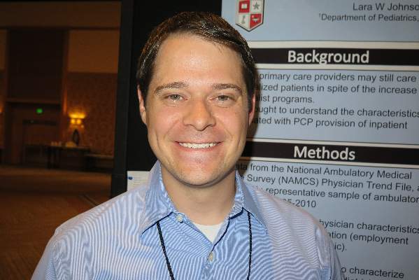
Most community pediatricians still practice in the hospital
SAN DIEGO – Nearly two-thirds of pediatricians provide inpatient hospital care in a given week of practice, results from a large national survey showed.
“We know that the number of overall hospitalists has rapidly increased in the United States, but we wanted to know how many of the non-hospitalists were continuing to provide inpatient care,” Dr. Shaughn M. Nunez said in an interview at the annual meeting of the American Academy of Pediatrics.

Dr. Nunez and his associates used the National Ambulatory Medical Care Survey (NAMCS), a nationally representative sample of ambulatory visits conducted by the National Center for Health Statistics. They defined providing hospital care as answering “yes” to the question regarding making any hospital visit in the last normal week of practice, and generated descriptive statistics for practice, physician and community characteristics. Next, they used bivariate tests of association to explore the relationship between practice characteristics and provision of hospital care.
Of 634 pediatricians who participated in the survey from 2005-2010, nearly two-thirds of respondents (62%) reported providing hospital care in the previous week, reported Dr. Nunez, a hospitalist at Texas Tech University Health Sciences Center, Lubbock. Pediatricians were more likely to report providing inpatient care, compared with family physicians (42%) and internists (58%), (P < .001).
The proportion of pediatricians who provided inpatient care ranged from 67% in 2005 to 58% in 2010. Those with more than 75% of patients on Medicaid were less likely to provide inpatient care (42.6%, P < .007). In addition, those who practiced in a community-clinic setting provided inpatient care less frequently, compared with those in a private practice setting (35% vs. 67%, respectively; P < .001).
Physician employment status also weighed in. Those who owned their practice reported providing inpatient care 68% of the time, compared with 55% of physician-employees and 47% of physicians working on a contract basis (P = .012). Practice location in a metropolitan area and region of the country were not associated with the provision of inpatient care.
Dr. Nunez acknowledged certain limitations of the study, including the fact that the researchers were unable to distinguish if pediatricians were providing newborn nursery care or inpatient care. He reported having no financial disclosures.
SAN DIEGO – Nearly two-thirds of pediatricians provide inpatient hospital care in a given week of practice, results from a large national survey showed.
“We know that the number of overall hospitalists has rapidly increased in the United States, but we wanted to know how many of the non-hospitalists were continuing to provide inpatient care,” Dr. Shaughn M. Nunez said in an interview at the annual meeting of the American Academy of Pediatrics.
Dr. Nunez and his associates used the National Ambulatory Medical Care Survey (NAMCS), a nationally representative sample of ambulatory visits conducted by the National Center for Health Statistics. They defined providing hospital care as answering “yes” to the question regarding making any hospital visit in the last normal week of practice, and generated descriptive statistics for practice, physician and community characteristics. Next, they used bivariate tests of association to explore the relationship between practice characteristics and provision of hospital care.
Of 634 pediatricians who participated in the survey from 2005-2010, nearly two-thirds of respondents (62%) reported providing hospital care in the previous week, reported Dr. Nunez, a hospitalist at Texas Tech University Health Sciences Center, Lubbock. Pediatricians were more likely to report providing inpatient care, compared with family physicians (42%) and internists (58%), (P < .001).
The proportion of pediatricians who provided inpatient care ranged from 67% in 2005 to 58% in 2010. Those with more than 75% of patients on Medicaid were less likely to provide inpatient care (42.6%, P < .007). In addition, those who practiced in a community-clinic setting provided inpatient care less frequently, compared with those in a private practice setting (35% vs. 67%, respectively; P < .001).
Physician employment status also weighed in. Those who owned their practice reported providing inpatient care 68% of the time, compared with 55% of physician-employees and 47% of physicians working on a contract basis (P = .012). Practice location in a metropolitan area and region of the country were not associated with the provision of inpatient care.
Dr. Nunez acknowledged certain limitations of the study, including the fact that the researchers were unable to distinguish if pediatricians were providing newborn nursery care or inpatient care. He reported having no financial disclosures.
AT THE AAP NATIONAL CONFERENCE
Key clinical point: Many pediatricians provide inpatient care.
Major finding: Nearly two-thirds of pediatricians (62%) provide inpatient care in a given week of practice.
Data source: A survey of 634 pediatricians who participated in the National Ambulatory Medical Care Survey from 2005-2010.
Disclosures: Dr. Nunez reported having no financial disclosures.
Look to presentation, not pathology, for dermatomyositis diagnosis
VANCOUVER, B.C.– On histology, pathologists are unable to reliably differentiate between cutaneous lupus erythematosus and dermatomyositis. So it falls to clinicians to sort out what’s what based on the patient’s clinical presentation.
“Dermatomyositis is a disease of muscle and skin, but as dermatologists we should view this as primarily a dermatologic disorder, because the diagnosis is made on the skin,” Dr. Jan Dutz said at the annual meeting of the Pacific Dermatologic Association. “The pathology is not diagnostic. Therefore, it’s up to you to make the diagnosis based on the characteristic patterns of dermatomyositis: the changes of the violaceous erythema on the face and arms, the Gottron’s papules, and the changes in blood vessels.”

Enlarged and decreased capillaries are a hallmark clinical feature of dermatomyositis, which can be observed by examining the proximal nail folds. “It turns out that you can look at those capillaries and predict the severity of the disease, so that those who have improvement in the capillaries following early treatment usually have less severe disease,” said Dr. Dutz, professor of dermatology and skin science at the University of British Columbia, Vancouver.
Like cutaneous lupus erythematosus, dermatomyositis is a type 1 interferon-mediated disease. The genes inducible by type 1 interferon alpha have increased expression in muscle and in peripheral blood lymphocyte, and interferon alpha administration can induce disease. Researchers recently described a new subset of dermatomyositis based on an investigation of 77 patients with the disorder (J. Am. Acad. Dermatol. 2011;65:25-34). Of these, 10 (13%) had anti-MDA-5 (melanoma differentiation associated gene 5) antibodies, which are antibodies to a protein related to the interferon pathway. Most of the patients (8 of 10) tested negative for antinuclear antibody. The clinical features were palmar papules, ulcerations (usually on the back of the hands), MDA-5 antibody, and an increased risk of interstitial lung disease. “Ulceration had an odds ratio of almost 20 with being associated with this antibody and this syndrome,” said Dr. Dutz, who was not involved with the study. “So if you see these papules and you see these ulcerations on the hands, it increases the chance of interstitial lung disease.”
These clinical features also have been observed in cases of juvenile dermatomyositis (Arthritis Res. Ther. 2014;16:R138). “These patients get skin ulcerations, oral ulcerations, and have a high incidence for arthritis, mild muscle disease, and interstitial lung disease,” he said.
Dr. Dutz said that he often turns to antimalarial agents as the first treatment option for patients with dermatomyositis. If they don’t respond, he typically switches them to methotrexate. “If that doesn’t work, I usually try mycophenolate mofetil or IVIG [intravenous immunoglobulin], which is one of the most effective treatments for dermatomyositis,” he said. “Studies keep on emphasizing this. There’s also some interest in using rituximab in patients with resistant disease.”
Dr. Dutz disclosed that he is an advisory board member for Janssen, AbbVie, Amgen, Leo Pharma, Roche, and Novartis. He has also conducted clinical trials for Centocor and Ono Pharmaceutical Co.
On Twitter @dougbrunk
VANCOUVER, B.C.– On histology, pathologists are unable to reliably differentiate between cutaneous lupus erythematosus and dermatomyositis. So it falls to clinicians to sort out what’s what based on the patient’s clinical presentation.
“Dermatomyositis is a disease of muscle and skin, but as dermatologists we should view this as primarily a dermatologic disorder, because the diagnosis is made on the skin,” Dr. Jan Dutz said at the annual meeting of the Pacific Dermatologic Association. “The pathology is not diagnostic. Therefore, it’s up to you to make the diagnosis based on the characteristic patterns of dermatomyositis: the changes of the violaceous erythema on the face and arms, the Gottron’s papules, and the changes in blood vessels.”
Enlarged and decreased capillaries are a hallmark clinical feature of dermatomyositis, which can be observed by examining the proximal nail folds. “It turns out that you can look at those capillaries and predict the severity of the disease, so that those who have improvement in the capillaries following early treatment usually have less severe disease,” said Dr. Dutz, professor of dermatology and skin science at the University of British Columbia, Vancouver.
Like cutaneous lupus erythematosus, dermatomyositis is a type 1 interferon-mediated disease. The genes inducible by type 1 interferon alpha have increased expression in muscle and in peripheral blood lymphocyte, and interferon alpha administration can induce disease. Researchers recently described a new subset of dermatomyositis based on an investigation of 77 patients with the disorder (J. Am. Acad. Dermatol. 2011;65:25-34). Of these, 10 (13%) had anti-MDA-5 (melanoma differentiation associated gene 5) antibodies, which are antibodies to a protein related to the interferon pathway. Most of the patients (8 of 10) tested negative for antinuclear antibody. The clinical features were palmar papules, ulcerations (usually on the back of the hands), MDA-5 antibody, and an increased risk of interstitial lung disease. “Ulceration had an odds ratio of almost 20 with being associated with this antibody and this syndrome,” said Dr. Dutz, who was not involved with the study. “So if you see these papules and you see these ulcerations on the hands, it increases the chance of interstitial lung disease.”
These clinical features also have been observed in cases of juvenile dermatomyositis (Arthritis Res. Ther. 2014;16:R138). “These patients get skin ulcerations, oral ulcerations, and have a high incidence for arthritis, mild muscle disease, and interstitial lung disease,” he said.
Dr. Dutz said that he often turns to antimalarial agents as the first treatment option for patients with dermatomyositis. If they don’t respond, he typically switches them to methotrexate. “If that doesn’t work, I usually try mycophenolate mofetil or IVIG [intravenous immunoglobulin], which is one of the most effective treatments for dermatomyositis,” he said. “Studies keep on emphasizing this. There’s also some interest in using rituximab in patients with resistant disease.”
Dr. Dutz disclosed that he is an advisory board member for Janssen, AbbVie, Amgen, Leo Pharma, Roche, and Novartis. He has also conducted clinical trials for Centocor and Ono Pharmaceutical Co.
On Twitter @dougbrunk
EXPERT ANALYSIS FROM PDA 2014

Transanal extraction found effective in rectal cancer surgery
Patients who underwent laparoscopic total mesorectal excision with coloanal anastomosis for rectal cancer had similar rates of mortality and morbidity, regardless of whether the extraction was performed transanally or transabdominally, a long-term single-center study showed.
“There are few data of full laparoscopic coloanal anastomosis for rectal cancer, including small series and short follow-up,” authors led by Dr. Quentin Denost of the department of surgery at Saint-André Hospital, Bordeau, France, wrote. “Moreover, the risk of anastomotic or perineal recurrence induced by transanal extraction of the rectal specimen is not known.” In addition, they continued, functional outcomes of laparoscopic coloanal anastomosis “have never been reported, and therefore the potential risk of anal incontinence related to transanal specimen extraction has never been discussed.”
In an effort to investigate the long-term outcome of laparoscopic coloanal anastomosis for rectal cancer, the researchers evaluated records of 220 patients who underwent laparoscopic total mesorectal excision and coloanal anastomosis for rectal cancer at Saint-André Hospital between 2000 and 2010 (Ann. Surg. 2014 Sept. 1 [doi:10.1097/SLA.0000000000000855]). Study endpoints of interest were circumferential margin, mesorectal grade, local recurrence, survival, and functional outcome obtained by a questionnaire sent to patients free of disease with at least 1 year of follow-up after stoma closure.
More than half of the patients (63%) were male, their median age was 64, and their median body mass index was 25 kg/m2. The tumors were a median of 4 cm from the anal verge and 1 cm from the anal ring, and 82% of the patients had stage T3 or T4 disease.
The authors reported that the overall mortality and surgical morbidity rates were 0.5% and 17%, respectively, the rate of positive circumferential resection margin was 9%, and the median anal continence score was 6 (range, 0-20). After a median follow-up of 51 months, the local recurrence rate was 4%, while at 5 years, the overall survival and disease-free survival rates were 83% and 70%, respectively.
When the authors evaluated results by extraction site, no significant differences were observed between the transanal extraction and transabdomonal extraction groups in the rate of overall mortality (0.8% vs. 0%, respectively; P = 1.000), overall morbidity (34% vs. 43%), positive circumferential margin (7% vs. 11%; P = .324), mesorectal grade, local recurrence (4% vs. 5%; P = .98), and disease-free survival (72% vs. 68%; P = .63). The median continence score was 6 in both groups (P = .92).
The findings demonstrated that pelvic control and survival “were not compromised by the association between mini invasive surgery and ultralow sphincter preservation,” the authors concluded. “Moreover, we demonstrated the safety and efficacy of transanal extraction of the rectal specimen with similar oncologic and functional outcome than the conventional abdominal extraction. Because of the wound advantages of transanal extraction, in terms of abdominal wall preservation, transanal extraction can be recommended in laparoscopic surgical management of low rectal cancer.”
They acknowledged certain limitations of the study, including the fact that BMI was slightly lower in the transanal group, compared with the transabdomonal group (24.3 vs. 25.8 kg/m2, respectively; P = .01). This suggests “that some obese patients probably received transabdominal instead of transanal extraction,” Dr. Denost and associates wrote. “Therefore, as we recommend preventing excessive stretching of the anal sphincter during rectal extraction, we also recommend to be cautious when performing transanal extraction in obese patients with wide mesorectal specimen, especially to avoid mesorectal injury and tumor spillage.”
The authors reported having no relevant financial disclosures.
On Twitter @dougbrunk
FROM ANNALS OF SURGERY
Key clinical point: Transanal extraction can be recommended in the laparoscopic surgical management of low rectal cancer.
Major finding: During laparoscopic total mesorectal excision, no significant differences were observed between patients who underwent transanal extraction or transabdomonal extraction in the rate of overall mortality (0.8% vs. 0%, respectively; P = 1.000), overall morbidity (34% vs. 43%), or local recurrence (4% vs. 5%; P = .98).
Data source: A single-center study of 220 patients who underwent laparoscopic total mesorectal excision and coloanal anastomosis for rectal cancer at Saint-André Hospital in Bordeaux, France, between 2000 and 2010.
Disclosures: The authors reported having no relevant financial disclosures.
Postoperative complications increase risk of death in CRC patients
Patients who develop infectious complications after undergoing curative surgery for colorectal cancer face a significantly increased risk of death, results from a large retrospective study showed.
“The association of postoperative complications with long-term survival after major surgery has been suggested by several studies in mixed populations and is to some degree expected and intuitive,” authors led by Dr. Avo Artinyan, of the surgery department at Baylor College of Medicine, Houston, wrote online Sept. 1 in Annals of Surgery. “It has been difficult, however, to determine a specific cause-effect relationship, particularly because this association is noted even in patients who suffer late mortality, that is, those who presumably recover from postoperative complications.”

In an effort to investigate the effect of postoperative complications on long-term survival after colorectal cancer resection, the researchers evaluated the records of 12,075 patients from the Veterans Affairs Surgical Quality Improvement Program and the Central Cancer Registry databases who underwent resection for nonmetastatic colorectal cancer from 1999 to 2009 (Ann. Surg. 2014 Sept. 1 [doi: 10.1097/SLA.0000000000000854]). They categorized patients by presence of any complication within 30 days and by type of complication (infectious vs. noninfectious); excluded patients who died within 90 days of the procedure; and performed univariate and multivariate analyses adjusted for patient, disease, and treatment factors.
The average age of the cohort was 69 years, 98% were men, more than two-thirds (69%) had an American Society of Anesthesiologists (ASA) classification score of 3, and 61% had stage 1 or 2 disease. Dr. Artinyan and his associates found that the overall morbidity and infectious complication rates were 27.8% and 22.5%, respectively.
Compared with patients who had no postoperative complications, those who did were older and had lower postoperative serum albumin, worse functional status, and higher ASA scores (P less than .001). Multivariate analysis revealed that the presence of any complication was associated with a 24% increased hazard of death (hazard ratio, 1.24; P less that .001). When the analysis was limited to the type of complication, patients with infectious complications (in particular, surgical site infections) had an increased hazard of death (HR, 1.31), predominately those with severe infections (HR, 1.41).
“To our knowledge, this is the largest single study to examine the association of postoperative complications with long-term survival for CRC,” the authors wrote. “Similar to other groups, we have demonstrated that postoperative complications occur in a significant proportion of patients after CRC resection and that most patients with postoperative morbidity have at least one infectious complication.”
They acknowledged certain limitations of the study, including its retrospective design and the potential for selection bias. “Additional limitations include the absence of margin data – which may have a considerable impact on both the risk of organ-space infections and disease recurrence – and the inability to calculate cancer-specific survival and other cancer-specific outcomes,” they wrote.
“Overall all-cause survival, however, is still a commonly used and useful outcome measure, and we have attempted to mitigate the effect of early non–cancer-related mortality with the exclusion of early deaths.”
The authors reported having no financial disclosures.
On Twitter @dougbrunk
Patients who develop infectious complications after undergoing curative surgery for colorectal cancer face a significantly increased risk of death, results from a large retrospective study showed.
“The association of postoperative complications with long-term survival after major surgery has been suggested by several studies in mixed populations and is to some degree expected and intuitive,” authors led by Dr. Avo Artinyan, of the surgery department at Baylor College of Medicine, Houston, wrote online Sept. 1 in Annals of Surgery. “It has been difficult, however, to determine a specific cause-effect relationship, particularly because this association is noted even in patients who suffer late mortality, that is, those who presumably recover from postoperative complications.”
In an effort to investigate the effect of postoperative complications on long-term survival after colorectal cancer resection, the researchers evaluated the records of 12,075 patients from the Veterans Affairs Surgical Quality Improvement Program and the Central Cancer Registry databases who underwent resection for nonmetastatic colorectal cancer from 1999 to 2009 (Ann. Surg. 2014 Sept. 1 [doi: 10.1097/SLA.0000000000000854]). They categorized patients by presence of any complication within 30 days and by type of complication (infectious vs. noninfectious); excluded patients who died within 90 days of the procedure; and performed univariate and multivariate analyses adjusted for patient, disease, and treatment factors.
The average age of the cohort was 69 years, 98% were men, more than two-thirds (69%) had an American Society of Anesthesiologists (ASA) classification score of 3, and 61% had stage 1 or 2 disease. Dr. Artinyan and his associates found that the overall morbidity and infectious complication rates were 27.8% and 22.5%, respectively.
Compared with patients who had no postoperative complications, those who did were older and had lower postoperative serum albumin, worse functional status, and higher ASA scores (P less than .001). Multivariate analysis revealed that the presence of any complication was associated with a 24% increased hazard of death (hazard ratio, 1.24; P less that .001). When the analysis was limited to the type of complication, patients with infectious complications (in particular, surgical site infections) had an increased hazard of death (HR, 1.31), predominately those with severe infections (HR, 1.41).
“To our knowledge, this is the largest single study to examine the association of postoperative complications with long-term survival for CRC,” the authors wrote. “Similar to other groups, we have demonstrated that postoperative complications occur in a significant proportion of patients after CRC resection and that most patients with postoperative morbidity have at least one infectious complication.”
They acknowledged certain limitations of the study, including its retrospective design and the potential for selection bias. “Additional limitations include the absence of margin data – which may have a considerable impact on both the risk of organ-space infections and disease recurrence – and the inability to calculate cancer-specific survival and other cancer-specific outcomes,” they wrote.
“Overall all-cause survival, however, is still a commonly used and useful outcome measure, and we have attempted to mitigate the effect of early non–cancer-related mortality with the exclusion of early deaths.”
The authors reported having no financial disclosures.
On Twitter @dougbrunk
FROM ANNALS OF SURGERY
Key clinical point: Postoperative complications after colorectal cancer surgery are associated with decreased long-term survival.
Major finding: The presence of any complication after CRC surgery was associated with a 24% increased hazard of death (hazard ratio, 1.24; P less than .001).
Data source: A retrospective evaluation of 12,075 patients who underwent resection for nonmetastatic CRC from 1999-2009.
Disclosures: The authors reported having no financial disclosures.

Ergonomic solutions limit Mohs surgeons’ aches and pains
VANCOUVER, B.C. – Attention to ergonomics may reduce risks for a wide range of musculoskeletal problems and headaches that afflict Mohs surgeons, Dr. Mariusz Sapijaszko said at the annual meeting of the Pacific Dermatologic Association.
The demands of performing Mohs surgery were first detailed in a study of 17 Mohs surgeons at the Mayo Clinic. Nearly two-thirds (59%) had chronic neck pain, half had shoulder pain (53%), and nearly half had lower back pain (41%). About a third experienced eye fatigue and one-fourth had headaches (Dermatol. Surg. 2007;33:1304-13).

Since then, a survey of American College of Mohs Surgery members found that 90% reported some type of musculoskeletal symptoms or injuries (Dermatol. Surg. 2012;38:240-8).
Dr. Sapijaszko of the Western Canada Dermatology Institute, Edmonton, Alberta, offered the following ergonomic tips based on his own clinical practice as well as recommendations offered by researchers who conducted the 2007 Mayo Clinic study and those focused on optimizing the operating theater environment (ANZ J. Surg. 2010;80:917-24).
To reduce neck-related symptoms:
• Keep your gaze angle between 15 and 30 degrees below horizontal.
• Position the patient close to you.
• Take short surgery breaks to stretch and adjust your posture.
• Use a stool with sternal support or a sit/stand stool.
To avoid lower back pain:
• Change positions frequently.
• Use a foot rest or foot rail.
• Use a stool with sternal support.
To prevent eye fatigue:
• Decrease the intensity of surgical lighting with a dimmer switch.
• Use goggles or glasses that contain antiglare film.
• Use brushed steel instead of polished steel instruments.
To minimize peripheral edema:
• Wear compression stockings.
• Use a foot rest or foot rail.
• Use gel insoles, or antifatigue floor mats.
To reduce headaches:
• Keep ambient noise below 56 dB.
• Select music based on the preference of the surgeon, patient, and other OR staff.
To optimize your comfort, optimize patient comfort:
• Select a procedure table that has adjustable positions for knee, hip, and neck angles as well as good lower back and lumbar support and a comfortable pillow type and position.
"You need to lie down on your own table and find out how good or bad it feels," Dr. Sapijaszko advised. He favors fully adjustable tables such as those used in the massage industry, and recommends a 12-degree tilt for the patient’s head.
On Twitter @dougbrunk
VANCOUVER, B.C. – Attention to ergonomics may reduce risks for a wide range of musculoskeletal problems and headaches that afflict Mohs surgeons, Dr. Mariusz Sapijaszko said at the annual meeting of the Pacific Dermatologic Association.
The demands of performing Mohs surgery were first detailed in a study of 17 Mohs surgeons at the Mayo Clinic. Nearly two-thirds (59%) had chronic neck pain, half had shoulder pain (53%), and nearly half had lower back pain (41%). About a third experienced eye fatigue and one-fourth had headaches (Dermatol. Surg. 2007;33:1304-13).
Since then, a survey of American College of Mohs Surgery members found that 90% reported some type of musculoskeletal symptoms or injuries (Dermatol. Surg. 2012;38:240-8).
Dr. Sapijaszko of the Western Canada Dermatology Institute, Edmonton, Alberta, offered the following ergonomic tips based on his own clinical practice as well as recommendations offered by researchers who conducted the 2007 Mayo Clinic study and those focused on optimizing the operating theater environment (ANZ J. Surg. 2010;80:917-24).
To reduce neck-related symptoms:
• Keep your gaze angle between 15 and 30 degrees below horizontal.
• Position the patient close to you.
• Take short surgery breaks to stretch and adjust your posture.
• Use a stool with sternal support or a sit/stand stool.
To avoid lower back pain:
• Change positions frequently.
• Use a foot rest or foot rail.
• Use a stool with sternal support.
To prevent eye fatigue:
• Decrease the intensity of surgical lighting with a dimmer switch.
• Use goggles or glasses that contain antiglare film.
• Use brushed steel instead of polished steel instruments.
To minimize peripheral edema:
• Wear compression stockings.
• Use a foot rest or foot rail.
• Use gel insoles, or antifatigue floor mats.
To reduce headaches:
• Keep ambient noise below 56 dB.
• Select music based on the preference of the surgeon, patient, and other OR staff.
To optimize your comfort, optimize patient comfort:
• Select a procedure table that has adjustable positions for knee, hip, and neck angles as well as good lower back and lumbar support and a comfortable pillow type and position.
"You need to lie down on your own table and find out how good or bad it feels," Dr. Sapijaszko advised. He favors fully adjustable tables such as those used in the massage industry, and recommends a 12-degree tilt for the patient’s head.
On Twitter @dougbrunk
EXPERT ANALYSIS AT THE PDA ANNUAL MEETING

Early treatment key to halting progression of cicatricial alopecia
VANCOUVER, B.C. – Cicatricial alopecia should be considered a "trichologic emergency," Dr. Jerry Shapiro advised at the annual meeting of the Pacific Dermatologic Association. The hair loss may be permanent, "so it’s important to get on top of this, and not just sit out the condition."
Cicatricial alopecia is characterized by a loss of follicular ostia with replacement by fibrous tissue. Primary cicatricial alopecia targets the hair follicle and involves preferential destruction of the follicular epithelium with sparing of interfollicular dermis. In secondary cicatricial alopecia, "the follicle is an innocent bystander" that is destroyed in the course of infectious tinea capitis, infiltrative diseases such as metastatic disease and sarcoidosis, trauma such as radiation exposure and burns, and inflammatory conditions such as pemphigus vulgaris.
The North American Hair Research Society consensus classification of cicatricial alopecia is based on the infiltrate: lymphocytic, neutrophilic, mixed, or nonspecific.
"We determine our treatment decisions based on the infiltrate," said Dr. Shapiro, whose New York– and Vancouver–based practices are devoted exclusively to hair and scalp disorders. "A biopsy can be crucial when you’re managing these patients, so you know how actively inflamed the lesions are and what the primary infiltrate is. We always take a 4-mm punch biopsy, and we may take two. We’ll do one for transverse sectioning and another one for longitudinal sectioning."
"Sometimes there’s (histologic) overlap between lupus erythematosus or lichen planopilaris," he said. The clinical and pathologic findings can be variable, so the differential diagnosis is difficult. "There are no cures, and the cause of these conditions is unknown."
Discoid lupus erythematosus often presents with atrophy and extensive hair loss. A hallmark feature is central follicular hyperkeratosis. If less than 10% of the scalp is involved, Dr. Shapiro uses ultrapotent topical corticosteroids such as clobetasol with or without triamcinolone acetonide (TCA) injections (10 mg/cc for a maximum total of 2 ccs per sitting once a month). "We inject into the areas that are red as well as the surrounding areas so that it doesn’t spread," he said.
If a patient responds to this regimen he continues to use it on an as-needed basis. For nonresponders, Dr. Shapiro resorts to hydroxychloroquine 200 mg b.i.d. or isotretinoin 40 mg b.i.d. Other alternatives can include topical tacrolimus. Others have used tazarotene and imiquimod, he said, but "I find the latter two too irritating."
For patients with 10% or greater scalp involvement, Dr. Shapiro uses hydroxychloroquine plus ultrapotent topical steroids, plus TCA injections, plus bridging therapy with prednisone, usually with 40 mg prednisone tapered.
Patients with lichen planopilaris typically present with peripheral hyperkeratosis, or frontal fibrosing alopecia, which is marked by loss of a strip of hair at the front and/or the sides of the scalp. Frontal fibrosing alopecia is "a silent epidemic. In Vancouver, I’m seeing about seven cases per day of this, and three to four cases per day in New York. Sometimes, the eyebrows are the first sign of hair loss. Many times it can go completely around the scalp, so it’s [considered] marginally fibrosing," Dr. Shapiro said.
Frontal fibrosing alopecia primarily affects postmenopausal women. "We think there is a hormonal component that is causing this, but there is also a trigger in the environment," Dr. Shapiro said. "We don’t know what the trigger is, but certain countries have less of it. China does not see that much of it, nor does Saudi Arabia."
Dr. Shapiro and his associates recently published a retrospective study of 62 cases of frontal fibrosing alopecia seen between January 2004 and March 2012. Of the 62 patients, 61 were women (Int. J. Dermatol. 2014 [doi:10.1111/ijd.12479]). Their average age was 61 years and the age of onset ranged from 18 to 81 years. In terms of symptoms, 22 (35%) patients were asymptomatic, 42 (68%) had a history of female pattern hair loss, and 50 (81%) patients’ eyebrows were affected.
In Dr. Shapiro’s experience, intralesional TCA with or without oral tetracycline or hydroxychloroquine may help to halt or slow the progression of frontal fibrosing alopecia. He typically uses intralesional Kenalog 2.5 mL/cc. "I do 30 injections for 3 ccs total: 0.1 cc/injection site and I go from one ear to the other," Dr. Shapiro said. "We’ll also use clobetasol solution at the beginning and taper to a betamethasone solution. Other things to consider are tacrolimus, Cetaphil cleanser, and finasteride."
Dr. Shapiro disclosed that he is a consultant to Johnson & Johnson, GSK/Stiefel, Allergan, MSD, and Applied Biology.
On Twitter @dougbrunk
EXPERT ANALYSIS AT THE PDA ANNUAL MEETING
Androgen deprivation should be continued indefinitely in men with metastatic castration-resistant prostate cancer
Pharmaceutical or surgical androgen deprivation should be continued indefinitely in men with metastatic castration-resistant prostate cancer, and abiraterone acetate/prednisone, enzalutamide, or radium-223 should also be offered, according to updated guidelines published online Sept. 8 in the Journal of Clinical Oncology.
The question posed to the panel of experts convened by the American Society of Clinical Oncology and Cancer Care Ontario (CCO) was simple: Which systemic therapies improve outcomes in men with metastatic castration-resistant prostate cancer? The effort builds on previous recommendations from both groups (BMC Cancer 2006;6:112 and Clin. Oncol. [R. Coll. Radiol.] 2013; 25:406-30).
"For men with androgen-sensitive metastatic disease, continuous androgen-deprivation therapy is the current standard of care," wrote the panel of authors led by Dr. Ethan Basch of University of North Carolina, Chapel Hill. "Ultimately, many of these men will develop castration-resistant prostate cancer (CRPC), warranting additional lines of therapy added to androgen-deprivation therapy."
The panel conducted a systematic review of medical literature published through June 2014 to search for trials that could inform recommendations. Studies were included if they were randomized, controlled trials or summaries based on such trials; if they included men with metastatic CRPC; if they compared systemic therapy, alone or in combination with other agents, versus placebo or other drug regimens; and if they were published English-language reports.
The researchers excluded articles if they involved only androgen-deprivation therapy, bone targeted agents, or radionuclides. Trials were required to have at least 50 patients per study arm, and in mixed study populations at least 90% of men were required to have metastases.
Based on analysis of 26 randomized, controlled trials, the panel determined that when added to androgen deprivation, three treatments demonstrated strong evidence of survival as well as QOL benefit, with a favorable benefit-risk profile: abiraterone acetate, enzalutamide, and radium-223 (for patients with predominantly bone metastases). "The toxicity profile of radium-223 is favorable when historically compared with other radiopharmaceuticals used in this population," the panel wrote.
"It is not known whether combining radium-223 with other treatments improves clinical outcomes. Radium-223 should be considered for patients with bone disease, whereas abiraterone or enzalutamide should be considered for those with bone and/or soft tissue disease. Use of any of these agents in combination should be restricted to the context of prospective clinical trials," they said (J. Clin. Oncol. 2014 Sept. 8 [doi:10.1200/jco.2013.54.8404]).
The panel also found that docetaxel with prednisone remains an acceptable therapy to offer patients based on strong evidence of improved survival, pain, and overall QOL, "but it has a relatively higher risk of toxicity when compared historically with abiraterone, enzalutamide, or radium-223. Potential harms of this therapy should be discussed with patients at the time of decision making in relation to the apparently lower risk of harms associated with other options and to the patient’s individual circumstances. If administered, docetaxel should be given every 3 weeks, which has been shown to be superior to a once-per-week schedule."
For men who are asymptomatic or mildly asymptomatic, sipuleucel-T "remains an option" as risks of harm are "relatively low," though impact on QOL has not been formally studied.
For men who have previously received docetaxel, cabazitaxel with prednisone offers a statistically significant overall survival benefit, "although it does not seem to substantially improve pain; impact on QOL is not known, but it carries a substantial toxicity profile," the panel wrote. A high toxicity risk is associated with the use of mitoxanthrone after docetaxel. No survival benefit has been observed with this agent, but it "may be commercially available where others are not and is generally less costly."
Clinical trials of several products have been conducted in men with CRPC without evidence of overall benefit in clinically meaningful patient outcomes, including atrasentan, bevacizumab, calcitriol, GVAX, orteronel, satraplatin, sunitinib, and zibotentan. "Products have been tested alone or in addition to docetaxel," the panel wrote. "Of these, bevacizumab and sunitinib have been approved by regulatory authorities for other indications."
The panel emphasized that patients "should be fully informed about the extent of potential benefit as well as harm and cost of treatment before making decisions. ASCO and CCO consider it essential to good clinical practice to [ensure] that this information and the goals of care are understood. Many patients misunderstand the goals of care in the setting of metastatic disease to be curative rather than palliative. This may lead to decisions to accept excess toxicity or cost based on incorrect assumptions about potential benefit. Palliative care should be offered to all patients, particularly to those exhibiting symptoms or QOL decrements," the panel said.
Members of the panel reported consultancy or advisory roles and funding from numerous pharmaceutical companies.
On Twitter @dougbrunk
FROM THE JOURNAL OF CLINICAL ONCOLOGY
PIK3CA mutations linked to lower pCR rates in women with HER2-positive breast cancer
Breast cancer patients with newly diagnosed human epidermal growth factor receptor 2–positive tumors with a PIK3CA mutation are less likely to achieve a pathologic complete response after neoadjuvant anthracycline-taxane–based chemotherapy with anti-HER2 treatment, results from a German study demonstrated.
"This effect remains, even if a dual anti-HER2 treatment is given and rates of pCR [pathologic complete response] are lowest in the HR-positive/HER2-positive tumors harboring the PIK3CA mutation," German Breast Group researchers led by Dr. Sibylle Loibl wrote in a study published online Sept. 8 in the Journal of Clinical Oncology. "The PIK3CA mutation data could be combined with downstream markers such as p4EBP1 using class prediction algorithms to improve characterization of the tumors and select appropriate tumors. Our data suggest that investigation of alternative therapies (such as a PI3K inhibitor) in PIK3CA-mutant HER2-positive breast cancers is warranted."
Although mutations of the PIK3CA gene are found in about 20% of all breast cancers, the frequency of such mutations is not equally distributed among the different biologic subtypes. Dr. Loibl and her associates set out to investigate the association between PIK3CA genotype and pCR rates in HER2-positive breast cancer treated with either dual or single anti-HER2 treatments in addition to neoadjuvant chemotherapy. They evaluated 504 tumor samples from participants in the GeparQuattro, GeparQuinto, and GeparSixto studies, in which all HER2-positive patients received either trastuzumab or lapatinib or the combination plus anthracycline-taxane chemotherapy (J. Clin. Oncol. 2014 Sept. 8 [Epub doi:10.1200/JCO.2014.55.7876]). PIK3CA mutations were evaluated in tumor material from formalin-fixed, paraffin-embedded tissue core biopsies taken before therapy with a tumor content of at least 20% determined by using classical Sanger sequencing of hot-spot mutations in exon 9 and exon 20.
Of the 504 patients, 291 had hormone receptor (HR)–positive tumors and 213 had HR-negative tumors. During a median follow-up period of 42 months, the researchers observed no statistically significant differences in disease-free survival or in overall survival between patients with or without a PIK3CA mutation (hazard ratios of 1.07 and 0.59, respectively). More than one in five of the patients (21.4%) harbored a PIK3CA mutation, and detection of a PIK3CA mutation was associated with a significantly lower pCR (19.4%, compared with 32.8% among those in the PIK3CA wild-type group [odds ratio, 0.49; P = .008]), Dr. Loibl, professor at the University of Frankfurt, Germany, and her associates reported.
Among the 291 HR-positive tumors, the pCR rate was 11.3% in patients with a PIK3CA mutation, compared with 27.5% in the PIK3CA wild-type group (OR, 0.34; P = .011). Among the 213 HR-negative tumors, the pCR rate was 30.4% in patients with a PIK3CA mutation, compared with 40.1% in those without the mutation (OR, 0.65; P = .223).
Multivariate analysis adjusted for age, tumor stage, nodal status, histologic type, tumor grade, study, and anti-HER2 treatment revealed that, in patients with a PIK3CA mutation, the pCR rates with lapatinib, trastuzumab, and the combination were 16%, 24.3%, and 17.4%, respectively (P = .654), while those rates in the wild-type group were with 18.2%, 33.8%, and 37.1% (P = .017).
"In this study, it seemed that HER2-positive patients with PIK3CA mutations derived long-term benefit from adjuvant trastuzumab, indicating sensitivity rather than resistance to anti-HER2 treatment, although numbers of patients were small," the researchers wrote.
"In the metastatic setting, data sets are small, conflicting, and without clear treatment effects. Indeed, the lower pCR rate in the HR-positive/HER2-positive PIK3CA mutant cohort could reflect lower chemosensitivity or resistance to anti-HER2 therapy. HR-positive/HER2-positive patients historically had a lower pCR rate with chemotherapy alone. In our data set, patients with PIK3CA mutations had similar pCR rates across all anti-HER2 treatment arms. In past studies, this pCR rate was as low as that of HER2-positive patients receiving chemotherapy without trastuzumab. However, because trastuzumab is now the standard of care, we cannot make any comments based on our data regarding the impact of PIK3CA genotype on patients who did not receive anti-HER2 therapy," they said.
The study was supported by a grant from the RESPONSIFY Consortium. Members of the research team disclosed numerous financial relationships with industry.
On Twitter @dougbrunk
Investigators from the German Breast Group have evaluated the association between the presence of PIK3CA mutations and response to anti-HER2 therapy by analyzing specimens collected from patients who participated in three previously conducted prospective neoadjuvant clinical trials. Their data indicate that the presence of PIK3CA mutations is associated with a poorer pathologic complete response (pCR) in patients who received trastuzumab, lapatinib, or a combination of the two. Although intriguing, the exact clinical application of these data requires a thoughtful discussion of their meaning.
Substantial attention has been paid to the development and testing of new therapeutic agents, but less rigor has been applied when identifying and validating tumor biomarkers. Recent efforts to generate the same sense of value for tumor biomarkers that we have for anticancer treatments have gained traction. It is important to understand that a tumor biomarker is an indication of cancer biology and behavior but that there may be one or more tests for that biomarker and they may not provide identical, or even similar, results. In this regard, a tumor biomarker test should not be incorporated into routine clinical care until it has cleared a number of hurdles, best articulated by the Evaluation of Genomic Applications in Practice and Prevention Working Group of the Centers for Disease Control and Prevention.
First, a tumor biomarker must be demonstrated to have analytic validity. In other words, the assay must accurately, reliably, and reproducibly measure what it is intended to measure. Second, the tumor biomarker test must have clinical (or biologic) validity, which implies that it separates one population into two or more groups with distinctly different outcomes. For a tumor biomarker test to be incorporated into standard of care, it also must be shown to have clinical utility. Establishment of clinical utility requires a high level of evidence from either prospective or prospective-retrospective studies demonstrating that application of the marker to direct therapy improves patient outcomes with sufficient magnitude to justify testing all eligible patients.
So, can we apply the intriguing data that were generated by Dr. Loibl and her associates to routine clinical care? Can mutations in PIK3CA be used to withhold anti-HER2 therapy in patients with HER2-positive breast cancer? Our answer is a resounding no, and the authors themselves do not suggest such a use of this assay. Indeed, there was no difference in disease-free survival or overall survival between the cohorts with PIK3CA wild-type versus mutant tumors, although follow-up was relatively short at only 3.5 years. Given the enormous disease-free survival and overall survival benefit of adjuvant trastuzumab, the data need to be much clearer about the lack of benefit of HER2-directed therapies before we would recommend against treatment with these lifesaving and relatively tolerable therapies.
Dr. N. Lynn Henry, Dr. Anne F. Schott, and Dr. Daniel F. Hayes are with the University of Michigan Comprehensive Cancer Center in Ann Arbor. These comments were extracted from an accompanying editorial published online J. Clin. Oncol. 2014 Sept. 8 [doi:10.1200/JCO.2014.57.6132]). Dr. Henry disclosed having received research funding from sanofi-aventis, BioMarin, and Celldex. Dr. Schott disclosed having received research funding from Dompé and Dr. Hayes disclosed having received research funding from Janssen. Dr. Hayes also disclosed a consulting or advisory role with Pfizer as well as stock ownership with Oncimmune and Inbiomotion.
Breast cancer patients with newly diagnosed human epidermal growth factor receptor 2–positive tumors with a PIK3CA mutation are less likely to achieve a pathologic complete response after neoadjuvant anthracycline-taxane–based chemotherapy with anti-HER2 treatment, results from a German study demonstrated.
"This effect remains, even if a dual anti-HER2 treatment is given and rates of pCR [pathologic complete response] are lowest in the HR-positive/HER2-positive tumors harboring the PIK3CA mutation," German Breast Group researchers led by Dr. Sibylle Loibl wrote in a study published online Sept. 8 in the Journal of Clinical Oncology. "The PIK3CA mutation data could be combined with downstream markers such as p4EBP1 using class prediction algorithms to improve characterization of the tumors and select appropriate tumors. Our data suggest that investigation of alternative therapies (such as a PI3K inhibitor) in PIK3CA-mutant HER2-positive breast cancers is warranted."
Although mutations of the PIK3CA gene are found in about 20% of all breast cancers, the frequency of such mutations is not equally distributed among the different biologic subtypes. Dr. Loibl and her associates set out to investigate the association between PIK3CA genotype and pCR rates in HER2-positive breast cancer treated with either dual or single anti-HER2 treatments in addition to neoadjuvant chemotherapy. They evaluated 504 tumor samples from participants in the GeparQuattro, GeparQuinto, and GeparSixto studies, in which all HER2-positive patients received either trastuzumab or lapatinib or the combination plus anthracycline-taxane chemotherapy (J. Clin. Oncol. 2014 Sept. 8 [Epub doi:10.1200/JCO.2014.55.7876]). PIK3CA mutations were evaluated in tumor material from formalin-fixed, paraffin-embedded tissue core biopsies taken before therapy with a tumor content of at least 20% determined by using classical Sanger sequencing of hot-spot mutations in exon 9 and exon 20.
Of the 504 patients, 291 had hormone receptor (HR)–positive tumors and 213 had HR-negative tumors. During a median follow-up period of 42 months, the researchers observed no statistically significant differences in disease-free survival or in overall survival between patients with or without a PIK3CA mutation (hazard ratios of 1.07 and 0.59, respectively). More than one in five of the patients (21.4%) harbored a PIK3CA mutation, and detection of a PIK3CA mutation was associated with a significantly lower pCR (19.4%, compared with 32.8% among those in the PIK3CA wild-type group [odds ratio, 0.49; P = .008]), Dr. Loibl, professor at the University of Frankfurt, Germany, and her associates reported.
Among the 291 HR-positive tumors, the pCR rate was 11.3% in patients with a PIK3CA mutation, compared with 27.5% in the PIK3CA wild-type group (OR, 0.34; P = .011). Among the 213 HR-negative tumors, the pCR rate was 30.4% in patients with a PIK3CA mutation, compared with 40.1% in those without the mutation (OR, 0.65; P = .223).
Multivariate analysis adjusted for age, tumor stage, nodal status, histologic type, tumor grade, study, and anti-HER2 treatment revealed that, in patients with a PIK3CA mutation, the pCR rates with lapatinib, trastuzumab, and the combination were 16%, 24.3%, and 17.4%, respectively (P = .654), while those rates in the wild-type group were with 18.2%, 33.8%, and 37.1% (P = .017).
"In this study, it seemed that HER2-positive patients with PIK3CA mutations derived long-term benefit from adjuvant trastuzumab, indicating sensitivity rather than resistance to anti-HER2 treatment, although numbers of patients were small," the researchers wrote.
"In the metastatic setting, data sets are small, conflicting, and without clear treatment effects. Indeed, the lower pCR rate in the HR-positive/HER2-positive PIK3CA mutant cohort could reflect lower chemosensitivity or resistance to anti-HER2 therapy. HR-positive/HER2-positive patients historically had a lower pCR rate with chemotherapy alone. In our data set, patients with PIK3CA mutations had similar pCR rates across all anti-HER2 treatment arms. In past studies, this pCR rate was as low as that of HER2-positive patients receiving chemotherapy without trastuzumab. However, because trastuzumab is now the standard of care, we cannot make any comments based on our data regarding the impact of PIK3CA genotype on patients who did not receive anti-HER2 therapy," they said.
The study was supported by a grant from the RESPONSIFY Consortium. Members of the research team disclosed numerous financial relationships with industry.
On Twitter @dougbrunk
FROM THE JOURNAL OF ONCOLOGY
Major finding: PIK3CA mutations are associated with lower rates of pathologic complete response to anti–human epidermal growth factor receptor 2 therapy in primary HER2-positive breast cancer.
Data source: An evaluation of 504 tumor samples from participants in the neoadjuvant GeparQuattro, GeparQuinto, and GeparSixto studies.
Disclosures: The study was supported by a grant from the RESPONSIFY Consortium. Members of the research team disclosed numerous financial conflicts with industry.
Anticoagulation options in trauma are expanding
SAN DIEGO – In the next 5-10 years, reaching for a powdered form of plasma may become the normative first-line treatment for trauma patients who present with severe bleeding. That’s because the current standard of administering fresh frozen plasma is riddled with problems, Dr. Martin Schreiber said at the University of California, San Diego, Critical Care Summer Session.
For one thing, frozen plasma takes 35 minutes to thaw. "That’s a problem, and the clotting factor function of plasma deteriorates as you freeze it and thaw it," said Dr. Schreiber, professor of surgery at Oregon Health & Science University, Portland. "Also, you need it in large volumes and that’s not good for patients with congestive heart failure on Coumadin, and the availability is limited, especially in rural settings."

Enter lyophilized plasma (LP), a process developed by HemCon Medical Technologies in which whole blood is sterilely removed, and the plasma component is separated and turned into a powder. The powdered plasma is returned and reconstituted prior to transfusion. "You can put this stuff as a powder on a shelf in nearly any environment," Dr. Schreiber said. "It’s good for at least 3 years, it survives a broad range of temperatures, and you can restore it to plasma within a couple of minutes."
An initial study of LP showed encouraging results with the use of a freeze-dried form of plasma for resuscitation ( J. Trauma 2008;65[5]:975-85). A later study by researchers including Dr. Schreiber evaluated the effects of the lyophilization process on plasma clotting factor levels in swine, by adding the antioxidant ascorbic acid (vitamin C) to the reconstitution solution, and by comparing the efficacy of LP with that of fresh frozen plasma and that of plasma and packed red blood cells in a 1:1 ratio (Arch. Surg. 2009;144[9]:829-34). "What we found was that if we gave LP with packed red blood cells in a 1:1 ratio we had 14% less blood loss than if it’s given as FFP with packed cells, which was significant," Dr. Schreiber said.
"The LP was better in terms of stopping hemorrhage. We also noticed that with the vitamin C, we suppressed inflammation and got reduced IL-6 [interleukin- 6] expression. Now, the Germans, the Dutch, and the French are using LP in their military settings. Our special forces people are also using it. It’s under current development for common use in your hospital in 5-10 years. I think this stuff is good anywhere. With LP we can always maintain a 1:1 ratio, and we don’t have to worry about the thawing process."
Tranexamic acid, a synthetic derivative of lysine, is another anticoagulant therapy that is likely to be used with increasing frequency, he predicted. This agent "binds plasminogen so plasminogen can’t break down fibrin so you can’t get fibrinolysis," said Dr. Schreiber, who has been deployed three times as a combat surgeon.
"This drug has been around forever and is extremely inexpensive. It’s approved by the FDA for use in tooth extraction and the oral form is approved for menorrhagia." Tranexamic acid has also been studied in 53 prospective, randomized studies involving some 3,800 subjects, mostly cardiac patients. "They show that if you use tranexamic acid, you use less blood. It reduces the amount of blood necessary for transfusing people in high-bleeding settings, but no difference in mortality, thrombotic events, myocardial infarction, or stroke. It does not seem to produce a hypercoagulable state."
One study of tranexamic acid use by British surgeons during Afghanistan combat operations found that soldiers who received tranexamic acid were seven times more likely to live, compared with those who did not receive the agent (Arch. Surg. 2012;147:113-9). "There was a survival of 85% in tranexamic acid group, compared with about 70% in those who did not receive it," said Dr. Schreiber. "This has resulted in a change in practice in civilian trauma centers where it is being used widely."
Another anticoagulant being used is the prothrombin complex concentrate known as Kcentra, which contains all four vitamin K–dependent coagulation factors. Distributed by CSL Behring, Kcentra is approved for warfarin reversal in adult patients with acute major bleeding and for those who require emergency surgery. The max dose is 50 units/kg. "That’s about $4,445 for a 70-kg person," Dr. Schreiber said. "Why is it so good? It’s rapidly available, you don’t have to give a lot of fluid, there’s no infectious risk, and you can very rapidly increase coagulation factor function. This is where we’re headed in the future for trauma patients."
Dr. Schreiber said that he had no relevant financial conflicts to disclose.
dbrunk@frontlinemedcom.com
On Twitter @dougbrunk

|
|
Dr. Frank Podbielski, FCCP, comments: Replacement of blood clotting factors in trauma (and other medical settings) has long been a challenge given the logistics of transfusion and shelf life of blood products. Advances in technology that have yielded powdered forms of blood clotting factors offer clinicians a greater degree of latitude in resuscitation of these patients.
Dr. Francis J. Podbielski, FCCP, is Visiting Clinical Associate Professor of Surgery at the University of Illinois at Chicago - College of Medicine and the Medical Director of the lung cancer program at Jordan Hospital in Plymouth, Massachusetts.
|
|
|
Dr. Frank Podbielski, FCCP, comments: Replacement of blood clotting factors in trauma (and other medical settings) has long been a challenge given the logistics of transfusion and shelf life of blood products. Advances in technology that have yielded powdered forms of blood clotting factors offer clinicians a greater degree of latitude in resuscitation of these patients.
Dr. Francis J. Podbielski, FCCP, is Visiting Clinical Associate Professor of Surgery at the University of Illinois at Chicago - College of Medicine and the Medical Director of the lung cancer program at Jordan Hospital in Plymouth, Massachusetts.
SAN DIEGO – In the next 5-10 years, reaching for a powdered form of plasma may become the normative first-line treatment for trauma patients who present with severe bleeding. That’s because the current standard of administering fresh frozen plasma is riddled with problems, Dr. Martin Schreiber said at the University of California, San Diego, Critical Care Summer Session.
For one thing, frozen plasma takes 35 minutes to thaw. "That’s a problem, and the clotting factor function of plasma deteriorates as you freeze it and thaw it," said Dr. Schreiber, professor of surgery at Oregon Health & Science University, Portland. "Also, you need it in large volumes and that’s not good for patients with congestive heart failure on Coumadin, and the availability is limited, especially in rural settings."
Enter lyophilized plasma (LP), a process developed by HemCon Medical Technologies in which whole blood is sterilely removed, and the plasma component is separated and turned into a powder. The powdered plasma is returned and reconstituted prior to transfusion. "You can put this stuff as a powder on a shelf in nearly any environment," Dr. Schreiber said. "It’s good for at least 3 years, it survives a broad range of temperatures, and you can restore it to plasma within a couple of minutes."
An initial study of LP showed encouraging results with the use of a freeze-dried form of plasma for resuscitation ( J. Trauma 2008;65[5]:975-85). A later study by researchers including Dr. Schreiber evaluated the effects of the lyophilization process on plasma clotting factor levels in swine, by adding the antioxidant ascorbic acid (vitamin C) to the reconstitution solution, and by comparing the efficacy of LP with that of fresh frozen plasma and that of plasma and packed red blood cells in a 1:1 ratio (Arch. Surg. 2009;144[9]:829-34). "What we found was that if we gave LP with packed red blood cells in a 1:1 ratio we had 14% less blood loss than if it’s given as FFP with packed cells, which was significant," Dr. Schreiber said.
"The LP was better in terms of stopping hemorrhage. We also noticed that with the vitamin C, we suppressed inflammation and got reduced IL-6 [interleukin- 6] expression. Now, the Germans, the Dutch, and the French are using LP in their military settings. Our special forces people are also using it. It’s under current development for common use in your hospital in 5-10 years. I think this stuff is good anywhere. With LP we can always maintain a 1:1 ratio, and we don’t have to worry about the thawing process."
Tranexamic acid, a synthetic derivative of lysine, is another anticoagulant therapy that is likely to be used with increasing frequency, he predicted. This agent "binds plasminogen so plasminogen can’t break down fibrin so you can’t get fibrinolysis," said Dr. Schreiber, who has been deployed three times as a combat surgeon.
"This drug has been around forever and is extremely inexpensive. It’s approved by the FDA for use in tooth extraction and the oral form is approved for menorrhagia." Tranexamic acid has also been studied in 53 prospective, randomized studies involving some 3,800 subjects, mostly cardiac patients. "They show that if you use tranexamic acid, you use less blood. It reduces the amount of blood necessary for transfusing people in high-bleeding settings, but no difference in mortality, thrombotic events, myocardial infarction, or stroke. It does not seem to produce a hypercoagulable state."
One study of tranexamic acid use by British surgeons during Afghanistan combat operations found that soldiers who received tranexamic acid were seven times more likely to live, compared with those who did not receive the agent (Arch. Surg. 2012;147:113-9). "There was a survival of 85% in tranexamic acid group, compared with about 70% in those who did not receive it," said Dr. Schreiber. "This has resulted in a change in practice in civilian trauma centers where it is being used widely."
Another anticoagulant being used is the prothrombin complex concentrate known as Kcentra, which contains all four vitamin K–dependent coagulation factors. Distributed by CSL Behring, Kcentra is approved for warfarin reversal in adult patients with acute major bleeding and for those who require emergency surgery. The max dose is 50 units/kg. "That’s about $4,445 for a 70-kg person," Dr. Schreiber said. "Why is it so good? It’s rapidly available, you don’t have to give a lot of fluid, there’s no infectious risk, and you can very rapidly increase coagulation factor function. This is where we’re headed in the future for trauma patients."
Dr. Schreiber said that he had no relevant financial conflicts to disclose.
dbrunk@frontlinemedcom.com
On Twitter @dougbrunk

Jury still out on effect of systemic psoriasis meds on MI risk
VANCOUVER, B.C. – Results from some studies have concluded that treatment with conventional systemic or biologic therapy improves the elevated risk of cardiovascular disease in patients with psoriasis, but the association is not yet definitive, according to Dr. Robert Kalb.
"Current evidence is suggestive, but certainly our patients with psoriasis need intensive management of cardiovascular risk factors and appropriate psoriasis therapy, which may also produce benefits from a cardiovascular standpoint," Dr. Kalb said at the annual meeting of the Pacific Dermatologic Association.

Researchers who conducted the earliest study to explore the association between psoriasis and increased risk for cardiovascular death found that when they controlled for risk factors including age, sex, smoking, diabetes, hypertension, and hyperlipidemia, having psoriasis led to a hazard ratio of 1.57 for cardiovascular death (Eur. Heart J. 2010;31:1000-6). Since that time, six meta-analyses have appeared in the medical literature showing that psoriasis is linked to an increased risk of cardiovascular disease. Most of these studies defined severe psoriasis as patients who had received a systemic agent. Body surface area or other objective measures of psoriasis were not part of the definition, said Dr. Kalb, a dermatologist in group practice in Buffalo.
Emerging evidence supports the idea that increased risk factors are associated with the severity of psoriasis. As part of the landmark Incident Health Outcomes and Psoriasis Events (iHope) study, investigators found that patients with disease affecting 10% or more body surface area had an increased risk of myocardial infarction. One study defined psoriasis severity by body surface area (JAMA Dermatol. 2013;149:1173-9). The investigators found that the burdens of MI and other comorbid diseases increase with increasing disease severity, particularly in those with 10% body surface area or more affected.
Investigators are also working to determine if systemic therapy reduces the risk of MI in psoriasis patients. The Consortium of Rheumatology Researchers of North America (CORRONA) registry database found that in patients with rheumatoid arthritis, anti–tumor necrosis factor (TNF) agents reduced the risk of cardiovascular events (HR, 0.39), compared with nonbiologic disease-modifying antirheumatic drugs. Methotrexate did not reduce the risk of cardiovascular events, while the use of prednisone increased the risk (Ann. Rheum. Dis. 2011;70:576-82). Another review showed that anti-TNF therapy decreased the risk, but the use of methotrexate decreased the risk slightly more (Rheumatology 2011;50:518-31). "Reassuringly, there was no increased risk of congestive heart failure, which has always been [included] in the label of [anti-]TNF agents," Dr. Kalb said.
A cohort study of 25,554 patients with moderate to severe psoriasis used U.S. administrative and pharmacy claims data to examine the risk of acute myocardial infarction in those who underwent systemic therapy, compared with those who underwent phototherapy. The investigators found a trend toward an increased risk of MI in those who received systemic therapy, but there were no significant differences in risk between the two treatment groups (Br. J. Dermatol. 2011;165:1066-73). On the other hand, a retrospective, longitudinal cohort study of 2,400 patients with severe psoriasis in Denmark showed that the use of biologic agents and methotrexate was associated with fewer cardiovascular events (hazard ratios, 0.48 and 0.50, respectively), but the use of other antipsoriatic therapies – including cyclosporine, retinoids, phototherapy, and topicals – were not (J. Intern Med. 2013;273:197-204).
A retrospective cohort study of 8,845 Kaiser Permanente psoriasis patients set out to determine whether treatment with TNF inhibitors was associated with a decreased risk of MI, compared with those who did not receive TNF inhibitors (Arch. Dermatol. 2012;148:1244-50) . After adjusting for MI risk factors, the researchers found that those who received TNF inhibitors had a significantly lower risk of MI, compared with those who received topical therapy (adjusted HR, 0.50).
"Why do different studies reach different conclusions?" Dr. Kalb asked. "Different populations were studied, and there were differences in terms of how reference groups were defined and in the methods for categorizing therapy and severity of disease. Data from population studies are not yet sufficient to determine whether [anti-]TNF agents will reduce the incidence of MI."
One conundrum for researchers is how to measure this decreased risk. "What surrogate markers should we use?" Dr. Kalb asked. "One study showed a significant decrease in CRP [C-reactive protein] and ESR [erythrocyte sedimentation rate] in patients receiving therapy. The gold standard of decrease in mortality in this type of study will not be done because it requires thousands of patients studied over at least 10 years." However, in cardiology, 18-fluorodeoxyglucose PET scanning shows decreased inflammation in patients taking statins. Whether the same effect can be shown in psoriasis patients taking systemic medications for their disease remains unknown, but prospective studies are ongoing.
Dr. Kalb disclosed that he is an investigator and/or consultant for AbbVie, Amgen, Celgene, Dermira, Janssen, Leo, Merck, Pfizer, and Taro. He is also on the dermatology safety monitoring board for ApoPharma and Eli Lilly.
On Twitter @dougbrunk
VANCOUVER, B.C. – Results from some studies have concluded that treatment with conventional systemic or biologic therapy improves the elevated risk of cardiovascular disease in patients with psoriasis, but the association is not yet definitive, according to Dr. Robert Kalb.
"Current evidence is suggestive, but certainly our patients with psoriasis need intensive management of cardiovascular risk factors and appropriate psoriasis therapy, which may also produce benefits from a cardiovascular standpoint," Dr. Kalb said at the annual meeting of the Pacific Dermatologic Association.
Researchers who conducted the earliest study to explore the association between psoriasis and increased risk for cardiovascular death found that when they controlled for risk factors including age, sex, smoking, diabetes, hypertension, and hyperlipidemia, having psoriasis led to a hazard ratio of 1.57 for cardiovascular death (Eur. Heart J. 2010;31:1000-6). Since that time, six meta-analyses have appeared in the medical literature showing that psoriasis is linked to an increased risk of cardiovascular disease. Most of these studies defined severe psoriasis as patients who had received a systemic agent. Body surface area or other objective measures of psoriasis were not part of the definition, said Dr. Kalb, a dermatologist in group practice in Buffalo.
Emerging evidence supports the idea that increased risk factors are associated with the severity of psoriasis. As part of the landmark Incident Health Outcomes and Psoriasis Events (iHope) study, investigators found that patients with disease affecting 10% or more body surface area had an increased risk of myocardial infarction. One study defined psoriasis severity by body surface area (JAMA Dermatol. 2013;149:1173-9). The investigators found that the burdens of MI and other comorbid diseases increase with increasing disease severity, particularly in those with 10% body surface area or more affected.
Investigators are also working to determine if systemic therapy reduces the risk of MI in psoriasis patients. The Consortium of Rheumatology Researchers of North America (CORRONA) registry database found that in patients with rheumatoid arthritis, anti–tumor necrosis factor (TNF) agents reduced the risk of cardiovascular events (HR, 0.39), compared with nonbiologic disease-modifying antirheumatic drugs. Methotrexate did not reduce the risk of cardiovascular events, while the use of prednisone increased the risk (Ann. Rheum. Dis. 2011;70:576-82). Another review showed that anti-TNF therapy decreased the risk, but the use of methotrexate decreased the risk slightly more (Rheumatology 2011;50:518-31). "Reassuringly, there was no increased risk of congestive heart failure, which has always been [included] in the label of [anti-]TNF agents," Dr. Kalb said.
A cohort study of 25,554 patients with moderate to severe psoriasis used U.S. administrative and pharmacy claims data to examine the risk of acute myocardial infarction in those who underwent systemic therapy, compared with those who underwent phototherapy. The investigators found a trend toward an increased risk of MI in those who received systemic therapy, but there were no significant differences in risk between the two treatment groups (Br. J. Dermatol. 2011;165:1066-73). On the other hand, a retrospective, longitudinal cohort study of 2,400 patients with severe psoriasis in Denmark showed that the use of biologic agents and methotrexate was associated with fewer cardiovascular events (hazard ratios, 0.48 and 0.50, respectively), but the use of other antipsoriatic therapies – including cyclosporine, retinoids, phototherapy, and topicals – were not (J. Intern Med. 2013;273:197-204).
A retrospective cohort study of 8,845 Kaiser Permanente psoriasis patients set out to determine whether treatment with TNF inhibitors was associated with a decreased risk of MI, compared with those who did not receive TNF inhibitors (Arch. Dermatol. 2012;148:1244-50) . After adjusting for MI risk factors, the researchers found that those who received TNF inhibitors had a significantly lower risk of MI, compared with those who received topical therapy (adjusted HR, 0.50).
"Why do different studies reach different conclusions?" Dr. Kalb asked. "Different populations were studied, and there were differences in terms of how reference groups were defined and in the methods for categorizing therapy and severity of disease. Data from population studies are not yet sufficient to determine whether [anti-]TNF agents will reduce the incidence of MI."
One conundrum for researchers is how to measure this decreased risk. "What surrogate markers should we use?" Dr. Kalb asked. "One study showed a significant decrease in CRP [C-reactive protein] and ESR [erythrocyte sedimentation rate] in patients receiving therapy. The gold standard of decrease in mortality in this type of study will not be done because it requires thousands of patients studied over at least 10 years." However, in cardiology, 18-fluorodeoxyglucose PET scanning shows decreased inflammation in patients taking statins. Whether the same effect can be shown in psoriasis patients taking systemic medications for their disease remains unknown, but prospective studies are ongoing.
Dr. Kalb disclosed that he is an investigator and/or consultant for AbbVie, Amgen, Celgene, Dermira, Janssen, Leo, Merck, Pfizer, and Taro. He is also on the dermatology safety monitoring board for ApoPharma and Eli Lilly.
On Twitter @dougbrunk
EXPERT ANALYSIS AT PDA 2014