User login
Doug Brunk is a San Diego-based award-winning reporter who began covering health care in 1991. Before joining the company, he wrote for the health sciences division of Columbia University and was an associate editor at Contemporary Long Term Care magazine when it won a Jesse H. Neal Award. His work has been syndicated by the Los Angeles Times and he is the author of two books related to the University of Kentucky Wildcats men's basketball program. Doug has a master’s degree in magazine journalism from the S.I. Newhouse School of Public Communications at Syracuse University. Follow him on Twitter @dougbrunk.
DPPOS: Metformin showed no effect on cognition
SAN FRANCISCO – There appear to be no effects on cognition from participating in the lifestyle and metformin arms of the Diabetes Prevention Program Outcomes Study, results from a large long-term analysis showed.
Diabetes was not related to cognition, but a worse hemoglobin A1c level was associated with a worse cognitive performance, Dr. José A. Luchsinger said at the annual scientific sessions of the American Diabetes Association. "Importantly, metformin is safe from a cognitive standpoint, contrary to some recent reports."

The Diabetes Prevention Program Outcomes Study (DPPOS) is a follow-up to the Diabetes Prevention Program (DPP), a randomized clinical trial that compared intensive lifestyle and metformin with placebo for the prevention of diabetes. On behalf of the DPP Research Group, Dr. Luchsinger presented findings from a study that set out to determine if the delay in or prevention of diabetes translated into cognitive benefits 12 years after randomization into the DPP.
"Diabetes in middle age and old age is related to cognitive impairment, both in amnestic and nonamnestic domains, and also in a range of severity from mild cognitive impairment to dementia," said Dr. Luchsinger of the department of medicine at Columbia University Medical Center, New York. "This association extends to the continuum of dysglycemia."
He and his associates hypothesized that diabetes prevention with metformin and lifestyle changes would be associated with better cognitive performance in the DPPOS. Their secondary hypotheses were that diabetes and dysglycemia are associated with worse cognitive function, and that metformin exposure is not associated with cognitive impairment. The researchers conducted cognitive assessments 12 and 14 years after DPP randomization. Global cognition was assessed with the six-item screener of the Mini-Mental State Exam, memory with the Spanish English Verbal Learning Test (SEVLT), and frontal executive function with an animal fluency (AF) and a letter fluency (LF) test and the Digit Symbol Substitution Test (DSST). They also calculated a composite of the SEVLT, AF, LF, and DSST results.
In all, 2,331 participated in the cognitive assessments. Their mean age was 64 years and 68% were women. There were no significant baseline differences in demographic and clinical characteristics by randomization group, but HbA1c level, diabetes prevalence, and diabetes duration were lower in the lifestyle and metformin groups.
Dr. Luchsinger reported that cognition was similar across all DPP treatment arms in year 8, while SEVLT improved from year 8 to year 10 in all arms (P = .0001). Changes in cognition were similar across DPP arms, but a worse HbA1c level was associated with a worse cognition composite score in models adjusted for age at randomization and assessment year (P = .0002). Exposure to metformin was not related to worse cognition.
"It’s important to consider that all DPP participants had impaired glucose tolerance at baseline," Dr. Luchsinger noted. "We had no people with normal glucose tolerance to compare them to; all had a high risk for developing diabetes. Also, the cohort is just entering the age of susceptibility of cognitive impairment. It may be that following them in older ages may show differences that we’re not seeing right now. A legacy effect is possible. By that I mean that there may be a long lag time from the intervention to cognitive effects."
The researchers plan to conduct a longer follow-up of the cohort with an assessment of brain structure function and amyloid load.
The study was supported by the National Institutes of Health, the Centers for Disease Control and Prevention, the Department of Veterans Affairs, the Indian Health Service, Bristol-Myers Squibb, Parke Davis, Lipha, and Lifescan. Dr. Luchsinger said he had no relevant financial conflicts.
On Twitter @dougbrunk
SAN FRANCISCO – There appear to be no effects on cognition from participating in the lifestyle and metformin arms of the Diabetes Prevention Program Outcomes Study, results from a large long-term analysis showed.
Diabetes was not related to cognition, but a worse hemoglobin A1c level was associated with a worse cognitive performance, Dr. José A. Luchsinger said at the annual scientific sessions of the American Diabetes Association. "Importantly, metformin is safe from a cognitive standpoint, contrary to some recent reports."
The Diabetes Prevention Program Outcomes Study (DPPOS) is a follow-up to the Diabetes Prevention Program (DPP), a randomized clinical trial that compared intensive lifestyle and metformin with placebo for the prevention of diabetes. On behalf of the DPP Research Group, Dr. Luchsinger presented findings from a study that set out to determine if the delay in or prevention of diabetes translated into cognitive benefits 12 years after randomization into the DPP.
"Diabetes in middle age and old age is related to cognitive impairment, both in amnestic and nonamnestic domains, and also in a range of severity from mild cognitive impairment to dementia," said Dr. Luchsinger of the department of medicine at Columbia University Medical Center, New York. "This association extends to the continuum of dysglycemia."
He and his associates hypothesized that diabetes prevention with metformin and lifestyle changes would be associated with better cognitive performance in the DPPOS. Their secondary hypotheses were that diabetes and dysglycemia are associated with worse cognitive function, and that metformin exposure is not associated with cognitive impairment. The researchers conducted cognitive assessments 12 and 14 years after DPP randomization. Global cognition was assessed with the six-item screener of the Mini-Mental State Exam, memory with the Spanish English Verbal Learning Test (SEVLT), and frontal executive function with an animal fluency (AF) and a letter fluency (LF) test and the Digit Symbol Substitution Test (DSST). They also calculated a composite of the SEVLT, AF, LF, and DSST results.
In all, 2,331 participated in the cognitive assessments. Their mean age was 64 years and 68% were women. There were no significant baseline differences in demographic and clinical characteristics by randomization group, but HbA1c level, diabetes prevalence, and diabetes duration were lower in the lifestyle and metformin groups.
Dr. Luchsinger reported that cognition was similar across all DPP treatment arms in year 8, while SEVLT improved from year 8 to year 10 in all arms (P = .0001). Changes in cognition were similar across DPP arms, but a worse HbA1c level was associated with a worse cognition composite score in models adjusted for age at randomization and assessment year (P = .0002). Exposure to metformin was not related to worse cognition.
"It’s important to consider that all DPP participants had impaired glucose tolerance at baseline," Dr. Luchsinger noted. "We had no people with normal glucose tolerance to compare them to; all had a high risk for developing diabetes. Also, the cohort is just entering the age of susceptibility of cognitive impairment. It may be that following them in older ages may show differences that we’re not seeing right now. A legacy effect is possible. By that I mean that there may be a long lag time from the intervention to cognitive effects."
The researchers plan to conduct a longer follow-up of the cohort with an assessment of brain structure function and amyloid load.
The study was supported by the National Institutes of Health, the Centers for Disease Control and Prevention, the Department of Veterans Affairs, the Indian Health Service, Bristol-Myers Squibb, Parke Davis, Lipha, and Lifescan. Dr. Luchsinger said he had no relevant financial conflicts.
On Twitter @dougbrunk
AT THE ADA ANNUAL SCIENTIFIC SESSIONS
Key clinical point: On long-term follow-up, there appear to be no cognitive differences among the treatment arms of the DPPOS.
Major finding: At year 8 of follow-up, cognition was similar among participants in the lifestyle and metformin arms of DPPOS.
Data source: A total of 2,331 cognitive assessments in the lifestyle and metformin arms of DPPOS.
Disclosures: The study was supported by the National Institutes of Health, the Centers for Disease Control and Prevention, the Department of Veterans Affairs, the Indian Health Service, Bristol-Myers Squibb, Parke Davis, Lipha, and Lifescan. Dr. Luchsinger said he had no relevant financial conflicts.

Field therapy preferred when treating actinic keratoses
VANCOUVER, B.C. – In the clinical opinion of Dr. Mariusz Sapijaszko, treating actinic keratosis without field therapy creates a disadvantage "because this is not an individual lesion disease," he maintained at the annual meeting of the Pacific Dermatologic Association.
Actinic keratosis "is a field concept disease. I tell patients ‘the sun has not been shining only on your left temple. It’s been shining all over your face and scalp, neck, and arms. ... It’s time to start looking after your skin with sun protection and lesion-directed field therapies.’"

An estimated 11% of all dermatologic visits in the United States are for actinic keratosis (AK) and "we worry about it because the natural course of AK lesions is unpredictable," said Dr. Sapijaszko of the Western Canada Dermatology Institute, Edmonton, Alta. It’s not easy to predict which lesions will progress to in situ or invasive squamous cell carcinomas (SCCs).
An estimated 40%-80% of cutaneous SCCs arise from, or near, an AK lesion, which supports the concept of field UV damage. AK lesions may persist, regress, or progress, depending on the patient’s immune status. Some lesions that regress will recur, from 32% within 1 year to 92% within 5 years. Progression can lead to hypertrophic AKs, in situ SCC, or invasive SCC. It can be difficult to distinguish AKs and early forms of SCC or even other nonmelanoma skin cancers, "so it’s important to treat all AKs," Dr. Sapijaszko said. Lesions that can progress to SCC include those that are hyperkeratotic, painful, have atypical features such as broader or deeper presentations, as well as those difficult to clear with standard therapies and those that occur in immunocompromised patients.
Locally destructive, mechanical ways to treat AKs include liquid nitrogen cryosurgery, electrodessication and curettage, and excision. "All of these treatments are highly operator dependent, because clearly if you use liquid nitrogen cryotherapy enough you will destroy that lesion but you will not destroy the surrounding DNA damage that has been present," he said.
Field-directed therapies, however, provide an opportunity for a more complete treatment effect. Options include 5-FU (5-fluorouracil), imiquimod, ingenol mebutate, and photodynamic therapy as well as chemical peels and laser resurfacing. Chemical peels and laser resurfacing "have less robust data, but they’re operator dependent, because you can do laser resurfacing with 100 microns or 300 microns," Dr. Sapijaszko said.
"That can depend on the technique you use, and the laser you use, and the patient in front of you." Some of the field treatment options are easier to apply than others. For example, 5-FU is applied twice daily, while imiquimod is applied twice weekly; yet all boast complete clearance rates in the 40%-50% range. "Side effects are also similar between these agents," he said. "Pain is not a big issue except with 5-FU; some patients experience a significant burning sensation."
The newest approved field therapy option, ingenol mebutate, has a dual mechanism of action: it causes cell death within 24 hours and it has been shown to reduce the number of UV-induced mutated p53 patches in mice. "This is important because we’re not just treating the lesions that we see, we want to treat the molecular changes that lead to the actual problem," Dr. Sapijaszko said. "Having decreased mutations is a huge advantage. Direct cell death leads to secondary inflammation. The immune response is characterized by cytokine release and activation of endothelial cells, leading to infiltration of lymphocytes and neutrophils, which contributes to clearance of tumor cells."
Before ingenol mebutate came on the market, investigators randomized patients with AKs to one of three treatment groups: imiquimod 5% cream, 5-FU 5% ointment, or cryotherapy (Br. J. Dermatol. 2007; 157 [suppl. 2]:34-40). Compared with their counterparts, patients in the imiquimod group fared significantly better in terms of sustained clearance of cleared lesions (73% vs. 54% in the 5-FU group, vs. 28% in the cryosurgery group (P less than .01).
"I wish we had comparative data to ingenol mebutate, but to me, of these three modalities, imiquimod stands out as the favorite," Dr. Sapijaszko said.
He went on to note that combining the available mechanical and field treatments for AK simultaneously or sequentially can lead to optimal outcomes. "Combination therapy, in particular cryotherapy, has been successfully used with a variety of topicals and has been shown to be highly advantageous, compared with placebo or to some of these agents alone," he said. "In addition, cryotherapy can be used with PDT, chemical peels, and laser resurfacing. Almost nobody in my practice gets one treatment, unless it’s a single individual lesion. Everybody gets recommendations on appropriate sun protection – just being sun smart."
Dr. Sapijaszko disclosed that he has received honoraria from and is an advisor to Galderma, Leo Pharma, and Valeant.
On Twitter @dougbrunk
VANCOUVER, B.C. – In the clinical opinion of Dr. Mariusz Sapijaszko, treating actinic keratosis without field therapy creates a disadvantage "because this is not an individual lesion disease," he maintained at the annual meeting of the Pacific Dermatologic Association.
Actinic keratosis "is a field concept disease. I tell patients ‘the sun has not been shining only on your left temple. It’s been shining all over your face and scalp, neck, and arms. ... It’s time to start looking after your skin with sun protection and lesion-directed field therapies.’"
An estimated 11% of all dermatologic visits in the United States are for actinic keratosis (AK) and "we worry about it because the natural course of AK lesions is unpredictable," said Dr. Sapijaszko of the Western Canada Dermatology Institute, Edmonton, Alta. It’s not easy to predict which lesions will progress to in situ or invasive squamous cell carcinomas (SCCs).
An estimated 40%-80% of cutaneous SCCs arise from, or near, an AK lesion, which supports the concept of field UV damage. AK lesions may persist, regress, or progress, depending on the patient’s immune status. Some lesions that regress will recur, from 32% within 1 year to 92% within 5 years. Progression can lead to hypertrophic AKs, in situ SCC, or invasive SCC. It can be difficult to distinguish AKs and early forms of SCC or even other nonmelanoma skin cancers, "so it’s important to treat all AKs," Dr. Sapijaszko said. Lesions that can progress to SCC include those that are hyperkeratotic, painful, have atypical features such as broader or deeper presentations, as well as those difficult to clear with standard therapies and those that occur in immunocompromised patients.
Locally destructive, mechanical ways to treat AKs include liquid nitrogen cryosurgery, electrodessication and curettage, and excision. "All of these treatments are highly operator dependent, because clearly if you use liquid nitrogen cryotherapy enough you will destroy that lesion but you will not destroy the surrounding DNA damage that has been present," he said.
Field-directed therapies, however, provide an opportunity for a more complete treatment effect. Options include 5-FU (5-fluorouracil), imiquimod, ingenol mebutate, and photodynamic therapy as well as chemical peels and laser resurfacing. Chemical peels and laser resurfacing "have less robust data, but they’re operator dependent, because you can do laser resurfacing with 100 microns or 300 microns," Dr. Sapijaszko said.
"That can depend on the technique you use, and the laser you use, and the patient in front of you." Some of the field treatment options are easier to apply than others. For example, 5-FU is applied twice daily, while imiquimod is applied twice weekly; yet all boast complete clearance rates in the 40%-50% range. "Side effects are also similar between these agents," he said. "Pain is not a big issue except with 5-FU; some patients experience a significant burning sensation."
The newest approved field therapy option, ingenol mebutate, has a dual mechanism of action: it causes cell death within 24 hours and it has been shown to reduce the number of UV-induced mutated p53 patches in mice. "This is important because we’re not just treating the lesions that we see, we want to treat the molecular changes that lead to the actual problem," Dr. Sapijaszko said. "Having decreased mutations is a huge advantage. Direct cell death leads to secondary inflammation. The immune response is characterized by cytokine release and activation of endothelial cells, leading to infiltration of lymphocytes and neutrophils, which contributes to clearance of tumor cells."
Before ingenol mebutate came on the market, investigators randomized patients with AKs to one of three treatment groups: imiquimod 5% cream, 5-FU 5% ointment, or cryotherapy (Br. J. Dermatol. 2007; 157 [suppl. 2]:34-40). Compared with their counterparts, patients in the imiquimod group fared significantly better in terms of sustained clearance of cleared lesions (73% vs. 54% in the 5-FU group, vs. 28% in the cryosurgery group (P less than .01).
"I wish we had comparative data to ingenol mebutate, but to me, of these three modalities, imiquimod stands out as the favorite," Dr. Sapijaszko said.
He went on to note that combining the available mechanical and field treatments for AK simultaneously or sequentially can lead to optimal outcomes. "Combination therapy, in particular cryotherapy, has been successfully used with a variety of topicals and has been shown to be highly advantageous, compared with placebo or to some of these agents alone," he said. "In addition, cryotherapy can be used with PDT, chemical peels, and laser resurfacing. Almost nobody in my practice gets one treatment, unless it’s a single individual lesion. Everybody gets recommendations on appropriate sun protection – just being sun smart."
Dr. Sapijaszko disclosed that he has received honoraria from and is an advisor to Galderma, Leo Pharma, and Valeant.
On Twitter @dougbrunk
EXPERT ANALYSIS AT THE PDA ANNUAL MEETING

Hemodialysis patients forget to take metabolic bone disease medications
LAS VEGAS – The most common reason why end-stage-renal disease patients missed doses of their metabolic bone disease medications was that they forgot to take them, a multicenter study showed.
"Medication adherence is a constant struggle for patients with end-stage renal disease," Maureen McKinley, M.S.W., said in an interview after a meeting sponsored by the National Kidney Foundation, where the study was presented. "Dialysis patients are prescribed an average of 21 pills a day which would be difficult for even a healthy person to track and manage. The average dialysis patient misses one out of every two medication doses, which has a direct impact on their health, frequency of hospitalizations and, in turn, on costs to our health care system."

Ms. McKinley, a clinical social worker at Irvine, Calif.–based DaVita HealthCare Partners, and her associates interviewed 50 patients across 17 hemodialysis clinics over a period of 12 weeks to determine root causes of missed metabolic bone disease (MBD) medication doses. Of the 50 patients, 70% reported having missed doses of their MBD medication over the 12-week period. Of these, 66% missed a dose fewer than five times while 10% missed doses five times or more.
The most frequent reason for missing doses was "forgot to take" (41%), followed by "ill and not eating that many meals" (10%), "don’t understand importance" (7%), and "difficulty swallowing" (7%). Other reasons included "side effects" (6%), "forgot to refill" (4%), and financial barriers (3%).
"From my background as a social worker, I was expecting financial reasons to be the most common cause for patients not taking all their prescribed medication," Ms. McKinley said. "After years of sitting with families struggling to make ends meet and stay on top of payments, I know the burden that dialysis can place on a patient and their loved ones. Yet, 41% of respondents cited "forgot to take" as the reason for not taking medications."
With the complexity of so many medications and the reality of the disease, "it’s all about keeping it simple and as easy as possible for our patients," Ms. McKinley continued. "Trying to assist with simple reminders, setting an alarm, putting pills where you can see them or carrying your phosphate binders with you. Having a support system is such a help for patients on dialysis. As clinicians, we try to assist in these ways, but having the support of loved ones is a huge benefit for these daily struggles."
The study was supported by DaVita Rx. Ms. McKinley is an employee of DaVita HealthCare Partners.
LAS VEGAS – The most common reason why end-stage-renal disease patients missed doses of their metabolic bone disease medications was that they forgot to take them, a multicenter study showed.
"Medication adherence is a constant struggle for patients with end-stage renal disease," Maureen McKinley, M.S.W., said in an interview after a meeting sponsored by the National Kidney Foundation, where the study was presented. "Dialysis patients are prescribed an average of 21 pills a day which would be difficult for even a healthy person to track and manage. The average dialysis patient misses one out of every two medication doses, which has a direct impact on their health, frequency of hospitalizations and, in turn, on costs to our health care system."
Ms. McKinley, a clinical social worker at Irvine, Calif.–based DaVita HealthCare Partners, and her associates interviewed 50 patients across 17 hemodialysis clinics over a period of 12 weeks to determine root causes of missed metabolic bone disease (MBD) medication doses. Of the 50 patients, 70% reported having missed doses of their MBD medication over the 12-week period. Of these, 66% missed a dose fewer than five times while 10% missed doses five times or more.
The most frequent reason for missing doses was "forgot to take" (41%), followed by "ill and not eating that many meals" (10%), "don’t understand importance" (7%), and "difficulty swallowing" (7%). Other reasons included "side effects" (6%), "forgot to refill" (4%), and financial barriers (3%).
"From my background as a social worker, I was expecting financial reasons to be the most common cause for patients not taking all their prescribed medication," Ms. McKinley said. "After years of sitting with families struggling to make ends meet and stay on top of payments, I know the burden that dialysis can place on a patient and their loved ones. Yet, 41% of respondents cited "forgot to take" as the reason for not taking medications."
With the complexity of so many medications and the reality of the disease, "it’s all about keeping it simple and as easy as possible for our patients," Ms. McKinley continued. "Trying to assist with simple reminders, setting an alarm, putting pills where you can see them or carrying your phosphate binders with you. Having a support system is such a help for patients on dialysis. As clinicians, we try to assist in these ways, but having the support of loved ones is a huge benefit for these daily struggles."
The study was supported by DaVita Rx. Ms. McKinley is an employee of DaVita HealthCare Partners.
AT SCM 14
Key clinical point: Discuss simple reminders, such as setting an alarm, to improve medication adherence for dialysis patients.
Major finding: Over a period of 12 weeks, 76% of patients being treated at hemodialysis clinics reported missing doses of their metabolic bone disease medications.
Data source: A survey of 50 patients at 17 hemodialysis clinics.
Disclosures: The study was supported by DaVita Rx. Ms. McKinley is an employee of DaVita HealthCare Partners.

Frozen or powdered? Anticoagulation options in trauma are expanding
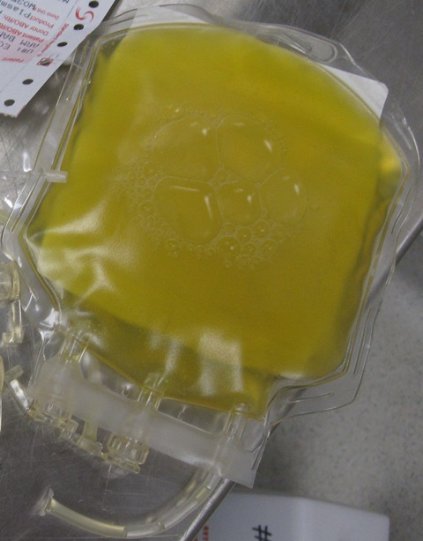
SAN DIEGO – In the next 5-10 years, reaching for a powdered form of plasma may become the normative first-line treatment for trauma patients who present with severe bleeding. That’s because the current standard of administering fresh frozen plasma is riddled with problems, Dr. Martin Schreiber said at the University of California, San Diego, Critical Care Summer Session.
For one thing, frozen plasma takes 35 minutes to thaw. "That’s a problem, and the clotting factor function of plasma deteriorates as you freeze it and thaw it," said Dr. Schreiber, professor of surgery at Oregon Health & Science University, Portland. "Also, you need it in large volumes and that’s not good for patients with congestive heart failure on Coumadin, and the availability is limited, especially in rural settings."

Enter lyophilized plasma (LP), a process developed by HemCon Medical Technologies in which whole blood is sterilely removed, and the plasma component is separated and turned into a powder. The powdered plasma is returned and reconstituted prior to transfusion. "You can put this stuff as a powder on a shelf in nearly any environment," Dr. Schreiber said. "It’s good for at least 3 years, it survives a broad range of temperatures, and you can restore it to plasma within a couple of minutes."
An initial study of LP showed encouraging results with the use of a freeze-dried form of plasma for resuscitation (J. Trauma 2008;65[5]:975-85). A later study by researchers including Dr. Schreiber evaluated the effects of the lyophilization process on plasma clotting factor levels in swine, by adding the antioxidant ascorbic acid (vitamin C) to the reconstitution solution, and by comparing the efficacy of LP with that of fresh frozen plasma and that of plasma and packed red blood cells in a 1:1 ratio (Arch. Surg. 2009;144[9]:829-34). "What we found was that if we gave LP with packed red blood cells in a 1:1 ratio we had 14% less blood loss than if it’s given as FFP with packed cells, which was significant," Dr. Schreiber said.
"The LP was better in terms of stopping hemorrhage. We also noticed that with the vitamin C, we suppressed inflammation and got reduced IL-6 [interleukin 6] expression. Now, the Germans, the Dutch, and the French are using LP in their military settings. Our special forces people are also using it. It’s under current development for common use in your hospital in 5-10 years. I think this stuff is good anywhere. With LP we can always maintain a 1:1 ratio and we don’t have to worry about the thawing process."
Tranexamic acid, a synthetic derivative of lysine, is another anticoagulant therapy that is likely to be used with increasing frequency, he predicted. This agent "binds plasminogen so plasminogen can’t break down fibrin so you can’t get fibrinolysis," said Dr. Schreiber, who has been deployed three times as a combat surgeon.
"This drug has been around forever and is extremely inexpensive. It’s approved by the FDA for use in tooth extraction and the oral form is approved for menorrhagia." Tranexamic acid has also been studied in 53 prospective, randomized studies involving some 3,800 subjects, mostly cardiac patients. "They show that if you use tranexamic acid, you use less blood. It reduces the amount of blood necessary for transfusing people in high-bleeding settings, but no difference in mortality, thrombotic events, myocardial infarction, or stroke. It does not seem to produce a hypercoagulable state."

One study of tranexamic acid use by British surgeons during Afghanistan combat operations found that soldiers who received tranexamic acid were seven times more likely to live, compared with those who did not receive the agent (Arch. Surg. 2012;147:113-19). "There was a survival of 85% in tranexamic acid group, compared with about 70% in those who did not receive it," said Dr. Schreiber. "This has resulted in a change in practice in civilian trauma centers where it is being used widely."
Another anticoagulant being used is the prothrombin complex concentrate known as Kcentra, which contains all four vitamin K–dependent coagulation factors. Distributed by CSL Behring, Kcentra is approved for warfarin reversal in adult patients with acute major bleeding and for those who require emergency surgery. The max dose is 50 units/kg. "That’s about $4,445 for a 70-kg person," Dr. Schreiber said. "Why is it so good? It’s rapidly available, you don’t have to give a lot of fluid, there’s no infectious risk, and you can very rapidly increase coagulation factor function. This is where we’re headed in the future for trauma patients."
Dr. Schreiber said that he had no relevant financial conflicts to disclose.
On Twitter @dougbrunk
SAN DIEGO – In the next 5-10 years, reaching for a powdered form of plasma may become the normative first-line treatment for trauma patients who present with severe bleeding. That’s because the current standard of administering fresh frozen plasma is riddled with problems, Dr. Martin Schreiber said at the University of California, San Diego, Critical Care Summer Session.
For one thing, frozen plasma takes 35 minutes to thaw. "That’s a problem, and the clotting factor function of plasma deteriorates as you freeze it and thaw it," said Dr. Schreiber, professor of surgery at Oregon Health & Science University, Portland. "Also, you need it in large volumes and that’s not good for patients with congestive heart failure on Coumadin, and the availability is limited, especially in rural settings."
Enter lyophilized plasma (LP), a process developed by HemCon Medical Technologies in which whole blood is sterilely removed, and the plasma component is separated and turned into a powder. The powdered plasma is returned and reconstituted prior to transfusion. "You can put this stuff as a powder on a shelf in nearly any environment," Dr. Schreiber said. "It’s good for at least 3 years, it survives a broad range of temperatures, and you can restore it to plasma within a couple of minutes."
An initial study of LP showed encouraging results with the use of a freeze-dried form of plasma for resuscitation (J. Trauma 2008;65[5]:975-85). A later study by researchers including Dr. Schreiber evaluated the effects of the lyophilization process on plasma clotting factor levels in swine, by adding the antioxidant ascorbic acid (vitamin C) to the reconstitution solution, and by comparing the efficacy of LP with that of fresh frozen plasma and that of plasma and packed red blood cells in a 1:1 ratio (Arch. Surg. 2009;144[9]:829-34). "What we found was that if we gave LP with packed red blood cells in a 1:1 ratio we had 14% less blood loss than if it’s given as FFP with packed cells, which was significant," Dr. Schreiber said.
"The LP was better in terms of stopping hemorrhage. We also noticed that with the vitamin C, we suppressed inflammation and got reduced IL-6 [interleukin 6] expression. Now, the Germans, the Dutch, and the French are using LP in their military settings. Our special forces people are also using it. It’s under current development for common use in your hospital in 5-10 years. I think this stuff is good anywhere. With LP we can always maintain a 1:1 ratio and we don’t have to worry about the thawing process."
Tranexamic acid, a synthetic derivative of lysine, is another anticoagulant therapy that is likely to be used with increasing frequency, he predicted. This agent "binds plasminogen so plasminogen can’t break down fibrin so you can’t get fibrinolysis," said Dr. Schreiber, who has been deployed three times as a combat surgeon.
"This drug has been around forever and is extremely inexpensive. It’s approved by the FDA for use in tooth extraction and the oral form is approved for menorrhagia." Tranexamic acid has also been studied in 53 prospective, randomized studies involving some 3,800 subjects, mostly cardiac patients. "They show that if you use tranexamic acid, you use less blood. It reduces the amount of blood necessary for transfusing people in high-bleeding settings, but no difference in mortality, thrombotic events, myocardial infarction, or stroke. It does not seem to produce a hypercoagulable state."
One study of tranexamic acid use by British surgeons during Afghanistan combat operations found that soldiers who received tranexamic acid were seven times more likely to live, compared with those who did not receive the agent (Arch. Surg. 2012;147:113-19). "There was a survival of 85% in tranexamic acid group, compared with about 70% in those who did not receive it," said Dr. Schreiber. "This has resulted in a change in practice in civilian trauma centers where it is being used widely."
Another anticoagulant being used is the prothrombin complex concentrate known as Kcentra, which contains all four vitamin K–dependent coagulation factors. Distributed by CSL Behring, Kcentra is approved for warfarin reversal in adult patients with acute major bleeding and for those who require emergency surgery. The max dose is 50 units/kg. "That’s about $4,445 for a 70-kg person," Dr. Schreiber said. "Why is it so good? It’s rapidly available, you don’t have to give a lot of fluid, there’s no infectious risk, and you can very rapidly increase coagulation factor function. This is where we’re headed in the future for trauma patients."
Dr. Schreiber said that he had no relevant financial conflicts to disclose.
On Twitter @dougbrunk
EXPERT ANALYSIS AT THE UCSD CRITICAL CARE SUMMER SESSION
Radiotherapy plus chemotherapy drive risk of pancreatic cancer in HL patients
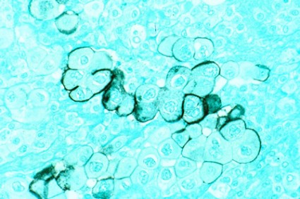
Hodgkin’s lymphoma survivors who undergo both radiotherapy and chemotherapy face an increased risk of subsequent pancreatic cancer, an intervention case-control study demonstrated. In fact, the risk was 18-fold among those who received subdiaphragmatic radiation delivered at 10 Gy or higher in addition to six or more cycles of chemotherapy that contained an alkylating agent.
"Several studies have reported significantly increased risks of pancreatic cancer among long-term HL [Hodgkin’s Lymphoma] survivors, but no prior study of HL survivors has assessed the risk of pancreatic cancer in relation to radiation dose or specific chemotherapeutic agents," researchers led by Dr. Graca M. Dores wrote in the July 25, 2014 issue of the Annals of Oncology. "In the general U.S. population, pancreatic cancer is the fourth most common cause of cancer death, with an overall 5-year relative survival of 5.8%," they noted.

In what the investigators characterized as the first analysis of its kind, Dr. Dores of the division of cancer epidemiology and genetics at the National Cancer Institute and associates drew from six population-based registries and data from main hospitals in the Netherlands to locate HL survivors who received a diagnosis of HL as their primary cancer between 1953 and 2003 and who had survived at least 5 years beyond the initial diagnosis. The cohort was comprised of 19,882 HL survivors, including 36 cases of pancreatic cancer and 70 matched controls. The researchers used logistic regression to estimate odds ratios for pancreatic cancer by comparing the histories of case patients to those of matched controls (Ann. Oncol. July 25 [doi:10.1093/annonc/mdu287]).
The median age at HL diagnosis was 47 years, and 73% had stage I or II disease. Among the 36 patients who developed pancreatic cancer, the median age of pancreatic cancer onset among cases was 61 years, a median of 19 years following the initial HL diagnosis. Dr. Dores and associates found that the risk of pancreatic cancer increased with increasing radiation dose to the location of the pancreatic tumor (P = .005) and increasing number of chemotherapy cycles that contained alkylating agents (P = .008). The risk of prostate cancer was 18-fold higher among those who received subdiaphragmatic radiation delivered at 10 Gy or higher in addition to six or more cycles of chemotherapy that contained an alkylating agent. "This risk was significantly greater than the OR of 3.8 predicted by an additive model (P = .041) and nonsignificantly greater than the OR of 5.4 predicted by a multiplicative model (P = 0.29)," the researchers wrote. "Analyses based on continuous variables yielded similar results."
In discussing the implications of the findings, Dr. Dores and associates acknowledged that treatment approaches for HL "have changed considerably over the past several decades in an effort to maximize efficacy and minimize toxicity. Although radiotherapy remains an important therapeutic modality, radiation volumes and doses have decreased considerably over time, and subdiaphragmatic radiotherapy is infrequently indicated. While the first-line therapy for many HL patients today includes doxorubicin and dacarbazine, procarbazine and cyclophosphamide continue to be used, although often with lower cumulative doses than used in the past. Our findings for topoisomerase II inhibitors are equivocal, but warrant further investigation."
They went on to note that the study extends the range of solid cancers associated with chemotherapy "and adds to the evidence that the combination of chemotherapy and radiotherapy can increase risks beyond those predicted by a multiplicative model. For HL patients, radiation dose-response relationships have now been demonstrated for second cancers of the lung, female breast, stomach, and pancreas and, with the exception of breast cancer, increased risks of these cancers have been observed after receipt of AA [alkylating agent]-containing therapy. Changes in HL therapy over time should reduce second cancer risks compared to those observed with past treatments. In the interim, health care providers caring for long-term HL survivors should be alert to this treatment sequela and encourage a healthy lifestyle to minimize additional cancer risk factors."
The study was supported by the Intramural Research Program of the National Cancer Institute, National Institutes of Health, Department of Health and Human Services, and National Cancer Institute contracts to Cancer Care Ontario, Toronto; Danish Cancer Society, Copenhagen; Finnish Cancer Registry, Helsinki; Information Management Services, Inc., Silver Spring, Md.; Karolinska Institute, Stockholm; University of Iowa; The University of Texas MD Anderson Cancer Center; and Westat, Inc., Rockville, Md. The Dutch study also was supported by the Lance Armstrong Foundation and the Dutch Cancer Society.
On Twitter @dougbrunk
Hodgkin’s lymphoma survivors who undergo both radiotherapy and chemotherapy face an increased risk of subsequent pancreatic cancer, an intervention case-control study demonstrated. In fact, the risk was 18-fold among those who received subdiaphragmatic radiation delivered at 10 Gy or higher in addition to six or more cycles of chemotherapy that contained an alkylating agent.
"Several studies have reported significantly increased risks of pancreatic cancer among long-term HL [Hodgkin’s Lymphoma] survivors, but no prior study of HL survivors has assessed the risk of pancreatic cancer in relation to radiation dose or specific chemotherapeutic agents," researchers led by Dr. Graca M. Dores wrote in the July 25, 2014 issue of the Annals of Oncology. "In the general U.S. population, pancreatic cancer is the fourth most common cause of cancer death, with an overall 5-year relative survival of 5.8%," they noted.
In what the investigators characterized as the first analysis of its kind, Dr. Dores of the division of cancer epidemiology and genetics at the National Cancer Institute and associates drew from six population-based registries and data from main hospitals in the Netherlands to locate HL survivors who received a diagnosis of HL as their primary cancer between 1953 and 2003 and who had survived at least 5 years beyond the initial diagnosis. The cohort was comprised of 19,882 HL survivors, including 36 cases of pancreatic cancer and 70 matched controls. The researchers used logistic regression to estimate odds ratios for pancreatic cancer by comparing the histories of case patients to those of matched controls (Ann. Oncol. July 25 [doi:10.1093/annonc/mdu287]).
The median age at HL diagnosis was 47 years, and 73% had stage I or II disease. Among the 36 patients who developed pancreatic cancer, the median age of pancreatic cancer onset among cases was 61 years, a median of 19 years following the initial HL diagnosis. Dr. Dores and associates found that the risk of pancreatic cancer increased with increasing radiation dose to the location of the pancreatic tumor (P = .005) and increasing number of chemotherapy cycles that contained alkylating agents (P = .008). The risk of prostate cancer was 18-fold higher among those who received subdiaphragmatic radiation delivered at 10 Gy or higher in addition to six or more cycles of chemotherapy that contained an alkylating agent. "This risk was significantly greater than the OR of 3.8 predicted by an additive model (P = .041) and nonsignificantly greater than the OR of 5.4 predicted by a multiplicative model (P = 0.29)," the researchers wrote. "Analyses based on continuous variables yielded similar results."
In discussing the implications of the findings, Dr. Dores and associates acknowledged that treatment approaches for HL "have changed considerably over the past several decades in an effort to maximize efficacy and minimize toxicity. Although radiotherapy remains an important therapeutic modality, radiation volumes and doses have decreased considerably over time, and subdiaphragmatic radiotherapy is infrequently indicated. While the first-line therapy for many HL patients today includes doxorubicin and dacarbazine, procarbazine and cyclophosphamide continue to be used, although often with lower cumulative doses than used in the past. Our findings for topoisomerase II inhibitors are equivocal, but warrant further investigation."
They went on to note that the study extends the range of solid cancers associated with chemotherapy "and adds to the evidence that the combination of chemotherapy and radiotherapy can increase risks beyond those predicted by a multiplicative model. For HL patients, radiation dose-response relationships have now been demonstrated for second cancers of the lung, female breast, stomach, and pancreas and, with the exception of breast cancer, increased risks of these cancers have been observed after receipt of AA [alkylating agent]-containing therapy. Changes in HL therapy over time should reduce second cancer risks compared to those observed with past treatments. In the interim, health care providers caring for long-term HL survivors should be alert to this treatment sequela and encourage a healthy lifestyle to minimize additional cancer risk factors."
The study was supported by the Intramural Research Program of the National Cancer Institute, National Institutes of Health, Department of Health and Human Services, and National Cancer Institute contracts to Cancer Care Ontario, Toronto; Danish Cancer Society, Copenhagen; Finnish Cancer Registry, Helsinki; Information Management Services, Inc., Silver Spring, Md.; Karolinska Institute, Stockholm; University of Iowa; The University of Texas MD Anderson Cancer Center; and Westat, Inc., Rockville, Md. The Dutch study also was supported by the Lance Armstrong Foundation and the Dutch Cancer Society.
On Twitter @dougbrunk
FROM ANNALS OF ONCOLOGY
Key clinical point: Both radiotherapy and chemotherapy increase pancreatic cancer risk among Hodgkin’s lymphoma survivors.
Major finding: Survivors of Hodgkin’s lymphoma treated with both subdiaphragmatic radiation and six or more cycles of alkylating agent-containing therapy were 18 times more likely to develop pancreatic cancer, compared with patients who received no such treatment.
Data source: An international case-control study within a cohort of 19,882 HL survivors diagnosed from 1953 to 2003, including 36 cases with pancreatic cancer and 70 matched controls.
Disclosures: The study was supported by the Intramural Research Program of the National Cancer Institute, National Institutes of Health, Department of Health and Human Services, and National Cancer Institute contracts to Cancer Care Ontario, Toronto; Danish Cancer Society, Copenhagen; Finnish Cancer Registry, Helsinki; Information Management Services, Inc., Silver Spring, Md.; Karolinska Institute, Stockholm; University of Iowa; The University of Texas MD Anderson Cancer Center; and Westat, Inc., Rockville, Md. The Dutch study also was supported by the Lance Armstrong Foundation and the Dutch Cancer Society.
Nurse practitioners playing bigger role in critical care
SAN DIEGO – Nurse practitioners are assuming expanded roles and responsibilities in the provision of critical care services, according to Thomas Farley, R.N., N.P.
At the University of California, San Diego, Critical Care Summer Session, Mr. Farley described the expanded role nurse practitioners (NPs) have played in critical care services for about a decade at the University of California, San Francisco (UCSF). In 2004, Dr. Michael Matthay, a pulmonologist, and Dr. Michael Gropper, a critical care anesthesiologist, spearheaded an effort to hire four NPs to work in the university’s medical-surgical intensive care unit (ICU). "At the time, there were increasing limitations on physician trainees, both in the number of trainees and their work-hour restrictions that were becoming more tight," recalled Mr. Farley of the UCSF School of Nursing. "The goal was to provide critical care at the bedside 24 hours per day, which is in line with the Leapfrog Group recommendations for critical care services. At the time, they had 60 adult ICU critical care beds."

Today, 18 NPs work in adult ICUs at UCSF, with expansion to 76 adult critical care beds, including the medical-surgical ICU, cardiothoracic ICU, and neuro ICU. "Initially the NPs were integrated with medical residents," Mr. Farley said. "Now there are teams that don’t have residents, so it’s an NP paired with an attending physician, or sometimes a fellow. There are always two NPs on service. Usually, during the day we have four NPs on, with two on at night." The program was described in 2011 (ICU Director 2011;2:16-19).
Practice trends driving the need for NPs in critical care include the increasing demand for intensivists and a reliance on a team-based approach to care delivery, "understanding that a single provider cannot provide all the needs that any individual critical care patient has," Mr. Farley explained. "I think we’re a little slow to incorporate the use of nurse practitioners on the West Coast. It’s certainly been done for quite some time on the East Coast."
He said that NPs are already incorporated into critical care delivery programs at Memorial Sloan Kettering Cancer Center and Columbia University, both in New York; Emory University, Atlanta; Henry Ford Hospital, Detroit; the Cleveland Clinic; and Vanderbilt University, Nashville, Tenn. Adopters out West include UCSF, UC Davis; UC Los Angeles; UC Fresno; and Oregon Health & Science University, Portland.
At UCSF, critical care NP duties include a consultative role to admitting services, a consultative role to bedside RNs, guidance of house staff, responding to code blue activations, assisting with rapid response consultations, serving on hospital-wide multidisciplinary committees, precepting adult care NP students, as well as attending morning teaching and monthly morbidity and mortality conferences. Procedures performed include insertion of central venous catheters, arterial catheters, chest tubes, and PICC (peripherally inserted central catheter) lines; lumbar puncture; suture and drain removal; and airway intubation.
The use of NPs in critical care "works because we have appropriate conduits for collaboration and supervision that are explained, understood, and taken to heart," Mr. Farley said. "The NPs need to know that they have supportive attending physicians who will assist them when questions arise. We’ve also had buy-in from the ICU nurses."
A growing body of medical literature suggests that this trend is having an effect. A retrospective study of 600 admissions at two ICUs in New York found that patients managed by nurse practitioner/physician assistant teams had no worse outcomes than patients managed by physicians (Chest 2011;139:1347-53). In addition, a recent survey of critical care program directors at academic medical centers found that 86% believe NPs and other advanced practice providers contribute to the continuity of care, 78% believe they save time during rounds and evaluating new patients, and 73% believe they assist in maintaining work flow (J. Crit. Care 2014; 29:112-5).
In the years ahead, Mr. Farley predicted that NPs and other advanced practice providers "are certainly going to be in the ICU, probably even in greater numbers and perhaps in broader roles than they are now."
Mr. Farley said he had no relevant financial conflicts.
On Twitter @dougbrunk
SAN DIEGO – Nurse practitioners are assuming expanded roles and responsibilities in the provision of critical care services, according to Thomas Farley, R.N., N.P.
At the University of California, San Diego, Critical Care Summer Session, Mr. Farley described the expanded role nurse practitioners (NPs) have played in critical care services for about a decade at the University of California, San Francisco (UCSF). In 2004, Dr. Michael Matthay, a pulmonologist, and Dr. Michael Gropper, a critical care anesthesiologist, spearheaded an effort to hire four NPs to work in the university’s medical-surgical intensive care unit (ICU). "At the time, there were increasing limitations on physician trainees, both in the number of trainees and their work-hour restrictions that were becoming more tight," recalled Mr. Farley of the UCSF School of Nursing. "The goal was to provide critical care at the bedside 24 hours per day, which is in line with the Leapfrog Group recommendations for critical care services. At the time, they had 60 adult ICU critical care beds."
Today, 18 NPs work in adult ICUs at UCSF, with expansion to 76 adult critical care beds, including the medical-surgical ICU, cardiothoracic ICU, and neuro ICU. "Initially the NPs were integrated with medical residents," Mr. Farley said. "Now there are teams that don’t have residents, so it’s an NP paired with an attending physician, or sometimes a fellow. There are always two NPs on service. Usually, during the day we have four NPs on, with two on at night." The program was described in 2011 (ICU Director 2011;2:16-19).
Practice trends driving the need for NPs in critical care include the increasing demand for intensivists and a reliance on a team-based approach to care delivery, "understanding that a single provider cannot provide all the needs that any individual critical care patient has," Mr. Farley explained. "I think we’re a little slow to incorporate the use of nurse practitioners on the West Coast. It’s certainly been done for quite some time on the East Coast."
He said that NPs are already incorporated into critical care delivery programs at Memorial Sloan Kettering Cancer Center and Columbia University, both in New York; Emory University, Atlanta; Henry Ford Hospital, Detroit; the Cleveland Clinic; and Vanderbilt University, Nashville, Tenn. Adopters out West include UCSF, UC Davis; UC Los Angeles; UC Fresno; and Oregon Health & Science University, Portland.
At UCSF, critical care NP duties include a consultative role to admitting services, a consultative role to bedside RNs, guidance of house staff, responding to code blue activations, assisting with rapid response consultations, serving on hospital-wide multidisciplinary committees, precepting adult care NP students, as well as attending morning teaching and monthly morbidity and mortality conferences. Procedures performed include insertion of central venous catheters, arterial catheters, chest tubes, and PICC (peripherally inserted central catheter) lines; lumbar puncture; suture and drain removal; and airway intubation.
The use of NPs in critical care "works because we have appropriate conduits for collaboration and supervision that are explained, understood, and taken to heart," Mr. Farley said. "The NPs need to know that they have supportive attending physicians who will assist them when questions arise. We’ve also had buy-in from the ICU nurses."
A growing body of medical literature suggests that this trend is having an effect. A retrospective study of 600 admissions at two ICUs in New York found that patients managed by nurse practitioner/physician assistant teams had no worse outcomes than patients managed by physicians (Chest 2011;139:1347-53). In addition, a recent survey of critical care program directors at academic medical centers found that 86% believe NPs and other advanced practice providers contribute to the continuity of care, 78% believe they save time during rounds and evaluating new patients, and 73% believe they assist in maintaining work flow (J. Crit. Care 2014; 29:112-5).
In the years ahead, Mr. Farley predicted that NPs and other advanced practice providers "are certainly going to be in the ICU, probably even in greater numbers and perhaps in broader roles than they are now."
Mr. Farley said he had no relevant financial conflicts.
On Twitter @dougbrunk
EXPERT ANALYSIS AT THE UCSD CRITICAL CARE SUMMER SESSION
Discovery of genes associated with schizophrenia called ‘historic’
A novel genomewide association study has revealed new biologic insights from 108 schizophrenia-associated genetic loci.
"As recently as 5 years ago, we still lacked proof that mental disorders had a genetic basis," one of the researchers, Dr. Nelson B. Freimer, a University of California, Los Angeles, professor of psychiatry and director of the UCLA Center for Neurobehavioral Genetics, said in a press release. "This work now demonstrates unequivocally that at least 100 distinct genes contribute to schizophrenia. By providing the strongest evidence to date for the biological underpinnings of mental illness, this study is of historic importance."
In a study published online in Nature, researchers from the Schizophrenia Working Group of the Psychiatric Genomics Consortium reported findings from a multistage schizophrenia genomewide association study of up to 36,989 cases and 113,075 controls (Nature 2014 July 22 [doi:10.1038/nature13595]). Noting that high heritability "points to a major role for inherited genetic variants in the etiology of schizophrenia," the working group cited current estimates suggesting that one-half to one-third of the genetic risk of schizophrenia "is indexed by common alleles genotyped by current genomewide association study (GWAS) arrays. Thus, GWAS is potentially an important tool for understanding the biological underpinnings of schizophrenia."
The researchers obtained genomewide data from 36,989 cases and 113,075 controls. From this data set, they identified 128 independent associations spanning 108 conservatively defined loci that had genomewide significance. Of these 108 loci, 83 have not been previously implicated in schizophrenia and therefore harbor "potential new biological insights into disease" etiology. Molecules that are the most current, or promising, target for therapy were associated with schizophrenia, including the dopamine D2 receptor (DRD2) gene as were many genes involved in glutamatergic neurotransmission and synaptic plasticity, including GRM3, GRIN2A, SRR, and GRIA1. Associations between schizophrenia and genes that encode voltage-gated calcium channel subunits, including CACNA1C, CACNB2, and CACNA11, also were observed. The observations "extend previous findings implicating members of this family of proteins in schizophrenia and other psychiatric disorders," the investigators wrote.
The working group found associations between schizophrenia and enhancers active in the brain; they also found associations between schizophrenia and enhancers active in tissues with important immune functions, especially B-lymphocyte lineages involved in acquired immunity, such as CD19 and CD20. "Epidemiological studies have long hinted at a role for immune dysregulation in schizophrenia, [and] the present findings provide genetic support for this hypothesis," the investigators wrote.
They also found significant overlap between genes in the schizophrenia GWAS-associated intervals and those with de novo nonsynonymous mutations in schizophrenia (P=.0061) "suggesting that mechanistic studies of rare genetic variation in schizophrenia will be informative for schizophrenia more widely." They concluded by noting that the way in which variation in these genes affects function to increase risk for schizophrenia "cannot be answered by genetics, but the overlap strongly suggests that common and rare variant studies are complementary rather than antagonistic, and that mechanistic studies driven by rare genetic variation will be informative for schizophrenia."
The National Institute of Mental Health provides core funding for the Psychiatric Genomics Consortium. The study also was supported by numerous grants from governmental and charitable organizations as well as philanthropic donations. Authors of the working group declared several competing financial conflicts of interest.
On Twitter @dougbrunk
FROM NATURE
Key clinical point: The discovery of genes associated with schizophrenia could lead to new treatments.
Major finding: A total of 108 specific locations in the human genome are associated with the risk for schizophrenia, 83 of which previously had not been linked to the disorder.
Data source: Findings from a multistage schizophrenia genomewide association study of up to 36,989 cases and 113,075 controls.
Disclosures: The National Institute of Mental Health provides core funding for the Psychiatric Genomics Consortium. The study also was supported by numerous grants from governmental and charitable organizations as well as philanthropic donations. Authors of the working group declared several competing financial conflicts of interest.

Intensive blood glucose control: No effect in hyperglycemic patients having CABG
SAN FRANCISCO – Intensive glucose control targeting a blood glucose of 100-140 mg/dL did not significantly reduce perioperative complications or mortality, compared with a less strict glucose target of 141-180 mg/dL in hyperglycemic patients undergoing coronary artery bypass graft surgery, a randomized trial showed.
"Inpatient hyperglycemia is associated with increased hospital complications and mortality," Dr. Guillermo E. Umpierrez said at the annual scientific sessions of the American Diabetes Association. "There have been a lot of controversies regarding what is the best target for glucose targeting in these patients in the perioperative period. There are studies suggesting that improved glycemic control improves outcomes, but others have failed to reproduce this data."

In an effort to address this question, Dr. Umpierrez and his associates at three hospitals in Atlanta conducted the open-label, randomized GLUCO-CABG trial to determine whether intensive glucose control (defined as a blood glucose target of 100-140 mg/dL) reduces perioperative complications, compared with conservative glucose control (defined as a glucose target of 141-180 mg/dL) in hyperglycemic patients undergoing CABG. Their hypothesis was that intensive therapy in the ICU would reduce perioperative complications, compared with a conservative insulin therapy, said Dr. Umpierrez, professor of medicine at Emory University in Atlanta.
The study population included 302 men and women aged 18-80 years with and without a history of diabetes who underwent CABG with or without valve surgery, and who had perioperative hyperglycemia greater than 140 mg/dL during their surgery or ICU stay. Half received intensive insulin therapy, and the other half received conservative insulin therapy. A computerized insulin infusion algorithm (Glytec’s Glucommander) was used to guide continuous IV infusion, which was given in the ICU until the patients were able to eat and/or be transferred to non-ICU services.
The mean age of the patients was 64 years, 72% were male, and their mean body mass index was 30.5 kg/m2. The mean ICU daily blood glucose levels were similar, at 132 mg/dL in the intensive group, compared with 154 mg/dL in the conservative group, and the hospital length of stay was similar between the two groups (11.4 vs. 9.5 days, respectively). In the ICU, a blood glucose level of less than 70 mg/dL occurred in 8% and 2% of the intensive and conservative groups, respectively, a significant difference, while no levels reached less than 40 mg/dL.
After ICU care, there were no differences between the intensive and conservative groups in mean daily blood glucose levels (143 vs. 141 mg/dL, respectively), percentage of patients with hypoglycemia (1% vs. 3%), or hospital readmissions (18% vs. 20%). There were also no differences between groups in rates of mortality, pneumonia, acute kidney injury, respiratory failure, or wound infection.
"The results of this study have significant clinical implications in the management of patients with hyperglycemia and diabetes in critical care units," Dr. Umpierrez said. "This study indicates that a target glucose of 141-180 mg/dL is as safe and effective and results in a lower rate of hypoglycemic events compared to a more intensive target of 100-140 mg/dL."
The study was funded by the National Institutes of Health and by a clinical research award from the American Diabetes Association. Glytec provided the Glucommander and Sanofi provided medications. Dr. Umpierrez has received research funding from and/or has served as an adviser to several pharmaceutical companies.
On Twitter @dougbrunk
SAN FRANCISCO – Intensive glucose control targeting a blood glucose of 100-140 mg/dL did not significantly reduce perioperative complications or mortality, compared with a less strict glucose target of 141-180 mg/dL in hyperglycemic patients undergoing coronary artery bypass graft surgery, a randomized trial showed.
"Inpatient hyperglycemia is associated with increased hospital complications and mortality," Dr. Guillermo E. Umpierrez said at the annual scientific sessions of the American Diabetes Association. "There have been a lot of controversies regarding what is the best target for glucose targeting in these patients in the perioperative period. There are studies suggesting that improved glycemic control improves outcomes, but others have failed to reproduce this data."
In an effort to address this question, Dr. Umpierrez and his associates at three hospitals in Atlanta conducted the open-label, randomized GLUCO-CABG trial to determine whether intensive glucose control (defined as a blood glucose target of 100-140 mg/dL) reduces perioperative complications, compared with conservative glucose control (defined as a glucose target of 141-180 mg/dL) in hyperglycemic patients undergoing CABG. Their hypothesis was that intensive therapy in the ICU would reduce perioperative complications, compared with a conservative insulin therapy, said Dr. Umpierrez, professor of medicine at Emory University in Atlanta.
The study population included 302 men and women aged 18-80 years with and without a history of diabetes who underwent CABG with or without valve surgery, and who had perioperative hyperglycemia greater than 140 mg/dL during their surgery or ICU stay. Half received intensive insulin therapy, and the other half received conservative insulin therapy. A computerized insulin infusion algorithm (Glytec’s Glucommander) was used to guide continuous IV infusion, which was given in the ICU until the patients were able to eat and/or be transferred to non-ICU services.
The mean age of the patients was 64 years, 72% were male, and their mean body mass index was 30.5 kg/m2. The mean ICU daily blood glucose levels were similar, at 132 mg/dL in the intensive group, compared with 154 mg/dL in the conservative group, and the hospital length of stay was similar between the two groups (11.4 vs. 9.5 days, respectively). In the ICU, a blood glucose level of less than 70 mg/dL occurred in 8% and 2% of the intensive and conservative groups, respectively, a significant difference, while no levels reached less than 40 mg/dL.
After ICU care, there were no differences between the intensive and conservative groups in mean daily blood glucose levels (143 vs. 141 mg/dL, respectively), percentage of patients with hypoglycemia (1% vs. 3%), or hospital readmissions (18% vs. 20%). There were also no differences between groups in rates of mortality, pneumonia, acute kidney injury, respiratory failure, or wound infection.
"The results of this study have significant clinical implications in the management of patients with hyperglycemia and diabetes in critical care units," Dr. Umpierrez said. "This study indicates that a target glucose of 141-180 mg/dL is as safe and effective and results in a lower rate of hypoglycemic events compared to a more intensive target of 100-140 mg/dL."
The study was funded by the National Institutes of Health and by a clinical research award from the American Diabetes Association. Glytec provided the Glucommander and Sanofi provided medications. Dr. Umpierrez has received research funding from and/or has served as an adviser to several pharmaceutical companies.
On Twitter @dougbrunk
AT THE ADA ANNUAL SCIENTIFIC SESSIONS
Key clinical point: A conservative target glucose of 141-180 mg/dL is as safe and effective as a more intensive target of 100-140 mg/dL, and results in a lower rate of hypoglycemic events.
Major finding: After ICU care, there were no differences between the intensive and conservative groups in mean daily blood glucose levels (143 vs. 141 mg/dL, respectively) or in the percentage of patients with hypoglycemia (1% vs. 3%; P = .68).
Data source: GLUCO-CABG, a randomized trial of 302 men and women aged 18-80 years who underwent CABG with or without valve surgery and who had perioperative hyperglycemia greater than 140 mg/dL during their surgery or ICU stay.
Disclosures: The study was funded by the National Institutes of Health and by a clinical research award from the American Diabetes Association. Glytec provided the Glucommander and Sanofi provided medications. Dr. Umpierrez has received research funding from and/or has served as an adviser to several pharmaceutical companies.

Be alert to less common, but dangerous, thoracic injuries in children
SAN DIEGO – Many thoracic injuries sustained by children and adolescents can be diagnosed with clinical assessment and chest x-ray, and many heal without surgical intervention, according to Dr. Timothy Fairbanks.
"When the pager goes off and says ‘3-year-old involved in a accident,’ that kid may be going home with a physical exam, stickers, and a high five; or he may require a life-saving procedure" in the emergency department. Dr. Fairbanks, of the division of pediatric surgery at Rady Children’s Hospital of San Diego, said at the University of California, San Diego, Critical Care Summer Session. "There’s complete variability in what we see and how serious the injuries can be."

According to data from the National Pediatric Trauma Registry, 86% of injuries are blunt and 14% are penetrating. Chest injuries "are the second leading cause of death to central nervous system injuries," Dr. Fairbanks said. "Mortality rates for blunt and penetrating injuries are similar. In blunt traumas, most kids don’t die from their blunt thoracic injury, but from a CNS injury that they acquired at the same time. Most of the kids who die from penetrating injuries die in the field before reaching the hospital."
Thoracic injuries occur in about 25% of patients who present to a Level I trauma center. While thoracic injuries are less common than abdominal and extremity injuries are, "they are of potentially higher risk and more lethal," he said. "Most can be treated successfully. Male to female injury rate is 2:1 to 3:1 in favor of males having more traumas."
He classifies thoracic trauma injuries by anatomy (rib, pulmonary, bronchial), blunt vs. penetrating, and threat to life (immediate or potentially life threatening). Motor vehicles are the most common mechanism of thoracic injury, and children have an increased risk in all traumas of auto vs. pedestrian, compared with adults. "When children become teenagers, their mechanisms of injuries approach those of the data for adults," Dr. Fairbanks explained. "We see more penetrating knife and gunshot wounds to the chest, although they are rare in our younger patient population. Tracheobronchial lacerations are more common in children, compared with adults. Aortic disruptions are less likely in children."
Common thoracic injuries that he and his associates see at Rady Children’s Hospital are lung contusion, pneumothorax, hemothorax, and fractures to the ribs, sternum, or scapula. Less common "but perhaps more dangerous injuries" are those to the heart, aorta, trachea, bronchi, and diaphragm. "The most common immediately life-threatening thoracic injuries in children are airway obstruction, pneumothorax, hemothorax, and cardiac tamponade," he said. "A large percentage of thoracic injuries have stable vital signs at presentation. Rib fractures are less common in children and are a marker for a significant mechanism of injury or force. Mediastinal structures are more mobile, making tension pneumothorax a bigger problem."
Diagnosis and treatment of thoracic injuries proceeds simultaneously, with an initial goal to rule out life-threatening injuries, which should be identified and treated during the initial resuscitation phase. This includes making sure the airway is clear and secure and providing supplemental oxygen. "If endotracheal intubation is needed, check the position," he advised. "You want to see bilateral chest rise and CO2 waveform on your monitor. It’s very common for a child to have a right mainstem intubation and no breath sounds on the left side." He also makes it a point to assess their ventilation and treat their pneumothorax. "Don’t wait for the chest x-ray if you have a good clinical suspicion," he said.
A chest x-ray can often guide your management decisions in cases of thoracic trauma, while a FAST scan – specifically an ultrasound – is good for seeing a cardiac tamponade.
Sometimes thoracic injuries require an immediate thoracotomy in the emergency department, but its indications "are controversial," Dr. Fairbanks said. "It’s a potentially life-threatening maneuver, especially with penetrating cardiac injuries." The indications for emergency department thoracotomy are post-traumatic arrest or near arrest in all cases of penetrating thoracic injuries; in blunt trauma with loss of vital signs in the ED; or in blunt trauma with loss of vital signs in route to the ED.
"One of the things we struggle with in pediatric trauma is the timing at which they lost the vital signs in the field," he said. "After 20 minutes the chance of survival is almost nil. However, the family is going to want to know that you did everything possible to save their 3- or 4-year-old child."
If the patient is stable, complete the physical exam. "You’re looking for tachypnea, tenderness to the chest wall and abrasions, or signs of thoracic trauma that require further investigation," he said. Important signs include cyanosis, dyspnea, tracheal deviation, hoarseness, jugular engagement, and subcutaneous emphysema.
Chest auscultation is recommended and an anteroposterior (AP) chest x-ray becomes valuable, "because it’s quick, simple, and yields a lot of information," he said. He characterized a thoracic CT scan as "an excellent study" that can help with the diagnosis of aortic and diaphragmatic injuries which can be missed on chest x-ray. "However, there is a lot more radiation with a chest CT," Dr. Fairbanks noted. "Only 1 in 200 chest CTs for trauma yields a new diagnosis." Other studies to consider include EKG, echocardiogram, bronchoscopy, video-assisted thoracic surgery, radionuclide bone scan, and MRI.
A thoracotomy is indicated when the patient is coding or near coding. The other indications are penetrating wound of the heart or great vessels; massive or continuous intrathoracic bleeding; open pneumothorax with major chest wall defect; aortogram indicating injury to the aorta or major branch; massive or continuing air leak, indicating injury to a major airway; cardiac tamponade; esophageal perforation, or diaphragmatic rupture.
Chest wall soft-tissue injuries are usually not clinically significant, "but they’re a marker for a more serious injury under a bruise," he said. "Rib fractures are less common in children. It’s an indicator of significant force. Treatment is pain control and prevention of atelectasis, which is not as big of a problem in kids as it is in adults. They will heal in 6 weeks. Consider child abuse, specifically in cases of multiple rib fractures and those that don’t make sense with the mechanism of reported injury, or rib fractures that are at different stages of healing."
Dr. Fairbanks said that he had no relevant financial conflicts to disclose.
SAN DIEGO – Many thoracic injuries sustained by children and adolescents can be diagnosed with clinical assessment and chest x-ray, and many heal without surgical intervention, according to Dr. Timothy Fairbanks.
"When the pager goes off and says ‘3-year-old involved in a accident,’ that kid may be going home with a physical exam, stickers, and a high five; or he may require a life-saving procedure" in the emergency department. Dr. Fairbanks, of the division of pediatric surgery at Rady Children’s Hospital of San Diego, said at the University of California, San Diego, Critical Care Summer Session. "There’s complete variability in what we see and how serious the injuries can be."
According to data from the National Pediatric Trauma Registry, 86% of injuries are blunt and 14% are penetrating. Chest injuries "are the second leading cause of death to central nervous system injuries," Dr. Fairbanks said. "Mortality rates for blunt and penetrating injuries are similar. In blunt traumas, most kids don’t die from their blunt thoracic injury, but from a CNS injury that they acquired at the same time. Most of the kids who die from penetrating injuries die in the field before reaching the hospital."
Thoracic injuries occur in about 25% of patients who present to a Level I trauma center. While thoracic injuries are less common than abdominal and extremity injuries are, "they are of potentially higher risk and more lethal," he said. "Most can be treated successfully. Male to female injury rate is 2:1 to 3:1 in favor of males having more traumas."
He classifies thoracic trauma injuries by anatomy (rib, pulmonary, bronchial), blunt vs. penetrating, and threat to life (immediate or potentially life threatening). Motor vehicles are the most common mechanism of thoracic injury, and children have an increased risk in all traumas of auto vs. pedestrian, compared with adults. "When children become teenagers, their mechanisms of injuries approach those of the data for adults," Dr. Fairbanks explained. "We see more penetrating knife and gunshot wounds to the chest, although they are rare in our younger patient population. Tracheobronchial lacerations are more common in children, compared with adults. Aortic disruptions are less likely in children."
Common thoracic injuries that he and his associates see at Rady Children’s Hospital are lung contusion, pneumothorax, hemothorax, and fractures to the ribs, sternum, or scapula. Less common "but perhaps more dangerous injuries" are those to the heart, aorta, trachea, bronchi, and diaphragm. "The most common immediately life-threatening thoracic injuries in children are airway obstruction, pneumothorax, hemothorax, and cardiac tamponade," he said. "A large percentage of thoracic injuries have stable vital signs at presentation. Rib fractures are less common in children and are a marker for a significant mechanism of injury or force. Mediastinal structures are more mobile, making tension pneumothorax a bigger problem."
Diagnosis and treatment of thoracic injuries proceeds simultaneously, with an initial goal to rule out life-threatening injuries, which should be identified and treated during the initial resuscitation phase. This includes making sure the airway is clear and secure and providing supplemental oxygen. "If endotracheal intubation is needed, check the position," he advised. "You want to see bilateral chest rise and CO2 waveform on your monitor. It’s very common for a child to have a right mainstem intubation and no breath sounds on the left side." He also makes it a point to assess their ventilation and treat their pneumothorax. "Don’t wait for the chest x-ray if you have a good clinical suspicion," he said.
A chest x-ray can often guide your management decisions in cases of thoracic trauma, while a FAST scan – specifically an ultrasound – is good for seeing a cardiac tamponade.
Sometimes thoracic injuries require an immediate thoracotomy in the emergency department, but its indications "are controversial," Dr. Fairbanks said. "It’s a potentially life-threatening maneuver, especially with penetrating cardiac injuries." The indications for emergency department thoracotomy are post-traumatic arrest or near arrest in all cases of penetrating thoracic injuries; in blunt trauma with loss of vital signs in the ED; or in blunt trauma with loss of vital signs in route to the ED.
"One of the things we struggle with in pediatric trauma is the timing at which they lost the vital signs in the field," he said. "After 20 minutes the chance of survival is almost nil. However, the family is going to want to know that you did everything possible to save their 3- or 4-year-old child."
If the patient is stable, complete the physical exam. "You’re looking for tachypnea, tenderness to the chest wall and abrasions, or signs of thoracic trauma that require further investigation," he said. Important signs include cyanosis, dyspnea, tracheal deviation, hoarseness, jugular engagement, and subcutaneous emphysema.
Chest auscultation is recommended and an anteroposterior (AP) chest x-ray becomes valuable, "because it’s quick, simple, and yields a lot of information," he said. He characterized a thoracic CT scan as "an excellent study" that can help with the diagnosis of aortic and diaphragmatic injuries which can be missed on chest x-ray. "However, there is a lot more radiation with a chest CT," Dr. Fairbanks noted. "Only 1 in 200 chest CTs for trauma yields a new diagnosis." Other studies to consider include EKG, echocardiogram, bronchoscopy, video-assisted thoracic surgery, radionuclide bone scan, and MRI.
A thoracotomy is indicated when the patient is coding or near coding. The other indications are penetrating wound of the heart or great vessels; massive or continuous intrathoracic bleeding; open pneumothorax with major chest wall defect; aortogram indicating injury to the aorta or major branch; massive or continuing air leak, indicating injury to a major airway; cardiac tamponade; esophageal perforation, or diaphragmatic rupture.
Chest wall soft-tissue injuries are usually not clinically significant, "but they’re a marker for a more serious injury under a bruise," he said. "Rib fractures are less common in children. It’s an indicator of significant force. Treatment is pain control and prevention of atelectasis, which is not as big of a problem in kids as it is in adults. They will heal in 6 weeks. Consider child abuse, specifically in cases of multiple rib fractures and those that don’t make sense with the mechanism of reported injury, or rib fractures that are at different stages of healing."
Dr. Fairbanks said that he had no relevant financial conflicts to disclose.
EXPERT ANALYSIS AT THE UCSD CRITICAL CARE SUMMER SESSION

Fatty Heart May Bring on Diabetes
A high volume of pericardial adipose tissue was associated with the prevalence of diabetes, independent of overall obesity, results from a long-term, diverse cohort study showed.
"Obesity is associated with an increased risk for cardiovascular disease and type 2 diabetes, and specific fat deposits may increase the risk more than others," Amy C. Alman, Ph.D., said at the annual scientific sessions of the American Diabetes Association.
The findings come from an analysis of year-25 exam data among 3,079 participants in CARDIA (Coronary Artery Risk Development in Young Adults), a longitudinal cohort study of the development of cardiovascular risk and disease that began in 1985 and was conducted at centers in Alabama, Minnesota, Illinois, and California.
Visceral adiposity "is metabolically active and more strongly associated with cardiovascular risk than subcutaneous adiposity. Ectopic adipose depots are metabolically active fat found in and around tissues and organs throughout the body, including the liver, muscles, and around the heart," explained Dr. Alman of the department of epidemiology and biostatistics at the University of South Florida, Tampa.
Pericardial adipose tissue (PAT) is an ectopic fat depot composed of epicardial adipose tissue deep to the pericardium and surrounding the coronary arteries and paracardial tissue, which is located along the surface of the parietal pericardium. She and her associates tested whether PAT was positively associated with prevalent diabetes at the year-25 exam of the CARDIA study.
Examinations including volume of PAT measures from chest CT scans were performed during 2010-2011, and the researchers used multivariable logistic regression to examine the relation between quartiles of PAT and diabetes. There were four PAT quartiles: less than 33.5 cm3 (quartile 1/referent volume), between 33.5 and less than 48.7 cm3 (quartile 2), between 48.7 and less than 71.7 cm3(quartile 3), and greater than 71.7 cm3 (quartile 4).
The mean age of the 3,079 study participants was 51 years, 44% were male, and their average body mass index was 31 kg/m2. Of these, 419 had prevalent diabetes. The prevalence of diabetes was highest in the fourth quartile of PAT volume (46% vs. 12% in the first quartile, 18% in the second, and 24% in the third), Dr. Alman reported.
In a logistic regression model of PAT volume on diabetes status by obesity, only PAT volume in the fourth quartile was significantly associated with diabetes status (odds ratio, 2.47), adjusted for field center, gender, age, race, systolic blood pressure, total cholesterol, triglycerides, and treatment with blood pressure– and cholesterol-lowering medications. A similar association was observed among nonobese patients (OR, 3.78).
"Deposition of a higher proportion of metabolically active ectopic fat is associated with diabetes in both obese and nonobese individuals," Dr. Alman concluded.
The analysis was "a very well characterized multicenter cohort study with a diverse racial cohort of middle-aged men and women," she added. Limitations of the study, she said, included its cross-sectional design, the fact that PAT was measured at a single point in time, and that it did not account for other ectopic fat depots such as liver fat. A longitudinal analysis of the participants is planned.
Dr. Alman had no relevant financial conflicts to disclose.