User login
Official Newspaper of the American College of Surgeons
Coming in July! New design for ACS Surgery News
ACS Surgery News, the official newspaper of the American College of Surgeons, has a new look. The July issue will have a new design, new colors, and an increased focus on visual accessibility for readers. The updated format is easier to read and has more graphic elements for quicker access to data. Future issues with the new design will feature opinion columns, updates on critical practice economics issues, and news coverage of cutting-edge surgical technology.
ACS Surgery News readership has surged over the past 2 years. The growing popularity of ACS Surgery News is based on quality content: lively commentary by thought leaders of the surgical profession, timely coverage of surgery meetings, and in-depth features on those issues most important to surgeons.
In addition, the publication website has a fresh look: better layout, easier access to the digital, interactive version of the publication, and more graphic elements.
The next issue of ACS Surgery News mails on July 17. This publication has thrived on commentary, critique, and contributions from readers. Take a look at the new design and let the editorial team know what you think.

Bariatric surgery beats lifestyle changes alone for type 2 diabetes
Among obese patients who underwent bariatric surgery 40% achieved at least partial remission of type 2 diabetes mellitus, compared with no patients who underwent a nonsurgical lifestyle intervention program, investigators reported online July 1 in JAMA Surgery.
The randomized clinical trial of 61 patients offers “further important evidence that at longer-term follow-up of 3 years, surgical treatments, including Roux-en-Y gastric bypass and laparoscopic adjustable gastric banding, are superior to lifestyle intervention alone for the remission of type 2 diabetes mellitus in obese individuals, including those with a body mass index (BMI) between 30 and 35 [kg/m2],” said Dr. Anita Courcoulas at the University of Pittsburgh Medical Center and her associates. But further studies will be needed to explore exactly how bariatric surgery affects diabetes and the effect of these procedures on the microvascular and macrovascular complications of diabetes, the investigators added (JAMA Surg. 2015 July 1 [doi:10.1001/jamasurg.2015.1534]).

Several studies have reported major improvements in type 2 diabetes mellitus (T2DM) after bariatric surgery, but did not assess long-term efficacy or safety compared with lifestyle and medical management, the researchers noted. To fill that gap, they randomized obese middle-aged adults with T2DM to either an intensive lifestyle weight loss program for 1 year followed by a 2-year low-level lifestyle intervention program, or to Roux-en-Y gastric bypass (RYGB) or laparoscopic adjustable gastric banding (LAGB) followed by the low-level lifestyle intervention program during years 2 and 3.
After 3 years, 40% of RYGB patients and 29% of LAGB patients were fully or partially remitted, compared with none of the control group (P = .004), the investigators reported. The bariatric surgery groups did not significantly differ in terms of complete remission, but 65% of RYGB patients and 33% of LAGB patients were able to stop all insulin and oral diabetes medications, compared with none of the control group (P < .001). Also, RYGB patients lost an average of 25% of their baseline body weight, compared with 15% for LAGB (P = .0002) and only 5.7% for the lifestyle-only control group (P < .0001).
At baseline, patients averaged 100.5 kg (standard deviation, 13.7 kg) in body weight, mean hemoglobin A1c level was 7.8% (standard deviation, 1.9%), average fasting plasma glucose level was 171.3 (72.5) mg per dL, the investigators said. “One important aspect of this study was that more than 40% of the sample were individuals with class I obesity (BMI ≥ 30), for whom data in the literature are largely lacking,” they added. Adverse events were uncommon after the first year, but RYGB was linked to significant drops in lean muscle and bone mass that will need further study, they noted.
The National Institutes of Health and the University of Pittsburgh Medical Center funded the study. Dr. Courcoulas reported research support from Nutrisystem, J&J Ethicon, and Covidien, and consulting relationships with Ethicon and Apollo Endosurgery. Two coauthors reported relationships with the Obesity Society/Nutrisystem, Jawbone/BodyMedia, and Weight Watchers. The other investigators declared no conflicts of interest.
We should consider the use of bariatric (metabolic) surgery in all severely obese patients with type 2 diabetes mellitus and start a mass treatment, similar to what was done with coronary artery bypass graft more than 50 years ago.
[The study findings are] reminiscent of the Swedish Obesity Study, a nonrandomized study of 1,658 obese patients who underwent bariatric surgery and 1,771 obese matched controls. None of these participants had diabetes mellitus at baseline. After 15 years, T2DM developed in 6.8 cases per 1,000 person-years and 28.4 cases per 1,000 person-years, respectively (P < .001). The effect of surgery was influenced by the presence of impaired fasting glucose, but not by body mass index. It was concluded that surgery appeared to be more efficient than the control [lifestyle intervention] in the prevention of T2DM.
In the Look AHEAD clinical trial, an intensive lifestyle intervention for weight loss was examined to determine the impact on cardiovascular events. The trial was stopped early based on a futility analysis. … Similarly, the TODAY clinical trial on adolescents with recent-onset T2DM demonstrated no benefits of intensive lifestyle intervention.
If surgery is more successful for these patients, which surgery should be done? It has been shown that malabsorption is better than restriction. … In a randomized clinical trial [of] severely obese patients with T2DM, at 2 years, diabetic remission occurred in none of the medical therapy patients versus 75% of the gastric bypass group and 95% of the biliary pancreatic diversion group (P < .001).
Dr. Michael Gagner is at Florida International University in Miami. He reported receiving honoraria from Ethicon, Covidien, Fore, MID, Olympus, and Boehringer Laboratories, and equity from Transenterix. These remarks are based on his accompanying editorial (JAMA Surg. 2015 July 1 [doi: 10.1001/jamasurg.2015.1542]).
Among obese patients who underwent bariatric surgery 40% achieved at least partial remission of type 2 diabetes mellitus, compared with no patients who underwent a nonsurgical lifestyle intervention program, investigators reported online July 1 in JAMA Surgery.
The randomized clinical trial of 61 patients offers “further important evidence that at longer-term follow-up of 3 years, surgical treatments, including Roux-en-Y gastric bypass and laparoscopic adjustable gastric banding, are superior to lifestyle intervention alone for the remission of type 2 diabetes mellitus in obese individuals, including those with a body mass index (BMI) between 30 and 35 [kg/m2],” said Dr. Anita Courcoulas at the University of Pittsburgh Medical Center and her associates. But further studies will be needed to explore exactly how bariatric surgery affects diabetes and the effect of these procedures on the microvascular and macrovascular complications of diabetes, the investigators added (JAMA Surg. 2015 July 1 [doi:10.1001/jamasurg.2015.1534]).
Several studies have reported major improvements in type 2 diabetes mellitus (T2DM) after bariatric surgery, but did not assess long-term efficacy or safety compared with lifestyle and medical management, the researchers noted. To fill that gap, they randomized obese middle-aged adults with T2DM to either an intensive lifestyle weight loss program for 1 year followed by a 2-year low-level lifestyle intervention program, or to Roux-en-Y gastric bypass (RYGB) or laparoscopic adjustable gastric banding (LAGB) followed by the low-level lifestyle intervention program during years 2 and 3.
After 3 years, 40% of RYGB patients and 29% of LAGB patients were fully or partially remitted, compared with none of the control group (P = .004), the investigators reported. The bariatric surgery groups did not significantly differ in terms of complete remission, but 65% of RYGB patients and 33% of LAGB patients were able to stop all insulin and oral diabetes medications, compared with none of the control group (P < .001). Also, RYGB patients lost an average of 25% of their baseline body weight, compared with 15% for LAGB (P = .0002) and only 5.7% for the lifestyle-only control group (P < .0001).
At baseline, patients averaged 100.5 kg (standard deviation, 13.7 kg) in body weight, mean hemoglobin A1c level was 7.8% (standard deviation, 1.9%), average fasting plasma glucose level was 171.3 (72.5) mg per dL, the investigators said. “One important aspect of this study was that more than 40% of the sample were individuals with class I obesity (BMI ≥ 30), for whom data in the literature are largely lacking,” they added. Adverse events were uncommon after the first year, but RYGB was linked to significant drops in lean muscle and bone mass that will need further study, they noted.
The National Institutes of Health and the University of Pittsburgh Medical Center funded the study. Dr. Courcoulas reported research support from Nutrisystem, J&J Ethicon, and Covidien, and consulting relationships with Ethicon and Apollo Endosurgery. Two coauthors reported relationships with the Obesity Society/Nutrisystem, Jawbone/BodyMedia, and Weight Watchers. The other investigators declared no conflicts of interest.
FROM JAMA SURGERY
Key clinical point: Bariatric surgery, especially Roux-en-Y gastric bypass, led to significant improvements in type 2 diabetes mellitus, compared with lifestyle changes alone.
Major finding: At year 3, 40% of RYGB patients and 29% of LAGB patients were fully or partially remitted, compared with none of the nonsurgical control group (P = .004).
Data source: Randomized, parallel-group clinical trial of 61 obese adults with type 2 diabetes mellitus.
Disclosures: The National Institutes of Health and the University of Pittsburgh Medical Center funded the study. Dr. Courcoulas reported research support from Nutrisystem, J&J Ethicon, and Covidien, and consulting relationships with Ethicon and Apollo Endosurgery. Two coauthors reported relationships with the Obesity Society/Nutrisystem, Jawbone/BodyMedia, and Weight Watchers. The other investigators declared no conflicts of interest.

Contrast imaging detects some lung tumors in surgery
Injecting patients with a molecular contrast agent before lung cancer surgery and then using fluorescent imaging during the operation proved safe and somewhat effective at finding lung adenocarcinomas and discovering tumor metastases in a pilot clinical trial of 50 patients.
Investigators from the University of Pennsylvania, Philadelphia, and Emory (Atlanta) and Purdue (West Lafayette, Ind.) universities reported their findings online in the Journal of Thoracic and Cardiovascular Surgery (2015 [doi:10.1016/j.jtcvs.2015.05.014]).
“We believe the most important finding in this study was the ability to systemically inject our contrast agent into patients prior to surgery and have 92% of the lung adenocarcinomas fluoresce in the operating room,” Dr. Olugbenga Okusanya of the University of Pennsylvania, Philadelphia, and his colleagues said. Their approach overcomes some of the limitations of other contrast agents for imaging of lung cancer.
The approach involves intravenous administration of 0.1 mg/kg of a fluorescent folate receptor-alpha (FR-alpha)–targeted molecule contrast agent 4 hours before surgery. The specific agent they used is a synthetic conjugate between folate and fluorescein isothiocyanate that binds to serum proteins.
They set out to determine if an optical-targeted molecular contrast agent to FR-alpha could bind lung adenocarcinoma and then if real-time optical imaging, either in situ or ex vivo during surgery, would show the lesions. The 50 study subjects all had biopsy-confirmed lung adenocarcinoma. In the OR, the researchers used a gantry-mounted fluorescent imaging system to capture images of the tumors.
With the operating room lights switched off, imaging of the cancer was captured by fluorescence and white light in situ. The lung was deflated during imaging to expose as much of the lung surface to the imaging as possible. The in situ optical imaging required on average eight minutes to perform. Only seven tumors (14%) appeared fluorescent in situ before they were excised. Computed tomography (CT) before surgery showed that all seven of these tumors were below the pleural surface and within 1.2 cm of the lung surface.
Among the other 43 tumors, 39 exhibited uniform fluorescence once the surgical team exposed their surfaces while 4 – all invasive adenocarcinomas – did not. Final pathology confirmed that all 50 tumors were cancerous.
“With further refinements, this tool may prove useful in locating adenocarcinomas deeper in the lung parenchyma, lymph nodes and at pleural and resection margins,” Dr. Okusanya and his coauthors said.
The investigators noted that one advantage of this imaging strategy is that it can locate tumors of varying sizes, subtypes and metabolic activity with tumor-to-background ratios “not markedly different” among them. “This finding demonstrates that intraoperative molecular imaging may be potentially broadly applicable to all FR-alpha lung tumors,” they said.
Other advantages is that wavelength fluorophore poses no radiation exposure and that surgeons readily understand optical imaging “with no need for special training to interpret or process the data.” They did acknowledge that a significant drawback of the molecular contrast agent was its lack of penetration into the lung parenchyma.
“New molecular tools are emerging to identify lung adenocarcinomas during pulmonary resection,” Dr. Okusanya and colleagues said. “This technology will permit precise visualization of tumor margins, localization of small malignant ground-glass opacities, and accurate selection of lymph nodes with metastatic cancer cells. With miniaturization of imaging devices, this method will be particularly useful in minimally invasive surgery.”
The pilot clinical trial was partially supported by grant from the Biomedical Laboratory Research & Development Service of the Veterans Affairs Office of Research and Development and by the CHEST Foundation One Breath Clinical Research Award (SS).
Dr. Shuming Nie is a coauthor and a consultant for Spectropath, a start-up company that is developing advanced instrumentation and nanoparticle contrast agents. Dr. Philip S. Low is a coauthor and a consultant and stakeholder in OnTarget Laboratories. None of the other coauthors had any relationships to disclose.
“Not unexpectedly, the Achilles’ heel of the technique is tumor depth beneath the pleural surface,” Dr. Michael I. Ebright of Columbia University, New York, said in his commentary of the results of the pilot study. He noted that only 7 of the 50 tumors were visible before resection – and those were all large and close to the pleural surface.
“As we move toward the detection of smaller lesions through lung cancer screening, as well as toward the use of minimally invasive surgical techniques challenging direct palpation, more practical techniques for intraoperative localization are sorely needed,” Dr. Ebright said in his invited commentary (J. Thorac. Cardiovasc. Surg. 2015 [doi:10.1016/j.jtcvs.2015.04.029]). But the techniques that have emerged are “time consuming, unreliable, or resource intensive.”
He likened the study results to a half-full glass of water: Those who see it half-empty will identify the weaknesses of the study because, among other findings, the technique does not image tumors more than 1 cm below the pleural surface; the half-full cohort will take the position that “new technology has to start somewhere, usually at proof of principle.”
But the in situ optical imaging approach is faster than the current localization options surgeons have, and the possibilities of the approach “are certainly exciting,” Dr. Ebright said.
“Imagination is an essential component of any technological progress,” Dr. Ebright said. “This study should be viewed as a launching pad rather than be judged solely on practicality in its current form.
Dr. Ebright is with the section of thoracic surgery, Columbia University Medical Center, New York.
Injecting patients with a molecular contrast agent before lung cancer surgery and then using fluorescent imaging during the operation proved safe and somewhat effective at finding lung adenocarcinomas and discovering tumor metastases in a pilot clinical trial of 50 patients.
Investigators from the University of Pennsylvania, Philadelphia, and Emory (Atlanta) and Purdue (West Lafayette, Ind.) universities reported their findings online in the Journal of Thoracic and Cardiovascular Surgery (2015 [doi:10.1016/j.jtcvs.2015.05.014]).
“We believe the most important finding in this study was the ability to systemically inject our contrast agent into patients prior to surgery and have 92% of the lung adenocarcinomas fluoresce in the operating room,” Dr. Olugbenga Okusanya of the University of Pennsylvania, Philadelphia, and his colleagues said. Their approach overcomes some of the limitations of other contrast agents for imaging of lung cancer.
The approach involves intravenous administration of 0.1 mg/kg of a fluorescent folate receptor-alpha (FR-alpha)–targeted molecule contrast agent 4 hours before surgery. The specific agent they used is a synthetic conjugate between folate and fluorescein isothiocyanate that binds to serum proteins.
They set out to determine if an optical-targeted molecular contrast agent to FR-alpha could bind lung adenocarcinoma and then if real-time optical imaging, either in situ or ex vivo during surgery, would show the lesions. The 50 study subjects all had biopsy-confirmed lung adenocarcinoma. In the OR, the researchers used a gantry-mounted fluorescent imaging system to capture images of the tumors.
With the operating room lights switched off, imaging of the cancer was captured by fluorescence and white light in situ. The lung was deflated during imaging to expose as much of the lung surface to the imaging as possible. The in situ optical imaging required on average eight minutes to perform. Only seven tumors (14%) appeared fluorescent in situ before they were excised. Computed tomography (CT) before surgery showed that all seven of these tumors were below the pleural surface and within 1.2 cm of the lung surface.
Among the other 43 tumors, 39 exhibited uniform fluorescence once the surgical team exposed their surfaces while 4 – all invasive adenocarcinomas – did not. Final pathology confirmed that all 50 tumors were cancerous.
“With further refinements, this tool may prove useful in locating adenocarcinomas deeper in the lung parenchyma, lymph nodes and at pleural and resection margins,” Dr. Okusanya and his coauthors said.
The investigators noted that one advantage of this imaging strategy is that it can locate tumors of varying sizes, subtypes and metabolic activity with tumor-to-background ratios “not markedly different” among them. “This finding demonstrates that intraoperative molecular imaging may be potentially broadly applicable to all FR-alpha lung tumors,” they said.
Other advantages is that wavelength fluorophore poses no radiation exposure and that surgeons readily understand optical imaging “with no need for special training to interpret or process the data.” They did acknowledge that a significant drawback of the molecular contrast agent was its lack of penetration into the lung parenchyma.
“New molecular tools are emerging to identify lung adenocarcinomas during pulmonary resection,” Dr. Okusanya and colleagues said. “This technology will permit precise visualization of tumor margins, localization of small malignant ground-glass opacities, and accurate selection of lymph nodes with metastatic cancer cells. With miniaturization of imaging devices, this method will be particularly useful in minimally invasive surgery.”
The pilot clinical trial was partially supported by grant from the Biomedical Laboratory Research & Development Service of the Veterans Affairs Office of Research and Development and by the CHEST Foundation One Breath Clinical Research Award (SS).
Dr. Shuming Nie is a coauthor and a consultant for Spectropath, a start-up company that is developing advanced instrumentation and nanoparticle contrast agents. Dr. Philip S. Low is a coauthor and a consultant and stakeholder in OnTarget Laboratories. None of the other coauthors had any relationships to disclose.
FROM THE JOURNAL OF THORACIC AND CARDIOVASCULAR SURGERY
Key clinical point: A pilot clinical trial reported that molecular contrast imaging during lung cancer surgery is safe and somewhat effective at isolating malignancies.
Major finding: Among 50 patients, the imaging technique detected seven tumors close to the lung surface while 39 others showed fluorescence in the operating room once they were removed.
Data source: Single-center population of 50 patients with biopsy-proven lung adenocarcinoma.
Disclosures: The pilot clinical trial was partially supported by grant from the Biomedical Laboratory Research & Development Service of the Veterans Affairs Office of Research and Development and by the CHEST Foundation One Breath Clinical Research Award (SS). Dr. Nie is a coauthor and a consultant for Spectropath, a start-up company that is developing advanced instrumentation and nanoparticle contrast agents. Dr. Low is a coauthor and a consultant and stakeholder in OnTarget Laboratories. None of the other coauthors had any relationships to disclose.
Post-CABG stroke risk same with one or two clamps
When performing on-pump coronary artery bypass grafting (CABG), cardiac surgeons can control very few factors to reduce the risk of stroke – with the exception of which method of aortic manipulation they use. Debate and controversy, however, have surrounded which aortic manipulation technique is best: single- or double-clamp occlusion.
A large retrospective study of almost 8,500 patients who had CABG at the Mayo Clinic in Rochester, Minn., over a 17-year period showed that, while use of the single-aortic cross-clamp (SC) technique steadily increased, the risk of stroke is virtually the same as it is with the partial aortic cross-clamp (PC), or double cross-clamp, technique. The study authors, led by Dr. Juan C. Araque, published their results online in the Journal of Thoracic and Cardiovascular Surgery (J. Thorac. Cardiovasc. Surg. 2015 [doi:10.1016/j.jtcvs.2015.04.010]).
“It is intuitive that less aortic manipulation would result in less risk of stroke,” Dr. Araque and colleagues said, but even off-pump CABG, which requires no aortic manipulation, is not without stroke risk.
“It is conceivable that there is some inherent risk of stroke associated with any cardiac operation, and that risk may increase with manipulation of the ascending aortic with the aortic cross clamp,” they wrote. “Our data would suggest, however, that the risk does not increase further with the additional aortic manipulation of the partial occlusion clamp.”
The study comes on the heels of a 2008 meta-analysis that found no benefit of SC in comparison to PC (Interact. Cardiovasc. Thorac. Surg. 2008;7:500-3), while another study in 2011 suggested that less aortic manipulation carried a significantly lower stroke risk (Heart Lung Circ. 2011;20:318-24).
The Mayo study evaluated the SC technique in 2,051 patients and PC in 6,446 patients who had isolated on-pump CABG between 1993 and 2010. The rate of stroke was 1.2% in the SC group and 1.5% among those who had PC. In two propensity-matched cohorts of 1,333 patients each, the stroke rate was 1.2% in each group. The investigators used the Society of Thoracic Surgeons’ risk calculator variables to create the propensity-matched cohorts.
The study group excluded high-risk patients, including those who had off-pump operations or previous cardiac surgeries or required replacement of a cross clamp during an unplanned operation.
The goal of the study was not to compare outcomes with the off-pump technique. “It is only to bring attention to the associated non-zero stroke rate with both techniques,” Dr. Araque and colleagues said.
Their findings are significant because on-pump CABG is the preferred operation of cardiac surgeons, accounting for more than 80% of the CABG operations in the SYNTAX study (N. Engl. J. Med. 2009;360:961-72). “The ‘anaortic’ off-pump technique may be a more specialized technique, representing less than 15% of operations in one large series,” Dr. Araque and coauthors said.
They acknowledged a few limitations resulting from the observational nature of the study, including that surgeons may have missed some strokes because they did not use a routine, standardized procedure for evaluating stroke signs along with the lack of documented assessment of the descending aorta. But they also stated that the large number of patients in the study, along with the use of propensity matching, addresses some of the bias inherent in an observational study.
The study authors disclosed no relationships.
“A randomized trial is needed to answer the question, ‘Can CABG [coronary artery bypass grafting] be safely performed with either one or two aortic clamps in all patients?’ ” Dr. Jennifer S. Lawton said in her invited commentary (J. Thorac. Cardiovasc. Surg. 2015 [doi:10.1016/j.jtcvs.2015.05.002]).
Dr. Lawton acknowledged the positions that advocates of both techniques have staked out: advocates of the single-clamp (SC) technique prefer the ability to perform the proximal anastomoses without the added space constraints and reduced visibility of the partial clamp and moving heart; proponents of the partial-clamp (PC) method cite advantages in the ability to determine graft length with the full heart and the likelihood to reanimate the heart earlier to reduce the risk of a heart attack.
The PC technique required longer cardiopulmonary bypass time, 88.2 minutes vs. 73.7 minutes, but the SC group had longer cross-clamp times, 54.5. vs. 50.7 minutes. “The longer clamp time did not alter the outcomes reported (stroke and mortality) – although specific outcomes of myocardial injury including need for inotropes, troponin levels, myocardial infarction, etc. were not reported,” Dr. Lawton said. “Thus, the question for the surgeon is, ‘What is more important, the brain or the heart?’ ”
The results from Dr. Araque’s study “are valuable” because of the large patient cohort and the suggestion that “the use of a second clamp is not likely to significantly alter outcomes of stroke and mortality,” she wrote.
But their study leaves a few questions remaining, Dr. Lawton said. “What is the best treatment of high-risk patients who may benefit from limited aortic manipulation the most? Can two clamps be safely applied to all types of aortas? And does the risk of dissection go up with the use of two clamps?”
Although a randomized trial would be difficult because of the low risk of stroke in on-pump CABG, such a trial could answer those questions if it involved routine epiaortic ultrasound, Dr. Lawton said.
Dr. Lawton is professor of surgery in the division of cardiothoracic surgery at Washington University, St. Louis.
When performing on-pump coronary artery bypass grafting (CABG), cardiac surgeons can control very few factors to reduce the risk of stroke – with the exception of which method of aortic manipulation they use. Debate and controversy, however, have surrounded which aortic manipulation technique is best: single- or double-clamp occlusion.
A large retrospective study of almost 8,500 patients who had CABG at the Mayo Clinic in Rochester, Minn., over a 17-year period showed that, while use of the single-aortic cross-clamp (SC) technique steadily increased, the risk of stroke is virtually the same as it is with the partial aortic cross-clamp (PC), or double cross-clamp, technique. The study authors, led by Dr. Juan C. Araque, published their results online in the Journal of Thoracic and Cardiovascular Surgery (J. Thorac. Cardiovasc. Surg. 2015 [doi:10.1016/j.jtcvs.2015.04.010]).
“It is intuitive that less aortic manipulation would result in less risk of stroke,” Dr. Araque and colleagues said, but even off-pump CABG, which requires no aortic manipulation, is not without stroke risk.
“It is conceivable that there is some inherent risk of stroke associated with any cardiac operation, and that risk may increase with manipulation of the ascending aortic with the aortic cross clamp,” they wrote. “Our data would suggest, however, that the risk does not increase further with the additional aortic manipulation of the partial occlusion clamp.”
The study comes on the heels of a 2008 meta-analysis that found no benefit of SC in comparison to PC (Interact. Cardiovasc. Thorac. Surg. 2008;7:500-3), while another study in 2011 suggested that less aortic manipulation carried a significantly lower stroke risk (Heart Lung Circ. 2011;20:318-24).
The Mayo study evaluated the SC technique in 2,051 patients and PC in 6,446 patients who had isolated on-pump CABG between 1993 and 2010. The rate of stroke was 1.2% in the SC group and 1.5% among those who had PC. In two propensity-matched cohorts of 1,333 patients each, the stroke rate was 1.2% in each group. The investigators used the Society of Thoracic Surgeons’ risk calculator variables to create the propensity-matched cohorts.
The study group excluded high-risk patients, including those who had off-pump operations or previous cardiac surgeries or required replacement of a cross clamp during an unplanned operation.
The goal of the study was not to compare outcomes with the off-pump technique. “It is only to bring attention to the associated non-zero stroke rate with both techniques,” Dr. Araque and colleagues said.
Their findings are significant because on-pump CABG is the preferred operation of cardiac surgeons, accounting for more than 80% of the CABG operations in the SYNTAX study (N. Engl. J. Med. 2009;360:961-72). “The ‘anaortic’ off-pump technique may be a more specialized technique, representing less than 15% of operations in one large series,” Dr. Araque and coauthors said.
They acknowledged a few limitations resulting from the observational nature of the study, including that surgeons may have missed some strokes because they did not use a routine, standardized procedure for evaluating stroke signs along with the lack of documented assessment of the descending aorta. But they also stated that the large number of patients in the study, along with the use of propensity matching, addresses some of the bias inherent in an observational study.
The study authors disclosed no relationships.
FROM THE JOURNAL OF THORACIC AND CARDIOVASCULAR SURGERY
Key clinical point: Two different approaches to aortic manipulation that surgeons use during on-pump coronary artery bypass graft surgery – single and double cross-clamp techniques – had similar rates of postoperative stroke.
Major finding: The single cross-clamp group had a stroke rate of 1.2% within 30 days of the operation, compared with 1.5% in the double cross-clamp group in unmatched cohorts.
Data source: Retrospective analysis of 8,497 patients treated with isolated on-pump coronary artery bypass grafting from 1993 to 2010.
Disclosures: The study authors reported having no financial disclosures.
Cefazolin ranks sixth as cause of drug-induced liver injury
A single intravenous infusion of cefazolin can cause drug-induced liver injury, and the antibiotic ranked sixth among pharmacologic causes of hepatic injury in an analysis of 1,212 patients.
“Cephalosporins appear to be a relatively common cause of antibiotic-associated liver injury,” said Dr. Saleh Alqahtani at the University of Texas Southwestern in Dallas and his associates. “The latency period is typically 1-3 weeks after exposure, and patients may not become symptomatic until after the antibiotic is stopped – this is particularly true in the unique clinical syndrome in which a single infusion of cefazolin leads to drug-induced liver injury.”
Cephalosporins have been reported as rare causes of drug-induced liver injury (DILI), but most data come from single case reports, the researchers said. To study causes of DILI, they analyzed cases from the Drug-Induced Livery Injury Network, an ongoing prospective study at eight U.S. medical centers. Enrolled patients had strong clinical suspicion for liver injury caused by a drug or an herbal agent. Liver injury was defined based on specific criteria for liver enzymes, alkaline phosphatase, or total bilirubin levels, or as an international normalized ratio greater than 1.5 that was accompanied by elevated liver enzymes or ALP. Patients were followed for at least 6 months after their baseline visit (Clin. Gastroenterol. Hepatol. 2014 Dec. 17 [doi: 10.1016/j.cgh.2015.01.010]).
Among the 1,212 cases of DILI in the analysis, one-third were linked to antimicrobial therapies, including 41 (3.3%) in which cephalosporins were implicated, the investigators reported. Nineteen of the cases were tied to a single dose of intravenous cefazolin given before surgery. These patients developed cholestatic or mixed hepatocellular-cholestatic injury 1-3 weeks after the cefazolin infusion. They almost always had jaundice and pruritus, and usually also had fever and nausea. Signs and symptoms were self-limiting, resolving within a few days to a few weeks.
“Because of confusion about the specific diagnosis, patients underwent substantial diagnostic testing (including multiple computed tomography scans, magnetic resonance imaging scans, endoscopic retrograde cholangiopancreatography exams, liver biopsies, and others), which often were unnecessary,” and in some cases led to severe complications, the investigators said.
The study also identified barriers to identifying cefazolin as a cause of DILI, they said. Patients often did not know they had received the antibiotic, and clinicians, including study investigators, often did not know that cefazolin could cause DILI. In more than half of cases, DILI was linked to cefazolin only after careful medical record reviews. “For these reasons, we speculate that cefazolin is and has been underappreciated as a cause of DILI,” the researchers noted. “The appearance of jaundice and pruritus 1-3 weeks after minor surgery should lead to a search of surgical records and medications that might have been given during surgery. These results also imply that the merits of routine use of cefazolin at the time of uncomplicated surgery should be reconsidered carefully.”
Two patients died after receiving cephalosporins other than cefazolin, and another patient developed severe liver injury, the researchers said. “However, in each of the fatal cases, patients had a complicated clinical course, with a severe hypersensitivity reaction on top of an underlying liver disease. Therefore, we urge caution in concluding that non-cefazolin cephalosporin-induced DILI may be severe or fatal,” they said. “Because cephalosporins are used commonly in clinical practice, it is likely that the overall mortality rate associated with cephalosporin use is low, but not nil, and it may be more likely in patients with underlying disorders.”
The study was funded by the National Institute of Diabetes and Digestive and Kidney Diseases, the National Institutes of Health, the National Cancer Institute, and by six Clinical and Translational Science Award grants. The investigators reported having no conflicts of interest.
FROM CLINICAL GASTROENTEROLOGY AND HEPATOLOGY
Key clinical point: A single dose of cefazolin can cause drug-induced liver injury (DILI), and the agent is implicated more often than previously thought.
Major finding: Cefazolin ranked sixth among causes of DILI, and signs and symptoms began 1-3 weeks after initial exposure.
Data source: Registry-based study of 1,212 cases of DILI.
Disclosures: The study was funded by the National Institute of Diabetes and Digestive and Kidney Diseases, the National Institutes of Health, the National Cancer Institute, and by Clinical and Translational Science Award grants. The investigators reported having no conflicts of interest.
Monitoring effectively identifies seizures in postbypass neonates
In the first report evaluating the impact of a clinical guideline that calls for the use of postoperative continuous electroencephalography (CEEG) on infants after they’ve had cardiopulmonary bypass surgery, investigators at Children’s Hospital of Philadelphia and the University of Pennsylvania validated the clinical utility of routine CEEG monitoring and found that clinical assessment for seizures without CEEG is not a reliable marker for diagnosis and treatment.
In a report online in the Journal of Thoracic and Cardiovascular Surgery (J. Thorac. Cardiovasc. Surg. 2015 [doi:10.1016/j.jtcvs.2015.03.045]), Dr. Maryam Naim and colleagues said that CEEG identified electroencephalographic seizures in 8% of newborns after cardiopulmonary bypass surgery. The study, conducted over 18 months, evaluated 172 newborns, none older than 1 month, with 161 (94%) having undergone postoperative CEEG. They had CEEG within 6 hours of their return to the cardiac intensive care unit.
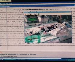
The study classified electroencephalographic seizures as EEG-only (also termed nonconvulsive seizures, with no observable clinical signs either at bedside or via video) or electroclinical seizures. Dr. Naim and colleagues said the majority of seizures they identified with CEEG would not have been noticed otherwise as they had no clinically obvious signs or symptoms.
The American Clinical Neurophysiology Society (ACNS) recommends that cardiac surgeons consider continuous CEEG monitoring in high-risk neonates with congenital heart disease (CHD) after bypass surgery, but Dr. Naim and coauthors raised the question of whether seizure incidence would justify routine CEEG for all neonates with CHD who’ve had bypass surgery, especially as health systems place greater emphasis on quality improvement programs and cost-effective strategies. The authors said that neonates with all types of congenital heart disease had seizures.
“In adult populations, CEEG has not been shown to significantly increase hospital costs, but cost-effectiveness analyses have not been performed in neonates with CHD,” the authors said.
So they attempted to identify at-risk populations of newborns who would benefit most from routine CEEG monitoring. In a multivariable model that the investigators used, both delayed sternal closure and longer deep hypothermic circulatory arrest (DHCA) during surgery seemed predictive of seizures, but the odds ratios for both were low, “suggesting the statistically significant findings may not be very useful in focusing CEEG implementation on a high-risk group.”
Previous studies have reported that identifying and treating seizures in newborns who have had bypass surgery may reduce secondary brain injury and improve outcomes (Pediatrics 2008;121:e759-67), and the Boston Circulatory Arrest Study showed an association between postoperative seizures and lower reading and math scores and lower cognitive and functional skills later in life (Circulation 2011;124:1361-1369). The authors cited other studies that showed older, critically ill children with “high seizure burdens” have had worse outcomes. (Critical Care Medicine 2013;31:215-23; Neurology 2014;82:396-404; Brain 2014;137:1429-38). They also pointed out increased risk if the seizure is not treated. “While occurrence of a seizure is a marker of brain injury, there may also be secondary injury if the seizure activity is not terminated,” Dr. Naim and coauthors said.
The investigators concluded that postoperative CEEG to identify seizures “is warranted,” and while they found some newborns may be at greater risk of postbypass seizures than others, they advocated for “widespread” monitoring strategies.
Their work also questioned the effectiveness of non-CEEG assessment. In the study, clinicians identified bedside events indicative of seizures – what the study termed “push-button events” – in 32 newborns, or about 18% of patients, but none of the events had an EEG correlate, so they were considered nonepileptic. When the authors looked more closely at those “push-button” events, they found they ranged from abnormal body movement in 14 and hypertension in 7 to tachycardia and abnormal face movements, among other characterizations, in lesser numbers.
“Furthermore, push-button events by bedside clinicians, including abnormal movements and hypertensive episodes concerning for possible seizures, did not have any EEG correlate, indicating that bedside clinical assessment for seizures without CEEG monitoring is unreliable,” Dr. Naim and colleagues said.
As to whether identifying and treating postbypass seizures in young newborns with CHD will improve long-term neurodevelopment in these children, the authors acknowledged that further study is needed.
They reported having no financial disclosures.
The findings of Dr. Maryam Naim and coauthors show that relying on physical examination alone is no longer adequate to rule out postoperative neurologic complications, Dr. Carl L. Backer and Dr. Bradley S. Marino said in their invited commentary on the study (J. Thorac. Cardiovasc. Surg. 2015 [doi:10.1016/j.jtcvs.2015.04.028]).
However, they noted that the level of “sophisticated monitoring” the investigators had at their disposal – 24-hour availability of EEG technologists, comprehensive 12-scalp electrode monitoring – is not available at all institutions. “What we need is a screening tool that is not as labor intensive,” Dr. Backer and Dr. Marino said – a screening CEEG monitor that would allow care teams to identify seizure activity at a minimal expense and serve as a basis for a full EEG for evaluation and avoid the expense and manpower for the vast majority of patients who do not have seizures.
Nonetheless, prevention of seizures in this newborn population is “critically important,” but that can only be achieved if the care team monitors for seizures and then assesses strategies, both during and after surgery, to eliminate development of seizures, the commentary authors said.
But the recent study points to the need for a multicenter, observational cross-sectional study using CEEG monitoring, Dr. Backer and Dr. Marino said.
Dr. Backer is a cardiovascular-thoracic surgeon and Dr. Marino is a cardiac surgeon at the Ann and Robert H. Lurie Children’s Hospital of Chicago.
In the first report evaluating the impact of a clinical guideline that calls for the use of postoperative continuous electroencephalography (CEEG) on infants after they’ve had cardiopulmonary bypass surgery, investigators at Children’s Hospital of Philadelphia and the University of Pennsylvania validated the clinical utility of routine CEEG monitoring and found that clinical assessment for seizures without CEEG is not a reliable marker for diagnosis and treatment.
In a report online in the Journal of Thoracic and Cardiovascular Surgery (J. Thorac. Cardiovasc. Surg. 2015 [doi:10.1016/j.jtcvs.2015.03.045]), Dr. Maryam Naim and colleagues said that CEEG identified electroencephalographic seizures in 8% of newborns after cardiopulmonary bypass surgery. The study, conducted over 18 months, evaluated 172 newborns, none older than 1 month, with 161 (94%) having undergone postoperative CEEG. They had CEEG within 6 hours of their return to the cardiac intensive care unit.
The study classified electroencephalographic seizures as EEG-only (also termed nonconvulsive seizures, with no observable clinical signs either at bedside or via video) or electroclinical seizures. Dr. Naim and colleagues said the majority of seizures they identified with CEEG would not have been noticed otherwise as they had no clinically obvious signs or symptoms.
The American Clinical Neurophysiology Society (ACNS) recommends that cardiac surgeons consider continuous CEEG monitoring in high-risk neonates with congenital heart disease (CHD) after bypass surgery, but Dr. Naim and coauthors raised the question of whether seizure incidence would justify routine CEEG for all neonates with CHD who’ve had bypass surgery, especially as health systems place greater emphasis on quality improvement programs and cost-effective strategies. The authors said that neonates with all types of congenital heart disease had seizures.
“In adult populations, CEEG has not been shown to significantly increase hospital costs, but cost-effectiveness analyses have not been performed in neonates with CHD,” the authors said.
So they attempted to identify at-risk populations of newborns who would benefit most from routine CEEG monitoring. In a multivariable model that the investigators used, both delayed sternal closure and longer deep hypothermic circulatory arrest (DHCA) during surgery seemed predictive of seizures, but the odds ratios for both were low, “suggesting the statistically significant findings may not be very useful in focusing CEEG implementation on a high-risk group.”
Previous studies have reported that identifying and treating seizures in newborns who have had bypass surgery may reduce secondary brain injury and improve outcomes (Pediatrics 2008;121:e759-67), and the Boston Circulatory Arrest Study showed an association between postoperative seizures and lower reading and math scores and lower cognitive and functional skills later in life (Circulation 2011;124:1361-1369). The authors cited other studies that showed older, critically ill children with “high seizure burdens” have had worse outcomes. (Critical Care Medicine 2013;31:215-23; Neurology 2014;82:396-404; Brain 2014;137:1429-38). They also pointed out increased risk if the seizure is not treated. “While occurrence of a seizure is a marker of brain injury, there may also be secondary injury if the seizure activity is not terminated,” Dr. Naim and coauthors said.
The investigators concluded that postoperative CEEG to identify seizures “is warranted,” and while they found some newborns may be at greater risk of postbypass seizures than others, they advocated for “widespread” monitoring strategies.
Their work also questioned the effectiveness of non-CEEG assessment. In the study, clinicians identified bedside events indicative of seizures – what the study termed “push-button events” – in 32 newborns, or about 18% of patients, but none of the events had an EEG correlate, so they were considered nonepileptic. When the authors looked more closely at those “push-button” events, they found they ranged from abnormal body movement in 14 and hypertension in 7 to tachycardia and abnormal face movements, among other characterizations, in lesser numbers.
“Furthermore, push-button events by bedside clinicians, including abnormal movements and hypertensive episodes concerning for possible seizures, did not have any EEG correlate, indicating that bedside clinical assessment for seizures without CEEG monitoring is unreliable,” Dr. Naim and colleagues said.
As to whether identifying and treating postbypass seizures in young newborns with CHD will improve long-term neurodevelopment in these children, the authors acknowledged that further study is needed.
They reported having no financial disclosures.
FROM THE JOURNAL OF THORACIC AND CARDIOVASCULAR SURGERY
Key clinical point: Electroencephalography is more effective than clinical observation in identifying seizures in infants immediately after they’ve had cardiopulmonary bypass surgery.
Major finding: Postoperative CEEG identified seizures in 8% of newborns with congenital heart disease after coronary bypass surgery.
Data source: Chart review involved 172 neonates from a single center. Multiple logistic regression analysis assessed seizures and clinical and predictive factors.
Disclosures: The authors reported having no financial disclosures.
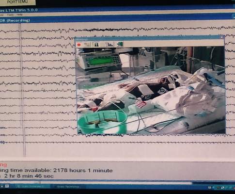
Elephant stent aorta repair – good outcomes, but is it too complex?
An acute aortic tear can be lethal, and more cardiac surgeons are favoring extended aortic arch replacement in these cases. Cardiac surgeons have tried many different arch replacement techniques, but en bloc repair and double- or triple-branch stent grafting carry significant risks, so a team of cardiac surgeons in Beijing has reported good 2-year results with a novel technique that combines stented elephant-trunk implantation with preservation of key vessels.
The technique accomplishes total arch replacement with the stent while preserving the autologous brachiocephalic vessels.
“This technique simplified hemostasis and anastomosis, reduced the size of the residual aortic patch wall, and preserved the autologous brachiocephalic vessels, yielding satisfactory surgical results,” wrote Dr. Li-Zhong Sun and colleagues at Beijing’s Capital Medical University (J. Thorac. Cardiovasc. Surg. 2015 [doi:10.1016/j.jtcvs.2015.03.002]).
There are four keys to the procedure:
• The use of forceps to grasp the stent-free sewing edge of the stented elephant trunk and straightening of the spiral shaped Dacron graft to approximately 3 cm.
• Preservation of the native brachiocephalic vessels.
• Creating a residual aortic wall containing the innominate artery and LCCA that’s as small as possible.
• An end-to-side anastomosis between the left subclavian artery (LSCA) and the left common carotid artery (LCCA), a key junction in their technique.
The 20 study subjects had surgery within 2 weeks of the onset of pain. All 20 were discharged after the procedure, and in a mean follow-up period of 26 months, 18 had good outcomes while 1 patient had thoracoabdominal aortic replacement 9 months after the initial surgery (1 patient was lost to follow-up).
The researchers used computed tomography to confirm patency of the anastomosis between the LSCA and LCCA.
In 2 of the 20 patients, the aorta was normal with aortic dissection limited to the descending aorta. In the remaining patients, the investigators observed thrombus obliteration of the false lumen around the surgical graft in 16, partial thrombosis in 1 and patency in 1.
The surgical technique exposes the right axillary artery through a right subclavicular incision and a median sternotomy, then dissects and exposes the brachiocephalic vessels and the transverse arch. Dissection of the LSCA and LCCA is the key step in making the end-to-end anastomosis between the two vessels. The researchers accomplished this by partially transecting the sternocleidomastoid muscle and other cervical muscles.
Dr. Sun and coauthors said that a separated graft technique offers a number of advantages over other techniques for aortic arch reconstruction. While en bloc repair preserves the native brachiocephalic vessels and, thus, results in long-term patency, the technique carries risk for postoperative rupture of the aortic patch containing the brachiocephalic vessels. Double- or triple-branched stent grafting has resulted in shifting or kinking of the graft and eventually graft occlusion or aortic disruption.
The authors acknowledged the study’s small sample size, and that the outcomes are “preliminary.” They said long-term follow-up would be required to confirm the outcomes.
They had no disclosures to report.
The Beijing study authors’ excellent postoperative outcomes show that alternative surgical techniques for elephant-trunk implantation can be employed safely, but their technique also raises questions about the use of advanced technology, Dr. Prashanth Vallabhajosyula and Dr. Wilson Y. Szeto of the University of Pennsylvania said in their commentary on the study (J. Thorac. Cardiovasc. Surg. 2015 [doi: 10.1016/j.jtcvs.2015.04.003]).
“But does this mean we should be doing [elephant-trunk] operation on every type A dissection patient?” wrote Dr. Vallabhajosyula and Dr. Szeto. If no primary tear appears in the aortic arch or the proximal descending thoracic aorta (DTA), “then should we empirically dissect the arch vessels and perform total arch replacement in an emergent situation?” They also questioned extensive dissection of the left subclavian artery (LSCA) by cutting into the muscles around the surgical site.
Elephant-trunk implantation is more complex than other aortic repair procedures, they noted. “So, if a total arch replacement is not required, then why do it?”
While they acknowledged advantages of total arch replacement, and elephant-trunk implantation in particular, most operations for type A dissection occur in smaller, community hospitals that are ill equipped to perform the procedure. “This raises the issue of wide clinical application of the [elephant-trunk] technique for acute type A dissection,” they said. The real issue may not be what type of anastomosis for the elephant-trunk technique surgeons should use, but rather what surgical technique – the elephant-trunk technique vs. transverse hemiarch reconstruction, they said. (Dr. Vallabhajosyula and Dr. Szeto mentioned that their institution has advocated for the latter.)
“To address this, a more comprehensive and meticulous approach is warranted based on parameters such as patient clinical picture, acuity, malperfusion, arch and DTA anatomy, and primary tear site location,” they said. But for now, Dr. Vallabhajosyula and Dr. Szeto said, the medical literature does not support total arch replacement over transverse hemiarch reconstruction.
Dr. Vallabhajosyula is assistant professor of surgery at the Hospital of the University of Pennsylvania; Dr. Szeto is associate professor of surgery in the division of cardiovascular surgery at the University of Pennsylvania Medical Center–Penn Presbyterian Medical Center.
An acute aortic tear can be lethal, and more cardiac surgeons are favoring extended aortic arch replacement in these cases. Cardiac surgeons have tried many different arch replacement techniques, but en bloc repair and double- or triple-branch stent grafting carry significant risks, so a team of cardiac surgeons in Beijing has reported good 2-year results with a novel technique that combines stented elephant-trunk implantation with preservation of key vessels.
The technique accomplishes total arch replacement with the stent while preserving the autologous brachiocephalic vessels.
“This technique simplified hemostasis and anastomosis, reduced the size of the residual aortic patch wall, and preserved the autologous brachiocephalic vessels, yielding satisfactory surgical results,” wrote Dr. Li-Zhong Sun and colleagues at Beijing’s Capital Medical University (J. Thorac. Cardiovasc. Surg. 2015 [doi:10.1016/j.jtcvs.2015.03.002]).
There are four keys to the procedure:
• The use of forceps to grasp the stent-free sewing edge of the stented elephant trunk and straightening of the spiral shaped Dacron graft to approximately 3 cm.
• Preservation of the native brachiocephalic vessels.
• Creating a residual aortic wall containing the innominate artery and LCCA that’s as small as possible.
• An end-to-side anastomosis between the left subclavian artery (LSCA) and the left common carotid artery (LCCA), a key junction in their technique.
The 20 study subjects had surgery within 2 weeks of the onset of pain. All 20 were discharged after the procedure, and in a mean follow-up period of 26 months, 18 had good outcomes while 1 patient had thoracoabdominal aortic replacement 9 months after the initial surgery (1 patient was lost to follow-up).
The researchers used computed tomography to confirm patency of the anastomosis between the LSCA and LCCA.
In 2 of the 20 patients, the aorta was normal with aortic dissection limited to the descending aorta. In the remaining patients, the investigators observed thrombus obliteration of the false lumen around the surgical graft in 16, partial thrombosis in 1 and patency in 1.
The surgical technique exposes the right axillary artery through a right subclavicular incision and a median sternotomy, then dissects and exposes the brachiocephalic vessels and the transverse arch. Dissection of the LSCA and LCCA is the key step in making the end-to-end anastomosis between the two vessels. The researchers accomplished this by partially transecting the sternocleidomastoid muscle and other cervical muscles.
Dr. Sun and coauthors said that a separated graft technique offers a number of advantages over other techniques for aortic arch reconstruction. While en bloc repair preserves the native brachiocephalic vessels and, thus, results in long-term patency, the technique carries risk for postoperative rupture of the aortic patch containing the brachiocephalic vessels. Double- or triple-branched stent grafting has resulted in shifting or kinking of the graft and eventually graft occlusion or aortic disruption.
The authors acknowledged the study’s small sample size, and that the outcomes are “preliminary.” They said long-term follow-up would be required to confirm the outcomes.
They had no disclosures to report.
FROM THE JOURNAL OF THORACIC AND CARDIOVASCULAR SURGERY
Key clinical point: Total aortic arch replacement with implantation of an elephant-trunk stent avoids risks of other more conventional approaches.
Major finding: Among 20 patients who had the elephant-trunk procedure, 18 had good results at a mean of 26 months after the operation (one had thoracoabdominal aortic arch replacement at 9 months and one was lost to follow-up).
Data source: Retrospective review of 20 patients with acute type A dissection who had total arch replacement at a single center.
Disclosures: The study authors reported having no financial disclosures.
Commentary: Molecular marker testing for diagnosis and prognosis – Where are we really?
An emerging market of molecular markers for thyroid cancer diagnostic and prognostic evaluation challenges current medical and surgical management; however, here is a strong word of caution; Unless the markers have been tested in the context of clinical and surgical management their true efficacy has not been accurately assessed.
We must always ask two questions before ordering these tests that can cost more than $3,000: 1) Does the use of a molecular test add any additional information that would otherwise change our recommendations; in other words, is the clinical information already known enough for appropriate clinical decision making? 2) What is the basis upon which recommendations for its use have been made; in other words, how were the studies conducted and interpreted? Are they valid?

By way of background, the gene expression clarifier (GEC) Afirma is marketed as a diagnostic tool for the differential diagnosis of indeterminate thyroid cytology (atypical cells of undetermined significance or follicular neoplasm, Bethesda categories III and IV, respectively).
Most importantly, it has been tested and is marketed as a negative predictor only. It is also only useful if used in the appropriate setting and if the test result is negative; a positive Afirma test can tell us only that the nodule is indeterminate, something, by definition, already determined by fine needle aspiration (FNA). Furthermore, any study examining its efficacy must be conducted within the context of clinical scenarios. For example, for a patient with an indeterminate lesion and a symptomatic multinodular goiter, neither a negative nor positive Afirma result would have any impact on surgical decision making and yet would have cost the patient more than $3,000. The latter patient simply needs a thyroidectomy. A patient who has a nodule that is approaching 4 cm or is increasing in size or a patient who has undergone several FNAs, ultrasounds, and endocrinology consultations similarly would likely choose surgery regardless of the GEC test result.
The only scenario in which Afirma testing is useful is in a patient with otherwise no indications for surgery except an indeterminate FNA; for instance for a patient with an asymptomatic < 4cm, stable, single nodule. In this scenario only a negative result could be helpful and would obviate the need for surgical intervention. One also must ask what is the likelihood of the patient undergoing repeat FNA, having repeat indeterminate cytology and repeat Afirma testing over the ensuing years and at what point should we consider these costs prohibitive compared with the patient undergoing a thyroidectomy instead?
Similar concerns exist with regard to somatic mutation panels, the most commonly marketed and used being Asuragen. The mutation panel consists of Ras and BRAF mutation and RET/PTC and PAX8/PPAR-gamma chromosomal rearrangement analyses. Importantly, the expanding literature on molecular markers for thyroid cancer, however, has lost track of a vast body of literature that reports Ras mutations present in up to 30% of benign thyroid nodules; RET/PTC in 24% and PAX8/PPAR-gamma in 14%, dramatically challenging the validity of the panel purported to accurately identify thyroid malignancy. We know that BRAF mutation is exclusively found in papillary thyroid cancer, but generally a BRAF-positive tumor is one in which a cytological diagnosis is already clear; it is either suspicious or definitively malignant on FNA. One might suggest that these aforementioned markers might indicate a harbinger of malignancy or identify a precancerous lesion; however, there is no study that supports this theory. Furthermore, it is important to note that even among pathology experts the diagnoses of follicular adenoma, follicular cancer, and follicular variant of papillary thyroid cancer have significant and documented inter- and intra- observer variability, thus challenging the gold standard upon which these markers have been tested.
With regard to BRAF, Ras, RET/PTC and PAX8/PPAR-gamma as prognostic markers there are many considerations, the least of which includes the differential diagnosis of follicular lesions addressed above and the known presence of these abnormalities in benign tumors. The majority of studies to date have conducted only univariate analyses without consideration of the clinical variables already available to us to make needed clinical decisions. In order to examine the usefulness of any marker panel a multivariable study is required, incorporating the available clinical variables such as size, lymph node metastases, and extra-thyroidal extension. For instance, the majority of studies examining the presence of lymph node metastases in BRAF-negative vs. BRAF-positive tumors were almost uniformly conducted on patients in whom routine lymph node dissection was not performed, thereby skewing the results. Only patients with suspicious nodes underwent a lymph node dissection; thus for those patients who had no suspicious nodes there was no information about their nodal status. Furthermore, several studies that included patients who underwent routine lymph node dissections and, importantly, included patients who underwent prophylactic dissections for whom a marker would be most useful, did not find that BRAF mutation was associated with aggressive features, including the presence of lymph node metastases. The same can be stated for a panel that includes Ras, RET/PTC, & PAX8/PPAR-gamma. “Follicular variant of papillary thyroid cancer and follicular cancer” will more likely harbor Ras and PAX8/PPAR-gamma mutations and will, by definition, include some tumors that would be considered benign by other pathologists.
There is an emerging market for molecular markers for thyroid cancer diagnosis and prognosis, but many of the studies driving this market are lacking in scientific rigor with regard to design and interpretation of results. Furthermore, the use of the marker panel requires scrutiny of its true impact on clinical management as well as its cost-effectiveness. GEC provides a negative predictor of malignancy and should be used to obviate surgery only; if the patient requires surgery its use provides no added information. BRAF is specific for thyroid cancer diagnosis, but is often not required as FNA evaluation in BRAF-positive tumors is usually already suspicious or malignant. Finally, molecular marker panels for thyroid cancer prognosis may be promising, but scrutiny for true accuracy and true clinical impact is necessary in order to make logical and useful recommendations for our patients.
Dr. Zeiger is professor of surgery, oncology, cellular, and molecular medicine, associate vice chair for faculty development, and associate dean for postdoctoral affairs at Johns Hopkins University in Baltimore. Dr. Zeiger consults for Interpace Diagnostics.
An emerging market of molecular markers for thyroid cancer diagnostic and prognostic evaluation challenges current medical and surgical management; however, here is a strong word of caution; Unless the markers have been tested in the context of clinical and surgical management their true efficacy has not been accurately assessed.
We must always ask two questions before ordering these tests that can cost more than $3,000: 1) Does the use of a molecular test add any additional information that would otherwise change our recommendations; in other words, is the clinical information already known enough for appropriate clinical decision making? 2) What is the basis upon which recommendations for its use have been made; in other words, how were the studies conducted and interpreted? Are they valid?
By way of background, the gene expression clarifier (GEC) Afirma is marketed as a diagnostic tool for the differential diagnosis of indeterminate thyroid cytology (atypical cells of undetermined significance or follicular neoplasm, Bethesda categories III and IV, respectively).
Most importantly, it has been tested and is marketed as a negative predictor only. It is also only useful if used in the appropriate setting and if the test result is negative; a positive Afirma test can tell us only that the nodule is indeterminate, something, by definition, already determined by fine needle aspiration (FNA). Furthermore, any study examining its efficacy must be conducted within the context of clinical scenarios. For example, for a patient with an indeterminate lesion and a symptomatic multinodular goiter, neither a negative nor positive Afirma result would have any impact on surgical decision making and yet would have cost the patient more than $3,000. The latter patient simply needs a thyroidectomy. A patient who has a nodule that is approaching 4 cm or is increasing in size or a patient who has undergone several FNAs, ultrasounds, and endocrinology consultations similarly would likely choose surgery regardless of the GEC test result.
The only scenario in which Afirma testing is useful is in a patient with otherwise no indications for surgery except an indeterminate FNA; for instance for a patient with an asymptomatic < 4cm, stable, single nodule. In this scenario only a negative result could be helpful and would obviate the need for surgical intervention. One also must ask what is the likelihood of the patient undergoing repeat FNA, having repeat indeterminate cytology and repeat Afirma testing over the ensuing years and at what point should we consider these costs prohibitive compared with the patient undergoing a thyroidectomy instead?
Similar concerns exist with regard to somatic mutation panels, the most commonly marketed and used being Asuragen. The mutation panel consists of Ras and BRAF mutation and RET/PTC and PAX8/PPAR-gamma chromosomal rearrangement analyses. Importantly, the expanding literature on molecular markers for thyroid cancer, however, has lost track of a vast body of literature that reports Ras mutations present in up to 30% of benign thyroid nodules; RET/PTC in 24% and PAX8/PPAR-gamma in 14%, dramatically challenging the validity of the panel purported to accurately identify thyroid malignancy. We know that BRAF mutation is exclusively found in papillary thyroid cancer, but generally a BRAF-positive tumor is one in which a cytological diagnosis is already clear; it is either suspicious or definitively malignant on FNA. One might suggest that these aforementioned markers might indicate a harbinger of malignancy or identify a precancerous lesion; however, there is no study that supports this theory. Furthermore, it is important to note that even among pathology experts the diagnoses of follicular adenoma, follicular cancer, and follicular variant of papillary thyroid cancer have significant and documented inter- and intra- observer variability, thus challenging the gold standard upon which these markers have been tested.
With regard to BRAF, Ras, RET/PTC and PAX8/PPAR-gamma as prognostic markers there are many considerations, the least of which includes the differential diagnosis of follicular lesions addressed above and the known presence of these abnormalities in benign tumors. The majority of studies to date have conducted only univariate analyses without consideration of the clinical variables already available to us to make needed clinical decisions. In order to examine the usefulness of any marker panel a multivariable study is required, incorporating the available clinical variables such as size, lymph node metastases, and extra-thyroidal extension. For instance, the majority of studies examining the presence of lymph node metastases in BRAF-negative vs. BRAF-positive tumors were almost uniformly conducted on patients in whom routine lymph node dissection was not performed, thereby skewing the results. Only patients with suspicious nodes underwent a lymph node dissection; thus for those patients who had no suspicious nodes there was no information about their nodal status. Furthermore, several studies that included patients who underwent routine lymph node dissections and, importantly, included patients who underwent prophylactic dissections for whom a marker would be most useful, did not find that BRAF mutation was associated with aggressive features, including the presence of lymph node metastases. The same can be stated for a panel that includes Ras, RET/PTC, & PAX8/PPAR-gamma. “Follicular variant of papillary thyroid cancer and follicular cancer” will more likely harbor Ras and PAX8/PPAR-gamma mutations and will, by definition, include some tumors that would be considered benign by other pathologists.
There is an emerging market for molecular markers for thyroid cancer diagnosis and prognosis, but many of the studies driving this market are lacking in scientific rigor with regard to design and interpretation of results. Furthermore, the use of the marker panel requires scrutiny of its true impact on clinical management as well as its cost-effectiveness. GEC provides a negative predictor of malignancy and should be used to obviate surgery only; if the patient requires surgery its use provides no added information. BRAF is specific for thyroid cancer diagnosis, but is often not required as FNA evaluation in BRAF-positive tumors is usually already suspicious or malignant. Finally, molecular marker panels for thyroid cancer prognosis may be promising, but scrutiny for true accuracy and true clinical impact is necessary in order to make logical and useful recommendations for our patients.
Dr. Zeiger is professor of surgery, oncology, cellular, and molecular medicine, associate vice chair for faculty development, and associate dean for postdoctoral affairs at Johns Hopkins University in Baltimore. Dr. Zeiger consults for Interpace Diagnostics.

ABA: Engineered skin substitutes trim pediatric burn mortality
CHICAGO – Autologous engineered skin substitutes (EES) reduce mortality and donor-site harvesting in children with extensive, deep burns, a randomized, paired-site comparison showed.
“Autologous ESS offer an alternative therapy for closure of extensive, excised, full-thickness burns,” Steven Boyce, Ph.D., said at the annual meeting of the American Burn Association.

The skin substitutes were engineered using split-thickness skin biopsies from 16 children with full-thickness burns covering an average total body surface area (TBSA) of 78% and a high of 95%.
The ESS were composed of autologous cultured keratinocytes and dermal fibroblasts attached to a collagen-based, biopolymer scaffold.
Clinicians are increasingly turning to tissue engineering as a way to address the shortfalls associated with the prevailing standard of care, autologous skin grafting, including lack of donor sites in larger burns, increased morbidity from autograft harvesting and skin grafts, and scarring.
At the 2014 ABA meeting, Canadian researchers reported on an autologous skin substitute that had both dermal and epidermal components. It took 2 months to prepare, whereas the Cincinnati model requires about 1 month to prepare and offers greater availability and stability of the closed wounds, Dr. Boyce said in an interview.
For the current study, Dr. Boyce and his associates compared mortality after treatment with ESS in 16 children with data from 1,008 patients from the National Burn Repository who were of similar ages, had 77% full-thickness TBSA burns, and were treated with meshed and expanded split-thickness skin autografts (AG). One patient died before ESS were prepared, leaving 15 patients in whom 2,056 ESS grafts were applied in 59 operations. Their average age was 6.3 years, 13 were male, and the average time to first ESS was 32 days.
Mortality was 6.25% (1/16) with the skin substitutes and 30.3% (305/1,008) with the autografts (P value < .05), said Dr. Boyce, a professor with the University of Cincinnati and director of the Skin Substitute Laboratories, Shriners Hospitals for Children, both in Cincinnati.
Rates of engraftment at postoperative day 14, however, significantly favored the AG group at 96.5 vs. 83.5% for ESS (P < .05).
One major reason for reduced rates of engraftment is lack of blood vessels in current models of engineered skin, he explained.
At day 28, the percentage of TBSA closed was approximately 30% for ESS and 47% for AG, while the ratio of closed-to-donor areas was significantly greater for ESS (108.7 vs. 4.0; P < .001).
The correlation between the percentage TBSA closed with ESS and TBSA burned generated an R-squared value of 0.65 (P < .001), Dr. Boyce said.
Regraftment rates were similar between graft types. Antibody formation to the biopolymer scaffold at postoperative day 28 was similar between patients receiving 1 or multiple ESS.
“Stable wound closure [with ESS] is interpreted to result from preservation of epidermal progenitor cells, formation in vitro of basement membrane between fibroblasts and keratinocytes, and generation of fibrovascular tissue by the biopolymer scaffold,” he said.
Tissue engineering offers patients the opportunity to survive with otherwise statistically lethal burns and “the data are very clear that happens with this product,” session comoderator and past ABA president Dr. David Ahrenholz said in an interview.
That said, “the product is very expensive, so I would not use it for patients in whom I had adequate donor sites, but it’s fairly easy to identify those patients on admission,” he added.
The study was supported by grants from the U.S. Department of Defense, National Institute of General Medical Sciences, and Shriners Hospitals for Children. The authors reported no current relevant financial disclosures.
On Twitter @pwendl
CHICAGO – Autologous engineered skin substitutes (EES) reduce mortality and donor-site harvesting in children with extensive, deep burns, a randomized, paired-site comparison showed.
“Autologous ESS offer an alternative therapy for closure of extensive, excised, full-thickness burns,” Steven Boyce, Ph.D., said at the annual meeting of the American Burn Association.
The skin substitutes were engineered using split-thickness skin biopsies from 16 children with full-thickness burns covering an average total body surface area (TBSA) of 78% and a high of 95%.
The ESS were composed of autologous cultured keratinocytes and dermal fibroblasts attached to a collagen-based, biopolymer scaffold.
Clinicians are increasingly turning to tissue engineering as a way to address the shortfalls associated with the prevailing standard of care, autologous skin grafting, including lack of donor sites in larger burns, increased morbidity from autograft harvesting and skin grafts, and scarring.
At the 2014 ABA meeting, Canadian researchers reported on an autologous skin substitute that had both dermal and epidermal components. It took 2 months to prepare, whereas the Cincinnati model requires about 1 month to prepare and offers greater availability and stability of the closed wounds, Dr. Boyce said in an interview.
For the current study, Dr. Boyce and his associates compared mortality after treatment with ESS in 16 children with data from 1,008 patients from the National Burn Repository who were of similar ages, had 77% full-thickness TBSA burns, and were treated with meshed and expanded split-thickness skin autografts (AG). One patient died before ESS were prepared, leaving 15 patients in whom 2,056 ESS grafts were applied in 59 operations. Their average age was 6.3 years, 13 were male, and the average time to first ESS was 32 days.
Mortality was 6.25% (1/16) with the skin substitutes and 30.3% (305/1,008) with the autografts (P value < .05), said Dr. Boyce, a professor with the University of Cincinnati and director of the Skin Substitute Laboratories, Shriners Hospitals for Children, both in Cincinnati.
Rates of engraftment at postoperative day 14, however, significantly favored the AG group at 96.5 vs. 83.5% for ESS (P < .05).
One major reason for reduced rates of engraftment is lack of blood vessels in current models of engineered skin, he explained.
At day 28, the percentage of TBSA closed was approximately 30% for ESS and 47% for AG, while the ratio of closed-to-donor areas was significantly greater for ESS (108.7 vs. 4.0; P < .001).
The correlation between the percentage TBSA closed with ESS and TBSA burned generated an R-squared value of 0.65 (P < .001), Dr. Boyce said.
Regraftment rates were similar between graft types. Antibody formation to the biopolymer scaffold at postoperative day 28 was similar between patients receiving 1 or multiple ESS.
“Stable wound closure [with ESS] is interpreted to result from preservation of epidermal progenitor cells, formation in vitro of basement membrane between fibroblasts and keratinocytes, and generation of fibrovascular tissue by the biopolymer scaffold,” he said.
Tissue engineering offers patients the opportunity to survive with otherwise statistically lethal burns and “the data are very clear that happens with this product,” session comoderator and past ABA president Dr. David Ahrenholz said in an interview.
That said, “the product is very expensive, so I would not use it for patients in whom I had adequate donor sites, but it’s fairly easy to identify those patients on admission,” he added.
The study was supported by grants from the U.S. Department of Defense, National Institute of General Medical Sciences, and Shriners Hospitals for Children. The authors reported no current relevant financial disclosures.
On Twitter @pwendl
AT THE ABA ANNUAL MEETING
Key clinical point: Availability of autologous engineered skin substitutes for treatment of extensive, deep burns may reduce time to wound closure, long-term morbidity, and mortality.
Major finding: Mortality was 6.25% with ESS and 30.3% with autografts (P < .05).
Data source: Randomized, paired-site comparison in 16 pediatric burn patients.
Disclosures: The study was supported by grants from the U.S. Department of Defense, National Institute of General Medical Sciences, and Shriners Hospitals for Children. The authors reported no current relevant financial disclosures.
SVS: Four easy preop variables predict mortality in ruptured AAAs
CHICAGO – Age greater than 76 years, plus preoperative creatinine greater than 2 mg/dL, blood pH less than 7.2, and systolic pressure at any point below 70 mm Hg collectively predicted 100% mortality with open or endovascular repair of ruptured abdominal aortic aneurysms, according to a new mortality risk score from Harborview Medical Center in Seattle.
Meeting all four criteria gives the maximum score of 4. Any one of the factors alone – a score of 1 – predicted 30% mortality with open repair and 9% with endovascular aneurysm repair (EVAR); a 2 predicted 80% mortality with open repair and 37% with EVAR; and a 3 predicted 82% mortality with open repair and 70% with EVAR.
Vascular surgeons at Harborview developed the system so they’d know whether to recommend transport or comfort care for ruptured abdominal aortic aneurysms (AAAs). The Level 1 trauma center serves more than a quarter of the U.S. landmass, and handles about 30-40 ruptured AAA’s annually. It’s not uncommon for patients to be flown in from Alaska.
Existing risk scores haven’t been validated for EVAR or rely on intraoperative variables, so they aren’t much help when counseling patients and referring physicians on what to do.
“Our ruptured AAA mortality risk score is based on four variables readily assessed preoperatively, allows accurate prediction of in-hospital mortality after repair of ruptured AAAs in the EVAR-first era, and does so better than any score thus published. It’s clinically relevant to the decision to transport and helps guide difficult discussions with patients and their families,” said investigator Dr. Ty Garland, chief vascular surgery resident at the University of Washington, Seattle.
When using the new risk score. “we don’t ever block transfer, but we have discussions with referring providers and in several cases with patients and their families over the telephone.” When the situation is hopeless, “we explain the data.” Twice in the past 6 months, patients have opted to spend their last hours at home with their families, Dr. Garland said at the Society for Vascular Surgery’s annual meeting.
To develop their system, the investigators culled through 37,000 variables from 303 ruptured AAA patients treated at Harborview from 2002-2013. Fifteen patients died in the emergency department, en route to surgery, or after choosing comfort care. Overall, 30-day mortality was 54% for open repair and 22% for EVAR.
On multivariate analysis, the team isolated the four preoperative variables most predictive of death. Preoperative creatinine greater than 2 mg/dL almost quadrupled the risk (odds ratio 3.7; P < .001); systolic blood pressure less than 70 mm Hg nearly tripled it (OR 2.7; P = .002); and pH less than 7.2 (OR 2.6; P = .009) and age greater than 76 years (OR 2.1; P = .011) more than doubled it.
The investigators then checked their results against the Vascular Study Group of New England Cardiac Index, Glasgow Aneurysm Score, and Edinburgh Ruptured Aneurysm Score. “Our preoperative risk score was most predictive of death, with an area under the curve of 0.76,” Dr. Garland said.
There was no outside funding for the work and Dr. Garland has no disclosures.
AT SVS 2015
Key clinical point: You can rely on preoperative variables to recommend surgery or comfort care for ruptured AAAs.
Major finding: Age greater than 76 years, plus preop creatinine greater than 2 mg/dL, blood pH less than 7.2, and systolic pressure at any point below 70 mm Hg predicted 100% mortality with open or endovascular repair of ruptured abdominal aortic aneurysms.
Data source: More than 300 ruptured AAA patients treated at Harborview Medical Center in Seattle from 2002-2013
Disclosures: There was no outside funding for the work, and the presenting investigator has no relevant disclosures.
