User login
Official Newspaper of the American College of Surgeons
How patient-reported outcomes may alter care
As Medicare and commercial payers rely increasingly on quality outcomes to determine reimbursement levels, clinicians focused on traditional clinical outcomes like length of hospital stay and readmission may be missing the point if they don’t take into consideration more patient-centric outcome. A team of investigators at MD Anderson Cancer Center in Houston recently evaluated that institution’s tool for measuring patient-reported outcomes in tracking results after thoracic surgery for lung cancer.
Christopher P. Fagundes, Ph.D., led the research team that reported their findings in the September issue of the Journal of Thoracic and Cardiovascular Surgery (2015 [doi:10.1016/j.jtcvs.2015.05.057]).
“We used the MD Anderson Symptom Inventory (MDASI) to elicit patient reports of the worst symptoms experienced after thoracic surgery,” Dr. Fagundes and his colleagues said in what may be the first study to use patient-reported outcomes to chart a recovery course after surgery. “This study demonstrates that the MDASI is a sensitive tool for detecting symptomatic recovery with an expected relationship among surgery type, preoperative performance status, and comorbid conditions,” the authors stated.
The MDASI measures the severity of 13 common cancer-related symptoms over the previous 24 hours and rates each on a scale from 0-10, with 10 as most severe. Patients in the study completed a written MDASI questionnaire, 60 upon enrollment, and 77 patients 3 and 5 days after surgery in the hospital. After discharge and for 3 months after surgery, patients received weekly calls from a computer/telephone interactive voice-response system that asked them to rate their symptoms.
All patients were newly diagnosed and treatment naive with early-stage non–small cell lung cancer (NSCLC) who had either standard open thoracotomy or video-assisted thoracoscopic surgery (VATS) lobectomy.
The investigators used a two-pronged approach to define recovery after surgery: symptoms returning to their baseline levels or to a mild level of severity. In the first week after surgery, pain, fatigue, and shortness of breath were “highly prevalent,” perhaps a combined effect from surgical insult and care during and after surgery. After a month, ratings for most symptoms returned to baseline, but fatigue remained the most-persistent symptom during the 3-month study.
NSCLC patients typically have a host of symptoms after major surgery, including inflammation, organ stress, and reaction to medications. In addition, up to a quarter have postsurgical complications that can amplify their symptoms and even delay their course of cancer treatment. The five most-severe postoperative symptoms patients in the study reported were fatigue, pain, shortness of breath, disturbed sleep, and drowsiness.
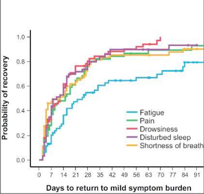
The median time to return to mild symptom severity for these five symptoms was shorter than return to baseline severity, with fatigue taking longer. Pain recovered significantly faster for patients who underwent VATS lobectomy vs. standard open thoracotomy (8 vs. 18 days, respectively; P = .022), according to the researchers. In addition, the researchers found that patients who had poor preoperative performance status or comorbidities reported significantly higher postoperative pain (P less than .05).
Having the ability to measure patient-reported outcomes allows physicians to identify patients at greatest risk for symptoms after surgery and to help caregivers develop pathways to speed up recovery and get patients to chemotherapy or other cancer treatments on schedule, Dr. Fagundes and his coauthors said.
“Using a straightforward, concise tool like the MDASI to obtain the patient’s perspective on how well he or she is recovering is a clinically relevant and user-friendly method for optimizing perioperative care,” the authors added.
Minimally invasive surgical techniques along with opioid-sparing analgesia have been incorporated into enhanced recovery programs to aim for better objective postoperative outcomes like fewer complications and shorter hospital stays. “Missing from these metrics is the voice of the patient, who is arguably the best source of information about what ‘recovery’ from surgery means,” Dr. Fagundes and colleagues said. “Lack of research on how to define and measure symptomatic and functional recovery after major cancer surgery from the patent’s perspective is an important gap in comprehensive postoperative care; it also compromises any comparison of ERP [enhanced recovery program] innovations against standard care.”
Study coauthors received funding from the National Cancer Institute of the National Institutes of Health, the MD Anderson Cancer Center Support Grant program and the American Cancer Society Research Scholar Grant program. The coauthors reported no conflicts of interest in this work.
“Whether ‘subjective’ or not, the measurements of five common postoperative symptoms appear to reflect the real world experience of many,” Dr. Robert B. Cameron of the University of California, Los Angeles, said in his invited commentary (J. Thorac. Cardiovasc. Surg. 2015 [doi: 10.1016/j.jtcvs.2015.06.017]).
What’s more, because the study investigators collected patient data after discharge, they eliminated opportunities to “game the system,” in Dr. Cameron’s words. One example of gaming the outcome measures would be to discharge patients early after surgery with a Heimlich chest tube, subjecting them to problems at home and requiring physicians visits and even emergency department care that can go unrecorded.
“Subjective” patient experiences are “real issues,” Dr. Cameron said. “After all, patients are the customers that the health care system serves.”
Patient-reported outcome measures could dramatically influence how physicians and health systems report patient outcomes. “While length of stay (LOS) and hospital complication rates appear to be valid outcome measures to report, in reality, they are outcomes only in a world when hospitals are the ‘customers,’ ” Dr. Cameron said. “In a patient-centric system, when patient outcomes matter, [patient-reported outcomes], such as those proposed by the MD Anderson investigators, are what truly matter.”
As Medicare and commercial payers rely increasingly on quality outcomes to determine reimbursement levels, clinicians focused on traditional clinical outcomes like length of hospital stay and readmission may be missing the point if they don’t take into consideration more patient-centric outcome. A team of investigators at MD Anderson Cancer Center in Houston recently evaluated that institution’s tool for measuring patient-reported outcomes in tracking results after thoracic surgery for lung cancer.
Christopher P. Fagundes, Ph.D., led the research team that reported their findings in the September issue of the Journal of Thoracic and Cardiovascular Surgery (2015 [doi:10.1016/j.jtcvs.2015.05.057]).
“We used the MD Anderson Symptom Inventory (MDASI) to elicit patient reports of the worst symptoms experienced after thoracic surgery,” Dr. Fagundes and his colleagues said in what may be the first study to use patient-reported outcomes to chart a recovery course after surgery. “This study demonstrates that the MDASI is a sensitive tool for detecting symptomatic recovery with an expected relationship among surgery type, preoperative performance status, and comorbid conditions,” the authors stated.
The MDASI measures the severity of 13 common cancer-related symptoms over the previous 24 hours and rates each on a scale from 0-10, with 10 as most severe. Patients in the study completed a written MDASI questionnaire, 60 upon enrollment, and 77 patients 3 and 5 days after surgery in the hospital. After discharge and for 3 months after surgery, patients received weekly calls from a computer/telephone interactive voice-response system that asked them to rate their symptoms.
All patients were newly diagnosed and treatment naive with early-stage non–small cell lung cancer (NSCLC) who had either standard open thoracotomy or video-assisted thoracoscopic surgery (VATS) lobectomy.
The investigators used a two-pronged approach to define recovery after surgery: symptoms returning to their baseline levels or to a mild level of severity. In the first week after surgery, pain, fatigue, and shortness of breath were “highly prevalent,” perhaps a combined effect from surgical insult and care during and after surgery. After a month, ratings for most symptoms returned to baseline, but fatigue remained the most-persistent symptom during the 3-month study.
NSCLC patients typically have a host of symptoms after major surgery, including inflammation, organ stress, and reaction to medications. In addition, up to a quarter have postsurgical complications that can amplify their symptoms and even delay their course of cancer treatment. The five most-severe postoperative symptoms patients in the study reported were fatigue, pain, shortness of breath, disturbed sleep, and drowsiness.
The median time to return to mild symptom severity for these five symptoms was shorter than return to baseline severity, with fatigue taking longer. Pain recovered significantly faster for patients who underwent VATS lobectomy vs. standard open thoracotomy (8 vs. 18 days, respectively; P = .022), according to the researchers. In addition, the researchers found that patients who had poor preoperative performance status or comorbidities reported significantly higher postoperative pain (P less than .05).
Having the ability to measure patient-reported outcomes allows physicians to identify patients at greatest risk for symptoms after surgery and to help caregivers develop pathways to speed up recovery and get patients to chemotherapy or other cancer treatments on schedule, Dr. Fagundes and his coauthors said.
“Using a straightforward, concise tool like the MDASI to obtain the patient’s perspective on how well he or she is recovering is a clinically relevant and user-friendly method for optimizing perioperative care,” the authors added.
Minimally invasive surgical techniques along with opioid-sparing analgesia have been incorporated into enhanced recovery programs to aim for better objective postoperative outcomes like fewer complications and shorter hospital stays. “Missing from these metrics is the voice of the patient, who is arguably the best source of information about what ‘recovery’ from surgery means,” Dr. Fagundes and colleagues said. “Lack of research on how to define and measure symptomatic and functional recovery after major cancer surgery from the patent’s perspective is an important gap in comprehensive postoperative care; it also compromises any comparison of ERP [enhanced recovery program] innovations against standard care.”
Study coauthors received funding from the National Cancer Institute of the National Institutes of Health, the MD Anderson Cancer Center Support Grant program and the American Cancer Society Research Scholar Grant program. The coauthors reported no conflicts of interest in this work.
FROM THE JOURNAL OF THORACIC AND CARDIOVASCULAR SURGERY
Key clinical point: Using the MD Anderson Symptom Inventory to elicit patient-reported symptom burden is a simple, clinically relevant way to optimize care after thoracic surgery for lung cancer.
Major finding: Assessing symptoms from the patient’s perspective throughout the postoperative recovery period is an effective strategy for evaluating perioperative care.
Data source: Sixty newly diagnosed patients with early-stage non–small cell lung cancer scheduled for thoracic surgery prospectively were recruited for evaluation.
Disclosures: Study coauthors received funding from the National Cancer Institute of the National Institutes of Health, the MD Anderson Cancer Center Support Grant program, and the American Cancer Society Research Scholar Grant program. The coauthors reported no conflicts of interest in this work.

CTS Innovation: The promise of tissue engineering for heart valve replacement and repair
Bioengineered tissues are showing promise in the treatment of valvular heart disease, according to the results of several early clinical and preclinical studies demonstrating the benefits of using heterologous substrates as scaffolding capable of promoting in vivo cell infiltration and remodeling.
Ultimately, cardiac tissue engineering seeks to develop the ideal material for replacement and repair of a variety of heart components – a material able to integrate, function, and, when necessary, grow and develop in a manner undifferentiated from normal cellular structures.
One of the most important areas of focus for such efforts is the development of heart valves and valve patches suitable for use in replacement or repair. The need is obvious: “The fact that literature continues to be published debating the best type of valve prosthesis is proof that, to date, the ideal valve substitute has not been found,” according to Cristian Rosu, Ph.D. and Dr. Edward G. Soltesz of the Cleveland Clinic (Semin Thorac Cardiovasc Surg. 2015 June 30 [doi: 10.1053/j.semtcvs.2015.06.007]).
Dr. Roşu and Dr. Soltesz go on to say that this lack of an ideal prosthetic heart valve leaves surgeons and their patients with a difficult choice at the time of valve surgery. The focus of their concern relates to how current bioprostheses are being used in younger and younger patients, and how these devices have a finite lifespan – requiring eventual reoperation and replacement, perhaps multiple times. In addition, calcification is a prominent and well-known risk of bioprosthetic valves, especially in children (Circulation. 2014 Jul 1;130[1]:51-60).
As for mechanical valves, although durable, they require a lifetime of anticoagulation therapy to prevent thrombosis. And the lack of growth potential found in both mechanical and current bioprosthetic devices is obviously a bane to pediatric valve therapy.
The latest efforts in tissue engineering therefore seek to develop valves and valve components that may be more permanent solutions by better mimicking natural valves.
But better mimicking of a natural valve is not an easy task, when such valves exist in “a near-perfect correlation of structure and function, enabling the valve to avoid excess stress on the cusps while simultaneously withstanding the wear and tear of 40-million repetitive deformations per year, equivalent to some 3 billion over a 75-year lifetime.” (“Principles of Tissue Engineering,” 4th ed. [London: Academic Press 2014, p. 813]).
The “holy grail” of tissue engineering, therefore, is to provide a completely in vitro–developed, autologous, fully cellularized, functional scaffolding for implantation that can live up to these requirements. In order to do this, extensive research into the search for the best cell sources and growth matrices and methods is underway, as illustrated by several recent reviews (Front Cell Dev Biol. 30 June 2015 [doi.org/10.3389/fcell.2015.00039] and Mater Sci Eng C. 2015 March 1;48:556-65).
Candidate cell types include a variety of embryonic stem cells, as well as adult cell types that have proven amenable to rejuvenation and redifferentiation (Adv Drug Deliv Rev. 2014;69-70:254-69).
In one example of the quest for completely in vitro human-tissue designed valves, Dr. Jean Dubé of Laval University, Quebec, and his colleagues reported research on a human tissue–engineered trileaflet heart valve assembled in vitro using human fibroblasts. These cells self-assembled into living tissue sheets when cultured in the presence of sodium ascorbate. These sheets could be layered together to create a thick construct, with the ultimate goal of replacing the use of bovine pericardium tissue implants with ones made of autologous cells from the patient (Acta Biomater. 2014 Aug;10[8]:3563-70).
Currently, however, many of the preclinical studies of fully tissue engineered heart valves have shown retraction of the heart valve leaflets as a major mechanism of functional failure. This retraction is caused by both passive and active cell stress and passive matrix stress, according to a review by Inge A.E.W. van Loosdregt, Ph.D., and her colleagues at the Eindhoven (the Netherlands) University of Technology (J Biomech. 2014 Jun 27;47[9]:2064-9).
While all of these developmental issues regarding the use of fully tissue-engineered valves are being worked out, early clinical applications are already being found for a new generation of valve prostheses that take an intermediate approach, one that uses an implanted scaffolding material that allows autologous cell infiltration and replacement in vivo.
Two of the most prominent examples of these scaffoldings currently in investigation and in early clinical use for heart valve repair or replacement are the bovine pericardium-derived CardioCel (Admedus, Brisbane, Australia) and the porcine intestinal submucosa-derived CorMatrix ECM(CorMatrix Cardiovascular, Roswell, Ga.). CorMatrix ECM was approved by the Food and Drug Administration in 2005 for pericardial repair and reconstruction, and in 2007 for cardiac tissue repair. The FDA approved CardioCel in 2014 for use in the United States in pericardial closure and for the repair of cardiac and vascular defects in both adults and children.
Both technologies rely on the concept of matrix infiltration by autologous cells after implantation in order to create a living mimic of the patient’s own tissues.
CardioCel is a highly treated, bovine pericardium-derived, decellularized collagen matrix. It showed significant resistance to calcification in mitral and pulmonary implants in a juvenile sheep model as reported by Dr. Christian P. Brizard at Royal Children’s Hospital, Melbourne, and his colleagues (J Thorac Cardiovasc Surg. 2014 Dec;148[6]:3194-201). These investigators replaced the posterior leaflet of the mitral valve and one of the pulmonary valve cusps with patches in 10-month-old ewes. They compared the use of CardioCel in six ewes to a control group of four ewes repaired with autologous pericardium that was treated intraoperatively with glutaraldehyde, which is the standard default used at their institution for more than 2 decades as the best material for valve repair in their pediatric patients.

The primary end points of the study were thickening and calcium content. They found that all animals survived with normal valve echocardiography until sacrifice at 7 months. They reported that the bovine pericardium patches allowed accurate valve repair at both systemic and pulmonary pressure with preserved mechanical properties and more-controlled healing and without calcification as compared with the controls. (Calcification is a known major risk factor for the eventual failure of bioprosthetic valves.) Additionally, the bovine pericardium patched valves showed the in vivo development of dense but thin cellularized outer layers of mature collagen I, compared with the controls, which had outer layers that were much less dense and showed the presence of immature collagen III.
In another case of valvular use of the CardioCel material, at the recent American Association for Thoracic Surgery Mitral Conclave 2015, M. Bonnie Ghosh-Dastidar, Ph.D., and her colleagues from the Royal Brompton Hospital, London, reported on a severely ill patient with significant mitral regurgitation and an infected mitral valve with large vegetations on the anterior leaflets. The infected tissue was resected and a large patch of CardioCel bovine pericardium was used to reconstruct the leaflets. Postoperative assessment showed a competent mitral valve with good area of leaflet coaptation, according to Dr. Ghosh-Dastidar.
In his invited commentary on the animal-model research by Dr. Brizard, Dr. Niv Ad, director of cardiac surgery research at Inova Heart and Vascular Institute, Falls Church, Va., said that despite the promise of the CardioCel patches, there were a number of alternative approaches being investigated. “Other engineered materials currently being study include processes such as lyophilization, which has shown promising results in reducing inflammation. Another proposed approach is the use of decellularized vessels and patches with the promise of normal remodeling and growth, such as extracellular matrix and its potential for tissue regeneration.”
Dr. Ad also stated that, “the key bioengineering challenge is to determine how biologic, structural, and mechanical factors interact and function in vivo. The understanding of these factors will prove critical to the development of a clinically viable tissue-engineered heart valve,” (J Thorac Cardiovasc Surg. 2014 Dec;148[6]:3202-3).
An example of the use of extracellular matrix material referred to by Dr. Ad is the CorMatrix ECM, which is an extracellular matrix material derived from porcine small-intestinal submucosa, processed and decellularized.
Dr. Marc W. Gerdisch and his colleagues from the Franciscan St. Francis Heart Center, Indianapolis, reported on the results of treating 19 patients with mitral valve disease using the CorMatrix patch material for partial or subtotal leaflet repair or extension. There were three deaths unrelated to the repair and no instances of perioperative or late stroke. Two patients with a history of cancer and cancer therapy experienced failure of the initial repair, requiring reintervention. However, the other mitral valve repair patients continued to show good valvular function and no calcification on echocardiographic follow-up of 4 days to 48 months (Thorac Cardiovasc Surg. 2014 Oct;148[4]:1370-8).
And in 2015, CorMatrix Cardiovascular announced FDA approval of an investigational device exemption for an early feasibility study of their CorMatrix ECM Tricuspid Heart Valve in up to 15 patients at 5 U.S. centers (NCT02397668). Indications are for the surgical management of tricuspid valve disease not amenable to annuloplasty or repair, including tricuspid valve disease secondary to congenital heart disease in pediatric patients and adult endocarditis patients. The CorMatrix ECM Tricuspid Valve is a flexible, unstented valve acting as a 3‐D scaffold designed to function immediately as a prosthetic, but one constructed to permit native cellular infiltration and remodeling.
Ultimately, both the CardioCel and the CorMatrix materials are just the tip of an iceberg of research into tissue engineering as a clinical tool. And success and the wider adoption of any current or developing technologies will require the results of far-more-extensive studies and long-term clinical results.
At some future date, engineered total organ substitutes or the routine injection of genetically engineered stem cells may provide a practical alternative for treating a diseased or damaged heart. But for now, the most-likely scenario is the continued exploration of tissue engineering to develop the most durable, growth-, stress-, and biologically compatible patches, valves, and vasculatures possible – defined tools that can be adapted to current surgical techniques, designed to obtain the best repair of congenital or acquired heart disease conditions.
As with any transformative technology in medicine, the path from laboratory success and optimistic short-term clinical results to durable, long-term postoperative benefits may be a rocky one. But given the current enthusiasm, it certainly looks as if it will be a well-traveled road.
Dr. Brizard reported consulting and lecturing fees from Admedus. His research was supported with the assistance of a grant from Admedus. Dr. Gerdisch reported consulting fees and equity ownership in CorMatrix Cardiovascular.
Bioengineered tissues are showing promise in the treatment of valvular heart disease, according to the results of several early clinical and preclinical studies demonstrating the benefits of using heterologous substrates as scaffolding capable of promoting in vivo cell infiltration and remodeling.
Ultimately, cardiac tissue engineering seeks to develop the ideal material for replacement and repair of a variety of heart components – a material able to integrate, function, and, when necessary, grow and develop in a manner undifferentiated from normal cellular structures.
One of the most important areas of focus for such efforts is the development of heart valves and valve patches suitable for use in replacement or repair. The need is obvious: “The fact that literature continues to be published debating the best type of valve prosthesis is proof that, to date, the ideal valve substitute has not been found,” according to Cristian Rosu, Ph.D. and Dr. Edward G. Soltesz of the Cleveland Clinic (Semin Thorac Cardiovasc Surg. 2015 June 30 [doi: 10.1053/j.semtcvs.2015.06.007]).
Dr. Roşu and Dr. Soltesz go on to say that this lack of an ideal prosthetic heart valve leaves surgeons and their patients with a difficult choice at the time of valve surgery. The focus of their concern relates to how current bioprostheses are being used in younger and younger patients, and how these devices have a finite lifespan – requiring eventual reoperation and replacement, perhaps multiple times. In addition, calcification is a prominent and well-known risk of bioprosthetic valves, especially in children (Circulation. 2014 Jul 1;130[1]:51-60).
As for mechanical valves, although durable, they require a lifetime of anticoagulation therapy to prevent thrombosis. And the lack of growth potential found in both mechanical and current bioprosthetic devices is obviously a bane to pediatric valve therapy.
The latest efforts in tissue engineering therefore seek to develop valves and valve components that may be more permanent solutions by better mimicking natural valves.
But better mimicking of a natural valve is not an easy task, when such valves exist in “a near-perfect correlation of structure and function, enabling the valve to avoid excess stress on the cusps while simultaneously withstanding the wear and tear of 40-million repetitive deformations per year, equivalent to some 3 billion over a 75-year lifetime.” (“Principles of Tissue Engineering,” 4th ed. [London: Academic Press 2014, p. 813]).
The “holy grail” of tissue engineering, therefore, is to provide a completely in vitro–developed, autologous, fully cellularized, functional scaffolding for implantation that can live up to these requirements. In order to do this, extensive research into the search for the best cell sources and growth matrices and methods is underway, as illustrated by several recent reviews (Front Cell Dev Biol. 30 June 2015 [doi.org/10.3389/fcell.2015.00039] and Mater Sci Eng C. 2015 March 1;48:556-65).
Candidate cell types include a variety of embryonic stem cells, as well as adult cell types that have proven amenable to rejuvenation and redifferentiation (Adv Drug Deliv Rev. 2014;69-70:254-69).
In one example of the quest for completely in vitro human-tissue designed valves, Dr. Jean Dubé of Laval University, Quebec, and his colleagues reported research on a human tissue–engineered trileaflet heart valve assembled in vitro using human fibroblasts. These cells self-assembled into living tissue sheets when cultured in the presence of sodium ascorbate. These sheets could be layered together to create a thick construct, with the ultimate goal of replacing the use of bovine pericardium tissue implants with ones made of autologous cells from the patient (Acta Biomater. 2014 Aug;10[8]:3563-70).
Currently, however, many of the preclinical studies of fully tissue engineered heart valves have shown retraction of the heart valve leaflets as a major mechanism of functional failure. This retraction is caused by both passive and active cell stress and passive matrix stress, according to a review by Inge A.E.W. van Loosdregt, Ph.D., and her colleagues at the Eindhoven (the Netherlands) University of Technology (J Biomech. 2014 Jun 27;47[9]:2064-9).
While all of these developmental issues regarding the use of fully tissue-engineered valves are being worked out, early clinical applications are already being found for a new generation of valve prostheses that take an intermediate approach, one that uses an implanted scaffolding material that allows autologous cell infiltration and replacement in vivo.
Two of the most prominent examples of these scaffoldings currently in investigation and in early clinical use for heart valve repair or replacement are the bovine pericardium-derived CardioCel (Admedus, Brisbane, Australia) and the porcine intestinal submucosa-derived CorMatrix ECM(CorMatrix Cardiovascular, Roswell, Ga.). CorMatrix ECM was approved by the Food and Drug Administration in 2005 for pericardial repair and reconstruction, and in 2007 for cardiac tissue repair. The FDA approved CardioCel in 2014 for use in the United States in pericardial closure and for the repair of cardiac and vascular defects in both adults and children.
Both technologies rely on the concept of matrix infiltration by autologous cells after implantation in order to create a living mimic of the patient’s own tissues.
CardioCel is a highly treated, bovine pericardium-derived, decellularized collagen matrix. It showed significant resistance to calcification in mitral and pulmonary implants in a juvenile sheep model as reported by Dr. Christian P. Brizard at Royal Children’s Hospital, Melbourne, and his colleagues (J Thorac Cardiovasc Surg. 2014 Dec;148[6]:3194-201). These investigators replaced the posterior leaflet of the mitral valve and one of the pulmonary valve cusps with patches in 10-month-old ewes. They compared the use of CardioCel in six ewes to a control group of four ewes repaired with autologous pericardium that was treated intraoperatively with glutaraldehyde, which is the standard default used at their institution for more than 2 decades as the best material for valve repair in their pediatric patients.
The primary end points of the study were thickening and calcium content. They found that all animals survived with normal valve echocardiography until sacrifice at 7 months. They reported that the bovine pericardium patches allowed accurate valve repair at both systemic and pulmonary pressure with preserved mechanical properties and more-controlled healing and without calcification as compared with the controls. (Calcification is a known major risk factor for the eventual failure of bioprosthetic valves.) Additionally, the bovine pericardium patched valves showed the in vivo development of dense but thin cellularized outer layers of mature collagen I, compared with the controls, which had outer layers that were much less dense and showed the presence of immature collagen III.
In another case of valvular use of the CardioCel material, at the recent American Association for Thoracic Surgery Mitral Conclave 2015, M. Bonnie Ghosh-Dastidar, Ph.D., and her colleagues from the Royal Brompton Hospital, London, reported on a severely ill patient with significant mitral regurgitation and an infected mitral valve with large vegetations on the anterior leaflets. The infected tissue was resected and a large patch of CardioCel bovine pericardium was used to reconstruct the leaflets. Postoperative assessment showed a competent mitral valve with good area of leaflet coaptation, according to Dr. Ghosh-Dastidar.
In his invited commentary on the animal-model research by Dr. Brizard, Dr. Niv Ad, director of cardiac surgery research at Inova Heart and Vascular Institute, Falls Church, Va., said that despite the promise of the CardioCel patches, there were a number of alternative approaches being investigated. “Other engineered materials currently being study include processes such as lyophilization, which has shown promising results in reducing inflammation. Another proposed approach is the use of decellularized vessels and patches with the promise of normal remodeling and growth, such as extracellular matrix and its potential for tissue regeneration.”
Dr. Ad also stated that, “the key bioengineering challenge is to determine how biologic, structural, and mechanical factors interact and function in vivo. The understanding of these factors will prove critical to the development of a clinically viable tissue-engineered heart valve,” (J Thorac Cardiovasc Surg. 2014 Dec;148[6]:3202-3).
An example of the use of extracellular matrix material referred to by Dr. Ad is the CorMatrix ECM, which is an extracellular matrix material derived from porcine small-intestinal submucosa, processed and decellularized.
Dr. Marc W. Gerdisch and his colleagues from the Franciscan St. Francis Heart Center, Indianapolis, reported on the results of treating 19 patients with mitral valve disease using the CorMatrix patch material for partial or subtotal leaflet repair or extension. There were three deaths unrelated to the repair and no instances of perioperative or late stroke. Two patients with a history of cancer and cancer therapy experienced failure of the initial repair, requiring reintervention. However, the other mitral valve repair patients continued to show good valvular function and no calcification on echocardiographic follow-up of 4 days to 48 months (Thorac Cardiovasc Surg. 2014 Oct;148[4]:1370-8).
And in 2015, CorMatrix Cardiovascular announced FDA approval of an investigational device exemption for an early feasibility study of their CorMatrix ECM Tricuspid Heart Valve in up to 15 patients at 5 U.S. centers (NCT02397668). Indications are for the surgical management of tricuspid valve disease not amenable to annuloplasty or repair, including tricuspid valve disease secondary to congenital heart disease in pediatric patients and adult endocarditis patients. The CorMatrix ECM Tricuspid Valve is a flexible, unstented valve acting as a 3‐D scaffold designed to function immediately as a prosthetic, but one constructed to permit native cellular infiltration and remodeling.
Ultimately, both the CardioCel and the CorMatrix materials are just the tip of an iceberg of research into tissue engineering as a clinical tool. And success and the wider adoption of any current or developing technologies will require the results of far-more-extensive studies and long-term clinical results.
At some future date, engineered total organ substitutes or the routine injection of genetically engineered stem cells may provide a practical alternative for treating a diseased or damaged heart. But for now, the most-likely scenario is the continued exploration of tissue engineering to develop the most durable, growth-, stress-, and biologically compatible patches, valves, and vasculatures possible – defined tools that can be adapted to current surgical techniques, designed to obtain the best repair of congenital or acquired heart disease conditions.
As with any transformative technology in medicine, the path from laboratory success and optimistic short-term clinical results to durable, long-term postoperative benefits may be a rocky one. But given the current enthusiasm, it certainly looks as if it will be a well-traveled road.
Dr. Brizard reported consulting and lecturing fees from Admedus. His research was supported with the assistance of a grant from Admedus. Dr. Gerdisch reported consulting fees and equity ownership in CorMatrix Cardiovascular.

More lawsuits against doctors? Overpayment ruling could be bad news
In a novel decision, the U.S. District Court for the Southern District of New York has ruled that the 60-day clock to return overpayments to the government begins ticking when a health provider receives notice a potential overpayment exists, not when an overpayment is conclusively ascertained.
Doctors should be concerned about the ruling, said Houston health law attorney Michael E. Clark, immediate past chair for the American Bar Association Health Law Section.

“This is a very troubling development because the judge has embraced the theory that certainty is not required as to what constitutes an identified overpayment,” Mr. Clark said in an interview. “Rather, knowledge can be established by recklessness under the facts. In short, practitioners must set up systems to alert them about potential overpayments so they can move quickly to avoid potentially ruinous False Claims Act liability.”
The Aug. 3 ruling in Kane v. Healthfirst is the first published decision to address the 60-day overpayment rule imposed under the Affordable Care Act and the Fraud Enforcement and Recovery Act (FERA). The rule requires that an overpayment be reported and returned by health providers within 60 days of the “date on which the overpayment was identified.” Health providers who retain an overpayment beyond that point are subject to liability under the False Claims Act (FCA).
In the Kane case, the federal government contends that three hospitals operated by Continuum Health Partners failed to report and return overpayments to Medicaid within 60 days of identification. Because of a computer glitch, Continuum billed both the government and a managed care organization for the same services, according to court documents. After the New York State Comptroller’s Office alerted Continuum to a possible overbilling, Continuum hired an employee, Robert P. Kane, to conduct an internal investigation into its billing. Mr. Kane – who was later fired – allegedly found 900 potentially improper Medicaid claims totaling $1 million, according to court documents. The government claims Continuum failed to repay the overpayments within 60 days and instead repaid only “small batches” of the affected claims over the next 2 years. Mr. Kane filed a whistleblower suit against Continuum, and the government intervened as a plaintiff.
But Continuum argued that the hospitals did not knowingly conceal the overpayments from the government, and that the overbillings had not been officially “identified.” The defendants were provided only notice of potential overpayments and did not identify actual overpayments so as to trigger the 60-day report and return clock, Continuum said in court documents. The health system requested the court throw out the government’s suit for lack of merit.
District Judge Edgardo Ramos agreed with the federal government and allowed the lawsuit to continue. Judge Ramos said the legislative history indicates that Congress intended for FCA liability to attach in circumstances where there is an established duty to pay money to the government, even if the precise amount due has yet to be determined.
“Here, after the comptroller alerted defendants to the software glitch and approached them with specific wrongful claims, and after Kane put defendants on notice of a set of claims likely to contain numerous overpayments, defendants had an established duty to report and return wrongly collected money,” Judge Ramos said in his opinion. “To allow defendants to evade liability because Kane’s email did not conclusively establish each erroneous claim and did not provide the specific amount owed to the government would contradict Congress’s intentions as expressed during the passage of the FERA.”

In an email, a spokesperson for the defendants said the hospitals are disappointed with the judge’s decision and will continue to vigorously defend its case in court. Attorneys for the government did not return messages seeking comment.
The judge’s ruling is encouraging to the federal government and for plaintiffs who wish to sue health providers for overbilling violations, said Joel M. Androphy, a Houston plaintiffs’ attorney.
“This is going to open the floodgates for lawyers now as part of their false claim and reporting practices to let the courts know about overpayment issues because they know the court and the government will be listening,” Mr. Androphy said in an interview. “It’s not going to be the sole basis for [a plaintiff’s] claim necessarily, but it could be an integral part.”
Mr. Androphy added that defendants can no longer complain they were confused by the 60-day overpayment rule and the meaning of “identification.” The judge’s ruling makes the regulation more clear and provides guidance to health providers about how the rule will be enforced, he said.
Washington health law attorney Robert T. Rhoad however, disagreed that the opinion clarifies application of the 60-day overpayment rule. The decision does not provide the bright lines for compliance that providers expect and need, said Mr. Rhoad.

“While the Kane decision provides an exposition of the etiology and perceived intent of the 60-day rule, its ultimate ruling was made through the narrow lens of the specific and arguably egregious [facts] alleged,” Mr. Rhoad said in an interview. “If anything, by finding that certainty is not required in identifying an overpayment triggering the 60-day rule, the decision may encourage the government and qui tam relators to come forward with expansive theories of what might constitute reckless disregard by a provider to identify an overpayment to invoke FCA liability by the running of the 60-day clock.”
To protect themselves from litigation, physicians should take prudent steps to conduct an appropriate investigation if faced with actual or constructive notice of a possible overpayment, Mr. Rhoad said. Showing that they acted with due diligence and without delay to investigate and, if identified, report an overpayment could help doctors withstand future governmental or judicial scrutiny.
On Twitter @legal_med
In a novel decision, the U.S. District Court for the Southern District of New York has ruled that the 60-day clock to return overpayments to the government begins ticking when a health provider receives notice a potential overpayment exists, not when an overpayment is conclusively ascertained.
Doctors should be concerned about the ruling, said Houston health law attorney Michael E. Clark, immediate past chair for the American Bar Association Health Law Section.
“This is a very troubling development because the judge has embraced the theory that certainty is not required as to what constitutes an identified overpayment,” Mr. Clark said in an interview. “Rather, knowledge can be established by recklessness under the facts. In short, practitioners must set up systems to alert them about potential overpayments so they can move quickly to avoid potentially ruinous False Claims Act liability.”
The Aug. 3 ruling in Kane v. Healthfirst is the first published decision to address the 60-day overpayment rule imposed under the Affordable Care Act and the Fraud Enforcement and Recovery Act (FERA). The rule requires that an overpayment be reported and returned by health providers within 60 days of the “date on which the overpayment was identified.” Health providers who retain an overpayment beyond that point are subject to liability under the False Claims Act (FCA).
In the Kane case, the federal government contends that three hospitals operated by Continuum Health Partners failed to report and return overpayments to Medicaid within 60 days of identification. Because of a computer glitch, Continuum billed both the government and a managed care organization for the same services, according to court documents. After the New York State Comptroller’s Office alerted Continuum to a possible overbilling, Continuum hired an employee, Robert P. Kane, to conduct an internal investigation into its billing. Mr. Kane – who was later fired – allegedly found 900 potentially improper Medicaid claims totaling $1 million, according to court documents. The government claims Continuum failed to repay the overpayments within 60 days and instead repaid only “small batches” of the affected claims over the next 2 years. Mr. Kane filed a whistleblower suit against Continuum, and the government intervened as a plaintiff.
But Continuum argued that the hospitals did not knowingly conceal the overpayments from the government, and that the overbillings had not been officially “identified.” The defendants were provided only notice of potential overpayments and did not identify actual overpayments so as to trigger the 60-day report and return clock, Continuum said in court documents. The health system requested the court throw out the government’s suit for lack of merit.
District Judge Edgardo Ramos agreed with the federal government and allowed the lawsuit to continue. Judge Ramos said the legislative history indicates that Congress intended for FCA liability to attach in circumstances where there is an established duty to pay money to the government, even if the precise amount due has yet to be determined.
“Here, after the comptroller alerted defendants to the software glitch and approached them with specific wrongful claims, and after Kane put defendants on notice of a set of claims likely to contain numerous overpayments, defendants had an established duty to report and return wrongly collected money,” Judge Ramos said in his opinion. “To allow defendants to evade liability because Kane’s email did not conclusively establish each erroneous claim and did not provide the specific amount owed to the government would contradict Congress’s intentions as expressed during the passage of the FERA.”
In an email, a spokesperson for the defendants said the hospitals are disappointed with the judge’s decision and will continue to vigorously defend its case in court. Attorneys for the government did not return messages seeking comment.
The judge’s ruling is encouraging to the federal government and for plaintiffs who wish to sue health providers for overbilling violations, said Joel M. Androphy, a Houston plaintiffs’ attorney.
“This is going to open the floodgates for lawyers now as part of their false claim and reporting practices to let the courts know about overpayment issues because they know the court and the government will be listening,” Mr. Androphy said in an interview. “It’s not going to be the sole basis for [a plaintiff’s] claim necessarily, but it could be an integral part.”
Mr. Androphy added that defendants can no longer complain they were confused by the 60-day overpayment rule and the meaning of “identification.” The judge’s ruling makes the regulation more clear and provides guidance to health providers about how the rule will be enforced, he said.
Washington health law attorney Robert T. Rhoad however, disagreed that the opinion clarifies application of the 60-day overpayment rule. The decision does not provide the bright lines for compliance that providers expect and need, said Mr. Rhoad.
“While the Kane decision provides an exposition of the etiology and perceived intent of the 60-day rule, its ultimate ruling was made through the narrow lens of the specific and arguably egregious [facts] alleged,” Mr. Rhoad said in an interview. “If anything, by finding that certainty is not required in identifying an overpayment triggering the 60-day rule, the decision may encourage the government and qui tam relators to come forward with expansive theories of what might constitute reckless disregard by a provider to identify an overpayment to invoke FCA liability by the running of the 60-day clock.”
To protect themselves from litigation, physicians should take prudent steps to conduct an appropriate investigation if faced with actual or constructive notice of a possible overpayment, Mr. Rhoad said. Showing that they acted with due diligence and without delay to investigate and, if identified, report an overpayment could help doctors withstand future governmental or judicial scrutiny.
On Twitter @legal_med

Ellmers’ bill seeks delay for Stage 3 meaningful use
A bill delaying the implementation of Stage 3 meaningful use until at least 2017 has been introduced in the House.
The legislation calls for a delay until the Centers for Medicare & Medicaid Services finalizes Merit-based Incentive Payment System (MIPS) program regulations or at least 75% of doctors and hospitals are successfully meeting Stage 2 meaningful use criteria.

Under the current proposed federal rule for meaningful use, all providers would need to be meeting stage 3 in 2018. CMS has not yet finalized the Stage 3 criteria.
“Only 19% of providers have met Stage 2 attestations – a clear sign that physicians, hospitals, and health care providers are challenged in meeting CMS’ onerous requirements,” Rep. Renee Ellmers (R-N.C.), who introduced the Flex-IT 2 Act (H.R. 3309), said in a statement. She added that 48% of hospitals are meeting Stage 2.
“Given this basic fact, I’m uncertain why CMS would continue to push forward with the Stage 3 rule. From my conversations with doctors back home, it is clear they are eager for relief,” Rep. Ellmers continued.
Rep. Ellmers has been a proponent of smart implementation of the meaningful use rule and earlier this year had legislation ready to move to shorten the attestation period in 2015 for Stage 2 to 90 days from the full year. CMS acted to shorten the period before legislative action was necessary.
The legislation, formally known as the Further Flexibility in HIT Reporting and Advancing Interoperability Act of 2015 would codify the 90-day attestation period, regardless of stage or program experience. It also would harmonizes reporting requirements across various programs to remove duplicative measurements and streamline requirements, encourages interoperability and expands hardship exemptions.
The bill’s introduction came amid calls from the American Medical Association to delay Stage 3 in order to sync its requirements with MIPS.
A bill delaying the implementation of Stage 3 meaningful use until at least 2017 has been introduced in the House.
The legislation calls for a delay until the Centers for Medicare & Medicaid Services finalizes Merit-based Incentive Payment System (MIPS) program regulations or at least 75% of doctors and hospitals are successfully meeting Stage 2 meaningful use criteria.
Under the current proposed federal rule for meaningful use, all providers would need to be meeting stage 3 in 2018. CMS has not yet finalized the Stage 3 criteria.
“Only 19% of providers have met Stage 2 attestations – a clear sign that physicians, hospitals, and health care providers are challenged in meeting CMS’ onerous requirements,” Rep. Renee Ellmers (R-N.C.), who introduced the Flex-IT 2 Act (H.R. 3309), said in a statement. She added that 48% of hospitals are meeting Stage 2.
“Given this basic fact, I’m uncertain why CMS would continue to push forward with the Stage 3 rule. From my conversations with doctors back home, it is clear they are eager for relief,” Rep. Ellmers continued.
Rep. Ellmers has been a proponent of smart implementation of the meaningful use rule and earlier this year had legislation ready to move to shorten the attestation period in 2015 for Stage 2 to 90 days from the full year. CMS acted to shorten the period before legislative action was necessary.
The legislation, formally known as the Further Flexibility in HIT Reporting and Advancing Interoperability Act of 2015 would codify the 90-day attestation period, regardless of stage or program experience. It also would harmonizes reporting requirements across various programs to remove duplicative measurements and streamline requirements, encourages interoperability and expands hardship exemptions.
The bill’s introduction came amid calls from the American Medical Association to delay Stage 3 in order to sync its requirements with MIPS.

Last-minute ICD-10 help for docs who are lagging
As the calendar winds down toward the Oct. 1 compliance date for ICD-10, be aware of options that can help medical practices meet requirements while systems are being upgraded.
[Physicians and their staffs] “may not have everything they need to do in the office all prepared and installed, but if they take advantage of other methods for billing under ICD-10, like free software that might be available from the government or from clearinghouses or using payer portals to enter claims” they should be able to get by, Jim Daley, director of IT for Blue Cross Blue Shield of South Carolina, said in an interview. “They can still use ICD-10 on the compliance date, even though it may not be ideal.”

Mr. Daley, who also is a past chair of the Workgroup for Electronic Data Interchange (WEDI), outlined a number of resources to help with the transition, even as the deadline is fast approaching.
“In particular, the CMS Road to 10 website is outstanding,” Mr. Daley said. “It may be overwhelming at times, but there are some very nice summaries out there.”
The Centers for Medicare & Medicaid Services also posted to its website a series of specialty-specific guides to help with the transition.
“Also, WEDI has some great resources for physicians to view,” Mr. Daley continued, stressing that physicians need to be updating office software to make sure it supports ICD-10.
He suggested working with training partners and clearinghouses, who also might have resources to help with the transition. “Use those resources that are available, and also don’t delay. There’s still time, although it’s getting short. There is a lot to do.”
Finally, he stressed that more detailed clinical documentation will be key to a smooth transition. “Understand enough about what’s required for ICD-10 to create good clinical documentation. Concepts such as left side or right side are very basic, but if that’s in your documentation, it makes it much easier to code them through ICD-10,” Mr. Daley said.
He continued, “If you are documenting the exact conditions and the exact processes that you performed when you saw the patient, which you should be doing anyway, then it should be much easier to pick the correct code. They are not as general as before. They are very specific. So if you have those details in your documentation, it will drive you down to picking the correct codes.”
As the calendar winds down toward the Oct. 1 compliance date for ICD-10, be aware of options that can help medical practices meet requirements while systems are being upgraded.
[Physicians and their staffs] “may not have everything they need to do in the office all prepared and installed, but if they take advantage of other methods for billing under ICD-10, like free software that might be available from the government or from clearinghouses or using payer portals to enter claims” they should be able to get by, Jim Daley, director of IT for Blue Cross Blue Shield of South Carolina, said in an interview. “They can still use ICD-10 on the compliance date, even though it may not be ideal.”
Mr. Daley, who also is a past chair of the Workgroup for Electronic Data Interchange (WEDI), outlined a number of resources to help with the transition, even as the deadline is fast approaching.
“In particular, the CMS Road to 10 website is outstanding,” Mr. Daley said. “It may be overwhelming at times, but there are some very nice summaries out there.”
The Centers for Medicare & Medicaid Services also posted to its website a series of specialty-specific guides to help with the transition.
“Also, WEDI has some great resources for physicians to view,” Mr. Daley continued, stressing that physicians need to be updating office software to make sure it supports ICD-10.
He suggested working with training partners and clearinghouses, who also might have resources to help with the transition. “Use those resources that are available, and also don’t delay. There’s still time, although it’s getting short. There is a lot to do.”
Finally, he stressed that more detailed clinical documentation will be key to a smooth transition. “Understand enough about what’s required for ICD-10 to create good clinical documentation. Concepts such as left side or right side are very basic, but if that’s in your documentation, it makes it much easier to code them through ICD-10,” Mr. Daley said.
He continued, “If you are documenting the exact conditions and the exact processes that you performed when you saw the patient, which you should be doing anyway, then it should be much easier to pick the correct code. They are not as general as before. They are very specific. So if you have those details in your documentation, it will drive you down to picking the correct codes.”

Online resource launched to prevent inpatient hospital falls
An online resource guide offers 21 targeted solutions for reducing the rate of falls in hospitals and urgent care settings, The Joint Commission Center for Transforming Healthcare announced in a statement.
Using the fall prevention methodology of the Targeted Solutions Tool, developed in collaboration with seven hospitals and five health care organizations, a typical 200-bed hospital could potentially reduce the number of patients injured from a fall from 117 to 45, avoiding approximately $1 million in costs annually, the agency claims.
Some of the recommendations for reducing in-hospital falls include:
• Creating awareness among staff.
• Using a validated fall risk assessment tool.
• Engaging patients and their families in the fall safety program.
• Hourly rounding with scheduled restroom use for patients.
• Engaging all hospital staff and patients to ensure no patient walks without assistance.
“Hundreds of thousands of patients fall in hospitals every year and many of these falls result in moderate to severe injuries that can prolong hospital stays and require the patient to undergo additional treatment,” Dr. Erin DuPree, vice president and chief medical officer of the Joint Commission Center for Transforming Healthcare, said in a statement.
The Joint Commission Center for Transforming Healthcare was created in 2008 as a nonprofit affiliate of The Joint Commission.
Check out the online resource here.
Perioperative cardiovascular assessment guidelines pose new challenges
ESTES PARK, COLO. – The latest American College of Cardiology/American Heart Association guidelines on perioperative cardiovascular evaluation in patients undergoing noncardiac surgery create several major new responsibilities for physicians doing the evaluations.
Some primary care physicians will be uncomfortable with several of the new recommendations. Perhaps that shouldn’t come as a surprise, since not a single primary care physician was included among the 17 members of the writing committee or the 38-member review committee. Cardiologists of all subspecialties, surgeons, anesthesiologists, hospitalists, a patient representative – all had a voice. But not primary care, Dr. Robert E. Burke noted at a conference on internal medicine sponsored by the University of Colorado.

He was quick to add, however, that the document (Circulation. 2014 Dec 9;130[24]:e278-333) is an impressive, thoughtful piece of work built upon 490 reference citations, and it deserves to be incorporated into daily clinical practice, albeit with a few caveats.
“I think the writing panel did a really good job, but the recommendations are only as good as the evidence – and there are a couple of areas where the evidence is not very strong ... It’s up to you and your patient to decide what to do in the controversial areas,” said Dr. Burke, assistant chief of hospital medicine at the Denver VA Medical Center and an internist at the university.
A big change from the previous 2007 ACC/AHA guidelines that Dr. Burke fully supports is that it’s no longer sufficient for the evaluating physician to formally consider only the level of risk posed by the patient’s planned noncardiac surgery, which can vary from a low-risk operation such as cataract surgery to a far-higher-risk procedure addressing, for example, an expanding abdominal aortic aneurysm.
Under the 2014 guidelines, it’s also important to incorporate the surgical risk with an estimate of the individual’s personalized risk of perioperative major adverse cardiovascular events. If the estimated risk is less than 1%, it’s appropriate to proceed straightaway with surgery. In contrast, a 1% or greater perioperative risk is considered high, and it’s then recommended to continue along a lengthy management algorithm. This involves sending the patient for cardiac stress testing provided two conditions are met: the surgeon has to be unwilling to operate without the stress test results and the patient must be willing to undergo CABG before the noncardiac surgery, should the stress test and follow-up cardiac catheterization prove positive.
The guidelines recommend using either of two validated risk-prediction tools for calculating patient risk: the Revised Cardiac Risk Index (Circulation 1999 Sep 7;100[10]:1043-9) or the American College of Surgeons’ National Surgical Quality Improvement Project (NSQIP) predictor. Dr. Burke prefers the NSQIP tool. It predicts multiple cardiac and noncardiac outcomes, including all-cause mortality, within 30 days of surgery, and it has been validated in 525 U.S. hospitals with more than 1 million operations. It’s also quite user-friendly, according to Dr. Burke.
In his view, the guidelines contain two major areas of controvery. One involves the much more aggressive approach to revascularization prior to noncardiac surgery in high-cardiovascular-risk patients compared with the 2007 guidelines. The current guidelines give a class I recommendation to revascularization before noncardiac surgery in patients with left main or triple-vessel disease, as well as two-vessel disease that includes the left anterior descending coronary artery. He said this is a leap that’s not supported by the best available evidence, which comes from the older CARP (Coronary-Artery Revascularization before Elective Major Vascular Surgery) trial.
In CARP, 510 military veterans with documented coronary artery disease and planned vascular surgery were randomized to preoperative revascularization or not. The only exclusions were left main disease, severe aortic stenosis, or a left ventricular ejection fraction below 20%. After 2.8 years of follow-up post surgery, preoperative revascularization wasn’t associated with any mortality benefit in any patient subgroup (N Engl J Med. 2004 Dec 30;351[27]:2795-804).
“I want to restate that in this well-done randomized controlled trial, they could not find any benefit in any outcome in any group within this very-high-risk group of patients. This is really the main trial that exists in perioperative care, and I’m very impressed with the results. The guidelines extrapolate from studies in other areas. This is one area where I think the cardiologists are more interventional than I would like. If I had a patient in my office I could not tell him with a straight face that revascularization before their planned surgery is going to offer them any benefit during up to nearly 3 years of follow-up unless they have left main disease, which was an exclusion in CARP,” Dr. Burke said.
The other main controversy in his view is the ACC/AHA stance on postoperative troponin measurement. It gets a class III rating – meaning no benefit, don’t do it – for routine screening in unselected patients without signs or symptoms suggestive of myocardial ischemia. This recommendation is based upon the absence to date of a prospective trial showing that acting on an elevated postoperative troponin in such patients improves outcomes. True enough, Dr. Burke conceded, but he finds persuasive the results of VISION (Vascular Events in Noncardiac Surgery Patients Cohort Evaluation), a prospective international study in which more than 15,000 patients undergoing noncardiac surgery underwent screening troponin measurements on the first 3 days after surgery.
Thirty-day mortality in the VISION study was 1% in those with a peak fourth-generation troponin of 0.01 ng/mL or less. Mortality was significantly increased in the nearly 12% of patients with a troponin of at least 0.02 ng/mL, climbing to a peak mortality rate of 16.9% in those with a postoperative troponin of 0.30 ng/mL or more (JAMA. 2012 Jun 6;307[21]:2295-304).
“I don’t know of any other test in the perioperative setting that’s as good as this in terms of risk-stratifying patients. We can’t just sit by and let this sort of 30-day mortality rate happen. We should do something. I think that if you treat these asymptomatic patients with an elevated postop troponin as if they had an MI – get them on optimal medical therapy and really push smoking cessation – it’s likely to have a benefit on outcomes,” said Dr. Burke.
He reported having no financial conflicts regarding his presentation.
ESTES PARK, COLO. – The latest American College of Cardiology/American Heart Association guidelines on perioperative cardiovascular evaluation in patients undergoing noncardiac surgery create several major new responsibilities for physicians doing the evaluations.
Some primary care physicians will be uncomfortable with several of the new recommendations. Perhaps that shouldn’t come as a surprise, since not a single primary care physician was included among the 17 members of the writing committee or the 38-member review committee. Cardiologists of all subspecialties, surgeons, anesthesiologists, hospitalists, a patient representative – all had a voice. But not primary care, Dr. Robert E. Burke noted at a conference on internal medicine sponsored by the University of Colorado.
He was quick to add, however, that the document (Circulation. 2014 Dec 9;130[24]:e278-333) is an impressive, thoughtful piece of work built upon 490 reference citations, and it deserves to be incorporated into daily clinical practice, albeit with a few caveats.
“I think the writing panel did a really good job, but the recommendations are only as good as the evidence – and there are a couple of areas where the evidence is not very strong ... It’s up to you and your patient to decide what to do in the controversial areas,” said Dr. Burke, assistant chief of hospital medicine at the Denver VA Medical Center and an internist at the university.
A big change from the previous 2007 ACC/AHA guidelines that Dr. Burke fully supports is that it’s no longer sufficient for the evaluating physician to formally consider only the level of risk posed by the patient’s planned noncardiac surgery, which can vary from a low-risk operation such as cataract surgery to a far-higher-risk procedure addressing, for example, an expanding abdominal aortic aneurysm.
Under the 2014 guidelines, it’s also important to incorporate the surgical risk with an estimate of the individual’s personalized risk of perioperative major adverse cardiovascular events. If the estimated risk is less than 1%, it’s appropriate to proceed straightaway with surgery. In contrast, a 1% or greater perioperative risk is considered high, and it’s then recommended to continue along a lengthy management algorithm. This involves sending the patient for cardiac stress testing provided two conditions are met: the surgeon has to be unwilling to operate without the stress test results and the patient must be willing to undergo CABG before the noncardiac surgery, should the stress test and follow-up cardiac catheterization prove positive.
The guidelines recommend using either of two validated risk-prediction tools for calculating patient risk: the Revised Cardiac Risk Index (Circulation 1999 Sep 7;100[10]:1043-9) or the American College of Surgeons’ National Surgical Quality Improvement Project (NSQIP) predictor. Dr. Burke prefers the NSQIP tool. It predicts multiple cardiac and noncardiac outcomes, including all-cause mortality, within 30 days of surgery, and it has been validated in 525 U.S. hospitals with more than 1 million operations. It’s also quite user-friendly, according to Dr. Burke.
In his view, the guidelines contain two major areas of controvery. One involves the much more aggressive approach to revascularization prior to noncardiac surgery in high-cardiovascular-risk patients compared with the 2007 guidelines. The current guidelines give a class I recommendation to revascularization before noncardiac surgery in patients with left main or triple-vessel disease, as well as two-vessel disease that includes the left anterior descending coronary artery. He said this is a leap that’s not supported by the best available evidence, which comes from the older CARP (Coronary-Artery Revascularization before Elective Major Vascular Surgery) trial.
In CARP, 510 military veterans with documented coronary artery disease and planned vascular surgery were randomized to preoperative revascularization or not. The only exclusions were left main disease, severe aortic stenosis, or a left ventricular ejection fraction below 20%. After 2.8 years of follow-up post surgery, preoperative revascularization wasn’t associated with any mortality benefit in any patient subgroup (N Engl J Med. 2004 Dec 30;351[27]:2795-804).
“I want to restate that in this well-done randomized controlled trial, they could not find any benefit in any outcome in any group within this very-high-risk group of patients. This is really the main trial that exists in perioperative care, and I’m very impressed with the results. The guidelines extrapolate from studies in other areas. This is one area where I think the cardiologists are more interventional than I would like. If I had a patient in my office I could not tell him with a straight face that revascularization before their planned surgery is going to offer them any benefit during up to nearly 3 years of follow-up unless they have left main disease, which was an exclusion in CARP,” Dr. Burke said.
The other main controversy in his view is the ACC/AHA stance on postoperative troponin measurement. It gets a class III rating – meaning no benefit, don’t do it – for routine screening in unselected patients without signs or symptoms suggestive of myocardial ischemia. This recommendation is based upon the absence to date of a prospective trial showing that acting on an elevated postoperative troponin in such patients improves outcomes. True enough, Dr. Burke conceded, but he finds persuasive the results of VISION (Vascular Events in Noncardiac Surgery Patients Cohort Evaluation), a prospective international study in which more than 15,000 patients undergoing noncardiac surgery underwent screening troponin measurements on the first 3 days after surgery.
Thirty-day mortality in the VISION study was 1% in those with a peak fourth-generation troponin of 0.01 ng/mL or less. Mortality was significantly increased in the nearly 12% of patients with a troponin of at least 0.02 ng/mL, climbing to a peak mortality rate of 16.9% in those with a postoperative troponin of 0.30 ng/mL or more (JAMA. 2012 Jun 6;307[21]:2295-304).
“I don’t know of any other test in the perioperative setting that’s as good as this in terms of risk-stratifying patients. We can’t just sit by and let this sort of 30-day mortality rate happen. We should do something. I think that if you treat these asymptomatic patients with an elevated postop troponin as if they had an MI – get them on optimal medical therapy and really push smoking cessation – it’s likely to have a benefit on outcomes,” said Dr. Burke.
He reported having no financial conflicts regarding his presentation.
EXPERT ANALYSIS FROM THE ANNUAL INTERNAL MEDICINE PROGRAM

CMS releases specialty-specific ICD-10 transition guides
The Centers for Medicare & Medicaid Services is offering a number of specialty-specific guidebooks to help with the transition to ICD-10.
“To help physicians and other providers get quickly up to speed, CMS has launched the ICD-10 Clinical Concepts Series for specialties,” according to a post on the agency’s ICD-10 news website. “Each guide in the series compiles key information from the Road to 10 online tool in a PDF format that can be readily shared, emailed, posted to websites, and printed. The guides include common ICD-10 codes, clinical documentation tips, clinical scenarios, and links to Road to 10.”

The specialty guidebooks include: family practice, internal medicine, cardiology, ob.gyn., orthopedics, and pediatrics.
Road to 10 includes tools to help build an ICD-10 action plan, interactive case studies to see how your coding selections compare with those of other physicians, quick reference documents, and an archive of webcasts created by and for medical professionals.
All health care professionals must start using ICD-10 on Oct. 1; however, the CMS has announced a 1-year transition period to help ensure physicians and other providers continue to get paid as they begin filing claims using the new code set.
The Centers for Medicare & Medicaid Services is offering a number of specialty-specific guidebooks to help with the transition to ICD-10.
“To help physicians and other providers get quickly up to speed, CMS has launched the ICD-10 Clinical Concepts Series for specialties,” according to a post on the agency’s ICD-10 news website. “Each guide in the series compiles key information from the Road to 10 online tool in a PDF format that can be readily shared, emailed, posted to websites, and printed. The guides include common ICD-10 codes, clinical documentation tips, clinical scenarios, and links to Road to 10.”
The specialty guidebooks include: family practice, internal medicine, cardiology, ob.gyn., orthopedics, and pediatrics.
Road to 10 includes tools to help build an ICD-10 action plan, interactive case studies to see how your coding selections compare with those of other physicians, quick reference documents, and an archive of webcasts created by and for medical professionals.
All health care professionals must start using ICD-10 on Oct. 1; however, the CMS has announced a 1-year transition period to help ensure physicians and other providers continue to get paid as they begin filing claims using the new code set.

Modifiable risk factors foretell colonic anastomotic leak
CHICAGO – Several modifiable risk factors predicted the development of anastomotic leak following elective colon resection, a large national analysis found.
“Preoperative smoking cessation, preoperative administration of oral antibiotic bowel preparation, and laparoscopic approach are modifiable factors that could reduce the risk of anastomotic leak,” Dr. Cindy Wu said at the American College of Surgeons (ACS) National Surgical Quality Improvement Program (NSQIP) National Conference.

Anastomotic leakage results in increased morbidity and mortality, yet the current literature analyzing risk factors for this complication is generally limited to retrospective studies of single institutions, she said.
To examine data from a larger sample of colectomy patients from multiple centers, the investigators used the NSQIP Participant Use Data File specifically targeted to colectomy to identify 14,848 patients who underwent elective colon resection from 2012 to 2013. Chi-square, Wald chi-square, and logistic regression analyses were performed examining patient factors (sex, race, comorbidities, smoking status, American Society of Anesthesiologists class, functional status, steroid use, and preoperative albumin), oncologic factors (chemotherapy, tumor stage, and presence or absence of disseminated cancer), and operative factors (wound class, mechanical bowel preparation, oral antibiotic preparation, surgical approach, colectomy site, surgical indication, and operative time).
In all, 3.4%, or 498 patients, experienced an anastomotic leak, which is consistent with the literature, Dr. Wu of Temple University in Philadelphia said. Of these patients, 101 required no intervention, while 272 required surgery and 125 needed percutaneous drainage. The mean age of the patients was 60.7 years and 57% were male.
In a univariate analysis, male sex (chi-square = 17.4; P less than .01), diabetes controlled with either oral medication or insulin (X2 = 9.5; P less than .01), and smoking within the last year (X2 = 20.4; P less than .01) were associated with a greater incidence of anastomotic leak.
Other risk factors that were significant in additional univariate analysis were ASA class (X2 = 23.3; P = .0001), functional status (X2 = 9.15; P = .01), 10% weight loss over the last 6 months (X2 = 5.83; P = .02), wound class (X2 = 10.8; P = .01), mechanical bowel preparation (X2 = 5.89; P = .01), lack of oral antibiotic preparation (X2 = 17.5; P less than .0001), open vs. laparoscopic/minimally invasive surgery (X2 = 60.0; P less than .0001), chemotherapy in the last 90 days (X2 = 23.1; P less than .0001), and presence of disseminated cancer (X2 = 7.41; P = .01), Dr. Wu said.
With all of these factors taken into account in multivariate analysis, independent predictors of an increased risk of anastomotic leak were male sex (odds ratio, 1.74; P = .01), tobacco use (OR, 1.73; P = .03), and lack of a preoperative oral antibiotic bowel preparation (OR, 1.79; P less than .01).
Interestingly, use of a laparoscopic technique was protective against the development of anastomotic leakage (OR, 0.54; P less than .01), she said.
The authors reported having no relevant financial disclosures.
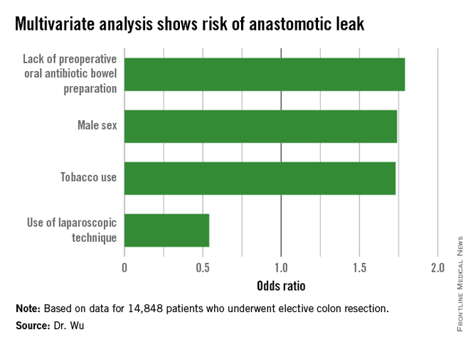
CHICAGO – Several modifiable risk factors predicted the development of anastomotic leak following elective colon resection, a large national analysis found.
“Preoperative smoking cessation, preoperative administration of oral antibiotic bowel preparation, and laparoscopic approach are modifiable factors that could reduce the risk of anastomotic leak,” Dr. Cindy Wu said at the American College of Surgeons (ACS) National Surgical Quality Improvement Program (NSQIP) National Conference.
Anastomotic leakage results in increased morbidity and mortality, yet the current literature analyzing risk factors for this complication is generally limited to retrospective studies of single institutions, she said.
To examine data from a larger sample of colectomy patients from multiple centers, the investigators used the NSQIP Participant Use Data File specifically targeted to colectomy to identify 14,848 patients who underwent elective colon resection from 2012 to 2013. Chi-square, Wald chi-square, and logistic regression analyses were performed examining patient factors (sex, race, comorbidities, smoking status, American Society of Anesthesiologists class, functional status, steroid use, and preoperative albumin), oncologic factors (chemotherapy, tumor stage, and presence or absence of disseminated cancer), and operative factors (wound class, mechanical bowel preparation, oral antibiotic preparation, surgical approach, colectomy site, surgical indication, and operative time).
In all, 3.4%, or 498 patients, experienced an anastomotic leak, which is consistent with the literature, Dr. Wu of Temple University in Philadelphia said. Of these patients, 101 required no intervention, while 272 required surgery and 125 needed percutaneous drainage. The mean age of the patients was 60.7 years and 57% were male.
In a univariate analysis, male sex (chi-square = 17.4; P less than .01), diabetes controlled with either oral medication or insulin (X2 = 9.5; P less than .01), and smoking within the last year (X2 = 20.4; P less than .01) were associated with a greater incidence of anastomotic leak.
Other risk factors that were significant in additional univariate analysis were ASA class (X2 = 23.3; P = .0001), functional status (X2 = 9.15; P = .01), 10% weight loss over the last 6 months (X2 = 5.83; P = .02), wound class (X2 = 10.8; P = .01), mechanical bowel preparation (X2 = 5.89; P = .01), lack of oral antibiotic preparation (X2 = 17.5; P less than .0001), open vs. laparoscopic/minimally invasive surgery (X2 = 60.0; P less than .0001), chemotherapy in the last 90 days (X2 = 23.1; P less than .0001), and presence of disseminated cancer (X2 = 7.41; P = .01), Dr. Wu said.
With all of these factors taken into account in multivariate analysis, independent predictors of an increased risk of anastomotic leak were male sex (odds ratio, 1.74; P = .01), tobacco use (OR, 1.73; P = .03), and lack of a preoperative oral antibiotic bowel preparation (OR, 1.79; P less than .01).
Interestingly, use of a laparoscopic technique was protective against the development of anastomotic leakage (OR, 0.54; P less than .01), she said.
The authors reported having no relevant financial disclosures.
AT THE ACS NSQIP NATIONAL CONFERENCE
Key clinical point: Altering specific patient and operative factors can modify the risk of anastomotic leakage after colectomy.
Major finding: Male sex (OR, 1.74), tobacco use (OR, 1.73), and lack of an oral antibiotic bowel preparation (OR, 1.79) predicted anastomotic leak.
Data source: A retrospective study of 14,848 elective colectomies.
Disclosures: The authors reported having no relevant financial disclosures.
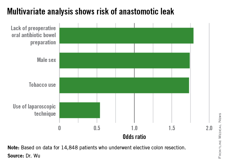
Inpatient mortality down for high-volume conditions
Inpatient mortality for pneumonia, acute MI, heart failure, and stroke each fell significantly from 2002 to 2012, the Agency for Healthcare Research and Quality reported.
Over that period, mortality among adults hospitalized with pneumonia went from 65 per 1,000 admissions to 35.8 per 1,000 for a drop of 45% – largest of the four high-volume conditions. Corresponding declines for the others were 41% for acute MI, 29% for heart failure, and 27% for stroke, the AHRQ noted.

Since “death following discharge from a hospital is not reflected in these data,” the report said, measures of inpatient mortality “can reflect both improvements in health care and shifts in where end-of-life care takes place over time.”
The estimates in the report are based on data from the Nationwide Inpatient Sample (2002-2011) and State Inpatient Databases (2012).
Inpatient mortality for pneumonia, acute MI, heart failure, and stroke each fell significantly from 2002 to 2012, the Agency for Healthcare Research and Quality reported.
Over that period, mortality among adults hospitalized with pneumonia went from 65 per 1,000 admissions to 35.8 per 1,000 for a drop of 45% – largest of the four high-volume conditions. Corresponding declines for the others were 41% for acute MI, 29% for heart failure, and 27% for stroke, the AHRQ noted.
Since “death following discharge from a hospital is not reflected in these data,” the report said, measures of inpatient mortality “can reflect both improvements in health care and shifts in where end-of-life care takes place over time.”
The estimates in the report are based on data from the Nationwide Inpatient Sample (2002-2011) and State Inpatient Databases (2012).