User login
Chip may allow for early cancer detection
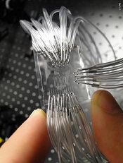
Institute of Photonic Sciences
Scientists say they’ve developed a lab-on-a-chip device capable of detecting protein markers for cancer.
The device can detect very low concentrations of protein markers in the blood, enabling cancer diagnosis in its earliest stages, the team says.
Romain Quidant, PhD, of The Institute of Photonic Sciences in Barcelona, Spain, and his colleagues described the device in Nano Letters.
The lab on a chip hosts 32 sensing sites distributed across a network of 8 fluidic microchannels that enables it to conduct multiple analyses.
Gold nanoparticles lie on the surface of the chip and are chemically programed with an antibody receptor in such a way that they are capable of specifically attracting the protein markers circulating in blood.
When a drop of blood is injected into the chip, it circulates through the microchannels, and, if cancer markers are present in the blood, they will stick to the nanoparticles located on the microchannels as they pass by, setting off changes in what is known as the plasmonic resonance.
The device monitors these changes, the magnitude of which is directly related to the concentration/number of markers in the patient’s blood. In this way, it provides a direct assessment of the patient’s risk of developing cancer.
“The most fascinating finding is that we are capable of detecting extremely low concentrations of this protein in a matter of minutes, making this device an ultra-high-sensitivity, state-of-the-art, powerful instrument that will benefit early detection and treatment monitoring of cancer,” Dr Quidant said. 
Institute of Photonic Sciences
Scientists say they’ve developed a lab-on-a-chip device capable of detecting protein markers for cancer.
The device can detect very low concentrations of protein markers in the blood, enabling cancer diagnosis in its earliest stages, the team says.
Romain Quidant, PhD, of The Institute of Photonic Sciences in Barcelona, Spain, and his colleagues described the device in Nano Letters.
The lab on a chip hosts 32 sensing sites distributed across a network of 8 fluidic microchannels that enables it to conduct multiple analyses.
Gold nanoparticles lie on the surface of the chip and are chemically programed with an antibody receptor in such a way that they are capable of specifically attracting the protein markers circulating in blood.
When a drop of blood is injected into the chip, it circulates through the microchannels, and, if cancer markers are present in the blood, they will stick to the nanoparticles located on the microchannels as they pass by, setting off changes in what is known as the plasmonic resonance.
The device monitors these changes, the magnitude of which is directly related to the concentration/number of markers in the patient’s blood. In this way, it provides a direct assessment of the patient’s risk of developing cancer.
“The most fascinating finding is that we are capable of detecting extremely low concentrations of this protein in a matter of minutes, making this device an ultra-high-sensitivity, state-of-the-art, powerful instrument that will benefit early detection and treatment monitoring of cancer,” Dr Quidant said.
Institute of Photonic Sciences
Scientists say they’ve developed a lab-on-a-chip device capable of detecting protein markers for cancer.
The device can detect very low concentrations of protein markers in the blood, enabling cancer diagnosis in its earliest stages, the team says.
Romain Quidant, PhD, of The Institute of Photonic Sciences in Barcelona, Spain, and his colleagues described the device in Nano Letters.
The lab on a chip hosts 32 sensing sites distributed across a network of 8 fluidic microchannels that enables it to conduct multiple analyses.
Gold nanoparticles lie on the surface of the chip and are chemically programed with an antibody receptor in such a way that they are capable of specifically attracting the protein markers circulating in blood.
When a drop of blood is injected into the chip, it circulates through the microchannels, and, if cancer markers are present in the blood, they will stick to the nanoparticles located on the microchannels as they pass by, setting off changes in what is known as the plasmonic resonance.
The device monitors these changes, the magnitude of which is directly related to the concentration/number of markers in the patient’s blood. In this way, it provides a direct assessment of the patient’s risk of developing cancer.
“The most fascinating finding is that we are capable of detecting extremely low concentrations of this protein in a matter of minutes, making this device an ultra-high-sensitivity, state-of-the-art, powerful instrument that will benefit early detection and treatment monitoring of cancer,” Dr Quidant said.
CHF screening guidelines need another look, group says

patient and her father
Credit: Rhoda Baer
New research suggests a need to revisit cardiac screening guidelines for survivors of childhood cancers.
The study indicates that less frequent screening for early signs of impending congestive heart failure (CHF) may yield a similar clinical benefit as current screening recommendations.
Furthermore, some survivors might be better served by a different method of screening than the one currently used. And early treatment of patients at high risk of CHF may be beneficial.
The researchers reported these findings in the Annals of Internal Medicine.
Current CHF screening guidelines recommend that childhood cancer survivors treated with chemotherapeutic agents known to affect long-term heart health be screened as often as every year, with a schedule dependent on their level of CHF risk.
The Children’s Oncology Group (COG) recommends that survivors undergo screening by echocardiography for asymptomatic left ventricular dysfunction (ALVD). If left untreated, this clinically silent condition can progress to CHF, so clinicians typically prescribe beta blockers and ACE inhibitors to patients with signs of ALVD.
The COG recommends that patients at high risk of developing CHF be screened every year or 2 and those at low risk be screened every 2 or 5 years
“It is important to monitor survivors so we can reduce the late effects of treatment whenever possible, but we may be asking them to be tested too often, which burdens both individuals and the healthcare system,” said study author Lisa Diller, MD, of the Dana-Farber/Boston Children’s Cancer and Blood Disorders Center in Massachusetts. “We think it is worthwhile to review the current CHF screening guidelines.”
To estimate the clinical benefits and cost-effectiveness of the current heart screening guidelines, Dr Diller and her colleagues constructed a computer model of a virtual cohort of 15-year-olds who had survived cancer at least 5 years.
Using data from the Childhood Cancer Survivors Study and the Framingham Heart Study, the researchers modeled the cohort’s CHF risk and clinical progression over the course of survivors’ lifetimes. Results suggested that routine screening may prevent as many as 1 in 12 cases of CHF.
The team then used Medicare data to estimate the costs and value (expressed in cost per quality-adjusted life-year [QALY]) of different screening schedules—every 1, 2, 5, or 10 years—and methods—echocardiography vs cardiac magnetic resonance imaging (cMRI)—for the different CHF risk groups.
At a cost-effectiveness threshold of $100,000/QALY, the model’s results indicated that echocardiographic screening might not be the best value for resources invested to reduce lifetime CHF risk among survivors at low risk of developing the disease.
On the other hand, the data suggested that biennial echocardiography screening may be a high-value strategy for high-risk survivors.
The simulation’s data also suggested that cMRI may be preferable to echocardiography as a screening method, with cMRI’s greater cost per test balanced by its greater sensitivity. According to the model, cMRI-based screening of low-risk survivors every 10 years and high-risk survivors every 5 years was more cost-effective than any echocardiography-based schedule.
Lastly, the data suggested it may be most beneficial to treat high-risk survivors before signs of ALVD even appear. For instance, proactively treating all high-risk patients in the virtual cohort with ACE inhibitors and beta blockers reduced their lifetime CHF risk more than if they received an echocardiograph every 2 years.
The researchers relied on simulation modeling using the best available clinical and epidemiologic data because of the logistical obstacles to conducting a prospective, randomized, clinical trial.
They said enrolling the number of survivors needed for such a study would be challenging, given how rare childhood cancers are. Yet guidance on the health benefits associated with current recommendations is needed.
“Our findings suggest that there is a long-term benefit in screening survivors at elevated risk for CHF,” said study author Jennifer Yeh, PhD, of the Harvard School of Public Health in Boston.
“Yet less frequent screening than currently recommended may be reasonable when other factors are considered. We hope these results can help inform the ongoing discussion about screening childhood cancer survivors.”
patient and her father
Credit: Rhoda Baer
New research suggests a need to revisit cardiac screening guidelines for survivors of childhood cancers.
The study indicates that less frequent screening for early signs of impending congestive heart failure (CHF) may yield a similar clinical benefit as current screening recommendations.
Furthermore, some survivors might be better served by a different method of screening than the one currently used. And early treatment of patients at high risk of CHF may be beneficial.
The researchers reported these findings in the Annals of Internal Medicine.
Current CHF screening guidelines recommend that childhood cancer survivors treated with chemotherapeutic agents known to affect long-term heart health be screened as often as every year, with a schedule dependent on their level of CHF risk.
The Children’s Oncology Group (COG) recommends that survivors undergo screening by echocardiography for asymptomatic left ventricular dysfunction (ALVD). If left untreated, this clinically silent condition can progress to CHF, so clinicians typically prescribe beta blockers and ACE inhibitors to patients with signs of ALVD.
The COG recommends that patients at high risk of developing CHF be screened every year or 2 and those at low risk be screened every 2 or 5 years
“It is important to monitor survivors so we can reduce the late effects of treatment whenever possible, but we may be asking them to be tested too often, which burdens both individuals and the healthcare system,” said study author Lisa Diller, MD, of the Dana-Farber/Boston Children’s Cancer and Blood Disorders Center in Massachusetts. “We think it is worthwhile to review the current CHF screening guidelines.”
To estimate the clinical benefits and cost-effectiveness of the current heart screening guidelines, Dr Diller and her colleagues constructed a computer model of a virtual cohort of 15-year-olds who had survived cancer at least 5 years.
Using data from the Childhood Cancer Survivors Study and the Framingham Heart Study, the researchers modeled the cohort’s CHF risk and clinical progression over the course of survivors’ lifetimes. Results suggested that routine screening may prevent as many as 1 in 12 cases of CHF.
The team then used Medicare data to estimate the costs and value (expressed in cost per quality-adjusted life-year [QALY]) of different screening schedules—every 1, 2, 5, or 10 years—and methods—echocardiography vs cardiac magnetic resonance imaging (cMRI)—for the different CHF risk groups.
At a cost-effectiveness threshold of $100,000/QALY, the model’s results indicated that echocardiographic screening might not be the best value for resources invested to reduce lifetime CHF risk among survivors at low risk of developing the disease.
On the other hand, the data suggested that biennial echocardiography screening may be a high-value strategy for high-risk survivors.
The simulation’s data also suggested that cMRI may be preferable to echocardiography as a screening method, with cMRI’s greater cost per test balanced by its greater sensitivity. According to the model, cMRI-based screening of low-risk survivors every 10 years and high-risk survivors every 5 years was more cost-effective than any echocardiography-based schedule.
Lastly, the data suggested it may be most beneficial to treat high-risk survivors before signs of ALVD even appear. For instance, proactively treating all high-risk patients in the virtual cohort with ACE inhibitors and beta blockers reduced their lifetime CHF risk more than if they received an echocardiograph every 2 years.
The researchers relied on simulation modeling using the best available clinical and epidemiologic data because of the logistical obstacles to conducting a prospective, randomized, clinical trial.
They said enrolling the number of survivors needed for such a study would be challenging, given how rare childhood cancers are. Yet guidance on the health benefits associated with current recommendations is needed.
“Our findings suggest that there is a long-term benefit in screening survivors at elevated risk for CHF,” said study author Jennifer Yeh, PhD, of the Harvard School of Public Health in Boston.
“Yet less frequent screening than currently recommended may be reasonable when other factors are considered. We hope these results can help inform the ongoing discussion about screening childhood cancer survivors.”
patient and her father
Credit: Rhoda Baer
New research suggests a need to revisit cardiac screening guidelines for survivors of childhood cancers.
The study indicates that less frequent screening for early signs of impending congestive heart failure (CHF) may yield a similar clinical benefit as current screening recommendations.
Furthermore, some survivors might be better served by a different method of screening than the one currently used. And early treatment of patients at high risk of CHF may be beneficial.
The researchers reported these findings in the Annals of Internal Medicine.
Current CHF screening guidelines recommend that childhood cancer survivors treated with chemotherapeutic agents known to affect long-term heart health be screened as often as every year, with a schedule dependent on their level of CHF risk.
The Children’s Oncology Group (COG) recommends that survivors undergo screening by echocardiography for asymptomatic left ventricular dysfunction (ALVD). If left untreated, this clinically silent condition can progress to CHF, so clinicians typically prescribe beta blockers and ACE inhibitors to patients with signs of ALVD.
The COG recommends that patients at high risk of developing CHF be screened every year or 2 and those at low risk be screened every 2 or 5 years
“It is important to monitor survivors so we can reduce the late effects of treatment whenever possible, but we may be asking them to be tested too often, which burdens both individuals and the healthcare system,” said study author Lisa Diller, MD, of the Dana-Farber/Boston Children’s Cancer and Blood Disorders Center in Massachusetts. “We think it is worthwhile to review the current CHF screening guidelines.”
To estimate the clinical benefits and cost-effectiveness of the current heart screening guidelines, Dr Diller and her colleagues constructed a computer model of a virtual cohort of 15-year-olds who had survived cancer at least 5 years.
Using data from the Childhood Cancer Survivors Study and the Framingham Heart Study, the researchers modeled the cohort’s CHF risk and clinical progression over the course of survivors’ lifetimes. Results suggested that routine screening may prevent as many as 1 in 12 cases of CHF.
The team then used Medicare data to estimate the costs and value (expressed in cost per quality-adjusted life-year [QALY]) of different screening schedules—every 1, 2, 5, or 10 years—and methods—echocardiography vs cardiac magnetic resonance imaging (cMRI)—for the different CHF risk groups.
At a cost-effectiveness threshold of $100,000/QALY, the model’s results indicated that echocardiographic screening might not be the best value for resources invested to reduce lifetime CHF risk among survivors at low risk of developing the disease.
On the other hand, the data suggested that biennial echocardiography screening may be a high-value strategy for high-risk survivors.
The simulation’s data also suggested that cMRI may be preferable to echocardiography as a screening method, with cMRI’s greater cost per test balanced by its greater sensitivity. According to the model, cMRI-based screening of low-risk survivors every 10 years and high-risk survivors every 5 years was more cost-effective than any echocardiography-based schedule.
Lastly, the data suggested it may be most beneficial to treat high-risk survivors before signs of ALVD even appear. For instance, proactively treating all high-risk patients in the virtual cohort with ACE inhibitors and beta blockers reduced their lifetime CHF risk more than if they received an echocardiograph every 2 years.
The researchers relied on simulation modeling using the best available clinical and epidemiologic data because of the logistical obstacles to conducting a prospective, randomized, clinical trial.
They said enrolling the number of survivors needed for such a study would be challenging, given how rare childhood cancers are. Yet guidance on the health benefits associated with current recommendations is needed.
“Our findings suggest that there is a long-term benefit in screening survivors at elevated risk for CHF,” said study author Jennifer Yeh, PhD, of the Harvard School of Public Health in Boston.
“Yet less frequent screening than currently recommended may be reasonable when other factors are considered. We hope these results can help inform the ongoing discussion about screening childhood cancer survivors.”
Test detects PE with greater specificity than D-dimer

Credit: Andre E.X. Brown
SAN DIEGO—New research suggests a urine test can be used to detect a pulmonary embolism (PE), providing similar sensitivity and greater specificity than the D-dimer test.
The urine test measures levels of fibrinopeptide B (FPB), a peptide released when a thrombus forms.
“The urine FPB test offers advantages over other screening methods because it doesn’t require blood to be drawn, and it can provide more accurate results than the D-dimer test,” said Timothy Fernandes, MD, of the University of California, San Diego.
Dr Fernandes and his colleagues presented results observed with the FPB test at the American Thoracic Society’s 2014 International Conference (abstract 53841).
The researchers tested samples taken from 344 patients who participated in the Pulmonary Embolism Diagnosis Study (PEDS), a multicenter study of patients considered likely to have an acute PE. Sixty-one of these patients (18%) had a confirmed PE, and 283 (83%) did not.
For all urine samples, the researchers measured the FPB concentration and evaluated the sensitivity and specificity of the test at various cut-off points in relation to its ability to predict the presence of PE.
The team found that, at concentrations of 2.5 ng/mL, urine FPB demonstrated sensitivity comparable to previously published values for plasma latex and whole-blood D-dimer levels, but with greater specificity.
Specifically, at a threshold of 2.5 ng/mL, urine FPB could predict PE with sensitivity of 75.4%, specificity of 28.9%, and a negative likelihood ratio of 0.18. Sensitivity was lower at thresholds of 5 ng/mL and 7.5 ng/mL, at 55.7% and 42.6%, respectively.
The researchers noted that the FPB test has the potential for greater specificity than the D-dimer test because FPB can reflect ongoing clot activity, while D-dimer can only be measured once a thrombus has already become degraded.
“The results of our study indicate that urine FPB tests may be a useful complement to current biomarkers such as D-dimer to measure for the
presence and activity of venous thromboembolism,” Dr Fernandes said.
He and his colleagues are now planning to develop a urine dipstick test for FBP. The patent for the urine FPB test is held by the University of California Board of Regents.
The researchers are also planning studies to assess urine FPB in other settings where D-dimer is used, including the use of urine FPB after anticoagulation to determine the risk of recurrent venous thromboembolism.
Credit: Andre E.X. Brown
SAN DIEGO—New research suggests a urine test can be used to detect a pulmonary embolism (PE), providing similar sensitivity and greater specificity than the D-dimer test.
The urine test measures levels of fibrinopeptide B (FPB), a peptide released when a thrombus forms.
“The urine FPB test offers advantages over other screening methods because it doesn’t require blood to be drawn, and it can provide more accurate results than the D-dimer test,” said Timothy Fernandes, MD, of the University of California, San Diego.
Dr Fernandes and his colleagues presented results observed with the FPB test at the American Thoracic Society’s 2014 International Conference (abstract 53841).
The researchers tested samples taken from 344 patients who participated in the Pulmonary Embolism Diagnosis Study (PEDS), a multicenter study of patients considered likely to have an acute PE. Sixty-one of these patients (18%) had a confirmed PE, and 283 (83%) did not.
For all urine samples, the researchers measured the FPB concentration and evaluated the sensitivity and specificity of the test at various cut-off points in relation to its ability to predict the presence of PE.
The team found that, at concentrations of 2.5 ng/mL, urine FPB demonstrated sensitivity comparable to previously published values for plasma latex and whole-blood D-dimer levels, but with greater specificity.
Specifically, at a threshold of 2.5 ng/mL, urine FPB could predict PE with sensitivity of 75.4%, specificity of 28.9%, and a negative likelihood ratio of 0.18. Sensitivity was lower at thresholds of 5 ng/mL and 7.5 ng/mL, at 55.7% and 42.6%, respectively.
The researchers noted that the FPB test has the potential for greater specificity than the D-dimer test because FPB can reflect ongoing clot activity, while D-dimer can only be measured once a thrombus has already become degraded.
“The results of our study indicate that urine FPB tests may be a useful complement to current biomarkers such as D-dimer to measure for the
presence and activity of venous thromboembolism,” Dr Fernandes said.
He and his colleagues are now planning to develop a urine dipstick test for FBP. The patent for the urine FPB test is held by the University of California Board of Regents.
The researchers are also planning studies to assess urine FPB in other settings where D-dimer is used, including the use of urine FPB after anticoagulation to determine the risk of recurrent venous thromboembolism.
Credit: Andre E.X. Brown
SAN DIEGO—New research suggests a urine test can be used to detect a pulmonary embolism (PE), providing similar sensitivity and greater specificity than the D-dimer test.
The urine test measures levels of fibrinopeptide B (FPB), a peptide released when a thrombus forms.
“The urine FPB test offers advantages over other screening methods because it doesn’t require blood to be drawn, and it can provide more accurate results than the D-dimer test,” said Timothy Fernandes, MD, of the University of California, San Diego.
Dr Fernandes and his colleagues presented results observed with the FPB test at the American Thoracic Society’s 2014 International Conference (abstract 53841).
The researchers tested samples taken from 344 patients who participated in the Pulmonary Embolism Diagnosis Study (PEDS), a multicenter study of patients considered likely to have an acute PE. Sixty-one of these patients (18%) had a confirmed PE, and 283 (83%) did not.
For all urine samples, the researchers measured the FPB concentration and evaluated the sensitivity and specificity of the test at various cut-off points in relation to its ability to predict the presence of PE.
The team found that, at concentrations of 2.5 ng/mL, urine FPB demonstrated sensitivity comparable to previously published values for plasma latex and whole-blood D-dimer levels, but with greater specificity.
Specifically, at a threshold of 2.5 ng/mL, urine FPB could predict PE with sensitivity of 75.4%, specificity of 28.9%, and a negative likelihood ratio of 0.18. Sensitivity was lower at thresholds of 5 ng/mL and 7.5 ng/mL, at 55.7% and 42.6%, respectively.
The researchers noted that the FPB test has the potential for greater specificity than the D-dimer test because FPB can reflect ongoing clot activity, while D-dimer can only be measured once a thrombus has already become degraded.
“The results of our study indicate that urine FPB tests may be a useful complement to current biomarkers such as D-dimer to measure for the
presence and activity of venous thromboembolism,” Dr Fernandes said.
He and his colleagues are now planning to develop a urine dipstick test for FBP. The patent for the urine FPB test is held by the University of California Board of Regents.
The researchers are also planning studies to assess urine FPB in other settings where D-dimer is used, including the use of urine FPB after anticoagulation to determine the risk of recurrent venous thromboembolism.
Team unearths surprising sepsis finding
Credit: NHLBI
SAN DIEGO—Results of a retrospective study suggest sepsis may contribute to as many as half of all hospital deaths in the US.
Researchers analyzed information from 6.5 million hospital discharge records and found that, of all hospital deaths, as many as 52% occurred in patients with sepsis.
“We were surprised to find that as many as 1 in 2 patients dying in US hospitals had sepsis,” said Vincent Liu, MD, of the Kaiser Permanente Northern California Division of Research.
He and his colleagues presented this finding at the American Thoracic Society’s 2014 International Conference (abstract 50626). The results have also been published in JAMA.
The researchers analyzed data from 6.5 million hospital discharge records derived from the Healthcare Cost and Utilization Project Nationwide Inpatient Sample (NIS) in 2010. The NIS is the largest publicly available, all-payer inpatient database in the US, containing data from 100% of hospital discharges from a stratified sample of 1051 community hospitals.
The team also evaluated records for 482,828 adult patients admitted to 21 Kaiser Permanente Northern California (KPNC) hospitals.
Using diagnosis and procedure codes, the researchers identified hospital admissions and deaths of patients with sepsis and estimated the percentage of total hospital charges associated with sepsis hospitalizations.
They used 2 approaches to identify patients with sepsis from International Statistical Classification of Diseases, Ninth Revision, Clinical Modification codes. The explicit approach identified patients with codes 038 (septicemia), 995.91 (sepsis), 995.92 (severe sepsis), or 785.52 (septic shock). The team also used an implicit approach, which included patients with evidence of both infection and acute organ failure.
In the NIS cohort, there were 280,663 explicit and 717,718 implicit sepsis hospitalizations. Sepsis hospitalizations accounted for 4.3% (explicit) to 10.9% (implicit) of all hospitalizations.
There were 143,312 deaths in the NIS cohort, and 34.7% (explicit) to 52.0% (implicit) occurred among patients with sepsis.
In the KPNC cohort, there were 55,008 explicit and 80,678 implicit sepsis hospitalizations. Sepsis hospitalizations accounted for 11.4% (explicit) to 16.7% (implicit) of all hospitalizations.
There were 14,206 inpatient deaths in this cohort, and 36.9% (explicit) to 55.9% (implicit) occurred among patients with sepsis.
“[W]e found that most patients already had sepsis at the time of hospital admission,” Dr Liu said. “There was also a large number of patients with less severe sepsis, a group for whom treatment guidelines are less well-defined. The results of our study suggest that improved care for sepsis patients of all severity levels and in all hospital settings could result in many future lives saved.”
Credit: NHLBI
SAN DIEGO—Results of a retrospective study suggest sepsis may contribute to as many as half of all hospital deaths in the US.
Researchers analyzed information from 6.5 million hospital discharge records and found that, of all hospital deaths, as many as 52% occurred in patients with sepsis.
“We were surprised to find that as many as 1 in 2 patients dying in US hospitals had sepsis,” said Vincent Liu, MD, of the Kaiser Permanente Northern California Division of Research.
He and his colleagues presented this finding at the American Thoracic Society’s 2014 International Conference (abstract 50626). The results have also been published in JAMA.
The researchers analyzed data from 6.5 million hospital discharge records derived from the Healthcare Cost and Utilization Project Nationwide Inpatient Sample (NIS) in 2010. The NIS is the largest publicly available, all-payer inpatient database in the US, containing data from 100% of hospital discharges from a stratified sample of 1051 community hospitals.
The team also evaluated records for 482,828 adult patients admitted to 21 Kaiser Permanente Northern California (KPNC) hospitals.
Using diagnosis and procedure codes, the researchers identified hospital admissions and deaths of patients with sepsis and estimated the percentage of total hospital charges associated with sepsis hospitalizations.
They used 2 approaches to identify patients with sepsis from International Statistical Classification of Diseases, Ninth Revision, Clinical Modification codes. The explicit approach identified patients with codes 038 (septicemia), 995.91 (sepsis), 995.92 (severe sepsis), or 785.52 (septic shock). The team also used an implicit approach, which included patients with evidence of both infection and acute organ failure.
In the NIS cohort, there were 280,663 explicit and 717,718 implicit sepsis hospitalizations. Sepsis hospitalizations accounted for 4.3% (explicit) to 10.9% (implicit) of all hospitalizations.
There were 143,312 deaths in the NIS cohort, and 34.7% (explicit) to 52.0% (implicit) occurred among patients with sepsis.
In the KPNC cohort, there were 55,008 explicit and 80,678 implicit sepsis hospitalizations. Sepsis hospitalizations accounted for 11.4% (explicit) to 16.7% (implicit) of all hospitalizations.
There were 14,206 inpatient deaths in this cohort, and 36.9% (explicit) to 55.9% (implicit) occurred among patients with sepsis.
“[W]e found that most patients already had sepsis at the time of hospital admission,” Dr Liu said. “There was also a large number of patients with less severe sepsis, a group for whom treatment guidelines are less well-defined. The results of our study suggest that improved care for sepsis patients of all severity levels and in all hospital settings could result in many future lives saved.”
Credit: NHLBI
SAN DIEGO—Results of a retrospective study suggest sepsis may contribute to as many as half of all hospital deaths in the US.
Researchers analyzed information from 6.5 million hospital discharge records and found that, of all hospital deaths, as many as 52% occurred in patients with sepsis.
“We were surprised to find that as many as 1 in 2 patients dying in US hospitals had sepsis,” said Vincent Liu, MD, of the Kaiser Permanente Northern California Division of Research.
He and his colleagues presented this finding at the American Thoracic Society’s 2014 International Conference (abstract 50626). The results have also been published in JAMA.
The researchers analyzed data from 6.5 million hospital discharge records derived from the Healthcare Cost and Utilization Project Nationwide Inpatient Sample (NIS) in 2010. The NIS is the largest publicly available, all-payer inpatient database in the US, containing data from 100% of hospital discharges from a stratified sample of 1051 community hospitals.
The team also evaluated records for 482,828 adult patients admitted to 21 Kaiser Permanente Northern California (KPNC) hospitals.
Using diagnosis and procedure codes, the researchers identified hospital admissions and deaths of patients with sepsis and estimated the percentage of total hospital charges associated with sepsis hospitalizations.
They used 2 approaches to identify patients with sepsis from International Statistical Classification of Diseases, Ninth Revision, Clinical Modification codes. The explicit approach identified patients with codes 038 (septicemia), 995.91 (sepsis), 995.92 (severe sepsis), or 785.52 (septic shock). The team also used an implicit approach, which included patients with evidence of both infection and acute organ failure.
In the NIS cohort, there were 280,663 explicit and 717,718 implicit sepsis hospitalizations. Sepsis hospitalizations accounted for 4.3% (explicit) to 10.9% (implicit) of all hospitalizations.
There were 143,312 deaths in the NIS cohort, and 34.7% (explicit) to 52.0% (implicit) occurred among patients with sepsis.
In the KPNC cohort, there were 55,008 explicit and 80,678 implicit sepsis hospitalizations. Sepsis hospitalizations accounted for 11.4% (explicit) to 16.7% (implicit) of all hospitalizations.
There were 14,206 inpatient deaths in this cohort, and 36.9% (explicit) to 55.9% (implicit) occurred among patients with sepsis.
“[W]e found that most patients already had sepsis at the time of hospital admission,” Dr Liu said. “There was also a large number of patients with less severe sepsis, a group for whom treatment guidelines are less well-defined. The results of our study suggest that improved care for sepsis patients of all severity levels and in all hospital settings could result in many future lives saved.”
Protein may be therapeutic target for AML
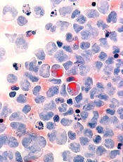
Credit: Lance Liotta
Researchers have found evidence to suggest that the phosphoinositide (PI) modulator PIP4K2A plays a key role in acute myeloid leukemia (AML).
PIs appear to control the transformation of hematopoietic stem cells into leukemic cells.
Some of these PIs switch on specific cell-signaling pathways, resulting in rapid growth and enhanced survival. Regulation of the PIs is carried out by a variety of proteins known as PI modulators.
“Little is known about the role of PI modulators in leukemia,” said Tim Somervaille, PhD, of The University of Manchester in the UK.
“We wanted to find out which ones were responsible for cell growth or survival in acute myeloid leukemia.”
He and his colleagues described this research in Oncogene.
The researchers performed a targeted knockdown screen of PI modulator genes in human AML cells, looking for genes required to sustain proliferation or prevent apoptosis.
They found that one PI modulator, PIP4K2A, was essential for the growth of leukemia. PIP4K2A knockdown resulted in leukemic cell death. This occurred in both murine MLL-AF9 AML cells and primary human AML cells.
Additional investigation showed that PIP4K2A knockdown resulted in the accumulation of the cyclin-dependent kinase inhibitors CDKN1A and CDKN1B, as well as G1 cell-cycle arrest and apoptosis. CDKN1A accumulation and apoptosis were partially dependent on activation of the mTOR pathway.
Fortunately, PIP4K2A knockdown did not adversely affect normal hematopoietic stem and progenitor cells from mice or human subjects. Neither clonogenic nor multilineage differentiation potential was affected.
“This makes [PIP4K2A] an ideal target for future drug development in leukemia,” Dr Somervaille concluded.
Credit: Lance Liotta
Researchers have found evidence to suggest that the phosphoinositide (PI) modulator PIP4K2A plays a key role in acute myeloid leukemia (AML).
PIs appear to control the transformation of hematopoietic stem cells into leukemic cells.
Some of these PIs switch on specific cell-signaling pathways, resulting in rapid growth and enhanced survival. Regulation of the PIs is carried out by a variety of proteins known as PI modulators.
“Little is known about the role of PI modulators in leukemia,” said Tim Somervaille, PhD, of The University of Manchester in the UK.
“We wanted to find out which ones were responsible for cell growth or survival in acute myeloid leukemia.”
He and his colleagues described this research in Oncogene.
The researchers performed a targeted knockdown screen of PI modulator genes in human AML cells, looking for genes required to sustain proliferation or prevent apoptosis.
They found that one PI modulator, PIP4K2A, was essential for the growth of leukemia. PIP4K2A knockdown resulted in leukemic cell death. This occurred in both murine MLL-AF9 AML cells and primary human AML cells.
Additional investigation showed that PIP4K2A knockdown resulted in the accumulation of the cyclin-dependent kinase inhibitors CDKN1A and CDKN1B, as well as G1 cell-cycle arrest and apoptosis. CDKN1A accumulation and apoptosis were partially dependent on activation of the mTOR pathway.
Fortunately, PIP4K2A knockdown did not adversely affect normal hematopoietic stem and progenitor cells from mice or human subjects. Neither clonogenic nor multilineage differentiation potential was affected.
“This makes [PIP4K2A] an ideal target for future drug development in leukemia,” Dr Somervaille concluded.
Credit: Lance Liotta
Researchers have found evidence to suggest that the phosphoinositide (PI) modulator PIP4K2A plays a key role in acute myeloid leukemia (AML).
PIs appear to control the transformation of hematopoietic stem cells into leukemic cells.
Some of these PIs switch on specific cell-signaling pathways, resulting in rapid growth and enhanced survival. Regulation of the PIs is carried out by a variety of proteins known as PI modulators.
“Little is known about the role of PI modulators in leukemia,” said Tim Somervaille, PhD, of The University of Manchester in the UK.
“We wanted to find out which ones were responsible for cell growth or survival in acute myeloid leukemia.”
He and his colleagues described this research in Oncogene.
The researchers performed a targeted knockdown screen of PI modulator genes in human AML cells, looking for genes required to sustain proliferation or prevent apoptosis.
They found that one PI modulator, PIP4K2A, was essential for the growth of leukemia. PIP4K2A knockdown resulted in leukemic cell death. This occurred in both murine MLL-AF9 AML cells and primary human AML cells.
Additional investigation showed that PIP4K2A knockdown resulted in the accumulation of the cyclin-dependent kinase inhibitors CDKN1A and CDKN1B, as well as G1 cell-cycle arrest and apoptosis. CDKN1A accumulation and apoptosis were partially dependent on activation of the mTOR pathway.
Fortunately, PIP4K2A knockdown did not adversely affect normal hematopoietic stem and progenitor cells from mice or human subjects. Neither clonogenic nor multilineage differentiation potential was affected.
“This makes [PIP4K2A] an ideal target for future drug development in leukemia,” Dr Somervaille concluded.
How cancer-fighting protein is held in check

Credit: A.T. Tikhonenko
A new study reveals how the protein p53 attaches to its regulatory molecule, BCL-xL.
Understanding how these molecular puzzle pieces fit together could help scientists design drugs that would unleash p53 to battle a range of cancers, according to study author Richard Kriwacki, PhD, of St Jude Children’s Research Hospital in Memphis, Tennessee.
He and his colleagues described this research in Nature Structural & Molecular Biology.
In guarding the cell against genetic damage, the p53 machinery functions both in the nucleus of the cell and in the cytosol. When this machinery detects irreparable damage to the cell, p53 is unleashed to trigger apoptosis.
In about half of all cancers, this machinery is rendered inoperable by mutation, enabling cancer cells to proliferate despite their genetic malfunctions. The protein BCL-xL is a central inhibitor of the p53 machinery, binding both p53 and BH3 proteins, which also drive apoptosis.
“The molecular details of how BCL-xl performs this dual inhibitory function were not understood,” Dr Kriwacki said. “Having those details has enabled us to determine exactly how BCL-xL can restrain or inhibit apoptosis through interactions with BH3-domain-containing proteins, as well as p53.”
He and his colleagues used a structural analysis technique called NMR spectroscopy to map the 3-D structure of p53 binding to BCL-xL.
Their experiments revealed how the DNA-binding domain of the p53 protein serves double duty in the machinery. It enables p53 to attach to DNA in the cell’s nucleus, helping the cell repair genetic damage. The same domain also acts as an attachment point for BCL-xL in the cytosol.
“The structural details that we report are novel,” Dr Kriwacki said. “And they provide the key insights for really dissecting the dual roles of BCL-xL in inhibiting apoptosis . . ., inhibiting the BH3-containing proteins on the one side and p53 on the other. Also, through these studies, we solidified the mechanistic understanding for how p53 functions in the cytosol, which complements its pro-apoptotic role in the nucleus.”
Dr Kriwacki added that these findings could help scientists design better anticancer agents. In many cancers, p53 is prevented from triggering apoptosis by its attachment to BCL-xL.
Drugs are currently being tested that bind to BCL-xL to free BH3 proteins to trigger apoptosis. However, Dr Kriwacki said new drugs could be developed that also block BCL-xL from binding p53.
“Our hypothesis is that many cancers have normal p53, but it is being tied up by BCL-xL,” he said. “If it could be released, it could play its role in triggering apoptosis. A drug that could block both of BCL-xL’s anti-apoptotic functions could potentially more profoundly induce apoptosis in cancer cells.”
Credit: A.T. Tikhonenko
A new study reveals how the protein p53 attaches to its regulatory molecule, BCL-xL.
Understanding how these molecular puzzle pieces fit together could help scientists design drugs that would unleash p53 to battle a range of cancers, according to study author Richard Kriwacki, PhD, of St Jude Children’s Research Hospital in Memphis, Tennessee.
He and his colleagues described this research in Nature Structural & Molecular Biology.
In guarding the cell against genetic damage, the p53 machinery functions both in the nucleus of the cell and in the cytosol. When this machinery detects irreparable damage to the cell, p53 is unleashed to trigger apoptosis.
In about half of all cancers, this machinery is rendered inoperable by mutation, enabling cancer cells to proliferate despite their genetic malfunctions. The protein BCL-xL is a central inhibitor of the p53 machinery, binding both p53 and BH3 proteins, which also drive apoptosis.
“The molecular details of how BCL-xl performs this dual inhibitory function were not understood,” Dr Kriwacki said. “Having those details has enabled us to determine exactly how BCL-xL can restrain or inhibit apoptosis through interactions with BH3-domain-containing proteins, as well as p53.”
He and his colleagues used a structural analysis technique called NMR spectroscopy to map the 3-D structure of p53 binding to BCL-xL.
Their experiments revealed how the DNA-binding domain of the p53 protein serves double duty in the machinery. It enables p53 to attach to DNA in the cell’s nucleus, helping the cell repair genetic damage. The same domain also acts as an attachment point for BCL-xL in the cytosol.
“The structural details that we report are novel,” Dr Kriwacki said. “And they provide the key insights for really dissecting the dual roles of BCL-xL in inhibiting apoptosis . . ., inhibiting the BH3-containing proteins on the one side and p53 on the other. Also, through these studies, we solidified the mechanistic understanding for how p53 functions in the cytosol, which complements its pro-apoptotic role in the nucleus.”
Dr Kriwacki added that these findings could help scientists design better anticancer agents. In many cancers, p53 is prevented from triggering apoptosis by its attachment to BCL-xL.
Drugs are currently being tested that bind to BCL-xL to free BH3 proteins to trigger apoptosis. However, Dr Kriwacki said new drugs could be developed that also block BCL-xL from binding p53.
“Our hypothesis is that many cancers have normal p53, but it is being tied up by BCL-xL,” he said. “If it could be released, it could play its role in triggering apoptosis. A drug that could block both of BCL-xL’s anti-apoptotic functions could potentially more profoundly induce apoptosis in cancer cells.”
Credit: A.T. Tikhonenko
A new study reveals how the protein p53 attaches to its regulatory molecule, BCL-xL.
Understanding how these molecular puzzle pieces fit together could help scientists design drugs that would unleash p53 to battle a range of cancers, according to study author Richard Kriwacki, PhD, of St Jude Children’s Research Hospital in Memphis, Tennessee.
He and his colleagues described this research in Nature Structural & Molecular Biology.
In guarding the cell against genetic damage, the p53 machinery functions both in the nucleus of the cell and in the cytosol. When this machinery detects irreparable damage to the cell, p53 is unleashed to trigger apoptosis.
In about half of all cancers, this machinery is rendered inoperable by mutation, enabling cancer cells to proliferate despite their genetic malfunctions. The protein BCL-xL is a central inhibitor of the p53 machinery, binding both p53 and BH3 proteins, which also drive apoptosis.
“The molecular details of how BCL-xl performs this dual inhibitory function were not understood,” Dr Kriwacki said. “Having those details has enabled us to determine exactly how BCL-xL can restrain or inhibit apoptosis through interactions with BH3-domain-containing proteins, as well as p53.”
He and his colleagues used a structural analysis technique called NMR spectroscopy to map the 3-D structure of p53 binding to BCL-xL.
Their experiments revealed how the DNA-binding domain of the p53 protein serves double duty in the machinery. It enables p53 to attach to DNA in the cell’s nucleus, helping the cell repair genetic damage. The same domain also acts as an attachment point for BCL-xL in the cytosol.
“The structural details that we report are novel,” Dr Kriwacki said. “And they provide the key insights for really dissecting the dual roles of BCL-xL in inhibiting apoptosis . . ., inhibiting the BH3-containing proteins on the one side and p53 on the other. Also, through these studies, we solidified the mechanistic understanding for how p53 functions in the cytosol, which complements its pro-apoptotic role in the nucleus.”
Dr Kriwacki added that these findings could help scientists design better anticancer agents. In many cancers, p53 is prevented from triggering apoptosis by its attachment to BCL-xL.
Drugs are currently being tested that bind to BCL-xL to free BH3 proteins to trigger apoptosis. However, Dr Kriwacki said new drugs could be developed that also block BCL-xL from binding p53.
“Our hypothesis is that many cancers have normal p53, but it is being tied up by BCL-xL,” he said. “If it could be released, it could play its role in triggering apoptosis. A drug that could block both of BCL-xL’s anti-apoptotic functions could potentially more profoundly induce apoptosis in cancer cells.”
Group finds cancer stem cells in MDS patients

Credit: NIH
A new study provides “conclusive evidence” to support the existence of cancer stem cells in patients with myelodysplastic syndromes (MDS), according to researchers.
The group analyzed malignant cells in the bone marrow of MDS patients and identified a distinct subset of cells that showed all the hallmarks of cancer stem cells.
Only these MDS stem cells—none of the other malignant cells—were able to propagate the disease.
The researchers reported these discoveries in Cancer Cell.
Petter Woll, PhD, of the University of Oxford in the UK, and his colleagues conducted this research, analyzing bone marrow cells from 15 patients with low- or intermediate-risk MDS.
The team set out to establish in which cells cancer-driving mutations originated. This led them to a rare subset of MDS cells—Lin-CD34+CD38-CD90+CD45RA- cells—that appeared to have all the properties of cancer stem cells.
These cells sat at the top of a hierarchy of MDS cells, could sustain themselves and replenish the other MDS cells, and were the origin of all stable DNA changes and mutations that drove the progression of MDS.
“This is conclusive evidence for the existence of cancer stem cells in myelodysplastic syndromes,” Dr Woll said. “We have identified a subset of cancer cells [and] shown that these rare cells are invariably the cells in which the cancer originates and also are the only cancer-propagating cells in the patients.”
Study author Sten Eirik W. Jacobsen, MD, PhD, also of the University of Oxford, noted that cancer stem cells have already been reported in a number of malignancies.
But previous findings have remained controversial, since the lab tests used to establish the identity of cancer stem cells have been shown to be unreliable.
“In our studies, we avoided the problem of unreliable lab tests by tracking the origin and development of cancer-driving mutations in MDS patients,” Dr Jacobsen said.
Dr Woll added that this research provides a target for the development of more efficient therapies for MDS.
“We need to understand more about what makes these cancer stem cells unique, what makes them different to all the other cancer cells,” he said. “If we can find biological pathways that are specifically dysregulated in cancer stem cells, we might be able to target them with new drugs.”
Credit: NIH
A new study provides “conclusive evidence” to support the existence of cancer stem cells in patients with myelodysplastic syndromes (MDS), according to researchers.
The group analyzed malignant cells in the bone marrow of MDS patients and identified a distinct subset of cells that showed all the hallmarks of cancer stem cells.
Only these MDS stem cells—none of the other malignant cells—were able to propagate the disease.
The researchers reported these discoveries in Cancer Cell.
Petter Woll, PhD, of the University of Oxford in the UK, and his colleagues conducted this research, analyzing bone marrow cells from 15 patients with low- or intermediate-risk MDS.
The team set out to establish in which cells cancer-driving mutations originated. This led them to a rare subset of MDS cells—Lin-CD34+CD38-CD90+CD45RA- cells—that appeared to have all the properties of cancer stem cells.
These cells sat at the top of a hierarchy of MDS cells, could sustain themselves and replenish the other MDS cells, and were the origin of all stable DNA changes and mutations that drove the progression of MDS.
“This is conclusive evidence for the existence of cancer stem cells in myelodysplastic syndromes,” Dr Woll said. “We have identified a subset of cancer cells [and] shown that these rare cells are invariably the cells in which the cancer originates and also are the only cancer-propagating cells in the patients.”
Study author Sten Eirik W. Jacobsen, MD, PhD, also of the University of Oxford, noted that cancer stem cells have already been reported in a number of malignancies.
But previous findings have remained controversial, since the lab tests used to establish the identity of cancer stem cells have been shown to be unreliable.
“In our studies, we avoided the problem of unreliable lab tests by tracking the origin and development of cancer-driving mutations in MDS patients,” Dr Jacobsen said.
Dr Woll added that this research provides a target for the development of more efficient therapies for MDS.
“We need to understand more about what makes these cancer stem cells unique, what makes them different to all the other cancer cells,” he said. “If we can find biological pathways that are specifically dysregulated in cancer stem cells, we might be able to target them with new drugs.”
Credit: NIH
A new study provides “conclusive evidence” to support the existence of cancer stem cells in patients with myelodysplastic syndromes (MDS), according to researchers.
The group analyzed malignant cells in the bone marrow of MDS patients and identified a distinct subset of cells that showed all the hallmarks of cancer stem cells.
Only these MDS stem cells—none of the other malignant cells—were able to propagate the disease.
The researchers reported these discoveries in Cancer Cell.
Petter Woll, PhD, of the University of Oxford in the UK, and his colleagues conducted this research, analyzing bone marrow cells from 15 patients with low- or intermediate-risk MDS.
The team set out to establish in which cells cancer-driving mutations originated. This led them to a rare subset of MDS cells—Lin-CD34+CD38-CD90+CD45RA- cells—that appeared to have all the properties of cancer stem cells.
These cells sat at the top of a hierarchy of MDS cells, could sustain themselves and replenish the other MDS cells, and were the origin of all stable DNA changes and mutations that drove the progression of MDS.
“This is conclusive evidence for the existence of cancer stem cells in myelodysplastic syndromes,” Dr Woll said. “We have identified a subset of cancer cells [and] shown that these rare cells are invariably the cells in which the cancer originates and also are the only cancer-propagating cells in the patients.”
Study author Sten Eirik W. Jacobsen, MD, PhD, also of the University of Oxford, noted that cancer stem cells have already been reported in a number of malignancies.
But previous findings have remained controversial, since the lab tests used to establish the identity of cancer stem cells have been shown to be unreliable.
“In our studies, we avoided the problem of unreliable lab tests by tracking the origin and development of cancer-driving mutations in MDS patients,” Dr Jacobsen said.
Dr Woll added that this research provides a target for the development of more efficient therapies for MDS.
“We need to understand more about what makes these cancer stem cells unique, what makes them different to all the other cancer cells,” he said. “If we can find biological pathways that are specifically dysregulated in cancer stem cells, we might be able to target them with new drugs.”
Protein inhibition confers radioprotective effects

Researchers believe they may have discovered a method for treating and preventing radiation-induced gastrointestinal toxicity.
The investigators found that inhibiting prolyl hydroxylase domain (PHD) proteins in mice could help protect them from radiation-induced toxicity and prolong their life spans.
“We were very surprised by the amount of protection the animals received,” said Amato Giaccia, PhD, of the Stanford University School of Medicine in California.
“The important thing to note is that we didn’t change the amount of damage the intestinal cells sustained as a result of the radiation. We simply changed the physiology of that tissue and how it responded to that damage.”
Dr Giaccia and his colleagues described this research in Science Translational Medicine.
The study began with an interest in hypoxia-inducible factor (HIF) proteins, which are known to help cells survive stressful conditions.
“Previous studies from our group and others have suggested that the HIF proteins are important in protecting cells from many types of stress,” Dr Giaccia said. “So we wondered whether stabilizing HIF proteins, and therefore increasing their levels within the cells, could also protect the intestine from the effects of radiation.”
The researchers inhibited the degradation of HIF proteins in 2 ways. In the first experiment, they engineered mice that were unable to express PHD isoforms, a group of 3 proteins that tag HIF proteins for destruction.
In another experiment, the investigators treated unmodified mice with a small molecule called dimethyloxyallyl glycine (DMOG), which also inhibits the activity of PHD proteins.
In both cases, the levels of HIF1 and HIF2 proteins increased significantly in the manipulated mice, as compared to controls.
In addition, 70% of the genetically modified mice lived for at least 30 days after receiving a normally lethal dose of abdominal radiation, and 27% survived at least 30 days after a normally lethal dose of whole-body radiation.
Sixty-seven percent of DMOG-treated mice survived for at least 60 days after receiving a normally lethal dose of abdominal radiation, and 40% lived for at least 30 days after a normally lethal dose of whole-body radiation.
The control mice in both experiments did not survive longer than 10 days after either type of radiation exposure.
Elucidating the mechanism
Further experiments showed that HIF2, rather than HIF1, is responsible for the radioprotection the researchers observed.
To determine the cause of the treated animals’ prolonged survival, the investigators looked directly at the epithelial cells lining the intestines.
Treated mice exhibited lower levels of cell death in response to abdominal radiation exposure and improved survival of crypts, which host the rapidly dividing stem cells necessary to accommodate the intestines’ need for repeated cell turnover.
The treated animals also experienced less diarrhea and fewer imbalances in fluid and electrolyte levels than untreated animals exposed to the same dose of radiation. And they quickly gained back the weight they had lost as a result of the exposure.
Treatment after radiation exposure
“The animals that survived the abdominal radiation have a life span that is similar to unexposed animals, which was very exciting to us,” Dr Giaccia said. “However, we realized it would be impossible to pretreat humans unexpectedly exposed to large amounts of radiation like at Chernobyl or Fukushima because those exposures are, by nature, unpredictable.”
So Dr Giaccia and his colleagues experimented with treating the mice with DMOG after abdominal radiation exposure. They found that, although the protective qualities of the molecule were diminished, it did help.
When DMOG was given 4 hours after radiation exposure, 45% of the treated mice, but no untreated mice, survived at least 10 days.
After 24 hours, the effect was more subtle. DMOG treatment showed little benefit at higher doses of radiation. But at a lower dose, 75% of the treated animals lived for at least 30 days, compared to 18.2% of the untreated animals.
“We found we were still able to rescue a significant proportion of the animals,” Dr Giaccia said.
Finally, the researchers tested the effect of DMOG treatment 24 hours after total-body irradiation.
They found that 37.5% of the treated mice survived for at least 30 days, but only if the mice were also given a bone marrow transplant to restore blood and immune stem cells killed by the radiation. None of the untreated mice lived beyond 10 days.
The investigators pointed out that, although this study suggests a possible way to mitigate the effects of therapeutic radiation exposure, more work remains. But the next steps are clear.
“There are a number of drug molecules that act in a manner similar to DMOG that are already in clinical trials for unrelated conditions,” Dr Giaccia said. “Our next step will be to test some of these molecules to see if they also offer radioprotection.”
Stanford University has filed a patent application, “Use of Prolyl Hydroxylase Inhibitors as a Radioprotective Drug for the Lower Gastrointestinal Tract” (international application No. PCT/US2012/052232), based on the results of this study.
Researchers believe they may have discovered a method for treating and preventing radiation-induced gastrointestinal toxicity.
The investigators found that inhibiting prolyl hydroxylase domain (PHD) proteins in mice could help protect them from radiation-induced toxicity and prolong their life spans.
“We were very surprised by the amount of protection the animals received,” said Amato Giaccia, PhD, of the Stanford University School of Medicine in California.
“The important thing to note is that we didn’t change the amount of damage the intestinal cells sustained as a result of the radiation. We simply changed the physiology of that tissue and how it responded to that damage.”
Dr Giaccia and his colleagues described this research in Science Translational Medicine.
The study began with an interest in hypoxia-inducible factor (HIF) proteins, which are known to help cells survive stressful conditions.
“Previous studies from our group and others have suggested that the HIF proteins are important in protecting cells from many types of stress,” Dr Giaccia said. “So we wondered whether stabilizing HIF proteins, and therefore increasing their levels within the cells, could also protect the intestine from the effects of radiation.”
The researchers inhibited the degradation of HIF proteins in 2 ways. In the first experiment, they engineered mice that were unable to express PHD isoforms, a group of 3 proteins that tag HIF proteins for destruction.
In another experiment, the investigators treated unmodified mice with a small molecule called dimethyloxyallyl glycine (DMOG), which also inhibits the activity of PHD proteins.
In both cases, the levels of HIF1 and HIF2 proteins increased significantly in the manipulated mice, as compared to controls.
In addition, 70% of the genetically modified mice lived for at least 30 days after receiving a normally lethal dose of abdominal radiation, and 27% survived at least 30 days after a normally lethal dose of whole-body radiation.
Sixty-seven percent of DMOG-treated mice survived for at least 60 days after receiving a normally lethal dose of abdominal radiation, and 40% lived for at least 30 days after a normally lethal dose of whole-body radiation.
The control mice in both experiments did not survive longer than 10 days after either type of radiation exposure.
Elucidating the mechanism
Further experiments showed that HIF2, rather than HIF1, is responsible for the radioprotection the researchers observed.
To determine the cause of the treated animals’ prolonged survival, the investigators looked directly at the epithelial cells lining the intestines.
Treated mice exhibited lower levels of cell death in response to abdominal radiation exposure and improved survival of crypts, which host the rapidly dividing stem cells necessary to accommodate the intestines’ need for repeated cell turnover.
The treated animals also experienced less diarrhea and fewer imbalances in fluid and electrolyte levels than untreated animals exposed to the same dose of radiation. And they quickly gained back the weight they had lost as a result of the exposure.
Treatment after radiation exposure
“The animals that survived the abdominal radiation have a life span that is similar to unexposed animals, which was very exciting to us,” Dr Giaccia said. “However, we realized it would be impossible to pretreat humans unexpectedly exposed to large amounts of radiation like at Chernobyl or Fukushima because those exposures are, by nature, unpredictable.”
So Dr Giaccia and his colleagues experimented with treating the mice with DMOG after abdominal radiation exposure. They found that, although the protective qualities of the molecule were diminished, it did help.
When DMOG was given 4 hours after radiation exposure, 45% of the treated mice, but no untreated mice, survived at least 10 days.
After 24 hours, the effect was more subtle. DMOG treatment showed little benefit at higher doses of radiation. But at a lower dose, 75% of the treated animals lived for at least 30 days, compared to 18.2% of the untreated animals.
“We found we were still able to rescue a significant proportion of the animals,” Dr Giaccia said.
Finally, the researchers tested the effect of DMOG treatment 24 hours after total-body irradiation.
They found that 37.5% of the treated mice survived for at least 30 days, but only if the mice were also given a bone marrow transplant to restore blood and immune stem cells killed by the radiation. None of the untreated mice lived beyond 10 days.
The investigators pointed out that, although this study suggests a possible way to mitigate the effects of therapeutic radiation exposure, more work remains. But the next steps are clear.
“There are a number of drug molecules that act in a manner similar to DMOG that are already in clinical trials for unrelated conditions,” Dr Giaccia said. “Our next step will be to test some of these molecules to see if they also offer radioprotection.”
Stanford University has filed a patent application, “Use of Prolyl Hydroxylase Inhibitors as a Radioprotective Drug for the Lower Gastrointestinal Tract” (international application No. PCT/US2012/052232), based on the results of this study.
Researchers believe they may have discovered a method for treating and preventing radiation-induced gastrointestinal toxicity.
The investigators found that inhibiting prolyl hydroxylase domain (PHD) proteins in mice could help protect them from radiation-induced toxicity and prolong their life spans.
“We were very surprised by the amount of protection the animals received,” said Amato Giaccia, PhD, of the Stanford University School of Medicine in California.
“The important thing to note is that we didn’t change the amount of damage the intestinal cells sustained as a result of the radiation. We simply changed the physiology of that tissue and how it responded to that damage.”
Dr Giaccia and his colleagues described this research in Science Translational Medicine.
The study began with an interest in hypoxia-inducible factor (HIF) proteins, which are known to help cells survive stressful conditions.
“Previous studies from our group and others have suggested that the HIF proteins are important in protecting cells from many types of stress,” Dr Giaccia said. “So we wondered whether stabilizing HIF proteins, and therefore increasing their levels within the cells, could also protect the intestine from the effects of radiation.”
The researchers inhibited the degradation of HIF proteins in 2 ways. In the first experiment, they engineered mice that were unable to express PHD isoforms, a group of 3 proteins that tag HIF proteins for destruction.
In another experiment, the investigators treated unmodified mice with a small molecule called dimethyloxyallyl glycine (DMOG), which also inhibits the activity of PHD proteins.
In both cases, the levels of HIF1 and HIF2 proteins increased significantly in the manipulated mice, as compared to controls.
In addition, 70% of the genetically modified mice lived for at least 30 days after receiving a normally lethal dose of abdominal radiation, and 27% survived at least 30 days after a normally lethal dose of whole-body radiation.
Sixty-seven percent of DMOG-treated mice survived for at least 60 days after receiving a normally lethal dose of abdominal radiation, and 40% lived for at least 30 days after a normally lethal dose of whole-body radiation.
The control mice in both experiments did not survive longer than 10 days after either type of radiation exposure.
Elucidating the mechanism
Further experiments showed that HIF2, rather than HIF1, is responsible for the radioprotection the researchers observed.
To determine the cause of the treated animals’ prolonged survival, the investigators looked directly at the epithelial cells lining the intestines.
Treated mice exhibited lower levels of cell death in response to abdominal radiation exposure and improved survival of crypts, which host the rapidly dividing stem cells necessary to accommodate the intestines’ need for repeated cell turnover.
The treated animals also experienced less diarrhea and fewer imbalances in fluid and electrolyte levels than untreated animals exposed to the same dose of radiation. And they quickly gained back the weight they had lost as a result of the exposure.
Treatment after radiation exposure
“The animals that survived the abdominal radiation have a life span that is similar to unexposed animals, which was very exciting to us,” Dr Giaccia said. “However, we realized it would be impossible to pretreat humans unexpectedly exposed to large amounts of radiation like at Chernobyl or Fukushima because those exposures are, by nature, unpredictable.”
So Dr Giaccia and his colleagues experimented with treating the mice with DMOG after abdominal radiation exposure. They found that, although the protective qualities of the molecule were diminished, it did help.
When DMOG was given 4 hours after radiation exposure, 45% of the treated mice, but no untreated mice, survived at least 10 days.
After 24 hours, the effect was more subtle. DMOG treatment showed little benefit at higher doses of radiation. But at a lower dose, 75% of the treated animals lived for at least 30 days, compared to 18.2% of the untreated animals.
“We found we were still able to rescue a significant proportion of the animals,” Dr Giaccia said.
Finally, the researchers tested the effect of DMOG treatment 24 hours after total-body irradiation.
They found that 37.5% of the treated mice survived for at least 30 days, but only if the mice were also given a bone marrow transplant to restore blood and immune stem cells killed by the radiation. None of the untreated mice lived beyond 10 days.
The investigators pointed out that, although this study suggests a possible way to mitigate the effects of therapeutic radiation exposure, more work remains. But the next steps are clear.
“There are a number of drug molecules that act in a manner similar to DMOG that are already in clinical trials for unrelated conditions,” Dr Giaccia said. “Our next step will be to test some of these molecules to see if they also offer radioprotection.”
Stanford University has filed a patent application, “Use of Prolyl Hydroxylase Inhibitors as a Radioprotective Drug for the Lower Gastrointestinal Tract” (international application No. PCT/US2012/052232), based on the results of this study.
Virotherapy shows activity against MM

Credit: CDC
Results of a proof-of-principle study suggest virotherapy can be effective against multiple myeloma (MM).
The study included 2 MM patients who each received a single dose of a measles virus engineered to target myeloma plasma cells (MV-NIS).
Both patients responded to the treatment, with initial reductions in M protein and complete resolution of bone marrow plasmacytosis. One of the patients achieved a complete remission that lasted 9 months.
The patients did experience adverse effects associated with MV-NIS, but all were resolved with appropriate treatment.
“This is the first study to establish the feasibility of systemic oncolytic virotherapy for disseminated cancer,” said Stephen Russell, MD, PhD, of the Mayo Clinic in Rochester, Minnesota.
“These patients were not responsive to other therapies and had experienced several recurrences of their disease.”
Dr Russell and his colleagues described this research in Mayo Clinic Proceedings.
Patient characteristics, treatment
The researchers explained that the 2 patients described in this report were the first to receive MV-NIS at the highest possible dose. They both received the virus at a dose of 1011 TCID50, infused into a superficial arm vein in 100 mL of normal saline over 60 minutes.
Both patients had limited previous exposure to measles (and therefore fewer antibodies to the virus) and essentially no remaining treatment options.
The first patient was a 49-year-old woman with heavily pretreated, light chain MM. Her last relapse was 9 months after her second autologous stem cell transplant, while she was not receiving therapy.
The second patient was a 65-year-old woman with relapsing IgA k MM that was refractory to all approved antimyeloma drugs. Her disease had progressed while she was receiving carfilzomib, pomalidomide, and dexamethasone therapy.
Adverse events
Patient 1 experienced a number of adverse effects related to MV-NIS, including a severe headache during treatment that required clinicians to temporarily stop her infusion.
This was followed by fever, tachycardia, hypotension, severe nausea and vomiting, and a superficial venous thrombosis extending from the wrist to the upper humerus. But all of these events responded to treatment.
Patient 2 also experienced adverse effects related to MV-NIS, including fever, tachycardia, hypotension, and headache. She responded to treatment for these events, and her recurring fever resolved spontaneously after a few hours.
Treatment response
Both patients’ disease responded to MV-NIS. They experienced initial reductions in M protein and complete resolution of bone marrow plasmacytosis at 6 weeks after treatment.
Patient 1 achieved a complete remission that lasted 9 months. A scan at 6 weeks showed the patient had experienced substantial improvement in all 5 of her previously identified lesions.
Although patient 2 initially responded to treatment, her plasmacytomas were progressing at the 6-week mark, and her free light chain level was increasing. Her 6-week scan revealed increased size and FDG uptake in most soft tissue lesions, although a few lesions did show varying degrees of improvement.
Credit: CDC
Results of a proof-of-principle study suggest virotherapy can be effective against multiple myeloma (MM).
The study included 2 MM patients who each received a single dose of a measles virus engineered to target myeloma plasma cells (MV-NIS).
Both patients responded to the treatment, with initial reductions in M protein and complete resolution of bone marrow plasmacytosis. One of the patients achieved a complete remission that lasted 9 months.
The patients did experience adverse effects associated with MV-NIS, but all were resolved with appropriate treatment.
“This is the first study to establish the feasibility of systemic oncolytic virotherapy for disseminated cancer,” said Stephen Russell, MD, PhD, of the Mayo Clinic in Rochester, Minnesota.
“These patients were not responsive to other therapies and had experienced several recurrences of their disease.”
Dr Russell and his colleagues described this research in Mayo Clinic Proceedings.
Patient characteristics, treatment
The researchers explained that the 2 patients described in this report were the first to receive MV-NIS at the highest possible dose. They both received the virus at a dose of 1011 TCID50, infused into a superficial arm vein in 100 mL of normal saline over 60 minutes.
Both patients had limited previous exposure to measles (and therefore fewer antibodies to the virus) and essentially no remaining treatment options.
The first patient was a 49-year-old woman with heavily pretreated, light chain MM. Her last relapse was 9 months after her second autologous stem cell transplant, while she was not receiving therapy.
The second patient was a 65-year-old woman with relapsing IgA k MM that was refractory to all approved antimyeloma drugs. Her disease had progressed while she was receiving carfilzomib, pomalidomide, and dexamethasone therapy.
Adverse events
Patient 1 experienced a number of adverse effects related to MV-NIS, including a severe headache during treatment that required clinicians to temporarily stop her infusion.
This was followed by fever, tachycardia, hypotension, severe nausea and vomiting, and a superficial venous thrombosis extending from the wrist to the upper humerus. But all of these events responded to treatment.
Patient 2 also experienced adverse effects related to MV-NIS, including fever, tachycardia, hypotension, and headache. She responded to treatment for these events, and her recurring fever resolved spontaneously after a few hours.
Treatment response
Both patients’ disease responded to MV-NIS. They experienced initial reductions in M protein and complete resolution of bone marrow plasmacytosis at 6 weeks after treatment.
Patient 1 achieved a complete remission that lasted 9 months. A scan at 6 weeks showed the patient had experienced substantial improvement in all 5 of her previously identified lesions.
Although patient 2 initially responded to treatment, her plasmacytomas were progressing at the 6-week mark, and her free light chain level was increasing. Her 6-week scan revealed increased size and FDG uptake in most soft tissue lesions, although a few lesions did show varying degrees of improvement.
Credit: CDC
Results of a proof-of-principle study suggest virotherapy can be effective against multiple myeloma (MM).
The study included 2 MM patients who each received a single dose of a measles virus engineered to target myeloma plasma cells (MV-NIS).
Both patients responded to the treatment, with initial reductions in M protein and complete resolution of bone marrow plasmacytosis. One of the patients achieved a complete remission that lasted 9 months.
The patients did experience adverse effects associated with MV-NIS, but all were resolved with appropriate treatment.
“This is the first study to establish the feasibility of systemic oncolytic virotherapy for disseminated cancer,” said Stephen Russell, MD, PhD, of the Mayo Clinic in Rochester, Minnesota.
“These patients were not responsive to other therapies and had experienced several recurrences of their disease.”
Dr Russell and his colleagues described this research in Mayo Clinic Proceedings.
Patient characteristics, treatment
The researchers explained that the 2 patients described in this report were the first to receive MV-NIS at the highest possible dose. They both received the virus at a dose of 1011 TCID50, infused into a superficial arm vein in 100 mL of normal saline over 60 minutes.
Both patients had limited previous exposure to measles (and therefore fewer antibodies to the virus) and essentially no remaining treatment options.
The first patient was a 49-year-old woman with heavily pretreated, light chain MM. Her last relapse was 9 months after her second autologous stem cell transplant, while she was not receiving therapy.
The second patient was a 65-year-old woman with relapsing IgA k MM that was refractory to all approved antimyeloma drugs. Her disease had progressed while she was receiving carfilzomib, pomalidomide, and dexamethasone therapy.
Adverse events
Patient 1 experienced a number of adverse effects related to MV-NIS, including a severe headache during treatment that required clinicians to temporarily stop her infusion.
This was followed by fever, tachycardia, hypotension, severe nausea and vomiting, and a superficial venous thrombosis extending from the wrist to the upper humerus. But all of these events responded to treatment.
Patient 2 also experienced adverse effects related to MV-NIS, including fever, tachycardia, hypotension, and headache. She responded to treatment for these events, and her recurring fever resolved spontaneously after a few hours.
Treatment response
Both patients’ disease responded to MV-NIS. They experienced initial reductions in M protein and complete resolution of bone marrow plasmacytosis at 6 weeks after treatment.
Patient 1 achieved a complete remission that lasted 9 months. A scan at 6 weeks showed the patient had experienced substantial improvement in all 5 of her previously identified lesions.
Although patient 2 initially responded to treatment, her plasmacytomas were progressing at the 6-week mark, and her free light chain level was increasing. Her 6-week scan revealed increased size and FDG uptake in most soft tissue lesions, although a few lesions did show varying degrees of improvement.
Device allows for single-cell analysis
Credit: Duke University
Using components similar to those that control electrons in microchips, engineers have designed a device that can sort, store, and retrieve individual cells for study.
The team hopes this chip-like device could be scaled up to sort and store hundreds of thousands of individual living cells in a matter of minutes.
Benjamin Yellen, PhD, of Duke University in Durham, North Carolina, and his colleagues described the device in Nature Communications.
The team created the device by printing thin electromagnetic components, like those found on microchips, onto a slide. These patterns create magnetic tracks and elements like switches, transistors, and diodes that guide magnetic beads and single cells tagged with magnetic nanoparticles through a thin, liquid film.
Like a series of small conveyer belts, localized rotating magnetic fields move the beads and cells along specific directions etched into a track, while built-in switches direct traffic to storage sites on the chip. The result is an integrated circuit that controls small magnetic objects much the way electrons are controlled on computer chips.
The engineers showed that a grid of 9 compartments—3 across by 3 down—allows the magnetic beads to enter but not leave. By tagging cells with magnetic particles and directing them to different compartments, the cells can be separated, sorted, stored, studied, and retrieved.
“You need to analyze thousands of cells to get the statistics necessary to understand which genes are being turned on and off in response to pharmaceuticals or other stimuli,” Dr Yellen said. “And if you’re looking for cells exhibiting rare behavior, which might be one cell out of a thousand, then you need arrays that can control hundreds of thousands of cells.”
As an example, Dr Yellen pointed to cells affected by cancers. Most afflicted cells are active and can be targeted by therapeutics. But a few rare cells remain dormant, biding their time and avoiding destruction before activating and bringing the disease out of remission.
With the new technology, Dr Yellen and his colleagues hope to watch millions of individual cells, pick out the few that become dormant, quickly retrieve them, and analyze their genetic activity.
“Our technology can offer new tools to improve our basic understanding of cancer metastasis at the single-cell level, how cancer cells respond to chemical and physical stimuli, and to test new concepts for gene delivery and metabolite transfer during cell division and growth,” said study author CheolGi Kim, PhD, of the Daegu Gyeongbuk Institute of Science and Technology in the Republic of Korea.
The researchers now plan to demonstrate a larger grid of 8-by-8 or 16-by-16 compartments with cells, and then to scale it up to hundreds of thousands of compartments.
“Our idea is a simple one,” Dr Kim said. “Because it is a system similar to electronics and is based on the same technology, it would be easy to fabricate. That makes the system relevant to commercialization.”
“There’s another technique paper we need to do as a follow-up before we get to actual biological applications,” Dr Yellen added. “But they’re on their way.”
Credit: Duke University
Using components similar to those that control electrons in microchips, engineers have designed a device that can sort, store, and retrieve individual cells for study.
The team hopes this chip-like device could be scaled up to sort and store hundreds of thousands of individual living cells in a matter of minutes.
Benjamin Yellen, PhD, of Duke University in Durham, North Carolina, and his colleagues described the device in Nature Communications.
The team created the device by printing thin electromagnetic components, like those found on microchips, onto a slide. These patterns create magnetic tracks and elements like switches, transistors, and diodes that guide magnetic beads and single cells tagged with magnetic nanoparticles through a thin, liquid film.
Like a series of small conveyer belts, localized rotating magnetic fields move the beads and cells along specific directions etched into a track, while built-in switches direct traffic to storage sites on the chip. The result is an integrated circuit that controls small magnetic objects much the way electrons are controlled on computer chips.
The engineers showed that a grid of 9 compartments—3 across by 3 down—allows the magnetic beads to enter but not leave. By tagging cells with magnetic particles and directing them to different compartments, the cells can be separated, sorted, stored, studied, and retrieved.
“You need to analyze thousands of cells to get the statistics necessary to understand which genes are being turned on and off in response to pharmaceuticals or other stimuli,” Dr Yellen said. “And if you’re looking for cells exhibiting rare behavior, which might be one cell out of a thousand, then you need arrays that can control hundreds of thousands of cells.”
As an example, Dr Yellen pointed to cells affected by cancers. Most afflicted cells are active and can be targeted by therapeutics. But a few rare cells remain dormant, biding their time and avoiding destruction before activating and bringing the disease out of remission.
With the new technology, Dr Yellen and his colleagues hope to watch millions of individual cells, pick out the few that become dormant, quickly retrieve them, and analyze their genetic activity.
“Our technology can offer new tools to improve our basic understanding of cancer metastasis at the single-cell level, how cancer cells respond to chemical and physical stimuli, and to test new concepts for gene delivery and metabolite transfer during cell division and growth,” said study author CheolGi Kim, PhD, of the Daegu Gyeongbuk Institute of Science and Technology in the Republic of Korea.
The researchers now plan to demonstrate a larger grid of 8-by-8 or 16-by-16 compartments with cells, and then to scale it up to hundreds of thousands of compartments.
“Our idea is a simple one,” Dr Kim said. “Because it is a system similar to electronics and is based on the same technology, it would be easy to fabricate. That makes the system relevant to commercialization.”
“There’s another technique paper we need to do as a follow-up before we get to actual biological applications,” Dr Yellen added. “But they’re on their way.”
Credit: Duke University
Using components similar to those that control electrons in microchips, engineers have designed a device that can sort, store, and retrieve individual cells for study.
The team hopes this chip-like device could be scaled up to sort and store hundreds of thousands of individual living cells in a matter of minutes.
Benjamin Yellen, PhD, of Duke University in Durham, North Carolina, and his colleagues described the device in Nature Communications.
The team created the device by printing thin electromagnetic components, like those found on microchips, onto a slide. These patterns create magnetic tracks and elements like switches, transistors, and diodes that guide magnetic beads and single cells tagged with magnetic nanoparticles through a thin, liquid film.
Like a series of small conveyer belts, localized rotating magnetic fields move the beads and cells along specific directions etched into a track, while built-in switches direct traffic to storage sites on the chip. The result is an integrated circuit that controls small magnetic objects much the way electrons are controlled on computer chips.
The engineers showed that a grid of 9 compartments—3 across by 3 down—allows the magnetic beads to enter but not leave. By tagging cells with magnetic particles and directing them to different compartments, the cells can be separated, sorted, stored, studied, and retrieved.
“You need to analyze thousands of cells to get the statistics necessary to understand which genes are being turned on and off in response to pharmaceuticals or other stimuli,” Dr Yellen said. “And if you’re looking for cells exhibiting rare behavior, which might be one cell out of a thousand, then you need arrays that can control hundreds of thousands of cells.”
As an example, Dr Yellen pointed to cells affected by cancers. Most afflicted cells are active and can be targeted by therapeutics. But a few rare cells remain dormant, biding their time and avoiding destruction before activating and bringing the disease out of remission.
With the new technology, Dr Yellen and his colleagues hope to watch millions of individual cells, pick out the few that become dormant, quickly retrieve them, and analyze their genetic activity.
“Our technology can offer new tools to improve our basic understanding of cancer metastasis at the single-cell level, how cancer cells respond to chemical and physical stimuli, and to test new concepts for gene delivery and metabolite transfer during cell division and growth,” said study author CheolGi Kim, PhD, of the Daegu Gyeongbuk Institute of Science and Technology in the Republic of Korea.
The researchers now plan to demonstrate a larger grid of 8-by-8 or 16-by-16 compartments with cells, and then to scale it up to hundreds of thousands of compartments.
“Our idea is a simple one,” Dr Kim said. “Because it is a system similar to electronics and is based on the same technology, it would be easy to fabricate. That makes the system relevant to commercialization.”
“There’s another technique paper we need to do as a follow-up before we get to actual biological applications,” Dr Yellen added. “But they’re on their way.”