User login
Study weakens link between nuclear facilities and cancer

Young people living near nuclear facilities in the UK since the 1990s are not at an increased risk of developing cancer, according to research published in the British Journal of Cancer.
Researchers studied cancer rates between 1963 and 2006 among individuals under age 25 who were living near Sellafield—a nuclear reprocessing site in Cumbria, England—or Dounreay, the site of 2 nuclear facilities in the highlands of Scotland—when diagnosed.
The team found no difference in cancer incidence from 1991 to 2006 between people living near the nuclear power plants and the general population.
However, the study confirmed an increased risk of cancers, particularly leukemia, already reported for earlier time periods.
“For many years, there have been concerns over the potential raised cancer risk among people—particularly children—who live near nuclear installations,” said study author Kathryn Bunch, of the University of Oxford in the UK.
“This study found that children, teenagers, and young adults living close to Sellafield and Dounreay are no longer at an increased risk of developing cancer. Furthermore, there is no evidence of any increased risk of cancer later in life for those who were born near these power plants.”
Sellafield analysis
The researchers performed a cross-sectional analysis using census data to derive age-specific estimates of cancer incidence for 3 areas:
- Seascale, the village closest to Sellafield
- The county districts of Allerdale and Copeland, which are relatively close to Sellafield; Seascale is located in Copeland, but this group excludes the Seascale ward
- The remainder of Cumbria.
Ages 0 to 14
There was a significantly increased risk of leukemia in the Seascale ward for patients aged 0 to 14 years from 1963 to 1983—standardized incidence ratio (SIR) of 9.85 (P<0.01)—and from 1963 to 2006—SIR of 6.85 (P<0.01).
There was also a significantly increased risk of all malignancies in the Seascale ward from 1963 to 1983—SIR of 4.12 (P<0.05)—and from 1963 to 2006—SIR of 3.58 (P<0.01).
There was no increased risk of leukemia or other malignancies in the Copeland and Allerdale county districts for any time period. However, there was an increased risk of leukemia from 1984 to 1990 for individuals living in the remainder of Cumbria—SIR 1.56 (P<0.05).
Ages 15 to 24
There was no increased risk in leukemia cases among 15-to-24-year-olds in the Seascale ward for any time period. However, there was an increased risk for other tumors—SIR 10.61 (P<0.05)—and all malignancies combined—SIR 9.25 (P<0.05)—from 1984 to 1990.
There was no increased risk of leukemia or other malignancies in Copeland and Allerdale county districts for any time period.
In the remainder of Cumbria, there was a decreased risk of leukemia and all malignancies combined from 1963 to 2006—SIRs of 0.58 and 0.85, respectively (P<0.05 for both).
Dounreay analysis
The researchers analyzed 2 geographical areas surrounding the Dounreay nuclear facilities. The area closest to Dounreay consists of the civil parishes of Thurso and Reay. The second area consists of the remaining civil parishes of Caithness, which is a much larger area but has a relatively sparse population.
For individuals aged 0 to 14, there was no increased incidence of leukemia or other malignancies for any time period or either geographic area.
In Thurso and Reay, there was an increased risk of leukemia among individuals aged 15 to 24, from 1984 to 1990—SIR of 9.22 (P<0.05).
In the remaining civil parishes of Caithness, the older age group had a decreased risk of all malignancies from 1963 to 2006—SIR of 0.55 (P<0.05).
The researchers said these results suggest that children, adolescents, and young adults living near Sellafield and Dounreay in recent years do not have an increased risk of leukemia or other cancers.
However, the analyses did indicate an increased incidence of leukemia and other cancers for earlier time periods.
“There has been a lot of concern that nuclear power stations could increase the risk of cancer, particularly leukemia,” said Julie Sharp, PhD, of Cancer Research UK, which funded this research.
“This study is reassuring for anyone who happens to be living near a power plant, as it shows no increased risk among children, teenagers, or young adults in recent years.” 
Young people living near nuclear facilities in the UK since the 1990s are not at an increased risk of developing cancer, according to research published in the British Journal of Cancer.
Researchers studied cancer rates between 1963 and 2006 among individuals under age 25 who were living near Sellafield—a nuclear reprocessing site in Cumbria, England—or Dounreay, the site of 2 nuclear facilities in the highlands of Scotland—when diagnosed.
The team found no difference in cancer incidence from 1991 to 2006 between people living near the nuclear power plants and the general population.
However, the study confirmed an increased risk of cancers, particularly leukemia, already reported for earlier time periods.
“For many years, there have been concerns over the potential raised cancer risk among people—particularly children—who live near nuclear installations,” said study author Kathryn Bunch, of the University of Oxford in the UK.
“This study found that children, teenagers, and young adults living close to Sellafield and Dounreay are no longer at an increased risk of developing cancer. Furthermore, there is no evidence of any increased risk of cancer later in life for those who were born near these power plants.”
Sellafield analysis
The researchers performed a cross-sectional analysis using census data to derive age-specific estimates of cancer incidence for 3 areas:
- Seascale, the village closest to Sellafield
- The county districts of Allerdale and Copeland, which are relatively close to Sellafield; Seascale is located in Copeland, but this group excludes the Seascale ward
- The remainder of Cumbria.
Ages 0 to 14
There was a significantly increased risk of leukemia in the Seascale ward for patients aged 0 to 14 years from 1963 to 1983—standardized incidence ratio (SIR) of 9.85 (P<0.01)—and from 1963 to 2006—SIR of 6.85 (P<0.01).
There was also a significantly increased risk of all malignancies in the Seascale ward from 1963 to 1983—SIR of 4.12 (P<0.05)—and from 1963 to 2006—SIR of 3.58 (P<0.01).
There was no increased risk of leukemia or other malignancies in the Copeland and Allerdale county districts for any time period. However, there was an increased risk of leukemia from 1984 to 1990 for individuals living in the remainder of Cumbria—SIR 1.56 (P<0.05).
Ages 15 to 24
There was no increased risk in leukemia cases among 15-to-24-year-olds in the Seascale ward for any time period. However, there was an increased risk for other tumors—SIR 10.61 (P<0.05)—and all malignancies combined—SIR 9.25 (P<0.05)—from 1984 to 1990.
There was no increased risk of leukemia or other malignancies in Copeland and Allerdale county districts for any time period.
In the remainder of Cumbria, there was a decreased risk of leukemia and all malignancies combined from 1963 to 2006—SIRs of 0.58 and 0.85, respectively (P<0.05 for both).
Dounreay analysis
The researchers analyzed 2 geographical areas surrounding the Dounreay nuclear facilities. The area closest to Dounreay consists of the civil parishes of Thurso and Reay. The second area consists of the remaining civil parishes of Caithness, which is a much larger area but has a relatively sparse population.
For individuals aged 0 to 14, there was no increased incidence of leukemia or other malignancies for any time period or either geographic area.
In Thurso and Reay, there was an increased risk of leukemia among individuals aged 15 to 24, from 1984 to 1990—SIR of 9.22 (P<0.05).
In the remaining civil parishes of Caithness, the older age group had a decreased risk of all malignancies from 1963 to 2006—SIR of 0.55 (P<0.05).
The researchers said these results suggest that children, adolescents, and young adults living near Sellafield and Dounreay in recent years do not have an increased risk of leukemia or other cancers.
However, the analyses did indicate an increased incidence of leukemia and other cancers for earlier time periods.
“There has been a lot of concern that nuclear power stations could increase the risk of cancer, particularly leukemia,” said Julie Sharp, PhD, of Cancer Research UK, which funded this research.
“This study is reassuring for anyone who happens to be living near a power plant, as it shows no increased risk among children, teenagers, or young adults in recent years.”
Young people living near nuclear facilities in the UK since the 1990s are not at an increased risk of developing cancer, according to research published in the British Journal of Cancer.
Researchers studied cancer rates between 1963 and 2006 among individuals under age 25 who were living near Sellafield—a nuclear reprocessing site in Cumbria, England—or Dounreay, the site of 2 nuclear facilities in the highlands of Scotland—when diagnosed.
The team found no difference in cancer incidence from 1991 to 2006 between people living near the nuclear power plants and the general population.
However, the study confirmed an increased risk of cancers, particularly leukemia, already reported for earlier time periods.
“For many years, there have been concerns over the potential raised cancer risk among people—particularly children—who live near nuclear installations,” said study author Kathryn Bunch, of the University of Oxford in the UK.
“This study found that children, teenagers, and young adults living close to Sellafield and Dounreay are no longer at an increased risk of developing cancer. Furthermore, there is no evidence of any increased risk of cancer later in life for those who were born near these power plants.”
Sellafield analysis
The researchers performed a cross-sectional analysis using census data to derive age-specific estimates of cancer incidence for 3 areas:
- Seascale, the village closest to Sellafield
- The county districts of Allerdale and Copeland, which are relatively close to Sellafield; Seascale is located in Copeland, but this group excludes the Seascale ward
- The remainder of Cumbria.
Ages 0 to 14
There was a significantly increased risk of leukemia in the Seascale ward for patients aged 0 to 14 years from 1963 to 1983—standardized incidence ratio (SIR) of 9.85 (P<0.01)—and from 1963 to 2006—SIR of 6.85 (P<0.01).
There was also a significantly increased risk of all malignancies in the Seascale ward from 1963 to 1983—SIR of 4.12 (P<0.05)—and from 1963 to 2006—SIR of 3.58 (P<0.01).
There was no increased risk of leukemia or other malignancies in the Copeland and Allerdale county districts for any time period. However, there was an increased risk of leukemia from 1984 to 1990 for individuals living in the remainder of Cumbria—SIR 1.56 (P<0.05).
Ages 15 to 24
There was no increased risk in leukemia cases among 15-to-24-year-olds in the Seascale ward for any time period. However, there was an increased risk for other tumors—SIR 10.61 (P<0.05)—and all malignancies combined—SIR 9.25 (P<0.05)—from 1984 to 1990.
There was no increased risk of leukemia or other malignancies in Copeland and Allerdale county districts for any time period.
In the remainder of Cumbria, there was a decreased risk of leukemia and all malignancies combined from 1963 to 2006—SIRs of 0.58 and 0.85, respectively (P<0.05 for both).
Dounreay analysis
The researchers analyzed 2 geographical areas surrounding the Dounreay nuclear facilities. The area closest to Dounreay consists of the civil parishes of Thurso and Reay. The second area consists of the remaining civil parishes of Caithness, which is a much larger area but has a relatively sparse population.
For individuals aged 0 to 14, there was no increased incidence of leukemia or other malignancies for any time period or either geographic area.
In Thurso and Reay, there was an increased risk of leukemia among individuals aged 15 to 24, from 1984 to 1990—SIR of 9.22 (P<0.05).
In the remaining civil parishes of Caithness, the older age group had a decreased risk of all malignancies from 1963 to 2006—SIR of 0.55 (P<0.05).
The researchers said these results suggest that children, adolescents, and young adults living near Sellafield and Dounreay in recent years do not have an increased risk of leukemia or other cancers.
However, the analyses did indicate an increased incidence of leukemia and other cancers for earlier time periods.
“There has been a lot of concern that nuclear power stations could increase the risk of cancer, particularly leukemia,” said Julie Sharp, PhD, of Cancer Research UK, which funded this research.
“This study is reassuring for anyone who happens to be living near a power plant, as it shows no increased risk among children, teenagers, or young adults in recent years.”
NICE expands recommended use for prasugrel

Credit: Mass. General Hospital
The UK’s National Institute for Health and Care Excellence (NICE) has decided to expand its recommendation for the antiplatelet agent prasugrel (Efient).
NICE’s new guidance recommends prasugrel in combination with aspirin to prevent thrombosis in patients with unstable angina, ST segment elevation myocardial infarction (STEMI), or non-ST segment elevation myocardial infarction (NSTEMI) who are undergoing percutaneous coronary intervention (PCI).
The previous guidance recommended prasugrel in combination with aspirin for patients with acute coronary syndromes undergoing PCI only when immediate primary PCI for STEMI was necessary, stent thrombosis occurred during clopidogrel treatment, or the patient had diabetes.
“[A NICE committee] assessed the clinical and cost effectiveness of prasugrel, noting that, since the original guidance was published in 2009, NICE has also published guidance on the use of ticagrelor for the same indication, and the price of another drug, clopidogrel, has reduced as generic versions have become available,” said Carole Longson, Director of the Centre for Health Technology Evaluation at NICE.
“Taking these factors into consideration, we are now recommending prasugrel as an option for more people with acute coronary syndromes than our previous guidance. The committee also heard from clinical experts that the faster action of prasugrel compared to clopidogrel could be an advantage for STEMI patients who need immediate percutaneous coronary intervention. The guidance also recommends prasugrel as an option for people with NSTEMI and unstable angina, with or without diabetes.”
The price of prasugrel is £47.56 per 28-tab pack (excluding value-added tax). The cost of treatment for 12 months is £628.48 (excluding value-added tax). Costs may vary in different settings because of negotiated procurement discounts.
The incremental cost-effectiveness ratios for all 4 of the patient subgroups reviewed (STEMI with diabetes, STEMI without diabetes, unstable angina or NSTEMI with diabetes, unstable angina and NSTEMI without diabetes) were lower than £20,000 per quality-adjusted life-year gained.
For patients with unstable angina or NSTEMI and diabetes, prasugrel proved more effective and less costly than clopidogrel.
Credit: Mass. General Hospital
The UK’s National Institute for Health and Care Excellence (NICE) has decided to expand its recommendation for the antiplatelet agent prasugrel (Efient).
NICE’s new guidance recommends prasugrel in combination with aspirin to prevent thrombosis in patients with unstable angina, ST segment elevation myocardial infarction (STEMI), or non-ST segment elevation myocardial infarction (NSTEMI) who are undergoing percutaneous coronary intervention (PCI).
The previous guidance recommended prasugrel in combination with aspirin for patients with acute coronary syndromes undergoing PCI only when immediate primary PCI for STEMI was necessary, stent thrombosis occurred during clopidogrel treatment, or the patient had diabetes.
“[A NICE committee] assessed the clinical and cost effectiveness of prasugrel, noting that, since the original guidance was published in 2009, NICE has also published guidance on the use of ticagrelor for the same indication, and the price of another drug, clopidogrel, has reduced as generic versions have become available,” said Carole Longson, Director of the Centre for Health Technology Evaluation at NICE.
“Taking these factors into consideration, we are now recommending prasugrel as an option for more people with acute coronary syndromes than our previous guidance. The committee also heard from clinical experts that the faster action of prasugrel compared to clopidogrel could be an advantage for STEMI patients who need immediate percutaneous coronary intervention. The guidance also recommends prasugrel as an option for people with NSTEMI and unstable angina, with or without diabetes.”
The price of prasugrel is £47.56 per 28-tab pack (excluding value-added tax). The cost of treatment for 12 months is £628.48 (excluding value-added tax). Costs may vary in different settings because of negotiated procurement discounts.
The incremental cost-effectiveness ratios for all 4 of the patient subgroups reviewed (STEMI with diabetes, STEMI without diabetes, unstable angina or NSTEMI with diabetes, unstable angina and NSTEMI without diabetes) were lower than £20,000 per quality-adjusted life-year gained.
For patients with unstable angina or NSTEMI and diabetes, prasugrel proved more effective and less costly than clopidogrel.
Credit: Mass. General Hospital
The UK’s National Institute for Health and Care Excellence (NICE) has decided to expand its recommendation for the antiplatelet agent prasugrel (Efient).
NICE’s new guidance recommends prasugrel in combination with aspirin to prevent thrombosis in patients with unstable angina, ST segment elevation myocardial infarction (STEMI), or non-ST segment elevation myocardial infarction (NSTEMI) who are undergoing percutaneous coronary intervention (PCI).
The previous guidance recommended prasugrel in combination with aspirin for patients with acute coronary syndromes undergoing PCI only when immediate primary PCI for STEMI was necessary, stent thrombosis occurred during clopidogrel treatment, or the patient had diabetes.
“[A NICE committee] assessed the clinical and cost effectiveness of prasugrel, noting that, since the original guidance was published in 2009, NICE has also published guidance on the use of ticagrelor for the same indication, and the price of another drug, clopidogrel, has reduced as generic versions have become available,” said Carole Longson, Director of the Centre for Health Technology Evaluation at NICE.
“Taking these factors into consideration, we are now recommending prasugrel as an option for more people with acute coronary syndromes than our previous guidance. The committee also heard from clinical experts that the faster action of prasugrel compared to clopidogrel could be an advantage for STEMI patients who need immediate percutaneous coronary intervention. The guidance also recommends prasugrel as an option for people with NSTEMI and unstable angina, with or without diabetes.”
The price of prasugrel is £47.56 per 28-tab pack (excluding value-added tax). The cost of treatment for 12 months is £628.48 (excluding value-added tax). Costs may vary in different settings because of negotiated procurement discounts.
The incremental cost-effectiveness ratios for all 4 of the patient subgroups reviewed (STEMI with diabetes, STEMI without diabetes, unstable angina or NSTEMI with diabetes, unstable angina and NSTEMI without diabetes) were lower than £20,000 per quality-adjusted life-year gained.
For patients with unstable angina or NSTEMI and diabetes, prasugrel proved more effective and less costly than clopidogrel.
FDA approves idelalisib for CLL, SLL and FL
The US Food and Drug Administration (FDA) has approved the PI3K delta inhibitor idelalisib (Zydelig) for the treatment of chronic lymphocytic leukemia (CLL), small lymphocytic lymphoma (SLL), and follicular lymphoma (FL).
The drug was granted traditional approval for use in combination with rituximab to treat patients with relapsed CLL who cannot receive rituximab alone.
Idelalisib has also received accelerated approval to treat patients with relapsed FL or SLL who have received at least 2 prior systemic therapies.
The FDA’s accelerated approval program allows for approval of a drug to treat a serious or life-threatening disease based on clinical data showing the drug has an effect on a surrogate endpoint that is reasonably likely to predict a clinical benefit to patients.
This program provides earlier patient access to a drug while the developer—in this case, Gilead Sciences—conducts trials confirming the drug’s benefit.
Idelalisib in CLL: Results of a phase 3 study
The approval of idelalisib in CLL is based on results of a phase 3 trial (Study 116), which was stopped early because idelalisib had a significant impact on progression-free survival.
The study included 220 CLL patients who could not receive chemotherapy. Half were randomized to receive idelalisib plus rituximab, and the other half were randomized to rituximab plus placebo.
Patients in the idelalisib arm had a much higher overall response rate than patients in the placebo arm—81% and 13%, respectively (P<0.001). But all responses were partial responses.
At 24 weeks, the rate of progression-free survival was 93% in the idelalisib arm and 46% in the placebo arm (P<0.001). The median progression-free survival was 5.5 months in the placebo arm and not reached in the idelalisib arm (P<0.001).
At 12 months, the overall survival rate was 92% in the idelalisib arm and 80% in the placebo arm (P=0.02).
Most adverse events, in either treatment group, were grade 2 or lower. The most common events in the idelalisib arm were pyrexia, fatigue, nausea, chills, and diarrhea. In the placebo arm, the most common events were infusion-related reactions, fatigue, cough, nausea, and dyspnea.
There were more serious adverse events in the idelalisib arm than in the placebo arm—40% and 35%, respectively. The most frequent serious events were pneumonia, pyrexia, and febrile neutropenia (in both treatment arms).
Idelalisib in FL and SLL: Results of a phase 2 study
Idelalisib’s accelerated approval in FL and SLL is supported by data from a single-arm, phase 2 trial (Study 101-09).
The drug was given as a single agent to patients who were refractory to rituximab and alkylating-agent-containing chemotherapy. Seventy-two patients had FL, and 26 had SLL.
The overall response rate was 54% in FL and 58% in SLL. Eight percent of FL responses were complete, and all responses in SLL patients were partial.
The median duration of response was 11.9 months in SLL patients (range, 0-14.7 months) but was not reached in FL patients (range, 0-14.8 months).
Improvements in patient survival or disease-related symptoms have not been established.
In all patients, the most common grade 3 or higher adverse events were neutropenia (27%), elevations in aminotransferase levels (13%), diarrhea (13%), and pneumonia (7%).
About idelalisib: Dosing, boxed warning and REMS
Idelalisib is an oral inhibitor of PI3K delta, a protein that plays a role in the activation, proliferation, and viability of B cells. PI3K delta signaling is active in many B-cell leukemias and lymphomas, and, by inhibiting the protein, idelalisib blocks several cellular signaling pathways that drive B-cell viability.
The drug is available as 150 mg and 100 mg tablets, administered orally twice-daily, but 150 mg is the recommended starting dose.
Idelalisib has a boxed warning on its label communicating the risks of fatal and serious toxicities, which include hepatic toxicity, severe diarrhea, colitis, pneumonitis, and intestinal perforation.
The drug is being approved with a risk evaluation and mitigation strategy (REMS) comprised of a communication plan to ensure healthcare providers are fully informed about these risks. For more information on this program, visit www.ZydeligREMS.com.
The US Food and Drug Administration (FDA) has approved the PI3K delta inhibitor idelalisib (Zydelig) for the treatment of chronic lymphocytic leukemia (CLL), small lymphocytic lymphoma (SLL), and follicular lymphoma (FL).
The drug was granted traditional approval for use in combination with rituximab to treat patients with relapsed CLL who cannot receive rituximab alone.
Idelalisib has also received accelerated approval to treat patients with relapsed FL or SLL who have received at least 2 prior systemic therapies.
The FDA’s accelerated approval program allows for approval of a drug to treat a serious or life-threatening disease based on clinical data showing the drug has an effect on a surrogate endpoint that is reasonably likely to predict a clinical benefit to patients.
This program provides earlier patient access to a drug while the developer—in this case, Gilead Sciences—conducts trials confirming the drug’s benefit.
Idelalisib in CLL: Results of a phase 3 study
The approval of idelalisib in CLL is based on results of a phase 3 trial (Study 116), which was stopped early because idelalisib had a significant impact on progression-free survival.
The study included 220 CLL patients who could not receive chemotherapy. Half were randomized to receive idelalisib plus rituximab, and the other half were randomized to rituximab plus placebo.
Patients in the idelalisib arm had a much higher overall response rate than patients in the placebo arm—81% and 13%, respectively (P<0.001). But all responses were partial responses.
At 24 weeks, the rate of progression-free survival was 93% in the idelalisib arm and 46% in the placebo arm (P<0.001). The median progression-free survival was 5.5 months in the placebo arm and not reached in the idelalisib arm (P<0.001).
At 12 months, the overall survival rate was 92% in the idelalisib arm and 80% in the placebo arm (P=0.02).
Most adverse events, in either treatment group, were grade 2 or lower. The most common events in the idelalisib arm were pyrexia, fatigue, nausea, chills, and diarrhea. In the placebo arm, the most common events were infusion-related reactions, fatigue, cough, nausea, and dyspnea.
There were more serious adverse events in the idelalisib arm than in the placebo arm—40% and 35%, respectively. The most frequent serious events were pneumonia, pyrexia, and febrile neutropenia (in both treatment arms).
Idelalisib in FL and SLL: Results of a phase 2 study
Idelalisib’s accelerated approval in FL and SLL is supported by data from a single-arm, phase 2 trial (Study 101-09).
The drug was given as a single agent to patients who were refractory to rituximab and alkylating-agent-containing chemotherapy. Seventy-two patients had FL, and 26 had SLL.
The overall response rate was 54% in FL and 58% in SLL. Eight percent of FL responses were complete, and all responses in SLL patients were partial.
The median duration of response was 11.9 months in SLL patients (range, 0-14.7 months) but was not reached in FL patients (range, 0-14.8 months).
Improvements in patient survival or disease-related symptoms have not been established.
In all patients, the most common grade 3 or higher adverse events were neutropenia (27%), elevations in aminotransferase levels (13%), diarrhea (13%), and pneumonia (7%).
About idelalisib: Dosing, boxed warning and REMS
Idelalisib is an oral inhibitor of PI3K delta, a protein that plays a role in the activation, proliferation, and viability of B cells. PI3K delta signaling is active in many B-cell leukemias and lymphomas, and, by inhibiting the protein, idelalisib blocks several cellular signaling pathways that drive B-cell viability.
The drug is available as 150 mg and 100 mg tablets, administered orally twice-daily, but 150 mg is the recommended starting dose.
Idelalisib has a boxed warning on its label communicating the risks of fatal and serious toxicities, which include hepatic toxicity, severe diarrhea, colitis, pneumonitis, and intestinal perforation.
The drug is being approved with a risk evaluation and mitigation strategy (REMS) comprised of a communication plan to ensure healthcare providers are fully informed about these risks. For more information on this program, visit www.ZydeligREMS.com.
The US Food and Drug Administration (FDA) has approved the PI3K delta inhibitor idelalisib (Zydelig) for the treatment of chronic lymphocytic leukemia (CLL), small lymphocytic lymphoma (SLL), and follicular lymphoma (FL).
The drug was granted traditional approval for use in combination with rituximab to treat patients with relapsed CLL who cannot receive rituximab alone.
Idelalisib has also received accelerated approval to treat patients with relapsed FL or SLL who have received at least 2 prior systemic therapies.
The FDA’s accelerated approval program allows for approval of a drug to treat a serious or life-threatening disease based on clinical data showing the drug has an effect on a surrogate endpoint that is reasonably likely to predict a clinical benefit to patients.
This program provides earlier patient access to a drug while the developer—in this case, Gilead Sciences—conducts trials confirming the drug’s benefit.
Idelalisib in CLL: Results of a phase 3 study
The approval of idelalisib in CLL is based on results of a phase 3 trial (Study 116), which was stopped early because idelalisib had a significant impact on progression-free survival.
The study included 220 CLL patients who could not receive chemotherapy. Half were randomized to receive idelalisib plus rituximab, and the other half were randomized to rituximab plus placebo.
Patients in the idelalisib arm had a much higher overall response rate than patients in the placebo arm—81% and 13%, respectively (P<0.001). But all responses were partial responses.
At 24 weeks, the rate of progression-free survival was 93% in the idelalisib arm and 46% in the placebo arm (P<0.001). The median progression-free survival was 5.5 months in the placebo arm and not reached in the idelalisib arm (P<0.001).
At 12 months, the overall survival rate was 92% in the idelalisib arm and 80% in the placebo arm (P=0.02).
Most adverse events, in either treatment group, were grade 2 or lower. The most common events in the idelalisib arm were pyrexia, fatigue, nausea, chills, and diarrhea. In the placebo arm, the most common events were infusion-related reactions, fatigue, cough, nausea, and dyspnea.
There were more serious adverse events in the idelalisib arm than in the placebo arm—40% and 35%, respectively. The most frequent serious events were pneumonia, pyrexia, and febrile neutropenia (in both treatment arms).
Idelalisib in FL and SLL: Results of a phase 2 study
Idelalisib’s accelerated approval in FL and SLL is supported by data from a single-arm, phase 2 trial (Study 101-09).
The drug was given as a single agent to patients who were refractory to rituximab and alkylating-agent-containing chemotherapy. Seventy-two patients had FL, and 26 had SLL.
The overall response rate was 54% in FL and 58% in SLL. Eight percent of FL responses were complete, and all responses in SLL patients were partial.
The median duration of response was 11.9 months in SLL patients (range, 0-14.7 months) but was not reached in FL patients (range, 0-14.8 months).
Improvements in patient survival or disease-related symptoms have not been established.
In all patients, the most common grade 3 or higher adverse events were neutropenia (27%), elevations in aminotransferase levels (13%), diarrhea (13%), and pneumonia (7%).
About idelalisib: Dosing, boxed warning and REMS
Idelalisib is an oral inhibitor of PI3K delta, a protein that plays a role in the activation, proliferation, and viability of B cells. PI3K delta signaling is active in many B-cell leukemias and lymphomas, and, by inhibiting the protein, idelalisib blocks several cellular signaling pathways that drive B-cell viability.
The drug is available as 150 mg and 100 mg tablets, administered orally twice-daily, but 150 mg is the recommended starting dose.
Idelalisib has a boxed warning on its label communicating the risks of fatal and serious toxicities, which include hepatic toxicity, severe diarrhea, colitis, pneumonitis, and intestinal perforation.
The drug is being approved with a risk evaluation and mitigation strategy (REMS) comprised of a communication plan to ensure healthcare providers are fully informed about these risks. For more information on this program, visit www.ZydeligREMS.com.
Combo appears safe and active in CLL, NHL

Credit: Linda Bartlett
KOHALA COAST, HAWAII—Early results of a small, phase 1 study suggest a novel combination treatment is active and generally well-tolerated in relapsed or refractory patients with chronic lymphocytic leukemia/small lymphocytic lymphoma (CLL/SLL) or non-Hodgkin lymphomas (NHLs).
The treatment consists of ublituximab (TG-1101), a monoclonal antibody that targets a unique epitope on the CD20 antigen, and TGR-1202, a next-generation PI3K delta inhibitor.
The combination appeared to be well tolerated overall, although infusion-related reactions were common, and nearly a quarter of patients experienced grade 3/4 neutropenia.
Four of 5 CLL/SLL patients experienced a partial response (PR), and the remaining patient had stable disease (SD). Among the 10 NHL patients, 1 had progressive disease, 7 had SD, and 2 achieved a PR.
Matthew Lunning, DO, of the University of Nebraska Medical Center in Omaha, and his colleagues presented these results in a poster at the 2014 Pan Pacific Lymphoma Conference.
The study is sponsored by TG Therapeutics, the company developing both drugs.
The researchers presented results from 8 patients with CLL/SLL, 7 patients with diffuse large B-cell lymphoma (DLBCL), 5 with follicular lymphoma (FL), and 1 patient with Richter’s syndrome (RS).
Patients had a median age of 64 years (range, 35-82), and they had received a median of 3 prior therapies (range, 1-9). Fifty-seven percent of patients had received 3 or more prior therapies, and 38% were refractory to their prior therapy.
The patients received escalating doses of TGR-1202, with a fixed dose of ublituximab—900 mg for patients with NHL and 600 mg for patients with CLL.
As of the data cutoff, all 21 patients were evaluable for safety, but only 15 were evaluable for efficacy.
Adverse events
The most common adverse event was infusion-related reactions, which occurred in 48% of patients. All of these events were manageable without dose reductions, and all but 1 event was grade 1 or 2 in severity.
Neutropenia was also common, occurring in 38% of patients. Grade 3/4 neutropenia occurred in 24% of patients. One CLL patient required a dose delay for neutropenia in cycle 1, which met the criteria for a dose-limiting toxicity.
No additional dose-limiting toxicities have been observed to date. Likewise, none of the patients has required dose reductions for either drug, and there were no drug-related AST/ALT elevations.
On the other hand, 1 patient did come off the study due to grade 1 itching that was possibly related to TGR-1202.
Other common adverse events associated with treatment included diarrhea (29%), nausea (29%), hoarseness (10%), muscle aches (10%), and fatigue (10%).
Activity in CLL/SLL
Of the 8 CLL/SLL patients enrolled to date, 5 were evaluable for efficacy. Four patients achieved a PR at the first efficacy assessment. The remaining patient, a CLL patient with both 17p and 11q del, achieved SD with a 44% nodal reduction at the first assessment.
All 5 patients achieved a greater-than-50% reduction in ALC by the first efficacy assessment. One patient achieved complete normalization of ALC (less than 4000/uL), and the other 4 patients achieved at least an 80% reduction by the first efficacy assessment.
The lymphocytosis generally observed in CLL patients treated with TGR-1202, similar to other PI3K delta and BTK inhibitors, appears to be mitigated by the addition of ublituximab.
Activity in NHL
Of the 13 patients in this group, 10 were evaluable for efficacy, including 5 with DLBCL, 4 with FL, and 1 with RS. Results were not as favorable in this group as they were among CLL/SLL patients, but, as the researchers pointed out, these patients were heavily pretreated.
Among the DLBCL patients, 2 achieved PRs with TGR-1202 and ublituximab. Both of these responses occurred at the higher dose of TGR-1202.
Two DLBCL patients had SD, and 1 patient progressed. DLBCL patients had a median of 3 prior treatment lines, and 3 patients had GCB DLBCL, with 1 patient classified as triple-hit lymphoma (overexpression of BCL2, BCL6, and MYC rearrangements).
In the FL group, all 4 patients had SD after treatment and exhibited a reduction in tumor mass at the first assessment. These patients had advanced disease and a median of 6 prior lines of therapy.
The RS patient also had SD following TGR-1202 and ublituximab.
“We have been very impressed with the safety profile and the level of activity observed to date in all patient groups with TGR-1202 in combination with ublituximab, particularly given the advanced stage of disease . . . ,” said Susan O’Brien, MD, a professor at MD Anderson Cancer Center in Houston and study chair for the CLL patient group.
“Of particular interest is the absence of observed elevations in AST/ALT with TGR-1202, which is a known adverse event associated with other PI3K delta inhibitors. We look forward to continuing enrollment at all trial centers of this exciting combination and presenting data on more patients at upcoming medical meetings.”
Credit: Linda Bartlett
KOHALA COAST, HAWAII—Early results of a small, phase 1 study suggest a novel combination treatment is active and generally well-tolerated in relapsed or refractory patients with chronic lymphocytic leukemia/small lymphocytic lymphoma (CLL/SLL) or non-Hodgkin lymphomas (NHLs).
The treatment consists of ublituximab (TG-1101), a monoclonal antibody that targets a unique epitope on the CD20 antigen, and TGR-1202, a next-generation PI3K delta inhibitor.
The combination appeared to be well tolerated overall, although infusion-related reactions were common, and nearly a quarter of patients experienced grade 3/4 neutropenia.
Four of 5 CLL/SLL patients experienced a partial response (PR), and the remaining patient had stable disease (SD). Among the 10 NHL patients, 1 had progressive disease, 7 had SD, and 2 achieved a PR.
Matthew Lunning, DO, of the University of Nebraska Medical Center in Omaha, and his colleagues presented these results in a poster at the 2014 Pan Pacific Lymphoma Conference.
The study is sponsored by TG Therapeutics, the company developing both drugs.
The researchers presented results from 8 patients with CLL/SLL, 7 patients with diffuse large B-cell lymphoma (DLBCL), 5 with follicular lymphoma (FL), and 1 patient with Richter’s syndrome (RS).
Patients had a median age of 64 years (range, 35-82), and they had received a median of 3 prior therapies (range, 1-9). Fifty-seven percent of patients had received 3 or more prior therapies, and 38% were refractory to their prior therapy.
The patients received escalating doses of TGR-1202, with a fixed dose of ublituximab—900 mg for patients with NHL and 600 mg for patients with CLL.
As of the data cutoff, all 21 patients were evaluable for safety, but only 15 were evaluable for efficacy.
Adverse events
The most common adverse event was infusion-related reactions, which occurred in 48% of patients. All of these events were manageable without dose reductions, and all but 1 event was grade 1 or 2 in severity.
Neutropenia was also common, occurring in 38% of patients. Grade 3/4 neutropenia occurred in 24% of patients. One CLL patient required a dose delay for neutropenia in cycle 1, which met the criteria for a dose-limiting toxicity.
No additional dose-limiting toxicities have been observed to date. Likewise, none of the patients has required dose reductions for either drug, and there were no drug-related AST/ALT elevations.
On the other hand, 1 patient did come off the study due to grade 1 itching that was possibly related to TGR-1202.
Other common adverse events associated with treatment included diarrhea (29%), nausea (29%), hoarseness (10%), muscle aches (10%), and fatigue (10%).
Activity in CLL/SLL
Of the 8 CLL/SLL patients enrolled to date, 5 were evaluable for efficacy. Four patients achieved a PR at the first efficacy assessment. The remaining patient, a CLL patient with both 17p and 11q del, achieved SD with a 44% nodal reduction at the first assessment.
All 5 patients achieved a greater-than-50% reduction in ALC by the first efficacy assessment. One patient achieved complete normalization of ALC (less than 4000/uL), and the other 4 patients achieved at least an 80% reduction by the first efficacy assessment.
The lymphocytosis generally observed in CLL patients treated with TGR-1202, similar to other PI3K delta and BTK inhibitors, appears to be mitigated by the addition of ublituximab.
Activity in NHL
Of the 13 patients in this group, 10 were evaluable for efficacy, including 5 with DLBCL, 4 with FL, and 1 with RS. Results were not as favorable in this group as they were among CLL/SLL patients, but, as the researchers pointed out, these patients were heavily pretreated.
Among the DLBCL patients, 2 achieved PRs with TGR-1202 and ublituximab. Both of these responses occurred at the higher dose of TGR-1202.
Two DLBCL patients had SD, and 1 patient progressed. DLBCL patients had a median of 3 prior treatment lines, and 3 patients had GCB DLBCL, with 1 patient classified as triple-hit lymphoma (overexpression of BCL2, BCL6, and MYC rearrangements).
In the FL group, all 4 patients had SD after treatment and exhibited a reduction in tumor mass at the first assessment. These patients had advanced disease and a median of 6 prior lines of therapy.
The RS patient also had SD following TGR-1202 and ublituximab.
“We have been very impressed with the safety profile and the level of activity observed to date in all patient groups with TGR-1202 in combination with ublituximab, particularly given the advanced stage of disease . . . ,” said Susan O’Brien, MD, a professor at MD Anderson Cancer Center in Houston and study chair for the CLL patient group.
“Of particular interest is the absence of observed elevations in AST/ALT with TGR-1202, which is a known adverse event associated with other PI3K delta inhibitors. We look forward to continuing enrollment at all trial centers of this exciting combination and presenting data on more patients at upcoming medical meetings.”
Credit: Linda Bartlett
KOHALA COAST, HAWAII—Early results of a small, phase 1 study suggest a novel combination treatment is active and generally well-tolerated in relapsed or refractory patients with chronic lymphocytic leukemia/small lymphocytic lymphoma (CLL/SLL) or non-Hodgkin lymphomas (NHLs).
The treatment consists of ublituximab (TG-1101), a monoclonal antibody that targets a unique epitope on the CD20 antigen, and TGR-1202, a next-generation PI3K delta inhibitor.
The combination appeared to be well tolerated overall, although infusion-related reactions were common, and nearly a quarter of patients experienced grade 3/4 neutropenia.
Four of 5 CLL/SLL patients experienced a partial response (PR), and the remaining patient had stable disease (SD). Among the 10 NHL patients, 1 had progressive disease, 7 had SD, and 2 achieved a PR.
Matthew Lunning, DO, of the University of Nebraska Medical Center in Omaha, and his colleagues presented these results in a poster at the 2014 Pan Pacific Lymphoma Conference.
The study is sponsored by TG Therapeutics, the company developing both drugs.
The researchers presented results from 8 patients with CLL/SLL, 7 patients with diffuse large B-cell lymphoma (DLBCL), 5 with follicular lymphoma (FL), and 1 patient with Richter’s syndrome (RS).
Patients had a median age of 64 years (range, 35-82), and they had received a median of 3 prior therapies (range, 1-9). Fifty-seven percent of patients had received 3 or more prior therapies, and 38% were refractory to their prior therapy.
The patients received escalating doses of TGR-1202, with a fixed dose of ublituximab—900 mg for patients with NHL and 600 mg for patients with CLL.
As of the data cutoff, all 21 patients were evaluable for safety, but only 15 were evaluable for efficacy.
Adverse events
The most common adverse event was infusion-related reactions, which occurred in 48% of patients. All of these events were manageable without dose reductions, and all but 1 event was grade 1 or 2 in severity.
Neutropenia was also common, occurring in 38% of patients. Grade 3/4 neutropenia occurred in 24% of patients. One CLL patient required a dose delay for neutropenia in cycle 1, which met the criteria for a dose-limiting toxicity.
No additional dose-limiting toxicities have been observed to date. Likewise, none of the patients has required dose reductions for either drug, and there were no drug-related AST/ALT elevations.
On the other hand, 1 patient did come off the study due to grade 1 itching that was possibly related to TGR-1202.
Other common adverse events associated with treatment included diarrhea (29%), nausea (29%), hoarseness (10%), muscle aches (10%), and fatigue (10%).
Activity in CLL/SLL
Of the 8 CLL/SLL patients enrolled to date, 5 were evaluable for efficacy. Four patients achieved a PR at the first efficacy assessment. The remaining patient, a CLL patient with both 17p and 11q del, achieved SD with a 44% nodal reduction at the first assessment.
All 5 patients achieved a greater-than-50% reduction in ALC by the first efficacy assessment. One patient achieved complete normalization of ALC (less than 4000/uL), and the other 4 patients achieved at least an 80% reduction by the first efficacy assessment.
The lymphocytosis generally observed in CLL patients treated with TGR-1202, similar to other PI3K delta and BTK inhibitors, appears to be mitigated by the addition of ublituximab.
Activity in NHL
Of the 13 patients in this group, 10 were evaluable for efficacy, including 5 with DLBCL, 4 with FL, and 1 with RS. Results were not as favorable in this group as they were among CLL/SLL patients, but, as the researchers pointed out, these patients were heavily pretreated.
Among the DLBCL patients, 2 achieved PRs with TGR-1202 and ublituximab. Both of these responses occurred at the higher dose of TGR-1202.
Two DLBCL patients had SD, and 1 patient progressed. DLBCL patients had a median of 3 prior treatment lines, and 3 patients had GCB DLBCL, with 1 patient classified as triple-hit lymphoma (overexpression of BCL2, BCL6, and MYC rearrangements).
In the FL group, all 4 patients had SD after treatment and exhibited a reduction in tumor mass at the first assessment. These patients had advanced disease and a median of 6 prior lines of therapy.
The RS patient also had SD following TGR-1202 and ublituximab.
“We have been very impressed with the safety profile and the level of activity observed to date in all patient groups with TGR-1202 in combination with ublituximab, particularly given the advanced stage of disease . . . ,” said Susan O’Brien, MD, a professor at MD Anderson Cancer Center in Houston and study chair for the CLL patient group.
“Of particular interest is the absence of observed elevations in AST/ALT with TGR-1202, which is a known adverse event associated with other PI3K delta inhibitors. We look forward to continuing enrollment at all trial centers of this exciting combination and presenting data on more patients at upcoming medical meetings.”
Fasting can have beneficial effects in cancer setting

in the bone marrow
New research indicates that cycles of prolonged fasting may prevent chemotherapy-induced immunosuppressive toxicity and induce regeneration of the hematopoietic system.
Long periods of fasting reduced damage in bone marrow stem and progenitor cells and protected both mice and humans from chemotoxicity.
In mice, the fasting cycles “flipped a regenerative switch,” changing the signaling pathways for hematopoietic stem cells (HSCs).
Researchers reported these results in Cell Stem Cell.
“We could not predict that prolonged fasting would have such a remarkable effect in promoting stem cell-based regeneration of the hematopoietic system,” said study author Valter Longo, PhD, of the University of Southern California in Los Angeles.
“When you starve, the system tries to save energy, and one of the things it can do to save energy is to recycle a lot of the immune cells that are not needed, especially those that may be damaged. What we started noticing in both our human work and animal work is that the white blood cell count goes down with prolonged fasting. Then, when you re-feed, the blood cells come back. So we started thinking, well, where does it come from?”
The researchers found that prolonged fasting reduced the enzyme PKA, which regulates HSC self-renewal and pluripotency.
“PKA is the key gene that needs to shut down in order for these stem cells to switch into regenerative mode,” Dr Longo said. “It gives the ‘okay’ for stem cells to go ahead and begin proliferating and rebuild the entire system.”
Prolonged fasting also lowered levels of IGF-1, a growth-factor hormone that has been linked to aging, tumor progression, and cancer risk.
In addition to downregulating the IGF-1/PKA pathway in HSCs, prolonged fasting protected hematopoietic cells from chemotoxicity and promoted HSC self-renewal to reverse immunosuppression.
Experiments revealed that inhibiting IGF-1 or PKA signaling mimicked the effects of prolonged fasting.
The researchers also analyzed a small group of patients from a pilot study evaluating the effects of fasting before chemotherapy. Fasting for 72 hours, but not 24 hours, ensured that patients had normal lymphocyte counts and maintained a normal lineage balance in white blood cells after chemotherapy.
Dr Longo’s lab is now conducting further research on controlled dietary interventions and stem cell regeneration in both animal and clinical studies.
in the bone marrow
New research indicates that cycles of prolonged fasting may prevent chemotherapy-induced immunosuppressive toxicity and induce regeneration of the hematopoietic system.
Long periods of fasting reduced damage in bone marrow stem and progenitor cells and protected both mice and humans from chemotoxicity.
In mice, the fasting cycles “flipped a regenerative switch,” changing the signaling pathways for hematopoietic stem cells (HSCs).
Researchers reported these results in Cell Stem Cell.
“We could not predict that prolonged fasting would have such a remarkable effect in promoting stem cell-based regeneration of the hematopoietic system,” said study author Valter Longo, PhD, of the University of Southern California in Los Angeles.
“When you starve, the system tries to save energy, and one of the things it can do to save energy is to recycle a lot of the immune cells that are not needed, especially those that may be damaged. What we started noticing in both our human work and animal work is that the white blood cell count goes down with prolonged fasting. Then, when you re-feed, the blood cells come back. So we started thinking, well, where does it come from?”
The researchers found that prolonged fasting reduced the enzyme PKA, which regulates HSC self-renewal and pluripotency.
“PKA is the key gene that needs to shut down in order for these stem cells to switch into regenerative mode,” Dr Longo said. “It gives the ‘okay’ for stem cells to go ahead and begin proliferating and rebuild the entire system.”
Prolonged fasting also lowered levels of IGF-1, a growth-factor hormone that has been linked to aging, tumor progression, and cancer risk.
In addition to downregulating the IGF-1/PKA pathway in HSCs, prolonged fasting protected hematopoietic cells from chemotoxicity and promoted HSC self-renewal to reverse immunosuppression.
Experiments revealed that inhibiting IGF-1 or PKA signaling mimicked the effects of prolonged fasting.
The researchers also analyzed a small group of patients from a pilot study evaluating the effects of fasting before chemotherapy. Fasting for 72 hours, but not 24 hours, ensured that patients had normal lymphocyte counts and maintained a normal lineage balance in white blood cells after chemotherapy.
Dr Longo’s lab is now conducting further research on controlled dietary interventions and stem cell regeneration in both animal and clinical studies.
in the bone marrow
New research indicates that cycles of prolonged fasting may prevent chemotherapy-induced immunosuppressive toxicity and induce regeneration of the hematopoietic system.
Long periods of fasting reduced damage in bone marrow stem and progenitor cells and protected both mice and humans from chemotoxicity.
In mice, the fasting cycles “flipped a regenerative switch,” changing the signaling pathways for hematopoietic stem cells (HSCs).
Researchers reported these results in Cell Stem Cell.
“We could not predict that prolonged fasting would have such a remarkable effect in promoting stem cell-based regeneration of the hematopoietic system,” said study author Valter Longo, PhD, of the University of Southern California in Los Angeles.
“When you starve, the system tries to save energy, and one of the things it can do to save energy is to recycle a lot of the immune cells that are not needed, especially those that may be damaged. What we started noticing in both our human work and animal work is that the white blood cell count goes down with prolonged fasting. Then, when you re-feed, the blood cells come back. So we started thinking, well, where does it come from?”
The researchers found that prolonged fasting reduced the enzyme PKA, which regulates HSC self-renewal and pluripotency.
“PKA is the key gene that needs to shut down in order for these stem cells to switch into regenerative mode,” Dr Longo said. “It gives the ‘okay’ for stem cells to go ahead and begin proliferating and rebuild the entire system.”
Prolonged fasting also lowered levels of IGF-1, a growth-factor hormone that has been linked to aging, tumor progression, and cancer risk.
In addition to downregulating the IGF-1/PKA pathway in HSCs, prolonged fasting protected hematopoietic cells from chemotoxicity and promoted HSC self-renewal to reverse immunosuppression.
Experiments revealed that inhibiting IGF-1 or PKA signaling mimicked the effects of prolonged fasting.
The researchers also analyzed a small group of patients from a pilot study evaluating the effects of fasting before chemotherapy. Fasting for 72 hours, but not 24 hours, ensured that patients had normal lymphocyte counts and maintained a normal lineage balance in white blood cells after chemotherapy.
Dr Longo’s lab is now conducting further research on controlled dietary interventions and stem cell regeneration in both animal and clinical studies.
Drug approved to treat NHL in Israel
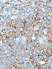
The Israeli Ministry of Health has granted approval for the antineoplastic agent pixantrone (Pixuvri).
The drug is now approved as monotherapy for adults with relapsed or refractory aggressive B-cell non-Hodgkin lymphoma (NHL) who have received fewer than 4 previous courses of treatment.
The benefit of pixantrone has not been established when used as fifth-line or greater treatment in patients who were refractory to their last therapy.
Pixantrone will be distributed in Israel by the Neopharm Group.
“The approval of Pixuvri in Israel provides patients with aggressive B-cell NHL who have failed second- or third-line therapy a new approved option, where none existed before, that can effectively treat their disease with manageable side effects,” said Abraham Avigdor, MD, of Tel Aviv University.
“Patients who have relapsed after second-line therapy have a poor survival outcome. It is vital to have additional treatment options available, like Pixuvri, so we can provide these patients the best care possible and help them battle their disease.”
The main study of pixantrone, the phase 3 EXTEND PIX301 trial, compared the drug to other chemotherapeutic agents in patients with relapsed or refractory NHL. The response rate was 20% in the pixantrone arm and 6% in the comparator arm.
In addition, patients receiving pixantrone had longer progression-free survival than patients in the comparator group, with a median of 10.2 months and 7.6 months, respectively.
However, grade 3/4 adverse events—including neutropenia, leukopenia, and thrombocytopenia—were more common in the pixantrone arm.
Pixantrone is already marketed in the European Union. In 2012, the European Commission granted conditional marketing authorization for the drug as monotherapy for adults with relapsed or refractory aggressive B-cell NHL.
Under the provisions of the conditional marketing authorization, Cell Therapeutics, Inc., the company developing pixantrone, will be required to complete a post-marketing study aimed at confirming the drug’s clinical benefit.
The European Medicines Agency’s Committee for Medicinal Products for Human Use has accepted PIX306, a randomized, phase 3 trial comparing pixantrone plus rituximab to gemcitabine plus rituximab in patients who have relapsed after 1 to 3 prior regimens for aggressive B-cell NHL and who are not eligible for autologous stem cell transplant.
As a condition of approval, Cell Therapeutics has agreed to have the trial data available by June 2015.
Pixantrone is not approved for use in the US.
The Israeli Ministry of Health has granted approval for the antineoplastic agent pixantrone (Pixuvri).
The drug is now approved as monotherapy for adults with relapsed or refractory aggressive B-cell non-Hodgkin lymphoma (NHL) who have received fewer than 4 previous courses of treatment.
The benefit of pixantrone has not been established when used as fifth-line or greater treatment in patients who were refractory to their last therapy.
Pixantrone will be distributed in Israel by the Neopharm Group.
“The approval of Pixuvri in Israel provides patients with aggressive B-cell NHL who have failed second- or third-line therapy a new approved option, where none existed before, that can effectively treat their disease with manageable side effects,” said Abraham Avigdor, MD, of Tel Aviv University.
“Patients who have relapsed after second-line therapy have a poor survival outcome. It is vital to have additional treatment options available, like Pixuvri, so we can provide these patients the best care possible and help them battle their disease.”
The main study of pixantrone, the phase 3 EXTEND PIX301 trial, compared the drug to other chemotherapeutic agents in patients with relapsed or refractory NHL. The response rate was 20% in the pixantrone arm and 6% in the comparator arm.
In addition, patients receiving pixantrone had longer progression-free survival than patients in the comparator group, with a median of 10.2 months and 7.6 months, respectively.
However, grade 3/4 adverse events—including neutropenia, leukopenia, and thrombocytopenia—were more common in the pixantrone arm.
Pixantrone is already marketed in the European Union. In 2012, the European Commission granted conditional marketing authorization for the drug as monotherapy for adults with relapsed or refractory aggressive B-cell NHL.
Under the provisions of the conditional marketing authorization, Cell Therapeutics, Inc., the company developing pixantrone, will be required to complete a post-marketing study aimed at confirming the drug’s clinical benefit.
The European Medicines Agency’s Committee for Medicinal Products for Human Use has accepted PIX306, a randomized, phase 3 trial comparing pixantrone plus rituximab to gemcitabine plus rituximab in patients who have relapsed after 1 to 3 prior regimens for aggressive B-cell NHL and who are not eligible for autologous stem cell transplant.
As a condition of approval, Cell Therapeutics has agreed to have the trial data available by June 2015.
Pixantrone is not approved for use in the US.
The Israeli Ministry of Health has granted approval for the antineoplastic agent pixantrone (Pixuvri).
The drug is now approved as monotherapy for adults with relapsed or refractory aggressive B-cell non-Hodgkin lymphoma (NHL) who have received fewer than 4 previous courses of treatment.
The benefit of pixantrone has not been established when used as fifth-line or greater treatment in patients who were refractory to their last therapy.
Pixantrone will be distributed in Israel by the Neopharm Group.
“The approval of Pixuvri in Israel provides patients with aggressive B-cell NHL who have failed second- or third-line therapy a new approved option, where none existed before, that can effectively treat their disease with manageable side effects,” said Abraham Avigdor, MD, of Tel Aviv University.
“Patients who have relapsed after second-line therapy have a poor survival outcome. It is vital to have additional treatment options available, like Pixuvri, so we can provide these patients the best care possible and help them battle their disease.”
The main study of pixantrone, the phase 3 EXTEND PIX301 trial, compared the drug to other chemotherapeutic agents in patients with relapsed or refractory NHL. The response rate was 20% in the pixantrone arm and 6% in the comparator arm.
In addition, patients receiving pixantrone had longer progression-free survival than patients in the comparator group, with a median of 10.2 months and 7.6 months, respectively.
However, grade 3/4 adverse events—including neutropenia, leukopenia, and thrombocytopenia—were more common in the pixantrone arm.
Pixantrone is already marketed in the European Union. In 2012, the European Commission granted conditional marketing authorization for the drug as monotherapy for adults with relapsed or refractory aggressive B-cell NHL.
Under the provisions of the conditional marketing authorization, Cell Therapeutics, Inc., the company developing pixantrone, will be required to complete a post-marketing study aimed at confirming the drug’s clinical benefit.
The European Medicines Agency’s Committee for Medicinal Products for Human Use has accepted PIX306, a randomized, phase 3 trial comparing pixantrone plus rituximab to gemcitabine plus rituximab in patients who have relapsed after 1 to 3 prior regimens for aggressive B-cell NHL and who are not eligible for autologous stem cell transplant.
As a condition of approval, Cell Therapeutics has agreed to have the trial data available by June 2015.
Pixantrone is not approved for use in the US.
Military technology has application for malaria
Credit: Peter H. Seeberger
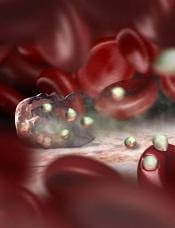
Researchers have used military technology to develop a test for detecting malaria parasites in the blood.
The team used a detector known as a focal plane array (FPA), which was originally developed for heat-seeking missiles.
The FPA gives highly detailed information on a sample area in minutes. The heat-seeking detector, which is coupled to an infrared imaging microscope, could detect the malaria parasite in a single red blood cell.
The infrared signature from the fatty acids of the parasites allowed the researchers to detect the parasite at its earliest stages and determine the number of parasites in a blood smear.
The team described the technology in Analyst.
“Our test detects malaria at its very early stages, so that doctors can stop the disease in its tracks before it takes hold and kills,” said study author Bayden Wood, PhD, of Monash University in Victoria, Australia. “We believe this sets the gold standard for malaria testing.”
“There are some excellent tests that diagnose malaria. However, the sensitivity is limited, and the best methods require hours of input from skilled microscopists, and that’s a problem in developing countries where malaria is most prevalent.”
The new test, on the other hand, gives an automatic diagnosis within 4 minutes and doesn’t require a specialist technician.
Study author Leann Tilley, PhD, of the University of Melbourne in Australia, said the test could make an impact in large-scale screening of malaria parasite carriers who do not present with the classic fever-type symptoms associated with the disease.
“In many countries, only people who display signs of malaria are treated,” Dr Tilley said. “But the problem with this approach is that some people don’t have typical flu-like symptoms associated with malaria, and this means a reservoir of parasites persists that can reemerge and spread very quickly within a community.”
“Our test works because it can detect the malaria parasite at the very early stages and can reliably detect it in an automated manner in a single red blood cell. No other test can do that.”
FPA detectors were originally developed for Javelin Portable anti-tank missiles in the 1990s. The heat-seeking detector is used on shoulder-fired missiles but can also be installed on tracked, wheeled, or amphibious vehicles, providing spatial and spectral information in a matter of seconds.
The FPA detector used in this project was coupled to a synchrotron source located at the InfraRed Environmental Imaging facility at the Synchrotron Radiation Center in Wisconsin.
For the next phase of this research, Dr Wood’s team is collaborating with Patcharee Jearanaikoon, PhD, of Kohn Kaen University in Thailand, to test the technology in clinics.
Credit: Peter H. Seeberger
Researchers have used military technology to develop a test for detecting malaria parasites in the blood.
The team used a detector known as a focal plane array (FPA), which was originally developed for heat-seeking missiles.
The FPA gives highly detailed information on a sample area in minutes. The heat-seeking detector, which is coupled to an infrared imaging microscope, could detect the malaria parasite in a single red blood cell.
The infrared signature from the fatty acids of the parasites allowed the researchers to detect the parasite at its earliest stages and determine the number of parasites in a blood smear.
The team described the technology in Analyst.
“Our test detects malaria at its very early stages, so that doctors can stop the disease in its tracks before it takes hold and kills,” said study author Bayden Wood, PhD, of Monash University in Victoria, Australia. “We believe this sets the gold standard for malaria testing.”
“There are some excellent tests that diagnose malaria. However, the sensitivity is limited, and the best methods require hours of input from skilled microscopists, and that’s a problem in developing countries where malaria is most prevalent.”
The new test, on the other hand, gives an automatic diagnosis within 4 minutes and doesn’t require a specialist technician.
Study author Leann Tilley, PhD, of the University of Melbourne in Australia, said the test could make an impact in large-scale screening of malaria parasite carriers who do not present with the classic fever-type symptoms associated with the disease.
“In many countries, only people who display signs of malaria are treated,” Dr Tilley said. “But the problem with this approach is that some people don’t have typical flu-like symptoms associated with malaria, and this means a reservoir of parasites persists that can reemerge and spread very quickly within a community.”
“Our test works because it can detect the malaria parasite at the very early stages and can reliably detect it in an automated manner in a single red blood cell. No other test can do that.”
FPA detectors were originally developed for Javelin Portable anti-tank missiles in the 1990s. The heat-seeking detector is used on shoulder-fired missiles but can also be installed on tracked, wheeled, or amphibious vehicles, providing spatial and spectral information in a matter of seconds.
The FPA detector used in this project was coupled to a synchrotron source located at the InfraRed Environmental Imaging facility at the Synchrotron Radiation Center in Wisconsin.
For the next phase of this research, Dr Wood’s team is collaborating with Patcharee Jearanaikoon, PhD, of Kohn Kaen University in Thailand, to test the technology in clinics.
Credit: Peter H. Seeberger
Researchers have used military technology to develop a test for detecting malaria parasites in the blood.
The team used a detector known as a focal plane array (FPA), which was originally developed for heat-seeking missiles.
The FPA gives highly detailed information on a sample area in minutes. The heat-seeking detector, which is coupled to an infrared imaging microscope, could detect the malaria parasite in a single red blood cell.
The infrared signature from the fatty acids of the parasites allowed the researchers to detect the parasite at its earliest stages and determine the number of parasites in a blood smear.
The team described the technology in Analyst.
“Our test detects malaria at its very early stages, so that doctors can stop the disease in its tracks before it takes hold and kills,” said study author Bayden Wood, PhD, of Monash University in Victoria, Australia. “We believe this sets the gold standard for malaria testing.”
“There are some excellent tests that diagnose malaria. However, the sensitivity is limited, and the best methods require hours of input from skilled microscopists, and that’s a problem in developing countries where malaria is most prevalent.”
The new test, on the other hand, gives an automatic diagnosis within 4 minutes and doesn’t require a specialist technician.
Study author Leann Tilley, PhD, of the University of Melbourne in Australia, said the test could make an impact in large-scale screening of malaria parasite carriers who do not present with the classic fever-type symptoms associated with the disease.
“In many countries, only people who display signs of malaria are treated,” Dr Tilley said. “But the problem with this approach is that some people don’t have typical flu-like symptoms associated with malaria, and this means a reservoir of parasites persists that can reemerge and spread very quickly within a community.”
“Our test works because it can detect the malaria parasite at the very early stages and can reliably detect it in an automated manner in a single red blood cell. No other test can do that.”
FPA detectors were originally developed for Javelin Portable anti-tank missiles in the 1990s. The heat-seeking detector is used on shoulder-fired missiles but can also be installed on tracked, wheeled, or amphibious vehicles, providing spatial and spectral information in a matter of seconds.
The FPA detector used in this project was coupled to a synchrotron source located at the InfraRed Environmental Imaging facility at the Synchrotron Radiation Center in Wisconsin.
For the next phase of this research, Dr Wood’s team is collaborating with Patcharee Jearanaikoon, PhD, of Kohn Kaen University in Thailand, to test the technology in clinics.
Anticoagulation preferable to CDT, study suggests

Credit: CDC
New research indicates that anticoagulant therapy alone may be safer than catheter-directed thrombolysis (CDT) for treating deep vein thrombosis (DVT).
The study revealed similar rates of in-hospital mortality among patients treated with CDT and those receiving anticoagulation alone.
However, patients who received CDT were more likely to develop pulmonary emboli, experience intracranial hemorrhaging, and require blood transfusions.
Several previous studies have suggested that CDT can reduce the incidence of post-thrombotic syndrome in DVT patients. But CDT is controversial, with conflicting directives on its use because of inconclusive comparative safety outcomes.
So Riyaz Bashir, MD, of Temple University School of Medicine in Philadelphia, Pennsylvania, and his colleagues set out to compare CDT with anticoagulation alone.
They reported their findings in JAMA Internal Medicine.
The researchers examined in-hospital mortality, as well as secondary outcomes of bleeding complications, length of stay, and hospital charges, in a group of 90,618 patients hospitalized for DVT from 2005 through 2010 as part of the Nationwide Inpatient Sample database.
In all, 3649 patients (4.1%) underwent CDT. The CDT utilization rate increased from 2.3% in 2005 to 5.9% in 2010.
The in-hospital mortality rates were not significantly different in the CDT and anticoagulation-only groups, at 1.2% and 0.9%, respectively (P=0.15).
However, rates of adverse events were higher among patients treated with CDT. This included blood transfusion (11.1% vs 6.5%, P<0.001), pulmonary embolism (17.9% vs 11.4%, P<0.001), intracranial hemorrhage (0.9% vs 0.3%, P=0.03), and vena cava filter placement (34.8% vs 15.6%, P<0.001).
Patients in the CDT group also had longer average lengths of stay (7.2 vs 5 days, P<0.001) and higher hospital charges ($85,094 vs $28,164, P<0.001) compared with the anticoagulation-only group.
The researchers pointed out that their results are based on observational data, so the findings could be subject to residual confounding. Therefore, randomized trials are needed to better evaluate the effects of CDT.
However, the team also said that, as we don’t yet have this information, it may be reasonable to restrict CDT use to those patients who have a low bleeding risk and a high risk for post-thrombotic syndrome.
Credit: CDC
New research indicates that anticoagulant therapy alone may be safer than catheter-directed thrombolysis (CDT) for treating deep vein thrombosis (DVT).
The study revealed similar rates of in-hospital mortality among patients treated with CDT and those receiving anticoagulation alone.
However, patients who received CDT were more likely to develop pulmonary emboli, experience intracranial hemorrhaging, and require blood transfusions.
Several previous studies have suggested that CDT can reduce the incidence of post-thrombotic syndrome in DVT patients. But CDT is controversial, with conflicting directives on its use because of inconclusive comparative safety outcomes.
So Riyaz Bashir, MD, of Temple University School of Medicine in Philadelphia, Pennsylvania, and his colleagues set out to compare CDT with anticoagulation alone.
They reported their findings in JAMA Internal Medicine.
The researchers examined in-hospital mortality, as well as secondary outcomes of bleeding complications, length of stay, and hospital charges, in a group of 90,618 patients hospitalized for DVT from 2005 through 2010 as part of the Nationwide Inpatient Sample database.
In all, 3649 patients (4.1%) underwent CDT. The CDT utilization rate increased from 2.3% in 2005 to 5.9% in 2010.
The in-hospital mortality rates were not significantly different in the CDT and anticoagulation-only groups, at 1.2% and 0.9%, respectively (P=0.15).
However, rates of adverse events were higher among patients treated with CDT. This included blood transfusion (11.1% vs 6.5%, P<0.001), pulmonary embolism (17.9% vs 11.4%, P<0.001), intracranial hemorrhage (0.9% vs 0.3%, P=0.03), and vena cava filter placement (34.8% vs 15.6%, P<0.001).
Patients in the CDT group also had longer average lengths of stay (7.2 vs 5 days, P<0.001) and higher hospital charges ($85,094 vs $28,164, P<0.001) compared with the anticoagulation-only group.
The researchers pointed out that their results are based on observational data, so the findings could be subject to residual confounding. Therefore, randomized trials are needed to better evaluate the effects of CDT.
However, the team also said that, as we don’t yet have this information, it may be reasonable to restrict CDT use to those patients who have a low bleeding risk and a high risk for post-thrombotic syndrome.
Credit: CDC
New research indicates that anticoagulant therapy alone may be safer than catheter-directed thrombolysis (CDT) for treating deep vein thrombosis (DVT).
The study revealed similar rates of in-hospital mortality among patients treated with CDT and those receiving anticoagulation alone.
However, patients who received CDT were more likely to develop pulmonary emboli, experience intracranial hemorrhaging, and require blood transfusions.
Several previous studies have suggested that CDT can reduce the incidence of post-thrombotic syndrome in DVT patients. But CDT is controversial, with conflicting directives on its use because of inconclusive comparative safety outcomes.
So Riyaz Bashir, MD, of Temple University School of Medicine in Philadelphia, Pennsylvania, and his colleagues set out to compare CDT with anticoagulation alone.
They reported their findings in JAMA Internal Medicine.
The researchers examined in-hospital mortality, as well as secondary outcomes of bleeding complications, length of stay, and hospital charges, in a group of 90,618 patients hospitalized for DVT from 2005 through 2010 as part of the Nationwide Inpatient Sample database.
In all, 3649 patients (4.1%) underwent CDT. The CDT utilization rate increased from 2.3% in 2005 to 5.9% in 2010.
The in-hospital mortality rates were not significantly different in the CDT and anticoagulation-only groups, at 1.2% and 0.9%, respectively (P=0.15).
However, rates of adverse events were higher among patients treated with CDT. This included blood transfusion (11.1% vs 6.5%, P<0.001), pulmonary embolism (17.9% vs 11.4%, P<0.001), intracranial hemorrhage (0.9% vs 0.3%, P=0.03), and vena cava filter placement (34.8% vs 15.6%, P<0.001).
Patients in the CDT group also had longer average lengths of stay (7.2 vs 5 days, P<0.001) and higher hospital charges ($85,094 vs $28,164, P<0.001) compared with the anticoagulation-only group.
The researchers pointed out that their results are based on observational data, so the findings could be subject to residual confounding. Therefore, randomized trials are needed to better evaluate the effects of CDT.
However, the team also said that, as we don’t yet have this information, it may be reasonable to restrict CDT use to those patients who have a low bleeding risk and a high risk for post-thrombotic syndrome.
Transfusion strategy appears to impact death patterns
Credit: UAB Hospital
A change in transfusion protocol has resulted in fewer potentially preventable deaths among soldiers, researchers have reported in JAMA Surgery.
In 2006, hospitals treating soldiers from Iraq and Afghanistan implemented a protocol called damage control resuscitation (DCR).
It involves administering blood products early and in a balanced ratio, using an aggressive approach to correct coagulopathy, and minimizing the use of crystalloid fluids.
The research showed that soldiers who died in the hospital after DCR was implemented were more likely than their predecessors to be severely injured and have a severe brain injury.
And this is consistent with a decrease in potentially preventable deaths, according to study author Nicholas R. Langan, MD, of the Madigan Army Medical Center in Tacoma, Washington, and his colleagues.
To conduct the study, the researchers reviewed data from the Joint Theater Trauma Registry of US forward combat hospitals. This included 57,179 soldiers, 2565 (4.5%) of whom died in the hospital. Seventy-four percent of these patients were severely injured, and 80% died within 24 hours of admission.
To assess the impact of DCR, the researchers divided patients into 2 groups: those treated before DCR implementation, from 2002 through 2005, and those treated with the DCR protocol, from 2006 through 2011.
The analysis showed that DCR policies were successfully implemented. There was a significant decrease in mean crystalloid infusion volume in the first 24 hours after hospitalization, from 6.1 L to 3.2 L (P<0.05).
There was a significant increase in fresh-frozen plasma use—from 3.2 U to 10.1 U (P=0.01)—and packed red blood cell use—from 8.4 U to 11.4 U (P=0.01)—in the first 24 hours after hospitalization.
And the mean ratio of packed red blood cells to fresh-frozen plasma changed from 2.6:1 in the pre-DCR period to 1.4:1 during the DCR period (P<0.01).
On the other hand, there was no significant difference in cryoprecipitate use, platelet use, or the ratio of packed red blood cells to cryoprecipitate or platelets.
The change in treatment protocol was associated with a change in the incidence of early and late, but not intermediate, deaths. The incidence of early death (within the first 24 hours) increased from 77% pre-DCR to 80% during DCR (P=0.02).
The incidence of late death (more than 7 days after injury) decreased from 10% pre-DCR to 6% during DCR (P<0.01). And the rate of intermediate death (1-7 days after injury) measured 13% for both periods (P=0.95).
The percentage of patients with any severe injury increased significantly from the pre-DCR period to the DCR period, from 64% to 80% (P<0.05). And the percentage of patients with severe head injuries increased significantly, from 57% to 73% (P<0.05).
As patients who died during the DCR period were more likely to have such “nonsurvivable” wounds, the researchers said this suggests that DCR is associated with a decrease in deaths among potentially salvageable patients.
Credit: UAB Hospital
A change in transfusion protocol has resulted in fewer potentially preventable deaths among soldiers, researchers have reported in JAMA Surgery.
In 2006, hospitals treating soldiers from Iraq and Afghanistan implemented a protocol called damage control resuscitation (DCR).
It involves administering blood products early and in a balanced ratio, using an aggressive approach to correct coagulopathy, and minimizing the use of crystalloid fluids.
The research showed that soldiers who died in the hospital after DCR was implemented were more likely than their predecessors to be severely injured and have a severe brain injury.
And this is consistent with a decrease in potentially preventable deaths, according to study author Nicholas R. Langan, MD, of the Madigan Army Medical Center in Tacoma, Washington, and his colleagues.
To conduct the study, the researchers reviewed data from the Joint Theater Trauma Registry of US forward combat hospitals. This included 57,179 soldiers, 2565 (4.5%) of whom died in the hospital. Seventy-four percent of these patients were severely injured, and 80% died within 24 hours of admission.
To assess the impact of DCR, the researchers divided patients into 2 groups: those treated before DCR implementation, from 2002 through 2005, and those treated with the DCR protocol, from 2006 through 2011.
The analysis showed that DCR policies were successfully implemented. There was a significant decrease in mean crystalloid infusion volume in the first 24 hours after hospitalization, from 6.1 L to 3.2 L (P<0.05).
There was a significant increase in fresh-frozen plasma use—from 3.2 U to 10.1 U (P=0.01)—and packed red blood cell use—from 8.4 U to 11.4 U (P=0.01)—in the first 24 hours after hospitalization.
And the mean ratio of packed red blood cells to fresh-frozen plasma changed from 2.6:1 in the pre-DCR period to 1.4:1 during the DCR period (P<0.01).
On the other hand, there was no significant difference in cryoprecipitate use, platelet use, or the ratio of packed red blood cells to cryoprecipitate or platelets.
The change in treatment protocol was associated with a change in the incidence of early and late, but not intermediate, deaths. The incidence of early death (within the first 24 hours) increased from 77% pre-DCR to 80% during DCR (P=0.02).
The incidence of late death (more than 7 days after injury) decreased from 10% pre-DCR to 6% during DCR (P<0.01). And the rate of intermediate death (1-7 days after injury) measured 13% for both periods (P=0.95).
The percentage of patients with any severe injury increased significantly from the pre-DCR period to the DCR period, from 64% to 80% (P<0.05). And the percentage of patients with severe head injuries increased significantly, from 57% to 73% (P<0.05).
As patients who died during the DCR period were more likely to have such “nonsurvivable” wounds, the researchers said this suggests that DCR is associated with a decrease in deaths among potentially salvageable patients.
Credit: UAB Hospital
A change in transfusion protocol has resulted in fewer potentially preventable deaths among soldiers, researchers have reported in JAMA Surgery.
In 2006, hospitals treating soldiers from Iraq and Afghanistan implemented a protocol called damage control resuscitation (DCR).
It involves administering blood products early and in a balanced ratio, using an aggressive approach to correct coagulopathy, and minimizing the use of crystalloid fluids.
The research showed that soldiers who died in the hospital after DCR was implemented were more likely than their predecessors to be severely injured and have a severe brain injury.
And this is consistent with a decrease in potentially preventable deaths, according to study author Nicholas R. Langan, MD, of the Madigan Army Medical Center in Tacoma, Washington, and his colleagues.
To conduct the study, the researchers reviewed data from the Joint Theater Trauma Registry of US forward combat hospitals. This included 57,179 soldiers, 2565 (4.5%) of whom died in the hospital. Seventy-four percent of these patients were severely injured, and 80% died within 24 hours of admission.
To assess the impact of DCR, the researchers divided patients into 2 groups: those treated before DCR implementation, from 2002 through 2005, and those treated with the DCR protocol, from 2006 through 2011.
The analysis showed that DCR policies were successfully implemented. There was a significant decrease in mean crystalloid infusion volume in the first 24 hours after hospitalization, from 6.1 L to 3.2 L (P<0.05).
There was a significant increase in fresh-frozen plasma use—from 3.2 U to 10.1 U (P=0.01)—and packed red blood cell use—from 8.4 U to 11.4 U (P=0.01)—in the first 24 hours after hospitalization.
And the mean ratio of packed red blood cells to fresh-frozen plasma changed from 2.6:1 in the pre-DCR period to 1.4:1 during the DCR period (P<0.01).
On the other hand, there was no significant difference in cryoprecipitate use, platelet use, or the ratio of packed red blood cells to cryoprecipitate or platelets.
The change in treatment protocol was associated with a change in the incidence of early and late, but not intermediate, deaths. The incidence of early death (within the first 24 hours) increased from 77% pre-DCR to 80% during DCR (P=0.02).
The incidence of late death (more than 7 days after injury) decreased from 10% pre-DCR to 6% during DCR (P<0.01). And the rate of intermediate death (1-7 days after injury) measured 13% for both periods (P=0.95).
The percentage of patients with any severe injury increased significantly from the pre-DCR period to the DCR period, from 64% to 80% (P<0.05). And the percentage of patients with severe head injuries increased significantly, from 57% to 73% (P<0.05).
As patients who died during the DCR period were more likely to have such “nonsurvivable” wounds, the researchers said this suggests that DCR is associated with a decrease in deaths among potentially salvageable patients.
Protein map may point to new cancer treatments

the endoplasmic reticulum
in green, mitochondria in red,
and chromosomes in blue
Credit: Wellcome Images
Researchers say they’ve uncovered the structure of one of the most important protein complexes involved in cell division, and their finding has implications for cancer treatment.
The team mapped the anaphase-promoting complex (APC/C), which performs a wide range of tasks associated with mitosis.
The researchers believe that seeing APC/C in unprecedented detail could transform our understanding of how cells divide and reveal binding sites for future cancer drugs.
“It’s very rewarding to finally tie down the detailed structure of this important protein, which is both one of the most important and most complicated found in all of nature,” said David Barford, DPhil, of the Medical Research Council Laboratory of Molecular Biology in Cambridge, UK.
“We hope our discovery will open up whole new avenues of research that increase our understanding of the process of mitosis and ultimately lead to the discovery of new cancer drugs.”
Dr Barford and his colleagues detailed their discovery in Nature.
The researchers reconstituted human APC/C and used a combination of electron microscopy and imaging software to visualize it at a resolution of less than a billionth of a meter.
The resolution was so fine that it allowed the team to see the secondary structure—the set of basic building blocks that combine to form every protein. Alpha-helix rods and folded beta-sheet constructions were clearly visible within the 20 subunits of the APC/C, defining the overall architecture of the complex.
Previous studies led by the same team had shown a globular structure for APC/C in much lower resolution, but the secondary structure had not been mapped.
Each of the APC/C’s subunits bond and mesh with other units at different points in the cell cycle, allowing it to control a range of mitotic processes, including the initiation of DNA replication, the segregation of chromosomes along spindles, and cytokinesis. Disrupting each of these processes could selectively kill cancer cells or prevent them from dividing.
“The fantastic insights into molecular structure provided by this study are a vivid illustration of the critical role played by fundamental cell biology in cancer research,” said Paul Workman, PhD, of The Institute of Cancer Research, London.
“The new study is a major step forward in our understanding of cell division. When this process goes awry, it is a critical difference that separates cancer cells from their healthy counterparts. Understanding exactly how cancer cells divide inappropriately is crucial to the discovery of innovative cancer treatments to improve outcomes for cancer patients.”
the endoplasmic reticulum
in green, mitochondria in red,
and chromosomes in blue
Credit: Wellcome Images
Researchers say they’ve uncovered the structure of one of the most important protein complexes involved in cell division, and their finding has implications for cancer treatment.
The team mapped the anaphase-promoting complex (APC/C), which performs a wide range of tasks associated with mitosis.
The researchers believe that seeing APC/C in unprecedented detail could transform our understanding of how cells divide and reveal binding sites for future cancer drugs.
“It’s very rewarding to finally tie down the detailed structure of this important protein, which is both one of the most important and most complicated found in all of nature,” said David Barford, DPhil, of the Medical Research Council Laboratory of Molecular Biology in Cambridge, UK.
“We hope our discovery will open up whole new avenues of research that increase our understanding of the process of mitosis and ultimately lead to the discovery of new cancer drugs.”
Dr Barford and his colleagues detailed their discovery in Nature.
The researchers reconstituted human APC/C and used a combination of electron microscopy and imaging software to visualize it at a resolution of less than a billionth of a meter.
The resolution was so fine that it allowed the team to see the secondary structure—the set of basic building blocks that combine to form every protein. Alpha-helix rods and folded beta-sheet constructions were clearly visible within the 20 subunits of the APC/C, defining the overall architecture of the complex.
Previous studies led by the same team had shown a globular structure for APC/C in much lower resolution, but the secondary structure had not been mapped.
Each of the APC/C’s subunits bond and mesh with other units at different points in the cell cycle, allowing it to control a range of mitotic processes, including the initiation of DNA replication, the segregation of chromosomes along spindles, and cytokinesis. Disrupting each of these processes could selectively kill cancer cells or prevent them from dividing.
“The fantastic insights into molecular structure provided by this study are a vivid illustration of the critical role played by fundamental cell biology in cancer research,” said Paul Workman, PhD, of The Institute of Cancer Research, London.
“The new study is a major step forward in our understanding of cell division. When this process goes awry, it is a critical difference that separates cancer cells from their healthy counterparts. Understanding exactly how cancer cells divide inappropriately is crucial to the discovery of innovative cancer treatments to improve outcomes for cancer patients.”
the endoplasmic reticulum
in green, mitochondria in red,
and chromosomes in blue
Credit: Wellcome Images
Researchers say they’ve uncovered the structure of one of the most important protein complexes involved in cell division, and their finding has implications for cancer treatment.
The team mapped the anaphase-promoting complex (APC/C), which performs a wide range of tasks associated with mitosis.
The researchers believe that seeing APC/C in unprecedented detail could transform our understanding of how cells divide and reveal binding sites for future cancer drugs.
“It’s very rewarding to finally tie down the detailed structure of this important protein, which is both one of the most important and most complicated found in all of nature,” said David Barford, DPhil, of the Medical Research Council Laboratory of Molecular Biology in Cambridge, UK.
“We hope our discovery will open up whole new avenues of research that increase our understanding of the process of mitosis and ultimately lead to the discovery of new cancer drugs.”
Dr Barford and his colleagues detailed their discovery in Nature.
The researchers reconstituted human APC/C and used a combination of electron microscopy and imaging software to visualize it at a resolution of less than a billionth of a meter.
The resolution was so fine that it allowed the team to see the secondary structure—the set of basic building blocks that combine to form every protein. Alpha-helix rods and folded beta-sheet constructions were clearly visible within the 20 subunits of the APC/C, defining the overall architecture of the complex.
Previous studies led by the same team had shown a globular structure for APC/C in much lower resolution, but the secondary structure had not been mapped.
Each of the APC/C’s subunits bond and mesh with other units at different points in the cell cycle, allowing it to control a range of mitotic processes, including the initiation of DNA replication, the segregation of chromosomes along spindles, and cytokinesis. Disrupting each of these processes could selectively kill cancer cells or prevent them from dividing.
“The fantastic insights into molecular structure provided by this study are a vivid illustration of the critical role played by fundamental cell biology in cancer research,” said Paul Workman, PhD, of The Institute of Cancer Research, London.
“The new study is a major step forward in our understanding of cell division. When this process goes awry, it is a critical difference that separates cancer cells from their healthy counterparts. Understanding exactly how cancer cells divide inappropriately is crucial to the discovery of innovative cancer treatments to improve outcomes for cancer patients.”