User login
Lab-generated platelets seem just like the real thing
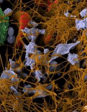
Credit: Andre E.X. Brown
A next-generation bioreactor can produce fully functional platelets, according to research published in Blood.
The bioreactor recapitulates human bone marrow and blood vessel microenvironments.
And when the researchers introduced megakaryocytes derived from human induced pluripotent stem cell cultures (hiPSC-MKs), the bioreactor produced platelets with the structural and functional properties of natural platelets.
The team said this work is a major advancement that could help address blood transfusion needs worldwide.
“The ability to generate an alternative source of functional human platelets with virtually no disease transmission represents a paradigm shift in how we collect platelets that may allow us to meet the growing need for blood transfusions,” said lead study author Jonathan Thon, PhD, of Brigham and Women’s Hospital in Boston.
His group’s bioreactor uses biologically inspired engineering to fully integrate the major components of bone marrow, modeling both its composition and blood flow characteristics.
The bioreactor recapitulates features such as bone marrow stiffness, extracellular matrix composition, micro-channel size, and blood flow stability under high-resolution live-cell microscopy to make human platelets.
“[B]eing able to develop a device that successfully models bone marrow represents a crucial bridge connecting our understanding of the physiological triggers of platelet formation to support drug development and scale platelet production,” said senior study author Joseph Italiano, Jr, PhD, also of Brigham and Women’s Hospital.
He and his colleagues showed that physiological shear stresses in the bioreactor triggered proplatelet initiation, reproduced ex vivo bone marrow proplatelet production, and generated functional platelets.
In static culture, hiPSC-MKs began producing proplatelets at 6 hours post-isolation and reached maximal production at 18 hours. However, hiPSC-MKs under physiological shear stress began producing proplatelets immediately upon trapping and extended/released proplatelets within the first 2 hours of culture.
About 90% of hiPSC-MKs under shear stress produced proplatelets, compared to 10% of hiPSC-MKs in static cultures.
The hiPSC-MK-derived bioreactor platelets displayed forward scatter, side scatter, and surface biomarker expression characteristic of human platelets. Electron microscopy showed the 2 types of platelets were ultrastructurally indistinguishable from one another.
Furthermore, bioreactor platelets displayed morphology and microtubule expression comparable to human platelets. And bioreactor platelets spread normally upon contact-activation with glass, forming both filpodia and lamellipodia.
“Bioreactor-derived platelets theoretically have several advantages over conventional, donor-derived platelets in terms of safety and resource utilization,” said William Savage, MD, PhD, medical director at Kraft Family Blood Donor Center at Brigham and Women’s Hospital, who did not contribute to the study.
“A major factor that has limited our ability to compare bioreactor platelets to donor platelets is the inefficiency of growing platelets, a problem that slows progress of clinical research. This study addresses that gap, while contributing to our understanding of platelet biology at the same time.”
Based on the promising results of this study, the researchers would like to begin clinical trials testing the bioreactor platelets in 2017.
“The regulatory bar is appropriately set high for blood products,” Dr Thon said. “And it is important to us that we show platelet quality, function, and safety over these next 3 years, since we’ll likely be recipients of these platelets ourselves at some point.” 
Credit: Andre E.X. Brown
A next-generation bioreactor can produce fully functional platelets, according to research published in Blood.
The bioreactor recapitulates human bone marrow and blood vessel microenvironments.
And when the researchers introduced megakaryocytes derived from human induced pluripotent stem cell cultures (hiPSC-MKs), the bioreactor produced platelets with the structural and functional properties of natural platelets.
The team said this work is a major advancement that could help address blood transfusion needs worldwide.
“The ability to generate an alternative source of functional human platelets with virtually no disease transmission represents a paradigm shift in how we collect platelets that may allow us to meet the growing need for blood transfusions,” said lead study author Jonathan Thon, PhD, of Brigham and Women’s Hospital in Boston.
His group’s bioreactor uses biologically inspired engineering to fully integrate the major components of bone marrow, modeling both its composition and blood flow characteristics.
The bioreactor recapitulates features such as bone marrow stiffness, extracellular matrix composition, micro-channel size, and blood flow stability under high-resolution live-cell microscopy to make human platelets.
“[B]eing able to develop a device that successfully models bone marrow represents a crucial bridge connecting our understanding of the physiological triggers of platelet formation to support drug development and scale platelet production,” said senior study author Joseph Italiano, Jr, PhD, also of Brigham and Women’s Hospital.
He and his colleagues showed that physiological shear stresses in the bioreactor triggered proplatelet initiation, reproduced ex vivo bone marrow proplatelet production, and generated functional platelets.
In static culture, hiPSC-MKs began producing proplatelets at 6 hours post-isolation and reached maximal production at 18 hours. However, hiPSC-MKs under physiological shear stress began producing proplatelets immediately upon trapping and extended/released proplatelets within the first 2 hours of culture.
About 90% of hiPSC-MKs under shear stress produced proplatelets, compared to 10% of hiPSC-MKs in static cultures.
The hiPSC-MK-derived bioreactor platelets displayed forward scatter, side scatter, and surface biomarker expression characteristic of human platelets. Electron microscopy showed the 2 types of platelets were ultrastructurally indistinguishable from one another.
Furthermore, bioreactor platelets displayed morphology and microtubule expression comparable to human platelets. And bioreactor platelets spread normally upon contact-activation with glass, forming both filpodia and lamellipodia.
“Bioreactor-derived platelets theoretically have several advantages over conventional, donor-derived platelets in terms of safety and resource utilization,” said William Savage, MD, PhD, medical director at Kraft Family Blood Donor Center at Brigham and Women’s Hospital, who did not contribute to the study.
“A major factor that has limited our ability to compare bioreactor platelets to donor platelets is the inefficiency of growing platelets, a problem that slows progress of clinical research. This study addresses that gap, while contributing to our understanding of platelet biology at the same time.”
Based on the promising results of this study, the researchers would like to begin clinical trials testing the bioreactor platelets in 2017.
“The regulatory bar is appropriately set high for blood products,” Dr Thon said. “And it is important to us that we show platelet quality, function, and safety over these next 3 years, since we’ll likely be recipients of these platelets ourselves at some point.”
Credit: Andre E.X. Brown
A next-generation bioreactor can produce fully functional platelets, according to research published in Blood.
The bioreactor recapitulates human bone marrow and blood vessel microenvironments.
And when the researchers introduced megakaryocytes derived from human induced pluripotent stem cell cultures (hiPSC-MKs), the bioreactor produced platelets with the structural and functional properties of natural platelets.
The team said this work is a major advancement that could help address blood transfusion needs worldwide.
“The ability to generate an alternative source of functional human platelets with virtually no disease transmission represents a paradigm shift in how we collect platelets that may allow us to meet the growing need for blood transfusions,” said lead study author Jonathan Thon, PhD, of Brigham and Women’s Hospital in Boston.
His group’s bioreactor uses biologically inspired engineering to fully integrate the major components of bone marrow, modeling both its composition and blood flow characteristics.
The bioreactor recapitulates features such as bone marrow stiffness, extracellular matrix composition, micro-channel size, and blood flow stability under high-resolution live-cell microscopy to make human platelets.
“[B]eing able to develop a device that successfully models bone marrow represents a crucial bridge connecting our understanding of the physiological triggers of platelet formation to support drug development and scale platelet production,” said senior study author Joseph Italiano, Jr, PhD, also of Brigham and Women’s Hospital.
He and his colleagues showed that physiological shear stresses in the bioreactor triggered proplatelet initiation, reproduced ex vivo bone marrow proplatelet production, and generated functional platelets.
In static culture, hiPSC-MKs began producing proplatelets at 6 hours post-isolation and reached maximal production at 18 hours. However, hiPSC-MKs under physiological shear stress began producing proplatelets immediately upon trapping and extended/released proplatelets within the first 2 hours of culture.
About 90% of hiPSC-MKs under shear stress produced proplatelets, compared to 10% of hiPSC-MKs in static cultures.
The hiPSC-MK-derived bioreactor platelets displayed forward scatter, side scatter, and surface biomarker expression characteristic of human platelets. Electron microscopy showed the 2 types of platelets were ultrastructurally indistinguishable from one another.
Furthermore, bioreactor platelets displayed morphology and microtubule expression comparable to human platelets. And bioreactor platelets spread normally upon contact-activation with glass, forming both filpodia and lamellipodia.
“Bioreactor-derived platelets theoretically have several advantages over conventional, donor-derived platelets in terms of safety and resource utilization,” said William Savage, MD, PhD, medical director at Kraft Family Blood Donor Center at Brigham and Women’s Hospital, who did not contribute to the study.
“A major factor that has limited our ability to compare bioreactor platelets to donor platelets is the inefficiency of growing platelets, a problem that slows progress of clinical research. This study addresses that gap, while contributing to our understanding of platelet biology at the same time.”
Based on the promising results of this study, the researchers would like to begin clinical trials testing the bioreactor platelets in 2017.
“The regulatory bar is appropriately set high for blood products,” Dr Thon said. “And it is important to us that we show platelet quality, function, and safety over these next 3 years, since we’ll likely be recipients of these platelets ourselves at some point.”
Two blood cancer patients cleared of HIV

Credit: CDC
MELBOURNE—Two men with hematologic malignancies who were also HIV-positive appear to be free of the virus after receiving stem cell transplants.
The patients have undetectable levels of HIV and remain free of their cancers—acute myeloid leukemia and non-Hodgkin lymphoma—more than 3 years after their transplants.
Importantly, the patients’ stem cell donors were not homozygous for CCR5-delta 32, a mutation that affords protection against HIV.
The researchers said these results herald a new direction in HIV research and provide hope for HIV-positive patients with leukemia and lymphoma.
The work was presented at the “Towards an HIV Cure Symposium,” which is part of the 20th International AIDS Conference.
Both patients were treated at St Vincent’s Hospital in partnership with the University of New South Wales’s Kirby Institute in Sydney, Australia.
One patient underwent a transplant in 2010 to treat his non-Hodgkin lymphoma, and his donor had 1 copy of CCR5-delta 32.
The second patient underwent a similar procedure for acute myeloid leukemia in 2011, and his donor did not have any copies of CCR5-delta 32.
Nevertheless, both patients were successfully cleared of HIV, although they remain on antiretroviral therapy as a protective measure.
“We’re so pleased that both patients are doing reasonably well years after the treatment for their cancers and remain free of both the original cancer and the HIV virus,” said David Cooper, MBBS, MD, DSc, of the Kirby Institute and St Vincent’s Hospital.
Until now, the only person considered to have cleared HIV is an American man, Timothy Ray Brown, who underwent 2 stem cell transplants in Berlin (in 2007 and 2008).
The cells in his second transplant included both copies of CCR5-delta 32, which affords protection against HIV and is found in less than 1% of the population. The man is no longer on antiretroviral therapy and remains free of HIV.
In Boston, 2 other patients underwent similar transplants in 2012, but the donor cells did not contain CCR5-delta 32. In both cases, HIV returned after antiretroviral treatment was stopped.
“It is very difficult to find a match for bone marrow donors and even more so to find one that affords protective immunity against HIV,” Dr Cooper said.
While his group’s results are a significant development, the researchers stressed that transplants are not a general functional “cure” for the up to 38.8 million people infected with HIV worldwide.
“This is a terrific, unexpected result for people with malignancy and HIV,” said Sam Milliken, MBBS, of St Vincent’s Hospital. “It may well give us a whole new insight into HIV, using the principles of stem cell transplantation.”
“It is important to caution that, at this stage, this form of treatment is far too dangerous for treating patients with HIV alone, but there may be potential for using transplants as an effective treatment modality for HIV down the track.”
The researchers said the 2 Sydney patients will be the subject of investigations to determine where any residual virus might be hiding and how it can be controlled. And the patients’ results point to a new direction for HIV research.
“We still don’t know why these patients have undetectable viral loads,” said Kersten Koelsch, MD, of the Kirby Institute. “One theory is that the induction therapy helps to destroy the cells in which the virus is hiding and that any remaining infected cells are destroyed by the patient’s new immune system.”
“We need more research to establish why and how bone marrow transplantation clears the virus. We also want to explore the predictors of sustained viral clearance and how this might be able to be exploited without the need for bone marrow transplantation.”
For the time being, the results mean that more patients who are eligible for transplant might be able to participate in clinical trials to determine the value of this procedure in HIV.
Credit: CDC
MELBOURNE—Two men with hematologic malignancies who were also HIV-positive appear to be free of the virus after receiving stem cell transplants.
The patients have undetectable levels of HIV and remain free of their cancers—acute myeloid leukemia and non-Hodgkin lymphoma—more than 3 years after their transplants.
Importantly, the patients’ stem cell donors were not homozygous for CCR5-delta 32, a mutation that affords protection against HIV.
The researchers said these results herald a new direction in HIV research and provide hope for HIV-positive patients with leukemia and lymphoma.
The work was presented at the “Towards an HIV Cure Symposium,” which is part of the 20th International AIDS Conference.
Both patients were treated at St Vincent’s Hospital in partnership with the University of New South Wales’s Kirby Institute in Sydney, Australia.
One patient underwent a transplant in 2010 to treat his non-Hodgkin lymphoma, and his donor had 1 copy of CCR5-delta 32.
The second patient underwent a similar procedure for acute myeloid leukemia in 2011, and his donor did not have any copies of CCR5-delta 32.
Nevertheless, both patients were successfully cleared of HIV, although they remain on antiretroviral therapy as a protective measure.
“We’re so pleased that both patients are doing reasonably well years after the treatment for their cancers and remain free of both the original cancer and the HIV virus,” said David Cooper, MBBS, MD, DSc, of the Kirby Institute and St Vincent’s Hospital.
Until now, the only person considered to have cleared HIV is an American man, Timothy Ray Brown, who underwent 2 stem cell transplants in Berlin (in 2007 and 2008).
The cells in his second transplant included both copies of CCR5-delta 32, which affords protection against HIV and is found in less than 1% of the population. The man is no longer on antiretroviral therapy and remains free of HIV.
In Boston, 2 other patients underwent similar transplants in 2012, but the donor cells did not contain CCR5-delta 32. In both cases, HIV returned after antiretroviral treatment was stopped.
“It is very difficult to find a match for bone marrow donors and even more so to find one that affords protective immunity against HIV,” Dr Cooper said.
While his group’s results are a significant development, the researchers stressed that transplants are not a general functional “cure” for the up to 38.8 million people infected with HIV worldwide.
“This is a terrific, unexpected result for people with malignancy and HIV,” said Sam Milliken, MBBS, of St Vincent’s Hospital. “It may well give us a whole new insight into HIV, using the principles of stem cell transplantation.”
“It is important to caution that, at this stage, this form of treatment is far too dangerous for treating patients with HIV alone, but there may be potential for using transplants as an effective treatment modality for HIV down the track.”
The researchers said the 2 Sydney patients will be the subject of investigations to determine where any residual virus might be hiding and how it can be controlled. And the patients’ results point to a new direction for HIV research.
“We still don’t know why these patients have undetectable viral loads,” said Kersten Koelsch, MD, of the Kirby Institute. “One theory is that the induction therapy helps to destroy the cells in which the virus is hiding and that any remaining infected cells are destroyed by the patient’s new immune system.”
“We need more research to establish why and how bone marrow transplantation clears the virus. We also want to explore the predictors of sustained viral clearance and how this might be able to be exploited without the need for bone marrow transplantation.”
For the time being, the results mean that more patients who are eligible for transplant might be able to participate in clinical trials to determine the value of this procedure in HIV.
Credit: CDC
MELBOURNE—Two men with hematologic malignancies who were also HIV-positive appear to be free of the virus after receiving stem cell transplants.
The patients have undetectable levels of HIV and remain free of their cancers—acute myeloid leukemia and non-Hodgkin lymphoma—more than 3 years after their transplants.
Importantly, the patients’ stem cell donors were not homozygous for CCR5-delta 32, a mutation that affords protection against HIV.
The researchers said these results herald a new direction in HIV research and provide hope for HIV-positive patients with leukemia and lymphoma.
The work was presented at the “Towards an HIV Cure Symposium,” which is part of the 20th International AIDS Conference.
Both patients were treated at St Vincent’s Hospital in partnership with the University of New South Wales’s Kirby Institute in Sydney, Australia.
One patient underwent a transplant in 2010 to treat his non-Hodgkin lymphoma, and his donor had 1 copy of CCR5-delta 32.
The second patient underwent a similar procedure for acute myeloid leukemia in 2011, and his donor did not have any copies of CCR5-delta 32.
Nevertheless, both patients were successfully cleared of HIV, although they remain on antiretroviral therapy as a protective measure.
“We’re so pleased that both patients are doing reasonably well years after the treatment for their cancers and remain free of both the original cancer and the HIV virus,” said David Cooper, MBBS, MD, DSc, of the Kirby Institute and St Vincent’s Hospital.
Until now, the only person considered to have cleared HIV is an American man, Timothy Ray Brown, who underwent 2 stem cell transplants in Berlin (in 2007 and 2008).
The cells in his second transplant included both copies of CCR5-delta 32, which affords protection against HIV and is found in less than 1% of the population. The man is no longer on antiretroviral therapy and remains free of HIV.
In Boston, 2 other patients underwent similar transplants in 2012, but the donor cells did not contain CCR5-delta 32. In both cases, HIV returned after antiretroviral treatment was stopped.
“It is very difficult to find a match for bone marrow donors and even more so to find one that affords protective immunity against HIV,” Dr Cooper said.
While his group’s results are a significant development, the researchers stressed that transplants are not a general functional “cure” for the up to 38.8 million people infected with HIV worldwide.
“This is a terrific, unexpected result for people with malignancy and HIV,” said Sam Milliken, MBBS, of St Vincent’s Hospital. “It may well give us a whole new insight into HIV, using the principles of stem cell transplantation.”
“It is important to caution that, at this stage, this form of treatment is far too dangerous for treating patients with HIV alone, but there may be potential for using transplants as an effective treatment modality for HIV down the track.”
The researchers said the 2 Sydney patients will be the subject of investigations to determine where any residual virus might be hiding and how it can be controlled. And the patients’ results point to a new direction for HIV research.
“We still don’t know why these patients have undetectable viral loads,” said Kersten Koelsch, MD, of the Kirby Institute. “One theory is that the induction therapy helps to destroy the cells in which the virus is hiding and that any remaining infected cells are destroyed by the patient’s new immune system.”
“We need more research to establish why and how bone marrow transplantation clears the virus. We also want to explore the predictors of sustained viral clearance and how this might be able to be exploited without the need for bone marrow transplantation.”
For the time being, the results mean that more patients who are eligible for transplant might be able to participate in clinical trials to determine the value of this procedure in HIV.
FDA warns public of non-sterile products

Credit: Bill Branson
The US Food and Drug Administration (FDA) is alerting healthcare professionals and consumers not to use sterile drugs produced by Downing Labs LLC, also known as NuVision Pharmacy, as the products may be contaminated.
Healthcare professionals should immediately check their medical supplies and quarantine any sterile products from NuVision.
Administration of a non-sterile drug product may result in serious and potentially life-threatening infections or death.
NuVision’s products were distributed nationwide. Most of the product labels say, “NuVision Pharmacy, Dallas TX, 75244 1-800-914-7435.”
FDA investigators inspected NuVision and observed unsanitary conditions that result in a lack of sterility assurance of purportedly sterile products, which puts patients at risk.
The inspection revealed sterility failures in 19 lots of products intended to be sterile, endotoxin failures in 3 lots of products, and inadequate or no investigation of these failures. Endotoxins are substances found in certain bacteria that cause a variety of serious reactions such as fever, shock, and changes in blood pressure and other circulatory functions.
The FDA is not aware of recent reports of illness associated with the use of these products.
Patients who have received any product produced by NuVision and have concerns should contact their healthcare professional.
Healthcare professionals and consumers can report adverse events associated with the use of NuVision’s products to the FDA’s MedWatch Adverse Event Reporting Program.
Credit: Bill Branson
The US Food and Drug Administration (FDA) is alerting healthcare professionals and consumers not to use sterile drugs produced by Downing Labs LLC, also known as NuVision Pharmacy, as the products may be contaminated.
Healthcare professionals should immediately check their medical supplies and quarantine any sterile products from NuVision.
Administration of a non-sterile drug product may result in serious and potentially life-threatening infections or death.
NuVision’s products were distributed nationwide. Most of the product labels say, “NuVision Pharmacy, Dallas TX, 75244 1-800-914-7435.”
FDA investigators inspected NuVision and observed unsanitary conditions that result in a lack of sterility assurance of purportedly sterile products, which puts patients at risk.
The inspection revealed sterility failures in 19 lots of products intended to be sterile, endotoxin failures in 3 lots of products, and inadequate or no investigation of these failures. Endotoxins are substances found in certain bacteria that cause a variety of serious reactions such as fever, shock, and changes in blood pressure and other circulatory functions.
The FDA is not aware of recent reports of illness associated with the use of these products.
Patients who have received any product produced by NuVision and have concerns should contact their healthcare professional.
Healthcare professionals and consumers can report adverse events associated with the use of NuVision’s products to the FDA’s MedWatch Adverse Event Reporting Program.
Credit: Bill Branson
The US Food and Drug Administration (FDA) is alerting healthcare professionals and consumers not to use sterile drugs produced by Downing Labs LLC, also known as NuVision Pharmacy, as the products may be contaminated.
Healthcare professionals should immediately check their medical supplies and quarantine any sterile products from NuVision.
Administration of a non-sterile drug product may result in serious and potentially life-threatening infections or death.
NuVision’s products were distributed nationwide. Most of the product labels say, “NuVision Pharmacy, Dallas TX, 75244 1-800-914-7435.”
FDA investigators inspected NuVision and observed unsanitary conditions that result in a lack of sterility assurance of purportedly sterile products, which puts patients at risk.
The inspection revealed sterility failures in 19 lots of products intended to be sterile, endotoxin failures in 3 lots of products, and inadequate or no investigation of these failures. Endotoxins are substances found in certain bacteria that cause a variety of serious reactions such as fever, shock, and changes in blood pressure and other circulatory functions.
The FDA is not aware of recent reports of illness associated with the use of these products.
Patients who have received any product produced by NuVision and have concerns should contact their healthcare professional.
Healthcare professionals and consumers can report adverse events associated with the use of NuVision’s products to the FDA’s MedWatch Adverse Event Reporting Program.
Gene editing strategy may control insect-borne diseases

Credit: James Gathany
Scientists have proposed that gene drives might be used to combat malaria and other insect-borne diseases, control invasive species, and promote sustainable agriculture.
Engineered gene drives are genetic systems that circumvent traditional rules of sexual reproduction and greatly increase the odds that the drive will be passed on to offspring.
This enables the spread of specified genetic alterations through targeted wild populations over many generations.
Gene drives represent a potentially powerful tool to confront regional or global challenges, including the control of invasive species and eradication of insect-borne diseases such as malaria and dengue.
The idea is not new, but a team of researchers has now outlined a technically feasible way to build gene drives that might spread almost any genomic change through populations of sexually reproducing species.
“We all rely on healthy ecosystems and share a responsibility to keep them intact for future generations,” said Kevin Esvelt, PhD, of the Wyss Institute at Harvard University in Boston.
“Given the broad potential of gene drives to address ecological problems, we hope to initiate a transparent, inclusive, and informed public discussion—well in advance of any testing—to collectively decide how we might use this technology for the betterment of humanity and the environment.”
Dr Esvelt and his colleagues have initiated this discussion by publishing papers on gene drives in Science and eLife.
The eLife paper describes the proposed technical methods of building gene drives in different species, defines their theoretical capabilities and limitations, and outlines possible applications.
The Science paper provides an initial assessment of potential environmental and security effects, an analysis of regulatory coverage, and recommendations to ensure responsible development and testing prior to use.
The new technical work in eLife builds upon research by Austin Burt, PhD, of Imperial College London in the UK, who, more than a decade ago, first proposed using a type of gene drive based on cutting DNA to alter populations.
The authors noted that the gene editing tool CRISPR—which is used to precisely insert, replace, and regulate genes—now makes it feasible to create gene drives that work in many different species.
“Our proposal represents a potentially powerful ecosystem management tool for global sustainability, but one that carries with it new concerns, as with any emerging technology,” said George Church, PhD, also of the Wyss Institute.
Dr Esvelt noted that the genomic changes made by gene drives should be reversible. The team has outlined in the eLife publication numerous precautionary measures intended to guide the safe and responsible development of gene drives, many of which were not possible with earlier technologies.
“If the public ever considers making use of a gene drive, we will need to develop appropriate safeguards,” he said. “Ensuring that we have a working reversal drive on hand to quickly undo the proposed genomic change would be one such precaution.”
Because the drives can spread traits only over generations, they will be most effective in species that reproduce quickly or can be released in large numbers, the researchers noted.
For insects, it could take only a couple of years to see a desired change in the population at large, while slower-reproducing organisms would require much longer. Altering human populations would require many centuries.
Gene drives could strike a powerful blow against malaria by altering mosquito populations so they can no longer spread the disease, according to the researchers.
Gene drives might also be used to rid local environments of invasive species or to pave the way toward more sustainable agriculture by reversing mutations that allow particular weed species, such as horseweed, to resist herbicides that are important for no-till farming.
However, the innovative nature of gene drives poses regulatory challenges.
“Simply put, gene drives do not fit comfortably within existing US regulations and international conventions,” said Kenneth Oye, PhD, of the Massachusetts Institute of Technology in Cambridge.
“For example, animal applications of gene drives would be regulated by the FDA as veterinary medicines. Potential implications of gene drives fall beyond the purview of the lists of bacteriological and viral agents that now define security regimes. We’ll need both regulatory reform and public engagement before we can consider beneficial uses. That is why we are excited about getting the conversation on gene drives going early.”
Credit: James Gathany
Scientists have proposed that gene drives might be used to combat malaria and other insect-borne diseases, control invasive species, and promote sustainable agriculture.
Engineered gene drives are genetic systems that circumvent traditional rules of sexual reproduction and greatly increase the odds that the drive will be passed on to offspring.
This enables the spread of specified genetic alterations through targeted wild populations over many generations.
Gene drives represent a potentially powerful tool to confront regional or global challenges, including the control of invasive species and eradication of insect-borne diseases such as malaria and dengue.
The idea is not new, but a team of researchers has now outlined a technically feasible way to build gene drives that might spread almost any genomic change through populations of sexually reproducing species.
“We all rely on healthy ecosystems and share a responsibility to keep them intact for future generations,” said Kevin Esvelt, PhD, of the Wyss Institute at Harvard University in Boston.
“Given the broad potential of gene drives to address ecological problems, we hope to initiate a transparent, inclusive, and informed public discussion—well in advance of any testing—to collectively decide how we might use this technology for the betterment of humanity and the environment.”
Dr Esvelt and his colleagues have initiated this discussion by publishing papers on gene drives in Science and eLife.
The eLife paper describes the proposed technical methods of building gene drives in different species, defines their theoretical capabilities and limitations, and outlines possible applications.
The Science paper provides an initial assessment of potential environmental and security effects, an analysis of regulatory coverage, and recommendations to ensure responsible development and testing prior to use.
The new technical work in eLife builds upon research by Austin Burt, PhD, of Imperial College London in the UK, who, more than a decade ago, first proposed using a type of gene drive based on cutting DNA to alter populations.
The authors noted that the gene editing tool CRISPR—which is used to precisely insert, replace, and regulate genes—now makes it feasible to create gene drives that work in many different species.
“Our proposal represents a potentially powerful ecosystem management tool for global sustainability, but one that carries with it new concerns, as with any emerging technology,” said George Church, PhD, also of the Wyss Institute.
Dr Esvelt noted that the genomic changes made by gene drives should be reversible. The team has outlined in the eLife publication numerous precautionary measures intended to guide the safe and responsible development of gene drives, many of which were not possible with earlier technologies.
“If the public ever considers making use of a gene drive, we will need to develop appropriate safeguards,” he said. “Ensuring that we have a working reversal drive on hand to quickly undo the proposed genomic change would be one such precaution.”
Because the drives can spread traits only over generations, they will be most effective in species that reproduce quickly or can be released in large numbers, the researchers noted.
For insects, it could take only a couple of years to see a desired change in the population at large, while slower-reproducing organisms would require much longer. Altering human populations would require many centuries.
Gene drives could strike a powerful blow against malaria by altering mosquito populations so they can no longer spread the disease, according to the researchers.
Gene drives might also be used to rid local environments of invasive species or to pave the way toward more sustainable agriculture by reversing mutations that allow particular weed species, such as horseweed, to resist herbicides that are important for no-till farming.
However, the innovative nature of gene drives poses regulatory challenges.
“Simply put, gene drives do not fit comfortably within existing US regulations and international conventions,” said Kenneth Oye, PhD, of the Massachusetts Institute of Technology in Cambridge.
“For example, animal applications of gene drives would be regulated by the FDA as veterinary medicines. Potential implications of gene drives fall beyond the purview of the lists of bacteriological and viral agents that now define security regimes. We’ll need both regulatory reform and public engagement before we can consider beneficial uses. That is why we are excited about getting the conversation on gene drives going early.”
Credit: James Gathany
Scientists have proposed that gene drives might be used to combat malaria and other insect-borne diseases, control invasive species, and promote sustainable agriculture.
Engineered gene drives are genetic systems that circumvent traditional rules of sexual reproduction and greatly increase the odds that the drive will be passed on to offspring.
This enables the spread of specified genetic alterations through targeted wild populations over many generations.
Gene drives represent a potentially powerful tool to confront regional or global challenges, including the control of invasive species and eradication of insect-borne diseases such as malaria and dengue.
The idea is not new, but a team of researchers has now outlined a technically feasible way to build gene drives that might spread almost any genomic change through populations of sexually reproducing species.
“We all rely on healthy ecosystems and share a responsibility to keep them intact for future generations,” said Kevin Esvelt, PhD, of the Wyss Institute at Harvard University in Boston.
“Given the broad potential of gene drives to address ecological problems, we hope to initiate a transparent, inclusive, and informed public discussion—well in advance of any testing—to collectively decide how we might use this technology for the betterment of humanity and the environment.”
Dr Esvelt and his colleagues have initiated this discussion by publishing papers on gene drives in Science and eLife.
The eLife paper describes the proposed technical methods of building gene drives in different species, defines their theoretical capabilities and limitations, and outlines possible applications.
The Science paper provides an initial assessment of potential environmental and security effects, an analysis of regulatory coverage, and recommendations to ensure responsible development and testing prior to use.
The new technical work in eLife builds upon research by Austin Burt, PhD, of Imperial College London in the UK, who, more than a decade ago, first proposed using a type of gene drive based on cutting DNA to alter populations.
The authors noted that the gene editing tool CRISPR—which is used to precisely insert, replace, and regulate genes—now makes it feasible to create gene drives that work in many different species.
“Our proposal represents a potentially powerful ecosystem management tool for global sustainability, but one that carries with it new concerns, as with any emerging technology,” said George Church, PhD, also of the Wyss Institute.
Dr Esvelt noted that the genomic changes made by gene drives should be reversible. The team has outlined in the eLife publication numerous precautionary measures intended to guide the safe and responsible development of gene drives, many of which were not possible with earlier technologies.
“If the public ever considers making use of a gene drive, we will need to develop appropriate safeguards,” he said. “Ensuring that we have a working reversal drive on hand to quickly undo the proposed genomic change would be one such precaution.”
Because the drives can spread traits only over generations, they will be most effective in species that reproduce quickly or can be released in large numbers, the researchers noted.
For insects, it could take only a couple of years to see a desired change in the population at large, while slower-reproducing organisms would require much longer. Altering human populations would require many centuries.
Gene drives could strike a powerful blow against malaria by altering mosquito populations so they can no longer spread the disease, according to the researchers.
Gene drives might also be used to rid local environments of invasive species or to pave the way toward more sustainable agriculture by reversing mutations that allow particular weed species, such as horseweed, to resist herbicides that are important for no-till farming.
However, the innovative nature of gene drives poses regulatory challenges.
“Simply put, gene drives do not fit comfortably within existing US regulations and international conventions,” said Kenneth Oye, PhD, of the Massachusetts Institute of Technology in Cambridge.
“For example, animal applications of gene drives would be regulated by the FDA as veterinary medicines. Potential implications of gene drives fall beyond the purview of the lists of bacteriological and viral agents that now define security regimes. We’ll need both regulatory reform and public engagement before we can consider beneficial uses. That is why we are excited about getting the conversation on gene drives going early.”
Teams find new way to kill malaria parasite
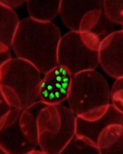
red blood cell; Credit: St Jude
Children’s Research Hospital
Two groups of researchers have found they can kill the malaria parasite by targeting a protein complex.
The research showed that a protein complex known as the Plasmodium translocon of exported proteins (PTEX) is needed for the export of malaria-parasite proteins into the cytoplasm of infected red blood cells, and such export is essential for parasite survival.
When the researchers disrupted passage of the proteins in cell cultures, malaria parasites stopped growing and died.
“The malaria parasite secretes hundreds of diverse proteins to seize control of red blood cells,” said Josh R. Beck, PhD, of the Washington University School of Medicine in St Louis.
“We’ve been searching for a single step that all those various proteins have to take to be secreted, and this looks like just such a bottleneck.”
He and his colleagues detailed their findings in a letter to Nature.
The researchers focused on heat shock protein 101 (HSP101), a component of PTEX. Previous studies had suggested that HSP101 might be involved in protein secretion.
So Dr Beck and his colleagues disabled HSP101 in cell cultures, expecting to block the discharge of some malarial proteins. To their surprise, they stopped all of them.
“We think this is a very promising target for drug development,” said study author Daniel Goldberg, MD, PhD, also of Washington University.
“We’re a long way from getting a new drug, but, in the short term, we may look at screening a variety of compounds to see if they have the potential to block HSP101.”
The researchers think HSP101 may ready malarial proteins for secretion through a pore that opens into the red blood cell. Part of this preparation may involve unfolding the proteins into a linear form that allows them to more easily pass through the pore. HSP101 may also give the proteins a biochemical kick that pushes them through the pore.
A separate study published in the same issue of Nature also highlights the importance of PTEX to the malaria parasite’s survival.
Brendan Elsworth, of the Macfarlane Burnet Institute for Medical Research and Public Health in Melbourne, Australia, and his colleagues neutralized the malaria parasite by disabling either HSP101 or PTEX150, another component of PTEX.
“That suggests there are multiple components of the process that we may be able to target with drugs,” Dr Beck said. “In addition, many of the proteins involved in secretion are unlike any human proteins, which means we may be able to disable them without adversely affecting important human proteins.”
red blood cell; Credit: St Jude
Children’s Research Hospital
Two groups of researchers have found they can kill the malaria parasite by targeting a protein complex.
The research showed that a protein complex known as the Plasmodium translocon of exported proteins (PTEX) is needed for the export of malaria-parasite proteins into the cytoplasm of infected red blood cells, and such export is essential for parasite survival.
When the researchers disrupted passage of the proteins in cell cultures, malaria parasites stopped growing and died.
“The malaria parasite secretes hundreds of diverse proteins to seize control of red blood cells,” said Josh R. Beck, PhD, of the Washington University School of Medicine in St Louis.
“We’ve been searching for a single step that all those various proteins have to take to be secreted, and this looks like just such a bottleneck.”
He and his colleagues detailed their findings in a letter to Nature.
The researchers focused on heat shock protein 101 (HSP101), a component of PTEX. Previous studies had suggested that HSP101 might be involved in protein secretion.
So Dr Beck and his colleagues disabled HSP101 in cell cultures, expecting to block the discharge of some malarial proteins. To their surprise, they stopped all of them.
“We think this is a very promising target for drug development,” said study author Daniel Goldberg, MD, PhD, also of Washington University.
“We’re a long way from getting a new drug, but, in the short term, we may look at screening a variety of compounds to see if they have the potential to block HSP101.”
The researchers think HSP101 may ready malarial proteins for secretion through a pore that opens into the red blood cell. Part of this preparation may involve unfolding the proteins into a linear form that allows them to more easily pass through the pore. HSP101 may also give the proteins a biochemical kick that pushes them through the pore.
A separate study published in the same issue of Nature also highlights the importance of PTEX to the malaria parasite’s survival.
Brendan Elsworth, of the Macfarlane Burnet Institute for Medical Research and Public Health in Melbourne, Australia, and his colleagues neutralized the malaria parasite by disabling either HSP101 or PTEX150, another component of PTEX.
“That suggests there are multiple components of the process that we may be able to target with drugs,” Dr Beck said. “In addition, many of the proteins involved in secretion are unlike any human proteins, which means we may be able to disable them without adversely affecting important human proteins.”
red blood cell; Credit: St Jude
Children’s Research Hospital
Two groups of researchers have found they can kill the malaria parasite by targeting a protein complex.
The research showed that a protein complex known as the Plasmodium translocon of exported proteins (PTEX) is needed for the export of malaria-parasite proteins into the cytoplasm of infected red blood cells, and such export is essential for parasite survival.
When the researchers disrupted passage of the proteins in cell cultures, malaria parasites stopped growing and died.
“The malaria parasite secretes hundreds of diverse proteins to seize control of red blood cells,” said Josh R. Beck, PhD, of the Washington University School of Medicine in St Louis.
“We’ve been searching for a single step that all those various proteins have to take to be secreted, and this looks like just such a bottleneck.”
He and his colleagues detailed their findings in a letter to Nature.
The researchers focused on heat shock protein 101 (HSP101), a component of PTEX. Previous studies had suggested that HSP101 might be involved in protein secretion.
So Dr Beck and his colleagues disabled HSP101 in cell cultures, expecting to block the discharge of some malarial proteins. To their surprise, they stopped all of them.
“We think this is a very promising target for drug development,” said study author Daniel Goldberg, MD, PhD, also of Washington University.
“We’re a long way from getting a new drug, but, in the short term, we may look at screening a variety of compounds to see if they have the potential to block HSP101.”
The researchers think HSP101 may ready malarial proteins for secretion through a pore that opens into the red blood cell. Part of this preparation may involve unfolding the proteins into a linear form that allows them to more easily pass through the pore. HSP101 may also give the proteins a biochemical kick that pushes them through the pore.
A separate study published in the same issue of Nature also highlights the importance of PTEX to the malaria parasite’s survival.
Brendan Elsworth, of the Macfarlane Burnet Institute for Medical Research and Public Health in Melbourne, Australia, and his colleagues neutralized the malaria parasite by disabling either HSP101 or PTEX150, another component of PTEX.
“That suggests there are multiple components of the process that we may be able to target with drugs,” Dr Beck said. “In addition, many of the proteins involved in secretion are unlike any human proteins, which means we may be able to disable them without adversely affecting important human proteins.”
Group takes first step toward treating rare pediatric disease

Credit: Petr Kratochvil
Investigators say they’ve discovered the genetic defect that underlies STING-associated vasculopathy with onset in infancy (SAVI), which has led to a potential treatment for this rare condition.
The team found that SAVI patients have a mutation in a gene that encodes the protein STING, a signaling molecule whose activation leads to interferon production.
So it followed that JAK inhibitors, which block the interferon pathway, showed activity in samples from SAVI patients.
And based on these results, the investigators are enrolling SAVI patients on a compassionate use protocol for the JAK1/2 inhibitor baricitinib.
Raphaela Goldbach-Mansky, MD, of the National Institute of Arthritis and Musculoskeletal and Skin Diseases in Bethesda, Maryland, and her colleagues described the results in NEJM.
The research began in 2004, when Dr Goldbach-Mansky was called upon to advise on a patient with a baffling problem. The 10-year-old girl had signs of systemic inflammation, especially in the blood vessels, and she had not responded to any treatments.
She had blistering rashes on her fingers, toes, ears, nose, and cheeks, and she had lost parts of her fingers to the disease. The child also had severe scarring in her lungs and was having trouble breathing. She had shown signs of the disease as an infant and had progressively worsened. She died a few years later.
By 2010, Dr Goldbach-Mansky had seen 2 other patients with the same symptoms. She suspected that all 3 had the same disease, and it was caused by a genetic defect that arose in the children, as their parents were not affected.
Her hunch suggested a strategy for identifying the genetic defect. By comparing the DNA of an affected child with the DNA of the child’s parents, scientists would be able to spot the differences and possibly identify the disease-causing mutation.
The DNA comparison revealed a novel mutation in TMEM173, the gene encoding STING, a protein whose activation leads to the production of interferon. When overproduced, interferon can trigger inflammation.
“Blood tests on the affected children had shown high levels of interferon-induced proteins, so we were not surprised when the mutated gene turned out to be related to interferon signaling,” Dr Goldbach-Mansky said.
When they tested the DNA of 5 other patients with similar symptoms, the investigators found mutations in the same gene, confirming STING’s role in the disease. The excessive inflammation observed in the patients, along with other evidence of interferon pathway activation, indicated that mutations in STING boosted the protein’s activity.
The investigators found that STING was present in high levels in the cells lining the blood vessels and the lungs, which would likely explain why these tissues are predominantly affected by SAVI.
Dr Goldbach-Mansky’s team next looked for ways to dampen the inflammatory response in patients with SAVI.
“When mutations that cause autoinflammatory conditions hit an important pathway, the outcome for patients can be dismal,” Dr Goldbach-Mansky said. “But because SAVI is caused by a single gene defect and interferon has such a strong role, I’m optimistic that we’ll be able to target the pathway and potentially make a huge difference in the lives of these children.”
The JAK inhibitors tofacitinib, ruxolitinib, and baricitinib are known to work by blocking the interferon pathway, so the investigators reasoned the drugs might be effective in patients with SAVI as well.
When they tested the effect of the drugs on SAVI patients’ blood cells in the lab, the team saw a marked reduction in interferon-pathway activation.
The investigators are now enrolling SAVI patients in a compassionate use protocol for baricitinib.
Dr Goldbach-Mansky’s team is also planning to investigate STING’s exact role in the interferon pathway and examine how the mutations that cause SAVI lead to interferon overproduction.
Credit: Petr Kratochvil
Investigators say they’ve discovered the genetic defect that underlies STING-associated vasculopathy with onset in infancy (SAVI), which has led to a potential treatment for this rare condition.
The team found that SAVI patients have a mutation in a gene that encodes the protein STING, a signaling molecule whose activation leads to interferon production.
So it followed that JAK inhibitors, which block the interferon pathway, showed activity in samples from SAVI patients.
And based on these results, the investigators are enrolling SAVI patients on a compassionate use protocol for the JAK1/2 inhibitor baricitinib.
Raphaela Goldbach-Mansky, MD, of the National Institute of Arthritis and Musculoskeletal and Skin Diseases in Bethesda, Maryland, and her colleagues described the results in NEJM.
The research began in 2004, when Dr Goldbach-Mansky was called upon to advise on a patient with a baffling problem. The 10-year-old girl had signs of systemic inflammation, especially in the blood vessels, and she had not responded to any treatments.
She had blistering rashes on her fingers, toes, ears, nose, and cheeks, and she had lost parts of her fingers to the disease. The child also had severe scarring in her lungs and was having trouble breathing. She had shown signs of the disease as an infant and had progressively worsened. She died a few years later.
By 2010, Dr Goldbach-Mansky had seen 2 other patients with the same symptoms. She suspected that all 3 had the same disease, and it was caused by a genetic defect that arose in the children, as their parents were not affected.
Her hunch suggested a strategy for identifying the genetic defect. By comparing the DNA of an affected child with the DNA of the child’s parents, scientists would be able to spot the differences and possibly identify the disease-causing mutation.
The DNA comparison revealed a novel mutation in TMEM173, the gene encoding STING, a protein whose activation leads to the production of interferon. When overproduced, interferon can trigger inflammation.
“Blood tests on the affected children had shown high levels of interferon-induced proteins, so we were not surprised when the mutated gene turned out to be related to interferon signaling,” Dr Goldbach-Mansky said.
When they tested the DNA of 5 other patients with similar symptoms, the investigators found mutations in the same gene, confirming STING’s role in the disease. The excessive inflammation observed in the patients, along with other evidence of interferon pathway activation, indicated that mutations in STING boosted the protein’s activity.
The investigators found that STING was present in high levels in the cells lining the blood vessels and the lungs, which would likely explain why these tissues are predominantly affected by SAVI.
Dr Goldbach-Mansky’s team next looked for ways to dampen the inflammatory response in patients with SAVI.
“When mutations that cause autoinflammatory conditions hit an important pathway, the outcome for patients can be dismal,” Dr Goldbach-Mansky said. “But because SAVI is caused by a single gene defect and interferon has such a strong role, I’m optimistic that we’ll be able to target the pathway and potentially make a huge difference in the lives of these children.”
The JAK inhibitors tofacitinib, ruxolitinib, and baricitinib are known to work by blocking the interferon pathway, so the investigators reasoned the drugs might be effective in patients with SAVI as well.
When they tested the effect of the drugs on SAVI patients’ blood cells in the lab, the team saw a marked reduction in interferon-pathway activation.
The investigators are now enrolling SAVI patients in a compassionate use protocol for baricitinib.
Dr Goldbach-Mansky’s team is also planning to investigate STING’s exact role in the interferon pathway and examine how the mutations that cause SAVI lead to interferon overproduction.
Credit: Petr Kratochvil
Investigators say they’ve discovered the genetic defect that underlies STING-associated vasculopathy with onset in infancy (SAVI), which has led to a potential treatment for this rare condition.
The team found that SAVI patients have a mutation in a gene that encodes the protein STING, a signaling molecule whose activation leads to interferon production.
So it followed that JAK inhibitors, which block the interferon pathway, showed activity in samples from SAVI patients.
And based on these results, the investigators are enrolling SAVI patients on a compassionate use protocol for the JAK1/2 inhibitor baricitinib.
Raphaela Goldbach-Mansky, MD, of the National Institute of Arthritis and Musculoskeletal and Skin Diseases in Bethesda, Maryland, and her colleagues described the results in NEJM.
The research began in 2004, when Dr Goldbach-Mansky was called upon to advise on a patient with a baffling problem. The 10-year-old girl had signs of systemic inflammation, especially in the blood vessels, and she had not responded to any treatments.
She had blistering rashes on her fingers, toes, ears, nose, and cheeks, and she had lost parts of her fingers to the disease. The child also had severe scarring in her lungs and was having trouble breathing. She had shown signs of the disease as an infant and had progressively worsened. She died a few years later.
By 2010, Dr Goldbach-Mansky had seen 2 other patients with the same symptoms. She suspected that all 3 had the same disease, and it was caused by a genetic defect that arose in the children, as their parents were not affected.
Her hunch suggested a strategy for identifying the genetic defect. By comparing the DNA of an affected child with the DNA of the child’s parents, scientists would be able to spot the differences and possibly identify the disease-causing mutation.
The DNA comparison revealed a novel mutation in TMEM173, the gene encoding STING, a protein whose activation leads to the production of interferon. When overproduced, interferon can trigger inflammation.
“Blood tests on the affected children had shown high levels of interferon-induced proteins, so we were not surprised when the mutated gene turned out to be related to interferon signaling,” Dr Goldbach-Mansky said.
When they tested the DNA of 5 other patients with similar symptoms, the investigators found mutations in the same gene, confirming STING’s role in the disease. The excessive inflammation observed in the patients, along with other evidence of interferon pathway activation, indicated that mutations in STING boosted the protein’s activity.
The investigators found that STING was present in high levels in the cells lining the blood vessels and the lungs, which would likely explain why these tissues are predominantly affected by SAVI.
Dr Goldbach-Mansky’s team next looked for ways to dampen the inflammatory response in patients with SAVI.
“When mutations that cause autoinflammatory conditions hit an important pathway, the outcome for patients can be dismal,” Dr Goldbach-Mansky said. “But because SAVI is caused by a single gene defect and interferon has such a strong role, I’m optimistic that we’ll be able to target the pathway and potentially make a huge difference in the lives of these children.”
The JAK inhibitors tofacitinib, ruxolitinib, and baricitinib are known to work by blocking the interferon pathway, so the investigators reasoned the drugs might be effective in patients with SAVI as well.
When they tested the effect of the drugs on SAVI patients’ blood cells in the lab, the team saw a marked reduction in interferon-pathway activation.
The investigators are now enrolling SAVI patients in a compassionate use protocol for baricitinib.
Dr Goldbach-Mansky’s team is also planning to investigate STING’s exact role in the interferon pathway and examine how the mutations that cause SAVI lead to interferon overproduction.
FDA approves new product for chronic ITP

Credit: Octapharma USA
The US Food and Drug Administration (FDA) has approved an intravenous immunoglobulin product (octagam 10%) for the treatment of chronic immune thrombocytopenia (ITP).
The product is a solvent/detergent-treated, sterile preparation of highly purified immunoglobulin G derived from large pools of human plasma.
It is intended to raise platelet counts to control or prevent bleeding.
The approval of octagam 10% is based on results of a phase 3 trial (Robak et al, Hematology, Oct. 2010). The trial included 66 patients with chronic ITP and 49 with newly diagnosed ITP.
Among the chronic ITP patients, 81.8% attained the primary efficacy endpoint of clinical response—a platelet count of at least 50×109/L within 7 days of dosing.
Among chronic ITP patients with bleeding at baseline (n=45), 77.7% reported no bleeding at day 7 after treatment.
There were no unexpected tolerability issues, even at the maximum infusion rate of 0.12 mL/kg/minute (720 mg/kg/hour).
The most common treatment-related adverse events in the entire patient cohort were headache (25%), fever (15%), and increased heart rate (11%). The most serious adverse event was headache.
octagam 10% has a black box warning detailing the risk of thrombosis, renal dysfunction, and acute renal failure associated with use of the product. For patients at risk of thrombosis, renal dysfunction, or renal failure, octagam 10% should be given at the minimum infusion rate practicable.
Healthcare providers should ensure adequate hydration in these patients before administering octagam 10%. Providers should also monitor patients for signs and symptoms of thrombosis and assess blood viscosity in patients at risk for hyperviscosity.
octagam 10% is contraindicated in patients who have a history of severe systemic hypersensitivity reactions, such as anaphylaxis, to human immunoglobulin. The product contains trace amounts of IgA (average 106 µg/mL in a 10% solution). It is contraindicated in IgA-deficient patients with antibodies against IgA and a history of hypersensitivity.
For more details, see the full prescribing information.
The makers of octagam 10%, Octapharma USA, said the product should be available in the US in September.
Credit: Octapharma USA
The US Food and Drug Administration (FDA) has approved an intravenous immunoglobulin product (octagam 10%) for the treatment of chronic immune thrombocytopenia (ITP).
The product is a solvent/detergent-treated, sterile preparation of highly purified immunoglobulin G derived from large pools of human plasma.
It is intended to raise platelet counts to control or prevent bleeding.
The approval of octagam 10% is based on results of a phase 3 trial (Robak et al, Hematology, Oct. 2010). The trial included 66 patients with chronic ITP and 49 with newly diagnosed ITP.
Among the chronic ITP patients, 81.8% attained the primary efficacy endpoint of clinical response—a platelet count of at least 50×109/L within 7 days of dosing.
Among chronic ITP patients with bleeding at baseline (n=45), 77.7% reported no bleeding at day 7 after treatment.
There were no unexpected tolerability issues, even at the maximum infusion rate of 0.12 mL/kg/minute (720 mg/kg/hour).
The most common treatment-related adverse events in the entire patient cohort were headache (25%), fever (15%), and increased heart rate (11%). The most serious adverse event was headache.
octagam 10% has a black box warning detailing the risk of thrombosis, renal dysfunction, and acute renal failure associated with use of the product. For patients at risk of thrombosis, renal dysfunction, or renal failure, octagam 10% should be given at the minimum infusion rate practicable.
Healthcare providers should ensure adequate hydration in these patients before administering octagam 10%. Providers should also monitor patients for signs and symptoms of thrombosis and assess blood viscosity in patients at risk for hyperviscosity.
octagam 10% is contraindicated in patients who have a history of severe systemic hypersensitivity reactions, such as anaphylaxis, to human immunoglobulin. The product contains trace amounts of IgA (average 106 µg/mL in a 10% solution). It is contraindicated in IgA-deficient patients with antibodies against IgA and a history of hypersensitivity.
For more details, see the full prescribing information.
The makers of octagam 10%, Octapharma USA, said the product should be available in the US in September.
Credit: Octapharma USA
The US Food and Drug Administration (FDA) has approved an intravenous immunoglobulin product (octagam 10%) for the treatment of chronic immune thrombocytopenia (ITP).
The product is a solvent/detergent-treated, sterile preparation of highly purified immunoglobulin G derived from large pools of human plasma.
It is intended to raise platelet counts to control or prevent bleeding.
The approval of octagam 10% is based on results of a phase 3 trial (Robak et al, Hematology, Oct. 2010). The trial included 66 patients with chronic ITP and 49 with newly diagnosed ITP.
Among the chronic ITP patients, 81.8% attained the primary efficacy endpoint of clinical response—a platelet count of at least 50×109/L within 7 days of dosing.
Among chronic ITP patients with bleeding at baseline (n=45), 77.7% reported no bleeding at day 7 after treatment.
There were no unexpected tolerability issues, even at the maximum infusion rate of 0.12 mL/kg/minute (720 mg/kg/hour).
The most common treatment-related adverse events in the entire patient cohort were headache (25%), fever (15%), and increased heart rate (11%). The most serious adverse event was headache.
octagam 10% has a black box warning detailing the risk of thrombosis, renal dysfunction, and acute renal failure associated with use of the product. For patients at risk of thrombosis, renal dysfunction, or renal failure, octagam 10% should be given at the minimum infusion rate practicable.
Healthcare providers should ensure adequate hydration in these patients before administering octagam 10%. Providers should also monitor patients for signs and symptoms of thrombosis and assess blood viscosity in patients at risk for hyperviscosity.
octagam 10% is contraindicated in patients who have a history of severe systemic hypersensitivity reactions, such as anaphylaxis, to human immunoglobulin. The product contains trace amounts of IgA (average 106 µg/mL in a 10% solution). It is contraindicated in IgA-deficient patients with antibodies against IgA and a history of hypersensitivity.
For more details, see the full prescribing information.
The makers of octagam 10%, Octapharma USA, said the product should be available in the US in September.
Nanoparticles could improve cancer diagnosis
Self-assembling nanoparticles may help physicians diagnose cancers earlier, according to a study published in Angewandte Chemie.
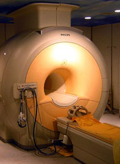
The nanoparticles boost the effectiveness of magnetic resonance imaging (MRI) by specifically seeking out CXCR4 receptors, which are found in cancerous cells.
The iron oxide nanoparticles are coated with peptide ligands that target tumor sites. When the particles find a tumor, they begin to interact with the cancerous cells.
Cancer-specific matrix metalloproteinase biomarkers prompt the nanoparticles to self-assemble into larger particles. And these larger particles are more visible on an MRI scan.
Researchers used cancer cells and mouse models to compare the effects of the self-assembling nanoparticles in MRI scanning against commonly used imaging agents. The nanoparticles produced a more powerful signal and created a clearer image of the tumor.
The team said the nanoparticles increase the sensitivity of MRI scans and could ultimately improve physicians’ ability to detect cancerous cells at much earlier stages of development.
“By improving the sensitivity of an MRI examination, our aim is to help doctors spot something that might be cancerous much more quickly,” said study author Nicholas Long, PhD, of Imperial College London in the UK. “This would enable patients to receive effective treatment sooner, which would hopefully improve survival rates from cancer.”
In addition to improving the sensitivity of MRI scans, the nanoparticles also appear to be safe. Before testing and injecting the particles into mice, the researchers had to ensure the particles would not become so big as to cause damage.
The team injected the particles into a saline solution inside a petri dish and monitored their growth over a 4-hour period. The nanoparticles grew from 100 nm to 800 nm, which was still small enough not to cause any harm.
Now, the researchers are working to enhance the effectiveness of the nanoparticles. And they hope to test their design in a human trial within the next 3 to 5 years.
“We would like to improve the design to make it even easier for doctors to spot a tumor and for surgeons to then operate on it,” Dr Long said. “We’re now trying to add an extra optical signal so that the nanoparticle would light up with a luminescent probe once it had found its target. So, combined with the better MRI signal, it will make it even easier to identify tumors.”
Self-assembling nanoparticles may help physicians diagnose cancers earlier, according to a study published in Angewandte Chemie.
The nanoparticles boost the effectiveness of magnetic resonance imaging (MRI) by specifically seeking out CXCR4 receptors, which are found in cancerous cells.
The iron oxide nanoparticles are coated with peptide ligands that target tumor sites. When the particles find a tumor, they begin to interact with the cancerous cells.
Cancer-specific matrix metalloproteinase biomarkers prompt the nanoparticles to self-assemble into larger particles. And these larger particles are more visible on an MRI scan.
Researchers used cancer cells and mouse models to compare the effects of the self-assembling nanoparticles in MRI scanning against commonly used imaging agents. The nanoparticles produced a more powerful signal and created a clearer image of the tumor.
The team said the nanoparticles increase the sensitivity of MRI scans and could ultimately improve physicians’ ability to detect cancerous cells at much earlier stages of development.
“By improving the sensitivity of an MRI examination, our aim is to help doctors spot something that might be cancerous much more quickly,” said study author Nicholas Long, PhD, of Imperial College London in the UK. “This would enable patients to receive effective treatment sooner, which would hopefully improve survival rates from cancer.”
In addition to improving the sensitivity of MRI scans, the nanoparticles also appear to be safe. Before testing and injecting the particles into mice, the researchers had to ensure the particles would not become so big as to cause damage.
The team injected the particles into a saline solution inside a petri dish and monitored their growth over a 4-hour period. The nanoparticles grew from 100 nm to 800 nm, which was still small enough not to cause any harm.
Now, the researchers are working to enhance the effectiveness of the nanoparticles. And they hope to test their design in a human trial within the next 3 to 5 years.
“We would like to improve the design to make it even easier for doctors to spot a tumor and for surgeons to then operate on it,” Dr Long said. “We’re now trying to add an extra optical signal so that the nanoparticle would light up with a luminescent probe once it had found its target. So, combined with the better MRI signal, it will make it even easier to identify tumors.”
Self-assembling nanoparticles may help physicians diagnose cancers earlier, according to a study published in Angewandte Chemie.
The nanoparticles boost the effectiveness of magnetic resonance imaging (MRI) by specifically seeking out CXCR4 receptors, which are found in cancerous cells.
The iron oxide nanoparticles are coated with peptide ligands that target tumor sites. When the particles find a tumor, they begin to interact with the cancerous cells.
Cancer-specific matrix metalloproteinase biomarkers prompt the nanoparticles to self-assemble into larger particles. And these larger particles are more visible on an MRI scan.
Researchers used cancer cells and mouse models to compare the effects of the self-assembling nanoparticles in MRI scanning against commonly used imaging agents. The nanoparticles produced a more powerful signal and created a clearer image of the tumor.
The team said the nanoparticles increase the sensitivity of MRI scans and could ultimately improve physicians’ ability to detect cancerous cells at much earlier stages of development.
“By improving the sensitivity of an MRI examination, our aim is to help doctors spot something that might be cancerous much more quickly,” said study author Nicholas Long, PhD, of Imperial College London in the UK. “This would enable patients to receive effective treatment sooner, which would hopefully improve survival rates from cancer.”
In addition to improving the sensitivity of MRI scans, the nanoparticles also appear to be safe. Before testing and injecting the particles into mice, the researchers had to ensure the particles would not become so big as to cause damage.
The team injected the particles into a saline solution inside a petri dish and monitored their growth over a 4-hour period. The nanoparticles grew from 100 nm to 800 nm, which was still small enough not to cause any harm.
Now, the researchers are working to enhance the effectiveness of the nanoparticles. And they hope to test their design in a human trial within the next 3 to 5 years.
“We would like to improve the design to make it even easier for doctors to spot a tumor and for surgeons to then operate on it,” Dr Long said. “We’re now trying to add an extra optical signal so that the nanoparticle would light up with a luminescent probe once it had found its target. So, combined with the better MRI signal, it will make it even easier to identify tumors.”
FDA approves product to treat attacks in HAE

The US Food and Drug Administration (FDA) has approved the first recombinant C1-esterase inhibitor product (Ruconest) for the treatment of acute attacks in adults and adolescents with hereditary angioedema (HAE).
HAE, which is caused by insufficient amounts of a plasma protein called C1-esterase inhibitor, affects approximately 6000 to 10,000 people in the US.
People with HAE can develop rapid swelling of the hands, feet, limbs, face, intestinal tract, or airway. These acute attacks can occur spontaneously or may be triggered by stress, surgery, or infection.
“Hereditary angioedema is a rare and potentially life-threatening disease,” said Karen Midthun, MD, director of the FDA’s Center for Biologics Evaluation and Research. “[The approval of Ruconest] provides an important treatment option for these patients.”
Ruconest is a human recombinant C1-esterase inhibitor purified from the milk of genetically modified rabbits. The product is intended to restore the level of functional C1-esterase inhibitor in a patient’s plasma, thereby treating the acute attack of swelling.
Trial results have suggested Ruconest is superior to placebo in treating most HAE attacks. However, due to the limited number of patients with laryngeal attacks, Ruconest has not been established as an effective treatment for these attacks.
The FDA approval of Ruconest to treat HAE is based on results of a phase 3, randomized, controlled trial (RCT), which included an open-label extension (OLE) phase, and is supported by the results of 2 additional RCTs and 2 additional OLE studies.
The pivotal RCT and OLE studies included 44 subjects who experienced 170 HAE attacks. The primary efficacy endpoint was the time to the beginning of symptom relief, assessed using patient-reported responses to 2 questions about the change in overall severity of their HAE attack symptoms after the start of treatment.
The researchers assessed these responses at regular time points for each of the affected anatomical locations for up to 24 hours. To achieve the primary endpoint, a patient had to have a positive response to both questions, along with persistence of improvement at the next assessment time (ie, the same or a better response).
There was a statistically significant difference in the time to the beginning of symptom relief in the intent-to-treat population (n=75) between the Ruconest and placebo arms (P=0.031).
The median time to the beginning of symptom relief was 90 minutes for Ruconest-treated patients (n=44) and 152 minutes for placebo-treated patients (n=31).
The most common adverse events, reported in at least 2% of patients receiving Ruconest, were headache, nausea, and diarrhea.
Serious adverse events associated with the treatment include anaphylaxis and arterial and venous thromboembolic events in patients with risk factors, such as an indwelling venous catheter/access device, a prior history of thrombosis, underlying atherosclerosis, the use of oral contraceptives or certain androgens, morbid obesity, and immobility.
Ruconest is manufactured by Pharming Group NV, located in Leiden, the Netherlands, and will be distributed in the US by Santarus Inc., a wholly owned subsidiary of Salix Pharmaceuticals Inc., which is located in Raleigh, North Carolina.
Salix is planning to make Ruconest available to patients later this year.
The US Food and Drug Administration (FDA) has approved the first recombinant C1-esterase inhibitor product (Ruconest) for the treatment of acute attacks in adults and adolescents with hereditary angioedema (HAE).
HAE, which is caused by insufficient amounts of a plasma protein called C1-esterase inhibitor, affects approximately 6000 to 10,000 people in the US.
People with HAE can develop rapid swelling of the hands, feet, limbs, face, intestinal tract, or airway. These acute attacks can occur spontaneously or may be triggered by stress, surgery, or infection.
“Hereditary angioedema is a rare and potentially life-threatening disease,” said Karen Midthun, MD, director of the FDA’s Center for Biologics Evaluation and Research. “[The approval of Ruconest] provides an important treatment option for these patients.”
Ruconest is a human recombinant C1-esterase inhibitor purified from the milk of genetically modified rabbits. The product is intended to restore the level of functional C1-esterase inhibitor in a patient’s plasma, thereby treating the acute attack of swelling.
Trial results have suggested Ruconest is superior to placebo in treating most HAE attacks. However, due to the limited number of patients with laryngeal attacks, Ruconest has not been established as an effective treatment for these attacks.
The FDA approval of Ruconest to treat HAE is based on results of a phase 3, randomized, controlled trial (RCT), which included an open-label extension (OLE) phase, and is supported by the results of 2 additional RCTs and 2 additional OLE studies.
The pivotal RCT and OLE studies included 44 subjects who experienced 170 HAE attacks. The primary efficacy endpoint was the time to the beginning of symptom relief, assessed using patient-reported responses to 2 questions about the change in overall severity of their HAE attack symptoms after the start of treatment.
The researchers assessed these responses at regular time points for each of the affected anatomical locations for up to 24 hours. To achieve the primary endpoint, a patient had to have a positive response to both questions, along with persistence of improvement at the next assessment time (ie, the same or a better response).
There was a statistically significant difference in the time to the beginning of symptom relief in the intent-to-treat population (n=75) between the Ruconest and placebo arms (P=0.031).
The median time to the beginning of symptom relief was 90 minutes for Ruconest-treated patients (n=44) and 152 minutes for placebo-treated patients (n=31).
The most common adverse events, reported in at least 2% of patients receiving Ruconest, were headache, nausea, and diarrhea.
Serious adverse events associated with the treatment include anaphylaxis and arterial and venous thromboembolic events in patients with risk factors, such as an indwelling venous catheter/access device, a prior history of thrombosis, underlying atherosclerosis, the use of oral contraceptives or certain androgens, morbid obesity, and immobility.
Ruconest is manufactured by Pharming Group NV, located in Leiden, the Netherlands, and will be distributed in the US by Santarus Inc., a wholly owned subsidiary of Salix Pharmaceuticals Inc., which is located in Raleigh, North Carolina.
Salix is planning to make Ruconest available to patients later this year.
The US Food and Drug Administration (FDA) has approved the first recombinant C1-esterase inhibitor product (Ruconest) for the treatment of acute attacks in adults and adolescents with hereditary angioedema (HAE).
HAE, which is caused by insufficient amounts of a plasma protein called C1-esterase inhibitor, affects approximately 6000 to 10,000 people in the US.
People with HAE can develop rapid swelling of the hands, feet, limbs, face, intestinal tract, or airway. These acute attacks can occur spontaneously or may be triggered by stress, surgery, or infection.
“Hereditary angioedema is a rare and potentially life-threatening disease,” said Karen Midthun, MD, director of the FDA’s Center for Biologics Evaluation and Research. “[The approval of Ruconest] provides an important treatment option for these patients.”
Ruconest is a human recombinant C1-esterase inhibitor purified from the milk of genetically modified rabbits. The product is intended to restore the level of functional C1-esterase inhibitor in a patient’s plasma, thereby treating the acute attack of swelling.
Trial results have suggested Ruconest is superior to placebo in treating most HAE attacks. However, due to the limited number of patients with laryngeal attacks, Ruconest has not been established as an effective treatment for these attacks.
The FDA approval of Ruconest to treat HAE is based on results of a phase 3, randomized, controlled trial (RCT), which included an open-label extension (OLE) phase, and is supported by the results of 2 additional RCTs and 2 additional OLE studies.
The pivotal RCT and OLE studies included 44 subjects who experienced 170 HAE attacks. The primary efficacy endpoint was the time to the beginning of symptom relief, assessed using patient-reported responses to 2 questions about the change in overall severity of their HAE attack symptoms after the start of treatment.
The researchers assessed these responses at regular time points for each of the affected anatomical locations for up to 24 hours. To achieve the primary endpoint, a patient had to have a positive response to both questions, along with persistence of improvement at the next assessment time (ie, the same or a better response).
There was a statistically significant difference in the time to the beginning of symptom relief in the intent-to-treat population (n=75) between the Ruconest and placebo arms (P=0.031).
The median time to the beginning of symptom relief was 90 minutes for Ruconest-treated patients (n=44) and 152 minutes for placebo-treated patients (n=31).
The most common adverse events, reported in at least 2% of patients receiving Ruconest, were headache, nausea, and diarrhea.
Serious adverse events associated with the treatment include anaphylaxis and arterial and venous thromboembolic events in patients with risk factors, such as an indwelling venous catheter/access device, a prior history of thrombosis, underlying atherosclerosis, the use of oral contraceptives or certain androgens, morbid obesity, and immobility.
Ruconest is manufactured by Pharming Group NV, located in Leiden, the Netherlands, and will be distributed in the US by Santarus Inc., a wholly owned subsidiary of Salix Pharmaceuticals Inc., which is located in Raleigh, North Carolina.
Salix is planning to make Ruconest available to patients later this year.
Team uncovers secrets of prothrombin structure, function

Credit: Darren Baker
In recreating the structure of prothrombin, researchers have improved their understanding of how the clotting factor functions.
By deleting a disordered linker region, they were able to visualize the complete structure of prothrombin.
This deleted version was activated to thrombin much faster than the intact version of prothrombin.
And results suggested that cofactor Va enhances the activation of prothrombin by altering the architecture of the linker.
Enrico Di Cera, MD, of Saint Louis University in Missouri, and his colleagues reported these findings in PNAS.
Last year, Dr Di Cera’s team published the first structure of prothrombin. This structure lacked a domain responsible for interaction with membranes, and certain other sections were not detected by X-ray analysis.
Though the researchers were able to crystallize the protein, there were disordered regions in the structure they could not see.
Within prothrombin, there are 2 kringle domains connected by a linker region that intrigued the researchers because of its intrinsic disorder.
“We deleted this linker, and crystals grew in a few days instead of months, revealing, for the first time, the full architecture of prothrombin,” Dr Di Cera said.
The crystal structure revealed a contorted conformation where the domains are not vertically stacked, kringle-1 comes close to the protease domain, and the Gla-domain contacts kringle-2.
The researchers also found the deleted version of prothrombin is activated to thrombin much faster than intact prothrombin.
Specifically, deletion of the linker reduced the enhancement of thrombin generation by cofactor Va from the more than 3000-fold observed with wild-type prothrombin to 60-fold. So it appears that deletion of the linker mimics the effect of cofactor Va on prothrombin activation.
“It took us almost 2 years to discover that the disordered linker was the key,” Dr Di Cera said. “Finally, prothrombin revealed its secrets, and, with that, the molecular mechanism of a key reaction of blood clotting finally becomes amenable to rational drug design for therapeutic intervention.”
Credit: Darren Baker
In recreating the structure of prothrombin, researchers have improved their understanding of how the clotting factor functions.
By deleting a disordered linker region, they were able to visualize the complete structure of prothrombin.
This deleted version was activated to thrombin much faster than the intact version of prothrombin.
And results suggested that cofactor Va enhances the activation of prothrombin by altering the architecture of the linker.
Enrico Di Cera, MD, of Saint Louis University in Missouri, and his colleagues reported these findings in PNAS.
Last year, Dr Di Cera’s team published the first structure of prothrombin. This structure lacked a domain responsible for interaction with membranes, and certain other sections were not detected by X-ray analysis.
Though the researchers were able to crystallize the protein, there were disordered regions in the structure they could not see.
Within prothrombin, there are 2 kringle domains connected by a linker region that intrigued the researchers because of its intrinsic disorder.
“We deleted this linker, and crystals grew in a few days instead of months, revealing, for the first time, the full architecture of prothrombin,” Dr Di Cera said.
The crystal structure revealed a contorted conformation where the domains are not vertically stacked, kringle-1 comes close to the protease domain, and the Gla-domain contacts kringle-2.
The researchers also found the deleted version of prothrombin is activated to thrombin much faster than intact prothrombin.
Specifically, deletion of the linker reduced the enhancement of thrombin generation by cofactor Va from the more than 3000-fold observed with wild-type prothrombin to 60-fold. So it appears that deletion of the linker mimics the effect of cofactor Va on prothrombin activation.
“It took us almost 2 years to discover that the disordered linker was the key,” Dr Di Cera said. “Finally, prothrombin revealed its secrets, and, with that, the molecular mechanism of a key reaction of blood clotting finally becomes amenable to rational drug design for therapeutic intervention.”
Credit: Darren Baker
In recreating the structure of prothrombin, researchers have improved their understanding of how the clotting factor functions.
By deleting a disordered linker region, they were able to visualize the complete structure of prothrombin.
This deleted version was activated to thrombin much faster than the intact version of prothrombin.
And results suggested that cofactor Va enhances the activation of prothrombin by altering the architecture of the linker.
Enrico Di Cera, MD, of Saint Louis University in Missouri, and his colleagues reported these findings in PNAS.
Last year, Dr Di Cera’s team published the first structure of prothrombin. This structure lacked a domain responsible for interaction with membranes, and certain other sections were not detected by X-ray analysis.
Though the researchers were able to crystallize the protein, there were disordered regions in the structure they could not see.
Within prothrombin, there are 2 kringle domains connected by a linker region that intrigued the researchers because of its intrinsic disorder.
“We deleted this linker, and crystals grew in a few days instead of months, revealing, for the first time, the full architecture of prothrombin,” Dr Di Cera said.
The crystal structure revealed a contorted conformation where the domains are not vertically stacked, kringle-1 comes close to the protease domain, and the Gla-domain contacts kringle-2.
The researchers also found the deleted version of prothrombin is activated to thrombin much faster than intact prothrombin.
Specifically, deletion of the linker reduced the enhancement of thrombin generation by cofactor Va from the more than 3000-fold observed with wild-type prothrombin to 60-fold. So it appears that deletion of the linker mimics the effect of cofactor Va on prothrombin activation.
“It took us almost 2 years to discover that the disordered linker was the key,” Dr Di Cera said. “Finally, prothrombin revealed its secrets, and, with that, the molecular mechanism of a key reaction of blood clotting finally becomes amenable to rational drug design for therapeutic intervention.”