User login
Protein proves essential for hematopoietic recovery

in the bone marrow
New research suggests the cell survival protein MCL-1, a target for a number of new anticancer agents, is essential for hematopoietic recovery.
Investigators found that reducing MCL-1 levels hindered hematopoietic recovery after chemotherapy and radiotherapy caused extensive destruction of mature blood cells.
Reducing MCL-1 also impaired reconstitution of the bone marrow after hematopoietic stem cell transplant (HSCT).
“Our previous research has shown that targeting MCL-1 could be used with great success for treating certain blood cancers,” said Alex Delbridge, PhD, of the Walter and Eliza Hall Institute of Medical Research in Melbourne, Victoria, Australia.
“However, we have now shown that MCL-1 is also critical for emergency recovery of the blood cell system after cancer therapy-induced blood cell loss.”
Dr Delbridge and his colleagues reported these findings in Blood.
Experiments in mice revealed that loss of a single MCL-1 allele, which reduced MCL-1 protein levels, greatly compromised the immune system and hindered red blood cell recovery after treatment with 5-fluorouracil, γ-irradiation, or HSCT.
Further investigation showed that the pro-apoptotic gene PUMA plays a key role in this phenomenon, as MCL-1 inhibits PUMA. In mice, knocking out PUMA alleviated—but did not eliminate—the HSC survival defect caused by deletion of both MCL-1 alleles.
“This exquisite dependency on MCL-1 for emergency blood cell production has important implications for potential cancer treatments involving MCL-1 inhibitors,” Dr Delbridge said.
“If MCL-1 inhibitors are to be used in combination with other cancer therapies, careful monitoring of the blood cell system will be needed,” added Stephanie Grabow, PhD, also of the Walter and Eliza Hall Institute.
“Our institute colleagues are working to evaluate a potential new drug to treat blood cancers by targeting MCL-1. Our findings suggest that MCL-1 inhibitors and chemotherapeutic drugs should not be used simultaneously.”
Dr Delbridge said this research also offers insights that could help improve HSCT.
“Stem cell transplants can be dangerous because, until the blood cell system is functionally restored, patients are vulnerable to infection,” he said. “Our research suggests that increasing levels of MCL-1 or decreasing the activity of opposing proteins could be a viable strategy for speeding up the regeneration process and reducing the risk of infection after stem cell transplantation.” ![]()
Low doses of imatinib promote hematopoiesis

Image by Volker Brinkmann
Low doses of the tyrosine kinase inhibitor imatinib can promote hematopoiesis, according to research published in PLOS Pathogens.
Preclinical experiments revealed that the drug can induce differentiation in hematopoietic stem cells (HSCs) and progenitors in the bone marrow,
augment myelopoiesis, and increase the number of myeloid cells in the blood and spleen.
Researchers said these findings suggest imatinib or related drugs could be used to treat infections.
“We think that low doses of imatinib are mimicking ‘emergency hematopoiesis,’ a normal early response to infection,” said study author Daniel Kalman, PhD, of the Emory University School of Medicine in Atlanta, Georgia.
“This was surprising because there are reports that imatinib can be immunosuppressive in some patients. Our data suggest that, at subclinical doses, imatinib can stimulate bone marrow stem cells to produce several types of myeloid cells, such as neutrophils and macrophages, and trigger their exodus from the bone marrow. However, higher doses appear to inhibit this process.”
Dr Kalman and his colleagues observed a 4-fold increase of neutrophils and a 3-fold increase of monocytes in the bone marrow of imatinib-treated mice. However, these mice did not see a significant change in the number of mature B cells, T cells, dendritic cells, eosinophils, or natural killer cells.
Imatinib did not induce the accumulation of HSCs, but it did regulate the accumulation of multipotent progenitors. The drug also reduced the accumulation of HSCs upon infection, which suggests it may increase the flux of HSCs to progenitors.
Imatinib did not increase the number of transplantable HSCs, but it did induce an irreversible commitment of HSCs into progenitors that could differentiate into myeloid cells ex vivo.
Imatinib induced an irreversible differentiation of HSCs or progenitors into myeloid cells in a dose-dependent manner. The drug facilitated the exodus of myeloid cells from the bone marrow only at lower doses. It inhibited this same process at higher doses.
Despite increasing the number of neutrophils, low doses of imatinib did not activate neutrophils. However, the cells retained the capacity to activate upon infection.
In fact, an increase in the number of neutrophils was sufficient to reduce Mycobacterium marinum bacterial load. And imatinib reduced the bacterial load in mice infected with pathogenic Francisella species.
The researchers said these results suggest low doses of imatinib could potentially be used to treat a variety of infections and might prove particularly useful in immunocompromised patients. ![]()
Collaboration may help prevent CLABSIs

Collaborative relationships between nurses and physicians may help reduce the rates of healthcare-associated infections in critical care, according to research published in Critical Care Nurse.
Study investigators found lower rates of central line-associated bloodstream infections (CLABSIs) and ventilator-associated pneumonia (VAP) in critical care units in which nurses reported a more favorable perception of nurse-physician collaboration.
“Our findings suggest that raising the quality of collaboration and communication among nurses and physicians has the potential to improve patient safety,” said study author Christine Boev, RN, PhD, CCRN, of Wegmans School of Nursing at St John Fisher College in Rochester, New York.
Dr Boev and her colleagues analyzed 5 years of data from 671 surveys of nurses in 4 specialized intensive care units (ICUs) at a 750-bed New York hospital.
The investigators also collected patient outcome data from those units for the same period, focusing on patients with CLABSIs or VAP. And the team analyzed unit-level variables such as nurses’ skill mix, nursing hours per patient day, and voluntary turnover.
Results revealed a significant association between nurse-physician collaboration and both CLABSIs and VAP. For every 0.5 unit increase in collaboration, the rate of CLABSIs decreased by 2.98 (P=0.005), and the rate of VAP decreased by 1.13 (P=0.005).
In addition, ICUs with a higher proportion of certified nurses had significantly lower incidences of both CLABSIs and VAP—0.43 (P=0.02) and 0.17 (P=0.01), respectively. And ICUs with higher numbers of nursing hours per patient day had significantly lower rates of CLABSIs—0.42 (P=0.05).
However, there was no significant difference in VAP rates according to nursing hours. And there was no significant difference in the rate of either type of infection according to nurses’ skill mix or voluntary turnover.
Dr Boev said these results suggest that efforts to prevent healthcare-associated infections should include interventions to improve nurse-physician collaboration. Such interventions might include multidisciplinary daily patient rounds and interprofessional educational programs, such as shared simulation training. ![]()
Mouse model could aid study of malaria
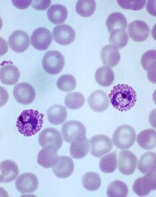
Plasmodium vivax infection
Image by Mae Melvin
Researchers say they have developed a human-chimeric mouse model that can advance the study of the malaria parasite Plasmodium vivax.
The model is engineered to grow human livers that can be infected with P vivax and used for investigations into parasite development, dormancy, and activation, as well as the effect certain drugs have on each aspect of the infection.
The researchers described this model in Cell Host & Microbe.
Stefan Kappe, PhD, of Seattle Biomedical Research Institute in Washington, and his colleagues noted that P vivax malaria parasites are more resistant to control and elimination than other malaria parasites because, after infection, they fall dormant within the liver for months to years.
When these dormant parasites eventually become activated, they replicate, infect the bloodstream, and cause relapsing malaria.
The lack of tractable P vivax animal models has made it difficult to examine P vivax liver-stage infection, but Dr Kappe and his colleagues found a solution in a model known as FRG KO huHep.
To create the FRG KO huHep model, the team transplanted human hepatocytes in the severely immunocompromised FRG KO mouse, which has deletions in fumarylacetoacetate hydrolase (FAH), recombinationactivating gene 2 (Rag2), and interleukin-2 receptor subunit gamma (Il2rg).
The researchers found that FRG KO huHep mice support P vivax sporozoite infection, liver-stage development, and the formation and persistence of dormant liver-stage parasites.
In addition, experiments with this model revealed that the antimalarial drug primaquine could prevent and eliminate liver-stage infection.
The researchers therefore concluded that P vivax-infected FRG KO huHep mice are a suitable model for studying liver-stage development and dormancy and may facilitate the discovery of drugs targeting relapsing malaria.
“This model is a real game-changer,” said Ivo Mueller, PhD, a malaria expert at the Walter and Eliza Hall Institute in Melbourne, Victoria, Australia, who was not involved in this research.
“For the first time, we now have a realistic opportunity that not only allows us to directly study this normally hidden P vivax life-stage, but also to test new drugs and vaccines. This new model is an essential resource in our quest to develop the new anti-vivax interventions that we will need to eliminate P vivax malaria.” ![]()
New administration option for antiplatelet drug

Photo courtesy of AstraZeneca
The US Food and Drug Administration (FDA) has approved a new administration option for the antiplatelet agent ticagrelor (Brilinta).
The agency has decided that, for patients with acute coronary syndrome (ACS) who cannot swallow ticagrelor tablets whole, the pills may be crushed and administered in water by swallowing or via nasogastric tube.
Ticagrelor is the only P2Y12 inhibitor that is FDA-approved to be administered in this way.
“We know that some patients who experience a heart attack are unable to swallow medications whole, yet it is important for these patients to receive and continue their oral antiplatelet therapy,” said Steven Zelenkofske, DO, Vice President of US Medical Affairs, Cardiovascular, at AstraZeneca, the company developing ticagrelor.
Survey data have shown that 40% of adults in the general population experience problems swallowing pills, and this difficulty may increase with age. Moreover, some patients who experience a heart attack have difficulty swallowing medications in the emergency setting.
So the new administration option for ticagrelor is intended to give healthcare professionals flexibility in treating their ACS patients.
Ticagrelor is a direct-acting P2Y12 receptor antagonist that works by inhibiting platelet activation. The drug is available in 90 mg tablets, to be administered with a single 180 mg oral loading dose (two 90 mg tablets) followed by a twice daily, 90 mg maintenance dose.
Following an initial loading dose of aspirin, ticagrelor should be used with a maintenance dose of aspirin at 75 mg to 100 mg once daily (an 81 mg dose in the US).
Results of the PLATO trial showed that, in ACS patients, ticagrelor can reduce the rate of a combined endpoint of cardiovascular death, myocardial infarction, and stroke, when compared to clopidogrel. The difference between treatments was driven by cardiovascular death and myocardial infarction, with no difference in the rate of stroke.
For more information on ticagrelor, see the full prescribing information. ![]()
AML evolution proves unexpectedly complex

Results of single-cell genotyping suggest the evolution of acute myeloid leukemia (AML) is more complex than we thought.
In screening for mutations in 2 genes, researchers identified at least 9 distinct clonal populations, each harboring unique mutational patterns.
Some mutations seemed to arise not sequentially, but independently and at different points in time.
These results suggest single-cell analysis may be more effective than bulk-tumor analysis for assessing cancer evolution.
Carlo Maley, PhD, of Arizona State University in Tempe, and his colleagues recounted the results in Science Translational Medicine.
The researchers set out to provide a more accurate picture of what takes place at the genetic level when an AML patient experiences relapse or metastasis. So they examined individual cells, screening them for mutations in FLT3 and NPM1.
The results suggested the same mutation was occurring multiple times within an AML patient. The team examined individual cells from 6 AML patients, and the results showed all combinations of homozygous and heterozygous mutations of FLT3 and NPM1.
“There’s no way to explain that with each mutation only happening once,” Dr Maley said. “That’s scary because it means that these cancers have access to many mutations and can find the same mutation over and over.”
Dr Maley noted that the process of convergent evolution, in which separate lineages develop similar features, appears to account for some of the observed diversity. And influences from the environment may drive convergent evolution, but identical mutations can also arise through pure coincidence, simply by virtue of the enormous numbers involved.
For example, a 1 cm3 AML tumor may contain a billion cells, each containing some 3 billion base pairs in its genome. Mutations are estimated to occur at a rate of 1 mutation in every billion base pairs.
“That means every time the population of cells in a 1 cm3 tumor undergoes 1 generation, which we think takes just a couple days, every possible mutation of the genome is happening somewhere in that tumor,” Dr Maley said.
This alone would lead to the same mutation likely occurring independently multiple times.
Curbing cancer’s lethality
Given AML’s near-limitless capacity for creating novel variants, what can clinicians do to halt the disease’s advance? According to Dr Maley, one approach would be to use cancer’s ability to evolve to our advantage, rather than attempt to fight it head on.
This paradigm draws on a branch of ecology known as life history theory. The idea is to carefully study the environmental factors that may lead organisms to favor either a fast or slow reproducing strategy to maximize their ability to survive.
According to the theory, fast reproduction tends to occur in environments with high extrinsic mortality. Aggressive cancer treatment creates just such an environment, favoring those cells able to reproduce quickly, producing large numbers of daughter cells, with a few evading extrinsic mortality to repopulate the tumor.
On the other hand, a very stable environment often favors slow reproduction, because organisms reach a carrying capacity of their surrounding environment. In this case, the limiting factor becomes competition between like organisms. Here, a slow reproducing strategy favoring greater investment in maintenance and survivability wins the competition.
“This approach would say, ‘Let’s keep tumors as stable as possible and keep their resources limited,’” Dr Maley said. “If we are able to keep the tumor cells contained and let them fight it out, we would expect to see more competitively fit cells that are growing very slowly.” ![]()
‘Junk’ RNA produces lymphoma phenotype

New research suggests the non-coding BRAF pseudogene can produce a lymphoma phenotype in mice.
Pseudogenes, a sub-class of long non-coding RNA that developed from the genome’s protein-coding genes but lost the ability to produce proteins, have long been considered genomic “junk.”
Yet the retention of pseudogenes during evolution has suggested they may have biological functions and contribute to disease development.
Now, researchers have provided evidence that one of these pseudogenes does have a role in causing cancer, and they described the discovery in Cell.
The investigators found that, independent of any other mutations, abnormal amounts of the BRAF pseudogene led to the development of an aggressive, lymphoma-like disease in mice.
The team said this finding suggests pseudogenes may play a primary role in a variety of diseases, and the functional genome could be much larger than we thought.
“Our mouse model of the BRAF pseudogene developed cancer as rapidly and aggressively as it would if you were to express the protein-coding BRAF oncogene,” said study author Pier Paolo Pandolfi, MD, PhD, of Harvard Medical School in Boston, Massachusetts.
“It’s remarkable that this very aggressive phenotype, resembling human diffuse large B-cell lymphoma, was driven by a piece of so-called ‘junk RNA.’”
The researchers’ discovery hinges on the concept of competing endogenous RNAs (ceRNA), a functional capability for pseudogenes Dr Pandolfi and his colleagues described almost 5 years ago. The team discovered that pseudogenes and other noncoding RNAs could act as “decoys” to divert and sequester microRNAs away from their protein-coding counterparts to regulate gene expression.
“Our discovery of these ‘decoys’ revealed a new role for messenger RNA, demonstrating that, beyond serving as a genetic intermediary in the protein-making process, messenger RNAs could actually regulate expression of one another through this sophisticated new ceRNA ‘language,’” Dr Pandolfi said.
He and his colleagues showed that, when these decoys prevented microRNAs from fulfilling their regulatory function, there could be severe consequences, including making cancer cells more aggressive.
With their new research, the investigators wanted to determine if this same ceRNA cross-talk took place in a living organism and if it would result in similar consequences.
“We conducted a proof-of-principle experiment using the BRAF pseudogene,” explained Florian Karreth, PhD, a fellow in the Pandolfi lab. “We investigated whether this pseudogene exerts critical functions in the context of a whole organism and whether its disruption contributes to the development of disease.”
The researchers focused on the BRAF pseudogene because of its potential ability to regulate levels of the BRAF protein, a well-known proto-oncogene linked to numerous cancer types. In addition, the BRAF pseudogene is known to exist in both humans and mice.
The team began by testing the BRAF pseudogene in tissue culture. Their findings showed that, when overexpressed, the pseudogene operated as a microRNA decoy that increased amounts of the BRAF protein.
This, in turn, stimulated the MAPK signaling cascade, a pathway through which the BRAF protein controls cell proliferation, differentiation, and survival and which is commonly found to be hyperactive in cancer.
The investigators went on to create a mouse model in which the BRAF pseudogene was overexpressed. And they found these mice developed an aggressive, lymphoma-like cancer.
“This cancer of B-lymphocytes manifested primarily in the spleens of the animals but also infiltrated other organs, including the kidneys and liver,” Dr Karreth said. “We were particularly surprised by the development of such a dramatic phenotype in response to BRAF pseudogene overexpression alone since the development of full-blown cancer usually requires two or more mutational events.”
Mice overexpressing the BRAF pseudogene displayed higher levels of the BRAF protein and hyperactivation of the MAPK pathway, which suggests this axis is critical to cancer development.
The researchers confirmed this by inhibiting the MAPK pathway with GSK1120212, a MEK inhibitor that dramatically reduced the cancer cells’ ability to infiltrate the liver in transplantation experiments.
The investigators further validated the microRNA decoy function of the BRAF pseudogene by creating two additional transgenic mice, one overexpressing the front half of the BRAF pseudogene, and the other overexpressing the back half.
Both of these mouse models developed the same lymphoma phenotype as the mice overexpressing the full-length pseudogene, a result the researchers described as “astonishing.”
“We never expected that portions of the BRAF pseudogene could elicit a phenotype,” Dr Karreth said. “[W]hen both front and back halves induced lymphomas, we were certain the BRAF pseudogene was functioning as a microRNA decoy.”
The investigators also found the BRAF pseudogene is overexpressed in human B-cell lymphomas, and the genomic region containing the BRAF pseudogene is commonly amplified in a variety of human cancers. This suggests the group’s murine findings are of relevance to human cancer development.
Moreover, the researchers found that silencing the BRAF pseudogene in human cancer cell lines that expressed higher levels led to reduced cell proliferation. This reinforces the importance of the pseudogene and suggests a therapy that reduces BRAF pseudogene levels may be beneficial to certain cancer patients.
“While we have been busy focusing on the genome’s 20,000 coding genes, we have neglected perhaps as many as 100,000 noncoding genetic units,” Dr Pandolfi noted. “Our new findings not only tell us that we need to characterize the role of all of these non-coding pseudogenes in cancer, but, more urgently, suggest that we need to increase our understanding of the non-coding ‘junk’ of the genome and incorporate this information into our personalized medicine assays.”
“The game has to start now. We have to sequence and analyze the genome and the RNA transcripts from the non-coding space.” ![]()
New research suggests the non-coding BRAF pseudogene can produce a lymphoma phenotype in mice.
Pseudogenes, a sub-class of long non-coding RNA that developed from the genome’s protein-coding genes but lost the ability to produce proteins, have long been considered genomic “junk.”
Yet the retention of pseudogenes during evolution has suggested they may have biological functions and contribute to disease development.
Now, researchers have provided evidence that one of these pseudogenes does have a role in causing cancer, and they described the discovery in Cell.
The investigators found that, independent of any other mutations, abnormal amounts of the BRAF pseudogene led to the development of an aggressive, lymphoma-like disease in mice.
The team said this finding suggests pseudogenes may play a primary role in a variety of diseases, and the functional genome could be much larger than we thought.
“Our mouse model of the BRAF pseudogene developed cancer as rapidly and aggressively as it would if you were to express the protein-coding BRAF oncogene,” said study author Pier Paolo Pandolfi, MD, PhD, of Harvard Medical School in Boston, Massachusetts.
“It’s remarkable that this very aggressive phenotype, resembling human diffuse large B-cell lymphoma, was driven by a piece of so-called ‘junk RNA.’”
The researchers’ discovery hinges on the concept of competing endogenous RNAs (ceRNA), a functional capability for pseudogenes Dr Pandolfi and his colleagues described almost 5 years ago. The team discovered that pseudogenes and other noncoding RNAs could act as “decoys” to divert and sequester microRNAs away from their protein-coding counterparts to regulate gene expression.
“Our discovery of these ‘decoys’ revealed a new role for messenger RNA, demonstrating that, beyond serving as a genetic intermediary in the protein-making process, messenger RNAs could actually regulate expression of one another through this sophisticated new ceRNA ‘language,’” Dr Pandolfi said.
He and his colleagues showed that, when these decoys prevented microRNAs from fulfilling their regulatory function, there could be severe consequences, including making cancer cells more aggressive.
With their new research, the investigators wanted to determine if this same ceRNA cross-talk took place in a living organism and if it would result in similar consequences.
“We conducted a proof-of-principle experiment using the BRAF pseudogene,” explained Florian Karreth, PhD, a fellow in the Pandolfi lab. “We investigated whether this pseudogene exerts critical functions in the context of a whole organism and whether its disruption contributes to the development of disease.”
The researchers focused on the BRAF pseudogene because of its potential ability to regulate levels of the BRAF protein, a well-known proto-oncogene linked to numerous cancer types. In addition, the BRAF pseudogene is known to exist in both humans and mice.
The team began by testing the BRAF pseudogene in tissue culture. Their findings showed that, when overexpressed, the pseudogene operated as a microRNA decoy that increased amounts of the BRAF protein.
This, in turn, stimulated the MAPK signaling cascade, a pathway through which the BRAF protein controls cell proliferation, differentiation, and survival and which is commonly found to be hyperactive in cancer.
The investigators went on to create a mouse model in which the BRAF pseudogene was overexpressed. And they found these mice developed an aggressive, lymphoma-like cancer.
“This cancer of B-lymphocytes manifested primarily in the spleens of the animals but also infiltrated other organs, including the kidneys and liver,” Dr Karreth said. “We were particularly surprised by the development of such a dramatic phenotype in response to BRAF pseudogene overexpression alone since the development of full-blown cancer usually requires two or more mutational events.”
Mice overexpressing the BRAF pseudogene displayed higher levels of the BRAF protein and hyperactivation of the MAPK pathway, which suggests this axis is critical to cancer development.
The researchers confirmed this by inhibiting the MAPK pathway with GSK1120212, a MEK inhibitor that dramatically reduced the cancer cells’ ability to infiltrate the liver in transplantation experiments.
The investigators further validated the microRNA decoy function of the BRAF pseudogene by creating two additional transgenic mice, one overexpressing the front half of the BRAF pseudogene, and the other overexpressing the back half.
Both of these mouse models developed the same lymphoma phenotype as the mice overexpressing the full-length pseudogene, a result the researchers described as “astonishing.”
“We never expected that portions of the BRAF pseudogene could elicit a phenotype,” Dr Karreth said. “[W]hen both front and back halves induced lymphomas, we were certain the BRAF pseudogene was functioning as a microRNA decoy.”
The investigators also found the BRAF pseudogene is overexpressed in human B-cell lymphomas, and the genomic region containing the BRAF pseudogene is commonly amplified in a variety of human cancers. This suggests the group’s murine findings are of relevance to human cancer development.
Moreover, the researchers found that silencing the BRAF pseudogene in human cancer cell lines that expressed higher levels led to reduced cell proliferation. This reinforces the importance of the pseudogene and suggests a therapy that reduces BRAF pseudogene levels may be beneficial to certain cancer patients.
“While we have been busy focusing on the genome’s 20,000 coding genes, we have neglected perhaps as many as 100,000 noncoding genetic units,” Dr Pandolfi noted. “Our new findings not only tell us that we need to characterize the role of all of these non-coding pseudogenes in cancer, but, more urgently, suggest that we need to increase our understanding of the non-coding ‘junk’ of the genome and incorporate this information into our personalized medicine assays.”
“The game has to start now. We have to sequence and analyze the genome and the RNA transcripts from the non-coding space.”
Fish oil may cause chemoresistance

Consuming certain types of fish and taking fish oil supplements may induce chemoresistance, according to research published in JAMA Oncology.
Researchers found that herring and mackerel, as well as 6 different types of fish oil supplements, raised blood levels of the fatty acid 16:4(n-3).
And experiments in mice showed that small amounts of either purified 16:4(n-3) or fish oil induced resistance to the chemotherapy drug cisplatin.
Emile E. Voest, MD, PhD, of the Netherlands Cancer Institute in Amsterdam, and his colleagues conducted this multi-part study.
In one part, the team conducted a survey to determine the rate of fish oil use among patients undergoing cancer treatment (n=118). Thirty-five patients (30%) reported regular use of nutritional supplements, and 13 (11%) said they used supplements containing omega-3 fatty acids.
For another part of the study, the researchers evaluated 6 types of fish oil supplements. All of them contained relevant amounts of 16:4(n-3), ranging from 0.2 µM to 5.7 µM.
The team also recruited healthy volunteers to examine blood levels of 16:4(n-3) after the ingestion of fish oil supplements (n=30) and fish (n=20).
Volunteers had increased blood levels of 16:4(n-3) after the recommended daily amount of 10 mL of fish oil and after a 50 mL dose. Subjects had an almost-complete normalization of blood levels 8 hours after a 10 mL fish oil dose, but they had a more prolonged elevation of fatty acid levels after a 50 mL dose.
Eating 100 grams of herring and mackerel also increased blood levels of 16:4(n-3) compared with tuna, which did not affect blood levels, and salmon,
which resulted in a small, short-lived peak.
Finally, experiments in mice showed that as little as 2.5 pmol of purified 16:4(n-3) or 1 µL of fish oil was sufficient to induce resistance to the chemotherapy drug cisplatin.
The fish oil/cisplatin combination had no significant impact on mouse tumors when compared to vehicle control treatment. The estimated tumor volume difference was 44.1 mm3 (P >0.99).
When the researchers compared cisplatin alone to cisplatin plus 16:4(n-3), the estimated tumor volume difference was 95.5 mm3 (P=0 .04). When they compared vehicle control to cisplatin alone, there was an estimated tumor volume difference of 142.4 mm3 (P=0.001).
The team said these results suggest that simultaneous exposure to chemotherapy and fish oil may be detrimental to cancer patients. So until further data become available, patients should avoid consuming fish oil from the day before chemotherapy until the day after.
Japan approves pomalidomide for MM

Photo by Steven Harbour
Japan’s Ministry of Health, Labour and Welfare (MHLW) has granted full marketing authorization for pomalidomide (Pomalyst) to treat patients with relapsed or refractory multiple myeloma (MM) who have previously received lenalidomide and bortezomib.
Pomalidomide will be available in Japan through RevMate, a restricted distribution program that should ensure the proper use of pomalidomide and prevent fetal exposure to the drug.
The MHLW approved pomalidomide based on results of trials in patients with previously treated MM, including the phase 3 MM-003 trial and the phase 2 MM-011 trial.
Phase 2 trial
In the MM-011 trial, which has not yet been published, researchers evaluated the safety and efficacy of pomalidomide in combination with dexamethasone in 36 patients with relapsed/refractory MM.
The patients had received at least 2 prior therapies, including lenalidomide and bortezomib, and had disease progression within 60 days of completing their last therapy.
Twenty-five percent of patients responded to treatment with pomalidomide and dexamethasone, 1 with a complete response and 8 with a partial response.
About 89% of patients (32/36) experienced adverse events, including lab test abnormalities. The most common events were neutropenia (69.4%), thrombocytopenia (33.3%), rash (22.2%), leukocytopenia (13.9%), pyrexia (13.9%), anemia (11.1%), lymphopenia (11.1%), and constipation (11.1%).
Phase 3 trial
The MM-003 study included 455 patients with relapsed or refractory MM. They were randomized to receive either pomalidomide plus low-dose dexamethasone (POM-LoDEX, n=302) or high-dose dexamethasone (HiDEX, n=153). The median follow-up was 10 months.
The overall response rate was 31% (n=95) in the POM-LoDEX arm and 10% (n=15) in the HiDEX arm. The median duration of response was 7.0 months (range, 6 to 9 months) and 6.1 months (range, 1.4 to 8.5 months), respectively.
The median progression-free survival was 4.0 months in the POM-LoDEX arm and 1.9 months in the HiDEX arm (P<0.001). And the median overall survival was 12.7 months in the POM-LoDEX arm and 8.1 months in the HiDEX arm (P=0.028).
Patients in the POM-LoDEX arm experienced more grade 3/4 neutropenia than patients in the HiDEX arm. But rates of grade 3/4 anemia and thrombocytopenia were similar between the 2 arms.
Rates of grade 3/4 non-hematologic toxicities were also similar between the arms and included infection, pneumonia, hemorrhage, glucose intolerance, and fatigue. Other adverse events of note included venous thromboembolism and peripheral neuropathy, which occurred at similar rates in both arms.
Test predicts DLBCL relapse better than CT, team says

Photo by Larry Young
Surveillance of circulating tumor DNA (ctDNA) can help predict relapse in most patients with diffuse large B-cell lymphoma before there is clinical evidence of the disease, according to a study published in The Lancet Oncology.
Investigators analyzed ctDNA using the clonoSEQ minimal residual disease (MRD) test and found they could predict relapse with a positive predictive value (PPV) of 88% and a negative predictive value (NPV) of 98%.
The test detected relapse a median of 3.5 months quicker than computed tomography (CT) scans.
“Patients with DLBCL with low amounts of disease at relapse have better survival than those with more disease, which is the rationale for surveillance CT scans,” said study author Wyndham Wilson, MD, PhD, of the National Cancer Institute in Bethesda, Maryland.
“Because the ctDNA test detects disease at a molecular level, it detects microscopic disease, which cannot be detected by CT scans, and may improve patient survival. Furthermore, ctDNA is non-invasive and can be employed as frequently needed, unlike surveillance CT scans, which expose patients to radiation and intravenous contrast.”
For this study, Dr Wilson and his colleagues evaluated 126 DLBCL patients who had participated in clinical trials from May 1993 to June 2013 and were followed for a median of 11 years post-treatment.
Surveillance monitoring
To investigate whether ctDNA monitoring could overcome the limitations of standard imaging techniques, the researchers compared serial ctDNA samples to CT scans taken at the same time post-treatment in patients who had achieved complete remission. This was known as “surveillance monitoring.”
The investigators performed surveillance monitoring of ctDNA in 107 patients who achieved complete remission.
The hazard ratio for clinical disease progression was 228 for patients who had detectable ctDNA during surveillance, when compared to patients with undetectable ctDNA (P<0.0001).
Surveillance ctDNA had a PPV of 88.2% and an NPV of 97.8%. And it revealed the risk of recurrence at a median of 3.5 months (range, 0-200 months) before there was evidence of clinical disease.
Interim monitoring
The researchers also analyzed whether the presence of ctDNA at the beginning of the third cycle of treatment predicted relapse, regardless of whether patients achieved complete remission by the end of treatment. This was known as “interim monitoring.”
Of the 108 patients included in the interim monitoring analysis, ctDNA was detected in 24 patients, 15 of whom eventually relapsed. Only 17 of the 84 patients with undetectable interim ctDNA relapsed.
Five years after the interim serum samples were taken, 80.2% of the patients who were negative for ctDNA were relapse-free, as were 41.7% of patients who were positive for ctDNA (P<0.0001).
Detectable interim ctDNA had a PPV of 62.5% and an NPV of 79.8%.
Fourteen of the 15 patients with detectable ctDNA who relapsed did so within 6 months of the end of treatment, as did 7 of the 17 patients without interim ctDNA.
Based on these results, the investigators concluded that surveillance monitoring of ctDNA identifies DLBCL patients at risk of disease recurrence before clinical evidence of disease in most patients, and interim monitoring of ctDNA is a promising biomarker to identify patients at high risk of treatment failure.
This research was funded by Adaptive Biotechnologies, the company developing the clonoSEQ MRD test, as well as the National Cancer Institute.