User login
Uninsured cancer patients pay more

Photo by Rhoda Baer
Uninsured patients are asked to pay much more for cancer treatments than Medicare and private insurers pay, according to research published in Health Affairs.
Researchers reviewed newly available Medicare data on what physicians charged for intravenous chemotherapy drugs in 2012.
And the results showed that uninsured cancer patients were asked to pay anywhere from 2 to 43 times what Medicare would pay and 2 to 5 times as
much as private insurance would pay.
For example, uninsured patients who did not negotiate the billed amounts could expect to pay $6711 for an infusion of the colorectal cancer drug oxaliplatin. But Medicare and private health plans only pay $3090 and $3616, respectively.
“Patients with Medicare and private insurance don’t pay the sticker price of healthcare,” said study author Stacie Dusetzina, PhD, of the University of North Carolina at Chapel Hill.
“They pay a discounted rate. However, uninsured patients don’t have the bargaining power, or they may not try to negotiate for a better price.”
In addition to estimating costs for infused chemotherapy drugs, Dr Dusetzina and her colleagues looked at what cancer patients were asked to pay for a doctor visit.
Uninsured patients were billed between $129 and $391, depending on the complexity of the visit. Medicare paid between $65 and $188, and private insurance paid $78 to $246 for the same visits.
“This is unreasonable,” Dr Dusetzina said. “There needs to be more transparency and less variability in healthcare pricing.”
Under the Affordable Care Act, everyone in the US must be insured or face a tax penalty. However, many Americans remain without insurance. So differences in what the uninsured are charged really matter, Dr Dusetzina said.
“In states like North Carolina that didn’t expand Medicaid, there is a large group of people who can get insurance on the federal exchange but cannot get subsidies because the law assumed they would be covered by Medicaid,” she noted.
This population earns at or near the federal poverty level. Without subsidies to help pay for private insurance, many are likely to remain uninsured.
These drug pricing discrepancies could become even more important depending on the outcome of the pending Supreme Court case King vs Burwell, which challenges the legitimacy of federal healthcare subsidies and could leave as many as 8 million Americans without subsidies and uninsured in 2016. ![]()
FDA grants drug orphan designation for SCD
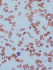
Image by Graham Beards
The US Food and Drug Administration (FDA) has granted orphan drug designation for the bovine PEGylated carboxyhemoglobin product Sanguinate to treat sickle cell disease (SCD).
Through its anti-vaso-constrictive properties, Sanguinate facilitates the transfer of oxygen to oxygen-deprived cells and tissues.
By correcting oxygen levels and downregulating inflammation, the drug could potentially treat many of the comorbidities associated with SCD.
Trials of Sanguinate
The company developing Sanguinate, Prolong Pharmaceuticals, has several clinical studies underway to determine the safety and efficacy of the drug in SCD and other diseases caused by the effects of oxygen deprivation.
In a phase 1 trial, Sanguinate proved safe and well-tolerated in healthy volunteers. Three cohorts of 8 subjects received single, ascending doses of Sanguinate at 80 mg/kg, 120 mg/kg, or 160 mg/kg. Two volunteers in each cohort were control subjects who received saline.
There were no serious adverse events reported with Sanguinate. Subjects experienced decreases in serum haptoglobin, but this did not appear to be dose-related. Sanguinate’s half-life was dose-dependent and ranged from 7.9 hours to 13.8 hours.
A phase 1 study of Sanguinate in SCD patients has been completed, and researchers are now conducting a phase 2 study testing the drug for the reduction or prevention of delayed cerebral ischemia following subarachnoid hemorrhage.
Phase 2 trials are also planned for vaso-occlusive crisis and leg ulcers secondary to SCD, as well as for preventing delayed graft function following kidney transplant. Sanguinate is also being evaluated for the treatment of beta-thalassemia.
About orphan designation
The FDA grants orphan designation to diseases affecting fewer than 200,000 people in the US.
Orphan designation provides the company developing a drug with certain benefits and incentives, including a 7-year period of marketing exclusivity upon regulatory approval, potential tax credits for certain activities, eligibility for orphan drug grants, and the waiver of certain administrative fees. ![]()
Environmental factors affect bloodstream infections

New research suggests environmental factors can affect the development of catheter-related bloodstream infections (CRBSIs) in patients who
receive parenteral nutrition therapy at home.
Using a peripherally inserted central venous catheter (PICC) for 1 additional infusion day per week significantly reduced the amount of time before a first CRBSI, and using a tunneled vascular access device managed by a home care nurse increased the mean incidence of CRBSIs.
Laura Fuglsang Bech, of Aalborg University in Denmark, and her colleagues reported these results in the Journal of Parenteral and Enteral Nutrition.
The researchers set out to determine if environmental factors play a role in the development of bloodstream infections among patients receiving parenteral nutrition therapy via vascular access devices or PICCs, the 2 most commonly used catheters.
The team looked at factors such as smoking, catheter management by a home care nurse, colectomy with stoma, number of infusion days per week, and C-reactive protein values at catheter insertion day.
Adult patients suffering from intestinal failure and receiving home parenteral nutrition were included in the study. There were 295 catheters—169 tunneled vascular access devices and 126 PICCs—used in 136 patients.
The researchers found that using a PICC for 1 additional infusion day per week significantly reduced the amount of time before a first bloodstream infection. The time to first CRBSI decreased by a factor of 2.47 with 1 additional infusion day per week (P=0.04).
The team also found that using a tunneled vascular access device managed by a home care nurse increased the mean incidence of bloodstream infections. The mean CRBSI incidence per 1000 catheter days was 1.45± 0.68 for catheters managed by a home care nurse and 0.56 ± 0.24 for catheters that were not (P<0.001).
None of the other factors the researchers analyzed had any significant impact on the timing or incidence of CRBSIs.
Based on these results, the researchers recommended revisions to current home care guidelines. They advised using PICCs only for short-term home therapy and when few infusion days per week are needed. And they said management of tunneled vascular access devices by home care nurses should be further specialized. ![]()
Team discovers transmissible leukemia in clams
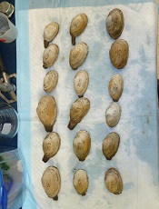
Photo by Michael J. Metzger
Results of a new study indicate that leukemia may be contagious—at least in clams.
Researchers found that outbreaks of leukemia in soft-shell clams along the east coast of North America can be explained by the spread of cancerous cells from one clam to another.
“The evidence indicates that the tumor cells themselves are contagious—that the cells can spread from one animal to another in the ocean,” said Stephen Goff, PhD, of Columbia University in New York, New York.
“We know this must be true because the genotypes of the tumor cells do not match those of the host animals that acquire the disease, but instead all derive from a single lineage of tumor cells.”
In other words, a leukemia that has killed many clams can be traced to one incidence of disease. The cancer originated in some unfortunate clam and has persisted ever since, as those cancerous cells divide, break free, and make their way to other clams.
Dr Goff and his colleagues described this discovery in Cell.
The researchers noted that only 2 other examples of transmissible cancer are known in the wild. These include the canine transmissible venereal tumor, transmitted by sexual contact, and the Tasmanian devil facial tumor disease, transmitted through biting.
In early studies of the leukemia in clams, Dr Goff and his colleagues found high levels of a particular sequence of DNA they named “Steamer.” While normal cells contain only 2 to 5 copies of Steamer, leukemic cells can have 150 copies. The researchers initially thought this difference was the result of a genetic amplification process occurring within each individual clam.
But when the team analyzed the genomes of leukemia cells collected in New York, Maine, and Prince Edward Island, they discovered something else entirely. The cancerous cells they had collected from clams living at different locations were nearly identical to one another at the genetic level.
“We were astonished to realize that the tumors did not arise from the cells of their diseased host animals, but rather from a rogue clonal cell line spreading over huge geographical distances,” Dr Goff said.
The results showed the cells can survive in seawater long enough to reach and sicken a new host. It is not yet known whether this leukemia can spread to other molluscs or whether there are mechanisms that recognize the malignant cells as foreign invaders and attack them.
Dr Goff said there is plenty the researchers don’t know about this leukemia, including when it first arose and how it spreads from one clam to another. They don’t know what role Steamer played in the cancer’s origin, if any. And they don’t know how often these sorts of cancers might arise in molluscs or other marine animals.
But the findings do suggest that transmissible cancers are more common than anyone suspected. ![]()
Regulatory monocytes can inhibit GVHD

Image courtesy of PLOS ONE
Researchers believe they have identified a population of monocytes that can protect against graft-vs-host disease (GVHD).
While analyzing peripheral blood stem cells from healthy donors, the team found CD34+ cells with the features of mature monocytes.
They said these cells are transcriptionally distinct from myeloid and monocytic precursors, but they are similar to mature monocytes and endowed with immunosuppressive properties.
Maud D’Aveni, MD, of INSERM in Paris, France, and colleagues described these cells and their abilities in Science Translational Medicine.
The researchers found that granulocyte colony-stimulating factor mobilized the monocytes, which strongly suppressed the activation of donor T cells.
In fact, patients who received peripheral blood stem cells containing high levels of the monocytes had lower rates of GVHD.
Experiments in mice revealed that the monocytes release nitric oxide, which triggers reactive T cells to self-destruct and activates a subset of T cells that suppress the immune system to produce immune tolerance.
The researchers said these results indicate that boosting this population of monocytes could help prevent GVHD in transplant recipients. ![]()
RBC age doesn’t affect outcomes, trial suggests

Photo by Elise Amendola
Results of the RECESS trial suggest the duration of red blood cell (RBC) storage does not affect clinical outcomes in patients undergoing cardiac surgery.
Patients who received older RBCs (stored for 21 days or more) did not have significantly higher multi-organ dysfunction scores, mortality rates, or rates of serious adverse events, when compared to patients who received newer RBCs (stored for 10 days or fewer).
Marie E. Stein, MD, of the University of Minnesota in Minneapolis, and her colleagues reported these results in NEJM. Dr Stein presented the same data last October at the AABB Annual Meeting 2014. ![]()
How vitamin D fights lymphoma

to engulf two particles
Vitamin D can stimulate macrophages to kill lymphoma cells, according to research published in Science Translational Medicine.
The researchers found that activation of the vitamin D signaling pathway activates the antitumor activity of tumor-associated macrophages and improves the efficacy of antibody-dependent cellular cytotoxicity.
The team said these results support the use of vitamin D supplements to boost the effectiveness of existing lymphoma therapies.
Heiko Bruns, PhD, of the University Hospital Erlangen in Germany, and his colleagues knew that vitamin D plays a central role in regulating macrophages, and macrophages often fail to kill tumor cells, partly because of cancer’s ability to evade immune detection.
Previous research has shown that lymphoma patients with low vitamin D levels do not respond to chemotherapy or immunotherapy as well as their peers. And this prompted the recommendation that such patients should take vitamin D supplements before and during treatment.
To uncover the mechanism behind vitamin D’s potential benefits, Dr Bruns and his colleagues analyzed how the vitamin affects macrophages’ ability to fight lymphoma cells.
The researchers found that vitamin D stimulated macrophages to secrete a peptide called cathelicidin, which kills lymphoma cells by damaging their mitochondria.
Macrophages from lymphoma patients were unable to properly metabolize vitamin D. Therefore, the macrophages produced fewer cathelicidin peptides and failed to kill the lymphoma cells.
Treating the macrophages with vitamin D boosted the production of cathelicidin and, in turn, lymphoma cell death.
Similarly, the researchers found that, in healthy individuals with vitamin D deficiency, vitamin D supplements triggered macrophages to release more cathelicidin, making them more effective against cultured lymphoma cells.
Furthermore, treating macrophages from lymphoma patients with both vitamin D and rituximab killed lymphoma cells more effectively than treatment with rituximab alone.
The researchers said these results suggest vitamin D can potentially enhance immunotherapy to more effectively treat lymphoma. ![]()
to engulf two particles
Vitamin D can stimulate macrophages to kill lymphoma cells, according to research published in Science Translational Medicine.
The researchers found that activation of the vitamin D signaling pathway activates the antitumor activity of tumor-associated macrophages and improves the efficacy of antibody-dependent cellular cytotoxicity.
The team said these results support the use of vitamin D supplements to boost the effectiveness of existing lymphoma therapies.
Heiko Bruns, PhD, of the University Hospital Erlangen in Germany, and his colleagues knew that vitamin D plays a central role in regulating macrophages, and macrophages often fail to kill tumor cells, partly because of cancer’s ability to evade immune detection.
Previous research has shown that lymphoma patients with low vitamin D levels do not respond to chemotherapy or immunotherapy as well as their peers. And this prompted the recommendation that such patients should take vitamin D supplements before and during treatment.
To uncover the mechanism behind vitamin D’s potential benefits, Dr Bruns and his colleagues analyzed how the vitamin affects macrophages’ ability to fight lymphoma cells.
The researchers found that vitamin D stimulated macrophages to secrete a peptide called cathelicidin, which kills lymphoma cells by damaging their mitochondria.
Macrophages from lymphoma patients were unable to properly metabolize vitamin D. Therefore, the macrophages produced fewer cathelicidin peptides and failed to kill the lymphoma cells.
Treating the macrophages with vitamin D boosted the production of cathelicidin and, in turn, lymphoma cell death.
Similarly, the researchers found that, in healthy individuals with vitamin D deficiency, vitamin D supplements triggered macrophages to release more cathelicidin, making them more effective against cultured lymphoma cells.
Furthermore, treating macrophages from lymphoma patients with both vitamin D and rituximab killed lymphoma cells more effectively than treatment with rituximab alone.
The researchers said these results suggest vitamin D can potentially enhance immunotherapy to more effectively treat lymphoma.
Inhibitor gets orphan designation for DLBCL

The US Food and Drug Administration (FDA) has granted orphan drug designation to CUDC-907 for the treatment of diffuse large B-cell lymphoma (DLBCL).
CUDC-907 is an oral, dual inhibitor of histone deacetylase and phosphoinositide 3-kinase enzymes. It is currently under investigation in phase 1 trials in patients with relapsed or refractory lymphomas, multiple myeloma, and advanced/relapsed solid tumors.
The FDA grants orphan status to products intended for treating diseases that affect fewer than 200,000 people in the US. Orphan designation qualifies the drug’s developer—in this case, Curis, Inc.—with incentives such as tax credits for qualified trials, the ability to apply for annual grant funding, and 7 years of market exclusivity once the drug is approved.
Phase 1 data
At the 2013 ASH Annual Meeting, researchers presented interim data from a phase 1, dose-escalation trial of CUDC-907 in patients with advanced lymphoma or multiple myeloma.
Thirteen patients had received CUDC-907 on either once-daily (QD) or twice-weekly (BIW) schedules at doses of 30 mg QD (n=7), 60 mg QD (n=3), or 60 mg BIW (n=3).
Dose-limiting toxicities (DLTs) of grade 3 diarrhea and grade 4 hyperglycemia were reported in 1 patient at the 60 mg QD dose. The most frequent grade 3 or 4 adverse events reported in 2 or more patients included thrombocytopenia, diarrhea, and neutropenia. Tolerability limited the ability to further dose escalate using the QD schedule.
No DLTs or dose interruptions were reported for patients enrolled on the BIW schedule. So dose escalation is ongoing with the BIW schedule as well as a separate, thrice-weekly treatment schedule.
Of the 13 patients treated, 11 were evaluable for response. One patient with mixed follicular lymphoma/DLBCL achieved a partial response, with a 70% reduction in a single target lesion observed at the 30 mg QD dose level.
Seven other patients met criteria for stable disease, including 4 with stable disease lasting at least 4 cycles of treatment.
Following the ASH presentation, Curis reported additional data from a subset of patients that suggest CUDC-907 has antitumor activity in patients with DLBCL.
For the November 10, 2014, data cutoff period, 8 patients with DLBCL were evaluable. One of these patients had a complete response, 2 had a partial response (tumor shrinkage greater than 50%), and 4 had tumor shrinkage ranging from 5% to 46%.
The dose-escalation stage of the trial is nearing completion, and Curis expects to present full data this year. Curis has also initiated an expansion cohort to further evaluate CUDC-907 in patients with DLBCL or multiple myeloma at the recommended phase 2 dose.
Blood products can transmit food allergies
![]()
In rare cases, children can develop allergies to previously tolerated foods after receiving blood products via transfusion, according to a case study published in Canadian Medical Association Journal.
“It is very unusual to identify someone who experienced passive transfer of allergy from blood products,” said study author Julia Upton, MD, of The Hospital for Sick Children in Toronto, Ontario, Canada.
“Importantly, this condition has an excellent prognosis and typically resolves within a few months.”
Dr Upton and her colleagues found that blood donors who have food allergies can transfer immunoglobulin E, an antibody that reacts against allergens, from blood products such as platelets, although this is rare.
The researchers said it is important for parents and physicians to be aware of this event in case children have anaphylactic reactions after receiving blood products, particularly after eating peanuts, tree nuts, and fish, foods they could previously consume without reaction.
These reactions—with symptoms such as facial swelling, throat discomfort, or sudden fatigue—should be treated immediately at an emergency department.
When there is passive transfer of allergies after blood transfusion, physicians should follow up with the family after a few months to decide the timing of careful reintroduction of the temporary allergens into a child’s diet.
Physicians should report suspected cases of passive transfer of allergies to the hospital’s transfusion service to investigate the cause and ensure the safety of the country’s blood supply.
Drug approved to treat CML, ALL in Canada

Photo courtesy of the FDA
Health Canada has approved ponatinib hydrochloride (Iclusig) to treat adults with any phase of chronic myeloid leukemia (CML) or Philadelphia
chromosome-positive acute lymphoblastic leukemia (Ph+ ALL) for whom other tyrosine kinase inhibitor (TKI) therapy is not appropriate, including CML or Ph+ ALL patients with the T315I mutation and those who have exhibited prior TKI resistance or intolerance.
Ponatinib is approved under the Notice of Compliance with Conditions policy based on promising evidence of clinical effectiveness.
Products approved under this policy are intended for the treatment, prevention, or diagnosis of a serious, life-threatening, or severely debilitating illness. The products must have demonstrated promising benefit, be of high quality, and possess an acceptable safety profile based on a benefit/risk assessment.
These products either respond to a serious unmet medical need in Canada or have demonstrated a significant improvement in the benefit/risk profile over existing therapies.
Ponatinib will be made available in Canada through a controlled distribution program. Prescribers who have completed the certification procedure will be able to prescribe the drug. Trained pharmacies will verify the prescriber’s certified status prior to dispensing ponatinib to the patient.
Health Canada’s decision to approve ponatinib was based on 2-year data from the phase 2 PACE trial.
A trial set to begin in mid-2015 will serve as the confirmatory trial for the Health Canada approval. Investigators will evaluate 3 starting doses of ponatinib in patients with refractory, chronic-phase CML who are resistant to at least 2 approved TKIs.
PACE trial
Researchers conducted this trial in patients with CML or Ph+ ALL who were resistant or intolerant to prior TKI therapy, or who had the T315I mutation.
Ponatinib demonstrated anti-leukemic activity in these patients, prompting a major cytogenetic response (MCyR) in 56% of chronic-phase CML patients and in 70% of patients with the T315I mutation. MCyR within the first 12 months of treatment was the primary endpoint for chronic-phase patients.
In patients with advanced disease, 57% of accelerated-phase CML patients and 31% of blast-phase CML patients achieved a major hematologic response (MaHR). MaHR within the first 6 months was the primary endpoint for patients with advanced disease. In patients with Ph+ ALL, 41% achieved MaHR.
Common non-hematologic adverse events included rash (38%), abdominal pain (38%), headache (35%), dry skin (35%), constipation (34%), fatigue (27%), pyrexia (27%), nausea (26%), arthralgia (25%), hypertension (21%), increased lipase (19%), and increased amylase (7%).
Hematologic events of any grade included thrombocytopenia (42%), neutropenia (24%), and anemia (20%). Serious adverse events of arterial thromboembolism, including arterial stenosis, occurred in patients with cardiovascular risk factors.
Extended follow-up data from the PACE trial, collected in 2013, suggested ponatinib can increase the risk of thrombotic events. When these data came to light, officials in the European Union and the US, where ponatinib had already been approved, began to investigate the drug.
Ponatinib was pulled from the US market for a little over 2 months, and trials of the drug were placed on partial hold while the Food and Drug Administration evaluated the drug’s safety. Ponatinib went back on the market in January 2014, with new safety measures in place.
The drug was not pulled from the market in the European Union, but the European Medicine’s Agency released recommendations for safer use of ponatinib. The Committee for Medicinal Products for Human Use reviewed data on ponatinib and decided the drug’s benefits outweigh its risks.