User login
Chemotherapy drugs recalled in US

Photo by Bill Branson
The pharmaceutical company Mylan is conducting a US-wide recall of several injectable chemotherapy drugs.
Testing of retention samples revealed foreign particulate matter in lots of gemcitabine, carboplatin, methotrexate, and cytarabine. So Mylan issued a
recall of these lots to the hospital/user level.
To date, Mylan has not received any reports of adverse events related to this recall. However, administering injectables that contain foreign particulates can have severe consequences.
Intrathecal administration could result in a life-threatening adverse event or permanent impairment of a body function. Intravenous administration has the potential to damage and/or obstruct blood vessels, which could induce emboli, particularly in the lungs. Intravenous injection can also result in local inflammation, phlebitis, allergic response, and/or embolization in the body and infection.
Intra-arterial administration could result in damage to blood vessels in the distal extremities or organs. And intramuscular administration could result in foreign-body inflammatory response, with local pain, swelling, and possible long-term granuloma formation.
Recall details
The following drugs are included in this recall:
- Gemcitabine for Injection, USP 200 mg; 10 mL; NDC number: 67457-464-20; Lot number: 7801396; Expiration date: 08/2016
- Gemcitabine for Injection, USP 200 mg; 10 mL; NDC number: 67457-464-20; Lot number: 7801401; Expiration date: 08/2016
- Gemcitabine for Injection, USP 200 mg; 10 mL; NDC number: 0069-3857-10; Lot number: 7801089; Expiration date: 07/2015
- Gemcitabine for Injection, USP 2 g; 100 mL; NDC number: 67457-463-02; Lot number: 7801222; Expiration date: 03/2016
- Gemcitabine for Injection, USP 1 g; 50 mL; NDC number: 67457-462-01; Lot number: 7801273; Expiration date: 05/2016
- Carboplatin Injection 10 mg/mL; 100 mL; NDC number: 67457-493-46; Lot number: 7801312; Expiration date: 06/2015
- Methotrexate Injection, USP 25 mg/mL; 2 mL (5 x 2 mL); NDC number: 0069-0146-02; Lot number: 7801082; Expiration date: 07/2015
- Cytarabine Injection 20 mg/mL; 5 mL (10 x 5mL); NDC number: 0069-0152-02; Lot number: 7801050; Expiration date: 05/2015.
Gemcitabine for Injection, USP 200 mg is an intravenously administered product indicated for the treatment of ovarian cancer, breast cancer, non-small cell lung cancer, and pancreatic cancer. These lots were distributed in the US between February 18, 2014, and December 19, 2014, and were manufactured and packaged by Agila Onco Therapies Limited, a Mylan company. Lot 7801089 is packaged with a Pfizer Injectable label.
Carboplatin Injection 10 mg/mL is an intravenously administered product indicated for the treatment of advanced ovarian carcinoma. The lot was distributed in the US between August 11, 2014, and October 7, 2014, and was packaged by Agila Onco Therapies Limited, a Mylan company, with a Mylan Institutional label.
Methotrexate Injection, USP 25 mg/mL can be administered intramuscularly, intravenously, intra-arterially, or intrathecally and is indicated for certain neoplastic diseases, severe psoriasis, and adult rheumatoid arthritis. The lot was distributed in the US between January 16, 2014, and March 25, 2014, and was packaged by Agila Onco Therapies Limited, a Mylan company, with a Pfizer Injectables label.
Cytarabine Injection can be administered intravenously or intrathecally and in combination with other approved anticancer drugs. Cytarabine is indicated for remission induction in acute non-lymphocytic leukemia in adults and pediatric patients. The lot was distributed in the US between May 02, 2014, and July 24, 2014, and was manufactured and packaged by Agila Onco Therapies Limited, a Mylan company located in Bangalore, India, and is packaged with a Pfizer Injectables label.
Mylan is notifying its distributors and customers by letter and is arranging for the return of all recalled products. Distributors, retailers, hospitals, clinics, and physicians with the recalled products should stop using them and return them to the place of purchase.
Consumers with questions regarding this recall can contact Mylan Customer Relations at 1-800-796-9526 or customer.service@mylan.com, Monday through Friday from 8 am to 5 pm EST.
Consumers should contact their physicians or healthcare providers if they have experienced any problems that may be related to using these drugs.
Adverse reactions or quality problems related to the use of these product may be reported to the US Food and Drug Administration’s MedWatch Adverse Event Reporting Program. ![]()
CDK inhibitor proves active against AML, ALL

PHILADELPHIA—Preclinical research suggests a cyclin-dependent kinase (CDK) inhibitor is active against acute leukemias, particularly those with mixed-lineage leukemia rearrangements (MLL-r).
CYC065 selectively inhibits CDK2, which drives cell-cycle transition and activates major DNA double-strand break repair pathways; CDK5, which drives metastatic spread; and CDK9, which regulates the transcription of genes important for the proliferation and survival of malignant cells.
Experiments have shown that CYC065 exhibits activity against acute myeloid leukemia (AML) and acute lymphoblastic leukemia (ALL), with and without MLL-r.
Daniella Zheleva, PhD, and her colleagues described these experiments in a poster at the AACR Annual Meeting 2015 (abstract 1650). All of the researchers are employees of Cyclacel Ltd., the company developing CYC065.
The researchers tested CYC065 in a panel of AML cell lines with wild-type MLL (HEL, HL60, Kasumi-1, KG-1, OCI-AML5, and PL21) and MLL-r (EOL-1, ML-2, MOLM-13, MV4-11, Nomo-1, OCI-AML2, and THP-1).
They found that MLL-r cell lines were “highly sensitive” to CYC065, but the sensitivity of cell lines with wild-type MLL correlated with the level of Bcl-2 family proteins. In the wild-type cell lines, IC50/70/90 values were correlated with BclXL and inversely correlated with Bak.
Six-hour pulse treatment of CYC065 at 0.5 µM to 1 µM was sufficient to cause 90% or greater cell death in sensitive cell lines. And cell lines with reduced sensitivity to the drug could be targeted by exposure to 10-hour pulse treatments of CYC065, or to CYC065 in combination with short pulses of Bcl-2 inhibitors.
The researchers observed “potent antitumor activity” when they administered CYC065 in AML xenograft models.
In an EOL-1 model, the median tumor growth inhibition on day 19 was 97% for mice that received CYC065 at 40 mg/kg (every day on days 1-5 and 8-12), 95% for mice that received CYC065 at 20 mg/kg every day on days 1-5 and 8-12), and 41% for mice that received cytarabine at 100 mg/kg (every day on days 1-5).
In the HL60 model, the median tumor growth inhibition on day 11 was 90% for mice that received CYC065 at 70 mg/kg (every day on days 1-5 and 8-12). And 2 mice achieved a complete response to treatment.
The researchers also found that CYC065 synergizes with cytarabine, particularly when CYC065 is given first. In fact, the combination could overcome the cytarabine resistance observed in the MV4-11 cell line.
CYC065 was “strongly synergistic” with Bcl2/BclXL inhibitors as well, the researchers said. CYC065 synergized with ABT-199, ABT-263, and ABT-737 in both AML cell lines (THP-1 and HEL) and ALL cell lines (Jurkat and SEM).
The researchers said the potent in vitro and in vivo activity of CYC065 and the ability to combine the drug with other antileukemic agents suggest that it may have therapeutic potential in AML and ALL. ![]()
Drug that fell short in prostate cancer could treat MM

PHILADELPHIA—A drug that has fallen short of expectations in clinical trials of prostate cancer may be effective for treating multiple myeloma (MM), according to research presented at the AACR Annual Meeting 2015.
The drug, tasquinimod, inhibits the function of S100A9, a pro-inflammatory protein that is elevated in MM, prostate cancer, and other malignancies.
Researchers found that tasquinimod can reduce tumor growth and improve survival in mouse models of MM. And these effects are associated with reduced angiogenesis in the bone marrow.
Cindy Lin, PhD, of the Wistar Institute in Philadelphia, Pennsylvania, and her colleagues presented these findings as abstract 1364.* The research was supported by Active Biotech and Ipsen, the companies developing tasquinimod.
Dr Lin noted that tasquinimod has already been tested in clinical trials of prostate cancer and initially appeared to be very effective. However, recent results from a phase 3 trial suggested the drug does not confer a favorable risk-benefit ratio for this population.
So Active Biotech and Ipsen decided to discontinue all trials of tasquinimod in prostate cancer. But the preclinical results observed in MM suggest tasquinimod may hold promise for treating these patients.
Activity in MM
Dr Lin said previous preclinical experiments revealed that myeloid-derived suppressor cells are involved in regulating MM progression, and these cells produce S100A9.
Tasquinimod is a quinoline-3-carboxamide derivative that binds to S100A9 and inhibits interaction with its receptors. So Dr Lin and her colleagues decided to investigate the antitumor effect of the drug in mouse models of MM.
The researchers initially tested tasquinimod in a syngeneic MM model, randomizing mice to treatment or control. Mice in the treatment group received tasquinimod at 30 mg/kg/day in their drinking water for 28 days.
Tasquinimod significantly improved survival in this model (P<0.005). All control mice died within 30 days of tumor inoculation, but about 40% of tasquinimod-treated mice were still alive more than 80 days out.
The researchers then tested tasquinimod in xenograft models of human MM. The drug significantly reduced tumor size in both H929 (P=0.0042) and RPMI-8226 models (P=0.0003). Treatment significantly improved survival in the H929 (P=0.0008) and RPMI-8226 models as well (P=0.0243).
Dr Lin said she and her colleagues did not see any side effects of treatment in any of the mice.
Investigating the mechanism
To determine if the antitumor effect of tasquinimod is, in fact, mediated through inhibition of S100A9, the researchers administered the drug to S100A9-knockout mice with MM. Tasquinimod did not improve survival in these mice, which suggests its anti-MM effects are mediated through S100A9 inhibition.
“To try and investigate some of the mechanisms of how survival is improved in tasquinimod-treated, tumor-bearing mice, we looked at a variety of different things, including angiogenesis,” Dr Lin said.
“We used CD31 immunohistochemistry to look at angiogenesis, and, in the untreated mice, we didn’t see a lot of staining. But in the tumor-bearing mice, there was a lot more staining [P=0.0231]. And when we gave the mice tasquinimod, angiogenesis was significantly decreased [P<0.0001].”
The researchers also looked at different angiogenic factors. And they found that, compared to control-treated mice with MM, tumor-bearing mice that received tasquinimod had a significant decrease in serum levels of VEGF, FGF2, tissue factor, and endoglin.
The team is now assessing the effects of S100A9 and tasquinimod on megakaryocytes and platelets, 2 of the major cell populations that promote angiogenesis. ![]()
*Information in the abstract differs from that presented at the meeting.
‘Watch and wait’ may be inadvisable for CLL

PHILADELPHIA—Withholding treatment from chronic lymphocytic leukemia (CLL) patients because they are of advanced age and have comorbidities may not be in their best interest, according to research presented at the AACR Annual Meeting 2015.
Most of the patients in this prospective, single-center study had 2 or more comorbidities, and their median age was 63.
But less than a quarter of the patients died of comorbidities, and none of them died of old age.
Most patients died of CLL progression or conditions possibly related to CLL.
Paolo Strati, MD, of the Mayo Clinic in Rochester, Minnesota, and his colleagues presented these findings in a poster at the meeting (abstract 5267).
The researchers evaluated 1174 CLL patients, starting within 9 months of CLL diagnosis, who consented to be studied between January 2002 and November 2014.
The patients’ median age was 63 (range, 23-89), 67% were male, and 98% were Caucasian. Fifty-two percent had a Rai stage of 0, 44% had stage I-II, and 4% had stage III-IV disease. Forty-four percent of patients were IGHV-unmutated, 40% had del13q, 9% had del11q, 5% had del17p.
“The baseline characteristics are what you generally see in a CLL population,” Dr Strati noted. “Most patients did have some form of other medical condition aside from CLL. In particular, 82% of patients, at the time of CLL diagnosis, had 2 or more comorbidities.”
Comorbidities included rheumatologic conditions (42%), hyperlipidemia (41%), hypertension (40%), genitourinary conditions (35%), gastrointestinal disorders (33%), obesity (32%), cardiac conditions (28%), other cancers (20%), respiratory conditions (18%), psychiatric diseases (17%), endocrine disorders (14%), diabetes (10%), substance abuse (5%), stroke (3%), venous thromboembolism (3%), and sexually transmitted infections (3%).
“If you are an average physician of CLL patients and see that they are old, with 2 or more comorbidities, you are very tempted not to do anything,” Dr Strati said. “You are assuming the patients are going to die of something other than CLL, and that’s actually an assumption across several countries.”
But Dr Strati and his colleagues found that was not the case for most of the patients they studied.
The researchers were able to determine the cause of death in 135 patients. Fifty-one percent of those patients died of progressive CLL, and an additional 26% died of causes potentially related to CLL, such as infections (5%) and second cancers (21%). Only 22% of patients died of comorbidities.
“We also looked into whether there was any association between baseline characteristics, baseline comorbidities, and causes of death, but there was not,” Dr Strati said, noting that this reinforces the idea that CLL patients are most likely to die of CLL progression.
Dr Strati and his colleagues are still investigating the influence of other comorbidities and clinical factors at diagnosis—such as smoking and the Charlson Comorbidity Index—on survival and the ultimate cause of death in CLL patients. The team plans to present these data at iwCLL 2015.
Still, Dr Strati said the data the researchers have collected thus far suggest physicians should consider treating CLL patients despite their advanced age and the presence of comorbidities, perhaps using biological agents if patients are unable to receive chemotherapy.
In addition, he said this research suggests patients should not be excluded from clinical trials due to advanced age or comorbidities. And he hopes these data will lead to a study comparing the outcomes of treating and not treating this patient population. ![]()
Meeting plasma transfusion guideline is feasible

Photo by Cristina Granados
High-volume trauma centers can provide consistent, rapid delivery of universal-donor plasma to massively hemorrhaging patients without excessive wastage, results of the PROPPR trial suggest.
For this study, researchers assessed the feasibility of the 2013 guidelines issued by the American College of Surgeons, which recommend that universal-donor products be immediately available upon the arrival of severely injured patients.
This recommendation may be outside the capabilities of many facilities, but it is likely to become the expected standard in the near future, the researchers said.
So Deborah Novak, MD, of the University of Arizona in Tucson, and her colleagues tested the feasibility of following the guidelines and reported their findings in Transfusion.
PROPPR was a randomized trial in which the researchers compared survival after the transfusion of 2 different blood component ratios in patients with traumatic massive hemorrhage. Transfusion services supporting the study were expected to provide thawed plasma, platelets, and red blood cells within 10 minutes of a request.
Twelve Level 1 trauma centers were involved in the trial. Participants collected data on the blood components transfused and the amount of time it took to deliver those products, but they focused primarily on plasma.
The researchers evaluated the adequacy of site plans by comparing the blood availability times to study goals and the American College of Surgeons guidelines.
The 680 patients in this trial received about 4700 units of plasma. Eleven of the sites consistently delivered 6 units of thawed, universal-donor plasma to their trauma-receiving unit within the required 10 minutes. The sites were able to deliver 12 units of plasma within 20 minutes.
Three sites used blood group A plasma instead of AB for massive transfusion and did not see any complications. None of the sites experienced shortages of AB plasma that limited enrollment. Two of the sites reported wasting nearly 25% of the AB plasma prepared.
“We hope the descriptions of the various ways in which centers fulfilled the requirement of delivering blood components to the bedside within 10 minutes inspire other facilities to devise the most effective way for their own circumstances,” Dr Novak said. ![]()
miR expression may predict long-term prognosis in DLBCL
Photo courtesy of NIH
MicroRNA (miR) expression may help us predict long-term prognosis in diffuse large B-cell lymphoma (DLBCL), according to a study published in
Investigators identified 8 miRs that were differently expressed in DLBCL patients with poor prognosis and patients with favorable prognosis.
However, many of the miRs that have been linked to DLBCL prognosis in previous studies were not associated with prognosis in this study.
“Our data are in agreement with previous findings showing that miR signature is predictive of prognosis for patients with DLBCL, although with different miRs achieving statistical significance,” said study author Meir Lahav, MD, of Tel Aviv University in Israel.
Dr Lahav and his colleagues analyzed miR signatures from tissue biopsies taken from 83 patients with DLBCL who were treated between 1995 and 2003.
Patients who relapsed within 9 months from the start of treatment were defined as poor prognosis (n=43), and patients with disease-free survival of at least 5 years were defined as good prognosis (n=40).
The investigators analyzed RNA using microarrays developed by Rosetta Genomics. To validate the microarray results, the team used quantitative real-time polymerase chain reaction (qRT-PCR) and an independent set of 13 samples.
They found that 4 miRs were upregulated in the poor-prognosis group compared to the good-prognosis group: hsa-miR-17-5p, hsa-miR-19b-3p, hsa-miR-20a-5p, and hsa-miR-106a-5p.
And 4 miRs were downregulated in the poor-prognosis group compared to the good-prognosis group: hsa-miR-150-5p, hsa-miR-342-3p, hsa-miR-181a-5p, and hsa-miR-140-3p.
The investigators said the strongest and most consistent correlation was for miR-342-3p and miR-150-5p, which discriminated between the 2 prognostic groups in the microarray analysis, qRT-PCR, and the independent validation set.
Several miRs that were found to have prognostic value in previous studies did not differentiate the prognostic groups in this study. These were miR-155-5p, miR-21-5p, miR-18a-5p, miR-221-3p, and miR-222-3p. However, one miR—miR-181a-5p—had prognostic value in a previous study and the current study.
The investigators said the differences in miRs might be explained by the fact that this study had a larger sample size and longer follow-up than previous studies.
The differences might also reflect prognostic changes with rituximab treatment, as the patients in this study did not receive rituximab (only CHOP).
Either way, the investigators said these results suggest that analyzing miR expression can potentially improve our ability to predict prognosis in DLBCL and may therefore have a significant clinical impact. ![]()
Malaria vaccine candidate proves somewhat effective
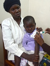
Photo by Caitlin Kleiboer
The malaria vaccine candidate RTS,S/AS01 is somewhat effective in young African children for up to 4 years after vaccination, according to final data from a phase 3 trial.
The vaccine proved more effective against clinical and severe malaria in children than in young infants, but efficacy waned over time in both age groups.
On the other hand, a booster dose of RTS,S/AS01 increased the average number of malaria cases prevented in children and infants.
“Despite the falling efficacy over time, there is still a clear benefit from RTS,S/AS01,” said Brian Greenwood, MD, of the London School of Hygiene & Tropical Medicine in the UK.
“An average 1363 cases of clinical malaria were prevented over 4 years of follow-up for every 1000 children vaccinated, and 1774 cases in those who also received a booster shot. Over 3 years of follow-up, an average 558 cases were averted for every 1000 infants vaccinated, and 983 cases in those also given a booster dose.”
Dr Greenwood and his colleagues disclosed these data in The Lancet. The research was funded by GlaxoSmithKline Biologicals SA, the company developing RTS,S/AS01, and the PATH Malaria Vaccine Initiative.
The trial included 15,459 young infants (aged 6 weeks to 12 weeks at first vaccination) and children (5 months to 17 months at first vaccination) from 11 sites across 7 sub-Saharan African countries (Burkina Faso, Gabon, Ghana, Kenya, Malawi, Mozambique, and United Republic of Tanzania) with varying levels of malaria transmission.
Earlier results from this trial, at 18 months of follow-up, showed efficacy of about 46% against clinical malaria in children and around 27% among young infants. Vaccine efficacy is defined as the reduction in the incidence of disease among participants who receive the vaccine compared to the incidence among participants who do not.
Dr Greenwood and his colleagues followed the infants and children for a further 20 to 30 months, respectively, and assessed the impact of a fourth booster dose.
Participants were each vaccinated 3 times with RTS,S/AS01, with or without a booster dose 18 months later, or given 4 doses of a comparator vaccine (control group).
In children who received 3 doses of RTS,S/AS01 plus a booster, the number of clinical episodes of malaria at 4 years was reduced by just over a third (36%). This is a drop in efficacy from the 50% protection against malaria seen in the first year.
Without a booster dose, the vaccine was not significantly effective against severe malaria in this age group. However, in children given a booster dose, the overall protective efficacy against severe malaria was 32% and 35% against malaria-associated hospitalizations.
In infants who received 3 doses of RTS,S/AS01 plus a booster, the vaccine reduced the risk of clinical episodes of malaria by 26% over 3 years of follow-up. There was no significant protection against severe disease in infants.
Meningitis occurred more frequently in children given RTS,S/AS01 than in children given the control vaccine. There were 11 cases of meningitis among children who received a booster, 10 cases among children who did not receive a booster, and 1 case among children in the control group.
RTS,S/AS02 produced more adverse reactions than the control vaccines. Convulsions following vaccination, although uncommon, occurred more frequently in children who received RTS,S/AS01. The incidence of other serious adverse events was similar in all the groups.
“The European Medicines Agency (EMA) will assess the quality, safety, and efficacy of the vaccine based on these final data,” Dr Greenwood said. “If the EMA gives a favorable opinion, WHO could recommend the use of RTS,S/AS01 as early as October this year. If licensed, RTS,S/AS01 would be the first licensed human vaccine against a parasitic disease.” ![]()
Device can test multiple cancer drugs in tumors

Image courtesy of
Presage Biosciences
A device that tests multiple cancer drugs in living tumor tissue could guide treatment selection in patients with lymphoma and other cancers, according to researchers.
They also believe the device, called CIVO, could help speed up drug development by testing the efficacy of candidate drugs in very small doses while sparing patients side effects.
CIVO is a handheld microinjection platform that can deliver small doses of up to 8 drugs or combinations of drugs into a tumor.
The device proved effective for testing multiple cancer drugs in xenograft mouse models, dogs, and humans with lymphoma.
Richard Klinghoffer, PhD, of Presage Biosciences in Seattle, Washington, and his colleagues described their research with CIVO in Science Translational Medicine. The research was funded by Presage Biosciences, the National Institutes of Health, and Seattle Children’s Hospital Neuro-Oncology Fund.
About CIVO
CIVO is designed for tumors near the skin surface, such as lymphoma or skin and breast cancer.
The technology enables the placement of multiple columns of drugs directly into the tumor along the needle axis, spanning the full depth of the tumor. This makes it possible to assess drug effects with multiple biomarkers and in multiple regions along the injection axis to capture the heterogeneity of response within the tumor.
Later (typically 24 to 72 hours after injection), the tumor is resected for subsequent analysis, and responses are measured with multiple immunohistochemistry-based assays and high-resolution scanning.
Results in mice
In xenograft lymphoma models, CIVO microinjection of well-characterized anticancer agents (vincristine, doxorubicin, mafosfamide, and prednisolone) induced spatially defined cellular changes around sites of drug exposure, specific to the known mechanisms of each drug. And the observed localized responses predicted responses to the same drugs systemically delivered in animals.
In pair-matched drug-resistant and drug-sensitive lymphoma models, CIVO correctly demonstrated tumor resistance to doxorubicin and vincristine.
The researchers also identified an unexpected enhanced sensitivity to the active form of cyclophosphamide in multidrug-resistant lymphomas compared with chemotherapy-naïve lymphomas.
And a CIVO-enabled in vivo screen of oncology agents revealed that a novel mTOR pathway inhibitor exhibits significantly increased tumor-killing activity in the drug-resistant setting compared with chemotherapy-naïve tumors.
Results in dogs and humans
Dogs with lymphoma showed no toxicity when injected with drugs via CIVO. And the researchers said they observed robust, easily tracked, drug-specific responses in the animals.
For lymphoma patients, the researchers used CIVO to inject microdoses of vincristine into the tumors in patients’ lymph nodes. Cells surrounding the injections died, and there were no serious adverse events, although patients did report mild discomfort.
“This analysis creates a comprehensive portrait of drug response that has never been seen before this early in the drug development process,” Dr Klinghoffer said. “Using this technology, we can assess how drugs, both as single agents and in combinations, impact the biology of tumor cells in the context of the native tumor microenvironment.”
“[T]ranslation of CIVO to the clinical setting has enabled assessment on all aspects of tumor biology, including drug effects on tumor-infiltrating immune cells. This sets the stage for a new type of pre-phase 1 clinical study in which multiple drugs or drug combinations can be tested simultaneously, directly in a patient’s own tumor, without toxicity associated with systemic drug delivery.” ![]()
Image courtesy of
Presage Biosciences
A device that tests multiple cancer drugs in living tumor tissue could guide treatment selection in patients with lymphoma and other cancers, according to researchers.
They also believe the device, called CIVO, could help speed up drug development by testing the efficacy of candidate drugs in very small doses while sparing patients side effects.
CIVO is a handheld microinjection platform that can deliver small doses of up to 8 drugs or combinations of drugs into a tumor.
The device proved effective for testing multiple cancer drugs in xenograft mouse models, dogs, and humans with lymphoma.
Richard Klinghoffer, PhD, of Presage Biosciences in Seattle, Washington, and his colleagues described their research with CIVO in Science Translational Medicine. The research was funded by Presage Biosciences, the National Institutes of Health, and Seattle Children’s Hospital Neuro-Oncology Fund.
About CIVO
CIVO is designed for tumors near the skin surface, such as lymphoma or skin and breast cancer.
The technology enables the placement of multiple columns of drugs directly into the tumor along the needle axis, spanning the full depth of the tumor. This makes it possible to assess drug effects with multiple biomarkers and in multiple regions along the injection axis to capture the heterogeneity of response within the tumor.
Later (typically 24 to 72 hours after injection), the tumor is resected for subsequent analysis, and responses are measured with multiple immunohistochemistry-based assays and high-resolution scanning.
Results in mice
In xenograft lymphoma models, CIVO microinjection of well-characterized anticancer agents (vincristine, doxorubicin, mafosfamide, and prednisolone) induced spatially defined cellular changes around sites of drug exposure, specific to the known mechanisms of each drug. And the observed localized responses predicted responses to the same drugs systemically delivered in animals.
In pair-matched drug-resistant and drug-sensitive lymphoma models, CIVO correctly demonstrated tumor resistance to doxorubicin and vincristine.
The researchers also identified an unexpected enhanced sensitivity to the active form of cyclophosphamide in multidrug-resistant lymphomas compared with chemotherapy-naïve lymphomas.
And a CIVO-enabled in vivo screen of oncology agents revealed that a novel mTOR pathway inhibitor exhibits significantly increased tumor-killing activity in the drug-resistant setting compared with chemotherapy-naïve tumors.
Results in dogs and humans
Dogs with lymphoma showed no toxicity when injected with drugs via CIVO. And the researchers said they observed robust, easily tracked, drug-specific responses in the animals.
For lymphoma patients, the researchers used CIVO to inject microdoses of vincristine into the tumors in patients’ lymph nodes. Cells surrounding the injections died, and there were no serious adverse events, although patients did report mild discomfort.
“This analysis creates a comprehensive portrait of drug response that has never been seen before this early in the drug development process,” Dr Klinghoffer said. “Using this technology, we can assess how drugs, both as single agents and in combinations, impact the biology of tumor cells in the context of the native tumor microenvironment.”
“[T]ranslation of CIVO to the clinical setting has enabled assessment on all aspects of tumor biology, including drug effects on tumor-infiltrating immune cells. This sets the stage for a new type of pre-phase 1 clinical study in which multiple drugs or drug combinations can be tested simultaneously, directly in a patient’s own tumor, without toxicity associated with systemic drug delivery.”
CCSs more likely to claim social security support

Photo courtesy of
Huntsman Cancer Institute
A new study indicates that childhood cancer survivors (CCSs) are more likely than individuals without a cancer history to enroll on federal programs that provide disability benefits.
CCSs diagnosed between 1970 and 1986 were about 2 to 5 times as likely as control subjects to utilize such a program.
“The long-term impact of cancer can affect other issues besides health outcomes,” said study author Anne Kirchhoff, PhD, of the Huntsman Cancer Institute at the University of Utah.
“We need to do a better job of helping people function throughout their lives, not just when they’re finishing their cancer therapy.”
Dr Kirchhoff and her colleagues conducted this research and detailed the results in the Journal of the National Cancer Institute.
The researchers looked at health insurance surveys completed in 2011 and 2012 by a random sample of 698 CCSs who were diagnosed between the ages of 0 and 20 years. Today, they range in age from 20s to early 60s.
The patients are part of a National Cancer Institute initiative, called the Childhood Cancer Survivor Study, which has followed more than 14,000 children and adolescents since 1994 who were diagnosed with cancer and survived at least 5 years after diagnosis. A comparison group of 210 siblings without cancer also responded to the survey and were used as controls.
Dr Kirchhoff and her colleagues looked at current or former enrollment on 2 federal disability programs:
- Supplemental security income (SSI), which is for people with limited income who have no prior work history
- Social security disability insurance (SSDI), which pays disability benefits to adults ages 18 years and older who have worked and paid social security taxes.
In all, 13.5% of CCSs reported being enrolled on SSI in the past or present, and 10% of survivors reported being enrolled on SSDI at some point. This was substantially higher than for the comparison group, in which 2.6% of patients reported SSI enrollment and 5.4% reported SSDI enrollment.
In addition, CCSs reported current enrollment in SSI more frequently than the US population, at rates of 7.3% and 2.5%, respectively.
Dr Kirchoff and her colleagues also identified survivor socio-demographic and treatment characteristics that were associated with a higher rate of enrollment in federal support programs.
“Survivors that were younger at diagnosis, age 4 or under, were about 7 times more likely to be on SSI than we see with survivors that were diagnosed in their adolescence,” she said.
SSI enrollment was more likely for female CCSs and for survivors with a history of cranial radiation treatment as well.
Dr Kirchhoff noted that, over the years, research on CCSs has caused hospitals to rethink how to better care for cancer survivors.
“There’s really a growing strategy to support survivors in the long-term,” she said. “For example, here at Huntsman Cancer Institute, we have a pediatric cancer late-effects clinic, which helps manage issues that might come up with childhood cancer survivors in the long term, including health-management support, health-behavior support, and access to providers to help them with other issues.”
Team images immune response

Photo by Aaron Logan
Researchers say they have devised an approach that allows for real-time imaging of the immune system’s response to tumors, without the need for blood draws or biopsies.
The method harnesses PET to identify areas of immune cell activity associated with inflammation or tumor development.
The researchers believe the approach offers a potential breakthrough in diagnostics and the ability to monitor the efficacy of cancer therapies.
The team described their method in PNAS.
Study author Hidde Ploegh, PhD, of the Whitehead Institute for Biomedical Research in Cambridge, Massachusetts, said that every experimental immunologist wants to monitor an ongoing immune response, but options are limited.
“One can look at blood, but blood is a vehicle of transport for immune cells and is not where immune responses occur,” he said. “Surgical biopsies are invasive and non-random, so, for example, a fine-needle aspirate of a tumor could miss a significant feature of that condition.”
In search of a better monitoring approach, Dr Ploegh and his colleagues leveraged two research tools that have become staples in his lab in recent years.
The first exploits single-domain antibodies known as VHHs, derived from the heavy chain-only antibodies made by the immune systems of animals in the camelid family. Dr Ploegh’s lab immunizes alpacas—his camelid of choice—to generate VHHs specific to immune cells of interest.
The second tool, sortagging, labels the VHHs in a site-specific fashion so that researchers can track the VHHs and their targets in a living animal.
Knowing that the tissue around tumors often contains immune cells such as neutrophils and macrophages, Dr Ploegh and his colleagues hypothesized that appropriately labeled VHHs might allow them to pinpoint tumor locations by finding the tumor-associated immune cells.
Dr Ploegh noted that VHHs’ extremely small size—approximately one-tenth that of conventional antibodies—are likely responsible for their superior tissue penetration and, thus, makes them particularly well suited for such use.
So the researchers generated VHHs that recognize mouse immune cells, then labeled these VHHs with radioisotopes and injected them into tumor-bearing mice. Subsequent PET imaging detected the location of immune cells around the tumor quickly and accurately.
“We were able to image tumors as small as 1 mm in size and within just a few days of their starting to grow,” said Mohammad Rashidian, PhD, a researcher in Dr Ploegh’s lab.
“We’re very excited about this because it’s a powerful approach to pick up inflammation in and around the tumor.”
Drs Rashidian and Ploegh believe that, with further refinement, the method could be used to monitor response to—and perhaps modify—cancer immunotherapy.
“To succeed with immunotherapy, we need more information about the tumor microenvironment,” Dr Rashidian said. “With this method, you could perhaps start immunotherapy and then, a few weeks later, image with VHHs to figure out progress and success of treatment.”
“PET imaging should allow a much more comprehensive look at the entire tumor in its environment,” Dr Ploegh added. “Then we can ask, ‘Did the tumor grow? Did immune cells invade? What has happened to the tumor?’ And to be able to see this without going in invasively is a significant achievement.”