User login
PET scans could prevent unnecessary RT in HL
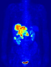
Image by Jens Langner
Performing PET scans immediately after chemotherapy may reveal which Hodgkin lymphoma (HL) patients need radiotherapy (RT).
A study published in NEJM showed similar rates of progression-free survival in HL patients who werePET-negative after chemotherapy, whether they received subsequent RT or not.
However, the investigators said longer follow-up is needed to determine if eliminating RT in PET-negative patients will lead to fewer late effects and improved overall survival.
The 602 patients who agreed to take part in this trial, known as RAPID, had a PET scan performed after chemotherapy. Patients who tested positive received RT.
Those who tested negative were divided into 2 groups. One group of 211 patients received no further treatment, and the other group of 209 patients had the standard RT.
At a median of 60 months of follow-up, the proportion of patients who were alive and disease-free was 94.6% in the RT group and 90.8% in the group that hadn’t received further treatment.
Eight patients in the RT group progressed, and 8 died (3 with disease progression, 1 of whom died from HL). Five of the deaths occurred in patients who did not ultimately receive RT.
In the untreated group, 20 patients progressed, and 4 patients died (2 with disease progression and none from HL).
“This research is an important step forward,” said study author John Radford, of The University of Manchester and The Christie NHS Foundation Trust in the UK.
“The results of RAPID show that, in early stage Hodgkin lymphoma, radiotherapy after initial chemotherapy marginally reduces the recurrence rate, but this is bought at the expense of exposing to radiation all patients with negative PET findings, most of whom are already cured.” ![]()
Decreased RBC clearance predicts ill health

New research suggests that increased red blood cell distribution width (RDW) is caused by reduced clearance of aging red blood cells (RBCs) from the bloodstream.
And previous studies showed that elevations in RDW predict the development, progression, and risk of death from many conditions.
“It appears that the human body slightly slows down the production and destruction of red blood cells in just about every major disease,” said John Higgins, MD, of Massachusetts General Hospital in Boston.
“If we can accurately measure the production or destruction rates, we might be able to identify many of these diseases in their earlier stages when they are most treatable. Existing measures of the production rate are far too imprecise to detect these subtle changes, but this paper shows how the destruction rate can be estimated using existing blood count data and a mathematical model.”
The paper has been published in the American Journal of Hematology.
Healthy adults generate RBCs at a rate of more than 2 million per second, and the cells circulate in the bloodstream for 100 to 120 days, during which their size decreases by around 30%. Aged RBCs are then cleared at about the same rate of 2 million per second.
Prior to the reports linking elevated RDW with an increased risk for many diseases, that measure had only been used to help distinguish between different forms of anemia.
To understand the correlation between RDW and disease prognosis, Dr Higgins and his colleagues analyzed raw data from more than 60,000 randomly selected blood samples. They used a mathematical model to replicate how RBC populations behave differently in health and in disease.
The researchers measured the extent to which RBCs in different phases of their life cycle contribute to increased RDW and found that the variance in size was strongly determined by mild increases in the numbers of the smallest and oldest cells.
Since the most important mechanism controlling the number of the oldest cells is the rate at which they are cleared from the bloodstream, the team made several predictions, which were validated by applying their model to clinical data from the blood samples and to data from several published studies.
The researchers found that increased RDW was associated with delayed RBC clearance, and increased RDW was associated with increased average age of RBCs.
Delayed RBC clearance was as strongly associated with overall risk of death, as was increased RDW, and delayed RBC clearance was associated with the presence of early signs of hidden diseases associated with increased RDW.
Patients with delayed RBC clearance had a greater risk of developing signs of those diseases in the future than patients with a typical clearance rate. In healthy patients, the rate of RBC clearance varied less than any other traditional blood-count characteristic.
Dr Higgins said there are many potential clinical applications of these findings, which need to be validated by future studies.
“Finding a reduced clearance rate in an apparently healthy person would likely mean that an underlying disease process was developing—such as the early stages of iron deficiency, kidney disease, colon cancer, or congestive heart failure—and would warrant further diagnostic evaluation,” Dr Higgins said.
“Based on this analysis of routine blood tests, a primary care physician could immediately consider appropriate follow-up diagnostic testing, instead of waiting for other signs and symptoms to appear as the condition progresses. In a patient with established disease, a reduced clearance rate could mean progression of disease or treatment failure, and imminent complications could be avoided or reduced by adjusting treatment right away or at least by more frequent monitoring.”
“In addition to confirming our findings in prospective studies that would follow a group of patients over time, we hope to identify the diseases for which an early warning provided by delayed clearance would lead to the most significant improvements in patient outcomes. We’d also like to understand more about the processes controlling red blood cell clearance and are actively developing similar models for populations of white blood cells and platelets.” ![]()
Gene therapy appears effective against WAS
![]()
Photo by Chad McNeeley
Results of a small study suggest gene therapy can lead to clinical improvements in children and teens with Wiskott-Aldrich syndrome (WAS).
The gene therapy—autologous, gene-corrected hematopoietic stem cells (HSCs) given along with chemotherapy—improved infectious complications, severe eczema, and symptoms of autoimmunity in 6 of the 7 patients studied.
The therapy also reduced patients’ use of blood products and the amount of time they spent in the hospital.
Marina Cavazzana, MD, PhD, of Necker Children’s Hospital in Paris, France, and colleagues reported these results in JAMA. The study was sponsored by Genethon.
The researchers noted that WAS is caused by loss-of-function mutations in the WAS gene. The condition is characterized by thrombocytopenia, eczema, and recurring infections. In the absence of definitive treatment, patients with classic WAS generally do not survive beyond their second or third decade of life.
Partially HLA-matched, allogeneic HSC transplant is often curative, but it is associated with a high incidence of complications. Dr Cavazzana and colleagues speculated that transplanting autologous, gene-corrected HSCs may be an effective and potentially safer alternative.
So the team assessed the outcomes and safety of autologous HSC gene therapy in 7 patients (age range, 0.8-15.5 years) with severe WAS who lacked HLA antigen-matched related or unrelated HSC donors.
Patients were enrolled in France and England and treated between December 2010 and January 2014. Follow-up ranged from 9 months to 42 months.
The treatment involved collecting mutated HSCs from patients and correcting the cells in the lab by introducing a healthy WAS gene using a lentiviral vector developed and produced by Genethon. The corrected cells were reinjected into patients who, in parallel, received chemotherapy to suppress their defective stem cells and autoimmune cells to make room for new, corrected cells.
Six of the 7 patients saw clinical improvements after this treatment. One patient died of pre-existing, treatment-refractory infectious disease.
In the 6 surviving patients, infectious complications resolved after gene therapy, and prophylactic antibiotic therapy was successfully discontinued in 3 cases. Severe eczema resolved in all affected patients, as did signs and symptoms of autoimmunity.
There were no severe bleeding episodes after treatment. And, at last follow-up, none of the 6 surviving patients required blood product support.
The median number of hospitalization days decreased from 25 during the 2 years before treatment to 0 during the 2 years after treatment.
“[T]he patients showed a significant clinical improvement due to the re-expression of the protein WASp in the cells of the immune system,” Dr
Cavazzana said.
However, the researchers also noted that the interpretation of these results is constrained by the small number of patients studied. So the team said they could not draw conclusions on long-term outcomes and safety without further follow-up and additional trials. ![]()
Interventions reduce bloodstream infections

A bundled intervention can considerably reduce central line-associated bloodstream infections (CLABSIs), according to research published in Infection Control & Hospital Epidemiology.
The intervention focused on evidence-based infection prevention practices, safety culture and teamwork, and scheduled measurement of infection rates.
By implementing these measures, intensive care units (ICUs) in Abu Dhabi achieved an overall 38% reduction in CLABSIs.
“These hospitals were able to show significant improvements in infection rates and have been able to sustain the improvements a year after we finished the project,” said study author Asad Latif, MBBS, MD, of the Johns Hopkins University School of Medicine in Baltimore, Maryland.
“Our results suggest that ICUs in disparate settings around the world could use this program and achieve similar results, significantly reducing the global morbidity, mortality, and excess costs associated with CLABSIs. In addition, this collaborative could serve as a model for future efforts to reduce other types of preventable medical harms in the Middle East and around the world.”
This study was a collaborative effort by the Armstrong Institute, Johns Hopkins Medicine International, and the Abu Dhabi Health Services Company (SEHA), which operates the government healthcare system in Abu Dhabi.
For the study, ICUs were instructed to assemble a comprehensive unit-based safety program (CUSP) team comprising local physician and nursing leaders, a senior executive, frontline healthcare providers, an infection control provider, and hospital quality and safety leaders.
The ICUs included 10 adult, 5 neonatal, and 3 pediatric ICUs, accounting for 77% of the adult, 74% of the neonatal, and 100% of the pediatric ICU beds in Abu Dhabi.
Starting in May 2012, the SEHA corporate quality team and ICU CUSP teams attended 14 weekly live webinars on CLABSI prevention conducted by Armstrong Institute faculty, followed by content and coaching webinars every 2 weeks for 24 months. The webinars were recorded by SEHA and posted on a local, shared computer drive, along with educational and training materials.
Armstrong faculty also conducted 4 site visits in Abu Dhabi at the beginning of the study, visiting each ICU to meet the CUSP team and tour the units. A year later, they conducted a 3-day patient safety workshop for participating hospitals.
CUSP teams implemented 3 interventions as part of the program: an effort to prevent CLABSIs that targeted clinicians’ use of evidence-based infection prevention recommendations from the Centers for Disease Control and Prevention, a CUSP process to improve safety culture and teamwork, and measurement of monthly CLABSI data and feedback to safety teams, senior leaders, and ICU staff.
The overall mean crude CLABSI rate for participating ICUs decreased from 2.56 infections per 1000 catheter days to 1.79 per 1,000 catheter days by the end of the study, corresponding to a 30% reduction.
By unit type, CLABSI rates decreased by 16% among adult ICUs, 48% among pediatric ICUs, and 47% in neonatal ICUs. The percentage of ICUs that achieved a quarterly CLABSI rate of less than 1 infection per 1000 catheter days increased from 44% to 61% after the interventions.
“Despite growing awareness, many hospitals around the world continue to struggle in their efforts to meaningfully reduce their CLABSI rates in a sustained manner,” said Sean M. Berenholtz, MD, of the Johns Hopkins University School of Medicine.
“In addition, hospitals and healthcare systems in the Middle East have unique barriers to implementing quality improvement programs, such as challenges with staff recruitment and retention, and personnel fearful of punitive repercussions from speaking up regarding patient safety concerns. In our study, bringing all stakeholders to the same table allowed everyone to share their concerns and ensure their voices were heard.” ![]()
Discovery could aid treatment of leukemia, lymphoma

Photo courtesy of IRCM
Researchers say they have uncovered a mechanism that could aid the development of therapies for lymphomas and leukemias.
The group’s research shed new light on a mechanism affecting activation-induced deaminase (AID), an enzyme that has proven crucial for immune response.
Javier Di Noia, PhD, of Institut de Recherches Cliniques de Montreal (IRCM) in Quebec, Canada, and his colleagues described this mechanism in The Journal of Experimental Medicine.
Dr Di Noia noted that, although AID is crucial for an efficient antibody response, high levels of the enzyme can have harmful effects and lead to cancer-causing mutations.
“The objective is to find the perfect level of AID activity to maximize the protection it provides to the body while reducing the risk of damage it can cause to cells,” he said.
Dr Di Noia and his colleagues previously found that heat-shock protein 90 (Hsp90) maintains the levels of AID by stabilizing it while it is still immature. In fact, they discovered that inhibiting Hsp90 significantly reduces the levels of AID in the cell.
“Through this new study, we identified another mechanism, controlled by the protein eEF1a [elongation factor eukaryotic elongation factor 1 α], that has the opposite effect,” said Stephen P. Methot, a PhD student in Dr Di Noia’s lab.
“The protein eEF1a retains AID in the cell’s cytoplasm, away from the genome. However, unlike Hsp90, it maintains AID in a ready-to-act state. We discovered that blocking the interaction between AID and eEF1a helps AID access the cell nucleus and thereby boosts AID activity. As a result, this could increase immune response and help fight infections, for instance.”
“We found the eEF1a mechanism is necessary to restrict AID activity in the cell. It acts as a buffer by allowing the cell to accumulate enough AID to be efficient but limits its activity to prevent the oncogenic or toxic effects that could result if too much AID is in continuous contact with the genome.”
The researchers also identified 2 existing drugs that can act on the eEF1a mechanism to release AID into the cell. The team said these drugs could potentially be used to boost AID activity and, thus, immune responses.
“With this discovery, we now understand mechanisms that can both reduce and increase the activity of AID by targeting different proteins,” Dr Di Noia said.
“This knowledge could eventually lead to new treatments to boost the immune system and help our aging population fight influenza, for example, as AID activity in our cells decreases with age. On the other hand, therapies could also be developed to lower toxic levels of AID in certain cancers such as B-cell lymphoma and leukemia.” ![]()
Epigenomic findings may help predict relapse in DLBCL

Photo by Rhoda Baer
Epigenomic heterogeneity at diagnosis may predict relapse in diffuse large B-cell lymphoma (DLBCL), according to research published in Nature Communications.
Investigators made this connection by reviewing biopsies taken from DLBCL patients before and after treatment.
The epigenome in these patients’ cancer cells changed greatly after treatment, and the global epigenome of pretreatment biopsies was substantially different in patients who relapsed and those who did not. There was more cell-to-cell heterogeneity in patients who relapsed.
“This is the first study I know of in cancer that looks at changes in the epigenome before and after treatment, and what we found could ultimately make traditional treatments much more effective,” said study author Olivier Elemento, PhD, of Weill Cornell Medical College in New York, New York.
To uncover the role of epigenetic involvement in DLBCL, Dr Elemento and his colleagues analyzed banked biopsies from patients. In each sample set, the investigators looked at sites in the epigenome where a methyl group was added or removed after DLBCL recurred.
They found a change in methylation that occurred between 39,808 and 1,035,960 specific methylation sites, depending on the sample. In addition, they identified between 78 and 13,162 differently methylated regions in the epigenome in relapsed disease.
“These are massive changes, given that the epigenome has 20 million methylation sites,” Dr Elemento said. “Our study shows that, in some cases, up to one-twentieth of the entire epigenome is changed after treatment. There are many more epigenetic changes than there are altered genes in DLBCL.”
“Once you have changes in methylation, the end result is an imbalanced expression of proteins,” added Giorgio Inghirami, MD, also of Weill Cornell.
“The tumor after chemotherapy is not the same as the tumor before treatment. This why it is so critical to have biopsies before any treatment of [primary or relapsed] lesions.”
The investigators hope this work will ultimately allow clinicians and researchers to predict treatment resistance in individual patients. ![]()
Measures improve use of VTE prophylaxis

Photo courtesy of CDC
Programs that give physicians real-time feedback and financial incentives may lead to improvements in hospital safety, according to a study published in the Journal of Hospital Medicine.
The study showed that hospitalists significantly improved their compliance with practice guidelines for preventing venous thromboembolism (VTE)
when they could get feedback on their compliance rates and a direct financial incentive for improving their performance.
“Our study confirms there is a real return on investment in such programs, not only for patient safety but also for hospitals,” said study author Henry Michtalik, MD, of Johns Hopkins Hospital in Baltimore, Maryland.
“Metrics such as the use of preventive drugs for [VTE] are already being monitored but only really improve a hospital’s quality of care when programs get data back to the people who are treating patients to directly improve care.”
Dr Michtalik and his team found that, by providing such information to physicians through web-based, real-time displays, monthly VTE prophylaxis compliance rates improved from 86% to 90% in 6 months.
Adding pay-for-performance to the real-time feedback for the following 18 months boosted compliance rates to 94%.
Dr Michtalik pointed out that “no one got wealthy off of the pay-for-performance program. Instead, we believe the money served more as a method to engage the providers.”
Payments ranged from $53 to $1244, with all but 2 of the incentive payments totaling under $1000. And it was during the 6-month feedback-only period that compliance rose the fastest.
The study involved 38 part-time and full-time academic hospitalists and the analysis of 3144 inpatients with a median stay of 3 days. The most common diagnoses were heart failure, acute kidney failure, temporary loss of consciousness (syncope), pneumonia, and chest pain.
Following the evidence-based guidelines of the American College of Chest Physicians for VTE prevention, physicians in the study were required to complete a VTE-risk assessment for each patient by using the hospital’s computerized provider order entry system.
“It sort of walks you through the thinking process” for making the VTE-risk assessment, Dr Michtalik said.
The system then prompted physicians with a risk-appropriate recommendation, but it was up to physicians to order the treatment itself. That allowed for physician discretion among types of prevention, as well as for patient and physician preference.
Before implementing the feedback system, the researchers established a 2-year baseline and found that physicians in the study prescribed inappropriate prophylaxis 7.9% of the time and did not prescribe prophylaxis when indicated 6.1% of the time.
Overall, the choice of inappropriate preventive treatment dropped to 6.2% with real-time feedback and to 2.6% with the addition of pay-for-performance. Lack of prophylaxis when indicated fell to 3.2% with feedback and to 3.1% with pay-for-performance.
Dr Michtalik noted that continuous improvements depend not only on the right kind of feedback but also on efforts to avoid “information overload,” especially now that an increasing amount of health and medical records are electronic.
“So you specifically target a few things that need to be improved,” he said, “and really incorporate them into the hospital’s culture.” ![]()
Photo courtesy of CDC
Programs that give physicians real-time feedback and financial incentives may lead to improvements in hospital safety, according to a study published in the Journal of Hospital Medicine.
The study showed that hospitalists significantly improved their compliance with practice guidelines for preventing venous thromboembolism (VTE)
when they could get feedback on their compliance rates and a direct financial incentive for improving their performance.
“Our study confirms there is a real return on investment in such programs, not only for patient safety but also for hospitals,” said study author Henry Michtalik, MD, of Johns Hopkins Hospital in Baltimore, Maryland.
“Metrics such as the use of preventive drugs for [VTE] are already being monitored but only really improve a hospital’s quality of care when programs get data back to the people who are treating patients to directly improve care.”
Dr Michtalik and his team found that, by providing such information to physicians through web-based, real-time displays, monthly VTE prophylaxis compliance rates improved from 86% to 90% in 6 months.
Adding pay-for-performance to the real-time feedback for the following 18 months boosted compliance rates to 94%.
Dr Michtalik pointed out that “no one got wealthy off of the pay-for-performance program. Instead, we believe the money served more as a method to engage the providers.”
Payments ranged from $53 to $1244, with all but 2 of the incentive payments totaling under $1000. And it was during the 6-month feedback-only period that compliance rose the fastest.
The study involved 38 part-time and full-time academic hospitalists and the analysis of 3144 inpatients with a median stay of 3 days. The most common diagnoses were heart failure, acute kidney failure, temporary loss of consciousness (syncope), pneumonia, and chest pain.
Following the evidence-based guidelines of the American College of Chest Physicians for VTE prevention, physicians in the study were required to complete a VTE-risk assessment for each patient by using the hospital’s computerized provider order entry system.
“It sort of walks you through the thinking process” for making the VTE-risk assessment, Dr Michtalik said.
The system then prompted physicians with a risk-appropriate recommendation, but it was up to physicians to order the treatment itself. That allowed for physician discretion among types of prevention, as well as for patient and physician preference.
Before implementing the feedback system, the researchers established a 2-year baseline and found that physicians in the study prescribed inappropriate prophylaxis 7.9% of the time and did not prescribe prophylaxis when indicated 6.1% of the time.
Overall, the choice of inappropriate preventive treatment dropped to 6.2% with real-time feedback and to 2.6% with the addition of pay-for-performance. Lack of prophylaxis when indicated fell to 3.2% with feedback and to 3.1% with pay-for-performance.
Dr Michtalik noted that continuous improvements depend not only on the right kind of feedback but also on efforts to avoid “information overload,” especially now that an increasing amount of health and medical records are electronic.
“So you specifically target a few things that need to be improved,” he said, “and really incorporate them into the hospital’s culture.”
Analyses reveal higher-than-expected pediatric cancer rates in Florida

Photo by Bill Branson
Several statistical analyses have revealed higher-than-expected rates of pediatric cancers in 2 regions of Florida—the Miami metro area and an area west of the Everglades.
The anomalous rates were detected by 5 different research teams—each using different epidemiological and statistical methodology—on a data set spanning the period from 2000 to 2010 that was provided by the Florida Association of Pediatric Tumor Programs (FAPTP).
Lance A. Waller, PhD, of Emory University in Atlanta, Georgia, reviewed the different analyses and described his findings in Statistics and Public Policy.
The research groups applied different analytical approaches to achieve the same goal: detect spatio-temporal pediatric cancer clusters. The analyses used familiar methods—scan statistics, classification, and hierarchical Bayesian modeling—as well as some ideas new to disease clustering: wombling and machine learning.
During their respective analyses of the FAPTP data, the research groups found several suggestive results. For instance, each approach identified local areas in which the observed pediatric cancer rate is significantly higher than the rate expected, given the number of people at risk.
While the precise areas of high reported risk differ between methods, the groups identified a few common results that overlap but are not identical.
For example, all 5 teams identified significantly elevated rates of pediatric cancers in an urban area within collections of ZIP code tabulation areas (ZCTAs) in the Miami metro area and in an area just west of the Everglades. (ZCTAs are geographic areas defined by the US Census Bureau to provide a link between census geography—blocks, block groups, and tracts—and US Postal Service ZIP code areas.)
One analysis suggested the local increase west of the Everglades is based on 2 cases, both classified as “other” race, while another analysis indicated that this cluster is limited to the year 2000.
The observed elevated rates near Miami involved a much larger population size and many more cases, factors that complicate the identification of any shared characteristics common to cases in the cluster.
The analyses also revealed other patterns in the data. Dr Waller said an analysis that revealed a statewide increase in the baseline pediatric cancer incidence rate occurring between 2005 and 2006 merits a closer look to see whether this result represents an overall increase in risk or a change in reporting, because the statistical analysis does not reveal potential reasons for the change.
There also are subtle differences between the specific clusters identified by the various analytical approaches. Comparisons across analyses revealed characteristics of the detected patterns, including the number of cases (2), types of cancer (leukemia or brain/central nervous system cancer), and the racial composition and timing of the cluster west of the Everglades.
As the methods the researchers used don’t completely agree on the precise location, boundaries, and make-up of the detected clusters, the findings suggest a single method may not prove sufficient for such analyses, Dr Waller said.
He added that the identified clusters are geographically quite large and therefore unlikely to provide clear links between particular environmental exposures to local risks.
“While the results do not identify a ‘smoking gun’ in the form of a shared environmental exposure in high-incidence areas, the results do provide epidemiologic insight into the local demographics of the incidence of pediatric cancer cases and suggest more detailed assessment of migration patterns in the Miami area,” Dr Waller said.
“Policy-wise, the results point to responsibly responsive next steps of detailed description of the cases and the at-risk population in the detected areas to summarize local features in the data, particularly the race of cases west of the Everglades and demographic descriptors of any shifts in the at-risk population in the Miami area during the study period.”
Policy responses by local and state health officials may involve more detailed follow-up, including additional data collection, exposure surveys, or in-depth investigation of case histories within a reported cluster.
Dr Waller added that estimated cancer rates consist of the local number of cases (reported by the FAPTP) and the local number of children at risk (reported by the decennial census). Higher-than-expected rates can result from unusually high numbers of reported cases or low numbers of reported local residents.
Since Miami, like many urban areas, often experiences rapid changes in population size and composition between decennial censuses, it is important to assess the accuracy of both data components. Dr Waller suggested, as a first step, assessing the accuracy of the case counts and the inter-census population projections defining the local rates.
“State and local health departments and public health agencies regularly respond to cluster reports from the public,” he said. “Typically, a responsive and effective response is not based on a detailed new epidemiologic study but, rather, is based on education, assessments of local concentrations of demographic risk factors associated with the reported cluster, and an assessment of the distribution of numbers of cases expected given the local demographics.”
Fake malaria drugs may be less common than we thought

Photo courtesy of the
London School of Hygiene
& Tropical Medicine
Investigations into antimalarial drug quality conducted in Cambodia and Tanzania uncovered no evidence of fake medicines.
Previous reports had suggested that up to a third of antimalarials might be falsified, or do not contain the stated active pharmaceutical ingredient.
The new research revealed no falsified drugs in either country, but it did unearth substandard antimalarial drugs, or genuine medicines that do not have the correct amount of the active ingredient.
These findings were published in 2 articles in the American Journal of Tropical Medicine and Hygiene.
“Although there have been alarming reports about the prevalence of fake antimalarials, our study provides ample data showing that the quality of drugs is not so bad based on comprehensive sampling and analysis presented here,” said Harparkash Kaur, PhD, of the London School of Hygiene & Tropical Medicine in the UK.
“The lack of falsified medicines in Cambodia and Tanzania are reassuring, but the presence of substandard medicines is definitely a concern.”
The researchers analyzed 2028 antimalarials from Tanzania and Cambodia at 3 independent laboratories in the UK and US. They classified drugs as acceptable, falsified, or substandard.
In Tanzania, the researchers used an “overt sampling” system, telling vendors they were going to analyze the quality of their medicines.
In Cambodia, the researchers used overt sampling as well as a “mystery client” approach, where actors pretended to be patients with malaria, or their relatives, and bought the medicines offered to them.
Both studies used a randomized approach to sampling of drug outlets, which differs from previous studies that mostly used non-representative methods for selecting drugs for analysis.
Neither study unearthed falsified drugs, but substandard drugs were found in 31% of samples in Cambodia and 12% of samples in Tanzania.
“Falsified medicines have received much attention globally, but substandard drugs are far more prevalent and of great concern,” said Shunmay Yeung, MBBS, PhD, also of the London School of Hygiene & Tropical Medicine.
“Not only do they leave patients with malaria undertreated, which could be fatal, but they may also contribute to the development of resistance to [artemisinin-based combination therapies], the most effective drugs for malaria. Generally, the fact that no falsified antimalarials were identified reflects the positive impact of the [countries’ efforts] to control drug quality.”
EBV-CTLs produce durable responses in EBV-LPD
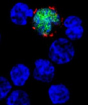
among uninfected cells (blue)
Image courtesy of NIH/
Benjamin Chaigne-Delalande
PHILADELPHIA—Cytotoxic T lymphocytes designed to target Epstein-Barr virus (EBV-CTLs) can elicit durable responses in patients
with EBV–associated lymphoproliferative disorder (EBV-LPD), according to data presented at the AACRAnnual Meeting 2015.
Results from two trials showed that EBV-CTLs derived from a patient’s transplant donor could produce a response rate of 62%, and EBV-CTLs derived from third-party donors could produce a response rate of 61%.
Study investigators noted that, with the achievement of complete response (CR), remission proved durable. And, unlike with chemotherapy, partial responses (PRs) to EBV-CTLs were durable as well.
The team presented these results as abstract CT107.*
“The purpose of our clinical trials was to see if giving T cells from a normal-immune individual that were expanded in culture and stimulated to respond to multiple proteins from the Epstein-Barr virus could provide a safe and effective treatment,” said Richard J. O’Reilly, MD, of Memorial Sloan Kettering Cancer Center in New York.
“The good news from our two clinical trials is that EBV-CTLs generated from either the patient’s transplant donor or from the bank of normal donor T cells developed at Memorial Sloan Kettering put aggressive EBV-LPD that had failed to respond to rituximab into long-lasting remission in more than 60% of patients.”
In the first trial, 26 patients with EBV-LPD received EBV-CTLs generated from their transplant donor. Thirteen of these patients had previously received rituximab, and 16 had high-risk disease.
Thirteen patients in this trial received HLA-matched, EBV-CTLs from the Memorial Sloan Kettering Cancer Center bank of EBV-CTLs generated from third-party, healthy donors. All 13 patients had high-risk disease, and 12 had received prior rituximab.
Dr O’Reilly noted that good results were observed with EBV-CTLs from both sources in this trial. And because EBV-CTLs from the bank are available immediately, he and his team used only EBV-CTLs from the bank when treating the 18 patients enrolled in the second trial.
Among the 39 patients enrolled in the first trial, 23 had a CR, none had a PR, and 2 had stable disease.
For patients who received EBV-CTLs from their primary donor, the combined rate of CR and PR was 62% (16 CRs). For patients who received third-party EBV-CTLs, the combined rate of CR and PR was 54% (7 CRs).
Sixteen of the patients who achieved a CR are still doing well, Dr O’Reilly said. Eight of these patients are alive more than 5 years after receiving EBV-CTLs, and 1 is alive more than 10 years after treatment.
Among the 18 patients enrolled in the second trial, 9 had a CR, 3 had a PR, and 1 had stable disease. The combined rate of CR and PR was 67%.
All of the patients who achieved a CR in this trial continue to do well, and the investigators will be following them long-term, Dr O’Reilly said.
He also noted that toxicities with EBV-CTLs were minimal, and there were no treatment-related deaths. None of the patients developed cytokine release syndrome or graft-vs-host disease requiring systemic therapy.
“The EBV-CTLs work well for the majority of recipients,” Dr O’Reilly said. “However, the responses became clinically evident only after the T cells expanded in vivo, which took about 7 to 14 days. We are rigorously pursuing the development of biomarkers or other tests to predict response earlier.”
Memorial Sloan Kettering Cancer Center has entered into an option agreement with Atara Biotherapeutics to further develop EBV-CTLs for clinical use. However, the data presented at AACR were accrued prior to that agreement.
Last month, the US Food and Drug Administration granted breakthrough therapy designation to EBV-CTLs generated from third-party donors for the treatment of patients with rituximab-refractory EBV-LPD.
*Information in the abstract differs from that presented at the meeting.