User login
Identifying artemisinin resistance not so straightforward

Photo by Juan D. Alfonso
The current method used to identify resistance to the antimalarial drug artemisinin is not entirely accurate, according to research published in PLOS Medicine.
Artemisinin rapidly clears malaria parasites from the blood of infected patients. When parasites develop resistance, clearance takes longer.
The best measure of parasite clearance is the parasite half-life in a patient’s blood, and a common cutoff used to denote artemisinin resistance is 5 hours.
Study author Lisa White, of Mahidol University in Bangkok, Thailand, and her colleagues found that parasite half-life predicts the likelihood of an artemisinin-resistant infection for individual patients. But the half-life is influenced by how common resistance is in the particular area.
The critical half-life varied between 3.5 hours in areas where resistance is rare to 5.5 hours in areas where resistance is common. This means there is no universal cutoff value in parasite half-life that can determine whether a particular infection is “sensitive” or “resistant” to artemisinin-based combination (ACT) therapy.
Because measuring the parasite half-life requires frequent blood sampling that is difficult to do in resource-limited settings, the World Health Organization (WHO) uses the following working definition for surveillance. Artemisinin resistance in a population is suspected if more than 10% of patients are still carrying parasites 3 days after the start of ACT.
Arguing that the cutoff used in the WHO’s working definition is based on limited data, the researchers examined how well the definition matches actual data from patients in areas with artemisinin-resistant parasites.
Applying a model specifically developed for this purpose, the team found that the WHO’s day-3 cutoff value of 10% is useful, but it would be more informative if the parasite load at the start of ACT was taken into account.
The researchers concluded that the WHO definition alone cannot be used to accurately predict the real proportion of artemisinin-resistant parasites, so a more detailed assessment is needed. ![]()
MKIs can overcome resistance in CML
PHILADELPHIA—Two multikinase inhibitors (MKIs) can treat chronic myeloid leukemia (CML) that is resistant to other inhibitors, according to preclinical research.
A series of in vitro experiments showed that the MKIs, sorafenib and axitinib, can overcome treatment resistance mediated by hyperactivation of the Src kinase Lyn, overexpression of the docking protein Gab2, and the presence of the Bcr-Abl T315I mutation.
Sebastian Halbach, of the University of Freiburg in Germany, and his colleagues presented these findings in a poster at the AACR Annual Meeting 2015 (abstract 2708).
Mechanisms of resistance
Halbach noted that CML is driven by the hyperactive fusion kinase Bcr-Abl, which builds up its own signaling network with various proteins, such as Gab2 and Lyn.
Resistance to tyrosine kinase inhibitors (TKIs) and MKIs can be caused by mutations in the Bcr-Abl oncogene, such as T315I, or by aberrant activity of components of the Bcr-Abl signaling network.
“We have previously shown in our lab that overexpression of the docking protein Gab2 . . . confers resistance against imatinib and dasatinib,” Halbach said. “And another mechanism of resistance is hyperactivation of the Src kinase Lyn.”
For the current study, Halbach and his colleagues investigated the role of Lyn by introducing imatinib, dasatinib, or DMSO to K562 cells (blast-phase CML), Lyn-transformed K562 cells, and Lyn-Y508F-transformed K562 cells.
They also compared imatinib and DMSO in Ba/F3 cells (a murine pro-B cell line), Lyn-transformed Ba/F3 cells, and Lyn-Y508F-transformed Ba/F3 cells.
The results of these experiments showed that hyperactive Lyn confers resistance to imatinib but not dasatinib.
“That’s not that surprising because dasatinib is a multikinase inhibitor which targets Src kinases,” Halbach noted. “Therefore, the hyperactivity of Lyn is directly targeted.”
Identifying new drugs
Having established that TKI and MKI resistance in CML can be mediated by Lyn and Gab2, as well as T315I, Halbach and his colleagues wanted to find drugs that would overcome this problem. They screened a panel of inhibitors and identified sorafenib and axitinib.
The researchers first evaluated the effects of sorafenib and axitinib against the T315I mutation. They tested the 2 MKIs—as well as imatinib, dasatinib, nilotinib, ponatinib, and DMSO—in the KBM5 cell line (blast-phase CML) and the KBM5-T315I cell line (imatinib-resistant CML).
Sorafenib and axitinib killed KBM5-T315I cells more effectively than any of the other inhibitors. The 2 MKIs also decreased the metabolic activity of T315I-positive cells more effectively than imatinib, dasatinib, or nilotinib, but not ponatinib, which produced similar results.
Next, Halbach and his colleagues tested sorafenib, axitinib, and the aforementioned inhibitors in K562 cells overexpressing Gab2. Overexpression of Gab2 conferred resistance to imatinib, dasatinib, nilotinib, and ponatinib, but not sorafenib and axitinib.
Both sorafenib and axitinib decreased the metabolic activity of Gab2-overexpressing cells more effectively than any of the other inhibitors.
Lastly, the researchers tested all of the inhibitors in Lyn-transformed K562 cells, Lyn-Y508F-transformed K562 cells, and K562 cells. They found that sorafenib and axitinib both overcame Lyn-Y508F-mediated resistance.
Sorafenib and axitinib killed K562 cells and Lyn-transformed K562 cells more effectively than any of the other inhibitors. The 2 MKIs also killed Lyn-Y508F-transformed K562 cells more effectively than imatinib and nilotinib, but not ponatinib or dasatinib.
Sorafenib decreased the metabolic activity of Lyn-Y508F-transformed K562 cells more effectively than all of the other inhibitors. But axitinib only proved more effective than imatinib in this regard.
Halbach said he hopes sorafenib and axitinib can one day serve as alternatives to ponatinib for CML patients, especially those with T315I mutations or high Gab2 levels.
For now, his team’s next step is to further analyze the influence of axitinib and sorafenib on the Bcr-Abl—Gab2 signaling complex. ![]()
Vena cava filter doesn’t reduce risk of recurrent PE
Photo from Business Wire
Retrievable inferior vena cava filters do not reduce the risk of recurrent pulmonary embolism (PE) or death in patients with PE, according to a study
published in JAMA.
The study showed no significant difference in outcomes between patients who received anticoagulation in conjunction with an inferior vena cava
filter and patients who received anticoagulant therapy alone.
Studies have shown an increase in the placement of inferior vena cava filters over the past few decades. However, a lack of reliable data on the use of these filters has meant the benefit-risk ratio of using them in patients at risk of recurrent venous thromboembolism (VTE) is uncertain.
To gain more insight, Patrick Mismetti, MD, PhD, of the Centre Hospitalier Universitaire de Saint-Etienne in France, and his colleagues conducted a prospective study.
They enrolled hospitalized patients with acute, symptomatic PE associated with lower-limb vein thrombosis who had at least 1 criterion for severity. Patients were randomized to receive anticoagulation and a retrievable inferior vena cava filter (n=200) or anticoagulation alone (n=199).
The follow-up period was 6 months. Patients received full-dose anticoagulation for at least 6 months, and filter retrieval was planned at 3 months from placement.
Filters were successfully inserted in 193 patients. Filter retrieval was successful in 153 of the 164 patients in whom retrieval was attempted.
In the anticoagulant-only group, 6 patients ultimately received a filter, 4 because they were undergoing surgery and needed to stop anticoagulation, and 2 because of bleeding complications.
By 3 months, 6 patients (3.0%) had recurrent PE in the filter group, as did 3 patients (1.5%) in the anticoagulant-only group (P=0.50). All episodes in the filter group and 2 in the anticoagulant group were fatal.
The researchers observed 1 additional PE recurrence in each treatment group between 3 and 6 months (P=0.54). And 3 patients developed filter thrombosis.
At 3 months, there was no significant difference between the 2 treatment groups with regard to recurrent deep vein thrombosis (P>0.99), recurrent VTE (P=0.36), major bleeding (P=0.63), or death from any cause (P=0.55).
At 6 months, there was no significant difference between the treatment groups with regard to recurrent deep vein thrombosis (P>0.99), recurrent VTE (P=0.59), major bleeding (P=0.69), or death from any cause (P=0.29).
The main cause of death in both treatment groups was cancer.
The researchers noted that the availability of retrievable inferior vena cava filters has probably contributed to their increased use for managing acute VTE, including their use in addition to full-dose anticoagulant therapy in patients with PE, a large clot burden, a poor cardiopulmonary reserve, or a suspected increased risk for recurrence, as advocated by several guidelines.
However, the team said the results of this study do not support the use of these filters in patients who can be treated with anticoagulation. ![]()
Team discovers mechanism behind malaria progression

a red blood cell
Photo courtesy of St. Jude
Children’s Research Hospital
Researchers say they have pinpointed one of the mechanisms responsible for the progression of malaria.
Computer modeling showed that the nanoscale knobs that form at the membrane of infected red blood cells cause the cell stiffening that is, in part, responsible for the reduced blood flow that can turn malaria deadly.
Subra Suresh, ScD, of Carnegie Mellon University in Pittsburgh, Pennsylvania, and his colleagues reported this finding in PNAS.
“Many of malaria’s symptoms are the result of impeded blood flow, which is directly tied to structural changes in infected red blood cells,” Dr Suresh said.
When a red blood cell is infected with malaria, the parasite releases proteins that interact with the cell membrane. The cell membrane undergoes a series of changes that result in stiffness and stickiness.
While researchers are fairly certain that the stickiness is caused by nanoscale knobs that protrude from the cell membrane, they were uncertain as to what caused the stiffness.
They hypothesized that the parasite protein/cell membrane interaction caused spectrin, a cytoskeletal protein that provides a scaffold for the cell membrane, to rearrange its networked structure to be more rigid.
However, the complexity of the cell membrane made it difficult for researchers to study and prove this hypothesis experimentally.
To visualize what happens at the cell membrane during malarial infection, Dr Suresh and his colleagues turned to a computer simulation technique called coarse-grained molecular dynamics (CGMD). CGMD has proven valuable for studying what happens at the cell membrane because it represents the membrane’s complex proteins and lipids with larger, simplified components rather than atom by atom.
Doing this requires less computing time and power than standard atomistic models, which allows scientists to run simulations for longer periods of time while still accurately recreating the behavior of the cell membrane.
Typically, researchers introduce different variables into the simulation and observe how the membrane reacts. In the current study, the researchers seeded the model membrane with proteins released by the malaria parasite Plasmodium falciparum.
From their simulation, the researchers found that the stiffening of the red blood cell membrane had little to do with the remodeling of spectrin.
Instead, the nanoscale knobs that cause the red blood cells to stick to the vein’s walls also cause the membrane to stiffen through a number of different mechanisms, including composite strengthening, strain hardening, and density-dependent vertical coupling effects.
According to the researchers, the discovery of this mechanism could provide a promising target for new antimalarial therapies. ![]()
Analysis helps quantify HU use in SCA

Photo by Zak Hubbard
Results of a retrospective study reinforce the idea that hydroxyurea (HU) is underused in US patients with sickle cell anemia (SCA).
Researchers analyzed data from an insurance claims database and identified 570 patients with probable SCA who were available for follow-up and likely would have benefitted from receiving HU.
Less than a quarter of those patients actually received the drug within a year of their third visit to a hospital seeking treatment for pain crises.
The researchers noted that these data may not be representative of the US population because the analysis does not include uninsured patients or publicly insured patients.
Nicolas Stettler, MD, of the Lewin Group in Falls Church, Virginia, and his colleagues conducted this research and reported the results in a letter to JAMA.
The researchers noted that the 2014 National Heart, Lung, and Blood Institute guidelines recommend using HU to treat all adults with SCA who experience 3 or more moderate-to-severe pain crises within a year. This is a “strong” recommendation based on high-quality evidence reviewed in 2008.
Despite this recommendation, it is thought that HU is underused, although the extent of its use has been unclear.
With that in mind, Dr Stettler and his colleagues examined the use of HU when indicated for SCA in the Optum Normative Health Informatics database. This database is a nationwide sample of commercial health and pharmacy claims from more than 36 million residents in all 50 states and Washington, DC.
The researchers identified adults ages 18 and older with 1 or more inpatient or outpatient claims for SCA between January 2009 and June 2013.
The team selected patients when they had 3 or more hospitalizations, emergency department visits, or both within 12 months that included 1 of the 5 most frequent diagnosis codes used for patients with SCA and pain crises.
Treatment was defined as filling 1 or more HU prescriptions during the 3, 6, or 12 months of continued enrollment following the third episode.
Of the enrolled population (n=26,631,901), the researchers identified 2086 adults with probable SCA. Of these patients, 677 had at least 3 pain-related hospitalizations or emergency department visits within 12 months, and 570 had at least 3 months of coverage after the third episode.
Among those 570 patients, 86 (15%) were treated with HU within 3 months of their third encounter. The percentage of treated patients increased slightly to 18% at 6 months and to 23% at 12 months.
The researchers noted that there are several barriers to HU treatment, including fear of adverse events, lack of clinician training, and failure to engage in shared decision-making.
The team also pointed out that their data do not include the uninsured or publicly insured population, which may have more limited access to healthcare or awareness of treatment options than the patients studied. So these findings may not be representative of the entire US population with SCA and may be a conservative estimate of the HU treatment gap.
To address this gap, it may be necessary to enhance patient outreach and clinician training and develop healthcare quality measures aimed at increasing the use of HU for all patients who would benefit, the researchers said. ![]()
Molecule increases TRAIL expression to fight NHL

Convention Center, site of the
AACR Annual Meeting 2015
PHILADELPHIA—When current treatment approaches failed to save a young patient with non-Hodgkin lymphoma (NHL), a researcher from The Children’s Hospital of Philadelphia was driven to investigate new therapeutic options.
The investigation led the researcher, Mala Talekar, MBBS, to ONC201 (formerly TIC10), a small molecule that induces apoptosis by increasing surface expression of tumor necrosis factor-related apoptosis-inducing ligand (TRAIL).
Preclinical experiments showed that ONC201 is active against NHL as a single agent, and it synergizes with chemotherapeutic drugs that are already used to treat NHL.
Dr Talekar and her colleagues described these experiments in a poster presented at the AACR Annual Meeting 2015 (abstract 5387). Some of the investigators involved in this research are employed by Oncoceutics, Inc., the company developing ONC201.
A researcher’s inspiration
“When I was doing my fellowship training, I had a teenage boy who had a rare form of non-Hodgkin’s lymphoma,” Dr Talekar explained. “He did not survive, despite receiving multiple treatments that are available for pediatric non-Hodgkin’s lymphoma.”
The boy’s death inspired Dr Talekar to seek new and better approaches to treat NHL. A search of the medical literature unearthed several articles detailing a TRAIL-based approach to treating lymphoma. So she decided to further investigate the effects of TRAIL in NHL.
“I first tried TRAIL in one lymphoma cell line,” she said. “And even though it did kill the cancer cells, it did not really give a satisfactory response.”
So Dr Talekar turned to the TRAIL agonist antibodies lexatumumab and mapatumumab, introducing each of them to human lymphoma cells. Although the antibodies caused more cell death than TRAIL itself, the response was still not satisfactory, she said.
“Fortunately for me, while I was working in the lab, one of the postdocs, Joshua Allen, discovered a new molecule called TRAIL-inducing compound 10, or TIC10,” Dr Talekar said. “So I tried TIC10—it is now called ONC201—and it gave a beautiful dose-response curve, causing complete cell death of the lymphoma cells.”
Dr Talekar was “very inspired” by this result and decided to test ONC201 in 8 different NHL cell lines—4 Burkitt lymphoma (Daudi, Raji, Ramos, and BJAB), 1 anaplastic large-cell lymphoma (Karpas299), and 3 mantle cell lymphoma (UPN2, Granta, and NCEB) cell lines.
“I found a beautiful dose-response curve,” Dr Talekar said, “suggesting that this molecule works in micromolar concentrations across all of the lymphoma cell lines.”
Elucidating the mechanism
Dr Talekar then set out to determine exactly how ONC201 causes cell death in NHL. Flow cytometry revealed that, as the dose of ONC201 increases, cell death increases, as does sub-G1 DNA content. This suggests the drug is causing cell death by apoptosis.
Next, Dr Talekar introduced ONC201 to NHL cell lines along with a pan-caspase inhibitor. She found the inhibitor blocked ONC201-induced apoptosis, which suggests ONC201 works via the caspase-mediated apoptotic pathway.
“The initial mechanism of action proposed for ONC201 was dual inactivation of two kinases, Akt and ERK,” Dr Talekar noted. “The dual inactivation causes dephosphorylation of Foxo3a. This causes its translocation to the nucleus and downstream upregulation of TRAIL, and, therefore, increased surface TRAIL expression. And we know increases in surface TRAIL cause cell death by apoptosis.”
With this in mind, Dr Talekar looked for increases in surface TRIAL after she incubated lymphoma cells with ONC201. She observed a dose-dependent increase in surface TRAIL and a linear correlation between the increase in TRAIL and apoptosis.
Then, she introduced ONC201 and a TRAIL-sequestering antibody, RIK-2, to lymphoma cells. RIK-2 inhibited apoptosis, which suggests ONC201 works as an anti-apoptotic agent via the TRAIL pathway.
Further testing
As a final step, Dr Talekar tested ONC201 in combination with chemotherapy drugs that are already used to treat pediatric NHL. She observed at least an additive effect, and sometimes a synergistic effect, between the drugs. The best responses occurred when she combined ONC201 with cytarabine, bortezomib, or doxorubicin.
Now, Dr Talekar is working on testing ONC201 in combination with cytarabine in a xenograft model of Burkitt lymphoma.
She noted that other in vivo research has suggested ONC201 has a “very benign safety profile.” In another poster presented at AACR 2015 (abstract 4479), researchers reported results indicating that ONC201 is safe.
“They have tested it in mice and dogs and found that, at 10-fold the therapeutic dose, you don’t see much toxicity at all,” Dr Talekar said.
ONC201 is also being tested in a phase 1 study of adults with advanced solid tumors. Phase 1 studies of the drug in relapsed or refractory NHL and relapsed or refractory acute leukemias and high-risk myelodysplastic syndromes are not yet recruiting patients. ![]()
Study sheds new light on decitabine
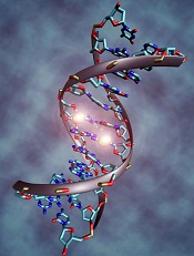
Image by Christoph Bock
A new study helps explain how the anticancer drug decitabine reverses cell damage and has revealed a potential biomarker that could indicate a
patient’s cancer stage and response to treatment.
Investigators found that decitabine combats some of cancer’s effects by taking the place of the nucleotide cytosine at specific locations on a replicating DNA strand.
In mimicking cytosine, the drug helps “tame” cancerous cells by turning on tumor suppressors and turning off oncogenes.
The investigators also found that decitabine causes an unexpected boost in the amount of a molecule known as 5-hydroxymethylcytosine (5hmC). Because many types of cancer cause 5hmC levels to plummet, an uptick in 5hmC could be a sign that cancer treatments are working.
“We think that the expression of 5hmC could be used as a biomarker to define the stage or the aggressiveness of cancer and to possibly indicate the effectiveness of cancer treatment,” said Joseph Irudayaraj, PhD, of Purdue University in West Lafayette, Indiana. “This could help us monitor the clinical success of patients receiving decitabine.”
Dr Irudayaraj and his colleagues reported these findings in Nature Scientific Reports.
Decitabine, one of the first epigenetic drugs, helps reverse the altered methylation patterns in cancerous cells, but its precise mode of action has not been clear.
Using a combination of models, Dr Irudayaraj and his colleagues showed that decitabine is taking the place of cytosine at strategic positions on replicating strands of DNA in cancer cells.
When an enzyme tries to add a methyl group to silence decitabine, the drug traps it in place, preventing methylation. This triggers another group of enzymes to transform a methylated cytosine on the parent DNA strand into 5hmC, a molecule whose biological function is not yet known.
The investigators confirmed the increase in 5hmC levels in decitabine-treated leukemia cells.
To better explain the team’s findings, Basudev Chowdhury, PhD, also of Purdue University, likened decitabine to a text editor that restores meaning to a garbled sentence and compared conventional chemotherapy to a delete button.
“Think of nucleotides as the alphabet with which our cells compose messages,” he said. “Epigenetics helps translate those messages into actions such as the production of proteins. But cancer can jumble the messages, making them nonsensical. Decitabine helps revise the messages so they can be understood.” ![]()
Image by Christoph Bock
A new study helps explain how the anticancer drug decitabine reverses cell damage and has revealed a potential biomarker that could indicate a
patient’s cancer stage and response to treatment.
Investigators found that decitabine combats some of cancer’s effects by taking the place of the nucleotide cytosine at specific locations on a replicating DNA strand.
In mimicking cytosine, the drug helps “tame” cancerous cells by turning on tumor suppressors and turning off oncogenes.
The investigators also found that decitabine causes an unexpected boost in the amount of a molecule known as 5-hydroxymethylcytosine (5hmC). Because many types of cancer cause 5hmC levels to plummet, an uptick in 5hmC could be a sign that cancer treatments are working.
“We think that the expression of 5hmC could be used as a biomarker to define the stage or the aggressiveness of cancer and to possibly indicate the effectiveness of cancer treatment,” said Joseph Irudayaraj, PhD, of Purdue University in West Lafayette, Indiana. “This could help us monitor the clinical success of patients receiving decitabine.”
Dr Irudayaraj and his colleagues reported these findings in Nature Scientific Reports.
Decitabine, one of the first epigenetic drugs, helps reverse the altered methylation patterns in cancerous cells, but its precise mode of action has not been clear.
Using a combination of models, Dr Irudayaraj and his colleagues showed that decitabine is taking the place of cytosine at strategic positions on replicating strands of DNA in cancer cells.
When an enzyme tries to add a methyl group to silence decitabine, the drug traps it in place, preventing methylation. This triggers another group of enzymes to transform a methylated cytosine on the parent DNA strand into 5hmC, a molecule whose biological function is not yet known.
The investigators confirmed the increase in 5hmC levels in decitabine-treated leukemia cells.
To better explain the team’s findings, Basudev Chowdhury, PhD, also of Purdue University, likened decitabine to a text editor that restores meaning to a garbled sentence and compared conventional chemotherapy to a delete button.
“Think of nucleotides as the alphabet with which our cells compose messages,” he said. “Epigenetics helps translate those messages into actions such as the production of proteins. But cancer can jumble the messages, making them nonsensical. Decitabine helps revise the messages so they can be understood.”
HL survivors have long-term risk of cardiovascular disease

Photo by Rhoda Baer
Survivors of Hodgkin lymphoma (HL) have an increased risk of developing cardiovascular diseases throughout their lives, according to a study published in JAMA Internal Medicine.
Previous research suggested that HL treatment is associated with an increased risk of cardiovascular diseases.
However, those studies did not determine how long the increased risk persists or pinpoint the risk factors for various cardiovascular diseases.
So Flora E. van Leeuwen, PhD, of the Netherlands Cancer Institute in Amsterdam, and her colleagues decided to investigate.
The team examined the risk for cardiovascular disease in HL survivors up to 40 years after they received treatment and compared that with the risk for cardiovascular disease in the general population. The researchers also studied treatment-related risk factors.
The study included 2524 Dutch patients who were diagnosed with HL when they were younger than 51 years of age. The patients’ median age was 27.3 years.
The patients were treated from 1965 through 1995 and had survived for at least 5 years after diagnosis. In all, 2052 patients (81.3%) had received mediastinal radiotherapy, and 773 (30.6%) had received chemotherapy containing an anthracycline.
At a median of 20.3 years of follow-up, there were 1713 cardiovascular events in 797 patients (31.6%), and 410 of those patients (51.4%) had experienced 2 events or more.
The most frequently occurring cardiovascular disease was coronary heart disease (CHD), with 401 patients developing it as their first event. This was followed by valvular heart disease (VHD, 374 events) and heart failure (HF, 140 events).
HL survivors had a 3.2-fold increased risk of developing CHD and a 6.8-fold increased risk of developing HF compared to the general population.
HL survivors who had been treated before age 25 had a 4.6-fold to 7.5-fold increased risk of CHD and a 10.9-fold to 40.5-fold increased risk of HF, depending on the age they ultimately attained.
HL survivors treated at 35 to 50 years of age had a 2.0-fold to 2.3-fold increased risk of CHD and a 3.1-fold to 5.2-fold increased risk of HF, depending on their attained age.
The risks of CHD and HF remained significantly increased beyond 35 years after HL treatment. The standardized incidence ratios were 3.9 and 5.8, respectively.
The median times between HL treatment and first cardiovascular disease events were 18 years for CHD, 24 years for VHD, and 19 years for HF.
The cumulative risk of any type of cardiovascular disease was 50% at 40 years after HL diagnosis. For patients who were treated for HL before they were 25, the cumulative risk of developing a cardiovascular disease at 60 years of age or older was 20% for CHD, 31% for VHD, and 11% for HF.
The study also suggested that mediastinal radiotherapy increased the risk of CHD, VHD, and HF. But anthracycline-containing chemotherapy only increased the risk of VHD and HF.
Dr van Leeuwen and her colleagues concluded that both physicians and patients should be aware that HL survivors have a persistently increased risk of developing cardiovascular diseases throughout their lives. The team also believes the results of their study may direct guidelines for follow-up in HL survivors.
A commentary related to this research is available in JAMA Internal Medicine as well.
MM drug met accelerated approval requirements

Photo courtesy of the CDC
Celgene Corporation has fulfilled the requirements for accelerated approval of pomalidomide (Pomalyst) in the US, based on results from the phase 3 MM-003 trial.
The trial showed that pomalidomide in combination with dexamethasone can improve survival in patients with relapsed or refractory multiple
myeloma (MM).
A drug can be granted accelerated approval in the US based on a surrogate endpoint thought to predict clinical benefit.
To retain approval from the US Food and Drug Administration (FDA), the drug must demonstrate an actual clinical benefit.
In 2013, the FDA granted pomalidomide accelerated approval for use in combination with dexamethasone to treat MM patients who have received at least 2 prior therapies, including lenalidomide and a proteasome inhibitor, and have demonstrated disease progression on or within 60 days of completing their last therapy.
The FDA’s approval was based on results from a phase 2 trial known as MM-002. The trial showed that pomalidomide plus dexamethasone can improve the overall response rate in relapsed/refractory MM patients when compared to pomalidomide alone.
About 29% of patients in the pomalidomide-dexamethasone arm achieved a partial response or better, compared to about 7% of patients in the pomalidomide-alone arm.
Now, results of the MM-003 trial have shown that pomalidomide plus low-dose dexamethasone can improve progression-free survival and overall survival in relapsed/refractory MM patients, when compared to high-dose dexamethasone alone.
The median progression-free survival was 3.6 months in the pomalidomide-dexamethasone arm and 1.8 months in the dexamethasone arm (P<0.001). And the median overall survival was 12.4 months and 8 months, respectively (P=0.009).
These outcomes suggest pomalidomide, in combination with dexamethasone, provides a clinical benefit for previously treated MM patients, which fulfills the requirements for accelerated approval. So the drug’s label has been updated to reflect his change.
For more details on pomalidomide, see the full prescribing information.
Drug can prevent bleeding in kids with hemophilia A
A recombinant factor VIII Fc fusion protein (rFVIIIFc/efmoroctocog alfa, Eloctate/Elocta) can prevent and control bleeding in previously treated children with severe hemophilia A, results of the phase 3 KIDS A-LONG study suggest.
Children who were previously receiving factor VIII prophylaxis saw their median annualized bleeding rate (ABR) decrease with rFVIIIFc, and close to half of the children on this study did not have any bleeding episodes while they were receiving rFVIIIFc.
None of the patients developed inhibitors to rFVIIIFc. And researchers said adverse events on this trial were typical of a pediatric hemophilia population.
The team reported these results in the Journal of Thrombosis and Haemostasis. The trial was sponsored by Biogen Idec and Sobi, the companies developing rFVIIIFc.
The study included 71 boys younger than 12 years of age who had severe hemophilia A. The patients had at least 50 prior exposure days to factor VIII and no history of factor VIII inhibitors.
The children were set to receive twice-weekly prophylactic infusions of rFVIIIFc, 25 IU/kg on day 1 and 50 IU/kg on day 4, but the researchers made adjustments to dosing as needed.
The median average weekly rFVIIIFc prophylactic dose was 88.11 IU kg. About 90% of the patients were on twice-weekly dosing at the end of the study. Seventy-four percent of patients were able to reduce their dosing frequency with rFVIIIFc compared to factor VIII prophylaxis.
About 46% of patients did not have any bleeding events on study. The median ABR was 1.96 overall and 0 for spontaneous and traumatic bleeding episodes, as well as for spontaneous joint bleeding episodes.
Among patients who were previously receiving factor VIII prophylaxis, their median ABR decreased with rFVIIIFc. For children younger than 6 years of age, the median ABR fell from 1.50 to 0. For children ages 6 through 11, the median ABR fell from 2.50 to 2.01.
About 86% of patients had at least one adverse event while on rFVIIIFc, but none of them discontinued treatment as a result.
Two non-serious events (myalgia and erythematous rash) were considered related to rFVIIIFc. And 5 patients experienced 7 serious adverse events that were not related to treatment.