User login
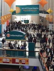
CDS systems often can’t tell if imaging is appropriate

Photo courtesy of NIH
Tools that help physicians decide whether to use diagnostic imaging can help reduce the use of unnecessary tests.
But new research suggests these tools may not be able to determine which tests are necessary most of the time.
The tools in question are computerized clinical decision support (CDS) systems, which match a patient’s characteristics against appropriateness
criteria to produce algorithmic treatment recommendations.
In a study published in JAMA, CDS systems did increase orders of imaging tests rated as “appropriate.”
However, the systems were not able to assign appropriateness ratings for a majority of tests because no appropriateness criteria were available for a particular test, or because the systems themselves were not able to find matching criteria.
“The increase in orders rated as appropriate is promising, but the number of tests that were not rated indicates there is room for further improvement of these tools,” said study author Peter S. Hussey, PhD, of the RAND Corporation in Boston, Massachusetts.
Study details
Dr Hussey and his colleagues used data from the Medicare Imaging Demonstration to evaluate the relationship of CDS system use with the proportion of imaging orders matched to appropriateness criteria, the appropriateness of ordered images, and the proportion of orders that changed after feedback.
The team compared 2 time periods during which clinicians used computerized radiology order entry systems and CDS systems for MRI, CT, and nuclear medicine procedures.
During a 6-month baseline period, the CDS systems tracked whether orders were linked with appropriateness criteria but did not provide clinicians with feedback on the appropriateness of orders.
During the 18-month intervention period, the CDS systems provided feedback indicating whether the order was linked to appropriateness criteria and, if so, the appropriateness rating, any recommendations for alternative orders, and a link to documentation supporting each rating.
National medical specialty societies developed the appropriateness criteria using expert panels that reviewed evidence and completed a structured rating process. The same appropriateness criteria were loaded into the CDS systems tools for all participating clinicians.
In all, 3340 clinicians placed 117,348 orders for advanced diagnostic imaging procedures.
Results
The CDS systems could not match most orders to appropriateness criteria. The systems did not identify relevant criteria for 63.3% of orders made during the baseline period and 66.5% of orders made during the intervention period.
Of the orders CDS systems could rate, 73.7% ordered during the baseline period and 81% ordered during the intervention period were rated as appropriate, and 11.1% and 6.4%, respectively, were rated inappropriate.
Of the orders that were initially rated as inappropriate, 4.8% were changed, and 1.9% were canceled.
When the CDS systems suggested an alternative for inappropriate orders, 9.9% of the orders were changed, and 0.4% were canceled. When the systems did not provide an alternative, 1.4% of inappropriate orders were changed, and 2.8% were canceled.
“In response to these findings, we recommend that clinical decision support efforts should focus on tools that help clinicians perform their work more efficiently and effectively,” said study author Katherine Kahn, MD, of the University of California, Los Angeles.
“We need a more comprehensive set of evidence-based guidelines that cover a greater proportion of advanced imaging orders for Medicare patients, and provide better methods for communicating feedback to clinicians.” ![]()
Scientists uncover structure of TOR complex 2

Photo courtesy of
University of Geneva
A group of researchers has developed a new tool to study the structure and function of target of rapamycin complex 2 (TORC2), which helps explain why rapamycin cannot access the TOR protein in this complex.
TOR is essential for the growth of normal cells but is hyperactive in tumor cells. Rapamycin is an immunosuppressant and anticancer agent that inactivates TOR in TORC1 but not in TORC2.
“In order to more easily study TORC2, we wanted to learn how to make this complex sensitive to rapamycin,” said Robbie Loewith, PhD, of the University of Geneva in Switzerland.
So Dr Loewith and a team of scientists from Switzerland, France, and the UK set out to elucidate how TORC2 works. The team reported their findings in Molecular Cell.
Using crosslinking-mass spectrometry and electron microscopy, they discovered that TORC2 has 3 features in common with TORC1: a rhomboid shape, 2-fold symmetry, and a central cavity delimited by the interface of its protein chains.
The 2 complexes differ markedly, however, in overall size, surface area of the interface, and the volume and shape of the central cavity.
By determining the structure of TORC2, the team could observe which subunit within TORC2 was obstructing the rapamycin binding site on TOR.
“By deleting part of this subunit, we generated a variant of TORC2 sensitive to rapamycin,” said Manoel Prouteau, PhD, also of the University of Geneva.
This allowed the researchers to study how TORC2 acts to stimulate cell growth.
Now, they hope to identify a specific inhibitor of endogenous TORC2 that could also be an effective anticancer agent.
“Our discovery that TORC2 inhibition alone is sufficient to block the cell cycle suggests that mTORC2-specific inhibitors may provide new and potentially better therapeutic alternatives,” the team concluded. ![]()
Drug prolongs PFS in indolent, refractory NHL
the 2015 ASCO Annual Meeting
CHICAGO—Adding obinutuzumab to treatment with bendamustine improves progression-free survival (PFS) in patients with rituximab-refractory, indolent non-Hodgkin lymphoma (NHL), interim results of the phase 3 GADOLIN trial suggest.
Study investigators said patients who received obinutuzumab and bendamustine followed by obinutuzumab maintenance had roughly double the PFS of patients who received bendamustine alone.
There was no significant difference between the treatment groups with regard to response rates or overall survival (OS), but the investigators said longer follow-up is needed to determine if obinutuzumab confers a benefit in OS.
This trial was stopped before its protocol-specified final analysis because of the PFS benefit observed in the obinutuzumab arm.
Laurie Sehn, MD, of the BC Cancer Agency in Vancouver, Canada, presented these results at the 2015 ASCO Annual Meeting (abstract LBA8502). Genentech Inc. and F. Hoffmann-La Roche Ltd. funded this research.
The trial included 413 patients with rituximab-refractory NHL, including follicular lymphoma (FL), marginal zone lymphoma (MZL), small lymphocytic lymphoma (SLL), and Waldenstrom’s macroglobulinemia (WM).
The patients were randomized to receive bendamustine alone (120 mg/m2/day on days 1 and 2 for up to six 28-day cycles) or a combination of bendamustine (90 mg/m2/day on days 1 and 2 for up to six 28-day cycles) plus obinutuzumab (1000 mg on days 1, 8, and 15 for cycle 1, followed by 1 dose for up to six 28-day cycles), followed by obinutuzumab maintenance (1000 mg every 2 months for 2 years or until progression).
Dr Sehn said there were no significant differences in baseline characteristics between the treatment arms. Patients in both arms had received a median of 2 prior treatments, and the median time from last treatment was about 4 months.
Of the 194 patients randomized to treatment in the obinutuzumab-bendamustine (OB) arm, 79.9% had FL, 13.9% had MZL, and 6.2% had SLL. Of the 202 patients randomized to the bendamustine-alone (control) arm, 82.2% had FL, 9.4% had MZL, 7.9% had SLL, and 0.5% had WM.
Ultimately, 156 patients completed induction in the OB arm, as did 129 patients in the control arm. Thirty-six patients completed maintenance with obinutuzumab, and 46 were still receiving maintenance at the time of analysis.
Safety results
Dr Sehn said there were no unexpected safety signals among patients in the OB arm.
About 99% of patients in the OB arm experienced at least 1 adverse event (AE), as did 98% of patients in the control arm. Severe AEs occurred in 38.1% and 32.8% of patients, respectively, and grade 3/4 AEs occurred in 67% and 62.1%, respectively.
AEs leading to treatment withdrawal occurred in 18% and 15.7% of patients, respectively. And AEs leading to death occurred in 6.2% and 6.1%, respectively.
Grade 3/4 AEs that occurred in at least 2% of patients in the OB and control arms, respectively, were neutropenia (33% vs 26.3%), thrombocytopenia (10.8% vs 16.2%), infusion-related reactions (10.8% vs 5.6%), anemia (7.7% vs 10.1%), febrile neutropenia (4.6% vs 3.5%), nausea (1% vs 3%), fatigue (1.5% vs 2.5%), diarrhea (1% vs 2.5%), and vomiting (2.1% vs 1%).
Response and survival
According to an independent radiology facility, 69.2% of patients in the OB arm had responded to treatment at the end of induction, as had 63% of the control arm. The best overall response by the 12-month mark was 78.7% and 76.6%, respectively.
The median follow-up was 21 months. At that point, the median PFS had not been reached in the OB arm but was 14.9 months in the control arm (P<0.0001), according to the independent radiology facility.
According to investigators, the median PFS was 29.2 months and 14 months, respectively (P<0.0001).
The median OS has not been reached in either arm (P=0.4017). Thirty-four patients (18%) in the OB arm died, as did 41 (20%) in the control arm.
Dr Sehn said longer follow-up is needed to determine the potential OS benefit associated with obinutuzumab, but the PFS benefit of OB is clinically meaningful.
“The fact that this new approach doubled average remission time marks a major step forward for our patients,” she said. “Obinutuzumab may offer patients the chance to stay well for a significantly longer period of time, putting off the need for additional chemotherapy.” ![]()
Breastfeeding may lower risk of ALL

Photo by Petr Kratochvil
Breastfeeding a child may reduce his risk of developing acute lymphoblastic leukemia (ALL) but perhaps not acute myeloid leukemia (AML), according to a review published in JAMA Pediatrics.
Researchers found that breastfeeding a child for 6 months or longer was associated with a 19% lower risk of childhood leukemia, compared with no
breastfeeding or breastfeeding for a shorter period of time.
And children who were breastfed for any amount of time had an 11% lower risk of childhood leukemia than children who were never breastfed.
However, when the researchers analyzed studies of ALL and AML separately, they found that breastfeeding was not associated with a significantly lower risk of AML.
Efrat L. Amitay, PhD, and Lital Keinan-Boker, MD, PhD, of the University of Haifa in Israel, conducted this research.
In a review of 18 studies, the pair found that breastfeeding a child for 6 months or longer was associated with a significantly lower risk of childhood leukemia, compared with no breastfeeding or breastfeeding for a shorter period of time (odds ratio [OR]=0.81).
And a separate analysis of 15 studies showed that children who were breastfed for any amount of time had a lower risk of childhood leukemia than children who were never breastfed (OR=0.89).
The researchers also conducted meta-analyses of AML studies and ALL studies separately—11 ALL and 6 AML studies. And they found a significant inverse association between breastfeeding for 6 months or more and ALL risk (OR=0.82) but no significant association for AML risk (OR=0.74).
The researchers said several biological mechanisms may explain the association between breastfeeding and a reduced risk of childhood leukemia. However, more high-quality studies are needed to clarify those mechanisms. ![]()
Newer anticoagulants may not be best for elderly, study shows

A meta-analysis of 92,816 people taking anticoagulants has shown that the risk of gastrointestinal (GI) bleeding related to the newer oral anticoagulants dabigatran and rivaroxaban is similar to that for warfarin.
But for patients older than 65, the risk of GI bleeding increases. By age 76, the risk may be 3 to 5 times higher when taking the newer anticoagulants compared to warfarin.
These findings were published in BMJ.
“The new anticoagulants have really been popular with patients who have previously only had one choice of oral anticoagulant,” said study author Neena S. Abraham, MD, of the Mayo Clinic in Scottsdale, Arizona.
“However, they may not be the right choice for everyone. Our findings definitely point toward important age-related risk that merit consideration when doctors are making treatment recommendations.”
Dr Abraham and her colleagues compared the risk of GI bleeding with newer anticoagulants and warfarin using national data available on privately insured patients and Medicare Advantage enrollees from the Optum Labs Data Warehouse.
Data on apixaban were not included in the study because there were too few patients prescribed apixaban in the dataset during the period of observation, from November 1, 2010, to September 30, 2013.
The cohort included 8578 (9.2%) patients on dabigatran, 16,253 (17.5%) on rivaroxaban, and 67,985 (73.2%) on warfarin. Patients were 18 years of age or older.
The researchers found that, among atrial fibrillation (AF) patients older than 75, the risk of GI bleeding was higher than with warfarin. The hazard ratios (HRs) were 2.49 (95% confidence interval [CI] 1.61 to 3.83) and 1.62 (95% CI 1.02 to 2.58), respectively.
However, among older patients without AF, the risk of GI bleeding was comparable with dabigatran and warfarin. The HRs were 1.56 (95% CI 0.42 to 5.80) and 2.73 (95% CI 0.83 to 8.94), respectively.
Older AF patients taking rivaroxaban had an increased risk of GI bleeding compared to patients taking warfarin. The HRs were 2.91 (95% CI 1.65 to 4.81) and 2.05 (95% CI 1.17 to 3.59), respectively.
And older patients without AF had an increased risk of GI bleeding with rivaroxaban compared to warfarin. The HRs were 4.58 (95% CI 2.40 to 8.72) and 4.40 (95% CI 2.43 to 7.96), respectively.
The researchers also found that, for those patients under 65, the newer agents seemed to confer a lower risk of GI bleeding than warfarin. ![]()
Inhibitor may fulfill unmet need in MF

© ASCO/Zach Boyden-Holmes
CHICAGO—The JAK2/FLT3 inhibitor pacritinib may fulfill an unmet need in the treatment of myelofibrosis (MF), according to a speaker at the 2015 ASCO Annual Meeting.
Results of the phase 3 PERSIST-1 trial indicate that pacritinib is safe and effective for MF patients with thrombocytopenia.
“Thrombocytopenia is a common feature in people with advanced [MF], and current treatment options have not been able to concurrently improve splenomegaly symptoms and cytopenias in these patients,” said study investigator Ruben A. Mesa, MD, of the Mayo Clinic Cancer Center in Scottsdale, Arizona.
But PERSIST-1 showed that pacritinib can accomplish this. And the drug proved more effective than best available therapy (BAT), excluding JAK inhibitors, in reducing spleen volume and alleviating MF symptoms in the entire cohort of MF patients.
Dr Mesa presented these results at ASCO as abstract LBA7006. The study was funded by CTI BioPharma Corp., the company developing pacritinib.
The trial included 327 patients who were randomized to receive pacritinib (n=220) or BAT (n=107).
Patients in the BAT arm received therapies that are routinely prescribed off-label for MF, such as erythropoietin-stimulating agents, immunomodulatory drugs, and hydroxyurea. Ruxolitinib was intentionally excluded from this trial because the study included patients with thrombocytopenia.
Dr Mesa said the patients’ baseline characteristics “demonstrate a group of individuals with advanced myelofibrosis, a heavy percentage of those with primary myelofibrosis, the vast majority having intermediate-2 or high-risk disease, with very significant splenomegaly, and the vast majority having the JAK2 mutation.”
“About half the individuals were anemic or transfusion-dependent,” he noted. “And a full third were thrombocytopenic, under 100,000 [platelets/µL], with 16% under 50,000 [platelets/µL]. This was the first phase 3 study of myelofibrosis that allowed individuals with a platelet count of less than 100,000 to be enrolled.”
Fifty-six percent of patients remained on pacritinib at the time of analysis, as did 8% of patients on BAT. Seventy-nine percent of patients crossed over from the BAT arm to the pacritinib arm.
Spleen reduction
The study’s primary endpoint was a reduction in spleen volume of 35% or greater.
In the intent-to-treat (ITT) population, 19.1% of patients in the pacritinib arm met this endpoint, as did 4.7% of patients in the BAT arm (P=0.0003). In the evaluable population—165 patients in the pacritinib arm and 85 patients in the BAT arm—the rates were 25% and 5.9%, respectively (P=0.0001).
Dr Mesa noted that pacritinib was able to reduce spleen volume in all subgroups of patients, including those with thrombocytopenia.
“Both the group [with platelet counts] under 100,000 as well as under 50,000 uniquely responded only on the pacritinib arm, with no responses on the BAT arm,” he said.
In the ITT population, 16.7% of patients with platelet counts under 100,000/µL and 22.9% of patients with platelet counts under 50,000/µL met the primary endpoint. The P values, in the comparison with the BAT arm, were 0.0451 and 0.0086, respectively.
In the evaluable population, 23.5% of patients with platelet counts under 100,000/µL and 33.3% of patients with platelet counts under 50,000/µL met the primary endpoint. The P values were 0.0370 and 0.0072, respectively.
“It is too early to know if pacritinib has an impact on survival, but that is clearly our expectation [based on the spleen responses observed],” Dr Mesa said.
TSS and transfusion
The study’s secondary endpoint was the proportion of patients with a 50% or greater reduction in Total Symptom Score (TSS) from baseline to week 24. TSS was measured by patient responses on the Myeloproliferative Neoplasm Symptom Assessment Form.
In the ITT population, 24.5% of pacritinib-treated patients and 6.5% of BAT-treated patients had a 50% or greater reduction in TSS score (P<0.0001). In the evaluable population, 40.9% and 9.9% of patients, respectively (P<0.0001), met this endpoint.
Dr Mesa also pointed out that 25.7% of pacritinib-treated patients who were severely anemic and transfusion-dependent—requiring at least 6 units of blood in the 90 days prior to study entry—became transfusion independent. But none of the BAT-treated patients did so (P<0.043).
Adverse events
“The most common adverse events [in the pacritinib arm] were consistent with the earlier studies,” Dr Mesa said. “Gastrointestinal toxicities were most common, although typically at low grades.”
“As expected, we saw very few individuals with any significant thrombocytopenia or anemia as drug-emergent. There were individuals who enrolled in the study as a grade 4, so some of those remained.”
The most common adverse events of any grade were diarrhea (53.2% in the pacritinib arm and 12.3% in the BAT arm), nausea (26.8% vs 6.6%), anemia (22.3% vs 19.8%), thrombocytopenia (16.8% vs 13.2%), and vomiting (15.9% vs 5.7%).
Ten percent of patients in the pacritinib arm required dose reductions due to adverse events. Diarrhea prompted dose interruptions in 13 patients and discontinuation in 3 patients. But pacritinib-associated diarrhea typically resolved in a little over a week.
“Based on these preliminary results, pacritinib may represent a very important agent for individuals with advanced disease and may have an impact on the disease course,” Dr Mesa concluded. ![]()
Drug improves upon standard therapy for relapsed CLL/SLL, speaker says

Chanan-Khan, MD
© ASCO/Zach Boyden-Holmes
CHICAGO—Interim results of the phase 3 HELIOS trial suggest that adding ibrutinib to treatment with bendamustine and rituximab (BR) improves outcomes for patients with relapsed chronic lymphocytic leukemia/small lymphocytic lymphoma (CLL/SLL).
Patients who received ibrutinib and BR had significantly higher response rates and a significantly longer progression-free survival than patients who received BR with placebo.
There was no significant difference between the arms with regard to overall survival, but the researchers said these results were confounded by the fact that 31% of patients in the placebo arm crossed over to the ibrutinib arm after they progressed.
“We found that ibrutinib can be safely paired with existing therapy to powerfully prolong remissions and improve patients’ well-being,” said study investigator Asher Alban Akmal Chanan-Khan, MD, of the Mayo Clinic in Jacksonville, Florida.
Dr Chanan-Khan presented these findings at the 2015 ASCO Annual Meeting (abstract LBA7005). The research was funded by Janssen Research & Development, LLC, the company co-developing ibrutinib with Pharmacyclics.
The study included 578 patients with previously treated CLL/SLL, excluding those with del(17p). The patients were randomized to receive 6 cycles of BR plus once-daily ibrutinib (n=289) or 6 cycles of BR plus placebo (n=289). Ibrutinib and placebo were given until disease progression or unacceptable toxicity.
Dr Chanan-Khan said baseline characteristics were comparable between the treatment arms. For each arm, the median number of prior treatments was 2, more than 50% of patients had bulky disease, and about 80% of patients had unmutated IGVH.
“[However,] advanced Rai-stage disease was observed in a slightly [greater] proportion of patients in the control arm versus the ibrutinib arm,” Dr Chanan-Khan noted. “Conversely, a higher proportion of patients with del(11q) was noted in the ibrutinib-containing arm.”
Ultimately, 81.9% (n=235) of patients in the ibrutinib arm and 77.4% (n=222) of those in the placebo arm received their assigned 6 cycles of BR. At the time of analysis, the rate of treatment discontinuation was 29.1% (n=84) in the ibrutinib arm and 64.7% in the placebo arm (n=187).
Those patients who progressed on placebo were allowed to cross over to the ibrutinib arm, and 90 patients had crossed over at the time of the interim analysis.
Response and survival
The study’s primary endpoint was progression-free survival, as assessed by an independent review committee (IRC), in the intent-to-treat population (n=289 in each arm).
At a median follow-up of 17 months, progression-free survival was 13.3 months in the placebo arm and was not reached in the ibrutinib arm (P<0.0001).
“The hazard ratio on this particular survival curve is 0.20, which translates into a reduced risk of progression or death by 80%,” Dr Chanan-Khan said. “This is remarkable. You cannot get a better hazard ratio than this.”
Dr Chanan-Khan also noted that the overall response rate was significantly higher in the ibrutinib arm than the placebo arm. The rates were 82.7% and 67.8%, respectively (P<0.0001), according to the IRC, and 86.2% and 68.9%, respectively (P<0.0001), according to investigator assessment.
The rate of complete response plus complete response with incomplete blood count recovery was 10.4% in the ibrutinib arm and 2.8% in the placebo arm, according to the IRC. According to investigator assessment, the rates were 21.4% and 5.9%, respectively.
The median overall survival was not reached in either arm, and the hazard ratio was 0.628 (P=0.0598).
Adverse events
Dr Chanan-Kahn said the safety profile of the ibrutinib-BR combination was consistent with the safety profiles of each individual drug.
The incidence of adverse events was 70.7% in the ibrutinib-BR arm and 70% in the placebo-BR arm. The most common events were neutropenia (58.2% and 54.7%, respectively), nausea (36.9% vs 35.2%), diarrhea (35.5% vs 23.7%), thrombocytopenia (30.7% vs 24.4%), pyrexia (24.7% vs 22%), anemia (22.6% vs 28.9%), and fatigue (21.6% vs 22.6%).
The incidence of grade 3/4 adverse events was 28.9% in the ibrutinib arm and 25% in the placebo arm. The most common of these were neutropenia (53.7% vs 50.5%) and thrombocytopenia (15% in both arms).
Atrial fibrillation was seen in 7.3% of patients in the ibrutinib arm and 2.8% in the placebo arm. Grade 3/4 atrial fibrillation occurred in 2.8% and 0.7% of patients, respectively. The incidence of tumor lysis syndrome was 3.5% in both arms.
The rate of bleeding was 31% in the ibrutinib arm and 14.6% in the placebo arm. And the rates of major hemorrhage were 3.8% and 1.7%, respectively.
Adverse events were the primary reason for discontinuation in patients who received ibrutinib—14.2%, compared to 11.8% of patients who received placebo. The primary reason for discontinuation in the placebo arm was progressive disease or relapse—45%, compared to 4.8% in the ibrutinib arm.
Taken together, the results of this trial suggest treatment with ibrutinib and BR is superior to standard BR therapy in patients with relapsed CLL/SLL, Dr Chanan-Kahn said.
“This was one of the most rigorous clinical trials ever conducted in CLL,” he said, “and it truly validates ibrutinib as an important drug for this cancer.” ![]()
Chanan-Khan, MD
© ASCO/Zach Boyden-Holmes
CHICAGO—Interim results of the phase 3 HELIOS trial suggest that adding ibrutinib to treatment with bendamustine and rituximab (BR) improves outcomes for patients with relapsed chronic lymphocytic leukemia/small lymphocytic lymphoma (CLL/SLL).
Patients who received ibrutinib and BR had significantly higher response rates and a significantly longer progression-free survival than patients who received BR with placebo.
There was no significant difference between the arms with regard to overall survival, but the researchers said these results were confounded by the fact that 31% of patients in the placebo arm crossed over to the ibrutinib arm after they progressed.
“We found that ibrutinib can be safely paired with existing therapy to powerfully prolong remissions and improve patients’ well-being,” said study investigator Asher Alban Akmal Chanan-Khan, MD, of the Mayo Clinic in Jacksonville, Florida.
Dr Chanan-Khan presented these findings at the 2015 ASCO Annual Meeting (abstract LBA7005). The research was funded by Janssen Research & Development, LLC, the company co-developing ibrutinib with Pharmacyclics.
The study included 578 patients with previously treated CLL/SLL, excluding those with del(17p). The patients were randomized to receive 6 cycles of BR plus once-daily ibrutinib (n=289) or 6 cycles of BR plus placebo (n=289). Ibrutinib and placebo were given until disease progression or unacceptable toxicity.
Dr Chanan-Khan said baseline characteristics were comparable between the treatment arms. For each arm, the median number of prior treatments was 2, more than 50% of patients had bulky disease, and about 80% of patients had unmutated IGVH.
“[However,] advanced Rai-stage disease was observed in a slightly [greater] proportion of patients in the control arm versus the ibrutinib arm,” Dr Chanan-Khan noted. “Conversely, a higher proportion of patients with del(11q) was noted in the ibrutinib-containing arm.”
Ultimately, 81.9% (n=235) of patients in the ibrutinib arm and 77.4% (n=222) of those in the placebo arm received their assigned 6 cycles of BR. At the time of analysis, the rate of treatment discontinuation was 29.1% (n=84) in the ibrutinib arm and 64.7% in the placebo arm (n=187).
Those patients who progressed on placebo were allowed to cross over to the ibrutinib arm, and 90 patients had crossed over at the time of the interim analysis.
Response and survival
The study’s primary endpoint was progression-free survival, as assessed by an independent review committee (IRC), in the intent-to-treat population (n=289 in each arm).
At a median follow-up of 17 months, progression-free survival was 13.3 months in the placebo arm and was not reached in the ibrutinib arm (P<0.0001).
“The hazard ratio on this particular survival curve is 0.20, which translates into a reduced risk of progression or death by 80%,” Dr Chanan-Khan said. “This is remarkable. You cannot get a better hazard ratio than this.”
Dr Chanan-Khan also noted that the overall response rate was significantly higher in the ibrutinib arm than the placebo arm. The rates were 82.7% and 67.8%, respectively (P<0.0001), according to the IRC, and 86.2% and 68.9%, respectively (P<0.0001), according to investigator assessment.
The rate of complete response plus complete response with incomplete blood count recovery was 10.4% in the ibrutinib arm and 2.8% in the placebo arm, according to the IRC. According to investigator assessment, the rates were 21.4% and 5.9%, respectively.
The median overall survival was not reached in either arm, and the hazard ratio was 0.628 (P=0.0598).
Adverse events
Dr Chanan-Kahn said the safety profile of the ibrutinib-BR combination was consistent with the safety profiles of each individual drug.
The incidence of adverse events was 70.7% in the ibrutinib-BR arm and 70% in the placebo-BR arm. The most common events were neutropenia (58.2% and 54.7%, respectively), nausea (36.9% vs 35.2%), diarrhea (35.5% vs 23.7%), thrombocytopenia (30.7% vs 24.4%), pyrexia (24.7% vs 22%), anemia (22.6% vs 28.9%), and fatigue (21.6% vs 22.6%).
The incidence of grade 3/4 adverse events was 28.9% in the ibrutinib arm and 25% in the placebo arm. The most common of these were neutropenia (53.7% vs 50.5%) and thrombocytopenia (15% in both arms).
Atrial fibrillation was seen in 7.3% of patients in the ibrutinib arm and 2.8% in the placebo arm. Grade 3/4 atrial fibrillation occurred in 2.8% and 0.7% of patients, respectively. The incidence of tumor lysis syndrome was 3.5% in both arms.
The rate of bleeding was 31% in the ibrutinib arm and 14.6% in the placebo arm. And the rates of major hemorrhage were 3.8% and 1.7%, respectively.
Adverse events were the primary reason for discontinuation in patients who received ibrutinib—14.2%, compared to 11.8% of patients who received placebo. The primary reason for discontinuation in the placebo arm was progressive disease or relapse—45%, compared to 4.8% in the ibrutinib arm.
Taken together, the results of this trial suggest treatment with ibrutinib and BR is superior to standard BR therapy in patients with relapsed CLL/SLL, Dr Chanan-Kahn said.
“This was one of the most rigorous clinical trials ever conducted in CLL,” he said, “and it truly validates ibrutinib as an important drug for this cancer.”
Team calls for change in warfarin dosing algorithms

Photo courtesy of NIGMS
Investigators have proposed the use of race-stratified algorithms to help clinicians better calculate the appropriate warfarin dose for a patient.
The team’s study, published in Blood, showed that clinical and genetic factors affecting warfarin dose requirements vary by race.
“As the outcomes of disease can vary by race, so can response to medications,” said Nita Limdi, PhD, PharmD, of the University of Alabama at Birmingham.
“Therefore, warfarin dosing equations that combine race groups for analysis (race-adjusted analysis) assume that the effect of variables—such as age and genetics—are the same across race groups, which may compromise dose prediction among patients of both races.”
To better understand how genetics and clinical factors influence warfarin dosing across race groups, Dr Limdi and her colleagues analyzed 1357 patients—595 African American and 762 European American—treated with warfarin.
The team calculated and compared dose recommendations according to both race-adjusted dosing models and race-stratified dosing models. They found that race-stratified analysis improved dose prediction in both racial groups, as compared to race- adjusted analysis.
Race-stratified analysis showed that European Americans with the CYP2C9*2 variant required less warfarin than European Americans with wild-type CYP2C9. But the same was not true for African Americans.
And although all participants who carried VKORC1 required lower doses, regardless of race, the proportional dose reduction was greater among European Americans.
The investigators therefore concluded that the influence of genetic and clinical factors on warfarin dose differs by race. So race-stratified algorithms, rather than race-adjusted algorithms, should be used to guide warfarin dosing.
“Our findings highlight the need for adequate racial representation in warfarin dosing studies to improve our understanding of how the factors that influence warfarin dose differ according to race,” Dr Limdi said. “This is the first step to developing race-specific algorithms to personalize therapy.”
Hodgkin lymphoma incidence on the decline worldwide
Photo courtesy of NIH
In trying to estimate the global cancer burden, researchers found that cases of Hodgkin lymphoma (HL) have decreased worldwide over the last 2 decades.
The team studied 28 cancer types in 188 countries, and HL was the only malignancy whose incidence decreased from 1990 to 2013.
And the number of HL deaths in 2013 was comparatively low. When the researchers ranked cancers according to the number of global deaths, HL was 26th on the list of 28.
The researchers disclosed these results in JAMA Oncology.
The team collected data from cancer registries, vital records, verbal autopsy reports, and other sources to estimate the global cancer burden.
The data suggested that, in 2013, there were 14.9 million new cancer cases and 8.2 million cancer deaths worldwide. The proportion of cancer deaths as part of all deaths increased from 12% in 1990 to 15% in 2013.
The most common malignancy in men was prostate cancer, with 1.4 million cases in 2013. For women, it was breast cancer, with 1.8 million cases in 2013.
Tracheal, bronchus, and lung cancers were the leading cause of cancer death in men and women, with 1.6 million deaths in 2013.
Hematologic malignancies
Globally, the age-standardized incidence of HL per 100,000 people decreased by 34% during the time period studied. Cases of HL fell from about 103,000 in 1990 to 93,000 in 2013.
When the researchers ranked cancer types according to the number of global deaths in 2013, HL came in 26th. There were about 24,000 HL deaths in 2013—14,000 among men and 10,000 among women.
Non-Hodgkin lymphoma (NHL) came in 11th for global cancer deaths in 2013. There were about 226,000 NHL deaths—133,000 among men and 92,000 among women.
In addition, the incidence of NHL more than doubled from 1990 to 2013, rising from about 227,000 to 465,000. According to 2013 data, 1 in 103 men and 1 in 151 women developed NHL between birth and 79 years of age.
The researchers observed an increase in cases of multiple myeloma (MM) as well, from about 63,000 in 1990 to 117,000 in 2013.
In 2013, there were about 79,000 MM deaths—42,000 among men and 37,000 among women. MM ranked 19th on the list of global cancer deaths in 2013.
Leukemia ranked 9th on the list. There were about 265,000 leukemia deaths in 2013—149,000 among men and 116,000 among women.
Cases of leukemia increased from 297,000 in 1990 to 414,000 in 2013. According to 2013 data, 1 in 127 men and 1 in 203 women developed leukemia between birth and 79 years of age.
This research shows that cancer remains a major threat to people’s health around the world, said study author Christina Fitzmaurice, MD, of the University of Washington in Seattle.
“Cancer prevention, screening, and treatment programs are costly,” she noted, “and it is very important for countries to know which cancers cause the highest disease burden in order to allocate scarce resources appropriately.”
DART molecule proves active against AML

Photo by Sakurai Midori
An artificial antibody that redirects T cells to target cancer cells shows promise for treating acute myeloid leukemia (AML), according to preclinical research.
The antibody, MGD006, induced tumor regression in mouse models of AML and was largely well-tolerated in cynomolgus monkeys.
Investigators say these results support clinical testing of MGD006 in AML, which is currently underway.
MGD006 is a humanized, dual-affinity re-targeting (DART) molecule that combines a portion of an antibody recognizing CD3, an activating molecule expressed by T cells, with an arm that recognizes CD123.
MGD006 redirects T cells to kill cells expressing CD123, which is upregulated in AML and other hematologic diseases.
Gurunadh Chichili, PhD, of MacroGenics, Inc., in Rockville, Maryland, and his colleagues described their work with MGD006 in Science Translational Medicine. MacroGenics, the company developing MGD006, funded this research.
Because MGD006 is designed to be cleared rapidly, it requires continuous delivery. So in mice, the investigators administered the molecule continuously for up to a week via peritoneally implanted osmotic pumps.
The NSG/b2m−/− mice had been reconstituted with human peripheral blood mononuclear cells and grafted with KG-1a cells, an AML-M0 line. The mice received MGD006 after tumors were allowed to grow to an average size of about 100 mm3.
Treated mice experienced significant tumor regression at all doses of MGD006 (P<0.005), but there was no activity in mice treated with a control DART molecule. The investigators found that 500 ng/kg of MGD006 per day was sufficient to completely eliminate leukemic cells.
The team also tested MGD006 in macaques and found the molecule binds to human and cynomolgus monkey antigens with similar affinities and redirects T cells from either species to kill CD123-expressing target cells.
The monkeys received continuous infusions of MGD006, starting at 0.1 mg/kg per day and escalating weekly to up to 1 mg/kg per day for a 4-week period. The treatment depleted circulating CD123-positive cells beginning at 72 hours and continuing throughout the infusion period.
The monkeys experienced cytokine release, but it was transient and most significant after the first dose of MDG006. After the first dose, IL-6 concentration returned to baseline by 72 hours, and the magnitude of IL-6 response decreased with each successive MGD006 infusion, even when the dose was increased.
The animals experienced reversible reductions in hematocrit and red cell mass at the highest doses of MDG006 but no neutropenia or thrombocytopenia.
“This research paved the way for our initiation of a phase 1 clinical study of MGD006 in 2014,” said Scott Koenig, MD, PhD, President and CEO of MacroGenics.
“MGD006 has demonstrated great promise as a T-cell-redirected cancer immunotherapy in preclinical studies. We are hopeful that these studies will translate into clinical trial results indicative of clinical improvement for patients with AML, myelodysplastic syndrome, and several other forms of leukemia and lymphoma.”