User login
CAR produces high CR rate in adults with rel/ref ALL

the 2015 ASCO Annual Meeting
© ASCO/Max Gersh
CHICAGO—A CD19-targeted chimeric antigen receptor (CAR) T-cell therapy can provide durable complete responses (CRs) or a bridge to allogeneic transplant in adults with relapsed or refractory acute lymphoblastic leukemia (ALL), updated results of a phase 1 study suggest.
The therapy, JCAR015, produced a CR rate of 87%, and 33% of these patients went on to transplant.
The median duration of response or relapse-free survival was 5.3 months. The median overall survival was 8.5 months.
Nearly a quarter of patients developed severe cytokine release syndrome (CRS), and nearly 30% experienced neurological toxicities. But researchers said these effects were largely treatable and reversible.
This study was temporarily placed on clinical hold last year, after 2 patients died from complications related to CRS. But the hold was soon lifted and enrollment and dosing criteria were changed in an attempt to prevent severe CRS.
Jae H. Park, MD, of Memorial Sloan Kettering Cancer Center in New York, presented updated results of this trial (NCT01044069) at the 2015 ASCO Annual Meeting (abstract 7010*). The study is sponsored by Memorial Sloan Kettering, but funding has also been provided by Juno Therapeutics, the company developing JCAR015.
Results from this trial have previously been reported in Science Translational Medicine (Davila et al 2014; Brentjens et al 2013), at AACR 2014, and at ASH 2014.
At ASCO, Dr Park presented results in 39 patients with relapsed/refractory, CD19+ ALL. All of them were evaluable for toxicity assessment, and 38 were evaluable for response with at least 1 month of follow-up.
There were 29 males, and the patients’ median age was 45 (range, 22-74). Thirty-three percent had Ph+ ALL, and 11% had the T315I mutation.
Forty-nine percent of patients had received 2 prior therapies, 23% had received 3, and 28% had received 4 or more. Thirty-six percent of patients had a prior allogeneic hematopoietic stem cell transplant (HSCT).
For this study, patients first underwent leukapheresis. While their T cells were being manufactured, they were allowed to receive salvage chemotherapy. Patients underwent repeat bone marrow biopsy to assess their disease status immediately prior to T-cell infusion.
Fifty-four percent of patients (n=21) had morphologic disease (>5% blasts in the bone marrow, median 52%) immediately prior to JCAR015 infusion, and the remaining patients (n=18) had minimal residual disease (MRD).
Two days after conditioning with cyclophosphamide, patients received an infusion of 1-3 x 10⁶ CAR T cells/kg. At day 28, the researchers assessed patients’ disease with a repeat bone marrow biopsy.
Treatment results
The median follow-up was 5.6 months (1 to >38 months). Six patients had more than a year of follow-up.
The CR rate was 87% (33/38), and 81% of evaluable patients (26/32) were MRD-negative. The median time to CR was 23 days, and the median duration of response or relapse-free survival was 5.3 months.
“We examined the CR rates by different subgroup,” Dr Park noted. “We looked at whether patients had a pre-T-cell disease burden: morphologic disease vs minimal residual disease, whether they had an allogeneic bone marrow transplant prior to CAR T-cell infusion, their Ph+ status, age at infusion, and prior lines of therapy. And there was no [significant] difference between these groups for CRs and MRD-negative CR rate.”
At the time of presentation, 14 patients were disease-free, 10 of whom had not gone on to HSCT. In all, 11 patients went on to allogeneic HSCT.
Fourteen patients relapsed during follow-up, 3 after HSCT. Two of these patients had CD19-negative bone marrow blasts.
The median overall survival was 8.5 months in all patients and 10.8 months in patients who were MRD-negative. The median overall survival was 9.9 months in patients who underwent allogeneic HSCT and 8.5 months in patients who did not.
Dr Park said key adverse events were CRS—clinically manifested by fever, hypotension, and respiratory insufficiency—and neurological changes such as delirium, global encephalopathy, aphasia, and seizures.
Twenty-three percent of patients (n=9) developed severe CRS, 28% (n=11) had grade 3/4 neurotoxicity, and 8% (n=3) had grade 5 toxicity. The patients with grade 5 toxicities died of ventricular arrhythmia, sepsis, and an unknown cause (although this patient suffered a seizure).
The severity of CRS correlated with disease burden, and CRS was managed with an IL-6R inhibitor (n=4), a steroid (n=2), or both (n=9). Neurological symptoms were reversible and could occur independently of CRS, Dr Park said. ![]()
*Information in the abstract differs from that presented at the meeting.
No survival difference with allo- or auto-SCT in PTCL
© ASCO/Max Gersh
CHICAGO—Allogeneic and autologous transplants produce similar survival rates when used as first-line therapy in younger patients with peripheral
T-cell lymphoma (PTCL), according to interim results of the AATT trial.
The study also showed that deaths among patients who received autologous stem cell transplants (auto-SCTs) were a result of relapse and salvage treatment, while deaths among allogeneic SCT (allo-SCT) recipients were transplant-related.
Norbert Schmitz, MD, PhD, of Asklepios Hospital St. Georg in Hamburg, Germany, presented these findings at the 2015 ASCO Annual Meeting (abstract 8507*).
Dr Schmitz noted that only previous study comparing auto-SCT with allo-SCT as first-line therapy in PTCL was not designed or powered to evaluate the differences between the transplant types.
So he and his colleagues conducted the AATT trial to determine the differences. The team hypothesized that allo-SCT would improve 3-year event-free survival from 35% to 60%, given an α of 5% and a power of 80%. They needed 140 patients to prove or disprove this theory.
Ultimately, the investigators enrolled 104 patients and performed an interim analysis when 58 patients were evaluable for response.
Of the 58 patients, 30 were randomized to the auto-SCT arm and 28 to the allo-SCT arm. Baseline characteristics were similar between the arms, including patients’ median ages (49 and 50, respectively), the proportion of patients with stage III/IV disease (87% and 93%), and the proportion with ECOG status greater than 1 (23% and 18%).
Most patients in both arms had PTCL not otherwise specified (36% in the auto-SCT arm and 50% in the allo-SCT arm). Other subtypes included angioimmunoblastic T-cell lymphoma (23% and 32%, respectively), ALK-negative anaplastic large-cell lymphoma (20% and 4%), and “other” PTCLs (20% and 8%). The other PTCLs were NK/T-cell lymphoma, intestinal T/NK-cell lymphoma, hepatosplenic γδ lymphoma, and subcutaneous panniculitis-like PTCL.
Treatment characteristics
Before undergoing transplant, patients in both arms received treatment with CHOEP (cyclophosphamide, doxorubicin, etoposide, vincristine, and prednisone) on days 1, 15, 29, and 43. If they experienced a complete response (CR), partial response, or no change, patients received DHAP (dexamethasone, cytarabine, and cisplatin) on day 64.
Patients in the auto-SCT arm received BEAM (carmustine, etoposide, cytarabine, and melphalan) prior to transplant. And patients in the allo-SCT arm received FBC (fludarabine, busulfan, and cyclophosphamide).
Overall, 36 patients (62%) completed treatment per protocol, 19 in the auto-SCT arm and 17 in the allo-SCT arm. Thirty-eight percent of all patients could not proceed to transplant per protocol, mostly because of early lymphoma progression.
Response and survival
The researchers observed CRs/unconfirmed CRs (CRus) in 33% (n=10) of patients in the auto-SCT arm and 39% (n=11) in the allo-SCT arm. CR/CRus and progressive disease within 2 months occurred in 3% (n=1) and 4% (n=1) of patients, respectively.
Partial responses were seen in 17% (n=5) of patients in the auto-SCT arm and 7% (n=2) in the allo-SCT arm. There was no change in 7% (n=2) and 0% of patients, respectively. And responses were unknown in 7% (n=2) of patients in the auto-SCT arm.
Progressive disease occurred in 33% (n=10) of patients in the auto-SCT arm and 36% (n=10) in the allo-SCT arm. And treatment-related death occurred in 0% (n=0) and 14% (n=4), respectively.
At the interim analysis, there was no significant difference between the treatment arms with regard to event-free survival (P=0.963) or overall survival (P=0.174).
“At that time, the decision was made to stop the study,” Dr Schmitz said.
He explained that a conditional power analysis showed a low probability that the primary endpoint—a 25% improvement in event-free survival with allo-SCT—could still be met. So the data safety monitoring board decided to stop enrollment.
An updated analysis, performed at a median observation time of 26 months, showed there was still no significant difference in overall survival between the treatment arms (P=0.362).
Cause of death
In the intent-to-treat population—30 patients in the auto-SCT arm and 28 in the allo-SCT arm—there were 16 lymphoma-related deaths, 10 in the auto-SCT arm and 6 in the allo-SCT arm.
There were 6 deaths related to study treatment (4 early and 2 late), all in the allo-SCT arm. One patient in the allo-SCT arm died of post-transplant lymphoproliferative disorder, and 1 patient in the same arm died of hemorrhage after salvage. One patient in each arm died as a result of salvage treatment.
Dr Schmitz and his colleagues also looked at the cause of death among patients who received a transplant—19 in the auto-SCT arm and 17 in the allo-SCT arm.
After SCT, there were 7 deaths in each arm. In the auto-SCT arm, there were 6 lymphoma-related deaths and 1 death related to salvage treatment. In the allo-SCT arm, there were 7 cases of non-relapse-related mortality, including 1 patient with post-transplant lymphoproliferative disorder.
“There certainly seems to be a [graft-vs-lymphoma] effect of allo-transplant in T-cell lymphoma that is, unfortunately, in some way, counterbalanced by high transplant-related mortality,” Dr Schmitz said.
He added that results of a final analysis of the 104 patients enrolled on this study should be available in 2017. ![]()
*Information in the abstract differs from that presented at the meeting.
Triplet shows early promise for relapsed CLL, NHL
©ASCO/Rodney White
CHICAGO—A 3-drug combination is safe and highly active in certain patients with relapsed B-cell malignancies, according to a speaker at the 2015 ASCO Annual Meeting.
The combination consists of the anti-CD20 monoclonal antibody ublituximab, the PI3Kδ inhibitor TGR-1202, and the BTK inhibitor ibrutinib.
In a small, phase 1 study, the triplet produced an overall response rate of 62%. It was particularly active in patients with chronic lymphocytic leukemia/small lymphocytic lymphoma (CLL/SLL) and those with mantle cell lymphoma (MCL).
The most common adverse events were infusion reactions, gastrointestinal events, rash, and fatigue. Grade 3 neutropenia and leukopenia occurred in 1 patient each.
Nathan Fowler, MD, of MD Anderson Cancer Center in Houston, Texas, presented these results at the meeting as abstract 8501.*
The trial enrolled 16 patients with CLL/SLL or non-Hodgkin lymphoma (NHL). Four had CLL, 1 had SLL, and 1 had Richter’s transformation. Four patients had follicular lymphoma (FL), 3 had diffuse large B-cell lymphoma (DLBCL), 2 had mantle cell lymphoma (MCL), and 1 had marginal zone lymphoma (MZL).
The patients’ median age was 63, and their median number of prior treatment regimens was 4 (range, 1-5). Fifty percent of patients (n=8) were refractory to prior therapy.
Treatment consisted of 900 mg of ublituximab, ibrutinib at either 420 mg (CLL/SLL) or 560 mg (NHL), and TGR-1202 at 3 different doses: 400 mg, 600 mg, or 800 mg. Ibrutinib and TGR-1202 were given once-daily beginning on day 1 of each cycle. Ublituximab was given on days 1, 8, and 15 of cycles 1 and 2, then on day 1 of cycles 4, 6, 9, and 12.
Safety and efficacy
Sixteen patients were evaluable for safety and 13 for efficacy. One of the patients was removed from the efficacy analysis at the discretion of the investigators, and it was too early to evaluate the other 2 patients.
The median time on study was 4 months (range, 1-9 months). The 5 CLL/SLL patients received TGR-1202 at 400 mg. One of these patients had a dose-limiting toxicity—reactivation of varicella zoster.
Of the NHL patients, 3 received TGR-1202 at 400 mg, 4 received 600 mg, and 4 received 800 mg. There were no dose-limiting toxicities in any of these patients.
Adverse events that were considered possibly related to treatment included infusion reactions (25%), diarrhea (19%), nausea (19%), fatigue (19%), rash (19%), anemia (13%), neutropenia (13%), leukopenia (13%), and insomnia (13%). Grade 3 events included neutropenia and leukopenia (6% each), and there were no grade 4 events.
“The majority of patients have demonstrated a response to the therapy,” Dr Fowler said. “All of the patients that have responded continue on treatment, and the longest [treatment duration] is about 10 months.”
The only complete response occurred in a patient with MCL. Partial responses occurred in all 3 CLL patients, 2 FL patients, the SLL patient, the MZL patient, and 1 MCL patient. One patient with FL, 2 with DLBCL, and 1 with Richter’s transformation did not respond to treatment.
In closing, Dr Fowler said this treatment was well-tolerated and showed significant early activity in this heavily pretreated patient population. Dose-escalation with TGR-1202 continues at 800 mg, and phase 2 studies of the triplet are planned. ![]()
*Information in the abstract differs from that presented at the meeting.
Resolution draws attention to sickle cell trait

and a normal one
Image by Betty Pace
Two members of the US House of Representatives have introduced a resolution calling for expanded government efforts to increase sickle cell trait (SCT) research and improve access to SCT screening and education.
Barbara Lee (D-CA) and Michael C. Burgess (R-TX) introduced the resolution, which was assigned to a congressional committee that will consider it before possibly sending it on to the House or Senate as a whole.
The resolution, H.Res.296, encourages the medical community as well as state and federal governments to work to ensure that all individuals are made aware of their SCT status by developing a common strategy for disseminating screening results and providing education and counseling to parents and families in collaboration with all 50 states’ newborn screening programs.
The resolution calls on the US Department of Health and Human Services to develop a public awareness campaign stressing the importance of knowing your SCT status and to expand access for screening and appropriate counseling for SCT carriers.
The resolution also says the House commits to ensuring support for research that expands our understanding of SCT and sickle cell disease.
The American Society of Hematology (ASH) said it applauds this bipartisan effort to advance public understanding of SCT, which is estimated to affect more than 300,000 people in the US, many of whom are unaware of their status.
ASH also said it has identified several priorities for research in this area and has collaborated with the US Centers for Disease Control and Prevention and the Sickle Cell Disease Association of America to develop a Sickle Cell Trait Toolkit for SCT carriers and their families.
To follow the progress of the resolution, H.Res.296 – Calling for Sickle Cell Trait Research, visit Congress.gov. ![]()
Blood test screens for many viruses simultaneously
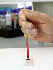
Photo by Максим Кукушкин
Scientists have reported that a new test can screen patients for current and past infection with more than 1000 viral strains, using a single drop of blood and for a cost of about $25 per blood sample.
The investigators used the test, known as VirScan, to screen more than 500 people from around the world and found that, on average, participants had been exposed to about 10 viral species over their lifetimes.
“Using this method, we can take a tiny drop of blood and determine what viruses a person has been infected with over the course of many years,” said Stephen Elledge, PhD, of Harvard Medical School in Boston, Massachusetts.
“What makes this so unique is the scale. Right now, a physician needs to guess what virus might be at play and individually test for it. With VirScan, we can look for virtually all viruses, even rare ones, with a single test.”
Dr Elledge and his colleagues described their work with VirScan in Science.
To develop VirScan, the group synthesized more than 93,000 short pieces of DNA encoding different segments of viral proteins. They introduced those pieces of DNA into bacteriophages.
Each bacteriophage manufactured a peptide and displayed it on the bacteriophage surface. As a group, the bacteriophages displayed all of the protein sequences found in the more than 1000 known strains of human viruses.
To perform the VirScan analysis, all of the peptide-displaying bacteriophages are allowed to mingle with a blood sample. Antiviral antibodies in the blood find and bind to their target epitopes within the displayed peptides.
The scientists then retrieve the antibodies and wash away everything except the few bacteriophages that cling to them. By sequencing the DNA of those bacteriophages, the team can identify which viral protein pieces are bound to antibodies in the blood sample.
That reveals which viruses a person’s immune system has previously encountered, either through infection or vaccination.
Dr Elledge estimated that it would take about 2 to 3 days to process 100 samples with VirScan, assuming sequencing is working optimally. He said he is optimistic the speed of the assay will increase with further development.
Putting VirScan to the test
To test VirScan, Dr Elledge and his colleagues used it to analyze blood samples from patients known to be infected with particular viruses, including HIV and hepatitis C.
“It turns out that it works really well,” Dr Elledge said. “We were in the sensitivity range of 95% to 100% for those, and the specificity was good. We didn’t falsely identify people who were negative. That gave us confidence that we could detect other viruses, and when we did see them, we would know they were real.”
So the investigators used VirScan to analyze the antibodies in 569 people from 4 countries (Peru, South Africa, Thailand, and the US), examining about 100 million potential antibody/epitope interactions.
The team found that, on average, each person had antibodies to 10 different species of viruses. As expected, antibodies against certain viruses were common among adults but not in children, suggesting that children had not yet been exposed to those viruses.
Individuals residing in South Africa, Peru, and Thailand tended to have antibodies against more viruses than people in the US. And people infected with HIV had antibodies against many more viruses than people without HIV.
Dr Elledge and his colleagues were surprised to find that antibody responses against specific viruses were similar between individuals, with different people’s antibodies recognizing identical amino acids in the viral peptides.
“In this paper alone, we identified more antibody/peptide interactions to viral proteins than had been identified in the previous history of all viral exploration,” Dr Elledge said.
The reproducibility of those interactions allowed the scientists to refine their analysis and improve the sensitivity of VirScan, Dr Elledge said, adding that the method will continue to improve as his team analyzes more samples.
The investigators also noted that their work is not limited to antiviral antibodies. They are now using it to look for antibodies that attack the body’s tissue in autoimmune diseases associated with cancers. A similar approach could be used to screen for antibodies against other types of pathogens as well. ![]()
LSC phenotypes correlate with prognosis in AML
© ASCO/Rodney White
CHICAGO—Researchers say they have identified 3 leukemia stem cell (LSC) phenotypes that are correlated with cytogenetic/molecular abnormalities and prognosis in acute myeloid leukemia (AML).
The investigators believe this knowledge could aid risk stratification of AML patients, particularly those without identifiable cytogenetic or molecular risk factors.
The findings may also pave the way for scientists to identify novel therapeutic targets on LSCs and monitor LSCs in response to therapy.
Jonathan Michael Gerber, MD, of Levine Cancer Institute in Charlotte, North Carolina, presented the findings at the 2015 ASCO Annual Meeting (abstract 7000*).
Via previous research, Dr Gerber and his colleagues identified 3 different LSC phenotypes in AML:
- LSCs that are CD34-negative
- LSCs that are CD34-positive, CD38-negative, and have intermediate levels of aldehyde dehydrogenase (ALDHint)
- LSCs that are CD34-positive, CD38-negative, and have high levels of ALDH (ALDHhigh).
With the current study, the researchers wanted to determine if these phenotypes correlate with cytogenetic/molecular features and treatment outcomes.
So they analyzed diagnostic samples from 98 patients with newly diagnosed AML who had normal or unfavorable cytogenetics. The patients were enrolled on a phase 2 trial comparing FLAM and 7+3 (Zeidner et al, haematologica 2015).
Dr Gerber and his colleagues identified 22 patients with CD34- LSCs, 43 with ALDHint LSCs, and 33 with ALDHhigh LSCs.
Risk factors
“We found that leukemia stem cell phenotype indeed correlated quite strongly with cytogenetic and molecular risk factors,” Dr Gerber said.
NPM1 mutations were more common in patients with CD34- LSCs (64%) than those with ALDHint LSCs (14%) or ALDHhigh LSCs (6%, P<0.001). NPM1 mutations were the sole abnormality in 50% of patients with CD34- LSCs.
Poor-risk cytogenetics and/or FLT3-ITD mutations were more common in patients with ALDHhigh LSCs (85%) than those with ALDHint LCSs (35%) or CD34- LSCs (18%, P<0.001).
Nine percent of patients in the CD34- LSC group fell into the European Leukemia Network poor-risk category, compared to 73% of patients in the ALDHhigh LSC group.
Only 2 patients had 11q23, and both had CD34- LSCs. Fifty-five percent of patients with ALDHhigh LSCs had prior myelodysplastic syndromes or myeloproliferative neoplasms.
Prognosis
“We found that leukemia stem cell phenotype correlated strongly with outcomes as well,” Dr Gerber said. “It turned out that CD34- patients fared more favorably overall.”
Patients with CD34- LSCs had the highest complete response rate (86%), followed by those with ALDHint LSCs (67%) and ALDHhigh LSCs (45%, P<0.01).
Patients in the CD34- group also had a higher rate of event-free survival at 2 years (46%) than patients in the ALDHint group (26%) or the ALDHhigh group (0%). The median event-free survival was 13 months, 11.3 months, and 2.2 months, respectively (P<0.01).
The rate of overall survival at 2 years was best for the CD34- group (76%), followed by the ALDHint group (38%) and the ALDHhigh group (34%). The median overall survival was not reached, 18.7 months, and 9.4 months, respectively (P=0.02).
Dr Gerber also noted that ALDHhigh patients fared much better if they underwent hematopoietic stem cell transplant.
“There is 0% leukemia-free survival at the 2-year mark for the ALDHhigh patients who were not transplanted,” he said. “Those that were transplanted fared about the same as everyone else in the series. So it was very striking that there were no chemotherapy survivors in that group.”
In closing, Dr Gerber said this research suggests the 3 LSC phenotypes are mutually exclusive and correlate with cytogenetic and molecular risk factors as well as outcomes in patients with AML.
“This [discovery] may allow for rapid risk stratification in this explosive disease, facilitate enrollment onto induction protocols . . . , and allow us to divert those ALDHhigh, very high-risk patients earlier to novel therapies and/or transplant, given that they’re not really helped much by conventional chemotherapy.” ![]()
*Information in the abstract differs from that presented at the meeting.
Biochemist Irwin Rose dies at 88

Photo courtesy of UCI
Biochemist and Nobel laureate Irwin “Ernie” Rose, PhD, has passed away at the age of 88.
Dr Rose and colleagues from Israel won the Nobel Prize in Chemistry in 2004 for their discovery of ubiquitin-mediated protein degradation.
This research has wide-ranging implications for medicine and led to the development of anticancer drugs such as bortezomib, which is approved in the US to treat multiple myeloma and mantle cell lymphoma.
According to his friends and colleagues, Dr Rose was humble, generous, and endlessly curious.
“Ernie was not interested in personal fame and was oblivious to the politics of science,” said Ann Skalka, PhD, of Fox Chase Cancer Center in Philadelphia, Pennsylvania.
“His total satisfaction came from solving intricate biochemical puzzles. Although Ernie was an intellectual leader on the project that ultimately won him the Nobel, he took no personal credit. He was rather surprised at being recognized, but all of us at Fox Chase knew that the Nobel Committee had gotten it right.”
Dr Rose was born in Brooklyn, New York, on July 16, 1926. His scientific ambitions began to take shape after he moved to Spokane, Washington, at 13. While in high school, he spent summers working at a local hospital. And this inspired him to pursue a career that involved “solving medical problems.”
Dr Rose attended Washington State College for his undergraduate work and went on to earn a doctoral degree at the University of Chicago, after a brief stint in the Navy. He spent the better part of his career as a research scientist at the Fox Chase Cancer Center.
There, during the late 1970s and early 1980s, Dr Rose helped reveal how ubiquitin molecules facilitate the breakdown of old and damaged proteins. The discovery of this process fostered a new understanding of the molecular activity present in cancers and other diseases.
For the work, Dr Rose shared the 2004 Nobel Prize in Chemistry with Avram Hershko, MD, PhD, and Aaron Ciechanover, MD, PhD, of the Israel Institute of Technology.
“Ernie had a genius for asking the right questions,” said Jonathan Chernoff, MD, PhD, of Fox Chase Cancer Center.
“In the mid-1950s, when many scientists were interested in how proteins are synthesized, Ernie became fascinated with the opposite issue—how are proteins degraded? With the collaboration of his Israeli colleagues, he cracked that problem with the discovery of the ubiquitin conjugating system.”
After retiring to Laguna Woods, California, in 1997, Dr Rose accepted a special research position with the University of California Irvine (UCI).
There, he studied the mechanisms of fumarase, an enzyme involved in the citric acid cycle, the cellular pathway by which higher organisms convert food into energy. And he quickly became a beloved colleague and mentor to students and faculty.
“[B]oth prior to and after winning the Nobel Prize, he would help any student or young postdoctoral researcher who was having a hard time with an experiment,” said Ralph Bradshaw, PhD, a former professor at UCI.
“It was a lot of fun working with him,” said James Nowick, PhD, of UCI. “He worked with his own hands, not relying on others, with old instrumentation, and was able to do literally superb science.”
“He was the quintessential scientist—perseverant, soft-spoken, and interested in science for science’s sake,” Dr Chernoff said. “We will miss him very much.”
Dr Rose died in his sleep on June 2 in Deerfield, Massachusetts. He is survived by his wife, Zelda; their sons, Howard, Frederic, and Robert; and 5 grandchildren. Dr Rose’s daughter, Sarah, died in 2005. ![]()
Photo courtesy of UCI
Biochemist and Nobel laureate Irwin “Ernie” Rose, PhD, has passed away at the age of 88.
Dr Rose and colleagues from Israel won the Nobel Prize in Chemistry in 2004 for their discovery of ubiquitin-mediated protein degradation.
This research has wide-ranging implications for medicine and led to the development of anticancer drugs such as bortezomib, which is approved in the US to treat multiple myeloma and mantle cell lymphoma.
According to his friends and colleagues, Dr Rose was humble, generous, and endlessly curious.
“Ernie was not interested in personal fame and was oblivious to the politics of science,” said Ann Skalka, PhD, of Fox Chase Cancer Center in Philadelphia, Pennsylvania.
“His total satisfaction came from solving intricate biochemical puzzles. Although Ernie was an intellectual leader on the project that ultimately won him the Nobel, he took no personal credit. He was rather surprised at being recognized, but all of us at Fox Chase knew that the Nobel Committee had gotten it right.”
Dr Rose was born in Brooklyn, New York, on July 16, 1926. His scientific ambitions began to take shape after he moved to Spokane, Washington, at 13. While in high school, he spent summers working at a local hospital. And this inspired him to pursue a career that involved “solving medical problems.”
Dr Rose attended Washington State College for his undergraduate work and went on to earn a doctoral degree at the University of Chicago, after a brief stint in the Navy. He spent the better part of his career as a research scientist at the Fox Chase Cancer Center.
There, during the late 1970s and early 1980s, Dr Rose helped reveal how ubiquitin molecules facilitate the breakdown of old and damaged proteins. The discovery of this process fostered a new understanding of the molecular activity present in cancers and other diseases.
For the work, Dr Rose shared the 2004 Nobel Prize in Chemistry with Avram Hershko, MD, PhD, and Aaron Ciechanover, MD, PhD, of the Israel Institute of Technology.
“Ernie had a genius for asking the right questions,” said Jonathan Chernoff, MD, PhD, of Fox Chase Cancer Center.
“In the mid-1950s, when many scientists were interested in how proteins are synthesized, Ernie became fascinated with the opposite issue—how are proteins degraded? With the collaboration of his Israeli colleagues, he cracked that problem with the discovery of the ubiquitin conjugating system.”
After retiring to Laguna Woods, California, in 1997, Dr Rose accepted a special research position with the University of California Irvine (UCI).
There, he studied the mechanisms of fumarase, an enzyme involved in the citric acid cycle, the cellular pathway by which higher organisms convert food into energy. And he quickly became a beloved colleague and mentor to students and faculty.
“[B]oth prior to and after winning the Nobel Prize, he would help any student or young postdoctoral researcher who was having a hard time with an experiment,” said Ralph Bradshaw, PhD, a former professor at UCI.
“It was a lot of fun working with him,” said James Nowick, PhD, of UCI. “He worked with his own hands, not relying on others, with old instrumentation, and was able to do literally superb science.”
“He was the quintessential scientist—perseverant, soft-spoken, and interested in science for science’s sake,” Dr Chernoff said. “We will miss him very much.”
Dr Rose died in his sleep on June 2 in Deerfield, Massachusetts. He is survived by his wife, Zelda; their sons, Howard, Frederic, and Robert; and 5 grandchildren. Dr Rose’s daughter, Sarah, died in 2005.
Nutrition source could improve platinum-based nanodrugs
A parenteral nutrition source can reduce the toxicity and increase the bioavailability of platinum-based anticancer nanodrugs, according to preclinical research published in Scientific Reports.
Many of the side effects of platinum-based drugs occur when they settle in healthy tissue.
To deliver these drugs in a more targeted way, researchers have created nanoscale delivery systems engineered to make the drugs accumulate at tumor sites.
However, tests of these nanodrugs show that between 1% and 10% of the drugs are delivered to the tumor site, with most of the remainder being diverted to the liver and spleen.
“The body’s immune system, especially the liver and spleen, has been one of the biggest stumbling blocks in developing nanoscale chemotherapy drug delivery systems,” said Chien Ho, PhD, of Carnegie Mellon University in Pittsburg, Pennsylvania.
“When the drugs collect in those organs, they become less available to treat the cancer and can also cause toxicity.”
But Dr Ho and his colleagues have found evidence to suggest that Intralipid, a fat emulsion used as a parenteral nutrition source, can help prevent that.
While developing cellular nanotags to help detect organ rejection, Dr Ho noticed that Intralipid reduced the amount of nanoparticles that were being cleared by the liver and spleen by about 50%. As a result, the nanoparticles remained in the bloodstream for longer periods of time.
So he and his colleagues decided to see if Intralipid had the same effect on platinum-based anticancer nanodrugs.
In the newly published study, the researchers administered a single, clinical dose of Intralipid to Sprague Dawley rats. One hour later, they administered a dose of a platinum-based chemotherapy drug that had been incorporated into a nanoparticle to both Intralipid-treated rats and controls.
Twenty-four hours after the drug was administered, rats pretreated with Intralipid had experienced reduced accumulation of the platinum-based drug compared to controls.
Drug accumulation decreased by 20.4% in the liver, 42.5% in the spleen, and 31.2% in the kidney. Consequently, in these organs, the toxic side effects of the nanodrug were significantly decreased compared to controls.
Furthermore, Intralipid pretreatment allowed more of the drug to remain available and active in the body for longer periods of time.
After 5 hours, the drug’s bioavailability increased by 18.7% in Intralipid-treated mice compared to controls. After 24 hours, bioavailability was 9.4% higher in Intralipid-treated mice than in controls.
The researchers believe this increased bioavailability will allow more of the drug to reach the tumor site and could perhaps allow clinicians to reduce the dosage needed to treat a patient. The team is now investigating the possibility of bringing this research to a clinical trial.
Single agent can treat resistant MM

© ASCO/Todd Buchanan
CHICAGO—The anti-CD38 monoclonal antibody daratumumab can be effective as a stand-alone therapy for some heavily pretreated patients with multiple myeloma (MM), results of an ongoing phase 2 trial suggest.
The study, known as SIRIUS or MMY2002, included more than 100 patients who had received 3 or more prior lines of therapy.
Roughly 30% of these subjects responded to daratumumab, with a median response duration of about 7 months.
The median progression-free survival was close to 4 months, and the estimated 1-year overall survival rate was 65%.
Serious adverse events (AEs) occurred in 30% of patients.
“These findings speak to the potential of daratumumab as an effective and tolerable option for people with multiple myeloma who have exhausted other available treatment options,” said study investigator Sagar Lonial, MD, of Emory University School of Medicine in Atlanta, Georgia.
Dr Lonial presented these findings at the 2015 ASCO Annual Meeting (abstract LBA8512). The research was funded by Janssen Research & Development, the company developing daratumumab.
In part 1 of this study, 34 patients were randomized to receive either 8 mg/kg of daratumumab once every 4 weeks or 16 mg/kg once a week for 8 weeks, then once every 2 weeks for 16 weeks and once every 4 weeks after that, until disease progression or unacceptable toxicity.
In part 2, an additional 90 patients were enrolled to receive 16 mg/kg of daratumumab on the same dosing schedule as in part 1.
Dr Lonial reported results for all patients in parts 1 and 2 who received 16 mg/kg of daratumumab. These 106 patients had received a median of 5 prior lines of therapy, including a proteasome inhibitor and an immunomodulatory drug.
According to an independent review committee, 29.2% of patients responded to daratumumab. Eighteen patients had a partial response, 10 had a very good partial response, and 3 had a stringent complete response. The median duration of response was 7.4 months.
“It is particularly noteworthy to see this level of response with a single-agent in this heavily pretreated population,” Dr Lonial said. “Ninety-seven percent of patients in this study were refractory to their last line of therapy, and 95% were double-refractory to both a [proteasome inhibitor] and an [immunomodulatory drug].”
The median overall survival has not been reached, and the estimated 1-year overall survival rate is 65%. The median progression-free survival was 3.7 months.
After a median follow up of 9.4 months, 45.2% of responders remain on therapy.
The most common AEs were fatigue (39.6%), anemia (33%), nausea (29.2%), thrombocytopenia (25.5%), neutropenia (22.6%), back pain (22.6%), and cough (20.8%).
Thirty percent of patients experienced serious AEs. And 4.7% of patients discontinued treatment due to AEs, none of which were considered drug-related.
Infusion-related reactions (IRR) were reported in 42.5% of patients and were predominantly grade 1 or 2 (4.7% grade 3; no grade 4). These occurred mainly during the first infusion.
The most common IRRs included nasal congestion (12%), throat irritation (7%), cough (6%), dyspnea (6%), chills (6%), and vomiting (6%)—all of which were treated with standard of care and slower infusion rates.
Cancer survivors mirror spouses’ moods

chemotherapy
Photo by Rhoda Baer
Cancer survivors’ moods are impacted—both positively and negatively—by their spouses’ moods, according to research published in Cancer Epidemiology, Biomarkers & Prevention.
In the study, cancer survivors whose spouses reported depressed moods were more likely to be depressed after about a year of follow-up, and survivors whose spouses reported better mental and physical health-related quality of life (HRQOL) were less likely to be depressed.
However, survivors’ moods did not have the same impact on their spouses.
“We were surprised that the effects of the spouses on the survivors were so much larger in this study than the effect of the survivors on their spouses,” said study author Kristin Litzelman, PhD, of the National Cancer Institute in Bethesda, Maryland. “We expected to see a more reciprocal relationship.”
Dr Litzelman and her colleagues conducted this research in an attempt to understand how cancer survivors and their families influence one another. The team hoped to identify ways to improve the healthcare both parties receive and thereby improve their health and well-being.
The researchers analyzed data from 910 cancer patients and their spouses, comparing them to 910 couples without any kind of cancer-related health problem.
The team used statistical models to assess how each spouse’s quality of life or depression at one time point was associated with his or her partner’s risk of depression around 11 months later. The researchers took into account a person’s previously reported mood, demographic characteristics, and other factors.
The results showed that, when spouses reported feeling depressed, cancer survivors were about 4 times more likely to report being depressed 11 months later (odds ratio [OR]=4.27). This association was stronger among female cancer survivors (OR=9.49) than male survivors (OR=3.98).
Cancer survivors whose spouses reported better HRQOL had a 30% decrease in depressed mood per 10-point improvement in HRQOL score. The ORs were 0.72 for mental health and 0.68 for physical health. The associations between spousal HRQOL and survivor depressed mood were similar for male and female survivors.
The researchers noted that cancer survivors’ moods did not have a significant impact on their spouses’ risk of depressed mood 11 months later.
And the team did not see mood associations in couples without any cancer-related health problems.
“This finding certainly needs to be backed up by other studies, but it highlights the importance of family well-being in cancer survivor outcomes,” Dr Litzelman said. “Our research highlights that spouses need to take care of themselves, not just for their own sake, but also for the sake of the cancer survivor.”
“Our findings also suggest that, when caring for cancer survivors, clinicians may want to assess the well-being of spousal caregivers. Future research could test whether including caregivers in the survivorship care plan might help to improve outcomes for both caregivers and for cancer survivors.”