User login
Urine test could reduce need for blood samples

Photo by Juan D. Alfonso
A new approach to urine testing could make the tests more versatile and therefore decrease the need for blood tests, according to researchers.
They believe the method could also reduce costs, produce results faster than current tests, and lower the volume of urine needed for a sample.
R. Kenneth Marcus, PhD, of Clemson University in South Carolina, and his colleagues described this method in Proteomics-Clinical Applications.
Dr Marcus noted that the trouble with testing urine is that it’s awash in salt, so it can be tricky to isolate the proteins that act as biomarkers.
To overcome this problem, he and his colleagues used a string made of capillary-channeled polymer fibers. The team packed the fibers into plastic tubes and then passed urine samples through the tubes by spinning them in a centrifuge for 30 seconds.
Then, the researchers ran de-ionized water through the tubes for a minute to wash off salt and other contaminants.
As proteins are hydrophobic, they remained stuck to the fibers. The team extracted the proteins by running a solvent through the tubes in the centrifuge for 30 seconds.
When this process was complete, the researchers were left with purified proteins that could be stored in a plastic vial and refrigerated until testing time.
The team was able to extract 12 samples in about 5 minutes, limited only by centrifuge capacity.
In addition to being faster and cheaper than current urine tests, the new testing method should also make it easier to test urine samples from infants, Dr Marcus said.
One of the challenges now is getting a large enough sample, but the new method requires only a few microliters of urine. ![]()
Understanding taste dysfunction in cancer
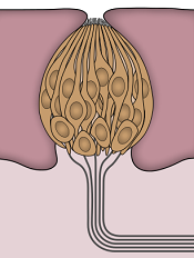
Image by Jonas Töle
Investigators have identified a molecular pathway that aids the renewal of taste buds, and they believe this discovery may have implications for cancer patients who suffer from an altered sense of taste during treatment.
“Taste dysfunction can . . . result from an alteration of the renewal capacities of taste buds and is often associated with psychological distress and malnutrition,” said Dany Gaillard, PhD, of the University of Colorado Anschutz Medical Campus in Aurora.
He and his colleagues decided to investigate this dysfunction using mouse models, and the group reported their findings in PLOS Genetics.
The investigators discovered that a protein in the Wnt pathway, ß-catenin, controls the renewal of taste cells by regulating separate stages of taste-cell turnover.
Previous research showed that Wnt/β-catenin signaling is crucial in developing taste buds in embryos and regulating the renewal of epithelial tissue in adults, including skin, hair follicles, the intestine, and the mouth.
“We show that activating this pathway directs the newly born cells to become, primarily, a specific taste- cell type whose role is to support the other taste cells and help them work efficiently,” said Linda Barlow, PhD, also of the University of Colorado Anschutz Medical Campus.
As chemotherapy destroys dividing precursor cells, including those that produce taste cells, the investigators believe that activating Wnt signaling may be a way to renew taste buds after chemotherapy.
New small-molecule drugs that specifically block the Wnt pathway are under development, and Drs Gaillard and Barlow predict these drugs could also cause taste dysfunction.
Dr Barlow said more research is needed to understand how taste is altered at the cellular level, but this research holds promise for developing new ways to improve cancer patients’ quality of life. ![]()
Bridge therapy may be detrimental for some VTE patients

Anticoagulant bridge therapy may be harmful for patients with a low to moderate risk of venous thromboembolism (VTE), according to research published in JAMA Internal Medicine.
The study included more than 1000 patients with VTE who had to stop receiving warfarin prior to a procedure.
Investigators compared outcomes in patients who received bridge therapy—a short-acting anticoagulant during the periprocedural period—and patients who did not.
Nathan Clark, PharmD, of Kaiser Permanente Colorado in Aurora, and his colleagues conducted this research.
They examined the electronic medical records of 1178 patients with VTE who underwent 1812 invasive diagnostic or surgical procedures between January 2006 and March 2012 that required the interruption of warfarin.
The investigators divided patients into 3 groups based on their annual risk of VTE recurrence without anticoagulant therapy. Seventy-nine percent of patients were categorized as low-risk, 17.9% as moderate-risk, and 3.1% as high-risk.
The team then divided patients according to the use of bridge therapy. Of the 1812 procedures, 555 included bridge therapy, and 1257 did not.
Patients in the bridged group had a significantly higher incidence of clinically relevant bleeding at 30 days than patients in the non-bridged group—2.7% and 0.2%, respectively (P=0.01).
When the investigators assessed patients according to VTE risk, they found the increased incidence of bleeding in the bridged group was significant among low-risk patients (2.0% vs 0.1%, P<0.001) and moderate-risk patients (4.6% vs 0%, P=0.004) but not high-risk patients (5.6% vs 4.8%, P=0.90).
On the other hand, there was no significant difference in VTE recurrence between the bridged and non-bridged groups—for all risk categories (0 vs 3 cases, P=0.56), low-risk patients (0 vs 2, P=0.37), moderate-risk patients (0 vs 1, P=0.48), or high-risk patients (0 vs 0, P>0.99)
The investigators said further research is needed to identify patient and procedure-related characteristics associated with the highest risk for perioperative VTE recurrence where targeted bridge therapy may be beneficial. ![]()
Reducing distress in caregivers of HSCT recipients
![]()
Photo by Chad McNeeley
Research has shown that caring for cancer patients after hematopoietic stem cell transplant (HSCT) can have negative psychological effects on the caregiver, but results of a new study suggest a psychosocial intervention could change that.
The trial showed that counseling sessions focused on stress management could significantly reduce stress, anxiety, depression, and mood disturbance among these caregivers.
“The first 100 days after a stem cell transplant is a critical period for patients, in which caregivers are called upon to deliver around-the-clock care, providing support for patients’ everyday needs and also patients’ emotional health, but who takes care of the caregivers?” asked Mark Laudenslager, PhD, of the University of Colorado Denver.
To address this problem, Dr Laudenslager and his colleagues studied 148 caregivers of patients who underwent allogeneic HSCT. The team described this research in Bone Marrow Transplantation.
The caregivers were randomized to a group that was offered a psychosocial intervention (n=74) and a group that received standard treatment, in which mental health support services were available but not required (n=74).
In the experimental group, caregivers attended 8 sessions on stress management. These one-on-one sessions focused on understanding stress and its physical consequences, changing roles as caregivers, cognitive behavioral stress management, pacing respiration, and identifying social support. The researchers call this intervention PsychoEducation, Paced Respiration and Relaxation (PEPRR).
After a patient underwent HSCT, Dr Laudenslager and his colleagues used several questionnaires to follow the trajectory of caregiver distress over time. The questionnaires were used to measure stress, depression, anxiety, mood disturbance, sleep quality, and other mental health outcomes.
There was no significant difference in stress or other mental health measures between the 2 treatment groups at baseline.
However, at 3 months after transplant, caregivers in the PEPRR group saw some significant improvements over caregivers in the standard treatment group.
The PEPRR group had less stress according to the Perceived Stress Scale (P=0.039), less depression according to the Center for Epidemiologic Studies Depression test (P=0.016), less anxiety according to the State-Trait Anxiety Inventory-State questionnaire (P=0.0009), and less mood disturbance according to the Profile of Mood States-Total Mood Disturbance test (P=0.039).
Overall caregiver distress (composite scores from the questionnaires) was significantly lower in the PEPRR group than the standard treatment group (P=0.019).
However, there was no significant difference in caregiver well-being (composite scores) or scores on the Caregiver Reaction Assessment, Pittsburgh Sleep Quality Index, Short Form 36 Health Survey, or Impact of Events scale.
Still, the other improvements caregivers experienced suggest PEPRR is a promising intervention, Dr Laudenslager said.
He and his colleagues are now recruiting subjects for a follow-up study (NCT02037568) focused on evaluating quality of life in allogeneic HSCT recipients whose caregivers participate in programs similar to the PEPRR intervention. ![]()
CAR T-cell therapy appears feasible in HL
LONDON—Results of a small, phase 1 trial suggest CD30-directed chimeric antigen receptor (CAR) T-cell therapy is feasible in patients with aggressive Hodgkin lymphoma (HL).
The trial included 7 patients with relapsed or refractory HL.
Five of the patients achieved stable disease or better after infusions of CAR T cells, and the researchers said treatment-related adverse events were manageable.
William (Wei) Cao, PhD, of Cellular Biomedicine Group, presented these results at the 10th Annual World Stem Cells & Regenerative Medicine Congress.
The research was funded by Cellular Biomedicine Group, the company developing the CAR T-cell therapy (known as CBM-C30.1), as well as by grants from the National Natural Science Foundation of China and the National Basic Science and Development Program of China.
The trial included 7 patients with progressive HL. Two patients had stage III disease, and 5 had stage IV. The patients had a median of 16 prior treatments (range, 8-24) and limited prognosis (several months to less than 2-year survival) with currently available therapies.
The patients received escalating doses of autologous T cells transduced with a CD30-directed CAR moiety for 3 to 5 days, following a conditioning regimen. The researchers measured the level of CAR transgenes in peripheral blood and biopsied tumor tissues by quantitative PCR.
Two patients achieved a partial response to CAR T-cell therapy, and 3 attained stable disease. So the therapy resulted in an overall disease control rate of 71.4% (5/7) and an objective response rate of 28.6% (2/7).
Stable disease lasted 2 months in 2 of the patients and more than 3.5 months in the third patient. Partial response lasted more than 2 months in 1 patient and more than 3.5 months in the other.
Dr Cao said adverse events consisted largely of fever and were manageable with medical intervention. One patient experienced 5-day self-limiting arthralgia, myalgia, and dual knee swelling 2 weeks after cell infusion. There were no delayed or severe adverse events.
“We are very encouraged by the efficacy and toxicity profile of our CAR-T CD30 technology,” Dr Cao said, “given that the [patients] were diagnosed with stage III and IV Hodgkin’s lymphoma.” ![]()
PBM program improves outcomes, study shows

Photo by Elise Amendola
A patient blood management (PBM) program can reduce transfusion use, cut costs, and improve outcomes in cardiac surgery patients, according to a single-center study.
A PBM program instituted at Eastern Maine Medical Center (EMMC) in Bangor substantially decreased the use of blood products, the loss of red blood cells, the length of hospital stays, the incidence of acute kidney injury, and direct costs.
Irwin Gross, MD, of EMMC, and his colleagues reported these results in Transfusion.
The team compared clinical and transfusion data from cardiac surgery patients treated at the center before the PBM program began (July 2006-March 2007) and after (April 2007-September 2012).
EMMC’s PBM initiative involved pre- and post-operative anemia management, a more restrictive transfusion threshold, the use of single-unit transfusions when necessary, and other measures.
The researchers analyzed data on 2662 patients, 387 treated before the PBM program began and 2275 treated after.
As expected, the rate of transfusions decreased after the PBM program began. The rate of red blood cell transfusion decreased from 39.3% to 20.8% (P<0.001), the rate of fresh-frozen plasma transfusion decreased from 18.3% to 6.5% (P<0.001), and the rate of platelet transfusion decreased from 17.8% to 9.8% (P<0.001).
Red blood cell loss decreased from a median of 721 mL to 552 mL (P<0.001), and pre-transfusion hemoglobin decreased from a mean of 7.2 ± 1.4 g/dL to 6.6 ± 1.2 g/dL (P<0.001).
Patients saw a decrease in the incidence of post-operative kidney injury from 7.6% to 5.0% (P=0.039) and a decrease in the median length of hospital stay from 10 days to 8 days (P<0.001).
Total adjusted direct costs decreased after the program began as well, falling from a median of $39,709 to $36,906 (P< 0.001).
There was no significant difference in the rate of hospital mortality or the incidence of cerebral vascular accident before and after the PBM program began. ![]()
Study quantifies VTE risk with different birth control pills

Results of a large, retrospective study support the association between newer contraceptive pills and a higher risk of venous thromboembolism (VTE).
The research showed that pills containing one of the newer types of progestogen—drospirenone, desogestrel, gestodene, and cyproterone—are associated with a nearly 2-fold higher risk of VTE than pills containing older progestogens—levonorgestrel, norethisterone, and norgestimate.
The researchers said this study has sufficient power to provide reliable comparative findings for different formulations of combined oral contraceptives. However, because it is an observational study, no definitive conclusions can be drawn about cause and effect.
The team described this research in BMJ alongside a related editorial.
Although the increased risk of VTE associated with combined oral contraceptives has been suggested previously, prior studies have used different methods to examine this link. So the relative risks associated with different combinations remain inconclusive.
Yana Vinogradova, of the University of Nottingham in the UK, and her colleagues tried to address these differences to help explain the range of results.
The team used prescription data from 2 large UK general practice databases to measure the associations between the use of combined oral contraceptives and the risk of VTE in women aged 15 to 49, adjusting for other known VTE risk factors.
The researchers matched 10,562 women with VTE to 42,034 control subjects and found that women who used any combined oral contraceptive within the past year had an increased risk of VTE compared with non-users of similar age and health status. The adjusted odds ratio was 2.97.
The risk of VTE was significantly higher for women who used the newer oral contraceptives than the older pills (P<0.001). The adjusted odds ratios were 4.28 for desogestrel, 4.27 for cyproterone, 4.12 for drospirenone, and 3.64 for gestodene, compared to 2.38 for levonorgestrel, 2.53 for norgestimate, and 2.56 for norethisterone.
The number of extra VTE cases per year per 10,000 treated women was lowest for levonorgestrel and norgestimate (6 cases for both) and highest for desogestrel and cyproterone (14 cases for both).
The researchers said that, although this is an observational study, it has produced the most reliable possible VTE risk estimates using currently available UK prescription data. ![]()
Results of a large, retrospective study support the association between newer contraceptive pills and a higher risk of venous thromboembolism (VTE).
The research showed that pills containing one of the newer types of progestogen—drospirenone, desogestrel, gestodene, and cyproterone—are associated with a nearly 2-fold higher risk of VTE than pills containing older progestogens—levonorgestrel, norethisterone, and norgestimate.
The researchers said this study has sufficient power to provide reliable comparative findings for different formulations of combined oral contraceptives. However, because it is an observational study, no definitive conclusions can be drawn about cause and effect.
The team described this research in BMJ alongside a related editorial.
Although the increased risk of VTE associated with combined oral contraceptives has been suggested previously, prior studies have used different methods to examine this link. So the relative risks associated with different combinations remain inconclusive.
Yana Vinogradova, of the University of Nottingham in the UK, and her colleagues tried to address these differences to help explain the range of results.
The team used prescription data from 2 large UK general practice databases to measure the associations between the use of combined oral contraceptives and the risk of VTE in women aged 15 to 49, adjusting for other known VTE risk factors.
The researchers matched 10,562 women with VTE to 42,034 control subjects and found that women who used any combined oral contraceptive within the past year had an increased risk of VTE compared with non-users of similar age and health status. The adjusted odds ratio was 2.97.
The risk of VTE was significantly higher for women who used the newer oral contraceptives than the older pills (P<0.001). The adjusted odds ratios were 4.28 for desogestrel, 4.27 for cyproterone, 4.12 for drospirenone, and 3.64 for gestodene, compared to 2.38 for levonorgestrel, 2.53 for norgestimate, and 2.56 for norethisterone.
The number of extra VTE cases per year per 10,000 treated women was lowest for levonorgestrel and norgestimate (6 cases for both) and highest for desogestrel and cyproterone (14 cases for both).
The researchers said that, although this is an observational study, it has produced the most reliable possible VTE risk estimates using currently available UK prescription data.
FDA clears test to detect bacteria in platelets
![]()
The US Food and Drug Administration (FDA) has expanded the authorized use of Verax Biomedical’s Platelet PGD Test, which detects bacteria in platelets intended for transfusion.
The FDA previously approved the test for leukocyte-reduced apheresis platelets (in 2007) and platelets derived from whole blood (in 2009).
Now, the test has been approved for pre-storage pooled platelets and apheresis platelets in platelet additive solution C (PAS-C) and plasma.
This makes the Platelet PGD Test the only rapid test on the market that can check every commonly distributed platelet type in the US, according to Verax Biomedical.
About the test
The Platelet PGD Test is an immunoassay used on the day of transfusion at the point of care—a hospital or transfusion service—to detect bacterial contamination in platelets to be transfused.
The test consists of a disposable plastic cartridge and 3 pretreatment reagents. To use, the tester pretreats a freshly collected platelet sample (500µL) and applies it to the sample well on the test cartridge.
Lights on the cartridge change from yellow to blue-violet when the test is ready to be interpreted, which is typically about 20 minutes after the sample is applied to the cartridge. The lights confirm that the appropriate volume of a sample was added and the testing is complete.
If the test is positive, a pink line will appear in 1 of the 2 windows on the cartridge. One window represents Gram-positive bacteria and the other Gram-negative. Non-reactive samples will have no line in either window.
Now that the FDA has expanded the indications for the Platelet PGD Test, it can be used as a quality control test for pools of up to 6 units of leukocyte-reduced and non-leukocyte-reduced whole-blood-derived platelets suspended in plasma that are pooled within 4 hours of transfusion.
The test can also be used within 24 hours of transfusion as a safety measure following testing with a growth-based, quality control test cleared by the FDA. For this indication, the Platelet PGD Test can be used with:
- Leukocyte-reduced apheresis platelets suspended in plasma
- Leukocyte-reduced apheresis platelets suspended in PAS-C and plasma
- Pre-storage pools of up to 6 leukocyte-reduced whole-blood-derived platelets suspended in plasma.
In studies conducted by Verax Biomedical (described in the summary document here), the Platelet PGD Test successfully detected bacteria in pre-storage pools of whole-blood derived platelets suspended in plasma and leukocyte-reduced apheresis platelets suspended in plasma or PAS-C and plasma.
Team says delayed cord clamping can’t hurt

Photo by Meutia Chaerani
and Indradi Soemardjan
New research suggests that delayed umbilical cord clamping in full-term infants may confer some minor long-term benefits and, at the very least, does not pose any harm.
Delayed clamping did not appear to have a significant effect on most of the mental and physical measures assessed in the study.
It was associated with improved scores in fine-motor skills and social skills at age 4, but these effects only occurred in boys.
Researchers reported these results in JAMA Pediatrics alongside a related editorial.
Previous research has shown that delaying umbilical cord clamping by 2 to 3 minutes after delivery allows fetal blood remaining in the placental circulation to be transfused back to the newborn, and this is associated with improved iron status at 4 to 6 months of age.
However, there is a lack of knowledge regarding the long-term effects of delayed clamping. So policymakers have been hesitant about making clear recommendations regarding cord clamping in full-term infants.
To gain more insight, Ola Andersson, MD, PhD, of Uppsala University in Sweden, and his colleagues performed follow-up assessments of 263 children who were previously enrolled in a randomized trial of cord clamping in full-term infants born in a Swedish hospital.
The team assessed the effects of delayed cord clamping on childhood development at age 4. Delayed clamping (n=141) was defined as occurring 3 or more minutes after delivery, and early clamping (n=122) was defined as occurring 10 seconds or fewer after delivery.
The researchers evaluated child behavior and development using parents’ responses on the Ages and Stages Questionnaire, Third Edition (ASQ), which is used to assess communication, motor skills, and other measures; and the Strengths and Difficulties Questionnaire, which is used to score children’s emotional difficulties, hyperactivity, and other difficulties.
A blinded psychologist also assessed children’s scores on the Wechsler Preschool and Primary Scale of Intelligence (WPPSI-III), which is used to assess IQ and similar measures, and the Movement Assessment Battery for Children (Movement ABC), which is used to assess manual dexterity and similar measures.
The researchers found no significant differences between the delayed and early clamping groups with regard to results on the WPPSI-III or the Movement ABC.
However, delayed clamping was associated with a significant improvement over early clamping in ASQ personal-social scores (adjusted mean difference [AMD]=2.8, P=0.006), fine-motor scores (AMD=2.1, P=0.03), and the Strengths and Difficulties Questionnaire prosocial subscale (AMD=0.5, P=0.05).
When the researchers assessed the children according to sex, they found that significant improvements associated with delayed clamping were only present in males.
Males in the delayed clamping group had significantly higher mean scores in tasks involving fine-motor function, including the WPPSI-III processing-speed quotient (AMD=4.2, P=0.02), the Movement ABC bicycle-trail task (AMD=0.8, P=0.03), and fine-motor scores on the ASQ (AMD=4.7, P=0.01). These boys also had significantly higher personal-social scores on the ASQ (AMD=4.9, P=0.004).
The researchers concluded that, although delayed cord clamping and early clamping resulted in similar overall neurodevelopment and behavior among 4-year-old children, there were differences in this study. And this suggests there are some positive, and no harmful, long-term effects of delayed cord clamping.
Histone variant may contribute to lymphoma

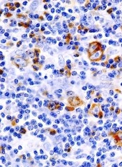
Image by Eric Smith
Researchers say they have identified histone chaperones that play an important role in the structure of chromatin.
The team believes this finding, published in Molecular Cell, could lead to a better understanding of lymphomas and other cancers.
“Maintaining an appropriate chromatin structure is essential for normal development, and, not surprisingly, defects in chromatin components can lead to several diseases,” said study author François Robert, PhD, of Institut de Recherches Cliniques de Montréal in Québec, Canada.
In studying chromatin, Dr Robert and his colleagues have been interested in a histone variant called H2A.Z.
The researchers knew that H2A.Z is incorporated into promoter regions of the gene by SWR-C-related chromatin remodeling complexes, but they wanted to determine if H2A.Z is actively excluded from non-promoter regions.
“With this study, we discovered that 2 other proteins, FACT and Spt6, play an important role in the location of H2A.Z,” said Célia Jeronimo, PhD, a research associate in Dr Robert’s lab.
The team found that FACT and SPt6 both help keep H2A.Z from accumulating in intragenic regions. When either histone chaperone is absent, H2A.Z is mislocalized, which alters chromatin composition and contributes to cryptic transcription.
“Inappropriate H2A.Z localization has previously been observed in cancer cells, but little was understood about the consequences of this phenomenon,” Dr Robert said.
“Although our study was performed in yeast cells, it suggests that mislocalization of H2A.Z may lead to cryptic transcription in some types of cancer such as lymphoma, and this may contribute to the disease. Our next step is therefore to investigate the possible role of H2A.Z and its associated gene expression defects in cancer cells.”