User login
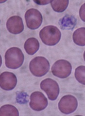
Researchers identify 11 subgroups of AML

Photo courtesy of NIGMS
Using patient samples from 3 prospective multicenter clinical trials of the German-Austrian AML Study Group, researchers have identified 5234 driver mutations involving 76 genes or regions in 1540 patients with acute myeloid leukemia (AML).
They say the sample size afforded a more comprehensive analysis than previously conducted, and as a result, they found patients were divided into at least 11 subgroups of AML.
They also identified 3 genomic categories beyond the existing World Health Organization (WHO) subgroups. These genomic categories are chromatin–spliceosome mutations, TP53–aneuploidy, and provisionally, IDH2R172 mutations.
Their study, led by scientists at the Wellcome Trust Sanger Institute and international collaborators, could have an impact on clinical trial design and improve the way patients are diagnosed and treated in the future.
The scientists stated in their published paper that 736 patients (48%) in their study would not have fit into the molecular groups included in the 2008 WHO classification of adult AML. This prompted them to reevaluate genomic classification of AML from the beginning.
“We have shown that AML is an umbrella term for a group of at least 11 different types of leukemia,” said Peter Campbell, MBChB, PhD, co-leader of the study. “We can now start to decode these genetics to shape clinical trials and develop diagnostics.”
The scientists found NPM1-mutated AML to be the largest class in their cohort, accounting for 27% of the patients.
The second largest subgroup--the chromatin-spliceosome group—accounted for 18% of patients, the TP53-aneuploidy group accounted for 13%, and the IDH2R172 mutations for 1%.
The study also showed that most patients had a unique combination of genetic changes driving their leukemia. This genetic complexity helps explain why AML shows such variability in survival rates among patients.
Under their new schema, the scientists were able to unambiguously classify 80% of the 1540 patients with driver mutations in a single subgroup. Fifty-six of the patients (4%) met criteria for 2 or more categories, primarily in the TP53-aneuploidy and chromatin-spliceosome groups.
They were not able to classify 11% of patients with driver mutations. They explained that these patients might have had mutations in drivers that were either not sequenced or had mutations that were missed.
The scientists applied their classification schema to an independent cohort from the Cancer Genome Atlas (TCGA). The new schema was able to replicate the absence of overlap among subgroups, and relative frequencies of the mutations were equivalent to those in the AML cohort.
“For the first time we untangled the genetic complexity seen in most AML cancer genomes into distinct evolutionary paths that lead to AML,” joint first author Elli Papaemmanuil, PhD, of Memorial Sloan Kettering Cancer Center in New York, said.
“By understanding these paths, we can help develop more appropriate treatments for individual patients with AML. We are now extending such studies across other leukemias."
The investigators recommend prospective clinical trials to further validate the schema.
The work was supported by the Wellcome Trust, Bundesministerium fur Bildung und Forschung, Deutsche Krebshilfe and Deutsche Forschungsgemeinschaft, the European Hematology Association, Amgen and the Kay Kendall Leukaemia Fund. ![]()
Corticosteroid prophylaxis reduces GVHD in high-risk patients

Image courtesy of PLOS ONE
A team of researchers has found that risk-stratified, low-dose corticosteroid prophylaxis can significantly reduce the incidence of acute graft-versus-host disease (GVHD), accelerate platelet recovery, and reduce adverse events without increasing infections in patients who undergo haploidentical transplantation.
They believe this is the first test of a novel risk stratification–directed prophylaxis strategy. The strategy effectively prevented acute GVHD among patients who were at high risk for GVHD, without unnecessarily exposing patients who were at low risk to excessive toxicity from additional immunosuppressive agents.
To evaluate the prophylaxis strategy, the team enrolled 228 patients who underwent haploidentical transplant onto this controlled, open-label, randomized trial.
Corresponding author Xiao-Jun Huang, MD, of Peking University in Beijing, China, and colleagues reported the results in JCO.
The team stratified patients as high risk or low risk for developing GVHD based on biomarkers.
They categorized patients as high risk if they had CD4:CD8 ratios of 1.15 or greater in bone marrow grafts or more than 1.9 x 10⁶/kg CD56bright NK cells in allogeneic grafts.
Patients who did not qualify for the high-risk group were considered to be low risk.
The team randomly assigned the high-risk patients to either receive or not receive the investigational intervention of methylprednisolone (MP).
All patients received cyclosporine, mycophenolate mofetil, and methotrexate for GVHD prevention.
Patients in any arm who developed acute GVHD higher than grade II received MP. If they did not respond, they received basiliximab.
Study population
Of the 228 patients enrolled, 83 were in the low-risk group, 72 in the high-risk experimental prophylaxis group, and 73 in the high-risk control group.
Patient characteristics were similar across all cohorts, including the conditioning regimens, except for their biomarker status.
Patients were a median age of about 30 years, (range 14 – 58), about half were male, and most had a diagnosis of acute myeloid leukemia or acute lymphoblastic leukemia.
GVHD incidence
The cumulative, 100-day incidence of acute GVHD grades II to IV was 21% in the high-risk prophylaxis arm.
This was similar to the incidence of 26% in the low-risk arm, P=0.43.
Both cohorts were significantly lower than the incidence of 48% in the high-risk control group, P<0.001.
Multivariate analysis showed that risk-stratified prophylaxis with low-dose corticosteroid significantly reduced the incidence of acute GVHD compared with no low-dose corticosteroid, with a hazard ratio of 0.66, P=0.007.
However, investigators found no differences between cohorts in the incidence of grades III to IV GVHD.
Corticosteroid-refractory acute GVHD developed in 14% of high-risk patients in the experimental arm, 18% in the control arm, and 23% in the low-risk group. Basiliximab, an anti-CD25 antibody, was used to treat it.
Hematopoietic and immune recovery
The median times to myeloid and platelet recovery were significantly shorter in the high-risk prophylaxis group (P<0.05 for both) and the low-risk group (P<0.05) than in the high-risk control group.
And immune reconstitution was comparable among all 3 cohorts.
Safety
Cumulative incidences among the low-risk, high-risk prophylaxis, and high-risk control groups, respectively, of cytomegalovirus reactivation (80%, 82%, 81%), Epstein-Barr virus reactivation (7%, 14%, 14%), post-transplantation lymphoproliferative disorder (0%, 1%, 4%), hemorrhagic cystitis (56%, 45%, 47%), bacteremia (13%, 10%, 11%), and invasive fungal infection (11%, 8%, 8%) were similar across the cohorts.
Osteonecrosis of the femoral head (P=0.034) and secondary hypertension (P=0.015) were significantly lower in the high-risk prophylaxis group than in the high-risk control group.
Transplant outcomes
Transplant outcomes, both relapse and non-relapse mortality at 100 days and 1 year, were similar among the 3 cohorts.
After a median follow-up of 505 days, the cumulative incidence of chronic GVHD was 44% in the high-risk prophylaxis group, 64% in the low-risk group, and 59% in the high-risk control group.
However, moderate to severe chronic GVHD was lower in the high-risk prophylaxis group, at 21%, compared to 50% in the low-risk group and 36% in the high-risk control group.
Thirty-two patients died of non-relapse causes, and the 3-year probabilities of leukemia-free survival and overall survival were similar among the 3 groups. ![]()
Better, cheaper test developed to identify BPDs
Investigators from more than 50 institutions across the globe have developed a high-throughput sequencing platform—the ThromboGenomics platform—targeting 63 genes relevant for inherited bleeding, thrombotic, and platelet disorders (BPDs).
The investigators say this platform provides a “comprehensive and cost-effective strategy” to diagnose BPDs and has a high sensitivity to detect and “shortlist” the causal variants when known to be in a BPD gene.
Molecular analysis is often unavailable for patients with BPDs, with the exception of hemophilia and von Willebrand disease, and this causes delays, often inconclusive molecular diagnoses, and compromises treatment.
So to address this unmet diagnostic need, the investigators sequenced 159 and 137 samples from cases with and without previously known causal BPD variants, respectively.
The platform, along with the processing and filtering methods, has a high sensitivity—100% based on the 159 samples with known causal variants.
The investigators report that the sensitivity remains high even when the variants are unknown—greater than 90% based on 61 samples.
The platform’s variant approach also has a high specificity of greater than 99.5% because it reduces the number of candidates requiring consideration.
The ThromboGenomics platform is already being used by the National Health Service in the United Kingdom.
The platform can identify single nucleotide variants, short insertions/deletions, and large copy number variants. They are then subjected to automated filtering and prioritized for diagnosis, resulting in an average of 5.34 candidate variants per individual.
However, the platform cannot identify inversions, and approximately 45% of severe hemophilia A cases are due to inversions. So the investigators recommend that a simple polymerase chain reaction-based test be performed to exclude those cases prior to high-throughput sequencing.
The investigators indicated that during validation of the ThromboGenomics platform, 13 new BPD genes were found.
Use of the platform, they believe, will reduce the diagnostic delay in reaching a conclusive molecular diagnosis for BPD patients.
And further, they believe that “by facilitating provision of a definitive diagnosis, our platform will bring substantial benefits to the estimated 2 million BPD cases worldwide.”
They described the development of the platform in Blood. ![]()
Most CML patients who stop nilotinib stay in remission
© ASCO/Matt Herp
CHICAGO—Nearly 60% of chronic myeloid leukemia (CML) patients who switch to nilotinib from imatinib maintain treatment-free remission for 48 weeks after stopping treatment, according to a new study, ENESTop, presented at the 2016 ASCO Annual Meeting (abstract 7054).
Treatment-free remission (TFR)—stopping tyrosine kinase inhibitor therapy after achieving a sustained deep molecular response—is an emerging treatment goal for patients with CML in chronic phase (CML-CP).
Results from Evaluating Nilotinib Efficacy and Safety in Clinical Trials–Complete Molecular Response (ENESTcmr) demonstrated that patients on long-term imatinib who had not achieved MR4.5 were more likely to achieve this response by switching to nilotinib than by remaining on imatinib.
“This suggests that, compared with remaining on imatinib, switching to nilotinib may enable more of these patients to reach a molecular response level required for attempting to achieve TFR in clinical trials,” said lead author Timothy Hughes, MD, of University of Adelaide in Australia.
ENESTop is the first study, providing the largest set of prospective TFR data to date, to specifically assess TFR in patients who achieved a sustained deep molecular response after switching from imatinib to nilotinib.
The trial evaluated 126 patients who were able to achieve a sustained deep molecular response with nilotinib, but not with prior imatinib therapy.
The study met its primary endpoint of the proportion of patients without confirmed loss of MR4.0 or loss of major molecular response (MMR) within 48 weeks of nilotinib discontinuation in the TFR phase.
Some 57.9% patients who achieved a sustained deep molecular response following at least three years of nilotinib therapy maintained a molecular response 48 weeks after stopping treatment.
Of the 51 patients with confirmed loss of MR4.0 or loss of MMR who restarted nilotinib, 98.0% regained at least MMR, with 94.1% regaining MR4.0 and 92.2% regaining MR4.5.
By weeks 12 and 13 of treatment reinitiation with nilotinib, half of retreated patients already achieved MR4.0 and MR4.5, respectively.
One patient entered the treatment reinitiation phase, but did not regain MMR by 20 weeks and discontinued the study.
“MR4.5 achieved following the switch from imatinib to nilotinib,” Dr Hughes said, “was durable in most patients; more than three quarters of enrolled patients were eligible to enter the TFR phase.”
No new safety signals were observed, Dr Hughes said. Consistent with reports in imatinib-treated patients, the rates of all grade musculoskeletal pain were 42.1% in the first year of the TFR phase versus 14.3% while still taking nilotinib in the consolidation phase.
Dr Hughes said the results suggest “TFR can be maintained in the majority of patients who achieve a sustained deep molecular response with nilotinib following switch from imatinib.”
He continued, “The results from ENESTop, together with those from ENESTcmr, show that a higher proportion of patients switching to nilotinib achieve MR 4.5, suggesting that a higher proportion of patients switching to nilotinib will achieve TFR compared with patients continuing on imatinib.”
Novartis is the sponsor of ENESTop and the manufacturer of imatinib (Gleevec) and nilotinib (Tasigna). Dr Hughes disclosed that he has received honoraria and research funding from Novartis. ![]()
Inotuzumab bests standard of care in adult ALL

Photo courtesy of MDACC
In multiple categories, the antibody-drug conjuagate inotuzumab ozogamicin achieved significantly better results than the standard of care in the treatment of adults with acute lymphoblastic leukemia (ALL).
Patients in the inotuzumab arm experienced a higher rate of complete remissions, a greater frequency of achieving minimal residual disease negativity, and longer progression-free survival and overall survival.
However, veno-occlusive liver disease occurred more frequently in the inotuzumab-treated patients.
Inotuzumab ozogamicin, an anti-CD22 antibody conjugated to calicheamicin, received breakthrough designation for ALL from the US Food and Drug Administration last October.
For this phase 3 trial, called INO-VATE, investigators randomized 326 patients to receive inotuzumab or the investigator’s choice of standard therapy. The first 218 patients, 109 in each arm, were included in the intent-to-treat analysis of complete remission.
Hagop M. Kantarjian, MD, of MD Anderson Cancer Center in Houston, Texas, presented the findings at the European Hematology Association meeting as abstract LB2233*. The study was simultaneously published in NEJM. Data cited here are based on the published paper.
Patients had to be 18 years of age or older and had to have relapsed or refractory disease with 5% or more blasts in the bone marrow. They had to be CD22-positive and could be either Philadelphia chromosome positive or negative. Patients had to be scheduled for their first or second salvage therapy.
No cross-over between the groups was allowed.
The primary endpoints were complete remission including complete remission with incomplete hematologic recovery, and overall survival.
Treatments
Patients in the inotuzumab arm received the drug intravenously at a starting dose of 1.8 mg/m2 per cycle for up to 6 cycles. Once a patient achieved complete remission or remission with incomplete hematologic recovery, the dose per cycle was reduced to 1.5 mg/m2.
Patients in the standard therapy arm could receive one of three regimens: FLAG (fludarabine, cytarabine, and granulocyte colony-stimulating factor), cytarabine plus mitoxantrone, or high-dose cytarabine. These regimens were chosen because they are commonly used for the treatment of relapsed or refractory ALL.
Patient characteristics
Patients in both arms were a median age of 47, range 18 – 79. And a little more than a third in each arm were 55 or older. Most patients were white, and about half had an ECOG performance status of 1.
Almost three quarters of the patients in each arm had bone marrow blasts of 50% or more.
Results
Patients in the inotuzumab arm received a median of 3 cycles of therapy and those in the standard therapy arm received a median of 1 cycle.
More patients in the inotuzumab arm received treatment for 2 or more cycles (73%) compared to the standard therapy arm (22%), a finding the investigators said was expected.
Dose reductions were more common in the inotuzumab arm (12%) compared with the standard therapy arm (3%).
More inotuzumab-treated patients discontinued therapy due to achieving complete remission (35%) than in the standard arm (15%).
And fewer patients in the inotuzumab arm (10%) discontinued treatment because of resistant disease than in the standard arm (40%).
Efficacy
The rate of complete remission, including incomplete hematologic recovery, was significantly higher in the inotuzumab group (80.7%) than in the standard group (29.4%), P<0.001.
In both groups, patients who achieved complete remission, including those with incomplete hematologic recovery, did so at the end of cycle 1.
"Standard chemotherapy regimens result in complete remission in 31 to 41 percent of patients who relapse earlier,” Dr Kantarjian noted, “and just 18 to 25 percent in those who relapse later."
"Patients in the inotuzumab ozogamicin study,” he continued, “had remission rates of 58% higher than previously reported, possibly due to patients being treated later in the disease course."
Among the complete responders, significantly more patients achieved minimal residual disease (MRD) negativity in the inotuzumab arm (78.4%) than in the standard therapy group (28.1%), P<0.001.
The median duration of remission was 4.6 months in the inotuzumab arm and 3.1 months in the standard therapy group, P=0.03.
And more patients treated with inotuzumab (41%) proceeded to stem cell transplant directly after treatment than in the standard therapy group (11%), P<0.001.
"Given that stem cell transplant is considered the only curative treatment option,” Dr Kantarjian said, “the ability of inotuzumab ozogamicin to increase the number of patients able to bridge to transplant is encouraging."
Survival
The intention-to-treat survival analysis included 164 patients in the inotuzumab arm and 162 in the standard therapy arm.
Progression-free survival (PFS) was significantly longer in the inotuzumab arm than in the standard therapy arm, a median of 5.0 months compared to 1.8 months, respectively, P<0.001.
The second primary objective of longer overall survival at the prespecified boundary of P=0.0208 was not met. Median overall survival was 7.7 months in the inotuzumab arm and 6.7 months in the standard therapy group, P=0.04.
Safety
In both treatment groups, the most common hematologic adverse events of any cause occurring during treatment were cytopenias.
Thrombocytopenia of grade 3 or higher was lower in the inotuzumab arm (37%) than in the standard therapy arm (59%).
Febrile neutropenia of grade 3 or higher occurred in 24% of inotuzumab-treated patients compared with 49% of patients in the standard therapy group.
In the inotuzumab group, the most common non-hematologic adverse events of any grade included nausea (32%), headache (28%), and pyrexia (27%). Grade 3 or higher nausea, headache, and pyrexia occurred in 2%, 1%, and 4%, respectively.
In the standard therapy arm, the most common non-hematologic events of any grade included nausea (47%), pyrexia (43%), and diarrhea (40%). Grade 3 or higher nausea, pyrexia, and diarrhea occurred in 0%, 5%, and 1%, respectively.
Febrile neutropenia was the most frequently reported serious adverse event, occurring in 12% of the inotuzumab-treated patients and 18% in the standard therapy group.
And liver-related adverse events were more common in the inotuzumab arm.
The most frequent liver-related adverse event of any grade was increased aspartate aminotransferase level, 20% in the inotuzumab group and 10% in the standard therapy group, hyperbilirubinemia, 15% and 10%, respectively, and increased alanine aminotransferase level, 14% and 11%, respectively.
Veno-occlusive liver disease (VOD) occurred more frequently with inotuzumab (11%, 15 patients) compared with standard therapy (1%, 1 patient). And cases were reported up to 2 years after randomization.
Five of the 15 patients developed VOD during or shortly after inotuzumab treatment. No cases of VOD occurred during the administration of standard therapy.
Seventeen deaths occurred during treatment in the inotuzumab arm and 11 in the standard therapy arm. Four deaths in the inotuzumab group and 2 in the standard therapy group were believed to be treatment-related.
The study was funded by Pfizer. ![]()
*Data in the abstract differ from those published in NEJM.
Increased lymphoma risk in patients with PIDD

© ASCO/Zach Boyden-Holmes
CHICAGO—Investigators have found an increased risk in cancer incidence for patients with primary immunodeficiency diseases (PIDD), and in particular, a significant increase in lymphoma cases.
Investigators reviewed records of patients registered in the United States Immune Deficiency Network (USIDNET) and found they had a 42% increase in cancer incidence overall compared to the general population in the Surveillance, Epidemiology and End Results (SEER) database.
And lymphoma incidence was 10 times higher among men and 8 times higher among women in the USIDNET registry.
The USIDNET registry collects information, including clinical, laboratory, and outcome data, on patients affected by PIDD. Site-specific cancer incidence rates are included in the registry.
Investigators compared data from the 2 registries based on age and gender. They abstracted data on 3665 patients from the USIDNET Registry from 2003 to 2015 and generated site-specific incidence rates for them. They also generated age adjusted incidence rates using the SEER database for comparison.
The investigators observed a 1.34-fold excess relative risk of cancer (P<0.001) in patients with PIDD compared to the age-adjusted SEER population.
They also discovered that in men, the relative risk increased to 1.8-fold (P<0.001), while in women, the excess relative risk of cancer was not significant.
Men also had a statistically significant increase in skin cancer (4.45-fold excess relative risk, P<0.001) and thyroid cancer (4-fold excess relative risk, P=0.002).
Women had a statistically significant increase in skin (3.19-fold excess relative risk, P<0.001) and stomach cancer (3-fold excess relative risk, P=0.015).
And both men and women had a statistically significant increase in lymphoma, at a significance of P<0.001 for each gender.
“This study found that patients with primary immunodeficiency disorders have a modest increase in overall cancer incidence,” senior author, Brahm Segal, MD, of Roswell Park Cancer Institute in Buffalo, New York, said, “driven by specific primary immunodeficiency disorders predisposing to specific cancers, particularly lymphoma.”
The investigators did not observe an increased risk for the most common solid tumor cancers, including breast, lung, prostate, and colon.
The investigators believe the findings point to a “restricted role of the immune system in protecting from specific cancers and question the role of immunosurveillance in incident risk of common solid tumor cancers.”
They reported their findings at the 2016 ASCO Annual Meeting as abstract 1520. ![]()
Team discovers 2 new subtypes of pediatric BCP ALL

Image courtesy Spencer Phillips
Using next-generation sequencing (NGS) technology, a team of researchers has discovered 2 new subtypes of pediatric B-cell precursor acute lymphoblastic leukemia (BCP ALL).
The 2 new subtypes, called DUX4-rearranged and ETV6/RUNX1-like, may improve risk stratification and targeted therapeutic options for treatment of the disease, the researchers say.
Previous studies have defined 6 major groups of ALL in children. The 2 new types can now be added to these groups.
The research team performed RNA sequencing in a population-based series of 195 pediatric BCP ALL cases, all under 18 years.
They found gene fusions present in 65% of BCP ALL cases. They also identified several new fusions along with the 2 novel subtypes.
The DUX4-rearranged subtype occurs “when a gene called DUX4, which is normally inactive in blood cells, becomes activated when the gene is relocated in the genome,” corresponding author Henrik Lilljebjörn, of Lund University in Sweden, said.
The DUX4-rearranged subtype was represented in 4% of the cases. It led to overexpression of DUX4 and frequently occurred together with intragenic ERG deletions.
The team observed a borderline significance (P=0.051) for age in cases with DUX4 rearrangements. Those with DUX4 rearrangements tended to be from older patients compared to cases lacking the fusion, with a median age of 6.5 year versus 4 years, respectively. The authors noted that this association needs to be confirmed in larger cohorts.
The ETV6/RUNX1-like subtype “resembles a previously known type of childhood leukemia,”Lilljebjörn said, “but is caused by other genetic mutations."
The ETV6/RUNX1-like subtype was represented in 3% of the BCP ALL cases.
According to the team, if all known subtypes are taken into consideration, “98% of the BCP ALL cases could be classified into distinct genetic subtypes with a known underlying driver mutation, or, less commonly, with a rare in-frame gene fusion,” they wrote.
“Finding the critical mutations in the diseased cells,” principal investigator Thoas Fioretos, MD, PhD, of Lund University, explained, “is an important condition for understanding the mechanisms of the disease and ultimately discovering new therapies.”
The investigators published their work in Nature Communications.
The research was funded mainly by The Swedish Childhood Cancer Foundation, The Swedish Cancer Society, The Swedish Research Council, the Faculty of Medicine at Lund University and Region Skåne. ![]()
Image courtesy Spencer Phillips
Using next-generation sequencing (NGS) technology, a team of researchers has discovered 2 new subtypes of pediatric B-cell precursor acute lymphoblastic leukemia (BCP ALL).
The 2 new subtypes, called DUX4-rearranged and ETV6/RUNX1-like, may improve risk stratification and targeted therapeutic options for treatment of the disease, the researchers say.
Previous studies have defined 6 major groups of ALL in children. The 2 new types can now be added to these groups.
The research team performed RNA sequencing in a population-based series of 195 pediatric BCP ALL cases, all under 18 years.
They found gene fusions present in 65% of BCP ALL cases. They also identified several new fusions along with the 2 novel subtypes.
The DUX4-rearranged subtype occurs “when a gene called DUX4, which is normally inactive in blood cells, becomes activated when the gene is relocated in the genome,” corresponding author Henrik Lilljebjörn, of Lund University in Sweden, said.
The DUX4-rearranged subtype was represented in 4% of the cases. It led to overexpression of DUX4 and frequently occurred together with intragenic ERG deletions.
The team observed a borderline significance (P=0.051) for age in cases with DUX4 rearrangements. Those with DUX4 rearrangements tended to be from older patients compared to cases lacking the fusion, with a median age of 6.5 year versus 4 years, respectively. The authors noted that this association needs to be confirmed in larger cohorts.
The ETV6/RUNX1-like subtype “resembles a previously known type of childhood leukemia,”Lilljebjörn said, “but is caused by other genetic mutations."
The ETV6/RUNX1-like subtype was represented in 3% of the BCP ALL cases.
According to the team, if all known subtypes are taken into consideration, “98% of the BCP ALL cases could be classified into distinct genetic subtypes with a known underlying driver mutation, or, less commonly, with a rare in-frame gene fusion,” they wrote.
“Finding the critical mutations in the diseased cells,” principal investigator Thoas Fioretos, MD, PhD, of Lund University, explained, “is an important condition for understanding the mechanisms of the disease and ultimately discovering new therapies.”
The investigators published their work in Nature Communications.
The research was funded mainly by The Swedish Childhood Cancer Foundation, The Swedish Cancer Society, The Swedish Research Council, the Faculty of Medicine at Lund University and Region Skåne.
Tandem ASCT for neuroblastoma comes with caveat
Photo by Chad McNeeley
CHICAGO—A phase 3 study of tandem autologous stem cell transplant (ASCT) for children with high-risk neuroblastoma has shown that tandem transplant as consolidation significantly improved event-free survival (EFS).
Nevertheless, the improvement comes “with an important caveat,” according to Julie R. Park, MD, “that this is in children who survive induction without disease progression after induction or severe induction-related toxicity.”
Of note, the tandem transplant did not increase toxicity or regimen-related mortality.
Dr Park, of the Seattle Children’s Hospital and University of Washington, presented the study data on behalf of the Children’s Oncology Group and the COG ANBL0532 Study Committee during the plenary session at the 2016 ASCO Annual Meeting (LBA3*).
Neuroblastoma, Dr Park explained, is a disease that occurs in young children and is the most common extracranial tumor of childhood. Patients with high-risk neuroblastoma account for 50% of all children diagnosed with the disease, and neuroblastoma accounts for more than 10% of all childhood cancer mortality.
The outcome for high-risk neuroblastoma patients is dismal: fewer than 50% survive following current multi-agent, aggressive therapy, she said.
Randomized clinical trials performed over the last 25 years “have taught us that dose intensification of therapy is important,” Dr Park said, “and the treatment of minimal residual disease with non-cross resistant therapies is equally important.”
Pilot studies of tandem ASCT demonstrated tolerable toxicity and suggested efficacy, and clinical trials demonstrated that collecting peripheral blood stem cells in small children with neuroblastoma was feasible.
So the investigators undertook the current study to improve 3-year EFS of high-risk neuroblastoma patients using a strategy of tandem transplant consolidation.
Eligibility
Patients with newly diagnosed high-risk neuroblastoma were eligible. They had to have metastatic disease (INSS stage 4) and be older than 18 months to be eligible for the trial.
They could be any age if they had INSS stage 2, 3, or 4 with MYCN amplification.
“Our prior studies had identified that the group with regional disease and toddlers with MYCN non-amplified metastatic disease had a better outcome compared to other children with high-risk neuroblastoma,” Dr Park commented.
“Therefore, these children were non-randomly assigned to single transplant and are not included in the randomization results provided today,” she explained.
All children had to have normal cardiac, liver, and renal function.
Trial design: Induction
Induction therapy consisted of 6 cycles of chemotherapy—2 cycles of cyclophosphamide/topotecan, followed by 4 alternating cycles of cisplatin/etoposide and cyclophosphamide/vincristine/doxorubicin.
Peripheral blood stem cells (PBSCs) were harvested after the first 2 cycles of dose intensive cyclophosphamide and topotecan.
Patients had surgery on the primary tumor after 5 cycles of induction.
Trial design: Consolidation
Following completion of induction therapy, patients were assessed for eligibility for consolidation. These criteria included sufficient PBSC harvest, adequate organ function, no evidence of disease progression, and consent for post induction therapy.
Children eligible for consolidation were randomized to either the standard transplant with carboplatin, etoposide, melphalan or tandem transplant with cyclophosphamide and thiotepa followed 6 – 8 weeks later by the second transplant with carboplatin, etoposide, and melphalan.
Patients in both arms received radiotherapy to their primary tumor site.
Trial Design: Post-consolidation
Patients then went on for post consolidation chemotherapy with isotretinoin.
“During the conduct of ANBL0532,” Dr Park pointed out, “there was an additional trial running within the Children’s Oncology Group for which patients were eligible to enroll.”
So from 2007 – 2009, the additional trial (ANBL0032) randomized patients to their then standard of isotretinoin or isotretinoin plus the anti-GD2 antibody dinutuximab and cytokines.
“In 2009, results of that study were released and the dinutuximab arm was superior,” she noted, so patients were no longer randomized to isotretinoin only.
Prior studies indicated that 40% of patients went off trial at the end of induction therapy, so the investigators enrolled 665 patients to ensure a randomization of at least 332 patients.
Data cut-off for the presentation was March 31, 2016.
Patient demographics
Of the 665 patients accrued, 13 were ineligible. Twenty-seven patients with favorable prognosis were non-randomly assigned to single ASCT.
Forty percent (229 patients) went off protocol therapy prior to randomization. The main reasons for this were death (8 patients), progressive disease (48 patients), and physician and patient discretion (206 patients).
Investigators randomized 355 patients, 179 to single ASCT and 176 to tandem ASCT.
Patients were a median age of 37.2 months, 38.2% had MYCN amplification, and 88% were INSS stage 4.
The randomized cohorts of patients were similar to the overall patient cohort in terms of age, MYCN amplification, and stage 4 disease.
The randomizations were also well balanced in terms of MYCN amplifications, stage 4 disease, and response to induction chemotherapy.
The investigators retrospectively reviewed the patient characteristics, including age at diagnosis, early response to induction, and assignment to receive immunotherapy, “and there was no statistically significant difference between the randomized cohorts for these characteristics,” Dr Park said.
Safety
The most commonly observed grade 3 or greater non-hematologic toxicities in the single and tandem arms included infection (18.3% and 17.9%, respectively), mucosal toxicities (17.2% and 13.6%, respectively), and hepatic toxicities, including sinusoidal obstructive disorder (6.5% and 6.2%, respectively).
The investigators observed no significant differences between the arms in regard to the rate of these toxicities. In addition, they observed no difference between the arms in the rate of regimen-related mortality, 4.1% in the single arm, and 1.2% in the tandem arm.
Efficacy
For all patients (n=652) from time of enrollment, the 3-year EFS was 51.0% and the 3-year OS was 68.3%.
In randomized patients (n=355), from the time of randomization, the 3-year EFS was 54.8% and the 3-year OS, 71.5%, with a median survival time of 4.6 years.
There was a statistically significant improvement in EFS for those patients assigned to tandem transplant, with a 3-year EFS of 61.4% as compared to those assigned to single transplant, who had a 3-year EFS of 48.4% (P=0.0081).
There was not a statistically significant difference in OS. However, Dr Park pointed out that the trial was not powered to detect a difference in overall survival.
When looking at the cohort of stage 4 patients who were older than 18 months, investigators again saw a statistically significant improvement in EFS. The 3-year EFS was 59%.1 for tandem transplants vs 45.5% for single transplants, P=0.0083.
Again, there was no difference in OS in the INSS stage 4 patients older than 18 months.
“And finally, when we analyzed the outcome for children who were assigned to receive immunotherapy,” Dr Park pointed out, “from the time of start of immunotherapy, there was a statistically significant improvement in both event-free survival and overall survival for those children who received tandem transplants.”
The 3-year EFS for patients who received a tandem transplant and immunotherapy was 73.7% compared to the 3-year EFS of 56.0% for patients who received a single transplant and immunotherapy (P=0.0033).
The 3-year overall survival was also significantly improved for patients who received tandem transplants and immunotherapy (83.7%) compared to those who received a single transplant and immunotherapy (74.4%) (P=0.0322).
Conclusions
“Tandem transplant consolidation improves outcome in patients with high-risk neuroblastoma,” Dr Park said, with the important caveat mentioned earlier.
The tandem transplant does not increase toxicity or regimen-related mortality, and the benefit of tandem ASCT remains following anti-GD2-directed immunotherapy.
*Data in the presentation differ slightly from the abstract
Brain changes in pediatric ALL associated with methotrexate
New research has found that higher blood concentrations of methotrexate in pediatric leukemia patients during treatment results in difficulties with mental flexibility, organization, and related skills for the long-term survivors.
And in patients with acute lymphoblastic leukemia (ALL) who had higher levels of methotrexate during treatment, brain imaging showed anatomical and functional changes in regions of the brain involved with executive functioning. Methotrexate is one of the few chemotherapy agents that crosses the blood–brain barrier.
“This study is the first to show a clear dose-response effect between methotrexate concentrations in the blood during treatment and executive functioning in survivors,” lead author Kevin Krull, PhD, of St Jude Children’s Research Hospital in Memphis, Tennessee, said.
“This information is essential for designing effective interventions to address the risk,” he said.
To examine the association between methotrexate exposure and neurocognitive outcomes, the investigators enrolled 218 long-term pediatric ALL survivors who participated in the St Jude Total Therapy XV clinical trial between 2000 and 2010.
The patients had been treated with multidrug chemotherapy according to the Total Therapy XV protocol, which included intrathecal treatments with methotrexate, hydrocortisone, and cytarabine in addition to other chemotherapeutic agents.
Researchers calculated methotrexate concentrations by measuring blood levels of the drug before, during, and after treatment. They also checked blood levels of the amino acid homocysteine, a marker of methotrexate activity, and the chemotherapy agent dexamethasone.
All patients had survived at least 5 years from their diagnosis and were at least 8 years old when this study was conducted.
Investigators performed neurocognitive testing, functional magnetic resonance imaging (MRI) during a task, and structural MRI with diffusion tensor imaging.
At long-term follow-up, survivors were an average of 13.8 years old and 7.7 years from diagnosis. Fifty-one percent were male, 74% were white, and 57% were in the low-risk treatment stratum.
Investigators found that the survivors’ intelligence was within normal limits compared with population expectations.
However, they found measures of executive function, processing speed, and memory to be less than population means, with a significance of P<0.02 after correction for false discovery rates.
And while impact of methotrexate varied, some survivors had executive functioning scores that indicated moderate to almost severe impairment.
Investigators also found higher plasma methotrexate to be associated with higher functional MRI activity, thicker cortex, and higher activity in frontal brain regions, regions often associated with executive function.
The investigators found neurocognitive impairment also to be associated with these imaging findings .
The increased activity in the frontal lobe region suggests that survivors’ brains may be working harder to compensate for impaired cognitive function, the investigators believe.
When they adjusted for age or dose of leucovorin rescue, these associations did not change.
And consistent with the methotrexate exposure, elevated homocysteine levels during therapy were associated with poorer cognitive flexibility.
The authors noted that they did not find an association between other chemotherapy agents and neurocognitive function.
“This information,” Dr Krull said, “is essential for designing effective intervention to address the risk.”
“Methotrexate has contributed to historically high cure rates for childhood leukemia,” Dr Krull said. “While physicians may look for opportunities to reduce concentrations of the drug in the future, interventions are already in development to enhance executive function in patients on therapy as well as long-term childhood cancer survivors.”
The investigators reported their findings in JCO.
Venetoclax + LDAC has potential in older AML patients

site of ASCO Annual Meeting
© ASCO/Todd Buchanan
CHICAGO—Investigators are pursuing the combination of the selective BCL-2 inhibitor venetoclax plus low-dose cytarabine (LDAC) in older, treatment-naïve patients with acute myeloid leukemia (AML) who are unfit for intensive chemotherapy.
These patients have few treatment options, and to date, the combination is achieving significant reduction in bone marrow and peripheral blast counts.
The combination has also achieved some complete responses, including those with incomplete marrow recovery, for a complete response (CR) rate of 54%. By comparison, expected CR rates with LDAC are about 10%.
Tara L. Lin, MD, of the University of Kansas Medical Center in Kansas City, reported the results of the non-randomized, open-label phase 1/2 dose-escalation/expansion study as abstract 7007* at the 2016 ASCO Annual Meeting.
Dr Lin reported on the 18 patients enrolled in the phase 1 portion and on an additional 8 patients treated in the phase 2 portion.
Objectives of the study were safety, efficacy, and exploratory for biomarkers predictive of outcome.
Dr Lin noted that the entire study had almost reached full enrollment early in May, and an additional 50 patients had been treated on the phase 2 portion to date.
Eligibility criteria
Patients 65 years or older with untreated AML were eligible to enroll. They could not be eligible for standard induction therapy, and they had to have ECOG performance status of 0 – 2.
Patients were excluded if they had received cytarabine previously for a pre-existing myeloid disorder, acute promyelocytic leukemia, or active central nervous system involvement with AML.
Dosing schedule
In the phase 1 portion, patients received venetoclax orally once daily on days 2 – 28 of cycle 1 and days 1 – 28 of subsequent cycles, which were 28 days.
They received LDAC at 20 mg/m2 subcutaneously on days 1 – 10 of all cycles.
The venetoclax dose escalated from 50 mg to 600 mg in 6 days for dose level 1, and from 100 mg to 800 mg in 6 days for dose level 2.
Every patient was hospitalized prior to the initiation of therapy and aggressive tumor lysis prophylaxis begun at least 48 hours prior to venetoclax administration during cycle 1 and 24 hours prior to start of LDAC.
Once the patients had received prophylaxis and had a white blood cell count <25,000/μL, they were able to begin therapy starting with LDAC on day 1 and continuing through day 10.
No patient received venetoclax on day 1, Dr Lin emphasized.
Instead venetoclax began 24 hours after the LDAC, starting on day 2, and dose escalated each day until patients reached the maximum dose that was designed for their cohort level, which was then continued on days 6 – 28.
“A dose-limiting toxicity of thrombocytopenia was identified in the phase 1 portion,” Dr Lin said, “which led to the phase 2 dose recommendation of 600 mg daily of venetoclax.”
Demographics
Twenty-six patients were evaluable at the time of the presentation, 16 in the venetoclax 600-mg dose group and 10 in the 800-mg dose group.
The patients were a median of 75 years (range 66 – 87).
Sixty-five percent were males, 62% were ECOG status 1, and 19% (5 patients) had received prior hypomethylating treatment for pre-existing myelodysplastic syndromes.
Thirty-eight percent had bone marrow blast counts of 51% or greater.
Safety
Treatment-emergent adverse events (TEAEs), excluding cytopenias, occurring in 30% or more of patients included nausea (77%), fatigue (42%), febrile neutropenia (38%), diarrhea (35%), and vomiting (31%).
Grade 3/4 TEAEs, excluding cytopenias, occurring in 10% or more of patients included febrile neutropenia (38%), hypertension (12%), hyponatremia (12%), and hypophosphatemia (12%).
“In general,” Dr Lin said, “the drug was very well tolerated and patients were not discontinuing therapy because of side effects.”
Pharmacokinetics
At day 10, which coincided with the end day of the co-administration of the 2 drugs, and again, at day 18, when patients were receiving venetoclax alone, no differences were seen in either the Cmax per dose or AUC per dose between day 10 and day 18.
So the co-administration of LDAC did not markedly affect venetoclax exposures.
Efficacy
The overall response rate, consisting of CR plus CRi plus partial responses (PR), totaled 58% (15/26) of all patients.
Nine patients (35%) had resistant or progressive disease; 2 had incomplete data due to discontinuation.
Most patients (79%)—19 of 24—had a decrease in bone marrow blast count of over 50%, and 88% (15/17) had a decrease in peripheral blast count of over 50%.
Responses of patients who had received hypomethylating agents did not differ from those who had not.
The investigators also evaluated the impact of a prior myeloproliferative neoplasm (MPN) (n=4) on outcome and found that none of these patients had a response to therapy.
However, patients who did not have a previous MPN had a response rate of 68%.
Survival
At 12 months, overall survival (OS) in all patients was 57.6%. If MPN patients were not included in the analysis, the OS increased to 70.5%.
The 11 non-responders had a median OS of 4 months, while for the 15 responders (CR, Cri, PR) the median OS has not yet been reached.
“This data, in terms of taking into account the safety data, how well it appears to have been tolerated by the patients, and these overall response data,” Dr Lin said, “certainly suggest that venetoclax plus low-dose araC appears to have significant activity in this older patient population and . . . is worth further study for this patient group.”
Venetoclax has demonstrated single-agent activity in heavily pretreated patients with relapsed/refractory AML. It received accelerated approval from the US Food and Drug Administration (FDA) for the treatment of chronic lymphocytic leukemia (CLL), and has 3 breakthrough therapy designations from the FDA—one in combination with hypomethylating agents for treatment-naïve AML, one in relapsed or refractory CLL, and one in combination with rituximab for relapsed/refractory CLL.
The European Commission also granted venetoclax orphan designation for AML.
Venetoclax is being developed by AbbVie in collaboration with Genentech. AbbVie and Genentech provided financial support for the study and participated in the design, study conduct, analysis, and interpretation of data.
*Data in the abstract differ from the presentation.