User login
TB meningitis cases in U.S. are fewer but more complicated
BOSTON – The number of cases of meningitis caused by tuberculosis has fallen dramatically in the United States in recent decades as TB itself has become less common, according to findings from a study presented at the annual meeting of the American Academy of Neurology.
However, these findings from patient hospitalizations during 1993-2013 in the Nationwide Inpatient Sample database also indicate that neurologic complications from TB meningitis are on the rise.
The findings suggest that neurologists need to become involved whenever a patient with TB shows signs of neurologic problems, said study lead author Alexander E. Merkler, MD, of Cornell University, New York, in an interview. “They’re at high risk, and some complications can be life threatening.”
According to Dr. Merkler, TB meningitis occurs when a patient’s case of TB invades the meninges surrounding the brain. “It can lead to seizures, stroke, hydrocephalus, and death,” he said at the meeting.
TB meningitis can affect anyone with TB, he said, but those who are immunocompromised and those with diabetes are especially vulnerable.
For their current study, Dr. Merkler and his associates used the Nationwide Inpatient Sample database to track patients hospitalized in the United States with TB meningitis from 1993 to 2013. They found 16,196 new cases over the 20-year period and uncovered a dramatic decrease in the rate of hospitalizations: The incidence fell from 6.2 to 1.9 hospitalizations per million people (rate difference, 4.3; 95% confidence interval, 2.1-6.5; P less than .001), and mortality during index hospitalization fell from 17.6% (95% CI, 12.0%-23.2%) to 7.6%, (95% CI, 2.2%-13.0%).
Dr. Merkler said that mortality appears to have declined as TB itself has become less common. According to the Centers for Disease Control and Prevention, the number of reported TB cases nationally was 9,557 in 2015, a rate of 3.0 cases per 100,000 persons. The total number of annual cases fell each year from 1993 to 2014, the CDC reported, although the rate leveled off at around 3.0/100,000 from 2013 to 2015.
“The fewer people have lung TB, the less they’ll have it going into meningitis and the brain,” Dr. Merkler said. “In terms of mortality, it is going down because we have better supportive care. We’re better at keeping these patients alive and giving them antibiotics sooner.”
However, the study found that the rates of the following complications in hospitalized TB meningitis patients rose over the 20-year period:
• Hydrocephalus, from 2.3% (95% confidence interval, 0.5%-4.2%) to 5.4% (95% CI, 2.3%-10.0%).
• Seizure, from 2.9% (95% CI, 0.3%-5.4%) to 14.1% (95% CI, 7.3%-21.0%).
• Stroke, from 2.9% (95% CI, 0.6%-5.3%) to 13.0% (95% CI, 6.3%-19.8%).
• Vision and hearing impairment, from 8.2% (95% CI, 4.8%-11.6%) to 10.9% (95% CI, 4.1%-17.6%), and from 1.1% (95% CI, 0.0%-2.3%) to 3.3% (95% CI, 0.0%-6.9%), respectively.
Dr. Merkler said it’s not clear why these rates are going up, but it may be because patients have more complications as a result of living longer. Another theory is that a form of drug-resistant TB is boosting the level of these complications, Dr. Merkler said, but he’s skeptical of that idea: “I don’t know why drug resistance would lead to more neurological complications.”
The study was funded by the National Institute of Neurological Disorders and Stroke and the Michael Goldberg Stroke Research Fund. Dr. Merkler reported no relevant financial disclosures.
BOSTON – The number of cases of meningitis caused by tuberculosis has fallen dramatically in the United States in recent decades as TB itself has become less common, according to findings from a study presented at the annual meeting of the American Academy of Neurology.
However, these findings from patient hospitalizations during 1993-2013 in the Nationwide Inpatient Sample database also indicate that neurologic complications from TB meningitis are on the rise.
The findings suggest that neurologists need to become involved whenever a patient with TB shows signs of neurologic problems, said study lead author Alexander E. Merkler, MD, of Cornell University, New York, in an interview. “They’re at high risk, and some complications can be life threatening.”
According to Dr. Merkler, TB meningitis occurs when a patient’s case of TB invades the meninges surrounding the brain. “It can lead to seizures, stroke, hydrocephalus, and death,” he said at the meeting.
TB meningitis can affect anyone with TB, he said, but those who are immunocompromised and those with diabetes are especially vulnerable.
For their current study, Dr. Merkler and his associates used the Nationwide Inpatient Sample database to track patients hospitalized in the United States with TB meningitis from 1993 to 2013. They found 16,196 new cases over the 20-year period and uncovered a dramatic decrease in the rate of hospitalizations: The incidence fell from 6.2 to 1.9 hospitalizations per million people (rate difference, 4.3; 95% confidence interval, 2.1-6.5; P less than .001), and mortality during index hospitalization fell from 17.6% (95% CI, 12.0%-23.2%) to 7.6%, (95% CI, 2.2%-13.0%).
Dr. Merkler said that mortality appears to have declined as TB itself has become less common. According to the Centers for Disease Control and Prevention, the number of reported TB cases nationally was 9,557 in 2015, a rate of 3.0 cases per 100,000 persons. The total number of annual cases fell each year from 1993 to 2014, the CDC reported, although the rate leveled off at around 3.0/100,000 from 2013 to 2015.
“The fewer people have lung TB, the less they’ll have it going into meningitis and the brain,” Dr. Merkler said. “In terms of mortality, it is going down because we have better supportive care. We’re better at keeping these patients alive and giving them antibiotics sooner.”
However, the study found that the rates of the following complications in hospitalized TB meningitis patients rose over the 20-year period:
• Hydrocephalus, from 2.3% (95% confidence interval, 0.5%-4.2%) to 5.4% (95% CI, 2.3%-10.0%).
• Seizure, from 2.9% (95% CI, 0.3%-5.4%) to 14.1% (95% CI, 7.3%-21.0%).
• Stroke, from 2.9% (95% CI, 0.6%-5.3%) to 13.0% (95% CI, 6.3%-19.8%).
• Vision and hearing impairment, from 8.2% (95% CI, 4.8%-11.6%) to 10.9% (95% CI, 4.1%-17.6%), and from 1.1% (95% CI, 0.0%-2.3%) to 3.3% (95% CI, 0.0%-6.9%), respectively.
Dr. Merkler said it’s not clear why these rates are going up, but it may be because patients have more complications as a result of living longer. Another theory is that a form of drug-resistant TB is boosting the level of these complications, Dr. Merkler said, but he’s skeptical of that idea: “I don’t know why drug resistance would lead to more neurological complications.”
The study was funded by the National Institute of Neurological Disorders and Stroke and the Michael Goldberg Stroke Research Fund. Dr. Merkler reported no relevant financial disclosures.
BOSTON – The number of cases of meningitis caused by tuberculosis has fallen dramatically in the United States in recent decades as TB itself has become less common, according to findings from a study presented at the annual meeting of the American Academy of Neurology.
However, these findings from patient hospitalizations during 1993-2013 in the Nationwide Inpatient Sample database also indicate that neurologic complications from TB meningitis are on the rise.
The findings suggest that neurologists need to become involved whenever a patient with TB shows signs of neurologic problems, said study lead author Alexander E. Merkler, MD, of Cornell University, New York, in an interview. “They’re at high risk, and some complications can be life threatening.”
According to Dr. Merkler, TB meningitis occurs when a patient’s case of TB invades the meninges surrounding the brain. “It can lead to seizures, stroke, hydrocephalus, and death,” he said at the meeting.
TB meningitis can affect anyone with TB, he said, but those who are immunocompromised and those with diabetes are especially vulnerable.
For their current study, Dr. Merkler and his associates used the Nationwide Inpatient Sample database to track patients hospitalized in the United States with TB meningitis from 1993 to 2013. They found 16,196 new cases over the 20-year period and uncovered a dramatic decrease in the rate of hospitalizations: The incidence fell from 6.2 to 1.9 hospitalizations per million people (rate difference, 4.3; 95% confidence interval, 2.1-6.5; P less than .001), and mortality during index hospitalization fell from 17.6% (95% CI, 12.0%-23.2%) to 7.6%, (95% CI, 2.2%-13.0%).
Dr. Merkler said that mortality appears to have declined as TB itself has become less common. According to the Centers for Disease Control and Prevention, the number of reported TB cases nationally was 9,557 in 2015, a rate of 3.0 cases per 100,000 persons. The total number of annual cases fell each year from 1993 to 2014, the CDC reported, although the rate leveled off at around 3.0/100,000 from 2013 to 2015.
“The fewer people have lung TB, the less they’ll have it going into meningitis and the brain,” Dr. Merkler said. “In terms of mortality, it is going down because we have better supportive care. We’re better at keeping these patients alive and giving them antibiotics sooner.”
However, the study found that the rates of the following complications in hospitalized TB meningitis patients rose over the 20-year period:
• Hydrocephalus, from 2.3% (95% confidence interval, 0.5%-4.2%) to 5.4% (95% CI, 2.3%-10.0%).
• Seizure, from 2.9% (95% CI, 0.3%-5.4%) to 14.1% (95% CI, 7.3%-21.0%).
• Stroke, from 2.9% (95% CI, 0.6%-5.3%) to 13.0% (95% CI, 6.3%-19.8%).
• Vision and hearing impairment, from 8.2% (95% CI, 4.8%-11.6%) to 10.9% (95% CI, 4.1%-17.6%), and from 1.1% (95% CI, 0.0%-2.3%) to 3.3% (95% CI, 0.0%-6.9%), respectively.
Dr. Merkler said it’s not clear why these rates are going up, but it may be because patients have more complications as a result of living longer. Another theory is that a form of drug-resistant TB is boosting the level of these complications, Dr. Merkler said, but he’s skeptical of that idea: “I don’t know why drug resistance would lead to more neurological complications.”
The study was funded by the National Institute of Neurological Disorders and Stroke and the Michael Goldberg Stroke Research Fund. Dr. Merkler reported no relevant financial disclosures.
AT AAN 2017
Key clinical point:
Major finding: The rate of TB meningitis hospitalizations fell from 6.2 to 1.9 per million people (rate difference, 4.3; 95% CI, 2.1-6.5; P less than .001).
Data source: The Nationwide Inpatient Sample database, which revealed 16,196 new cases of TB meningitis from 1993 to 2013.
Disclosures: The study was funded by the National Institute of Neurological Disorders and Stroke and the Michael Goldberg Stroke Research Fund. Dr. Merkler reported no relevant financial disclosures.
App allows monitoring of drug effects in Parkinson’s
BOSTON – The developers of a free app that tests mental and physical symptoms in patients with Parkinson’s disease (PD) report that their software allows the monitoring of responses to treatment.
“We can track markers of medication,” Larsson Omberg, PhD, vice president of systems biology at the nonprofit Sage Bionetworks, said in an interview. “We can statistically predict whether someone has taken their medication or not, and we can even subdivide populations into strong responders to l-dopa and individuals who don’t necessarily have strong responses to medication but have strong fluctuations based on the time of day.”

A total of 10,326 participants (86%) served as a control group of people who do not have PD. Their average age was 32 (interquartile range, 23-38). The other 14% of participants (n = 2,373) disclosed that they have PD. Their average age was 60 (IQR, 54-68), and they reported having the disease for an average of 8 years (IQR, 4-10).
Overall, 80% of the control participants were male, as were 64% of the PD participants.
Researchers found that 96% of the PD participants were taking PD medications, and 10% reported having had deep brain stimulation.
Participants were asked to use the app’s surveys and tests to measure things such as tremor, sleep quality, frequency of gait, tapping ability, and voice jitter.
“We ask them to perform these active tasks, things they might do in the clinic,” Dr. Omberg said. “The difference is that we are capturing measurements using the sensors in the phone. It’s relatively easy so we can track changes over a day and from day to day.”
For example, one of the tests measures how quickly users can tap the screen over a 20-second interval. “It is a two-finger tap between two fingers and two buttons on the screen,” Dr. Omberg said. “Finger tapping is associated with the severity of the disease.”
The researchers found that they could track patient variability throughout the day and connect the data to the times when patients took medication. In half of the PD patients, the data showed a correlation between medication use and performance of at least one of five types of activities – gait, balance, voice, memory, and tapping.
The app and the information it has provided have limitations, he said. While one participant has used the app at least three times a day for 2 years, many users tried it out for a short time and became inactive. And while users can access some of their own data, it’s not very helpful yet: “It’s very raw and probably not the most useful to a clinician,” Dr. Omberg said.
Regarding whether the app could be used to reveal early signs of PD, Dr. Omberg cautioned that it wasn’t designed for that purpose. Still, “I’m pretty sure applications like mPower would be technically able to do this at some point in the future,” he said. “But there is a lot of validation work that will have to be done first.”
For now, the app remains available for free for iOS devices, and researchers are working on a new version. Meanwhile, Dr. Omberg said the app is serving as a research tool to collect baseline and trial data in the Safety of Urate Elevation in Parkinson’s Disease (SURE-PD) study, which is examining the use of oral inosine to boost serum urate in PD patients.
The study was funded by the Robert Wood Johnson Foundation.
BOSTON – The developers of a free app that tests mental and physical symptoms in patients with Parkinson’s disease (PD) report that their software allows the monitoring of responses to treatment.
“We can track markers of medication,” Larsson Omberg, PhD, vice president of systems biology at the nonprofit Sage Bionetworks, said in an interview. “We can statistically predict whether someone has taken their medication or not, and we can even subdivide populations into strong responders to l-dopa and individuals who don’t necessarily have strong responses to medication but have strong fluctuations based on the time of day.”
A total of 10,326 participants (86%) served as a control group of people who do not have PD. Their average age was 32 (interquartile range, 23-38). The other 14% of participants (n = 2,373) disclosed that they have PD. Their average age was 60 (IQR, 54-68), and they reported having the disease for an average of 8 years (IQR, 4-10).
Overall, 80% of the control participants were male, as were 64% of the PD participants.
Researchers found that 96% of the PD participants were taking PD medications, and 10% reported having had deep brain stimulation.
Participants were asked to use the app’s surveys and tests to measure things such as tremor, sleep quality, frequency of gait, tapping ability, and voice jitter.
“We ask them to perform these active tasks, things they might do in the clinic,” Dr. Omberg said. “The difference is that we are capturing measurements using the sensors in the phone. It’s relatively easy so we can track changes over a day and from day to day.”
For example, one of the tests measures how quickly users can tap the screen over a 20-second interval. “It is a two-finger tap between two fingers and two buttons on the screen,” Dr. Omberg said. “Finger tapping is associated with the severity of the disease.”
The researchers found that they could track patient variability throughout the day and connect the data to the times when patients took medication. In half of the PD patients, the data showed a correlation between medication use and performance of at least one of five types of activities – gait, balance, voice, memory, and tapping.
The app and the information it has provided have limitations, he said. While one participant has used the app at least three times a day for 2 years, many users tried it out for a short time and became inactive. And while users can access some of their own data, it’s not very helpful yet: “It’s very raw and probably not the most useful to a clinician,” Dr. Omberg said.
Regarding whether the app could be used to reveal early signs of PD, Dr. Omberg cautioned that it wasn’t designed for that purpose. Still, “I’m pretty sure applications like mPower would be technically able to do this at some point in the future,” he said. “But there is a lot of validation work that will have to be done first.”
For now, the app remains available for free for iOS devices, and researchers are working on a new version. Meanwhile, Dr. Omberg said the app is serving as a research tool to collect baseline and trial data in the Safety of Urate Elevation in Parkinson’s Disease (SURE-PD) study, which is examining the use of oral inosine to boost serum urate in PD patients.
The study was funded by the Robert Wood Johnson Foundation.
BOSTON – The developers of a free app that tests mental and physical symptoms in patients with Parkinson’s disease (PD) report that their software allows the monitoring of responses to treatment.
“We can track markers of medication,” Larsson Omberg, PhD, vice president of systems biology at the nonprofit Sage Bionetworks, said in an interview. “We can statistically predict whether someone has taken their medication or not, and we can even subdivide populations into strong responders to l-dopa and individuals who don’t necessarily have strong responses to medication but have strong fluctuations based on the time of day.”
A total of 10,326 participants (86%) served as a control group of people who do not have PD. Their average age was 32 (interquartile range, 23-38). The other 14% of participants (n = 2,373) disclosed that they have PD. Their average age was 60 (IQR, 54-68), and they reported having the disease for an average of 8 years (IQR, 4-10).
Overall, 80% of the control participants were male, as were 64% of the PD participants.
Researchers found that 96% of the PD participants were taking PD medications, and 10% reported having had deep brain stimulation.
Participants were asked to use the app’s surveys and tests to measure things such as tremor, sleep quality, frequency of gait, tapping ability, and voice jitter.
“We ask them to perform these active tasks, things they might do in the clinic,” Dr. Omberg said. “The difference is that we are capturing measurements using the sensors in the phone. It’s relatively easy so we can track changes over a day and from day to day.”
For example, one of the tests measures how quickly users can tap the screen over a 20-second interval. “It is a two-finger tap between two fingers and two buttons on the screen,” Dr. Omberg said. “Finger tapping is associated with the severity of the disease.”
The researchers found that they could track patient variability throughout the day and connect the data to the times when patients took medication. In half of the PD patients, the data showed a correlation between medication use and performance of at least one of five types of activities – gait, balance, voice, memory, and tapping.
The app and the information it has provided have limitations, he said. While one participant has used the app at least three times a day for 2 years, many users tried it out for a short time and became inactive. And while users can access some of their own data, it’s not very helpful yet: “It’s very raw and probably not the most useful to a clinician,” Dr. Omberg said.
Regarding whether the app could be used to reveal early signs of PD, Dr. Omberg cautioned that it wasn’t designed for that purpose. Still, “I’m pretty sure applications like mPower would be technically able to do this at some point in the future,” he said. “But there is a lot of validation work that will have to be done first.”
For now, the app remains available for free for iOS devices, and researchers are working on a new version. Meanwhile, Dr. Omberg said the app is serving as a research tool to collect baseline and trial data in the Safety of Urate Elevation in Parkinson’s Disease (SURE-PD) study, which is examining the use of oral inosine to boost serum urate in PD patients.
The study was funded by the Robert Wood Johnson Foundation.
AT AAN 2017
Promising phase II results for Rytary reformulation for Parkinson’s
BOSTON – The results of a small phase II trial confirm that IPX203, an investigational reformulation of the extended-release carbidopa-levodopa combination called Rytary, holds the potential to greatly extend “on” time in Parkinson’s disease patients.
While it’s early in the research process, “this is a promising new formulation,” said Mark Stacy, MD, who led the trial that pitted IPX203 against immediate-release (IR) carbidopa-levodopa (CD-LD) and Rytary.
Dr. Stacy, professor of neurology and vice dean for clinical research at Duke University, Durham, N.C., and his colleagues released the phase II results at the annual meeting of the American Academy of Neurology.
Parkinson’s disease patients often aren’t adherent to their drugs despite the risk that they’ll see symptoms return. “It’s been demonstrated that they miss medicines as often as in any other disease,” Dr. Stacy said in an interview.
Rytary, which was approved by the Food and Drug Administration in 2015, has improved adherence in Parkinson’s disease patients by allowing them to reduce the number of doses they need to separately take every day to four. “If this new formulation is able to last 8 hours, you could essentially take that at bedtime and have medication in your system when you awaken,” Dr. Stacy said. “Then you could take it twice more during the day.”
The new, open-label, crossover trial randomly assigned 26 patients on stable IR regimens (at least 400 mg LD/day, at least 4 doses a day) to single doses of IR, Rytary, and IPX203. For 10 hours after dose, raters evaluated every 30 minutes whether the subjects were on (with troublesome dyskinesia) or off (without troublesome dyskinesia).
All but one subject finished the trial. Over the first hour, the patients fared about the same in terms of converting to on status. Overall, PX203 significantly decreased average off time (4.5 hours), compared with IR (7.2 hours; P less than .0001) and Rytary (5.4 hours; P less than .05).
Researchers also found that IPX203 had the highest duration of a 4-point improvement in the Unified Parkinson’s Disease Rating Scale Part III score at 6.1 hours, compared with IR (3.7 hours; P less than .0001) and Rytary (5.2 hours; P less than .05). In addition, 13-point improvements were higher for IPX203 at 4.8 hours, compared with IR (2.2 hours; P less than .0001) and Rytary (3.6 hours; P less than .05).
No serious adverse events were reported. Nausea, dizziness, and hypertension occurred in two or more patients in each treatment group and were more common with IR (28.0%) and IPX203 (19.2%) than with Rytary (8.0%).
The study was funded by Impax Laboratories. Dr. Stacy reported several disclosures, including consulting work for numerous drug makers.
BOSTON – The results of a small phase II trial confirm that IPX203, an investigational reformulation of the extended-release carbidopa-levodopa combination called Rytary, holds the potential to greatly extend “on” time in Parkinson’s disease patients.
While it’s early in the research process, “this is a promising new formulation,” said Mark Stacy, MD, who led the trial that pitted IPX203 against immediate-release (IR) carbidopa-levodopa (CD-LD) and Rytary.
Dr. Stacy, professor of neurology and vice dean for clinical research at Duke University, Durham, N.C., and his colleagues released the phase II results at the annual meeting of the American Academy of Neurology.
Parkinson’s disease patients often aren’t adherent to their drugs despite the risk that they’ll see symptoms return. “It’s been demonstrated that they miss medicines as often as in any other disease,” Dr. Stacy said in an interview.
Rytary, which was approved by the Food and Drug Administration in 2015, has improved adherence in Parkinson’s disease patients by allowing them to reduce the number of doses they need to separately take every day to four. “If this new formulation is able to last 8 hours, you could essentially take that at bedtime and have medication in your system when you awaken,” Dr. Stacy said. “Then you could take it twice more during the day.”
The new, open-label, crossover trial randomly assigned 26 patients on stable IR regimens (at least 400 mg LD/day, at least 4 doses a day) to single doses of IR, Rytary, and IPX203. For 10 hours after dose, raters evaluated every 30 minutes whether the subjects were on (with troublesome dyskinesia) or off (without troublesome dyskinesia).
All but one subject finished the trial. Over the first hour, the patients fared about the same in terms of converting to on status. Overall, PX203 significantly decreased average off time (4.5 hours), compared with IR (7.2 hours; P less than .0001) and Rytary (5.4 hours; P less than .05).
Researchers also found that IPX203 had the highest duration of a 4-point improvement in the Unified Parkinson’s Disease Rating Scale Part III score at 6.1 hours, compared with IR (3.7 hours; P less than .0001) and Rytary (5.2 hours; P less than .05). In addition, 13-point improvements were higher for IPX203 at 4.8 hours, compared with IR (2.2 hours; P less than .0001) and Rytary (3.6 hours; P less than .05).
No serious adverse events were reported. Nausea, dizziness, and hypertension occurred in two or more patients in each treatment group and were more common with IR (28.0%) and IPX203 (19.2%) than with Rytary (8.0%).
The study was funded by Impax Laboratories. Dr. Stacy reported several disclosures, including consulting work for numerous drug makers.
BOSTON – The results of a small phase II trial confirm that IPX203, an investigational reformulation of the extended-release carbidopa-levodopa combination called Rytary, holds the potential to greatly extend “on” time in Parkinson’s disease patients.
While it’s early in the research process, “this is a promising new formulation,” said Mark Stacy, MD, who led the trial that pitted IPX203 against immediate-release (IR) carbidopa-levodopa (CD-LD) and Rytary.
Dr. Stacy, professor of neurology and vice dean for clinical research at Duke University, Durham, N.C., and his colleagues released the phase II results at the annual meeting of the American Academy of Neurology.
Parkinson’s disease patients often aren’t adherent to their drugs despite the risk that they’ll see symptoms return. “It’s been demonstrated that they miss medicines as often as in any other disease,” Dr. Stacy said in an interview.
Rytary, which was approved by the Food and Drug Administration in 2015, has improved adherence in Parkinson’s disease patients by allowing them to reduce the number of doses they need to separately take every day to four. “If this new formulation is able to last 8 hours, you could essentially take that at bedtime and have medication in your system when you awaken,” Dr. Stacy said. “Then you could take it twice more during the day.”
The new, open-label, crossover trial randomly assigned 26 patients on stable IR regimens (at least 400 mg LD/day, at least 4 doses a day) to single doses of IR, Rytary, and IPX203. For 10 hours after dose, raters evaluated every 30 minutes whether the subjects were on (with troublesome dyskinesia) or off (without troublesome dyskinesia).
All but one subject finished the trial. Over the first hour, the patients fared about the same in terms of converting to on status. Overall, PX203 significantly decreased average off time (4.5 hours), compared with IR (7.2 hours; P less than .0001) and Rytary (5.4 hours; P less than .05).
Researchers also found that IPX203 had the highest duration of a 4-point improvement in the Unified Parkinson’s Disease Rating Scale Part III score at 6.1 hours, compared with IR (3.7 hours; P less than .0001) and Rytary (5.2 hours; P less than .05). In addition, 13-point improvements were higher for IPX203 at 4.8 hours, compared with IR (2.2 hours; P less than .0001) and Rytary (3.6 hours; P less than .05).
No serious adverse events were reported. Nausea, dizziness, and hypertension occurred in two or more patients in each treatment group and were more common with IR (28.0%) and IPX203 (19.2%) than with Rytary (8.0%).
The study was funded by Impax Laboratories. Dr. Stacy reported several disclosures, including consulting work for numerous drug makers.
AT AAN 2017
Key clinical point: Investigational drug IPX203, a reformulation of Rytary (extended release carbidopa-levodopa), holds promise as an improved extended-release Parkinson’s disease medication.
Major finding: After one dose, PX203 significantly decreased average off time (4.5 hours), compared with immediate-release carbidopa-levodopa (7.2 hours; P less than .0001) and Rytary (5.4 hours; P less than .05).
Data source: Open-label, rater-blinded, randomized, crossover study of 25 Parkinson’s disease patients
Disclosures: The study was funded by Impax Laboratories. The presenter reported several disclosures, including consulting work for numerous drug makers.
Dermatologist calls for paradigm shift on treating ocular rosacea
A partnership between ophthalmologists and dermatologists holds the potential to produce a better treatment for ocular rosacea, one of the few conditions that brings the two specialties together.
“Hopefully, the paradigm eventually shifts from treating symptoms and signs of ocular rosacea with oral antibiotics or reflexive referral to ophthalmology to approaching cutaneous treatment first,” said dermatologist Kara Capriotti, MD, who has been working with a team of ophthalmologists to develop a treatment that focuses on ocular rosacea as a “lid disease,” instead of as a primary ocular surface problem.

Eye scrubs, artificial tears, stress relief measures, and antibiotics are among the treatments used for the condition, which, however, can be treatment-resistant.
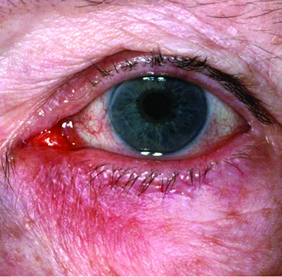
The key, she believes, is to move from a focus on ocular symptoms to an emphasis on lid and skin signs associated with ocular rosacea. “Almost all of the ophthalmology approaches to the disease start at the ocular surface, and these surface treatments are always a little bit [or very] toxic to the ocular surface,” she said. “By treating the lid signs first through a topical approach through the closed lid, we can eliminate a lot of the ocular surface symptoms without ever touching the ocular surface, thus preventing a lot of the associated toxicity and side effects you commonly see with topical ophthalmics.”
Dr. Capriotti and her colleagues have developed a topical treatment that contains dimethyl sulfoxide (DMSO) combined with dilute povidone iodine, which is applied to the lid margin of the closed eye.
In 2015, they published a case report describing the successful use of the treatment for a 78-year-old man with rosacea blepharoconjunctivitis, who had failed oral and topical treatments. He was treated with povidone iodine 10% solution in a DMSO vehicle, which was compounded into a topical gel by a pharmacy and administered twice a day, rubbed onto the lash line and eyelid.
After one week, “remarkable improvements were noted,” with reversal of much of the conjunctivitis, anterior lid erythema, and thickening, they wrote. One month later, after once daily application, “not only were the initial improvements conserved, but the posterior lid margin vessels and telangiectasias had begun to attenuate and involute. Moreover, meibomian capping was no longer present, secretions were less viscous, and tear break-up time normalized.”
DMSO is a skin penetration agent that is rarely used in ophthalmology and povidone iodine is a biocidal agent used in eye care, they pointed out in the case report. “This novel therapy may warrant further investigation in randomized, controlled clinical trials,” they concluded (Ophthalmol Ther. 2015 Dec;4[2]:143-50).
There are plans to start a clinical trial later in 2017, according to Dr. Capriottis, the cofounder of Veloce Biopharma, a company that is developing this product.
Until more specific treatments are available, what can dermatologists do now to improve their care of patients with ocular rosacea? “First and foremost, you have to be comfortable looking at the eyelids and ruling out anything that could be masquerading as blepharitis,” Dr. Capriotti commented. “We have a big disadvantage in dermatology because we don’t use slit-lamp biomicroscopy, but we are pretty good at looking at skin. If the patient has a lot of extraocular rosacea signs, I definitely think dermatologists can manage the prescription of a topical for the lids and evaluate the response.”
She cautioned, however, that referrals are in order in several situations: cases that are severe, those that involve the eye only, and those with which ocular symptoms seem to outweigh the lid signs.
Dr. Capriotti is also the senior vice president of dermatology at Veloce. In the study published in 2015, she and her three coauthors disclosed having financial interest in ALC Therapeutics, a company that was dissolved and restructured into Veloce.
A partnership between ophthalmologists and dermatologists holds the potential to produce a better treatment for ocular rosacea, one of the few conditions that brings the two specialties together.
“Hopefully, the paradigm eventually shifts from treating symptoms and signs of ocular rosacea with oral antibiotics or reflexive referral to ophthalmology to approaching cutaneous treatment first,” said dermatologist Kara Capriotti, MD, who has been working with a team of ophthalmologists to develop a treatment that focuses on ocular rosacea as a “lid disease,” instead of as a primary ocular surface problem.
Eye scrubs, artificial tears, stress relief measures, and antibiotics are among the treatments used for the condition, which, however, can be treatment-resistant.
The key, she believes, is to move from a focus on ocular symptoms to an emphasis on lid and skin signs associated with ocular rosacea. “Almost all of the ophthalmology approaches to the disease start at the ocular surface, and these surface treatments are always a little bit [or very] toxic to the ocular surface,” she said. “By treating the lid signs first through a topical approach through the closed lid, we can eliminate a lot of the ocular surface symptoms without ever touching the ocular surface, thus preventing a lot of the associated toxicity and side effects you commonly see with topical ophthalmics.”
Dr. Capriotti and her colleagues have developed a topical treatment that contains dimethyl sulfoxide (DMSO) combined with dilute povidone iodine, which is applied to the lid margin of the closed eye.
In 2015, they published a case report describing the successful use of the treatment for a 78-year-old man with rosacea blepharoconjunctivitis, who had failed oral and topical treatments. He was treated with povidone iodine 10% solution in a DMSO vehicle, which was compounded into a topical gel by a pharmacy and administered twice a day, rubbed onto the lash line and eyelid.
After one week, “remarkable improvements were noted,” with reversal of much of the conjunctivitis, anterior lid erythema, and thickening, they wrote. One month later, after once daily application, “not only were the initial improvements conserved, but the posterior lid margin vessels and telangiectasias had begun to attenuate and involute. Moreover, meibomian capping was no longer present, secretions were less viscous, and tear break-up time normalized.”
DMSO is a skin penetration agent that is rarely used in ophthalmology and povidone iodine is a biocidal agent used in eye care, they pointed out in the case report. “This novel therapy may warrant further investigation in randomized, controlled clinical trials,” they concluded (Ophthalmol Ther. 2015 Dec;4[2]:143-50).
There are plans to start a clinical trial later in 2017, according to Dr. Capriottis, the cofounder of Veloce Biopharma, a company that is developing this product.
Until more specific treatments are available, what can dermatologists do now to improve their care of patients with ocular rosacea? “First and foremost, you have to be comfortable looking at the eyelids and ruling out anything that could be masquerading as blepharitis,” Dr. Capriotti commented. “We have a big disadvantage in dermatology because we don’t use slit-lamp biomicroscopy, but we are pretty good at looking at skin. If the patient has a lot of extraocular rosacea signs, I definitely think dermatologists can manage the prescription of a topical for the lids and evaluate the response.”
She cautioned, however, that referrals are in order in several situations: cases that are severe, those that involve the eye only, and those with which ocular symptoms seem to outweigh the lid signs.
Dr. Capriotti is also the senior vice president of dermatology at Veloce. In the study published in 2015, she and her three coauthors disclosed having financial interest in ALC Therapeutics, a company that was dissolved and restructured into Veloce.
A partnership between ophthalmologists and dermatologists holds the potential to produce a better treatment for ocular rosacea, one of the few conditions that brings the two specialties together.
“Hopefully, the paradigm eventually shifts from treating symptoms and signs of ocular rosacea with oral antibiotics or reflexive referral to ophthalmology to approaching cutaneous treatment first,” said dermatologist Kara Capriotti, MD, who has been working with a team of ophthalmologists to develop a treatment that focuses on ocular rosacea as a “lid disease,” instead of as a primary ocular surface problem.
Eye scrubs, artificial tears, stress relief measures, and antibiotics are among the treatments used for the condition, which, however, can be treatment-resistant.
The key, she believes, is to move from a focus on ocular symptoms to an emphasis on lid and skin signs associated with ocular rosacea. “Almost all of the ophthalmology approaches to the disease start at the ocular surface, and these surface treatments are always a little bit [or very] toxic to the ocular surface,” she said. “By treating the lid signs first through a topical approach through the closed lid, we can eliminate a lot of the ocular surface symptoms without ever touching the ocular surface, thus preventing a lot of the associated toxicity and side effects you commonly see with topical ophthalmics.”
Dr. Capriotti and her colleagues have developed a topical treatment that contains dimethyl sulfoxide (DMSO) combined with dilute povidone iodine, which is applied to the lid margin of the closed eye.
In 2015, they published a case report describing the successful use of the treatment for a 78-year-old man with rosacea blepharoconjunctivitis, who had failed oral and topical treatments. He was treated with povidone iodine 10% solution in a DMSO vehicle, which was compounded into a topical gel by a pharmacy and administered twice a day, rubbed onto the lash line and eyelid.
After one week, “remarkable improvements were noted,” with reversal of much of the conjunctivitis, anterior lid erythema, and thickening, they wrote. One month later, after once daily application, “not only were the initial improvements conserved, but the posterior lid margin vessels and telangiectasias had begun to attenuate and involute. Moreover, meibomian capping was no longer present, secretions were less viscous, and tear break-up time normalized.”
DMSO is a skin penetration agent that is rarely used in ophthalmology and povidone iodine is a biocidal agent used in eye care, they pointed out in the case report. “This novel therapy may warrant further investigation in randomized, controlled clinical trials,” they concluded (Ophthalmol Ther. 2015 Dec;4[2]:143-50).
There are plans to start a clinical trial later in 2017, according to Dr. Capriottis, the cofounder of Veloce Biopharma, a company that is developing this product.
Until more specific treatments are available, what can dermatologists do now to improve their care of patients with ocular rosacea? “First and foremost, you have to be comfortable looking at the eyelids and ruling out anything that could be masquerading as blepharitis,” Dr. Capriotti commented. “We have a big disadvantage in dermatology because we don’t use slit-lamp biomicroscopy, but we are pretty good at looking at skin. If the patient has a lot of extraocular rosacea signs, I definitely think dermatologists can manage the prescription of a topical for the lids and evaluate the response.”
She cautioned, however, that referrals are in order in several situations: cases that are severe, those that involve the eye only, and those with which ocular symptoms seem to outweigh the lid signs.
Dr. Capriotti is also the senior vice president of dermatology at Veloce. In the study published in 2015, she and her three coauthors disclosed having financial interest in ALC Therapeutics, a company that was dissolved and restructured into Veloce.
Oral insulin matches glargine in phase II trial
SAN DIEGO – In an industry-funded phase II trial, researchers say they’ve shown for the first time that oral basal insulin tablets can safely decrease plasma glucose in patients with type 2 diabetes (T2DM).
“Oral basal insulin appears safe and efficacious in insulin-naive patients with type 2 [diabetes] insufficiently controlled with medications. It improves glycemic control to a similar extent as does glargine,” said study lead author Leona Plum-Mörschel, MD, CEO of the German clinical research organization Profil Mainz, in an oral presentation at the scientific sessions of the American Diabetes Association. A statement from Novo Nordisk, the maker of the investigational medication, says the company discontinued development of the product due to its low bioavailability and concerns about the costs of producing it.
According to Dr. Plum-Mörschel, researchers have been searching for an oral insulin treatment for almost a century without success.
In the new phase IIa study, researchers tested a basal insulin analog tablet called OI338GT. It uses a technology platform called Gipet by Merrion Pharmaceuticals that aims to boost the absorption of injectable drugs when they are given in oral form.
Researchers recruited 50 insulin-naive patients with T2DM (mean age 61 ± 7 years, mean BMI 30.5 ± 3.7 kg/m²) whose diabetes wasn’t properly controlled by metformin by itself or in conjunction with other drugs. All had hemoglobin A1c (HbA1c) levels between 7% and 10%.
The patients were randomly assigned (1:1) to the investigational oral medication or subcutaneous insulin glargine (IGlar). They took the drugs once a day for 8 weeks in addition to their existing drug regimen.
The researchers increased the doses on a weekly basis with a goal of reaching fasting plasma glucose (FPG) of 80-126 mg/dL. Daily doses of the investigational drug (nmol/kg) began at a mean 30 ± 11 and reached 114 ± 51 by the end of the study; daily mean doses of IGlar (U/kg) began at 0.11 ± 0.03 and ended at 0.33 ± 0.15.
Both medications boosted glycemic control at about the same level, the researchers report, based on levels of FPG, HbA1c, and fructosamine levels at 8 weeks.
Mean FPG levels (mg/dL), the primary endpoint, declined in investigational medication and IGlar from 175 ± 50 and 164 ± 31, respectively, at baseline to 129 ± 33 and 121 ± 17, respectively, at end of treatment. The treatment difference (investigational medication – IGlar) was 5.2 mg/dL [–8.8;19.1], 95% CI, P = .4567.
In a post hoc analysis, mean HbA1c levels declined from 8.1 ± 0.6% and 8.2 ± 0.8% at baseline in investigational medication and IGlar, respectively, to 7.3 ± 0.8% and 7.1 ± 0.6%, respectively. The treatment difference (investigational medication – IGlar) was 0.30% [–0.03;0.63], 95% CI, P = .0774.
In another post hoc analysis, mean fructosamine levels (micromol/L) dipped from 275 ± 44 and 273 ± 50 in investigational medication and IGlar, respectively, to 235 ± 45 and 223 ± 34, respectively. The treatment difference (investigational medication – IGlar)
was 9.6 micromol/L [–11.7;30.9], 95% CI, P = .3700. None of the patients reported severe hypoglycemia. In the investigational group, 6 subjects suffered 7 events of hypoglycemia that emerged or worsened during treatment; 11 events occurred in 6 subjects in the IGlar group. Researchers report similar levels of adverse events in both groups: 31 in 15 patients treated with the investigational drug and 37 in 17 patients treated with IGlar.
“After termination of treatment, plasma glucose rebounded to baseline levels within a couple of weeks,” Dr. Plum-Mörschel said.
Novo Nordisk funded the trial. Dr. Plum-Mörschel reports no disclosures.
SAN DIEGO – In an industry-funded phase II trial, researchers say they’ve shown for the first time that oral basal insulin tablets can safely decrease plasma glucose in patients with type 2 diabetes (T2DM).
“Oral basal insulin appears safe and efficacious in insulin-naive patients with type 2 [diabetes] insufficiently controlled with medications. It improves glycemic control to a similar extent as does glargine,” said study lead author Leona Plum-Mörschel, MD, CEO of the German clinical research organization Profil Mainz, in an oral presentation at the scientific sessions of the American Diabetes Association. A statement from Novo Nordisk, the maker of the investigational medication, says the company discontinued development of the product due to its low bioavailability and concerns about the costs of producing it.
According to Dr. Plum-Mörschel, researchers have been searching for an oral insulin treatment for almost a century without success.
In the new phase IIa study, researchers tested a basal insulin analog tablet called OI338GT. It uses a technology platform called Gipet by Merrion Pharmaceuticals that aims to boost the absorption of injectable drugs when they are given in oral form.
Researchers recruited 50 insulin-naive patients with T2DM (mean age 61 ± 7 years, mean BMI 30.5 ± 3.7 kg/m²) whose diabetes wasn’t properly controlled by metformin by itself or in conjunction with other drugs. All had hemoglobin A1c (HbA1c) levels between 7% and 10%.
The patients were randomly assigned (1:1) to the investigational oral medication or subcutaneous insulin glargine (IGlar). They took the drugs once a day for 8 weeks in addition to their existing drug regimen.
The researchers increased the doses on a weekly basis with a goal of reaching fasting plasma glucose (FPG) of 80-126 mg/dL. Daily doses of the investigational drug (nmol/kg) began at a mean 30 ± 11 and reached 114 ± 51 by the end of the study; daily mean doses of IGlar (U/kg) began at 0.11 ± 0.03 and ended at 0.33 ± 0.15.
Both medications boosted glycemic control at about the same level, the researchers report, based on levels of FPG, HbA1c, and fructosamine levels at 8 weeks.
Mean FPG levels (mg/dL), the primary endpoint, declined in investigational medication and IGlar from 175 ± 50 and 164 ± 31, respectively, at baseline to 129 ± 33 and 121 ± 17, respectively, at end of treatment. The treatment difference (investigational medication – IGlar) was 5.2 mg/dL [–8.8;19.1], 95% CI, P = .4567.
In a post hoc analysis, mean HbA1c levels declined from 8.1 ± 0.6% and 8.2 ± 0.8% at baseline in investigational medication and IGlar, respectively, to 7.3 ± 0.8% and 7.1 ± 0.6%, respectively. The treatment difference (investigational medication – IGlar) was 0.30% [–0.03;0.63], 95% CI, P = .0774.
In another post hoc analysis, mean fructosamine levels (micromol/L) dipped from 275 ± 44 and 273 ± 50 in investigational medication and IGlar, respectively, to 235 ± 45 and 223 ± 34, respectively. The treatment difference (investigational medication – IGlar)
was 9.6 micromol/L [–11.7;30.9], 95% CI, P = .3700. None of the patients reported severe hypoglycemia. In the investigational group, 6 subjects suffered 7 events of hypoglycemia that emerged or worsened during treatment; 11 events occurred in 6 subjects in the IGlar group. Researchers report similar levels of adverse events in both groups: 31 in 15 patients treated with the investigational drug and 37 in 17 patients treated with IGlar.
“After termination of treatment, plasma glucose rebounded to baseline levels within a couple of weeks,” Dr. Plum-Mörschel said.
Novo Nordisk funded the trial. Dr. Plum-Mörschel reports no disclosures.
SAN DIEGO – In an industry-funded phase II trial, researchers say they’ve shown for the first time that oral basal insulin tablets can safely decrease plasma glucose in patients with type 2 diabetes (T2DM).
“Oral basal insulin appears safe and efficacious in insulin-naive patients with type 2 [diabetes] insufficiently controlled with medications. It improves glycemic control to a similar extent as does glargine,” said study lead author Leona Plum-Mörschel, MD, CEO of the German clinical research organization Profil Mainz, in an oral presentation at the scientific sessions of the American Diabetes Association. A statement from Novo Nordisk, the maker of the investigational medication, says the company discontinued development of the product due to its low bioavailability and concerns about the costs of producing it.
According to Dr. Plum-Mörschel, researchers have been searching for an oral insulin treatment for almost a century without success.
In the new phase IIa study, researchers tested a basal insulin analog tablet called OI338GT. It uses a technology platform called Gipet by Merrion Pharmaceuticals that aims to boost the absorption of injectable drugs when they are given in oral form.
Researchers recruited 50 insulin-naive patients with T2DM (mean age 61 ± 7 years, mean BMI 30.5 ± 3.7 kg/m²) whose diabetes wasn’t properly controlled by metformin by itself or in conjunction with other drugs. All had hemoglobin A1c (HbA1c) levels between 7% and 10%.
The patients were randomly assigned (1:1) to the investigational oral medication or subcutaneous insulin glargine (IGlar). They took the drugs once a day for 8 weeks in addition to their existing drug regimen.
The researchers increased the doses on a weekly basis with a goal of reaching fasting plasma glucose (FPG) of 80-126 mg/dL. Daily doses of the investigational drug (nmol/kg) began at a mean 30 ± 11 and reached 114 ± 51 by the end of the study; daily mean doses of IGlar (U/kg) began at 0.11 ± 0.03 and ended at 0.33 ± 0.15.
Both medications boosted glycemic control at about the same level, the researchers report, based on levels of FPG, HbA1c, and fructosamine levels at 8 weeks.
Mean FPG levels (mg/dL), the primary endpoint, declined in investigational medication and IGlar from 175 ± 50 and 164 ± 31, respectively, at baseline to 129 ± 33 and 121 ± 17, respectively, at end of treatment. The treatment difference (investigational medication – IGlar) was 5.2 mg/dL [–8.8;19.1], 95% CI, P = .4567.
In a post hoc analysis, mean HbA1c levels declined from 8.1 ± 0.6% and 8.2 ± 0.8% at baseline in investigational medication and IGlar, respectively, to 7.3 ± 0.8% and 7.1 ± 0.6%, respectively. The treatment difference (investigational medication – IGlar) was 0.30% [–0.03;0.63], 95% CI, P = .0774.
In another post hoc analysis, mean fructosamine levels (micromol/L) dipped from 275 ± 44 and 273 ± 50 in investigational medication and IGlar, respectively, to 235 ± 45 and 223 ± 34, respectively. The treatment difference (investigational medication – IGlar)
was 9.6 micromol/L [–11.7;30.9], 95% CI, P = .3700. None of the patients reported severe hypoglycemia. In the investigational group, 6 subjects suffered 7 events of hypoglycemia that emerged or worsened during treatment; 11 events occurred in 6 subjects in the IGlar group. Researchers report similar levels of adverse events in both groups: 31 in 15 patients treated with the investigational drug and 37 in 17 patients treated with IGlar.
“After termination of treatment, plasma glucose rebounded to baseline levels within a couple of weeks,” Dr. Plum-Mörschel said.
Novo Nordisk funded the trial. Dr. Plum-Mörschel reports no disclosures.
AT THE ADA ANNUAL SCIENTIFIC SESSIONS
Key clinical point: In 8-week trial, oral insulin tablets produce similar glucose control to injectable glargine (IGlar) in patients with type 2 diabetes (T2DM).
Major finding: Mean levels fasting plasma glucose (FPG, mg/dL) declined from 175 ± 50 to 129 ± 33 (investigational medication) and from 164 ± 31 to 121 ± 17 (IGlar). The treatment difference (investigational medication – IGlar) was 5.2 mg/dL [–8.8;19.1], 95% CI, P = .4567.
Data source: 50 insulin-naive patients with T2DM randomly assigned (1:1) to daily doses of investigational oral medication or IGlar for 8 weeks, titrated to reach target FPG of 80-126 mg/dL.
Disclosures: Novo Nordisk funded the trial. Dr. Plum-Mörschel reports no disclosures.
Arab Americans reluctant to seek depression care from psychiatrists
SAN DIEGO – A new study, the first of its kind, finds that Arab Americans in a Michigan community are among least likely ethnic groups to seek help for depression from a mental health specialist. But they’re especially likely to look for assistance from a primary care physician.
Researchers aren’t sure why the discrepancy exists. “This may be because of the stigma associated with mental health in the community,” study lead author Florence J. Dallo, PhD, said in an interview. Whatever the case, she said, the finding points to the importance of paying close attention to the diagnosis and treatment of depression in Arab Americans.
Dr. Dallo presented her findings in an oral presentation at the annual meeting of the American Psychiatric Association. She told colleagues that Arab Americans are “largely invisible in health research.”
According to her, the sparse studies that do exist look at depression and posttraumatic stress disorder among Arab American refugees. But simply being Arab in the United States has its own challenges. “Let’s not forget the political climate in which we live, with a lot of discrimination, stress, and marginalization for this group,” Dr. Dallo said.
For the new study, she and her colleagues tracked a sample of patients who sought care at a large metropolitan hospital in southeast Michigan. This region of the country has a high Arab American population.
The researchers found that Arab Americans were less likely to complete a depression screening questionnaire than were whites, Asian Americans, African Americans, and Latinos.
Of those who did complete the questionnaire, 6.3% of Arab Americans screened positive for depression, compared with 5.6% of whites, 6.3% of African Americans, 7.7% of Latinos and 2.1% of Asian Americans.
Compared with the other ethnic groups, Arab Americans were especially likely to seek help from primary care physicians but especially unlikely to look to mental health specialists.
Why does this gap exist? In addition to the stigma surrounding mental illness in the community, Dr. Dallo said, religion might play a factor. “Arab Americans may feel their mental health condition is the will of God and that God will take care of them,” she said. “They may feel they do not need a psychiatrist or medication to help them manage their mental health condition.”
In an interview, Hikmet Jamil, MD, PhD, professor in the department of family medicine at Michigan State University, East Lansing, said there is a stigma surrounding mental health among Arab immigrants. “They don’t want to hear about psychiatry or psychology, because back home, if a person goes to a psychologist, he is a mad man,” he said. “They find it very hard to take advice from psychiatrists.”
But they still have a need for care, he said, because many suffer from PTSD and anxiety.
In some cases, this is tied to trauma suffered before they came to the United States, he said. However, “when they come here, they face another kind of trauma. They feel that there is discrimination against them, especially in terms of finding a job.”
According to Dr. Jamil, highly educated Arabs often face special challenges in finding jobs because their educational background does not always open doors in the United States – where they must take exams to get licenses. If they can’t get jobs in their chosen fields, he said, “they’ll take any kind of work to get money.”
How can clinicians serve this population? One approach is to cloak the fact that a patient is getting care for a mental issue. “They may go for primary health care but not mention depression or psychiatry,” Dr. Jamil said. “The physician can pick up on that and sometimes give them psychiatric advice without making it clear that they have some depression or psychiatric disorder.”
Dr. Dallo suggests a focus on primary care. “Given that Arab Americans are more likely to follow up with a primary care doctor rather than a behavioral specialist, perhaps the relationship with the primary care doctor can be encouraged and become regular,” she said. “The more comfortable the patient becomes with his or her primary care doctor, the more they may be more likely to see a behavioral specialist in the future.”
The study was funded by a grant from the Blue Cross Blue Shield of Michigan Foundation. Dr. Dallo and Dr. Jamil report no relevant disclosures.
SAN DIEGO – A new study, the first of its kind, finds that Arab Americans in a Michigan community are among least likely ethnic groups to seek help for depression from a mental health specialist. But they’re especially likely to look for assistance from a primary care physician.
Researchers aren’t sure why the discrepancy exists. “This may be because of the stigma associated with mental health in the community,” study lead author Florence J. Dallo, PhD, said in an interview. Whatever the case, she said, the finding points to the importance of paying close attention to the diagnosis and treatment of depression in Arab Americans.
Dr. Dallo presented her findings in an oral presentation at the annual meeting of the American Psychiatric Association. She told colleagues that Arab Americans are “largely invisible in health research.”
According to her, the sparse studies that do exist look at depression and posttraumatic stress disorder among Arab American refugees. But simply being Arab in the United States has its own challenges. “Let’s not forget the political climate in which we live, with a lot of discrimination, stress, and marginalization for this group,” Dr. Dallo said.
For the new study, she and her colleagues tracked a sample of patients who sought care at a large metropolitan hospital in southeast Michigan. This region of the country has a high Arab American population.
The researchers found that Arab Americans were less likely to complete a depression screening questionnaire than were whites, Asian Americans, African Americans, and Latinos.
Of those who did complete the questionnaire, 6.3% of Arab Americans screened positive for depression, compared with 5.6% of whites, 6.3% of African Americans, 7.7% of Latinos and 2.1% of Asian Americans.
Compared with the other ethnic groups, Arab Americans were especially likely to seek help from primary care physicians but especially unlikely to look to mental health specialists.
Why does this gap exist? In addition to the stigma surrounding mental illness in the community, Dr. Dallo said, religion might play a factor. “Arab Americans may feel their mental health condition is the will of God and that God will take care of them,” she said. “They may feel they do not need a psychiatrist or medication to help them manage their mental health condition.”
In an interview, Hikmet Jamil, MD, PhD, professor in the department of family medicine at Michigan State University, East Lansing, said there is a stigma surrounding mental health among Arab immigrants. “They don’t want to hear about psychiatry or psychology, because back home, if a person goes to a psychologist, he is a mad man,” he said. “They find it very hard to take advice from psychiatrists.”
But they still have a need for care, he said, because many suffer from PTSD and anxiety.
In some cases, this is tied to trauma suffered before they came to the United States, he said. However, “when they come here, they face another kind of trauma. They feel that there is discrimination against them, especially in terms of finding a job.”
According to Dr. Jamil, highly educated Arabs often face special challenges in finding jobs because their educational background does not always open doors in the United States – where they must take exams to get licenses. If they can’t get jobs in their chosen fields, he said, “they’ll take any kind of work to get money.”
How can clinicians serve this population? One approach is to cloak the fact that a patient is getting care for a mental issue. “They may go for primary health care but not mention depression or psychiatry,” Dr. Jamil said. “The physician can pick up on that and sometimes give them psychiatric advice without making it clear that they have some depression or psychiatric disorder.”
Dr. Dallo suggests a focus on primary care. “Given that Arab Americans are more likely to follow up with a primary care doctor rather than a behavioral specialist, perhaps the relationship with the primary care doctor can be encouraged and become regular,” she said. “The more comfortable the patient becomes with his or her primary care doctor, the more they may be more likely to see a behavioral specialist in the future.”
The study was funded by a grant from the Blue Cross Blue Shield of Michigan Foundation. Dr. Dallo and Dr. Jamil report no relevant disclosures.
SAN DIEGO – A new study, the first of its kind, finds that Arab Americans in a Michigan community are among least likely ethnic groups to seek help for depression from a mental health specialist. But they’re especially likely to look for assistance from a primary care physician.
Researchers aren’t sure why the discrepancy exists. “This may be because of the stigma associated with mental health in the community,” study lead author Florence J. Dallo, PhD, said in an interview. Whatever the case, she said, the finding points to the importance of paying close attention to the diagnosis and treatment of depression in Arab Americans.
Dr. Dallo presented her findings in an oral presentation at the annual meeting of the American Psychiatric Association. She told colleagues that Arab Americans are “largely invisible in health research.”
According to her, the sparse studies that do exist look at depression and posttraumatic stress disorder among Arab American refugees. But simply being Arab in the United States has its own challenges. “Let’s not forget the political climate in which we live, with a lot of discrimination, stress, and marginalization for this group,” Dr. Dallo said.
For the new study, she and her colleagues tracked a sample of patients who sought care at a large metropolitan hospital in southeast Michigan. This region of the country has a high Arab American population.
The researchers found that Arab Americans were less likely to complete a depression screening questionnaire than were whites, Asian Americans, African Americans, and Latinos.
Of those who did complete the questionnaire, 6.3% of Arab Americans screened positive for depression, compared with 5.6% of whites, 6.3% of African Americans, 7.7% of Latinos and 2.1% of Asian Americans.
Compared with the other ethnic groups, Arab Americans were especially likely to seek help from primary care physicians but especially unlikely to look to mental health specialists.
Why does this gap exist? In addition to the stigma surrounding mental illness in the community, Dr. Dallo said, religion might play a factor. “Arab Americans may feel their mental health condition is the will of God and that God will take care of them,” she said. “They may feel they do not need a psychiatrist or medication to help them manage their mental health condition.”
In an interview, Hikmet Jamil, MD, PhD, professor in the department of family medicine at Michigan State University, East Lansing, said there is a stigma surrounding mental health among Arab immigrants. “They don’t want to hear about psychiatry or psychology, because back home, if a person goes to a psychologist, he is a mad man,” he said. “They find it very hard to take advice from psychiatrists.”
But they still have a need for care, he said, because many suffer from PTSD and anxiety.
In some cases, this is tied to trauma suffered before they came to the United States, he said. However, “when they come here, they face another kind of trauma. They feel that there is discrimination against them, especially in terms of finding a job.”
According to Dr. Jamil, highly educated Arabs often face special challenges in finding jobs because their educational background does not always open doors in the United States – where they must take exams to get licenses. If they can’t get jobs in their chosen fields, he said, “they’ll take any kind of work to get money.”
How can clinicians serve this population? One approach is to cloak the fact that a patient is getting care for a mental issue. “They may go for primary health care but not mention depression or psychiatry,” Dr. Jamil said. “The physician can pick up on that and sometimes give them psychiatric advice without making it clear that they have some depression or psychiatric disorder.”
Dr. Dallo suggests a focus on primary care. “Given that Arab Americans are more likely to follow up with a primary care doctor rather than a behavioral specialist, perhaps the relationship with the primary care doctor can be encouraged and become regular,” she said. “The more comfortable the patient becomes with his or her primary care doctor, the more they may be more likely to see a behavioral specialist in the future.”
The study was funded by a grant from the Blue Cross Blue Shield of Michigan Foundation. Dr. Dallo and Dr. Jamil report no relevant disclosures.
EXPERT ANALYSIS FROM APA
Apomorphine pump dramatically decreases ‘off’ time in Parkinson’s
BOSTON – The most rigorous study of its kind suggests that apomorphine subcutaneous infusion (APO) via pump can substantially reduce ‘off’ time when conventional therapy fails to control dyskinesias in patients with Parkinson’s disease.
“When a patient´s off periods start to become refractory to adjustments to their oral medication, consider continuous dopaminergic drug delivery such as subcutaneous apomorphine infusion, which has proven efficacy and is generally well tolerated in this group of patients,” lead author Regina Katzenschlager, MD, head of the department of neurology and the Karl Landsteiner Institute for Neuroimmunological and Neurodegenerative Disorders at Danube Hospital in Vienna, said in an interview.
According to the study authors, previous open-label research has supported the use of apomorphine to reduce dyskinesias, off time, and the need for levodopa.
For the new study, researchers recruited 106 patients aged at least 30 years with a diagnosis of idiopathic Parkinson’s disease and motor fluctuations that were not adequately controlled by medication. They all were taking L-dopa and had an average off time of at least 3 hours a day.
The subjects, who came from 23 centers in seven countries, were randomly assigned to receive apomorphine infusion while they were awake (16 hours; 8 or less mg/hour) or placebo saline infusion.
“Apomorphine is administered via bespoke pumps, which are produced by several manufacturers,” Dr. Katzenschlager said. “The pump used in the trial is a mechanical delivery system and has been in clinical use in Europe for many years for this purpose. The pump is worn outside the body; no surgical procedure is required. It delivers the drug continuously into the subcutaneous fatty tissue, usually abdomen or thighs, via a tube and a thin metal or Teflon needle.”
The pump is about the size of a mobile phone, Dr. Katzenschlager said, and typically worn in a pouch attached to a belt.
The study abstract does not note the dose of apomorphine, and Dr. Katzenschlager declined to say how much of the drug is given to patients. The trial information on clinicaltrials.gov states only that a 5-mg/mL solution of APO-go was to be provided via prefilled syringe. The hourly flow rate of apomorphine and dosages of other Parkinson’s disease medications were adjusted during the first 4 weeks of the trial for efficacy and tolerability purposes.
Researchers report that daily off time declined over the 12-week trial (–0.58 hours for placebo vs. –2.47 hours for apomorphine), accounting for a difference of –1.89 hours (95% CI, –3.16 to –0.62; P = .0025).
“This was highly statistically significant. This degree of improvement has been proven to be clinically relevant to the patients,” Dr. Katzenschlager said. “This was reflected in the patients’ own assessment of the overall treatment effect in the study.”
The study examined Patient Global Impression of Change in apomorphine vs. placebo. The abstract released at the AAN meeting does not report the full results but says that the apomorphine patients had higher scores at the end of the study (P less than .001).
Why might this infusion approach work? “Apomorphine infusion achieves more than injections, which are intended as a rescue therapy for off periods in addition to the patients’ oral medication,” Dr. Katzenschlager said. “The continuous delivery of apomorphine resembles the natural state of dopamine levels in the brain in persons without Parkinson’s disease, where this is mostly stable. The administration via the pump system usually enables oral drugs to be reduced. This may contribute to the improvement in dyskinesias often observed and is a benefit to the patients in itself.”
The study authors noted that apomorphine “was generally well tolerated and no unexpected adverse events were observed.” Dr. Katzenschlager declined to discuss cost.
Britannia Pharmaceuticals, which manufactures APO-go, funded the study. Dr. Katzenschlager reports multiple disclosures, including research support and speaking/consulting fees from Brittania Pharmaceuticals.
BOSTON – The most rigorous study of its kind suggests that apomorphine subcutaneous infusion (APO) via pump can substantially reduce ‘off’ time when conventional therapy fails to control dyskinesias in patients with Parkinson’s disease.
“When a patient´s off periods start to become refractory to adjustments to their oral medication, consider continuous dopaminergic drug delivery such as subcutaneous apomorphine infusion, which has proven efficacy and is generally well tolerated in this group of patients,” lead author Regina Katzenschlager, MD, head of the department of neurology and the Karl Landsteiner Institute for Neuroimmunological and Neurodegenerative Disorders at Danube Hospital in Vienna, said in an interview.
According to the study authors, previous open-label research has supported the use of apomorphine to reduce dyskinesias, off time, and the need for levodopa.
For the new study, researchers recruited 106 patients aged at least 30 years with a diagnosis of idiopathic Parkinson’s disease and motor fluctuations that were not adequately controlled by medication. They all were taking L-dopa and had an average off time of at least 3 hours a day.
The subjects, who came from 23 centers in seven countries, were randomly assigned to receive apomorphine infusion while they were awake (16 hours; 8 or less mg/hour) or placebo saline infusion.
“Apomorphine is administered via bespoke pumps, which are produced by several manufacturers,” Dr. Katzenschlager said. “The pump used in the trial is a mechanical delivery system and has been in clinical use in Europe for many years for this purpose. The pump is worn outside the body; no surgical procedure is required. It delivers the drug continuously into the subcutaneous fatty tissue, usually abdomen or thighs, via a tube and a thin metal or Teflon needle.”
The pump is about the size of a mobile phone, Dr. Katzenschlager said, and typically worn in a pouch attached to a belt.
The study abstract does not note the dose of apomorphine, and Dr. Katzenschlager declined to say how much of the drug is given to patients. The trial information on clinicaltrials.gov states only that a 5-mg/mL solution of APO-go was to be provided via prefilled syringe. The hourly flow rate of apomorphine and dosages of other Parkinson’s disease medications were adjusted during the first 4 weeks of the trial for efficacy and tolerability purposes.
Researchers report that daily off time declined over the 12-week trial (–0.58 hours for placebo vs. –2.47 hours for apomorphine), accounting for a difference of –1.89 hours (95% CI, –3.16 to –0.62; P = .0025).
“This was highly statistically significant. This degree of improvement has been proven to be clinically relevant to the patients,” Dr. Katzenschlager said. “This was reflected in the patients’ own assessment of the overall treatment effect in the study.”
The study examined Patient Global Impression of Change in apomorphine vs. placebo. The abstract released at the AAN meeting does not report the full results but says that the apomorphine patients had higher scores at the end of the study (P less than .001).
Why might this infusion approach work? “Apomorphine infusion achieves more than injections, which are intended as a rescue therapy for off periods in addition to the patients’ oral medication,” Dr. Katzenschlager said. “The continuous delivery of apomorphine resembles the natural state of dopamine levels in the brain in persons without Parkinson’s disease, where this is mostly stable. The administration via the pump system usually enables oral drugs to be reduced. This may contribute to the improvement in dyskinesias often observed and is a benefit to the patients in itself.”
The study authors noted that apomorphine “was generally well tolerated and no unexpected adverse events were observed.” Dr. Katzenschlager declined to discuss cost.
Britannia Pharmaceuticals, which manufactures APO-go, funded the study. Dr. Katzenschlager reports multiple disclosures, including research support and speaking/consulting fees from Brittania Pharmaceuticals.
BOSTON – The most rigorous study of its kind suggests that apomorphine subcutaneous infusion (APO) via pump can substantially reduce ‘off’ time when conventional therapy fails to control dyskinesias in patients with Parkinson’s disease.
“When a patient´s off periods start to become refractory to adjustments to their oral medication, consider continuous dopaminergic drug delivery such as subcutaneous apomorphine infusion, which has proven efficacy and is generally well tolerated in this group of patients,” lead author Regina Katzenschlager, MD, head of the department of neurology and the Karl Landsteiner Institute for Neuroimmunological and Neurodegenerative Disorders at Danube Hospital in Vienna, said in an interview.
According to the study authors, previous open-label research has supported the use of apomorphine to reduce dyskinesias, off time, and the need for levodopa.
For the new study, researchers recruited 106 patients aged at least 30 years with a diagnosis of idiopathic Parkinson’s disease and motor fluctuations that were not adequately controlled by medication. They all were taking L-dopa and had an average off time of at least 3 hours a day.
The subjects, who came from 23 centers in seven countries, were randomly assigned to receive apomorphine infusion while they were awake (16 hours; 8 or less mg/hour) or placebo saline infusion.
“Apomorphine is administered via bespoke pumps, which are produced by several manufacturers,” Dr. Katzenschlager said. “The pump used in the trial is a mechanical delivery system and has been in clinical use in Europe for many years for this purpose. The pump is worn outside the body; no surgical procedure is required. It delivers the drug continuously into the subcutaneous fatty tissue, usually abdomen or thighs, via a tube and a thin metal or Teflon needle.”
The pump is about the size of a mobile phone, Dr. Katzenschlager said, and typically worn in a pouch attached to a belt.
The study abstract does not note the dose of apomorphine, and Dr. Katzenschlager declined to say how much of the drug is given to patients. The trial information on clinicaltrials.gov states only that a 5-mg/mL solution of APO-go was to be provided via prefilled syringe. The hourly flow rate of apomorphine and dosages of other Parkinson’s disease medications were adjusted during the first 4 weeks of the trial for efficacy and tolerability purposes.
Researchers report that daily off time declined over the 12-week trial (–0.58 hours for placebo vs. –2.47 hours for apomorphine), accounting for a difference of –1.89 hours (95% CI, –3.16 to –0.62; P = .0025).
“This was highly statistically significant. This degree of improvement has been proven to be clinically relevant to the patients,” Dr. Katzenschlager said. “This was reflected in the patients’ own assessment of the overall treatment effect in the study.”
The study examined Patient Global Impression of Change in apomorphine vs. placebo. The abstract released at the AAN meeting does not report the full results but says that the apomorphine patients had higher scores at the end of the study (P less than .001).
Why might this infusion approach work? “Apomorphine infusion achieves more than injections, which are intended as a rescue therapy for off periods in addition to the patients’ oral medication,” Dr. Katzenschlager said. “The continuous delivery of apomorphine resembles the natural state of dopamine levels in the brain in persons without Parkinson’s disease, where this is mostly stable. The administration via the pump system usually enables oral drugs to be reduced. This may contribute to the improvement in dyskinesias often observed and is a benefit to the patients in itself.”
The study authors noted that apomorphine “was generally well tolerated and no unexpected adverse events were observed.” Dr. Katzenschlager declined to discuss cost.
Britannia Pharmaceuticals, which manufactures APO-go, funded the study. Dr. Katzenschlager reports multiple disclosures, including research support and speaking/consulting fees from Brittania Pharmaceuticals.
AT AAN 2017
Key clinical point:
Major finding: Daily ‘off’ time declined over 12 weeks by –0.58 hours for placebo vs. –2.47 hours for apomorphine, accounting for a difference of –1.89 hours (95% CI, –3.16 to –0.62; P = .0025).
Data source: Double-blind, randomized, placebo-controlled, phase III study of 106 Parkinson’s disease patients.
Disclosures: Britannia Pharmaceuticals, which manufactures APO-go, funded the study.
Most veterans with schizophrenia or bipolar I report suicide attempts
SAN DIEGO – A new study reports that about half of assessed U.S. veterans with schizophrenia or bipolar I disorder have tried to kill themselves. Nearly 70% of those with schizophrenia had documented suicidal behavior or ideation, as did more than 82% of those with bipolar I disorder.
“The VA struggles to predict suicidal ideation and behavior,” said study lead author Philip D. Harvey, PhD, of the Carter VA Medical Center in Miami, in an interview. “These data suggest that having one of these diagnoses is a major risk factor. Regular assessment makes considerable sense.”
Dr. Harvey released the study findings in a poster at the annual meeting of the American Psychiatric Association.
For the study, Dr. Harvey and his colleagues examined findings from a VA research project into the genetics behind functional disability in schizophrenia and bipolar illness.
“We know that suicide risk is higher in veterans than in the general population. We also know that the current focus is on returning veterans who were deployed in combat operations,” said Dr. Harvey, who also is affiliated with the University of Miami. “We wanted to evaluate the risk for suicidal ideation and behavior in the segment of the veteran population who have recently or ever been exposed to military trauma.”
The project assessed VA patients with schizophrenia (N = 3,941) or bipolar I disorder (N = 5,414) through in-person evaluations regarding issues like cognitive and functional status, and history of posttraumatic stress disorder. All of the subjects were outpatients at 26 VA medical centers.
Combined, the mean age of the study participants was 53.6 years, plus or minus 11 years, and 86.2% were male. Whites made up 57.4% of the sample, followed by blacks (37.0%) and other (5.6%). A total of 27% had no comorbid psychiatric conditions.
The study authors found documented suicidal ideation or suicidal behavior in 69.9% of veterans with schizophrenia and 82.3% of those with bipolar disorder; the percentages who reported making actual suicide attempts was 46.1% schizophrenia and 54.5% bipolar disorder.
The risk of suicidal ideation was lower in schizophrenia vs. bipolar disorder (odds ratio, 0.82; 95% confidence interval, 0.71-0.95), as was suicidal behavior (OR, 0.81; 95% CI, 0.71-0.93).
Dr. Harvey said this is not surprising. “The combination of a history of euphoric mood and significant depression [characteristic of bipolar disorder] is very challenging.”
Other factors lowered risk: College education vs. high school or less (OR, 0.82; 95% CI, 0.67-1.00 for ideation; OR, 0.70; 95% CI, 0.58-0.84 for behavior). In addition, lower risk was found among black vs. white patients (OR, 0.72; 95% CI, 0.63-0.84 for ideation; OR, 0.82; 95% CI, 0.72-0.93, for behavior).
These factors boosted risk: multiple psychiatric comorbidities vs. none (OR, 2.61; 95% CI, 2.22-3.07 for ideation; OR, 3.82; 95% CI, 3.30-4.41, for behavior), and those with a history of being ever vs. never married (OR, 1.18; 95% CI, 1.02-1.37 for ideation; OR, 1.36; 95% CI, 1.19-1.55, for behavior). Most of those who had been married later were divorced.
“These findings underscore the need for continuous monitoring for suicidality in veteran populations, regardless of age or psychiatric diagnosis, and especially with multiple psychiatric comorbidities,” the authors wrote.
The study was funded by the Department of Veterans Affairs Cooperative Study Program. Dr. Harvey reported no relevant disclosures.
SAN DIEGO – A new study reports that about half of assessed U.S. veterans with schizophrenia or bipolar I disorder have tried to kill themselves. Nearly 70% of those with schizophrenia had documented suicidal behavior or ideation, as did more than 82% of those with bipolar I disorder.
“The VA struggles to predict suicidal ideation and behavior,” said study lead author Philip D. Harvey, PhD, of the Carter VA Medical Center in Miami, in an interview. “These data suggest that having one of these diagnoses is a major risk factor. Regular assessment makes considerable sense.”
Dr. Harvey released the study findings in a poster at the annual meeting of the American Psychiatric Association.
For the study, Dr. Harvey and his colleagues examined findings from a VA research project into the genetics behind functional disability in schizophrenia and bipolar illness.
“We know that suicide risk is higher in veterans than in the general population. We also know that the current focus is on returning veterans who were deployed in combat operations,” said Dr. Harvey, who also is affiliated with the University of Miami. “We wanted to evaluate the risk for suicidal ideation and behavior in the segment of the veteran population who have recently or ever been exposed to military trauma.”
The project assessed VA patients with schizophrenia (N = 3,941) or bipolar I disorder (N = 5,414) through in-person evaluations regarding issues like cognitive and functional status, and history of posttraumatic stress disorder. All of the subjects were outpatients at 26 VA medical centers.
Combined, the mean age of the study participants was 53.6 years, plus or minus 11 years, and 86.2% were male. Whites made up 57.4% of the sample, followed by blacks (37.0%) and other (5.6%). A total of 27% had no comorbid psychiatric conditions.
The study authors found documented suicidal ideation or suicidal behavior in 69.9% of veterans with schizophrenia and 82.3% of those with bipolar disorder; the percentages who reported making actual suicide attempts was 46.1% schizophrenia and 54.5% bipolar disorder.
The risk of suicidal ideation was lower in schizophrenia vs. bipolar disorder (odds ratio, 0.82; 95% confidence interval, 0.71-0.95), as was suicidal behavior (OR, 0.81; 95% CI, 0.71-0.93).
Dr. Harvey said this is not surprising. “The combination of a history of euphoric mood and significant depression [characteristic of bipolar disorder] is very challenging.”
Other factors lowered risk: College education vs. high school or less (OR, 0.82; 95% CI, 0.67-1.00 for ideation; OR, 0.70; 95% CI, 0.58-0.84 for behavior). In addition, lower risk was found among black vs. white patients (OR, 0.72; 95% CI, 0.63-0.84 for ideation; OR, 0.82; 95% CI, 0.72-0.93, for behavior).
These factors boosted risk: multiple psychiatric comorbidities vs. none (OR, 2.61; 95% CI, 2.22-3.07 for ideation; OR, 3.82; 95% CI, 3.30-4.41, for behavior), and those with a history of being ever vs. never married (OR, 1.18; 95% CI, 1.02-1.37 for ideation; OR, 1.36; 95% CI, 1.19-1.55, for behavior). Most of those who had been married later were divorced.
“These findings underscore the need for continuous monitoring for suicidality in veteran populations, regardless of age or psychiatric diagnosis, and especially with multiple psychiatric comorbidities,” the authors wrote.
The study was funded by the Department of Veterans Affairs Cooperative Study Program. Dr. Harvey reported no relevant disclosures.
SAN DIEGO – A new study reports that about half of assessed U.S. veterans with schizophrenia or bipolar I disorder have tried to kill themselves. Nearly 70% of those with schizophrenia had documented suicidal behavior or ideation, as did more than 82% of those with bipolar I disorder.
“The VA struggles to predict suicidal ideation and behavior,” said study lead author Philip D. Harvey, PhD, of the Carter VA Medical Center in Miami, in an interview. “These data suggest that having one of these diagnoses is a major risk factor. Regular assessment makes considerable sense.”
Dr. Harvey released the study findings in a poster at the annual meeting of the American Psychiatric Association.
For the study, Dr. Harvey and his colleagues examined findings from a VA research project into the genetics behind functional disability in schizophrenia and bipolar illness.
“We know that suicide risk is higher in veterans than in the general population. We also know that the current focus is on returning veterans who were deployed in combat operations,” said Dr. Harvey, who also is affiliated with the University of Miami. “We wanted to evaluate the risk for suicidal ideation and behavior in the segment of the veteran population who have recently or ever been exposed to military trauma.”
The project assessed VA patients with schizophrenia (N = 3,941) or bipolar I disorder (N = 5,414) through in-person evaluations regarding issues like cognitive and functional status, and history of posttraumatic stress disorder. All of the subjects were outpatients at 26 VA medical centers.
Combined, the mean age of the study participants was 53.6 years, plus or minus 11 years, and 86.2% were male. Whites made up 57.4% of the sample, followed by blacks (37.0%) and other (5.6%). A total of 27% had no comorbid psychiatric conditions.
The study authors found documented suicidal ideation or suicidal behavior in 69.9% of veterans with schizophrenia and 82.3% of those with bipolar disorder; the percentages who reported making actual suicide attempts was 46.1% schizophrenia and 54.5% bipolar disorder.
The risk of suicidal ideation was lower in schizophrenia vs. bipolar disorder (odds ratio, 0.82; 95% confidence interval, 0.71-0.95), as was suicidal behavior (OR, 0.81; 95% CI, 0.71-0.93).
Dr. Harvey said this is not surprising. “The combination of a history of euphoric mood and significant depression [characteristic of bipolar disorder] is very challenging.”
Other factors lowered risk: College education vs. high school or less (OR, 0.82; 95% CI, 0.67-1.00 for ideation; OR, 0.70; 95% CI, 0.58-0.84 for behavior). In addition, lower risk was found among black vs. white patients (OR, 0.72; 95% CI, 0.63-0.84 for ideation; OR, 0.82; 95% CI, 0.72-0.93, for behavior).
These factors boosted risk: multiple psychiatric comorbidities vs. none (OR, 2.61; 95% CI, 2.22-3.07 for ideation; OR, 3.82; 95% CI, 3.30-4.41, for behavior), and those with a history of being ever vs. never married (OR, 1.18; 95% CI, 1.02-1.37 for ideation; OR, 1.36; 95% CI, 1.19-1.55, for behavior). Most of those who had been married later were divorced.
“These findings underscore the need for continuous monitoring for suicidality in veteran populations, regardless of age or psychiatric diagnosis, and especially with multiple psychiatric comorbidities,” the authors wrote.
The study was funded by the Department of Veterans Affairs Cooperative Study Program. Dr. Harvey reported no relevant disclosures.
AT APA
Key clinical point: Roughly half of U.S. veterans with schizophrenia or bipolar I disorder have tried to kill themselves, and most of these veterans have histories of suicidal ideation or behavior.
Major finding: Suicide attempts are reported in 46.1% of patients with schizophrenia and 54.5% of those with bipolar I disorder. Documented suicidal ideation or behavior is reported in 69.9% of veterans with schizophrenia and 82.3% of those with bipolar disorder.
Data source: A genomic study with in-person assessments of VA patients with schizophrenia (N = 3,941) or bipolar disorder (N = 5,414). The mean age was 53.6 years, plus or minus 11 years; 86.2% were male, and 57.4% were white.
Disclosures: The study was funded by the Department of Veterans Affairs Cooperative Study Program.
Consistent weight benefits seen in empagliflozin use
SAN DIEGO – In a follow-up to the blockbuster trial results linking the type 2 diabetes drug empagliflozin (Jardiance) to a dramatically lower risk of cardiac death, researchers report that the drug improved weight-related measures in multiple groups.
Two daily doses of empagliflozin, 10 mg and 25 mg, “had consistent and robust effects on lowering weight, waist circumference, and other markers of body fat across most patients regardless of their age, sex, or degree of abdominal obesity,” study lead author Ian J. Neeland, MD, of the department of medicine at UT Southwestern Medical Center, Dallas, said in an interview. “Our next step is to determine if these effects may contribute to the improvement in cardiovascular risk seen with the drug.”

“In the EMPA-REG OUTCOME study, empagliflozin treatment significantly reduced the risk of cardiovascular death by 38%,” Dr. Neeland said. “We also observed that patients treated with empagliflozin had improvements in markers of body fatness such as weight, waist circumference, and estimated total body fat. Since we know that obesity is a major risk factor for cardiovascular disease, we were interested in finding out if the improvements in weight and other markers of body fatness may have contributed to the observed cardiovascular benefits of empagliflozin in the study. One part of this was to examine whether the drug had consistent effects on body fat according to other important cardiovascular risk factors.”
The researchers analyzed changes in body weight, waist circumference, index of central obesity, and estimated total body fat from baseline to week 164 in a study that randomly assigned participants with type 2 diabetes and cardiovascular disease to placebo or 10 mg or 25 mg of empagliflozin. The number of patients in the groups were 2,333, 2,345 and 2,342, respectively, and their mean baseline weight was around 86.0 kg.
In general, researchers found that across groups, weight measures improved more in drug-treated patients than those treated with placebo. The higher dose (25 mg) often had a greater effect; the two available doses of the drug are 10 mg and 20 mg.
For example, the placebo-adjusted mean reduction in weight was –1.70 kg in men (95% confidence interval, –2.14 to –1.27) in the 10-mg group and 2.18 kg (95% CI, –2.61 to –1.75) in the 25-mg group. For women, the reduction was –1.32 kg (95% CI, –2.02 to –0.62) in the 10-mg group and –1.44 kg (95% CI, –2.15 to –0.73) in the 25-mg group.
“Patients lost on average 1.5-2 kg of weight – about 4 pounds – with empagliflozin, compared with placebo,” Dr. Neeland said. “Although quality of life and other metrics of better health were not systematically collected, we do know that people who lose weight and waist circumference tend to feel better, have fewer health problems, and live longer, compared with people who remain obese.”
Dr. Neeland said researchers still need to understand whether the improvements in obesity markers contribute to the drug’s positive cardiac effects.
Study funding was not reported. The original EMPA-REG OUTCOME trial was funded by Boehringer Ingelheim and Eli Lilly. Dr. Neeland disclosed consultant/speakers bureau support from Boehringer Ingelheim. He is a scientific advisory board member for Advanced MR Analytics AB.
SAN DIEGO – In a follow-up to the blockbuster trial results linking the type 2 diabetes drug empagliflozin (Jardiance) to a dramatically lower risk of cardiac death, researchers report that the drug improved weight-related measures in multiple groups.
Two daily doses of empagliflozin, 10 mg and 25 mg, “had consistent and robust effects on lowering weight, waist circumference, and other markers of body fat across most patients regardless of their age, sex, or degree of abdominal obesity,” study lead author Ian J. Neeland, MD, of the department of medicine at UT Southwestern Medical Center, Dallas, said in an interview. “Our next step is to determine if these effects may contribute to the improvement in cardiovascular risk seen with the drug.”
“In the EMPA-REG OUTCOME study, empagliflozin treatment significantly reduced the risk of cardiovascular death by 38%,” Dr. Neeland said. “We also observed that patients treated with empagliflozin had improvements in markers of body fatness such as weight, waist circumference, and estimated total body fat. Since we know that obesity is a major risk factor for cardiovascular disease, we were interested in finding out if the improvements in weight and other markers of body fatness may have contributed to the observed cardiovascular benefits of empagliflozin in the study. One part of this was to examine whether the drug had consistent effects on body fat according to other important cardiovascular risk factors.”
The researchers analyzed changes in body weight, waist circumference, index of central obesity, and estimated total body fat from baseline to week 164 in a study that randomly assigned participants with type 2 diabetes and cardiovascular disease to placebo or 10 mg or 25 mg of empagliflozin. The number of patients in the groups were 2,333, 2,345 and 2,342, respectively, and their mean baseline weight was around 86.0 kg.
In general, researchers found that across groups, weight measures improved more in drug-treated patients than those treated with placebo. The higher dose (25 mg) often had a greater effect; the two available doses of the drug are 10 mg and 20 mg.
For example, the placebo-adjusted mean reduction in weight was –1.70 kg in men (95% confidence interval, –2.14 to –1.27) in the 10-mg group and 2.18 kg (95% CI, –2.61 to –1.75) in the 25-mg group. For women, the reduction was –1.32 kg (95% CI, –2.02 to –0.62) in the 10-mg group and –1.44 kg (95% CI, –2.15 to –0.73) in the 25-mg group.
“Patients lost on average 1.5-2 kg of weight – about 4 pounds – with empagliflozin, compared with placebo,” Dr. Neeland said. “Although quality of life and other metrics of better health were not systematically collected, we do know that people who lose weight and waist circumference tend to feel better, have fewer health problems, and live longer, compared with people who remain obese.”
Dr. Neeland said researchers still need to understand whether the improvements in obesity markers contribute to the drug’s positive cardiac effects.
Study funding was not reported. The original EMPA-REG OUTCOME trial was funded by Boehringer Ingelheim and Eli Lilly. Dr. Neeland disclosed consultant/speakers bureau support from Boehringer Ingelheim. He is a scientific advisory board member for Advanced MR Analytics AB.
SAN DIEGO – In a follow-up to the blockbuster trial results linking the type 2 diabetes drug empagliflozin (Jardiance) to a dramatically lower risk of cardiac death, researchers report that the drug improved weight-related measures in multiple groups.
Two daily doses of empagliflozin, 10 mg and 25 mg, “had consistent and robust effects on lowering weight, waist circumference, and other markers of body fat across most patients regardless of their age, sex, or degree of abdominal obesity,” study lead author Ian J. Neeland, MD, of the department of medicine at UT Southwestern Medical Center, Dallas, said in an interview. “Our next step is to determine if these effects may contribute to the improvement in cardiovascular risk seen with the drug.”
“In the EMPA-REG OUTCOME study, empagliflozin treatment significantly reduced the risk of cardiovascular death by 38%,” Dr. Neeland said. “We also observed that patients treated with empagliflozin had improvements in markers of body fatness such as weight, waist circumference, and estimated total body fat. Since we know that obesity is a major risk factor for cardiovascular disease, we were interested in finding out if the improvements in weight and other markers of body fatness may have contributed to the observed cardiovascular benefits of empagliflozin in the study. One part of this was to examine whether the drug had consistent effects on body fat according to other important cardiovascular risk factors.”
The researchers analyzed changes in body weight, waist circumference, index of central obesity, and estimated total body fat from baseline to week 164 in a study that randomly assigned participants with type 2 diabetes and cardiovascular disease to placebo or 10 mg or 25 mg of empagliflozin. The number of patients in the groups were 2,333, 2,345 and 2,342, respectively, and their mean baseline weight was around 86.0 kg.
In general, researchers found that across groups, weight measures improved more in drug-treated patients than those treated with placebo. The higher dose (25 mg) often had a greater effect; the two available doses of the drug are 10 mg and 20 mg.
For example, the placebo-adjusted mean reduction in weight was –1.70 kg in men (95% confidence interval, –2.14 to –1.27) in the 10-mg group and 2.18 kg (95% CI, –2.61 to –1.75) in the 25-mg group. For women, the reduction was –1.32 kg (95% CI, –2.02 to –0.62) in the 10-mg group and –1.44 kg (95% CI, –2.15 to –0.73) in the 25-mg group.
“Patients lost on average 1.5-2 kg of weight – about 4 pounds – with empagliflozin, compared with placebo,” Dr. Neeland said. “Although quality of life and other metrics of better health were not systematically collected, we do know that people who lose weight and waist circumference tend to feel better, have fewer health problems, and live longer, compared with people who remain obese.”
Dr. Neeland said researchers still need to understand whether the improvements in obesity markers contribute to the drug’s positive cardiac effects.
Study funding was not reported. The original EMPA-REG OUTCOME trial was funded by Boehringer Ingelheim and Eli Lilly. Dr. Neeland disclosed consultant/speakers bureau support from Boehringer Ingelheim. He is a scientific advisory board member for Advanced MR Analytics AB.
AT THE ADA ANNUAL SCIENTIFIC SESSIONS
Key clinical point:
Major finding: Placebo-adjusted mean reduction in weight was –1.70 kg in men for daily 10-mg dose group and –2.18 kg in daily 25-mg group. For women, the losses were –1.32 kg in the 10-mg group and –1.44 kg in the 25-mg group.
Data source: Secondary analysis of 164-week randomized, double-blind, placebo-controlled study of patients with type 2 diabetes and cardiovascular disease assigned to placebo or 10-mg or 25-mg doses of empagliflozin.
Disclosures: Study funding was not reported. The original EMPA-REG OUTCOME trial was funded by Boehringer Ingelheim and Eli Lilly.
Vets with TBIs are more likely to develop Parkinson’s
BOSTON – New research finds that military veterans who suffered mild traumatic brain injuries (TBIs) faced more than 1.5 times the risk of developing Parkinson’s disease (PD), compared with other veterans over up to 12 years of follow-up. The risk doubled for those who suffered moderate to severe TBIs.
The findings don’t confirm a link between brain injury and PD, and the number of PD diagnoses remained small even among those who’d suffered the worst TBIs.
Still, the findings suggest that “mild TBI may have long-term consequences, including PD,” said study lead author Raquel C. Gardner, MD, a neurologist whose findings were released at the annual meeting of the American Academy of Neurology. “We need to ramp up efforts to prevent TBI and also make sure we are carefully screening TBI-exposed patients for some of these long-term consequences, for which we may be able to offer therapies to improve quality of life.”
Most recently, a 2016 study found signs of a link between previous TBIs that caused more than an hour of unconsciousness and PD (hazard ratio, 3.56; 95% confidence interval, 1.52-8.28; JAMA Neurol. 2016;73[9]:1062-9).
As for mild TBI, a 2014 systematic review examined five studies and found that only one linked it to PD (OR, 1.5; 95% CI, 1.4-1.7).
For the new study, researchers analyzed records of patients served by the Veterans Health Administration from 2002 to 2014. They age matched 162,935 veterans who had suffered TBIs (half mild, half moderate to severe) to 162,935 veterans who had not (a 2% sample of all veterans served by the VHA).
Mild TBIs are defined as those that caused loss of consciousness of less than 30 minutes. Mild to moderate TBIs caused more than 30 minutes of unconsciousness.
The study participants hadn’t been diagnosed with PD or dementia at baseline or over the following year. Their average age was 48 years.
Compared with those who hadn’t suffered TBIs, those who did were more likely to be male (92% vs. 85%) and to suffer from hypertension (12% vs. 8%), cerebrovascular disease (4% vs. 1%), posttraumatic stress disorder (21% vs. 4%), and depression (24% vs. 9%; P less than .001).
“Prior studies have determined that TBI is a risk factor for depression and PTSD,” Dr. Gardner said. “Thus, higher rates of these outcomes among the patient with TBI in our study may represent sequelae of the TBI.”
Indications of education and income were similar among the two groups (P = .94 and P = .29, respectively). Those who suffered TBIs were more likely to be white than those who didn’t (73% vs. 67%) and less likely to be of other or unknown race (7% vs. 13%; P less than .001).
The percentages of veterans who developed PD were 0.31% (no TBI), 0.58% (any TBI), 0.47% (mild TBI), and 0.75% (moderate/severe TBI).
The unadjusted hazard ratios for PD were 1.81 (1.63-2.01) for any TBI, 1.59 (1.39-1.82) for mild TBI, and 2.01 (1.78-2.26) for moderate/severe TBI (P less than .0001).
Hazard ratios adjusted for demographics and comorbidities were 1.71 (1.53-1.92) for any TBI, 1.56 (1.35-1.80) for mild TBI, and 1.83 (1.61-2.07) for mild/moderate TBI (P less than .0001).
“The vast majority of people in this study did not develop PD,” Dr. Gardner said. “However, those with TBI had about a 50%-60% increased risk of PD that was statistically significant. While the P value is very small, the important numbers are really the confidence intervals around the estimate. According to our confidence intervals, we are very confident that the true estimate is between about 35% and 80% increased risk.”
Researchers also found that TBI sufferers who developed PD were 2 years younger at diagnosis than those who didn’t suffer TBIs (70 vs. 72; P = .003).
To limit the possibility of reverse causation, researchers tried excluding veterans who were diagnosed with PD within 4 years after baseline. The results remained similar.
The study has limitations. It’s not clear when the TBIs occurred. Also, the study doesn’t take the causes of TBIs into account. “In this veteran population, particularly among the younger veterans of Operation Iraqi Freedom and Operation Enduring Freedom, many are likely blast-related TBIs,” Dr. Gardner said.
The study is also limited because of the sample, said Paul K. Crane, MD, of the University of Washington, Seattle, in an interview. He was lead author of a 2016 study into links between TBI and PD and other neurodegenerative conditions (JAMA Neurol. 2016;73[9]:1062-9).
“People treated at the VA are not a representative sample of anyone other than people treated at the VA,” he said. “The ability to generalize beyond the large convenience sample is difficult.”
He added that “many people who do not have a diagnosis of mild TBI in the VA medical system nevertheless have had a mild TBI. Medical records for TBI are very incomplete. Perhaps this is especially true for veterans, who are at extremely high risk of TBI.”
Still, the research “reinforces the idea that TBI, including so-called ‘mild’ TBI – and in this case, that means mild TBI that has resulted in electronic data codes in a health records system – is definitely not innocuous, and, in particular, there is a relationship between TBI exposure and risk for Parkinson’s disease.”
How could this research be useful? Dr. Crane said it shouldn’t change practice. “We should avoid head injuries, but we should have done so before. We should diagnose Parkinson’s disease because it can be treated,” he said. “The individual risk for PD is not tons more among those with a history of head injury as defined in this paper, so I doubt we would find that heightened awareness of PD in that group is warranted.”
However, he added that “this kind of research is useful in helping us to conceptualize the downstream consequences of TBI and reinforce a strong and growing literature that finds links between TBI exposure and PD risk. Much remains to be learned.”
The study was supported by the National Institute of Neurological Disorders and Stroke, the National Institute on Aging, the American Federation for Aging Research, the Weill Institute for Neurosciences, and the U.S. Departments of Defense and Veterans Affairs. Dr. Gardner reported no relevant disclosures. Dr. Lane reported receiving funding from the Alzheimer’s Association, the National Institutes of Health, and the Department of Defense.
BOSTON – New research finds that military veterans who suffered mild traumatic brain injuries (TBIs) faced more than 1.5 times the risk of developing Parkinson’s disease (PD), compared with other veterans over up to 12 years of follow-up. The risk doubled for those who suffered moderate to severe TBIs.
The findings don’t confirm a link between brain injury and PD, and the number of PD diagnoses remained small even among those who’d suffered the worst TBIs.
Still, the findings suggest that “mild TBI may have long-term consequences, including PD,” said study lead author Raquel C. Gardner, MD, a neurologist whose findings were released at the annual meeting of the American Academy of Neurology. “We need to ramp up efforts to prevent TBI and also make sure we are carefully screening TBI-exposed patients for some of these long-term consequences, for which we may be able to offer therapies to improve quality of life.”
Most recently, a 2016 study found signs of a link between previous TBIs that caused more than an hour of unconsciousness and PD (hazard ratio, 3.56; 95% confidence interval, 1.52-8.28; JAMA Neurol. 2016;73[9]:1062-9).
As for mild TBI, a 2014 systematic review examined five studies and found that only one linked it to PD (OR, 1.5; 95% CI, 1.4-1.7).
For the new study, researchers analyzed records of patients served by the Veterans Health Administration from 2002 to 2014. They age matched 162,935 veterans who had suffered TBIs (half mild, half moderate to severe) to 162,935 veterans who had not (a 2% sample of all veterans served by the VHA).
Mild TBIs are defined as those that caused loss of consciousness of less than 30 minutes. Mild to moderate TBIs caused more than 30 minutes of unconsciousness.
The study participants hadn’t been diagnosed with PD or dementia at baseline or over the following year. Their average age was 48 years.
Compared with those who hadn’t suffered TBIs, those who did were more likely to be male (92% vs. 85%) and to suffer from hypertension (12% vs. 8%), cerebrovascular disease (4% vs. 1%), posttraumatic stress disorder (21% vs. 4%), and depression (24% vs. 9%; P less than .001).
“Prior studies have determined that TBI is a risk factor for depression and PTSD,” Dr. Gardner said. “Thus, higher rates of these outcomes among the patient with TBI in our study may represent sequelae of the TBI.”
Indications of education and income were similar among the two groups (P = .94 and P = .29, respectively). Those who suffered TBIs were more likely to be white than those who didn’t (73% vs. 67%) and less likely to be of other or unknown race (7% vs. 13%; P less than .001).
The percentages of veterans who developed PD were 0.31% (no TBI), 0.58% (any TBI), 0.47% (mild TBI), and 0.75% (moderate/severe TBI).
The unadjusted hazard ratios for PD were 1.81 (1.63-2.01) for any TBI, 1.59 (1.39-1.82) for mild TBI, and 2.01 (1.78-2.26) for moderate/severe TBI (P less than .0001).
Hazard ratios adjusted for demographics and comorbidities were 1.71 (1.53-1.92) for any TBI, 1.56 (1.35-1.80) for mild TBI, and 1.83 (1.61-2.07) for mild/moderate TBI (P less than .0001).
“The vast majority of people in this study did not develop PD,” Dr. Gardner said. “However, those with TBI had about a 50%-60% increased risk of PD that was statistically significant. While the P value is very small, the important numbers are really the confidence intervals around the estimate. According to our confidence intervals, we are very confident that the true estimate is between about 35% and 80% increased risk.”
Researchers also found that TBI sufferers who developed PD were 2 years younger at diagnosis than those who didn’t suffer TBIs (70 vs. 72; P = .003).
To limit the possibility of reverse causation, researchers tried excluding veterans who were diagnosed with PD within 4 years after baseline. The results remained similar.
The study has limitations. It’s not clear when the TBIs occurred. Also, the study doesn’t take the causes of TBIs into account. “In this veteran population, particularly among the younger veterans of Operation Iraqi Freedom and Operation Enduring Freedom, many are likely blast-related TBIs,” Dr. Gardner said.
The study is also limited because of the sample, said Paul K. Crane, MD, of the University of Washington, Seattle, in an interview. He was lead author of a 2016 study into links between TBI and PD and other neurodegenerative conditions (JAMA Neurol. 2016;73[9]:1062-9).
“People treated at the VA are not a representative sample of anyone other than people treated at the VA,” he said. “The ability to generalize beyond the large convenience sample is difficult.”
He added that “many people who do not have a diagnosis of mild TBI in the VA medical system nevertheless have had a mild TBI. Medical records for TBI are very incomplete. Perhaps this is especially true for veterans, who are at extremely high risk of TBI.”
Still, the research “reinforces the idea that TBI, including so-called ‘mild’ TBI – and in this case, that means mild TBI that has resulted in electronic data codes in a health records system – is definitely not innocuous, and, in particular, there is a relationship between TBI exposure and risk for Parkinson’s disease.”
How could this research be useful? Dr. Crane said it shouldn’t change practice. “We should avoid head injuries, but we should have done so before. We should diagnose Parkinson’s disease because it can be treated,” he said. “The individual risk for PD is not tons more among those with a history of head injury as defined in this paper, so I doubt we would find that heightened awareness of PD in that group is warranted.”
However, he added that “this kind of research is useful in helping us to conceptualize the downstream consequences of TBI and reinforce a strong and growing literature that finds links between TBI exposure and PD risk. Much remains to be learned.”
The study was supported by the National Institute of Neurological Disorders and Stroke, the National Institute on Aging, the American Federation for Aging Research, the Weill Institute for Neurosciences, and the U.S. Departments of Defense and Veterans Affairs. Dr. Gardner reported no relevant disclosures. Dr. Lane reported receiving funding from the Alzheimer’s Association, the National Institutes of Health, and the Department of Defense.
BOSTON – New research finds that military veterans who suffered mild traumatic brain injuries (TBIs) faced more than 1.5 times the risk of developing Parkinson’s disease (PD), compared with other veterans over up to 12 years of follow-up. The risk doubled for those who suffered moderate to severe TBIs.
The findings don’t confirm a link between brain injury and PD, and the number of PD diagnoses remained small even among those who’d suffered the worst TBIs.
Still, the findings suggest that “mild TBI may have long-term consequences, including PD,” said study lead author Raquel C. Gardner, MD, a neurologist whose findings were released at the annual meeting of the American Academy of Neurology. “We need to ramp up efforts to prevent TBI and also make sure we are carefully screening TBI-exposed patients for some of these long-term consequences, for which we may be able to offer therapies to improve quality of life.”
Most recently, a 2016 study found signs of a link between previous TBIs that caused more than an hour of unconsciousness and PD (hazard ratio, 3.56; 95% confidence interval, 1.52-8.28; JAMA Neurol. 2016;73[9]:1062-9).
As for mild TBI, a 2014 systematic review examined five studies and found that only one linked it to PD (OR, 1.5; 95% CI, 1.4-1.7).
For the new study, researchers analyzed records of patients served by the Veterans Health Administration from 2002 to 2014. They age matched 162,935 veterans who had suffered TBIs (half mild, half moderate to severe) to 162,935 veterans who had not (a 2% sample of all veterans served by the VHA).
Mild TBIs are defined as those that caused loss of consciousness of less than 30 minutes. Mild to moderate TBIs caused more than 30 minutes of unconsciousness.
The study participants hadn’t been diagnosed with PD or dementia at baseline or over the following year. Their average age was 48 years.
Compared with those who hadn’t suffered TBIs, those who did were more likely to be male (92% vs. 85%) and to suffer from hypertension (12% vs. 8%), cerebrovascular disease (4% vs. 1%), posttraumatic stress disorder (21% vs. 4%), and depression (24% vs. 9%; P less than .001).
“Prior studies have determined that TBI is a risk factor for depression and PTSD,” Dr. Gardner said. “Thus, higher rates of these outcomes among the patient with TBI in our study may represent sequelae of the TBI.”
Indications of education and income were similar among the two groups (P = .94 and P = .29, respectively). Those who suffered TBIs were more likely to be white than those who didn’t (73% vs. 67%) and less likely to be of other or unknown race (7% vs. 13%; P less than .001).
The percentages of veterans who developed PD were 0.31% (no TBI), 0.58% (any TBI), 0.47% (mild TBI), and 0.75% (moderate/severe TBI).
The unadjusted hazard ratios for PD were 1.81 (1.63-2.01) for any TBI, 1.59 (1.39-1.82) for mild TBI, and 2.01 (1.78-2.26) for moderate/severe TBI (P less than .0001).
Hazard ratios adjusted for demographics and comorbidities were 1.71 (1.53-1.92) for any TBI, 1.56 (1.35-1.80) for mild TBI, and 1.83 (1.61-2.07) for mild/moderate TBI (P less than .0001).
“The vast majority of people in this study did not develop PD,” Dr. Gardner said. “However, those with TBI had about a 50%-60% increased risk of PD that was statistically significant. While the P value is very small, the important numbers are really the confidence intervals around the estimate. According to our confidence intervals, we are very confident that the true estimate is between about 35% and 80% increased risk.”
Researchers also found that TBI sufferers who developed PD were 2 years younger at diagnosis than those who didn’t suffer TBIs (70 vs. 72; P = .003).
To limit the possibility of reverse causation, researchers tried excluding veterans who were diagnosed with PD within 4 years after baseline. The results remained similar.
The study has limitations. It’s not clear when the TBIs occurred. Also, the study doesn’t take the causes of TBIs into account. “In this veteran population, particularly among the younger veterans of Operation Iraqi Freedom and Operation Enduring Freedom, many are likely blast-related TBIs,” Dr. Gardner said.
The study is also limited because of the sample, said Paul K. Crane, MD, of the University of Washington, Seattle, in an interview. He was lead author of a 2016 study into links between TBI and PD and other neurodegenerative conditions (JAMA Neurol. 2016;73[9]:1062-9).
“People treated at the VA are not a representative sample of anyone other than people treated at the VA,” he said. “The ability to generalize beyond the large convenience sample is difficult.”
He added that “many people who do not have a diagnosis of mild TBI in the VA medical system nevertheless have had a mild TBI. Medical records for TBI are very incomplete. Perhaps this is especially true for veterans, who are at extremely high risk of TBI.”
Still, the research “reinforces the idea that TBI, including so-called ‘mild’ TBI – and in this case, that means mild TBI that has resulted in electronic data codes in a health records system – is definitely not innocuous, and, in particular, there is a relationship between TBI exposure and risk for Parkinson’s disease.”
How could this research be useful? Dr. Crane said it shouldn’t change practice. “We should avoid head injuries, but we should have done so before. We should diagnose Parkinson’s disease because it can be treated,” he said. “The individual risk for PD is not tons more among those with a history of head injury as defined in this paper, so I doubt we would find that heightened awareness of PD in that group is warranted.”
However, he added that “this kind of research is useful in helping us to conceptualize the downstream consequences of TBI and reinforce a strong and growing literature that finds links between TBI exposure and PD risk. Much remains to be learned.”
The study was supported by the National Institute of Neurological Disorders and Stroke, the National Institute on Aging, the American Federation for Aging Research, the Weill Institute for Neurosciences, and the U.S. Departments of Defense and Veterans Affairs. Dr. Gardner reported no relevant disclosures. Dr. Lane reported receiving funding from the Alzheimer’s Association, the National Institutes of Health, and the Department of Defense.
AT AAN 2017
Key clinical point:
Major finding: Veterans who’d suffered any TBI were more likely to develop PD (adjusted HR, 1.71; 95% CI, 1.53-1.92; P less than .0001).
Data source: A retrospective cohort study of age-matched veterans (162,935 who had suffered TBIs and 162,935 who had not) who received care from the Veterans Health Administration from 2002 to 2014.
Disclosures: The study was supported by the National Institute of Neurological Disorders and Stroke, the National Institute on Aging, the American Federation for Aging Research, the Weill Institute for Neurosciences, and the U.S. Departments of Defense and Veterans Affairs. Dr. Gardner reports no relevant disclosures. Dr. Lane reported receiving funding from the Alzheimer’s Association, the National Institutes of Health, and the Department of Defense.