User login
Cemiplimab for Unresectable Cutaneous Squamous Cell Carcinoma: Experience From a Tertiary Center
Cemiplimab for Unresectable Cutaneous Squamous Cell Carcinoma: Experience From a Tertiary Center
Cutaneous squamous cell carcinoma (cSCC) is the second most prevalent skin cancer and ranks sixth in prevalence among all cancers in the United Kingdom.1,2 The etiologic factors underlying cSCC are well established, with major efforts undertaken by governments and public health organizations over the past 2 decades to increase public awareness globally. Known risk factors for cSCC include chronic exposure to UV radiation, radiotherapy, chemical injury, and immunosuppression. The first 3 risk factors amplify risk by increasing accumulation of abnormal gene mutations. Immunosuppression hampers the immune system’s ability to eradicate cells bearing malignant genetic aberrations. Notable gene mutations implicated in cSCC include p53, p16, telomerase reverse transcriptase, NOTCH1, ROS1, mitogen-activated protein kinases, forkhead box M1, and cyclooxygenase 2, in addition to matrix metalloproteinases, which are most commonly associated with Marjolin ulcers.3
The incidence of cSCC continues to surge worldwide,3,4 with more patients presenting with advanced stages of disease and a notable increase in those presenting with unresectable cSCC due to either locally advanced disease or distant metastases.5 Existing therapies for cSCC include surgical excision (including Mohs micrographic surgery); radiotherapy (indicated for cosmetic reasons, locally advanced disease, and/or patient factors); and systemic treatments, encompassing chemotherapy (eg, 5-fluorouracil), and epidermal growth factor receptor inhibitors (indicated locally advanced disease or distant metastases).4
In recent years, immunotherapy has emerged as a potent and effective treatment modality for unresectable cSCC, both locally advanced and metastatic. The success of immunotherapy in cSCC treatment can be attributed to the unique tumor microenvironment of cSCC, which is characterized by high tumor mutational burden, increased density of tumor-infiltrating lymphocytes (TILs), and heightened programmed cell death ligand 1 (PD-L1) expression on neoplastic cells. The elevated TIL density enables a robust immune response, rendering checkpoint inhibitors particularly effective. Greater tumor mutational burden further augments this enhanced TIL activity, amplifying the response to checkpoint inhibitors. Additionally, heightened PD-L1 expression facilitates more effective unmasking by checkpoint inhibitors, thereby enhancing the immune response.6
Cemiplimab is a programmed cell death protein 1/PD-L1 that was approved by the US Food and Drug Administration in September 2018 for treatment of cSCC. It also gained a European Union endorsement in June 2019 and National Institute for Health and Care Excellence approval in August 2019 based on the highly promising results of a phase 2 trial that involved only 59 adult patients with metastatic cSCC.7 The trial reported an overall response rate (ORR) of 47%, durable disease control in 61% of patients, a median time to response of 1.9 months, and response duration exceeding 6 months in 57% of patients. The phase 2 trials reported an estimated 12-month progression-free survival (PFS) of 53% and an estimated 12-month overall survival (OS) of 81%.7
Despite the noteworthy response statistics demonstrated by these studies, it is imperative to recognize that immunotherapies, while potent, are not without challenges. They can precipitate severe immune-related adverse events (AEs), including myocarditis, adrenal failure, and pneumonitis, which can negatively impact patient health outcomes and lead to early treatment cessation. The initial trials reported high-grade AEs such as pneumonitis, pleural effusion, and, notably 11 total deaths, with 8 (72.7%) attributed to disease progression and 3 (27.3%) to AEs.7 Additionally, cost and access to immunotherapy are inherent limitations of the treatment; immunotherapy agents are expensive, and not all centers or patients are able to access them.
The aim of this study was to assess the efficacy of cemiplimab in patients with inoperable cSCC, including locally advanced and metastatic disease, treated at a tertiary referral center in the United Kingdom, and to compare outcomes with the pivotal phase 2 trial that supported regulatory approval of cemiplimab.7 The primary objective was ORR, with secondary objectives including PFS, OS, and AEs.
Methods
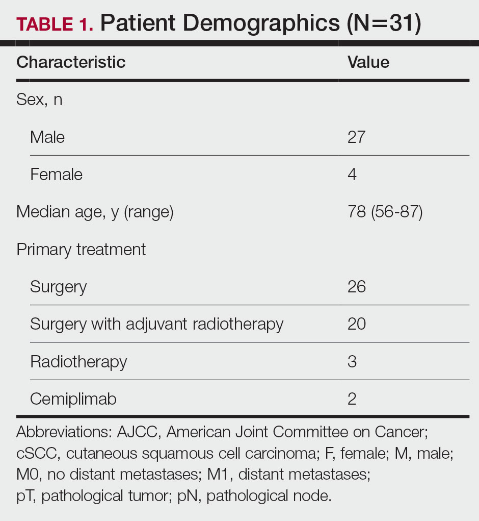
The patients included in this study had unresectable cSCC and therefore were not candidates for surgery or radiotherapy. Patient demographics are presented in Table 1. The main indications for cemiplimab in place of surgery or radiotherapy included local recurrence, locally advanced disease involving deep structures, advanced nodal disease, and distant metastatic disease. Patients meeting these criteria and the following inclusion criteria for cemiplimab treatment from November 2018 through March 2023 at a single tertiary referral center were included in the study:
- Age 18 years or older
- Histologically confirmed cSCC with locoregional recurrence after surgery or radiotherapy, or histologically confirmed advanced or metastatic disease deemed to be inoperable
- Eastern Cooperative Oncology Group performance status of 0 to 2
All enrolled patients received intravenous infusions of cemiplimab 3 times weekly at a dosage of 350 mg. Treatment was continued until complete response, unacceptable toxicity, or disease progression, with a maximum duration of 2 years or 35 cycles. Patients underwent regular follow-up, typically 3 weeks preceding each treatment cycle. Monitoring adhered to the Common Terminology Criteria for Adverse Events, version 4.0, as outlined by the National Cancer Institute.8 Response to treatment was reported according to the guidelines stipulated by the Response Evaluation Criteria in Solid Tumours, version 1.1.9 Written informed consent was obtained for all patients.
Comprehensive patient demographics, histologic profiles, and clinical data were meticulously captured on a retrospective basis. The primary objective centered on elucidating the ORR. Secondary objectives encompassed evaluating PFS, OS, and a comprehensive analysis of AEs. Progression-free survival and OS were calculated by generating Kaplan-Meier curves using Python 3.9 (Python Software Foundation).
Results
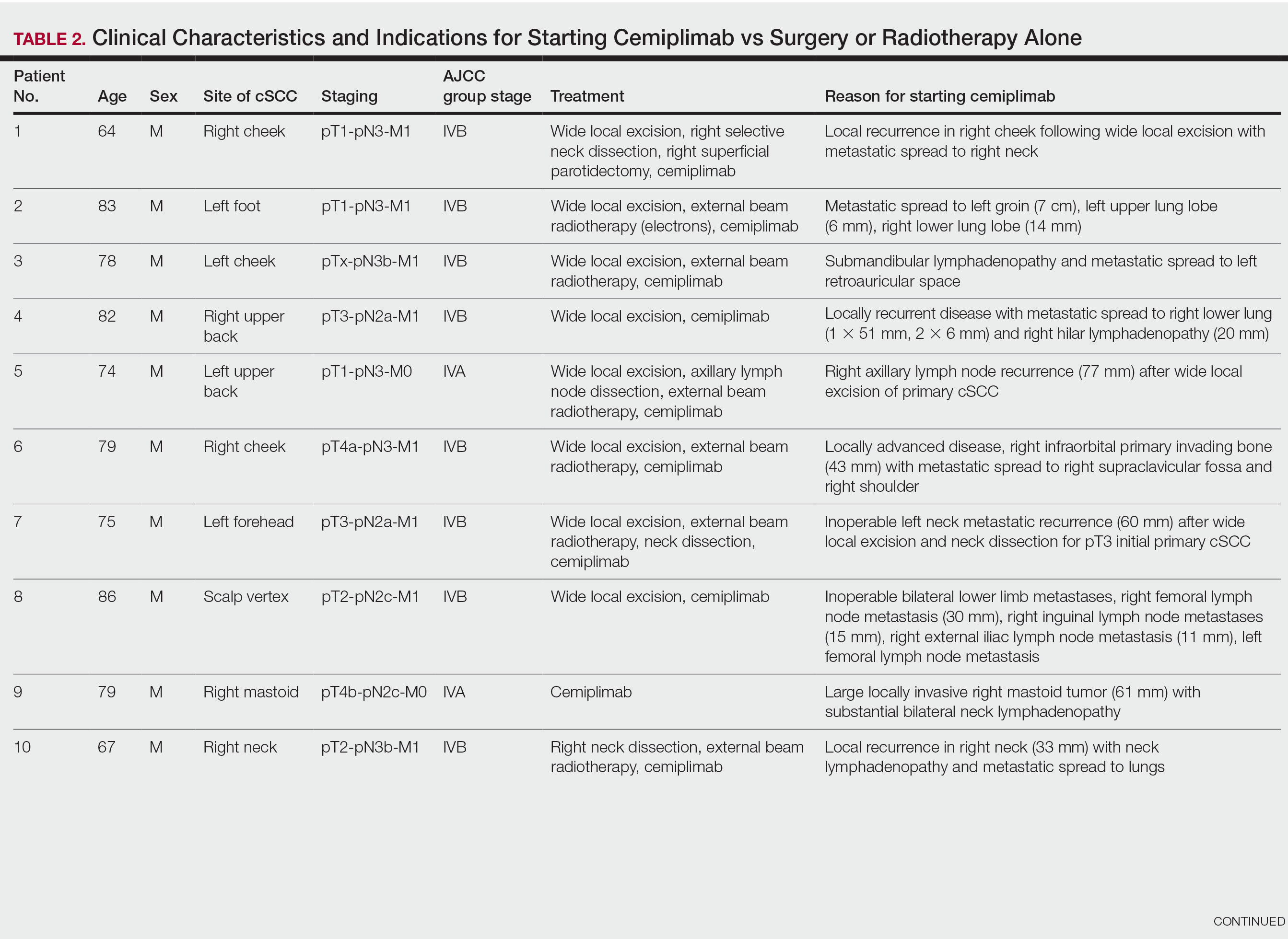
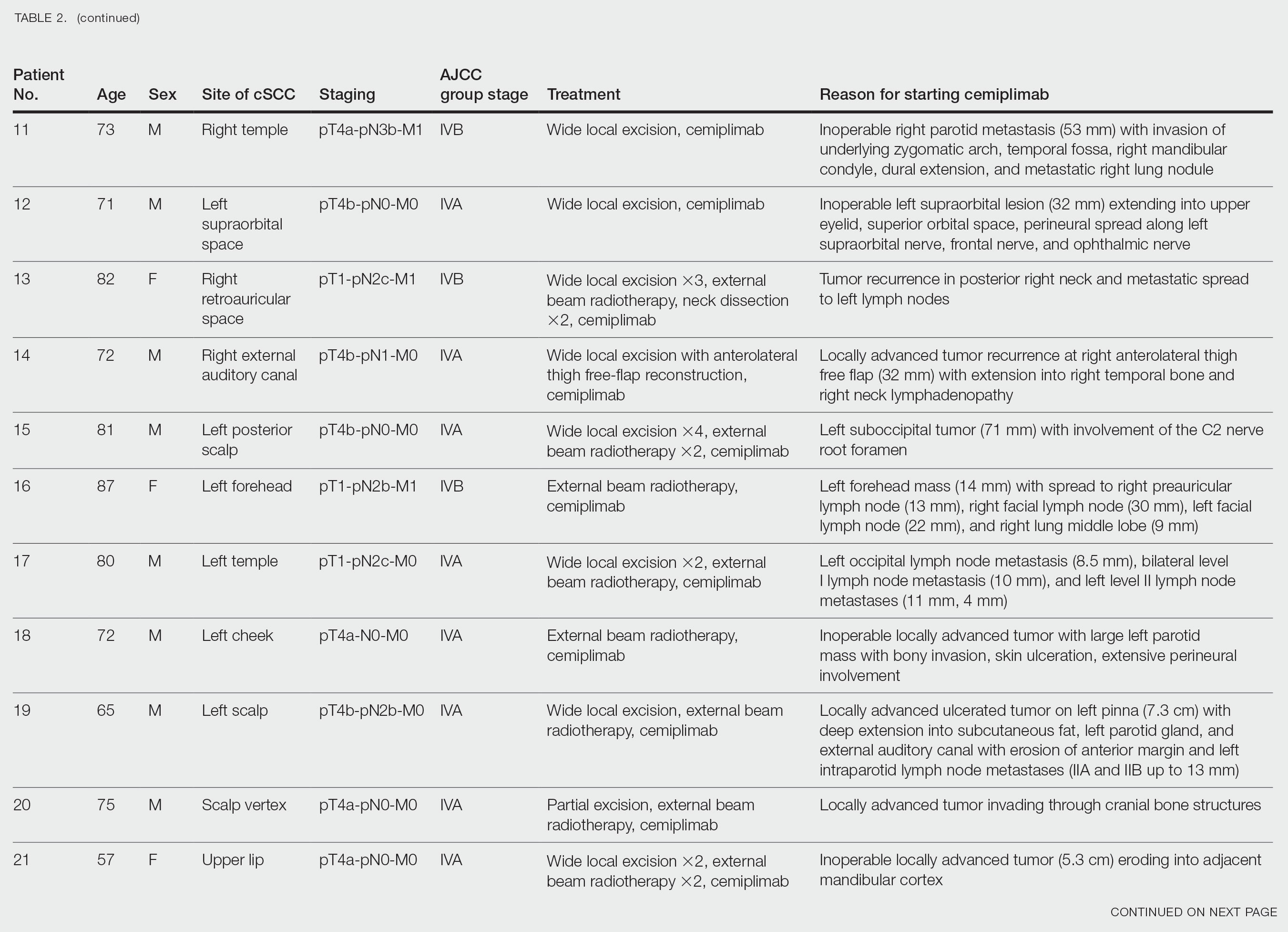
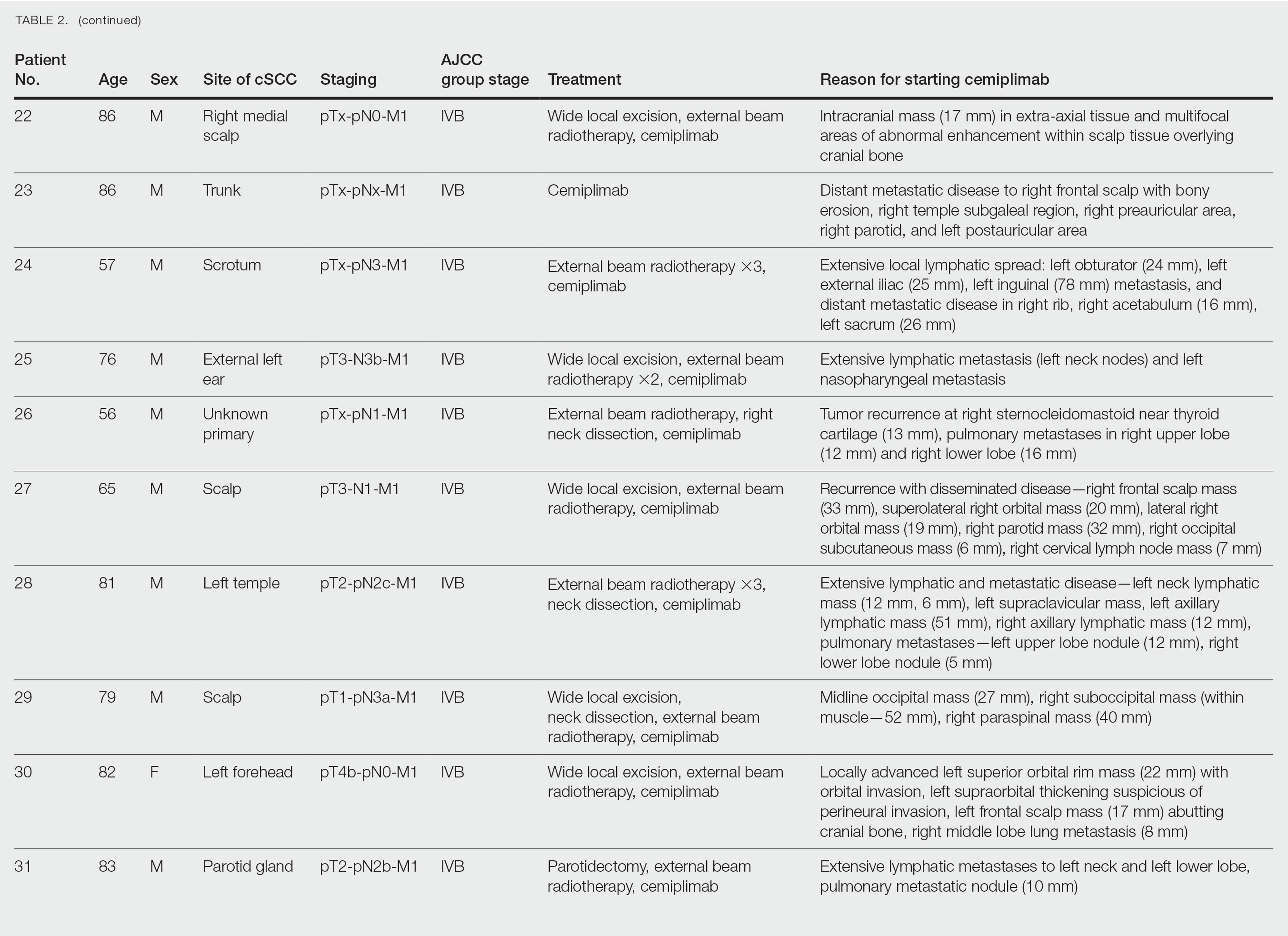
Patient Characteristics—From November 2018 through March 2023, a cohort of 31 patients with inoperable cSCC underwent treatment with cemiplimab at our tertiary referral center. The median duration of follow-up was 13 months. Clinical characteristics are outlined in the Table 2. Four (12.9%) patients successfully completed the full 2-year treatment course. Nine (29.0%) continued to receive cemiplimab therapy at the conclusion of this study in March 2023, with treatment courses ranging from 2 to 11 months since initiation. Ten (32.3%) patients discontinued treatment due to AEs, while 5 (16.1%) regrettably ceased treatment due to mortality. Two (6.5%) patients terminated treatment due to the COVID-19 pandemic, and 1 (3.2%) discontinued treatment as a result of disease progression.
Clinical Efficacy—Of the 31 enrolled patients, a substantial proportion experienced positive clinical outcomes, with 20 (64.5%) achieving complete response and 6 (19.4%) achieving partial response. A total of 26 patients achieved a response on cemiplimab, with an ORR of 83.9% (95% CI, 66.3%-94.6%). Regrettably, 2 (6.5%) patients experienced disease progression, while 3 (9.7%) died before response to cemiplimab could be assessed. Following a median follow-up period of 13 months, the median PFS and OS remained unreached, emphasizing the efficacy of cemiplimab in treating inoperable cSCC (Figures 1 and 2).
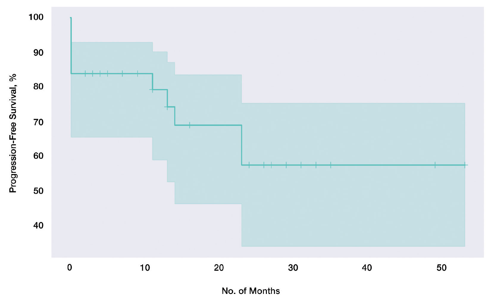
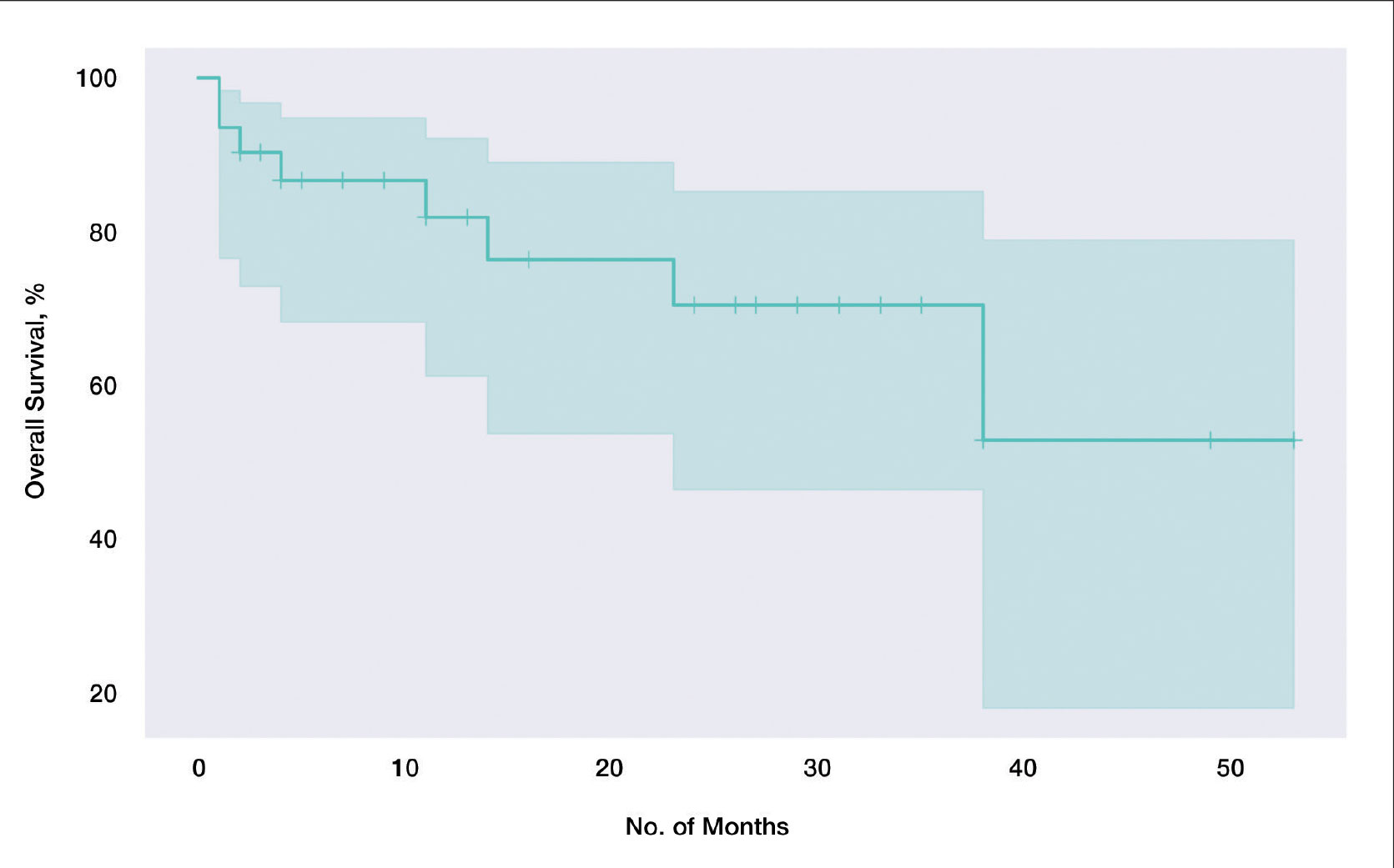
In our cohort, 2-year PFS was 57.5% (95% CI, 33.9%-75.5%) with cemiplimab and 2-year OS was 70.6% (95% CI, 46.5%-85.4%). For PFS, we observed the steepest drops at onset and at the 23-month mark (Figure 1), while for OS we observed the steepest drop at the 38-month mark (Figure 2). Clinically, we observed cemiplimab causing near-complete regression of previously large, ulcerating, fungating cSCC in patients who responded to cemiplimab, mirroring results seen elsewhere.7
Adverse Events and Treatment Cessation—A substantial proportion of patients (24/31 [77.4%]) reported AEs during treatment. Notably, treatment discontinuation was necessary in 10 (32.3%) patients due to a range of AEs, including myocarditis, atrial flutter, pneumonitis, nephritis, derangement of liver function tests, and arthritis. Additional relevant side effects included adrenal insufficiency (3/31 [9.7%]), fatigue (3/31 [9.7%]), diarrhea (2/31 [6.5%]), and type 1 diabetes 1/31 [3.2%]). These outcomes emphasize the importance of vigilance and monitoring when administering cemiplimab in the context of advanced cSCC.
Comment
Historically, advanced cSCC has had a bleak prognosis. The nature of the disease generally meant these patients could not be operated on due to metastatic spread or local invasion, and radiotherapy was not curative. The only option remaining was palliation, but new therapies have shown promise due to specific inherent characteristics of advanced cSCC; for example, the characteristic high mutation burden prevalent in advanced cSCC has paved the way for the emergence of immunotherapy as a promising avenue for intervention.10 Cemiplimab in particular has emerged as a feasible treatment for patients who would otherwise be confined to palliation. Our findings derived from a local cohort reinforce this notion, with a remarkable 83.9% (26/31) exhibiting a favorable response to cemiplimab. Although this local sample of 31 patients is small in absolute terms, in the context of the trial with 59 participants7 that gained global approval for the use of cemiplimab, our study adds a substantial amount of data to the growing body of evidence on the long-term efficacy of cemiplimab. Notably, our results emphasize the potential applicability of cemiplimab among elderly patients and individuals with lower performance statuses: populations historically excluded from immunotherapeutic considerations.
Immunotherapeutic AEs and Tolerance—As anticipated with immunotherapeutic agents, cemiplimab is associated with AEs that also are seen in its counterparts.11 A total of 77.4% (24/31) of our cohort reported immune-related AEs, although the severity warranted treatment discontinuation in only 10 (10/24 [41.7%]) patients, representing less than half of those who encountered side effects and less than a third of the entire cohort. Furthermore, most of these immune-related AEs were managed effectively with short courses of oral steroids, further substantiating the notion that cemiplimab is generally well tolerated across patients of diverse performance statuses. Even for patients who discontinued treatment early due to immune-related side effects, benefits persisted despite the partial course of cemiplimab. Of the 10 patients who discontinued treatment due to immune AEs, 6 (60%) demonstrated stable complete response, 2 (20%) experienced relapse after stopping cemiplimab, and 2 (20%) demonstrated a partial response with stable disease.
Challenges in the Most Vulnerable Patient—Of the 5 recorded mortalities, 2 (40%) were attributed to disease progression, while 3 (60%) occurred before response assessment could be undertaken. The 3 patients who died prior to response evaluation were among the most medically fragile in the cohort, characterized by extensive metastatic cSCC and major comorbidities that, in isolation, posed life-threatening risks. For individuals grappling with widespread metastatic cSCC and substantial life-threatening comorbidities, it is plausible that the necessary physiologic resilience necessary for cemiplimab therapy may be absent. We hypothesize that an immune reconstitution syndrome–like response may be responsible for this early mortality, and these patients may lack the necessary physiological resilience to tolerate this response. This subset of patients warrants careful consideration when considering therapy with cemiplimab.
Conclusion
In summary, our results underscore the efficacy of cemiplimab, as it supported a response in more than three-quarters of our patient cohort. Additionally, the associated AEs, similar to those with other programmed cell death protein 1 inhibitors, generally were manageable with medical intervention. Our findings corroborate earlier studies that have demonstrated the therapeutic potential of cemiplimab in advanced, inoperable cSCC management. In addition to efficacy, our results also suggest that cemiplimab holds promise as a therapeutic option for patients who might not be amenable to the stresses of general anesthesia, surgery, or prolonged hospitalization, although cemiplimab should likely be used with caution in patients with severe, life-threatening medical comorbidities and/or concurrent severe illness. Furthermore, our data demonstrate that the benefits persist not only beyond the completion of the full 2-year course, but also after partial treatment courses discontinued due to patient-specific factors. Future studies would be useful to better understand and optimize dose and duration of cemiplimab treatment to maximize therapeutic effectiveness while minimizing risk of immune-related AEs. Among individuals confronting advanced, inoperable cSCC, cemiplimab is emerging as a viable and beneficial intervention.
- Waldman A, Schmults C. Cutaneous squamous cell carcinoma. Hematol Oncol Clin North Am. 2019;33:1-12. doi:10.1016/j.hoc.2018.08.001
- Venables ZC, Nijsten T, Wong KF, et al. Epidemiology of basal and cutaneous squamous cell carcinoma in the U.K. 2013–15: a cohort study. Br J Dermatol. 2019;181:474-482. doi:10.1111/bjd.17873
- Que SKT, Zwald FO, Schmults CD. Cutaneous squamous cell carcinoma. J Am Acad Dermatol. 2018;78:237-247. doi:10.1016/j.jaad.2017.08.059
- Green AC, Olsen CM. Cutaneous squamous cell carcinoma: an epidemiological review. Br J Dermatol. 2017;177:373-381. doi:10.1111/bjd.15324
- Jovic’ M, Marinkovic’ M, Sud‐ecki B, et al. COVID-19 and cutaneous squamous cell carcinoma—impact of the pandemic on unequal access to healthcare. Healthcare (Basel). 2023;11:1994. doi:10.3390/healthcare11141994
- Ansary TM, Hossain MDR, Komine M, et al. Immunotherapy for the treatment of squamous cell carcinoma: potential benefits and challenges. Int J Mol Sci. 2022;23:8530. doi:10.3390/ijms23158530
- Migden MR, Rischin D, Schmults CD, et al. PD-1 blockade with cemiplimab in advanced cutaneous squamous-cell carcinoma. N Engl J Med. 2018;379:341-351. doi:10.1056/nejmoa1805131
- National Cancer Institute. Lead organizations: NCI network trial development and conduct. Updated September 29, 2025. Accessed March 10, 2026. https://dctd.cancer.gov/research/ctep-trials/trial-development#ctc_40
- Eisenhauer EA, Therasse P, Bogaerts J, et al. New response evaluation criteria in solid tumours: revised RECIST guideline (version 1.1). Eur J Cancer. 2009;45:228-247. doi:10.1016/j.ejca.2008.10.026
- Goodman AM, Kato S, Bazhenova L, et al. Tumor mutational burden as an independent predictor of response to immunotherapy in diverse cancers. Mol Cancer Ther. 2017;16:2598-2608. doi:10.1158/1535-7163.mct-17-0386
- Kroschinsky F, Stölzel F, von Bonin S, et al. New drugs, new toxicities: severe side effects of modern targeted and immunotherapy of cancer and their management. Crit Care. 2017;21:89. doi:10.1186/s13054-017-1678-1
Cutaneous squamous cell carcinoma (cSCC) is the second most prevalent skin cancer and ranks sixth in prevalence among all cancers in the United Kingdom.1,2 The etiologic factors underlying cSCC are well established, with major efforts undertaken by governments and public health organizations over the past 2 decades to increase public awareness globally. Known risk factors for cSCC include chronic exposure to UV radiation, radiotherapy, chemical injury, and immunosuppression. The first 3 risk factors amplify risk by increasing accumulation of abnormal gene mutations. Immunosuppression hampers the immune system’s ability to eradicate cells bearing malignant genetic aberrations. Notable gene mutations implicated in cSCC include p53, p16, telomerase reverse transcriptase, NOTCH1, ROS1, mitogen-activated protein kinases, forkhead box M1, and cyclooxygenase 2, in addition to matrix metalloproteinases, which are most commonly associated with Marjolin ulcers.3
The incidence of cSCC continues to surge worldwide,3,4 with more patients presenting with advanced stages of disease and a notable increase in those presenting with unresectable cSCC due to either locally advanced disease or distant metastases.5 Existing therapies for cSCC include surgical excision (including Mohs micrographic surgery); radiotherapy (indicated for cosmetic reasons, locally advanced disease, and/or patient factors); and systemic treatments, encompassing chemotherapy (eg, 5-fluorouracil), and epidermal growth factor receptor inhibitors (indicated locally advanced disease or distant metastases).4
In recent years, immunotherapy has emerged as a potent and effective treatment modality for unresectable cSCC, both locally advanced and metastatic. The success of immunotherapy in cSCC treatment can be attributed to the unique tumor microenvironment of cSCC, which is characterized by high tumor mutational burden, increased density of tumor-infiltrating lymphocytes (TILs), and heightened programmed cell death ligand 1 (PD-L1) expression on neoplastic cells. The elevated TIL density enables a robust immune response, rendering checkpoint inhibitors particularly effective. Greater tumor mutational burden further augments this enhanced TIL activity, amplifying the response to checkpoint inhibitors. Additionally, heightened PD-L1 expression facilitates more effective unmasking by checkpoint inhibitors, thereby enhancing the immune response.6
Cemiplimab is a programmed cell death protein 1/PD-L1 that was approved by the US Food and Drug Administration in September 2018 for treatment of cSCC. It also gained a European Union endorsement in June 2019 and National Institute for Health and Care Excellence approval in August 2019 based on the highly promising results of a phase 2 trial that involved only 59 adult patients with metastatic cSCC.7 The trial reported an overall response rate (ORR) of 47%, durable disease control in 61% of patients, a median time to response of 1.9 months, and response duration exceeding 6 months in 57% of patients. The phase 2 trials reported an estimated 12-month progression-free survival (PFS) of 53% and an estimated 12-month overall survival (OS) of 81%.7
Despite the noteworthy response statistics demonstrated by these studies, it is imperative to recognize that immunotherapies, while potent, are not without challenges. They can precipitate severe immune-related adverse events (AEs), including myocarditis, adrenal failure, and pneumonitis, which can negatively impact patient health outcomes and lead to early treatment cessation. The initial trials reported high-grade AEs such as pneumonitis, pleural effusion, and, notably 11 total deaths, with 8 (72.7%) attributed to disease progression and 3 (27.3%) to AEs.7 Additionally, cost and access to immunotherapy are inherent limitations of the treatment; immunotherapy agents are expensive, and not all centers or patients are able to access them.
The aim of this study was to assess the efficacy of cemiplimab in patients with inoperable cSCC, including locally advanced and metastatic disease, treated at a tertiary referral center in the United Kingdom, and to compare outcomes with the pivotal phase 2 trial that supported regulatory approval of cemiplimab.7 The primary objective was ORR, with secondary objectives including PFS, OS, and AEs.
Methods
The patients included in this study had unresectable cSCC and therefore were not candidates for surgery or radiotherapy. Patient demographics are presented in Table 1. The main indications for cemiplimab in place of surgery or radiotherapy included local recurrence, locally advanced disease involving deep structures, advanced nodal disease, and distant metastatic disease. Patients meeting these criteria and the following inclusion criteria for cemiplimab treatment from November 2018 through March 2023 at a single tertiary referral center were included in the study:
- Age 18 years or older
- Histologically confirmed cSCC with locoregional recurrence after surgery or radiotherapy, or histologically confirmed advanced or metastatic disease deemed to be inoperable
- Eastern Cooperative Oncology Group performance status of 0 to 2
All enrolled patients received intravenous infusions of cemiplimab 3 times weekly at a dosage of 350 mg. Treatment was continued until complete response, unacceptable toxicity, or disease progression, with a maximum duration of 2 years or 35 cycles. Patients underwent regular follow-up, typically 3 weeks preceding each treatment cycle. Monitoring adhered to the Common Terminology Criteria for Adverse Events, version 4.0, as outlined by the National Cancer Institute.8 Response to treatment was reported according to the guidelines stipulated by the Response Evaluation Criteria in Solid Tumours, version 1.1.9 Written informed consent was obtained for all patients.
Comprehensive patient demographics, histologic profiles, and clinical data were meticulously captured on a retrospective basis. The primary objective centered on elucidating the ORR. Secondary objectives encompassed evaluating PFS, OS, and a comprehensive analysis of AEs. Progression-free survival and OS were calculated by generating Kaplan-Meier curves using Python 3.9 (Python Software Foundation).
Results
Patient Characteristics—From November 2018 through March 2023, a cohort of 31 patients with inoperable cSCC underwent treatment with cemiplimab at our tertiary referral center. The median duration of follow-up was 13 months. Clinical characteristics are outlined in the Table 2. Four (12.9%) patients successfully completed the full 2-year treatment course. Nine (29.0%) continued to receive cemiplimab therapy at the conclusion of this study in March 2023, with treatment courses ranging from 2 to 11 months since initiation. Ten (32.3%) patients discontinued treatment due to AEs, while 5 (16.1%) regrettably ceased treatment due to mortality. Two (6.5%) patients terminated treatment due to the COVID-19 pandemic, and 1 (3.2%) discontinued treatment as a result of disease progression.
Clinical Efficacy—Of the 31 enrolled patients, a substantial proportion experienced positive clinical outcomes, with 20 (64.5%) achieving complete response and 6 (19.4%) achieving partial response. A total of 26 patients achieved a response on cemiplimab, with an ORR of 83.9% (95% CI, 66.3%-94.6%). Regrettably, 2 (6.5%) patients experienced disease progression, while 3 (9.7%) died before response to cemiplimab could be assessed. Following a median follow-up period of 13 months, the median PFS and OS remained unreached, emphasizing the efficacy of cemiplimab in treating inoperable cSCC (Figures 1 and 2).
In our cohort, 2-year PFS was 57.5% (95% CI, 33.9%-75.5%) with cemiplimab and 2-year OS was 70.6% (95% CI, 46.5%-85.4%). For PFS, we observed the steepest drops at onset and at the 23-month mark (Figure 1), while for OS we observed the steepest drop at the 38-month mark (Figure 2). Clinically, we observed cemiplimab causing near-complete regression of previously large, ulcerating, fungating cSCC in patients who responded to cemiplimab, mirroring results seen elsewhere.7
Adverse Events and Treatment Cessation—A substantial proportion of patients (24/31 [77.4%]) reported AEs during treatment. Notably, treatment discontinuation was necessary in 10 (32.3%) patients due to a range of AEs, including myocarditis, atrial flutter, pneumonitis, nephritis, derangement of liver function tests, and arthritis. Additional relevant side effects included adrenal insufficiency (3/31 [9.7%]), fatigue (3/31 [9.7%]), diarrhea (2/31 [6.5%]), and type 1 diabetes 1/31 [3.2%]). These outcomes emphasize the importance of vigilance and monitoring when administering cemiplimab in the context of advanced cSCC.
Comment
Historically, advanced cSCC has had a bleak prognosis. The nature of the disease generally meant these patients could not be operated on due to metastatic spread or local invasion, and radiotherapy was not curative. The only option remaining was palliation, but new therapies have shown promise due to specific inherent characteristics of advanced cSCC; for example, the characteristic high mutation burden prevalent in advanced cSCC has paved the way for the emergence of immunotherapy as a promising avenue for intervention.10 Cemiplimab in particular has emerged as a feasible treatment for patients who would otherwise be confined to palliation. Our findings derived from a local cohort reinforce this notion, with a remarkable 83.9% (26/31) exhibiting a favorable response to cemiplimab. Although this local sample of 31 patients is small in absolute terms, in the context of the trial with 59 participants7 that gained global approval for the use of cemiplimab, our study adds a substantial amount of data to the growing body of evidence on the long-term efficacy of cemiplimab. Notably, our results emphasize the potential applicability of cemiplimab among elderly patients and individuals with lower performance statuses: populations historically excluded from immunotherapeutic considerations.
Immunotherapeutic AEs and Tolerance—As anticipated with immunotherapeutic agents, cemiplimab is associated with AEs that also are seen in its counterparts.11 A total of 77.4% (24/31) of our cohort reported immune-related AEs, although the severity warranted treatment discontinuation in only 10 (10/24 [41.7%]) patients, representing less than half of those who encountered side effects and less than a third of the entire cohort. Furthermore, most of these immune-related AEs were managed effectively with short courses of oral steroids, further substantiating the notion that cemiplimab is generally well tolerated across patients of diverse performance statuses. Even for patients who discontinued treatment early due to immune-related side effects, benefits persisted despite the partial course of cemiplimab. Of the 10 patients who discontinued treatment due to immune AEs, 6 (60%) demonstrated stable complete response, 2 (20%) experienced relapse after stopping cemiplimab, and 2 (20%) demonstrated a partial response with stable disease.
Challenges in the Most Vulnerable Patient—Of the 5 recorded mortalities, 2 (40%) were attributed to disease progression, while 3 (60%) occurred before response assessment could be undertaken. The 3 patients who died prior to response evaluation were among the most medically fragile in the cohort, characterized by extensive metastatic cSCC and major comorbidities that, in isolation, posed life-threatening risks. For individuals grappling with widespread metastatic cSCC and substantial life-threatening comorbidities, it is plausible that the necessary physiologic resilience necessary for cemiplimab therapy may be absent. We hypothesize that an immune reconstitution syndrome–like response may be responsible for this early mortality, and these patients may lack the necessary physiological resilience to tolerate this response. This subset of patients warrants careful consideration when considering therapy with cemiplimab.
Conclusion
In summary, our results underscore the efficacy of cemiplimab, as it supported a response in more than three-quarters of our patient cohort. Additionally, the associated AEs, similar to those with other programmed cell death protein 1 inhibitors, generally were manageable with medical intervention. Our findings corroborate earlier studies that have demonstrated the therapeutic potential of cemiplimab in advanced, inoperable cSCC management. In addition to efficacy, our results also suggest that cemiplimab holds promise as a therapeutic option for patients who might not be amenable to the stresses of general anesthesia, surgery, or prolonged hospitalization, although cemiplimab should likely be used with caution in patients with severe, life-threatening medical comorbidities and/or concurrent severe illness. Furthermore, our data demonstrate that the benefits persist not only beyond the completion of the full 2-year course, but also after partial treatment courses discontinued due to patient-specific factors. Future studies would be useful to better understand and optimize dose and duration of cemiplimab treatment to maximize therapeutic effectiveness while minimizing risk of immune-related AEs. Among individuals confronting advanced, inoperable cSCC, cemiplimab is emerging as a viable and beneficial intervention.
- Waldman A, Schmults C. Cutaneous squamous cell carcinoma. Hematol Oncol Clin North Am. 2019;33:1-12. doi:10.1016/j.hoc.2018.08.001
- Venables ZC, Nijsten T, Wong KF, et al. Epidemiology of basal and cutaneous squamous cell carcinoma in the U.K. 2013–15: a cohort study. Br J Dermatol. 2019;181:474-482. doi:10.1111/bjd.17873
- Que SKT, Zwald FO, Schmults CD. Cutaneous squamous cell carcinoma. J Am Acad Dermatol. 2018;78:237-247. doi:10.1016/j.jaad.2017.08.059
- Green AC, Olsen CM. Cutaneous squamous cell carcinoma: an epidemiological review. Br J Dermatol. 2017;177:373-381. doi:10.1111/bjd.15324
- Jovic’ M, Marinkovic’ M, Sud‐ecki B, et al. COVID-19 and cutaneous squamous cell carcinoma—impact of the pandemic on unequal access to healthcare. Healthcare (Basel). 2023;11:1994. doi:10.3390/healthcare11141994
- Ansary TM, Hossain MDR, Komine M, et al. Immunotherapy for the treatment of squamous cell carcinoma: potential benefits and challenges. Int J Mol Sci. 2022;23:8530. doi:10.3390/ijms23158530
- Migden MR, Rischin D, Schmults CD, et al. PD-1 blockade with cemiplimab in advanced cutaneous squamous-cell carcinoma. N Engl J Med. 2018;379:341-351. doi:10.1056/nejmoa1805131
- National Cancer Institute. Lead organizations: NCI network trial development and conduct. Updated September 29, 2025. Accessed March 10, 2026. https://dctd.cancer.gov/research/ctep-trials/trial-development#ctc_40
- Eisenhauer EA, Therasse P, Bogaerts J, et al. New response evaluation criteria in solid tumours: revised RECIST guideline (version 1.1). Eur J Cancer. 2009;45:228-247. doi:10.1016/j.ejca.2008.10.026
- Goodman AM, Kato S, Bazhenova L, et al. Tumor mutational burden as an independent predictor of response to immunotherapy in diverse cancers. Mol Cancer Ther. 2017;16:2598-2608. doi:10.1158/1535-7163.mct-17-0386
- Kroschinsky F, Stölzel F, von Bonin S, et al. New drugs, new toxicities: severe side effects of modern targeted and immunotherapy of cancer and their management. Crit Care. 2017;21:89. doi:10.1186/s13054-017-1678-1
Cemiplimab for Unresectable Cutaneous Squamous Cell Carcinoma: Experience From a Tertiary Center
Cemiplimab for Unresectable Cutaneous Squamous Cell Carcinoma: Experience From a Tertiary Center
PRACTICE POINTS
- In a cohort of patients with advanced cutaneous squamous cell carcinoma not amenable to surgery or radiotherapy, cemiplimab achieved an 83.9% overall response rate, with 64.5% achieving complete response.
- Two-year overall survival was 73.5%, indicating cemiplimab can provide durable benefit and may improve prognosis in this difficult-to-treat group.
- Adverse events are an ongoing concern; 77.4% of patients experienced adverse events. While cemiplimab is effective, patients taking it need regular monitoring.
A Cost-Effectiveness and Psychological Evaluation of Early Skin Biopsies vs Later-Onset Surgeries in Melanoma Management
Compared to later-onset procedures, early diagnosis of melanoma using affordable skin biopsies can result in better patient outcomes, lower health care expenditures, and enhanced psychological well-being.1 Numerous research and economic evaluations that highlight the possible advantages of early intervention in melanoma care lend support to this strategy. In health care systems, the cost of early identification and screening for skin cancer is a critical factor.1 There has been debate in the literature regarding performing more frequent biopsies earlier for skin cancers, which may greatly improve patient outcomes at the expense of increased financial cost, compared with performing fewer biopsies, which reduces costs at the potential expense of managing later-onset melanoma.1,2 We sought to summarize the current literature and address some considerations that may help bring more clarity to this topic.
According to a study of a large health care system, the average cost of a skin cancer screening visit was $150, of which $105 (70%) went toward the costs of the office visit and $45 (30%) went toward the costs of the biopsy.1 In the changing health care landscape, it is crucial to take into account the possible compounded savings from early diagnosis and treatment. While biopsies do involve some expenses, consideration of immunotherapy costs for advanced melanoma also should be considered, as they provide an alternative viewpoint on the financial effects of melanoma treatment.2 The use of new systemic treatments such as immunotherapy has led to a notable rise in Medicare users’ first-year melanoma treatment expenses. The average expense of treating stage IV melanoma rose from $47,739 in 2007 through 2012 to $117,450 in 2018 through 2019. This sharp rise highlights how much more expensive treating advanced melanoma is than performing biopsies for early detection and treatment. Hundreds of biopsies might be carried out for the cost of a single advanced melanoma therapy, possibly identifying several cases at an earlier, more manageable stage.2
Patient quality of life and survival rates also can be considerably improved by early melanoma detection through screening.3 Compared to patients with later-stage diagnoses, those with early-stage melanoma reported a higher overall quality of life. Better physical functioning and reduced levels of anxiety and sadness were linked to early identification using skin biopsies. Patients with more advanced melanoma who had later-onset procedures, on the other hand, experience worsening psychological symptoms and physical health.3
A cost-effectiveness analysis using a Markov cohort model compared the long-term economic impact of early detection and primary prevention of melanoma. It found that daily use of sunscreen could prevent a substantial number of new skin tumors and melanoma deaths and reduce health care costs when compared to early detection strategies such as performing extra biopsies.4 There already are programs across the United States that aim to educate the public on the importance of wearing sunscreen; this has, in turn, reduced the prevalence of skin cancer in certain communities. Primary prevention resulted in just 1364 new melanomas and more than $430 million in expenditures per 100,000 individuals, whereas early diagnosis produced 2446 new melanomas and more than $660 million in economic expenses per 100,000 individuals.4 It is imperative to acknowledge that skin biopsies remain a vital tool for the early identification of melanoma, particularly in high-risk groups.
By using technologies such as teledermoscopy, the cost-effectiveness of skin cancer referral and consultation can be further enhanced; for example, teledermoscopy for skin cancer referral and triage would result in faster clinical resolution at an average cost of $54.64 per case. This method may reduce the need for redundant in-person consultations and increase the effectiveness of melanoma identification.5
Large-scale public health initiatives in skin cancer prevention and early detection have the potential to be very effective, as evidenced by the War on Melanoma project at Oregon Health & Science University (Portland, Oregon). This all-encompassing strategy, which uses cutting-edge technologies, public education, and health care professional training, has improved melanoma outcomes and decreased health care expenditures with encouraging results.6
A few tactics can be used to best balance the costs of later-onset procedures and early skin biopsies. These include using advanced technologies such as teledermoscopy and dermoscopy, provider training to increase diagnostic accuracy, public health campaigns to raise awareness and promote prevention, and a comprehensive strategy combining targeted early detection strategies with primary prevention.5,6 Health care systems can optimize the financial efficiency and clinical results of melanoma treatment by putting these principles into practice.
Compared to later-onset melanoma procedures, early skin biopsies typically are more cost-effective, produce better patient outcomes, and offer psychological advantages, even if they may have a higher initial cost. Health care systems can optimize the trade-off between early detection and cost effectiveness in melanoma management by putting sophisticated technology to use, enhancing provider training, and implementing focused screening programs.5,6 To support evidence-based policies and guidelines, future research should assess the long-term economic impact of different melanoma prevention and detection measures.
- Matsumoto M, Secrest A, Anderson A, et al. Estimating the cost of skin cancer detection by dermatology providers in a large health care system. J Am Acad Dermatol. 2018;78:701-709.e1. doi:10.1016/j.jaad.2017.11.033
- Gogebakan KC, Mukherjee K, Berry EG, et al. Impact of novel systemic therapies on the first-year costs of care for melanoma among Medicare beneficiaries. Cancer. 2021;127:2926-2933. doi:10.1002/cncr.33515
- Young JN, Griffith-Bauer K, Hill E, et al. The benefit of early-stage diagnosis: a registry-based survey evaluating the quality of life in patients with melanoma. Skin Health Dis. 2023;3:E237. doi:10.1002/ski2.237
- Gordon L, Olsen C, Whiteman DC, et al. Prevention versus early detection for long-term control of melanoma and keratinocyte carcinomas: a cost-effectiveness modelling study. BMJ Open. 2020;10:E034388. doi:10.1136/bmjopen-2019-034388
- Buja A, Rivera M, Girardi G, et al. Cost-effectiveness of a melanoma screening programme using whole disease modelling. J Med Screen. 2020;27:157-167. doi:10.1177/0969141319885998
- Gogebakan KC, Berry EG, Geller AC, et al. Strategizing screening for melanoma in an era of novel treatments: a model-based approach. Cancer Epidemiol Biomarkers Prev. 2020;29:2599-2607. doi:10.1158/1055-9965.EPI-20-0881
Compared to later-onset procedures, early diagnosis of melanoma using affordable skin biopsies can result in better patient outcomes, lower health care expenditures, and enhanced psychological well-being.1 Numerous research and economic evaluations that highlight the possible advantages of early intervention in melanoma care lend support to this strategy. In health care systems, the cost of early identification and screening for skin cancer is a critical factor.1 There has been debate in the literature regarding performing more frequent biopsies earlier for skin cancers, which may greatly improve patient outcomes at the expense of increased financial cost, compared with performing fewer biopsies, which reduces costs at the potential expense of managing later-onset melanoma.1,2 We sought to summarize the current literature and address some considerations that may help bring more clarity to this topic.
According to a study of a large health care system, the average cost of a skin cancer screening visit was $150, of which $105 (70%) went toward the costs of the office visit and $45 (30%) went toward the costs of the biopsy.1 In the changing health care landscape, it is crucial to take into account the possible compounded savings from early diagnosis and treatment. While biopsies do involve some expenses, consideration of immunotherapy costs for advanced melanoma also should be considered, as they provide an alternative viewpoint on the financial effects of melanoma treatment.2 The use of new systemic treatments such as immunotherapy has led to a notable rise in Medicare users’ first-year melanoma treatment expenses. The average expense of treating stage IV melanoma rose from $47,739 in 2007 through 2012 to $117,450 in 2018 through 2019. This sharp rise highlights how much more expensive treating advanced melanoma is than performing biopsies for early detection and treatment. Hundreds of biopsies might be carried out for the cost of a single advanced melanoma therapy, possibly identifying several cases at an earlier, more manageable stage.2
Patient quality of life and survival rates also can be considerably improved by early melanoma detection through screening.3 Compared to patients with later-stage diagnoses, those with early-stage melanoma reported a higher overall quality of life. Better physical functioning and reduced levels of anxiety and sadness were linked to early identification using skin biopsies. Patients with more advanced melanoma who had later-onset procedures, on the other hand, experience worsening psychological symptoms and physical health.3
A cost-effectiveness analysis using a Markov cohort model compared the long-term economic impact of early detection and primary prevention of melanoma. It found that daily use of sunscreen could prevent a substantial number of new skin tumors and melanoma deaths and reduce health care costs when compared to early detection strategies such as performing extra biopsies.4 There already are programs across the United States that aim to educate the public on the importance of wearing sunscreen; this has, in turn, reduced the prevalence of skin cancer in certain communities. Primary prevention resulted in just 1364 new melanomas and more than $430 million in expenditures per 100,000 individuals, whereas early diagnosis produced 2446 new melanomas and more than $660 million in economic expenses per 100,000 individuals.4 It is imperative to acknowledge that skin biopsies remain a vital tool for the early identification of melanoma, particularly in high-risk groups.
By using technologies such as teledermoscopy, the cost-effectiveness of skin cancer referral and consultation can be further enhanced; for example, teledermoscopy for skin cancer referral and triage would result in faster clinical resolution at an average cost of $54.64 per case. This method may reduce the need for redundant in-person consultations and increase the effectiveness of melanoma identification.5
Large-scale public health initiatives in skin cancer prevention and early detection have the potential to be very effective, as evidenced by the War on Melanoma project at Oregon Health & Science University (Portland, Oregon). This all-encompassing strategy, which uses cutting-edge technologies, public education, and health care professional training, has improved melanoma outcomes and decreased health care expenditures with encouraging results.6
A few tactics can be used to best balance the costs of later-onset procedures and early skin biopsies. These include using advanced technologies such as teledermoscopy and dermoscopy, provider training to increase diagnostic accuracy, public health campaigns to raise awareness and promote prevention, and a comprehensive strategy combining targeted early detection strategies with primary prevention.5,6 Health care systems can optimize the financial efficiency and clinical results of melanoma treatment by putting these principles into practice.
Compared to later-onset melanoma procedures, early skin biopsies typically are more cost-effective, produce better patient outcomes, and offer psychological advantages, even if they may have a higher initial cost. Health care systems can optimize the trade-off between early detection and cost effectiveness in melanoma management by putting sophisticated technology to use, enhancing provider training, and implementing focused screening programs.5,6 To support evidence-based policies and guidelines, future research should assess the long-term economic impact of different melanoma prevention and detection measures.
- Matsumoto M, Secrest A, Anderson A, et al. Estimating the cost of skin cancer detection by dermatology providers in a large health care system. J Am Acad Dermatol. 2018;78:701-709.e1. doi:10.1016/j.jaad.2017.11.033
- Gogebakan KC, Mukherjee K, Berry EG, et al. Impact of novel systemic therapies on the first-year costs of care for melanoma among Medicare beneficiaries. Cancer. 2021;127:2926-2933. doi:10.1002/cncr.33515
- Young JN, Griffith-Bauer K, Hill E, et al. The benefit of early-stage diagnosis: a registry-based survey evaluating the quality of life in patients with melanoma. Skin Health Dis. 2023;3:E237. doi:10.1002/ski2.237
- Gordon L, Olsen C, Whiteman DC, et al. Prevention versus early detection for long-term control of melanoma and keratinocyte carcinomas: a cost-effectiveness modelling study. BMJ Open. 2020;10:E034388. doi:10.1136/bmjopen-2019-034388
- Buja A, Rivera M, Girardi G, et al. Cost-effectiveness of a melanoma screening programme using whole disease modelling. J Med Screen. 2020;27:157-167. doi:10.1177/0969141319885998
- Gogebakan KC, Berry EG, Geller AC, et al. Strategizing screening for melanoma in an era of novel treatments: a model-based approach. Cancer Epidemiol Biomarkers Prev. 2020;29:2599-2607. doi:10.1158/1055-9965.EPI-20-0881
Practice Points
- Early melanoma detection via skin biopsy is generally more cost-effective than managing advanced-stage disease, largely due to the high costs associated with systemic therapies (eg, immunotherapy) used in later-stage melanoma.
- Earlier diagnosis is associated with improved patient outcomes, including better quality of life and reduced psychological distress, compared with later-stage melanoma diagnoses requiring more extensive intervention.
- Integrated prevention and early detection strategies—such as dermoscopy, teledermoscopy, and public health initiatives—may optimize melanoma outcomes while reducing overall health care expenditures.
Case Series of Patients With Cardiac Amyloidosis at VA New York Harbor Healthcare-Brooklyn
Case Series of Patients With Cardiac Amyloidosis at VA New York Harbor Healthcare-Brooklyn
Transthyretin amyloid cardiomyopathy (ATTR-CM) is caused by the misfolding of the TTR protein, which results in aggregation of amyloid fibrils that deposit in the myocardium and causes restrictive cardiomyopathy. Though it remains underdiagnosed, ATTR-CM is increasingly being recognized as a cause of heart failure in geriatric patients.1 There are 2 categories of ATTRCM: wild-type ATTR-CM (wtATTR-CM), in which there is no mutation in the TTR gene, and hereditary ATTR-CM (hATTR-CM), in which a mutation is present in the TTR gene. Research has shown that wtATTR-CM accounted for as many as 30% of cases of heart failure (HF) with preserved ejection fraction (HFpEF) in patients aged > 75 years.2 A significant percentage of the veteran patient population consists of older males. Given their age, these patients are at greater risk for ATTR diagnosis.3
Identifying red flags for patients within this population may allow clinicians to make earlier diagnoses and improve outcomes. A high index of suspicion is needed to diagnose ATTR because many early signs and symptoms are extracardiac, which leads to delayed diagnoses and worse outcomes. This article describes 8 cases of ATTR-CM within the US Department of Veterans Affairs (VA) New York Harbor Healthcare System-Brooklyn (VANYHHSB).
Methods
This retrospective case series was reviewed and approved by the VANYHHSB Institutional Review Board where it was conducted. Patients diagnosed with ATTR between 2017 and 2024 were identified using International Classification of Diseases, Tenth Revision codes. Eleven patients were identified; 3 were excluded due to insufficient medical records. The remaining 8 patient records were retrospectively reviewed and included.
Case 1
A 67-year-old male with a history of carpal tunnel syndrome (CTS) presented following a syncopal episode. Initial electrocardiogram (ECG) showed sinus rhythm, first-degree atrioventricular block, and a bifascicular block. Transthoracic echocardiogram (TTE) showed moderate asymmetric left ventricular (LV) hypertrophy (LVH) and biatrial enlargement with an ejection fraction (EF) > 55%. The patient was discharged with a loop recorder and an outpatient follow-up appointment scheduled. One month later, he presented with worsening dyspnea on exertion with clinical signs of hypervolemia. A repeat TTE showed global LV wall thickening, moderately reduced LV systolic function (EF 40%), and moderate pulmonary hypertension. Given these findings, the patient underwent cardiac magnetic resonance imaging (CMR), which suggested an infiltrative cardiomyopathy. Amyloid light-chain (AL) amyloidosis evaluation, technetium-99m (99mTC) pyrophosphate imaging, and a fat pad biopsy were unrevealing. An endomyocardial biopsy was performed with electron microscopy, which confirmed amyloidosis. Genetic testing was negative, and the patient began taking tafamidis. There were no later admissions for decompensated HF; however, the patient developed atrial fibrillation (AF) and an interval TTE demonstrated no improvement in his EF. He died at age 73 years.
Case 2
A 78-year-old male with a history of CTS presented with lightheadedness. Initial ECG showed rate-controlled AF and TTE revealed a moderately thickened LV wall with normal LV size, mild left atrial enlargement, and an EF of 65%. The patient was discharged with a scheduled outpatient CMR appointment; however, he defaulted from follow-up. Two years later, he presented with recurrent syncopal episodes and physical examination was consistent with hypervolemia. Repeat TTE revealed moderate LVH, biatrial enlargement, and an EF of 55%. An inpatient CMR was suggestive of cardiac amyloidosis, and a pyrophosphate scan was diagnostic for ATTR. The patient started taking tafamidis, but continued to have recurrent admissions for HF exacerbation. He died at age 81 years.
Case 3
A 71-year-old male with a history of CTS presented with exertional dyspnea. The initial ECG showed sinus rhythm with left atrial enlargement and left axis deviation. Subsequent TTE revealed severe LVH, mildly reduced LV cavity size, moderate to severe biatrial enlargement, and an EF of 25% to 30%. Outpatient 99mTC pyrophosphate imaging suggested cardiac amyloidosis, and laboratory testing showed no evidence of monoclonal proteins. The patient was started on tafamidis for ATTR. At 2-year follow-up, he had new AF and to date has had no further hospitalizations for acute decompensated HF.
Case 4
A 92-year-old male with a history of AF and bilateral CTS presented with lightheadedness. An ECG revealed AF with a slowed ventricular response. Subsequent Holter monitoring demonstrated pauses exceeding 3 seconds, and a permanent pacemaker was recommended. During his preoperative evaluation, TTE revealed severe concentric LVH with a speckled appearance of the myocardium, mild-tomoderate biatrial enlargement, and an EF of 50% to 55%. 99mTC pyrophosphate imaging was positive for amyloidosis and the patient started taking tafamidis. Recurrent hospital admissions for decompensated HF complicated his progression. The patient died at age 95 years.
Case 5
A 72-year-old male with a history of bilateral CTS and cervical spinal stenosis presented with dyspnea on exertion. An ECG revealed a normal sinus rhythm. A TTE found severely reduced systolic function with an EF ≤ 25%, mild concentric LVH, grade 3 diastolic dysfunction, mild-tomoderate biatrial enlargement, and moderate pulmonary hypertension (Figure 1). He was started on guideline-directed medical therapy (GDMT) for HF, which included sacubitril/valsartan, metoprolol succinate, and empagliflozin. The patient’s dyspnea improved, and a workup for nonischemic cardiomyopathy was initiated. 99mTC pyrophosphate imaging 1 year after his initial presentation was positive, leading to ATTR-CM diagnosis. The patient started taking tafamidis and he has since had a stable progression and continued to demonstrate good exercise tolerance with no hospitalizations. His most recent TTE indicated an EF of 40% to 45%.

(parasternal long axis view) of patient 5
demonstrating concentric left ventricular
hypertrophy with a speckled myocardium
and dilated left atrium.
Case 6
A 76-year-old male with a history of paroxysmal AF status after multiple ablations, bilateral CTS, and severe cervical spinal stenosis presented with dyspnea on exertion. The patient’s ECG showed normal sinus rhythm and left axis deviation with concern for left anterior hemiblock. A TTE revealed moderate LVH with a speckled appearance of the myocardium and grade 3 diastolic dysfunction with preserved EF. Before completing the workup for underlying cardiomyopathy, the patient underwent an interventional radiology procedure for an angiomyolipoma, and his postoperative course was complicated by pulmonary edema, requiring admission to the coronary care unit for diuresis. A repeat TTE revealed a reduced EF of 35%, and he was discharged on GDMT. No monoclonal protein was seen in the serum or urine. The patient’s progression was complicated by recurrent admissions for acute decompensated HF and supraventricular tachycardia despite being on amiodarone, which led to a delay in obtaining 99mTC pyrophosphate imaging. Due to hypotension, the patient was unable to tolerate GDMT. He eventually underwent 99mTC pyrophosphate imaging that confirmed ATTR-CM and was started on tafamidis. One year following initial presentation, the patient’s EF progressively declined to 20% to 25%, and he died shortly after discharge to subacute rehabilitation.
Case 7
A 95-year-old male with a history of longstanding persistent AF and bilateral CTS presented with dyspnea on exertion, bendopnea, and worsening bilateral pedal edema for a week. An ECG showed AF with a controlled ventricular response and low-voltage QRS waves (Figure 2). A TTE showed biatrial enlargement, LVH, and preserved EF > 55%. He started taking furosemide and was discharged with a diagnosis of HFpEF. The patient missed cardiology follow-up and presented 1 year later with decompensated HF. An amyloidosis workup was recommended, but due to intermittent periods of being lost to follow-up, the patient did not pursue this workup until 3 years after his initial presentation when 99mTC pyrophosphate imaging confirmed ATTR-CM. The patient declined tafamidis and continued to be followed by the cardiology team. His HF is managed with furosemide as needed due to intolerance to GDMT.
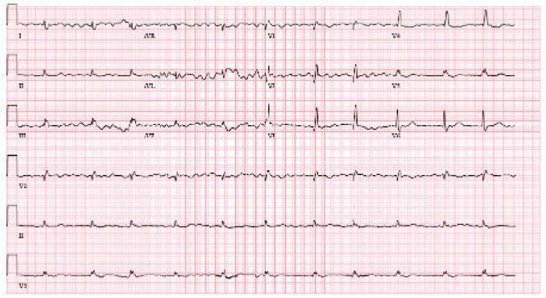
demonstrating atrial fibrillation with controlled
ventricular response and low-voltage QRS.
Case 8
A 74-year-old male with history of bilateral CTS presented with right-sided chest pain associated with shortness of breath, diaphoresis, dizziness, and worsening abdominal pain. He was found to have inferior wall myocardial infarction on ECG with later percutaneous coronary intervention to the left circumflex. His hospital course was complicated by decompensated HF (EF 45% to 50%) and AF with rapid ventricular response. He was treated and discharged with follow-up visits scheduled in the cardiology clinic. Multiple attempts were made to place him on GDMT; however, the patient was unable to tolerate these medications due to recurrent admissions for syncope. During a cardiology clinic visit 3 years after his initial presentation, an amyloidosis workup was initiated. 99mTC pyrophosphate imaging was positive for ATTR-CM, and he started taking tafamidis. Before this diagnosis, ECG indicated low-voltage QRS complexes. His progression has since been complicated by admissions for decompensated HF, recurrent episodes of AF requiring atrioventricular node ablation, and biventricular implantable cardioverter-defibrillator implantation after failed attempts at electrical cardioversion. He continued to follow up in the HF clinic.
Discussion
ATTR-CM is an underdiagnosed cause of cardiomyopathy, particularly in older adults. TTR is a transport protein produced in the liver, and misfolding can occur due to age-related instability of the wild-type protein (wtATTR) or pathologic variants in the TTR (hATTR). The misfolding leads to restrictive physiology, HF (often with preserved EF) arrhythmias, and conduction system disease.1 It is likely that the predominantly older male veteran population would be predisposed to wtATTR cardiomyopathy given that misfolding in the condition is believed to be age-related.
Current criteria for ATTR diagnosis require a combination of clinical suspicion, imaging, laboratory testing, and, in some cases, tissue biopsy confirmation and genetic testing.1,4,5 The diagnostic algorithm is as follows:
Clinical suspicion. Consider ATTR in patients with unexplained HFpEF, LV wall thickness ≥ 12 to 14 mm, discordance between ECG voltage and wall thickness, or associated extracardiac manifestations such as CTS, lumbar spinal stenosis, or peripheral/ autonomic neuropathy.
Exclusion of AL amyloidosis. Bone scintigraphy alone cannot distinguish between AL amyloidosis and ATTR, so patients with suspected amyloid cardiomyopathy should undergo serum and urine immunofixation electrophoresis and serum free light chain assay to rule out a monoclonal gammopathy as seen in AL amyloidosis.
Cardiac scintigraphy. Once AL amyloidosis is excluded, a positive 99mTC-labeled boneavid tracer image (such as a pyrophosphate scan) with grade 2 or grade 3 myocardial uptake is diagnostic of ATTR.
Tissue biopsy. If there is monoclonal gammopathy or equivocal imaging, tissue biopsy (eg, endomyocardial) with Congo red staining and amyloid typing by mass spectrometry or immunohistochemistry is necessary.
Genetic testing. Once ATTR is confirmed, genetic testing distinguishes hATTR from wtATTR, which impacts management and determines the need to screen family members.
Currently, there are 3 therapies approved by the US Food and Drug Administration (FDA) to treat ATTR-CM: tafamidis, acoramidis, and vutrisiran.6-8 Tafamidis and acoramidis stabilize the TTR tetramer, preventing amyloid formation. Vutrisiran uses RNA interference to silence the gene that produces TTR. Tafamidis has been found to improve cardiovascular outcomes in ATTR-CM. In the ATTR-ACT trial, it reduced all-cause mortality and cardiovascular hospitalizations in patients with ATTR-CM and New York Heart Association class I-III symptoms.6 There are other disease-modifying therapies, such the TTR gene silencers inotersen and patisiran; however, these are only FDA-approved for hATTR polyneuropathy and not for ATTRCM; ongoing trials are evaluating their cardiac efficacy.
The mean age of ATTR-CM diagnosis in the patients described in this case series was 79 years, which is older than the mean age of 74 years reported in prior research.4 All patients were male, and the most common presenting symptom was dyspnea on exertion. Table 1 outlines baseline characteristics and associated comorbidities of patients in this case series. The patients presented with many red-flag signs of ATTR-CM (Figure 3). Among them included syncope (4 of 8 patients), spinal stenosis (2 of 8 patients), arrhythmia (7 of 8 patients), heart failure (7 of 8 patients), and bilateral CTS (all patients).

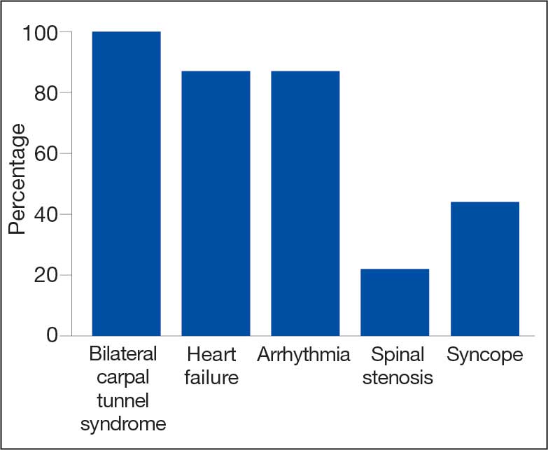
amyloidosis red-flag signs identified.
Bilateral CTS was diagnosed in all patients before their diagnosis of ATTR-CM. Patient 7 had a diagnosis of CTS 9 years before his ATTR-CM diagnosis, underscoring a subtle, yet important extracardiac sign that may increase clinical suspicion for ATTR-CM. Previous research found that the probability of having CTS is highest 5 to 9 years prior to the development of cardiomyopathy. Its presence is also a prognostic marker in ATTR, independent of cardiac involvement.9 The median interval between CTS diagnosis and cardiomyopathy diagnosis was 5 years in this series.
Although screening criteria for ATTR have been proposed, none have been incorporated into formal guidelines.10,11 We propose that a baseline ECG and screening TTE be obtained in any patient aged > 65 years with cardiac risk equivalents such as hypertension and diabetes, presenting with ≥ 1 extracardiac red-flag signs, such as bilateral CTS and spinal stenosis. This will likely facilitate an earlier diagnosis of ATTR-CM, leading to earlier treatment initiation and better patient outcomes. This initiative can be started in the primary care setting and facilitates early cardiology referral. This recommendation is based on literature supporting clinical patterns and observations the authors have made in clinical practice.
Obstructive sleep apnea (OSA) was a comorbidity present in 50% of the patients described in this case series. The literature describing this association is sparse; however, a prospective observational study reported that disorders of sleep inclusive of OSA are frequent in patients with cardiac amyloidosis.12 A theory behind this association is that amyloid deposits in the upper airway tissues lead to airway narrowing. 13 More research is needed to further assess the relationship between OSA and cardiac amyloidosis, particularly with respect to the timing of OSA prior to the development of cardiomyopathy as it may be a potential early sign for clinicians to acknowledge.
An important observation from this case series is the variability in the timing of tafamidis initiation relative to symptom onset and confirmed diagnosis of ATTR-CM (Table 2). Although tafamidis has been found to slow disease progression, several patients in this series began treatment at advanced stages or years after the onset of cardiac symptoms, potentially limiting its clinical benefit.
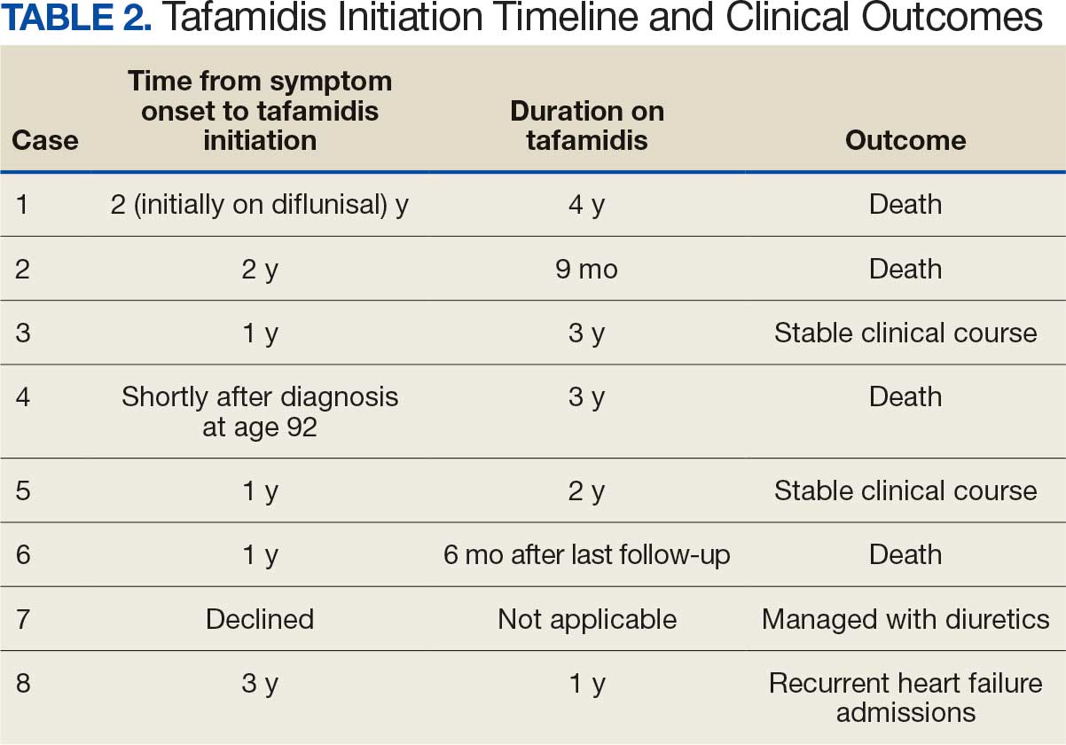
For example, patient 1 was diagnosed with ATTR 2 years before tafamidis became available on the market and was initially treated with diflunisal. Patients who started tafamidis earlier in the disease course (eg, patients 3 and 5) appeared to have better long-term outcomes, including an absence of heart failure hospitalizations after initiation. In contrast, patients with delayed treatment initiation (eg, patients 2, 6, and 8) experienced ongoing decompensations or early mortality. Patient 7 declined tafamidis, underscoring challenges in medication uptake among older adults. Randomized controlled trials are warranted to compare the effectiveness of tafamidis with other recently FDA-approved therapies, such as acoramidis and vutrisiran, particularly in terms of cardiovascular outcomes.
AF was the most common arrhythmia observed in these patients and may occur years before the development of HF symptoms in the setting of ATTR-CM. Due to inherent conduction system disease, AF in this population may have a controlled or slow ventricular response, as observed in patient 4. Patients with atrial fibrillation and cardiac amyloidosis should receive anticoagulation regardless of CHA2DS2- VASc score due to their high risk of intracardiac thrombus formation.4
Limitations
This case series lacked genetic testing following a confirmed ATTR-CM diagnosis. Although much of the treatment is the same regardless of the presence of a TTR mutation, knowing the specific subtype of ATTR-CM has implications for prognosis and for screening family members.
Conclusions
Following analysis of 8 patients diagnosed with ATTR, this case series could serve as a blueprint for research into ATTR in veterans. In clinical practice, following military service, veterans may not be routinely seen by an outpatient physician and may present with sequelae of advanced stages of ATTR. Early identification of red-flag symptoms can lead to a higher clinical suspicion, prompting early diagnostic evaluation and treatment initiation and ultimately mitigating adverse outcomes. Future research that includes genetic testing for those with confirmed ATTR-CM may prove useful as a foundation for detailed and informed discussions with patients and their families regarding prognosis and, if indicated, screening for family members.
- Kittleson MM, Maurer MS, Ambardekar AV, et al. Cardiac amyloidosis: evolving diagnosis and management: a scientific statement from the American Heart Association. Circulation. 2020;142:e7-e22. doi:10.1161/CIR.0000000000000792
- Dharmarajan K, Maurer MS. Transthyretin cardiac amyloidoses in older North Americans. J Am Geriatr Soc. 2012;60:765-774. doi:10.1111/j.1532-5415.2011.03868.x
- Nativi-Nicolau J, Redd A, Kelly N, et al. Increasing number of amyloidosis diagnosis in the Veterans Affairs populations. J Card Fail. 2019;25:S96.
- Ruberg FL, Grogan M, Hanna M, et al. Transthyretin amyloid cardiomyopathy: JACC state-of-the-art review. J Am Coll Cardiol. 2019;73:2872-2891. doi:10.1016/j.jacc.2019.04.003
- Gillmore JD, Maurer, Falk RH, et al. Nonbiopsy diagnosis of cardiac transthyretin amyloidosis. Circulation. 2016;133:2404-2412. doi:10.1161/CIRCULATIONAHA.116.021612
- Maurer MS, Schwartz JH, Gundapaneni B, et al. Tafamidis treatment for patients with transthyretin amyloid cardiomyopathy. N Engl J Med. 2018;379:1007-1016. doi:10.1056/NEJMoa1805689
- Gillmore JD, Judge DP, Cappelli F, et al. Efficacy and safety of acoramidis in transthyretin amyloid cardiomyopathy. N Engl J Med. 2024;390:132-142. doi:10.1056/NEJMoa2305434
- Fontana M, Berk JL, Gillmore JD, et al. Vutrisiran in patients with transthyretin amyloidosis with cardiomyopathy. N Engl J Med. 2025;392:33-44. doi:10.1056/NEJMoa2409134
- Milandri A, Farioli A, Gagliardi C, et al. Carpal tunnel syndrome in cardiac amyloidosis: implications for early diagnosis and prognostic role across the spectrum of aetiologies. Eur J Heart Fail. 2020;22:507-515. doi:10.1002/ejhf.1742
- Garcia-Pavia P, Rapezzi C, Adler Y, et al. Diagnosis and treatment of cardiac amyloidosis: a position statement of the ESC Working Group on Myocardial and Pericardial Diseases. Eur Heart J. 2021;42:1554-1568. doi:10.1093/eurheartj/ehab072
- Brito D, Albrecht FC, de Arenaza DP, et al. World Heart Federation consensus on transthyretin amyloidosis cardiomyopathy (ATTR-CM). Glob Heart. 2023;18:59. doi:10.5334/gh.1262
- Bodez D, Guellich A, Kharoubi M, et al. Prevalence, severity, and prognostic value of sleep apnea syndromes in cardiac amyloidosis. Sleep. 2016;39:1333-1341. doi:10.5665/sleep.5958
- Colaco B, Colaco C, Lipford M. A forgotten problem: sleep-disordered breathing in amyloidosis. Chest. 2016;150:1293A. doi:10.1016/j.chest.2016.08.1407
Transthyretin amyloid cardiomyopathy (ATTR-CM) is caused by the misfolding of the TTR protein, which results in aggregation of amyloid fibrils that deposit in the myocardium and causes restrictive cardiomyopathy. Though it remains underdiagnosed, ATTR-CM is increasingly being recognized as a cause of heart failure in geriatric patients.1 There are 2 categories of ATTRCM: wild-type ATTR-CM (wtATTR-CM), in which there is no mutation in the TTR gene, and hereditary ATTR-CM (hATTR-CM), in which a mutation is present in the TTR gene. Research has shown that wtATTR-CM accounted for as many as 30% of cases of heart failure (HF) with preserved ejection fraction (HFpEF) in patients aged > 75 years.2 A significant percentage of the veteran patient population consists of older males. Given their age, these patients are at greater risk for ATTR diagnosis.3
Identifying red flags for patients within this population may allow clinicians to make earlier diagnoses and improve outcomes. A high index of suspicion is needed to diagnose ATTR because many early signs and symptoms are extracardiac, which leads to delayed diagnoses and worse outcomes. This article describes 8 cases of ATTR-CM within the US Department of Veterans Affairs (VA) New York Harbor Healthcare System-Brooklyn (VANYHHSB).
Methods
This retrospective case series was reviewed and approved by the VANYHHSB Institutional Review Board where it was conducted. Patients diagnosed with ATTR between 2017 and 2024 were identified using International Classification of Diseases, Tenth Revision codes. Eleven patients were identified; 3 were excluded due to insufficient medical records. The remaining 8 patient records were retrospectively reviewed and included.
Case 1
A 67-year-old male with a history of carpal tunnel syndrome (CTS) presented following a syncopal episode. Initial electrocardiogram (ECG) showed sinus rhythm, first-degree atrioventricular block, and a bifascicular block. Transthoracic echocardiogram (TTE) showed moderate asymmetric left ventricular (LV) hypertrophy (LVH) and biatrial enlargement with an ejection fraction (EF) > 55%. The patient was discharged with a loop recorder and an outpatient follow-up appointment scheduled. One month later, he presented with worsening dyspnea on exertion with clinical signs of hypervolemia. A repeat TTE showed global LV wall thickening, moderately reduced LV systolic function (EF 40%), and moderate pulmonary hypertension. Given these findings, the patient underwent cardiac magnetic resonance imaging (CMR), which suggested an infiltrative cardiomyopathy. Amyloid light-chain (AL) amyloidosis evaluation, technetium-99m (99mTC) pyrophosphate imaging, and a fat pad biopsy were unrevealing. An endomyocardial biopsy was performed with electron microscopy, which confirmed amyloidosis. Genetic testing was negative, and the patient began taking tafamidis. There were no later admissions for decompensated HF; however, the patient developed atrial fibrillation (AF) and an interval TTE demonstrated no improvement in his EF. He died at age 73 years.
Case 2
A 78-year-old male with a history of CTS presented with lightheadedness. Initial ECG showed rate-controlled AF and TTE revealed a moderately thickened LV wall with normal LV size, mild left atrial enlargement, and an EF of 65%. The patient was discharged with a scheduled outpatient CMR appointment; however, he defaulted from follow-up. Two years later, he presented with recurrent syncopal episodes and physical examination was consistent with hypervolemia. Repeat TTE revealed moderate LVH, biatrial enlargement, and an EF of 55%. An inpatient CMR was suggestive of cardiac amyloidosis, and a pyrophosphate scan was diagnostic for ATTR. The patient started taking tafamidis, but continued to have recurrent admissions for HF exacerbation. He died at age 81 years.
Case 3
A 71-year-old male with a history of CTS presented with exertional dyspnea. The initial ECG showed sinus rhythm with left atrial enlargement and left axis deviation. Subsequent TTE revealed severe LVH, mildly reduced LV cavity size, moderate to severe biatrial enlargement, and an EF of 25% to 30%. Outpatient 99mTC pyrophosphate imaging suggested cardiac amyloidosis, and laboratory testing showed no evidence of monoclonal proteins. The patient was started on tafamidis for ATTR. At 2-year follow-up, he had new AF and to date has had no further hospitalizations for acute decompensated HF.
Case 4
A 92-year-old male with a history of AF and bilateral CTS presented with lightheadedness. An ECG revealed AF with a slowed ventricular response. Subsequent Holter monitoring demonstrated pauses exceeding 3 seconds, and a permanent pacemaker was recommended. During his preoperative evaluation, TTE revealed severe concentric LVH with a speckled appearance of the myocardium, mild-tomoderate biatrial enlargement, and an EF of 50% to 55%. 99mTC pyrophosphate imaging was positive for amyloidosis and the patient started taking tafamidis. Recurrent hospital admissions for decompensated HF complicated his progression. The patient died at age 95 years.
Case 5
A 72-year-old male with a history of bilateral CTS and cervical spinal stenosis presented with dyspnea on exertion. An ECG revealed a normal sinus rhythm. A TTE found severely reduced systolic function with an EF ≤ 25%, mild concentric LVH, grade 3 diastolic dysfunction, mild-tomoderate biatrial enlargement, and moderate pulmonary hypertension (Figure 1). He was started on guideline-directed medical therapy (GDMT) for HF, which included sacubitril/valsartan, metoprolol succinate, and empagliflozin. The patient’s dyspnea improved, and a workup for nonischemic cardiomyopathy was initiated. 99mTC pyrophosphate imaging 1 year after his initial presentation was positive, leading to ATTR-CM diagnosis. The patient started taking tafamidis and he has since had a stable progression and continued to demonstrate good exercise tolerance with no hospitalizations. His most recent TTE indicated an EF of 40% to 45%.
(parasternal long axis view) of patient 5
demonstrating concentric left ventricular
hypertrophy with a speckled myocardium
and dilated left atrium.
Case 6
A 76-year-old male with a history of paroxysmal AF status after multiple ablations, bilateral CTS, and severe cervical spinal stenosis presented with dyspnea on exertion. The patient’s ECG showed normal sinus rhythm and left axis deviation with concern for left anterior hemiblock. A TTE revealed moderate LVH with a speckled appearance of the myocardium and grade 3 diastolic dysfunction with preserved EF. Before completing the workup for underlying cardiomyopathy, the patient underwent an interventional radiology procedure for an angiomyolipoma, and his postoperative course was complicated by pulmonary edema, requiring admission to the coronary care unit for diuresis. A repeat TTE revealed a reduced EF of 35%, and he was discharged on GDMT. No monoclonal protein was seen in the serum or urine. The patient’s progression was complicated by recurrent admissions for acute decompensated HF and supraventricular tachycardia despite being on amiodarone, which led to a delay in obtaining 99mTC pyrophosphate imaging. Due to hypotension, the patient was unable to tolerate GDMT. He eventually underwent 99mTC pyrophosphate imaging that confirmed ATTR-CM and was started on tafamidis. One year following initial presentation, the patient’s EF progressively declined to 20% to 25%, and he died shortly after discharge to subacute rehabilitation.
Case 7
A 95-year-old male with a history of longstanding persistent AF and bilateral CTS presented with dyspnea on exertion, bendopnea, and worsening bilateral pedal edema for a week. An ECG showed AF with a controlled ventricular response and low-voltage QRS waves (Figure 2). A TTE showed biatrial enlargement, LVH, and preserved EF > 55%. He started taking furosemide and was discharged with a diagnosis of HFpEF. The patient missed cardiology follow-up and presented 1 year later with decompensated HF. An amyloidosis workup was recommended, but due to intermittent periods of being lost to follow-up, the patient did not pursue this workup until 3 years after his initial presentation when 99mTC pyrophosphate imaging confirmed ATTR-CM. The patient declined tafamidis and continued to be followed by the cardiology team. His HF is managed with furosemide as needed due to intolerance to GDMT.
demonstrating atrial fibrillation with controlled
ventricular response and low-voltage QRS.
Case 8
A 74-year-old male with history of bilateral CTS presented with right-sided chest pain associated with shortness of breath, diaphoresis, dizziness, and worsening abdominal pain. He was found to have inferior wall myocardial infarction on ECG with later percutaneous coronary intervention to the left circumflex. His hospital course was complicated by decompensated HF (EF 45% to 50%) and AF with rapid ventricular response. He was treated and discharged with follow-up visits scheduled in the cardiology clinic. Multiple attempts were made to place him on GDMT; however, the patient was unable to tolerate these medications due to recurrent admissions for syncope. During a cardiology clinic visit 3 years after his initial presentation, an amyloidosis workup was initiated. 99mTC pyrophosphate imaging was positive for ATTR-CM, and he started taking tafamidis. Before this diagnosis, ECG indicated low-voltage QRS complexes. His progression has since been complicated by admissions for decompensated HF, recurrent episodes of AF requiring atrioventricular node ablation, and biventricular implantable cardioverter-defibrillator implantation after failed attempts at electrical cardioversion. He continued to follow up in the HF clinic.
Discussion
ATTR-CM is an underdiagnosed cause of cardiomyopathy, particularly in older adults. TTR is a transport protein produced in the liver, and misfolding can occur due to age-related instability of the wild-type protein (wtATTR) or pathologic variants in the TTR (hATTR). The misfolding leads to restrictive physiology, HF (often with preserved EF) arrhythmias, and conduction system disease.1 It is likely that the predominantly older male veteran population would be predisposed to wtATTR cardiomyopathy given that misfolding in the condition is believed to be age-related.
Current criteria for ATTR diagnosis require a combination of clinical suspicion, imaging, laboratory testing, and, in some cases, tissue biopsy confirmation and genetic testing.1,4,5 The diagnostic algorithm is as follows:
Clinical suspicion. Consider ATTR in patients with unexplained HFpEF, LV wall thickness ≥ 12 to 14 mm, discordance between ECG voltage and wall thickness, or associated extracardiac manifestations such as CTS, lumbar spinal stenosis, or peripheral/ autonomic neuropathy.
Exclusion of AL amyloidosis. Bone scintigraphy alone cannot distinguish between AL amyloidosis and ATTR, so patients with suspected amyloid cardiomyopathy should undergo serum and urine immunofixation electrophoresis and serum free light chain assay to rule out a monoclonal gammopathy as seen in AL amyloidosis.
Cardiac scintigraphy. Once AL amyloidosis is excluded, a positive 99mTC-labeled boneavid tracer image (such as a pyrophosphate scan) with grade 2 or grade 3 myocardial uptake is diagnostic of ATTR.
Tissue biopsy. If there is monoclonal gammopathy or equivocal imaging, tissue biopsy (eg, endomyocardial) with Congo red staining and amyloid typing by mass spectrometry or immunohistochemistry is necessary.
Genetic testing. Once ATTR is confirmed, genetic testing distinguishes hATTR from wtATTR, which impacts management and determines the need to screen family members.
Currently, there are 3 therapies approved by the US Food and Drug Administration (FDA) to treat ATTR-CM: tafamidis, acoramidis, and vutrisiran.6-8 Tafamidis and acoramidis stabilize the TTR tetramer, preventing amyloid formation. Vutrisiran uses RNA interference to silence the gene that produces TTR. Tafamidis has been found to improve cardiovascular outcomes in ATTR-CM. In the ATTR-ACT trial, it reduced all-cause mortality and cardiovascular hospitalizations in patients with ATTR-CM and New York Heart Association class I-III symptoms.6 There are other disease-modifying therapies, such the TTR gene silencers inotersen and patisiran; however, these are only FDA-approved for hATTR polyneuropathy and not for ATTRCM; ongoing trials are evaluating their cardiac efficacy.
The mean age of ATTR-CM diagnosis in the patients described in this case series was 79 years, which is older than the mean age of 74 years reported in prior research.4 All patients were male, and the most common presenting symptom was dyspnea on exertion. Table 1 outlines baseline characteristics and associated comorbidities of patients in this case series. The patients presented with many red-flag signs of ATTR-CM (Figure 3). Among them included syncope (4 of 8 patients), spinal stenosis (2 of 8 patients), arrhythmia (7 of 8 patients), heart failure (7 of 8 patients), and bilateral CTS (all patients).
amyloidosis red-flag signs identified.
Bilateral CTS was diagnosed in all patients before their diagnosis of ATTR-CM. Patient 7 had a diagnosis of CTS 9 years before his ATTR-CM diagnosis, underscoring a subtle, yet important extracardiac sign that may increase clinical suspicion for ATTR-CM. Previous research found that the probability of having CTS is highest 5 to 9 years prior to the development of cardiomyopathy. Its presence is also a prognostic marker in ATTR, independent of cardiac involvement.9 The median interval between CTS diagnosis and cardiomyopathy diagnosis was 5 years in this series.
Although screening criteria for ATTR have been proposed, none have been incorporated into formal guidelines.10,11 We propose that a baseline ECG and screening TTE be obtained in any patient aged > 65 years with cardiac risk equivalents such as hypertension and diabetes, presenting with ≥ 1 extracardiac red-flag signs, such as bilateral CTS and spinal stenosis. This will likely facilitate an earlier diagnosis of ATTR-CM, leading to earlier treatment initiation and better patient outcomes. This initiative can be started in the primary care setting and facilitates early cardiology referral. This recommendation is based on literature supporting clinical patterns and observations the authors have made in clinical practice.
Obstructive sleep apnea (OSA) was a comorbidity present in 50% of the patients described in this case series. The literature describing this association is sparse; however, a prospective observational study reported that disorders of sleep inclusive of OSA are frequent in patients with cardiac amyloidosis.12 A theory behind this association is that amyloid deposits in the upper airway tissues lead to airway narrowing. 13 More research is needed to further assess the relationship between OSA and cardiac amyloidosis, particularly with respect to the timing of OSA prior to the development of cardiomyopathy as it may be a potential early sign for clinicians to acknowledge.
An important observation from this case series is the variability in the timing of tafamidis initiation relative to symptom onset and confirmed diagnosis of ATTR-CM (Table 2). Although tafamidis has been found to slow disease progression, several patients in this series began treatment at advanced stages or years after the onset of cardiac symptoms, potentially limiting its clinical benefit.
For example, patient 1 was diagnosed with ATTR 2 years before tafamidis became available on the market and was initially treated with diflunisal. Patients who started tafamidis earlier in the disease course (eg, patients 3 and 5) appeared to have better long-term outcomes, including an absence of heart failure hospitalizations after initiation. In contrast, patients with delayed treatment initiation (eg, patients 2, 6, and 8) experienced ongoing decompensations or early mortality. Patient 7 declined tafamidis, underscoring challenges in medication uptake among older adults. Randomized controlled trials are warranted to compare the effectiveness of tafamidis with other recently FDA-approved therapies, such as acoramidis and vutrisiran, particularly in terms of cardiovascular outcomes.
AF was the most common arrhythmia observed in these patients and may occur years before the development of HF symptoms in the setting of ATTR-CM. Due to inherent conduction system disease, AF in this population may have a controlled or slow ventricular response, as observed in patient 4. Patients with atrial fibrillation and cardiac amyloidosis should receive anticoagulation regardless of CHA2DS2- VASc score due to their high risk of intracardiac thrombus formation.4
Limitations
This case series lacked genetic testing following a confirmed ATTR-CM diagnosis. Although much of the treatment is the same regardless of the presence of a TTR mutation, knowing the specific subtype of ATTR-CM has implications for prognosis and for screening family members.
Conclusions
Following analysis of 8 patients diagnosed with ATTR, this case series could serve as a blueprint for research into ATTR in veterans. In clinical practice, following military service, veterans may not be routinely seen by an outpatient physician and may present with sequelae of advanced stages of ATTR. Early identification of red-flag symptoms can lead to a higher clinical suspicion, prompting early diagnostic evaluation and treatment initiation and ultimately mitigating adverse outcomes. Future research that includes genetic testing for those with confirmed ATTR-CM may prove useful as a foundation for detailed and informed discussions with patients and their families regarding prognosis and, if indicated, screening for family members.
- Kittleson MM, Maurer MS, Ambardekar AV, et al. Cardiac amyloidosis: evolving diagnosis and management: a scientific statement from the American Heart Association. Circulation. 2020;142:e7-e22. doi:10.1161/CIR.0000000000000792
- Dharmarajan K, Maurer MS. Transthyretin cardiac amyloidoses in older North Americans. J Am Geriatr Soc. 2012;60:765-774. doi:10.1111/j.1532-5415.2011.03868.x
- Nativi-Nicolau J, Redd A, Kelly N, et al. Increasing number of amyloidosis diagnosis in the Veterans Affairs populations. J Card Fail. 2019;25:S96.
- Ruberg FL, Grogan M, Hanna M, et al. Transthyretin amyloid cardiomyopathy: JACC state-of-the-art review. J Am Coll Cardiol. 2019;73:2872-2891. doi:10.1016/j.jacc.2019.04.003
- Gillmore JD, Maurer, Falk RH, et al. Nonbiopsy diagnosis of cardiac transthyretin amyloidosis. Circulation. 2016;133:2404-2412. doi:10.1161/CIRCULATIONAHA.116.021612
- Maurer MS, Schwartz JH, Gundapaneni B, et al. Tafamidis treatment for patients with transthyretin amyloid cardiomyopathy. N Engl J Med. 2018;379:1007-1016. doi:10.1056/NEJMoa1805689
- Gillmore JD, Judge DP, Cappelli F, et al. Efficacy and safety of acoramidis in transthyretin amyloid cardiomyopathy. N Engl J Med. 2024;390:132-142. doi:10.1056/NEJMoa2305434
- Fontana M, Berk JL, Gillmore JD, et al. Vutrisiran in patients with transthyretin amyloidosis with cardiomyopathy. N Engl J Med. 2025;392:33-44. doi:10.1056/NEJMoa2409134
- Milandri A, Farioli A, Gagliardi C, et al. Carpal tunnel syndrome in cardiac amyloidosis: implications for early diagnosis and prognostic role across the spectrum of aetiologies. Eur J Heart Fail. 2020;22:507-515. doi:10.1002/ejhf.1742
- Garcia-Pavia P, Rapezzi C, Adler Y, et al. Diagnosis and treatment of cardiac amyloidosis: a position statement of the ESC Working Group on Myocardial and Pericardial Diseases. Eur Heart J. 2021;42:1554-1568. doi:10.1093/eurheartj/ehab072
- Brito D, Albrecht FC, de Arenaza DP, et al. World Heart Federation consensus on transthyretin amyloidosis cardiomyopathy (ATTR-CM). Glob Heart. 2023;18:59. doi:10.5334/gh.1262
- Bodez D, Guellich A, Kharoubi M, et al. Prevalence, severity, and prognostic value of sleep apnea syndromes in cardiac amyloidosis. Sleep. 2016;39:1333-1341. doi:10.5665/sleep.5958
- Colaco B, Colaco C, Lipford M. A forgotten problem: sleep-disordered breathing in amyloidosis. Chest. 2016;150:1293A. doi:10.1016/j.chest.2016.08.1407
Case Series of Patients With Cardiac Amyloidosis at VA New York Harbor Healthcare-Brooklyn
Case Series of Patients With Cardiac Amyloidosis at VA New York Harbor Healthcare-Brooklyn
Atopic Dermatitis: New Insights and Expanded Treatment Options
Atopic Dermatitis: New Insights and Expanded Treatment Options
Atopic dermatitis (AD) is a chronic skin condition generally characterized by pruritic and erythematous papules and plaques.1 While AD commonly manifests in childhood, 1 in 4 patients living with AD report adult onset of the disease.2 The clinical presentation and prevalence of AD vary across age groups, skin tones, and racial and ethnic groups. Globally, AD is estimated to have a prevalence of 2.6%; however, rates vary widely by region.1 Morphology and distribution of AD lesions also vary by population; therefore, defining one classic presentation of AD is not sufficient in diverse patient populations.3
Epidemiology
The prevalence of AD ranges from 0.2% to 24.6% worldwide, with higher rates in Africa and Oceania and lower rates in India and Northern and Eastern Europe.1 In the United States, AD affects all racial and ethnic groups; however, prevalence and severity are increased in Black children compared with White children.4 In one prospective cohort study, Hispanic children and non-Hispanic Black children aged 3 years and younger had greater odds of AD persisting into mid childhood (approximately age 7 years) compared with non-Hispanic White children.5,6
Key Clinical Features
Clinical features of AD are heterogeneous and may include differences in color, morphology, and distribution. Brown, hyperpigmented, gray, and/or violaceous plaques may predominate in patients with skin of color (SOC) compared with the erythematous plaques commonly described in lighter skin tones.1,3 Established scoring systems for AD rely on erythema as a key diagnostic feature, but because erythema can be difficult to detect in darker skin tones, disease severity may be underestimated and diagnosis may be delayed in this population.4
Atopic dermatitis in SOC may manifest as lichenoid plaques,7 prurigo nodules,7,8 lichenification,1 and follicular accentuation.9 Lichen planus–like AD is a distinct variant characterized by lichenoid plaques with a predilection for the extensor surfaces and face in patients with darker skin tones1,8 occurring in approximately 9% of patients in one study.10
Other key clinical features of AD in patients with SOC include pityriasis alba,10 increased risk for postinflammatory pigment alteration (including hyperpigmentation and/or hypopigmentation),1 and greater trunk and extensor involvement.1,11
Worth Noting
The scientific landscape for AD has grown rapidly, increasing our understanding of its pathophysiology, treatment, and social impact. Nonsteroidal treatments available for pediatric and adult patients with AD have increased in recent years, including crisaborole (approved for use in those ages ≥ 3 months), tacrolimus (≥ 2 years), and pimecrolimus (≥ 2 years). Injectable options include dupilumab (≥ 6 months), lebrikizumab (≥ 12 years), nemolizumab (≥ 12 years), and tralokinumab (≥ 12 years). Oral options include abrocitinib (≥ 12 years) and upadacitinib (≥ 12 years).12 Topical options include roflumilast 0.15% cream (≥ 6 years)12 and 0.05% cream (≥ 2-5 years),13 ruxolitinib 1.5% cream (≥ 2 years),14 and tapinarof 1% cream (≥ 2 years).12
For some patients, postinflammatory pigment alteration associated with AD has a higher impact on quality of life than the AD itself.7 In a study of 260 US adults with AD, the emotional impact of pigmentary changes was greatest in Black patients, with 53.3% reporting that pigment changes bothered them “a lot” or “very much.”15
Genome-wide association studies have not identified a single determinant that explains racial and ethnic differences in susceptibility to AD.4 Instead, social determinants of health are thought to play a role in the difference in AD prevalence and severity across groups in the United States.16
Health Disparity Highlight
In an analysis of 20 US metropolitan cities, urban and inner-city residence was associated with approximately 1.7-fold increased odds of AD.4 Among pediatric patients with moderate to severe AD, Black children were more likely to be exposed to tobacco smoke17 and traffic-related air pollution.18 Low socioeconomic status and low income also have been associated with moderate16 and severe19 AD. At the same education level, Black individuals in the United States receive less income than their White counterparts and have markedly less wealth at equivalent incomes.20
In utero exposure to maternal stress is associated with AD.4 Increased IgE levels have been recorded in children who develop AD, with Black children having the highest IgE levels overall compared to other children.18
An analysis of medical records from an urban medical center in Baltimore, Maryland, from 2013 through 2018 showed that Black patients with AD were less likely to receive topical corticosteroids, topical calcineurin inhibitors, a topical phosphodiesterase 4 inhibitor, and a biologic compared to White patients with AD.21
Since the disproportionate burden experienced by patients with AD is not physiologic, it is imperative to address these systemic complexities and address the barriers impacting treatment availability to improve health outcomes for all patients living with AD.
- Kaufman BP, Guttman-Yassky E, Alexis AF. Atopic dermatitis in diverse racial and ethnic groups—variations in epidemiology, genetics, clinical presentation and treatment. Exp Dermatol. 2018;27:340-357.
- Lee HH, Patel KR, Singam V, et al. A systematic review and meta-analysis of the prevalence and phenotype of adult-onset atopic dermatitis. J Am Acad Dermatol. 2019;80:1526-1532.E7.
- Adawi W, Cornman H, Kambala A, et al. Diagnosing atopic dermatitis in skin of color. Dermatol Clin. 2023;41:417-429.
- Narla S, Silverberg JI. Current updates in the epidemiology and comorbidities of atopic dermatitis. Ann Allergy Asthma Immunol. 2025;135:511-520.
- Croce EA, Levy ML, Adamson AS, et al. Reframing racial and ethnic disparities in atopic dermatitis in Black and Latinx populations. J Allergy Clin Immunol. 2021;148:1104-1111.
- Kim Y, Blomberg M, Rifas-Shiman SL, et al. Racial/ethnic differences in incidence and persistence of childhood atopic dermatitis. J Invest Dermatol. 2019;139:827-834.
- Nomura T, Wu J, Kabashima K, et al. Endophenotypic variations of atopic dermatitis by age, race, and ethnicity. J Allergy Clin Immunol. 2020;8:1840-1852.
- McColl M, Boozalis E, Aguh C, et al. Pruritus in Black skin: unique molecular characteristics and clinical features. J Natl Med Assoc. 2021;114:30-38.
- Silverberg JI, Margolis DJ, Boguniewicz M, et al. Distribution of atopic dermatitis lesions in United States adults. J Eur Acad Dermatol Venereol. 2019;33:1341-1348.
- Summey BT, Bowen SE, Allen HB. Lichen planus-like atopic dermatitis: expanding the differential diagnosis of spongiotic dermatitis. J Cutan Pathol. 2008;35:311-314.
- Odhiambo JA, Williams HC, Clayton TO, et al; ISAAC Phase Three Study Group. Global variations in prevalence of eczema symptoms in children from ISAAC Phase Three. J Allergy Clin Immunol. 2009;124:1251-1258.E23.
- Gallagher K, Halperin-Goldstein S, Paller AS. New treatments in atopic dermatitis update. Ann Allergy Asthma Immunol. 2025;135:498-510.E10.
- Shaw ML. FDA expands roflumilast use for atopic dermatitis to children aged 2 to 5 years. Am J Managed Care. October 6, 2025. Accessed April 30, 2026. https://www.ajmc.com/view/fda-expands -roflumilast-use-for-atopic-dermatitis-to-children-aged-2-to-5-years
- Eichenfield LF, Stein Gold LF, Simpson EL, et al. Efficacy and safety of ruxolitinib cream in children aged 2 to 11 years with atopic dermatitis: results from TRuE-AD3, a phase 3, randomized double-blind study. J Am Acad of Dermatol. 2025;93:689-698.
- Heath CR, Dosono B, Shi VY, et al. Variability in skin tone changes by race and ethnicity among US adults with atopic dermatitis. Presented at: Skin of Color Update 2024, September 13-15, 2024, New York, NY.
- Tackett KJ, Jenkins F, Morrell DS, et al. Structural racism and its influence on the severity of atopic dermatitis in African American children. Pediatr Dermatol. 2020;37:142-146.
- Narla S, Silverberg JI. The role of environmental exposures in atopic dermatitis. Curr Allergy Asthma Rep. 2020;20:74.
- Bauer SJ, Spoer BR, Ehrman R, et al. A systematic review of historic neighborhood redlining and contemporary health outcomes. Public Health. 2025;238:181-187.
- Chung J, Simpson EL. The socioeconomics of atopic dermatitis. Ann Allergy Asthma Immunol. 2019;122:360-366.
- Martinez A, de la Rosa R, Mujahid M, et al. Structural racism and its pathways to asthma and atopic dermatitis. J Allergy Clin Immunol. 2021;148:1112-1120.
- Bell MA, Whang KA, Thomas J, et al. Racial and ethnic disparities in access to emerging and frontline therapies in common dermatological conditions: a cross-sectional study. J Natl Med Assoc. 2020;112:650-653.
Atopic dermatitis (AD) is a chronic skin condition generally characterized by pruritic and erythematous papules and plaques.1 While AD commonly manifests in childhood, 1 in 4 patients living with AD report adult onset of the disease.2 The clinical presentation and prevalence of AD vary across age groups, skin tones, and racial and ethnic groups. Globally, AD is estimated to have a prevalence of 2.6%; however, rates vary widely by region.1 Morphology and distribution of AD lesions also vary by population; therefore, defining one classic presentation of AD is not sufficient in diverse patient populations.3
Epidemiology
The prevalence of AD ranges from 0.2% to 24.6% worldwide, with higher rates in Africa and Oceania and lower rates in India and Northern and Eastern Europe.1 In the United States, AD affects all racial and ethnic groups; however, prevalence and severity are increased in Black children compared with White children.4 In one prospective cohort study, Hispanic children and non-Hispanic Black children aged 3 years and younger had greater odds of AD persisting into mid childhood (approximately age 7 years) compared with non-Hispanic White children.5,6
Key Clinical Features
Clinical features of AD are heterogeneous and may include differences in color, morphology, and distribution. Brown, hyperpigmented, gray, and/or violaceous plaques may predominate in patients with skin of color (SOC) compared with the erythematous plaques commonly described in lighter skin tones.1,3 Established scoring systems for AD rely on erythema as a key diagnostic feature, but because erythema can be difficult to detect in darker skin tones, disease severity may be underestimated and diagnosis may be delayed in this population.4
Atopic dermatitis in SOC may manifest as lichenoid plaques,7 prurigo nodules,7,8 lichenification,1 and follicular accentuation.9 Lichen planus–like AD is a distinct variant characterized by lichenoid plaques with a predilection for the extensor surfaces and face in patients with darker skin tones1,8 occurring in approximately 9% of patients in one study.10
Other key clinical features of AD in patients with SOC include pityriasis alba,10 increased risk for postinflammatory pigment alteration (including hyperpigmentation and/or hypopigmentation),1 and greater trunk and extensor involvement.1,11
Worth Noting
The scientific landscape for AD has grown rapidly, increasing our understanding of its pathophysiology, treatment, and social impact. Nonsteroidal treatments available for pediatric and adult patients with AD have increased in recent years, including crisaborole (approved for use in those ages ≥ 3 months), tacrolimus (≥ 2 years), and pimecrolimus (≥ 2 years). Injectable options include dupilumab (≥ 6 months), lebrikizumab (≥ 12 years), nemolizumab (≥ 12 years), and tralokinumab (≥ 12 years). Oral options include abrocitinib (≥ 12 years) and upadacitinib (≥ 12 years).12 Topical options include roflumilast 0.15% cream (≥ 6 years)12 and 0.05% cream (≥ 2-5 years),13 ruxolitinib 1.5% cream (≥ 2 years),14 and tapinarof 1% cream (≥ 2 years).12
For some patients, postinflammatory pigment alteration associated with AD has a higher impact on quality of life than the AD itself.7 In a study of 260 US adults with AD, the emotional impact of pigmentary changes was greatest in Black patients, with 53.3% reporting that pigment changes bothered them “a lot” or “very much.”15
Genome-wide association studies have not identified a single determinant that explains racial and ethnic differences in susceptibility to AD.4 Instead, social determinants of health are thought to play a role in the difference in AD prevalence and severity across groups in the United States.16
Health Disparity Highlight
In an analysis of 20 US metropolitan cities, urban and inner-city residence was associated with approximately 1.7-fold increased odds of AD.4 Among pediatric patients with moderate to severe AD, Black children were more likely to be exposed to tobacco smoke17 and traffic-related air pollution.18 Low socioeconomic status and low income also have been associated with moderate16 and severe19 AD. At the same education level, Black individuals in the United States receive less income than their White counterparts and have markedly less wealth at equivalent incomes.20
In utero exposure to maternal stress is associated with AD.4 Increased IgE levels have been recorded in children who develop AD, with Black children having the highest IgE levels overall compared to other children.18
An analysis of medical records from an urban medical center in Baltimore, Maryland, from 2013 through 2018 showed that Black patients with AD were less likely to receive topical corticosteroids, topical calcineurin inhibitors, a topical phosphodiesterase 4 inhibitor, and a biologic compared to White patients with AD.21
Since the disproportionate burden experienced by patients with AD is not physiologic, it is imperative to address these systemic complexities and address the barriers impacting treatment availability to improve health outcomes for all patients living with AD.
- Kaufman BP, Guttman-Yassky E, Alexis AF. Atopic dermatitis in diverse racial and ethnic groups—variations in epidemiology, genetics, clinical presentation and treatment. Exp Dermatol. 2018;27:340-357.
- Lee HH, Patel KR, Singam V, et al. A systematic review and meta-analysis of the prevalence and phenotype of adult-onset atopic dermatitis. J Am Acad Dermatol. 2019;80:1526-1532.E7.
- Adawi W, Cornman H, Kambala A, et al. Diagnosing atopic dermatitis in skin of color. Dermatol Clin. 2023;41:417-429.
- Narla S, Silverberg JI. Current updates in the epidemiology and comorbidities of atopic dermatitis. Ann Allergy Asthma Immunol. 2025;135:511-520.
- Croce EA, Levy ML, Adamson AS, et al. Reframing racial and ethnic disparities in atopic dermatitis in Black and Latinx populations. J Allergy Clin Immunol. 2021;148:1104-1111.
- Kim Y, Blomberg M, Rifas-Shiman SL, et al. Racial/ethnic differences in incidence and persistence of childhood atopic dermatitis. J Invest Dermatol. 2019;139:827-834.
- Nomura T, Wu J, Kabashima K, et al. Endophenotypic variations of atopic dermatitis by age, race, and ethnicity. J Allergy Clin Immunol. 2020;8:1840-1852.
- McColl M, Boozalis E, Aguh C, et al. Pruritus in Black skin: unique molecular characteristics and clinical features. J Natl Med Assoc. 2021;114:30-38.
- Silverberg JI, Margolis DJ, Boguniewicz M, et al. Distribution of atopic dermatitis lesions in United States adults. J Eur Acad Dermatol Venereol. 2019;33:1341-1348.
- Summey BT, Bowen SE, Allen HB. Lichen planus-like atopic dermatitis: expanding the differential diagnosis of spongiotic dermatitis. J Cutan Pathol. 2008;35:311-314.
- Odhiambo JA, Williams HC, Clayton TO, et al; ISAAC Phase Three Study Group. Global variations in prevalence of eczema symptoms in children from ISAAC Phase Three. J Allergy Clin Immunol. 2009;124:1251-1258.E23.
- Gallagher K, Halperin-Goldstein S, Paller AS. New treatments in atopic dermatitis update. Ann Allergy Asthma Immunol. 2025;135:498-510.E10.
- Shaw ML. FDA expands roflumilast use for atopic dermatitis to children aged 2 to 5 years. Am J Managed Care. October 6, 2025. Accessed April 30, 2026. https://www.ajmc.com/view/fda-expands -roflumilast-use-for-atopic-dermatitis-to-children-aged-2-to-5-years
- Eichenfield LF, Stein Gold LF, Simpson EL, et al. Efficacy and safety of ruxolitinib cream in children aged 2 to 11 years with atopic dermatitis: results from TRuE-AD3, a phase 3, randomized double-blind study. J Am Acad of Dermatol. 2025;93:689-698.
- Heath CR, Dosono B, Shi VY, et al. Variability in skin tone changes by race and ethnicity among US adults with atopic dermatitis. Presented at: Skin of Color Update 2024, September 13-15, 2024, New York, NY.
- Tackett KJ, Jenkins F, Morrell DS, et al. Structural racism and its influence on the severity of atopic dermatitis in African American children. Pediatr Dermatol. 2020;37:142-146.
- Narla S, Silverberg JI. The role of environmental exposures in atopic dermatitis. Curr Allergy Asthma Rep. 2020;20:74.
- Bauer SJ, Spoer BR, Ehrman R, et al. A systematic review of historic neighborhood redlining and contemporary health outcomes. Public Health. 2025;238:181-187.
- Chung J, Simpson EL. The socioeconomics of atopic dermatitis. Ann Allergy Asthma Immunol. 2019;122:360-366.
- Martinez A, de la Rosa R, Mujahid M, et al. Structural racism and its pathways to asthma and atopic dermatitis. J Allergy Clin Immunol. 2021;148:1112-1120.
- Bell MA, Whang KA, Thomas J, et al. Racial and ethnic disparities in access to emerging and frontline therapies in common dermatological conditions: a cross-sectional study. J Natl Med Assoc. 2020;112:650-653.
Atopic Dermatitis: New Insights and Expanded Treatment Options
Atopic Dermatitis: New Insights and Expanded Treatment Options
Assessment of False-Positive Fentanyl Results on Urine Drug Screens in Veterans
Assessment of False-Positive Fentanyl Results on Urine Drug Screens in Veterans
A urine drug screen (UDS) is commonly performed to evaluate illicit and prescribed drug use in patients to guide treatment decisions and ensure patient safety. Common uses include evaluating medication adherence, identifying ingested substances in cases of intoxication or overdose, ruling out substance-induced disorders, and screening for illicit drug use. There is a potential for false-positive or false-negative results due to the qualitative and nonspecific nature of UDSs.1 These results can be verified with confirmatory testing using gas chromatography/mass spectrometry or liquid chromatography/ tandem mass spectrometry by identifying specific molecular structures and quantifying the amount of drug or substance present in the sample.1
An April 2023 memorandum instructed all US Department of Veterans Affairs (VA) medical centers and community-based outpatient clinics (CBOC) to have fentanyl urine testing readily available.2 Some facilities added fentanyl to a standard UDS, while others created a separate quick order. The memorandum led to increased fentanyl testing. As a result, unexpected positive fentanyl UDS results are more common. Some facilities have an automatic fentanyl confirmation test that is ordered after a positive fentanyl UDS. However, a positive result for fentanyl on a UDS does not automatically result in confirmation testing at all VA facilities. Without automatic confirmation testing, a clinician must decide to order a fentanyl confirmation test following the positive result. Therefore, the true rate of false-positive results for fentanyl is unknown because confirmation testing is not ordered for every positive UDS.
False-positive results can have unintended consequences, including discontinuation of prescribed medications, patient stigma, and inappropriate recommendations for substance use treatment. False-positive results may contribute to unnecessary health care costs and adversely affect patients’ lives. Previous research has reported false-positive fentanyl UDS results for patients taking risperidone, ziprasidone, and labetalol.3-5 Studies have found that loperamide and high-concentration methamphetamine samples could cause false-positive fentanyl UDS results.6,7 Wang et al evaluated the performance of the SEFRIA fentanyl immunoassay using the 1 ng/mL cutoff cleared by the US Food and Drug Administration (FDA). The study of 410 patients found a 38% false-positive rate; concomitant use of trazodone, labetalol, and haloperidol accounted for 230 (56%) of the false-positive results.8 Limited data evaluating false-positive results for the current SEFRIA fentanyl testing assay suggest the need for additional research. This study aims to add to data on false-positive results for fentanyl on UDS samples and potential causes.
Methods
A retrospective, multicenter observational cohort study was conducted that included patients at 3 VA MidSouth Healthcare Network VA medical centers located in Tennessee with their associated CBOCs from August 1, 2023, to August 1, 2024 who had positive fentanyl UDS results. The primary outcome was the rate of false-positive fentanyl UDS results when confirmation testing was performed. Secondary outcomes included the rate of confirmation testing, prescribed medications used by patients with false-positive UDS results, and the rate of follow-up in the electronic health record (EHR) on results of confirmation testing. Confirmations were primarily obtained for positive results and not all UDSs. Therefore, it was not possible in this retrospective study to obtain the true measure of false-negative or true-negative results.
A structured query language query was performed to identify patients with a UDS positive for fentanyl from August 1, 2023, to August 1, 2024. Patients were enrolled if they were aged ≥ 18 years with a UDS positive for fentanyl. Patients were excluded from the primary outcome analysis if results for the confirmatory testing were unquantifiable or could not be found.
Study Intervention
This was a descriptive study with no comparator group. The rate of confirmed false-positive results for fentanyl, rate of confirmation testing for patients with positive fentanyl UDS results, rate of follow-up on confirmation results, and prescribed medications in patients with false-positive fentanyl results were evaluated. For true-positive results, follow-up was defined as documentation in the EHR reporting fentanyl use or illicit substance use likely to be laced with fentanyl at the time of the UDS or documentation of the confirmation result. For false-positive results, follow-up was defined as documentation in the EHR of the confirmation result.
Statistical Analysis
Descriptive statistics including means and percentages were used to analyze demographic data. Continuous variables and parametric data are presented as mean (SD) and nominal data as percentages. All statistical analyses were completed using Excel. The SEFRIA fentanyl immunoassay was used at each study site. Facilities 1 and 2 were combined for the primary outcome analysis because they used the same fentanyl immunoassay cutoff level of 1 ng/mL. Facility 3 used a cutoff level of 2 ng/mL and was analyzed separately.
Results
A total of 1228 UDS tests were positive for fentanyl, including 618 at facility 1, 308 at facility 2, and 302 at facility 3 (Figure 1). Patients were predominantly male and White, with a mean age of 55 years, though age and race varied by location (Table 1). Patients may have had ≥ 1 UDS. Of 1228 UDSs recorded in the EHR, 578 were sent for confirmation testing and 546 had confirmation results available in the EHR (84 at facility 1, 271 at facility 2, and 191 at facility 3). Of 546 confirmation tests, 186 were negative for fentanyl, indicating a false-positive rate of 34.1%. Most confirmation tests (43%) were requested for patients seen in a mental health clinic.
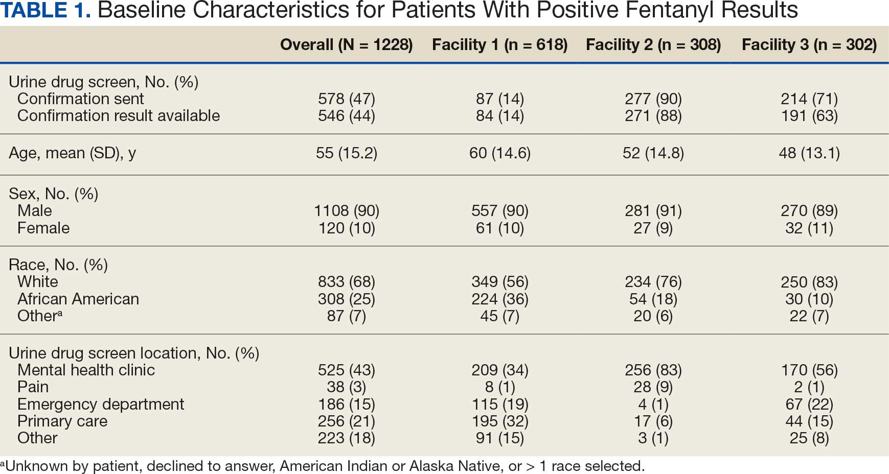
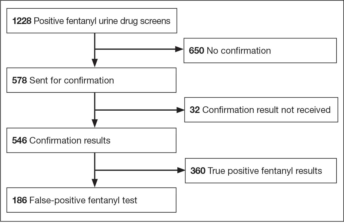
The combined false-positive rate was 49.9% for 355 UDS confirmation results at facilities 1 and 2 (70.2% and 43.5%, respectively) and 4.7% for 191 UDS confirmation results at facility 3, which used the higher 2 ng/mL cutoff level (Figure 2). Confirmation testing was ordered for 578 tests (47.1%). There were 87 confirmation tests (14.1%) at facility 1, 277 tests (89.9%) at facility 2, and 214 (70.9%) at facility 3 (Figure 3). Follow-up after confirmation tests was completed for 406 patients (74.4%): 56 follow-ups (66.7%) at facility 1, 190 follow-ups (70.1%) at facility 2, and 160 follow-ups (83.8%) at facility 3 (Figure 4). Trazodone was the most commonly prescribed medication for patients with false-positive fentanyl UDS results. Trazodone was prescribed to 153 patients (82,3%), followed by 116 patients (62.4%) prescribed naloxone, 86 patients (46.2%) prescribed or with reported use of acetaminophen, 72 patients (38.7%) prescribed nicotine replacement products, and 64 patients (34.4%) prescribed omeprazole (Table 2).
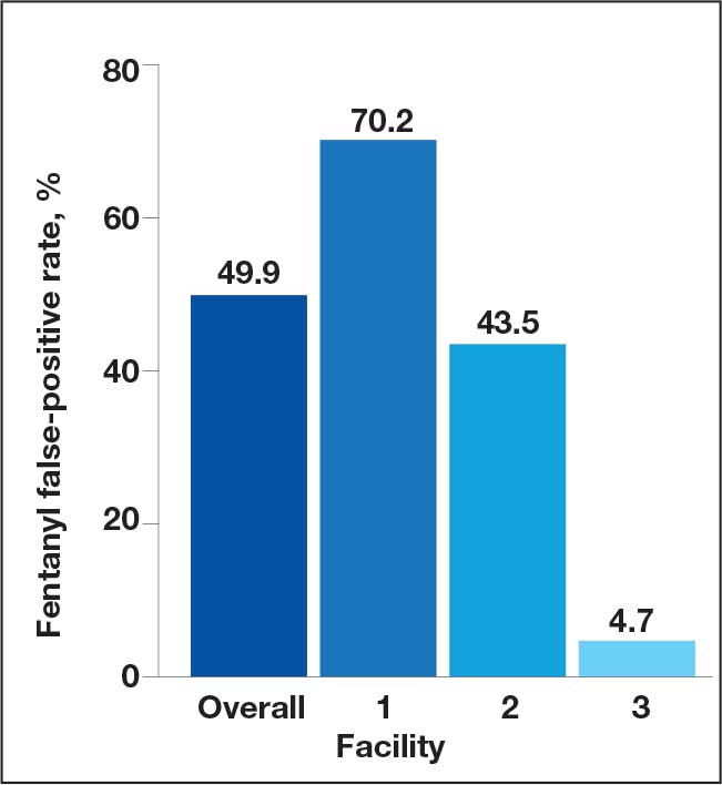
Confirmed Fentanyl False-Positive Rate
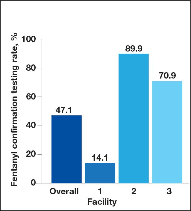
Confirmation Testing
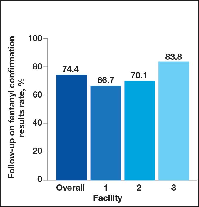
Fentanyl Confirmation Results
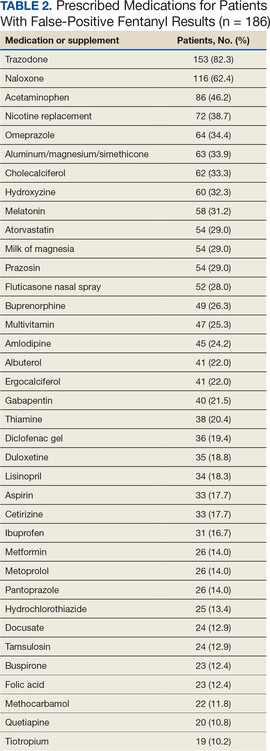
Discussion
There are several factors to note when interpreting the study results. First, facilities 1 and 2 used the FDA-cleared 1 ng/mL cutoff for positive results on the SEFRIA fentanyl immunoassay, whereas facility 3 used a cutoff level of 2 ng/mL. Second, during the study period, facilities 1 and 3 included fentanyl as part of their standard UDS; facility 2 required a separate fentanyl UDS order. Third, facility 2 had automatic confirmation testing for positive results on individually ordered fentanyl UDS tests. Finally, confirmation tests were primarily obtained for positive fentanyl results and not all UDSs, which limited the analyses that could be performed.
This study found a high rate of false-positive fentanyl UDS results at facilities 1 and 2 and a very low rate at facility 3, likely due to the higher cutoff level. Facility 3 used the higher cutoff level due to previously observed high rates of false-positive results. While a higher cutoff level can decrease the rate of false-positive results, it also may increase the rate of false-negative results.
Studies have found false-positive rates ranging from 3% to 45% with the SEFRIA immunoassay FDA-cleared 1 ng/mL cutoff. Increasing the cutoff to 1.3 ng/mL decreased the false-positive rate from 38% to 7.5% in a study by Wang et al.8-11 Manar et al evaluated fentanyl assays in 42 samples using a 2 ng/mL cutoff for the SEFRIA assay and reported a false-positive rate of 0 and a false-negative rate of 22.5%.12 Given the high rate of false-positive rates demonstrated in studies using the current FDA-recommended 1 ng/mL cutoff, additional studies evaluating different cutoff levels may be beneficial to determine the best cutoff level to reduce false-positive results without significantly increasing false-negative rates. While data on the impact of using a higher cutoff level are limited, the results of our study have led to discussions at VA MidSouth Healthcare Network facilities regarding use of different cutoff levels.
There was a low rate of confirmation testing at facility 1 compared with facilities 2 and 3. Only facility 2 had automatic confirmation testing during the study period. Pharmacists at facility 3 reviewed UDS results without needing a consultation and, during the study period, could order fentanyl UDS confirmations. Another factor that may have contributed to the disparity in confirmation testing between facilities is the location of the UDS order. Most UDS samples at facilities 2 and 3 were ordered for patients seen in mental health clinics, whereas many facility 1 orders were placed in primary care or the emergency department (ED).
Given these results, education may be indicated regarding the risk of false-positive results and the importance of confirmation testing in primary care and the ED. Facility 1 and 3 did not have automatic fentanyl confirmation testing during the study; however, facility 3 implemented automatic confirmation shortly after the study period and facility 1 implemented automatic confirmation testing for a positive fentanyl UDS result after evaluation of the study data.
Although follow-up on confirmation UDS results was fairly high, it was highest at facility 3, which does not require a consultation for pharmacist UDS result evaluations. Given the high rate of false-positive results for fentanyl, confirmation testing for a positive UDS and follow-up on confirmation results is an important step to consider. The higher rate of follow-up at the facility where pharmacists had more autonomous involvement shows the benefits of having pharmacists provide comprehensive patient care. Implementing similar protocols across all facilities may improve follow-up, which may improve patient care and safety given the implications of false-positive results.
Trazodone was prescribed in 82.3% of all patients with false-positive fentanyl tests. Even at facility 3, with the higher fentanyl immunoassay cutoff level, trazodone was prescribed in 77.8% of patients with false-positive results. While this retrospective study does not show causation, it does align with the findings reported by Wang et al, adding to the data implicating trazodone as a potential cause for false-positive fentanyl UDS results. The high incidence of trazodone prescriptions in patients with false-positive UDS results at facility 3 strengthens this association, indicating that even when using a higher cutoff level, trazodone may be implicated.
While there was a high rate of confirmed false-positive results in this study, there was also a potential for undetected true-positive results. The SEFRIA fentanyl immunoassay is sensitive to multiple fentanyl analogues. Williams et al showed that the SEFRIA immunoassay detected 57 of 58 fentanyl analogues tested; norsufentanil was the only analogue it did not detect.13 Most of the confirmatory tests reviewed during this study did not include all fentanyl analogues, only fentanyl and norfentanyl. Given the increased prevalence of synthetic fentanyl analogues, this is an important consideration because some identified false-positive results could potentially be undetected true-positive results for a fentanyl analogue. Switching to a more comprehensive confirmation test that includes more fentanyl analogues may reduce the risk of undetected positive results and, therefore, reduce the observed rate of false-positive UDS results.
Strengths and Limitations
Patient medications were only identified if they were documented in the EHR at the time of UDS results, which could have missed over-the-counter medications or medications prescribed outside the VA; this limits identification and implication of medications as possibly contributing to false-positive results. Only samples sent for confirmation were evaluated for true- or false-positive results; therefore, the true rate of false-positive results could not be determined. UDS confirmation tests only analyzed for fentanyl and norfentanyl, which left the potential for undetected true-positive results for other fentanyl analogues. Use of EHR data for the analysis leaves the potential for documentation errors and undetected bias.
This study adds to limited data on false-positive results for fentanyl on UDS samples. It included a large sample size of patients across multiple sites. Additionally, it included results using multiple cutoff levels on the SEFRIA fentanyl immunoassay, adding to limited data in this area.
Conclusions
This retrospective study found evidence that automatic confirmation testing should be considered for positive fentanyl UDS tests due to the high rate of false-positive results. Facility 1 began automatic confirmation testing due to the findings of this study. Facilities should consider switching to a more comprehensive confirmation test that includes more fentanyl analogues to reduce the risk of undetected true-positive results. This study also adds to the data implicating trazodone in fentanyl UDS false-positive results due to high incidence of trazodone prescriptions among patients in the study with false-positive UDS results. Future considerations include investigating different cutoff levels for the SEFRIA fentanyl immunoassay to reduce false-positive results as data are currently limited.
- Kale N. Urine drug tests: ordering and interpreting results. Am Fam Physician. 2019;99:33-39.
- Scavella E. US Department of Veterans Affairs, Assistant Under Secretary for Health for Clinical Services/Chief Medical Officer. Veterans Health Administration memorandum: urine toxicology screening (inpatient, residential, and outpatient substance use disorder [SUD] and mental health treatment programs) (VIEWS 9897520). April 18, 2023.
- Shroitman NK, Peles E, Even-Tov S, et al. Falsepositive fentanyl screening kit results duringWang D, Sun Q, Schneider R, et al. Understanding FDA-cleared fentanyl testing: a clinical evaluation of the SEFRIA fentanyl immunoassay. Drug Alcohol Depend. 2024;259:111287. doi:10.1016/j.drugalcdep.2024.111287 treatment with long-term injectable risperidone (Risperdal- Consta). Psychiatry Res. 2021;305:114246. doi:10.1016/j.psychres.2021.114246
- Waters K, Tewksbury A. A false-positive fentanyl result on urine drug screen in a patient treated with ziprasidone. J Am Pharm Assoc (2003). 2022;62:1707-1710. doi:10.1016/j.japh.2022.05.011
- Wanar A, Isley BC, Saia K, et al. False-positive fentanyl urine detection after initiation of labetalol treatment for hypertension in pregnancy: a case report. J Addict Med. 2022;16:e417-e419. doi:10.1097/ADM.0000000000001010
- Geno KA, Badea A, Lynch KL, et al. An opioid hiding in plain sight: loperamide-induced false-positive fentanyl and buprenorphine immunoassay results. J Appl Lab Med. 2022;7:1318-1328. doi:10.1093/jalm/jfac065
- Abbott DL, Limoges JF, Virkler KJ, et al. ELISA screens for fentanyl in urine are susceptible to false-positives in highconcentration methamphetamine samples. J Anal Toxicol. 2022;46:457-459. doi:10.1093/jat/bkab033
- Wang D, Sun Q, Schneider R, et al. Understanding FDA-cleared fentanyl testing: a clinical evaluation of the SEFRIA fentanyl immunoassay. Drug Alcohol Depend. 2024;259:111287. doi:10.1016/j.drugalcdep.2024.111287
- Mills CM, Dryja PC, Champion-Lyons E, et al. Performance of fentanyl immunoassays in an ED patient population. J Appl Lab Med. 2024;9:886-894. doi:10.1093/jalm/jfae022
- Feng S, Rutledge TJ, Manzoni M, et al. Performance of 2 fentanyl immunoassays against a liquid chromatography- tandem mass spectrometry method. J Anal Toxicol. 2021;45:117-123. doi:10.1093/jat/bkaa053
- Laryea ET, Nichols JH. Evaluation of a rapid drug test device for urine fentanyl compared with mass spectrometry and 2 urine fentanyl assays. J Appl Lab Med. 2024;9:1020-1024. doi:10.1093/jalm/jfae059
- Manar S, George B, Huang R. B-336 comparison of the LZI fentanyl enzyme immunoassay with ARKII and SEFRIA fentanyl assays on Beckman AU analyzer. Clin Chem. 2023;69:hvad097.655. doi:10.1093/clinchem/hvad097.655
- Williams GR, Akala M, Wolf CE. Detection of 58 fentanyl analogs using ARK fentanyl II and Immunalysis fentanyl immunoassays. Clin Biochem. 2023;113:45-51. doi:10.1016/j.clinbiochem.2023.01.001
A urine drug screen (UDS) is commonly performed to evaluate illicit and prescribed drug use in patients to guide treatment decisions and ensure patient safety. Common uses include evaluating medication adherence, identifying ingested substances in cases of intoxication or overdose, ruling out substance-induced disorders, and screening for illicit drug use. There is a potential for false-positive or false-negative results due to the qualitative and nonspecific nature of UDSs.1 These results can be verified with confirmatory testing using gas chromatography/mass spectrometry or liquid chromatography/ tandem mass spectrometry by identifying specific molecular structures and quantifying the amount of drug or substance present in the sample.1
An April 2023 memorandum instructed all US Department of Veterans Affairs (VA) medical centers and community-based outpatient clinics (CBOC) to have fentanyl urine testing readily available.2 Some facilities added fentanyl to a standard UDS, while others created a separate quick order. The memorandum led to increased fentanyl testing. As a result, unexpected positive fentanyl UDS results are more common. Some facilities have an automatic fentanyl confirmation test that is ordered after a positive fentanyl UDS. However, a positive result for fentanyl on a UDS does not automatically result in confirmation testing at all VA facilities. Without automatic confirmation testing, a clinician must decide to order a fentanyl confirmation test following the positive result. Therefore, the true rate of false-positive results for fentanyl is unknown because confirmation testing is not ordered for every positive UDS.
False-positive results can have unintended consequences, including discontinuation of prescribed medications, patient stigma, and inappropriate recommendations for substance use treatment. False-positive results may contribute to unnecessary health care costs and adversely affect patients’ lives. Previous research has reported false-positive fentanyl UDS results for patients taking risperidone, ziprasidone, and labetalol.3-5 Studies have found that loperamide and high-concentration methamphetamine samples could cause false-positive fentanyl UDS results.6,7 Wang et al evaluated the performance of the SEFRIA fentanyl immunoassay using the 1 ng/mL cutoff cleared by the US Food and Drug Administration (FDA). The study of 410 patients found a 38% false-positive rate; concomitant use of trazodone, labetalol, and haloperidol accounted for 230 (56%) of the false-positive results.8 Limited data evaluating false-positive results for the current SEFRIA fentanyl testing assay suggest the need for additional research. This study aims to add to data on false-positive results for fentanyl on UDS samples and potential causes.
Methods
A retrospective, multicenter observational cohort study was conducted that included patients at 3 VA MidSouth Healthcare Network VA medical centers located in Tennessee with their associated CBOCs from August 1, 2023, to August 1, 2024 who had positive fentanyl UDS results. The primary outcome was the rate of false-positive fentanyl UDS results when confirmation testing was performed. Secondary outcomes included the rate of confirmation testing, prescribed medications used by patients with false-positive UDS results, and the rate of follow-up in the electronic health record (EHR) on results of confirmation testing. Confirmations were primarily obtained for positive results and not all UDSs. Therefore, it was not possible in this retrospective study to obtain the true measure of false-negative or true-negative results.
A structured query language query was performed to identify patients with a UDS positive for fentanyl from August 1, 2023, to August 1, 2024. Patients were enrolled if they were aged ≥ 18 years with a UDS positive for fentanyl. Patients were excluded from the primary outcome analysis if results for the confirmatory testing were unquantifiable or could not be found.
Study Intervention
This was a descriptive study with no comparator group. The rate of confirmed false-positive results for fentanyl, rate of confirmation testing for patients with positive fentanyl UDS results, rate of follow-up on confirmation results, and prescribed medications in patients with false-positive fentanyl results were evaluated. For true-positive results, follow-up was defined as documentation in the EHR reporting fentanyl use or illicit substance use likely to be laced with fentanyl at the time of the UDS or documentation of the confirmation result. For false-positive results, follow-up was defined as documentation in the EHR of the confirmation result.
Statistical Analysis
Descriptive statistics including means and percentages were used to analyze demographic data. Continuous variables and parametric data are presented as mean (SD) and nominal data as percentages. All statistical analyses were completed using Excel. The SEFRIA fentanyl immunoassay was used at each study site. Facilities 1 and 2 were combined for the primary outcome analysis because they used the same fentanyl immunoassay cutoff level of 1 ng/mL. Facility 3 used a cutoff level of 2 ng/mL and was analyzed separately.
Results
A total of 1228 UDS tests were positive for fentanyl, including 618 at facility 1, 308 at facility 2, and 302 at facility 3 (Figure 1). Patients were predominantly male and White, with a mean age of 55 years, though age and race varied by location (Table 1). Patients may have had ≥ 1 UDS. Of 1228 UDSs recorded in the EHR, 578 were sent for confirmation testing and 546 had confirmation results available in the EHR (84 at facility 1, 271 at facility 2, and 191 at facility 3). Of 546 confirmation tests, 186 were negative for fentanyl, indicating a false-positive rate of 34.1%. Most confirmation tests (43%) were requested for patients seen in a mental health clinic.
The combined false-positive rate was 49.9% for 355 UDS confirmation results at facilities 1 and 2 (70.2% and 43.5%, respectively) and 4.7% for 191 UDS confirmation results at facility 3, which used the higher 2 ng/mL cutoff level (Figure 2). Confirmation testing was ordered for 578 tests (47.1%). There were 87 confirmation tests (14.1%) at facility 1, 277 tests (89.9%) at facility 2, and 214 (70.9%) at facility 3 (Figure 3). Follow-up after confirmation tests was completed for 406 patients (74.4%): 56 follow-ups (66.7%) at facility 1, 190 follow-ups (70.1%) at facility 2, and 160 follow-ups (83.8%) at facility 3 (Figure 4). Trazodone was the most commonly prescribed medication for patients with false-positive fentanyl UDS results. Trazodone was prescribed to 153 patients (82,3%), followed by 116 patients (62.4%) prescribed naloxone, 86 patients (46.2%) prescribed or with reported use of acetaminophen, 72 patients (38.7%) prescribed nicotine replacement products, and 64 patients (34.4%) prescribed omeprazole (Table 2).
Confirmed Fentanyl False-Positive Rate
Confirmation Testing
Fentanyl Confirmation Results
Discussion
There are several factors to note when interpreting the study results. First, facilities 1 and 2 used the FDA-cleared 1 ng/mL cutoff for positive results on the SEFRIA fentanyl immunoassay, whereas facility 3 used a cutoff level of 2 ng/mL. Second, during the study period, facilities 1 and 3 included fentanyl as part of their standard UDS; facility 2 required a separate fentanyl UDS order. Third, facility 2 had automatic confirmation testing for positive results on individually ordered fentanyl UDS tests. Finally, confirmation tests were primarily obtained for positive fentanyl results and not all UDSs, which limited the analyses that could be performed.
This study found a high rate of false-positive fentanyl UDS results at facilities 1 and 2 and a very low rate at facility 3, likely due to the higher cutoff level. Facility 3 used the higher cutoff level due to previously observed high rates of false-positive results. While a higher cutoff level can decrease the rate of false-positive results, it also may increase the rate of false-negative results.
Studies have found false-positive rates ranging from 3% to 45% with the SEFRIA immunoassay FDA-cleared 1 ng/mL cutoff. Increasing the cutoff to 1.3 ng/mL decreased the false-positive rate from 38% to 7.5% in a study by Wang et al.8-11 Manar et al evaluated fentanyl assays in 42 samples using a 2 ng/mL cutoff for the SEFRIA assay and reported a false-positive rate of 0 and a false-negative rate of 22.5%.12 Given the high rate of false-positive rates demonstrated in studies using the current FDA-recommended 1 ng/mL cutoff, additional studies evaluating different cutoff levels may be beneficial to determine the best cutoff level to reduce false-positive results without significantly increasing false-negative rates. While data on the impact of using a higher cutoff level are limited, the results of our study have led to discussions at VA MidSouth Healthcare Network facilities regarding use of different cutoff levels.
There was a low rate of confirmation testing at facility 1 compared with facilities 2 and 3. Only facility 2 had automatic confirmation testing during the study period. Pharmacists at facility 3 reviewed UDS results without needing a consultation and, during the study period, could order fentanyl UDS confirmations. Another factor that may have contributed to the disparity in confirmation testing between facilities is the location of the UDS order. Most UDS samples at facilities 2 and 3 were ordered for patients seen in mental health clinics, whereas many facility 1 orders were placed in primary care or the emergency department (ED).
Given these results, education may be indicated regarding the risk of false-positive results and the importance of confirmation testing in primary care and the ED. Facility 1 and 3 did not have automatic fentanyl confirmation testing during the study; however, facility 3 implemented automatic confirmation shortly after the study period and facility 1 implemented automatic confirmation testing for a positive fentanyl UDS result after evaluation of the study data.
Although follow-up on confirmation UDS results was fairly high, it was highest at facility 3, which does not require a consultation for pharmacist UDS result evaluations. Given the high rate of false-positive results for fentanyl, confirmation testing for a positive UDS and follow-up on confirmation results is an important step to consider. The higher rate of follow-up at the facility where pharmacists had more autonomous involvement shows the benefits of having pharmacists provide comprehensive patient care. Implementing similar protocols across all facilities may improve follow-up, which may improve patient care and safety given the implications of false-positive results.
Trazodone was prescribed in 82.3% of all patients with false-positive fentanyl tests. Even at facility 3, with the higher fentanyl immunoassay cutoff level, trazodone was prescribed in 77.8% of patients with false-positive results. While this retrospective study does not show causation, it does align with the findings reported by Wang et al, adding to the data implicating trazodone as a potential cause for false-positive fentanyl UDS results. The high incidence of trazodone prescriptions in patients with false-positive UDS results at facility 3 strengthens this association, indicating that even when using a higher cutoff level, trazodone may be implicated.
While there was a high rate of confirmed false-positive results in this study, there was also a potential for undetected true-positive results. The SEFRIA fentanyl immunoassay is sensitive to multiple fentanyl analogues. Williams et al showed that the SEFRIA immunoassay detected 57 of 58 fentanyl analogues tested; norsufentanil was the only analogue it did not detect.13 Most of the confirmatory tests reviewed during this study did not include all fentanyl analogues, only fentanyl and norfentanyl. Given the increased prevalence of synthetic fentanyl analogues, this is an important consideration because some identified false-positive results could potentially be undetected true-positive results for a fentanyl analogue. Switching to a more comprehensive confirmation test that includes more fentanyl analogues may reduce the risk of undetected positive results and, therefore, reduce the observed rate of false-positive UDS results.
Strengths and Limitations
Patient medications were only identified if they were documented in the EHR at the time of UDS results, which could have missed over-the-counter medications or medications prescribed outside the VA; this limits identification and implication of medications as possibly contributing to false-positive results. Only samples sent for confirmation were evaluated for true- or false-positive results; therefore, the true rate of false-positive results could not be determined. UDS confirmation tests only analyzed for fentanyl and norfentanyl, which left the potential for undetected true-positive results for other fentanyl analogues. Use of EHR data for the analysis leaves the potential for documentation errors and undetected bias.
This study adds to limited data on false-positive results for fentanyl on UDS samples. It included a large sample size of patients across multiple sites. Additionally, it included results using multiple cutoff levels on the SEFRIA fentanyl immunoassay, adding to limited data in this area.
Conclusions
This retrospective study found evidence that automatic confirmation testing should be considered for positive fentanyl UDS tests due to the high rate of false-positive results. Facility 1 began automatic confirmation testing due to the findings of this study. Facilities should consider switching to a more comprehensive confirmation test that includes more fentanyl analogues to reduce the risk of undetected true-positive results. This study also adds to the data implicating trazodone in fentanyl UDS false-positive results due to high incidence of trazodone prescriptions among patients in the study with false-positive UDS results. Future considerations include investigating different cutoff levels for the SEFRIA fentanyl immunoassay to reduce false-positive results as data are currently limited.
- Kale N. Urine drug tests: ordering and interpreting results. Am Fam Physician. 2019;99:33-39.
- Scavella E. US Department of Veterans Affairs, Assistant Under Secretary for Health for Clinical Services/Chief Medical Officer. Veterans Health Administration memorandum: urine toxicology screening (inpatient, residential, and outpatient substance use disorder [SUD] and mental health treatment programs) (VIEWS 9897520). April 18, 2023.
- Shroitman NK, Peles E, Even-Tov S, et al. Falsepositive fentanyl screening kit results duringWang D, Sun Q, Schneider R, et al. Understanding FDA-cleared fentanyl testing: a clinical evaluation of the SEFRIA fentanyl immunoassay. Drug Alcohol Depend. 2024;259:111287. doi:10.1016/j.drugalcdep.2024.111287 treatment with long-term injectable risperidone (Risperdal- Consta). Psychiatry Res. 2021;305:114246. doi:10.1016/j.psychres.2021.114246
- Waters K, Tewksbury A. A false-positive fentanyl result on urine drug screen in a patient treated with ziprasidone. J Am Pharm Assoc (2003). 2022;62:1707-1710. doi:10.1016/j.japh.2022.05.011
- Wanar A, Isley BC, Saia K, et al. False-positive fentanyl urine detection after initiation of labetalol treatment for hypertension in pregnancy: a case report. J Addict Med. 2022;16:e417-e419. doi:10.1097/ADM.0000000000001010
- Geno KA, Badea A, Lynch KL, et al. An opioid hiding in plain sight: loperamide-induced false-positive fentanyl and buprenorphine immunoassay results. J Appl Lab Med. 2022;7:1318-1328. doi:10.1093/jalm/jfac065
- Abbott DL, Limoges JF, Virkler KJ, et al. ELISA screens for fentanyl in urine are susceptible to false-positives in highconcentration methamphetamine samples. J Anal Toxicol. 2022;46:457-459. doi:10.1093/jat/bkab033
- Wang D, Sun Q, Schneider R, et al. Understanding FDA-cleared fentanyl testing: a clinical evaluation of the SEFRIA fentanyl immunoassay. Drug Alcohol Depend. 2024;259:111287. doi:10.1016/j.drugalcdep.2024.111287
- Mills CM, Dryja PC, Champion-Lyons E, et al. Performance of fentanyl immunoassays in an ED patient population. J Appl Lab Med. 2024;9:886-894. doi:10.1093/jalm/jfae022
- Feng S, Rutledge TJ, Manzoni M, et al. Performance of 2 fentanyl immunoassays against a liquid chromatography- tandem mass spectrometry method. J Anal Toxicol. 2021;45:117-123. doi:10.1093/jat/bkaa053
- Laryea ET, Nichols JH. Evaluation of a rapid drug test device for urine fentanyl compared with mass spectrometry and 2 urine fentanyl assays. J Appl Lab Med. 2024;9:1020-1024. doi:10.1093/jalm/jfae059
- Manar S, George B, Huang R. B-336 comparison of the LZI fentanyl enzyme immunoassay with ARKII and SEFRIA fentanyl assays on Beckman AU analyzer. Clin Chem. 2023;69:hvad097.655. doi:10.1093/clinchem/hvad097.655
- Williams GR, Akala M, Wolf CE. Detection of 58 fentanyl analogs using ARK fentanyl II and Immunalysis fentanyl immunoassays. Clin Biochem. 2023;113:45-51. doi:10.1016/j.clinbiochem.2023.01.001
Assessment of False-Positive Fentanyl Results on Urine Drug Screens in Veterans
Assessment of False-Positive Fentanyl Results on Urine Drug Screens in Veterans

VA Advanced Training for Clinician Researchers and Data Scientists in Mental Health
VA Advanced Training for Clinician Researchers and Data Scientists in Mental Health
The US Department of Veterans Affairs (VA) mission realizes President Abraham Lincoln’s promise to “to care for him who shall have borne the battle, and for his widow, and his orphan.”1 Evidence-based care fulfills this promise and is the backbone of Veterans Health Administration (VHA) mental health care.2,3 To ensure veterans receive state-of-the-art clinical care, a skilled workforce and investment in data-driven approaches are necessary to identify best treatments and strategies to implement them in practice.
Through scientific and clinical training tailored to VA, the 23 VA Advanced Fellowships have secured a steady flow of highly trained PhD professionals (ie, psychologists and other allied health professionals), and medical doctors (ie, psychiatrists and neurologists) into the VA workforce.4 The VA Advanced Fellows are funded by the Office of Academic Affiliations (OAA) and offer 2-year training opportunities for postresidency MDs and postdoctoral PhDs. This article describes a VA Advanced Fellowship in mental health as an example of how these programs can have a broad and positive impact on the VA health care system.
Advanced Fellows Program
The VA Advanced Fellowship in Mental Illness Research and Treatment (AF MIRT), formerly known as the VA Special Fellowship Program in Advanced Psychiatry and Psychology, educates and trains clinician and nonclinician researchers to meet VA priority mental health care needs.5 Clinical AF MIRT fellows dedicate 75% of their time to training and research activities and 25% to direct clinical services. Data science fellows complete projects that inform veteran clinical care through qualitative data collection, program evaluation, and analysis of large datasets. The full translational pathway to evidence-based clinical care is represented by fellow research spanning basic animal models, genetics, and neuroimaging to implementation science and applied clinical care for veterans.
In 2025, AF MIRT marked its 25th year of training postdoctoral-level mental health scientific practitioners and scholars. This investment in clinical research training has had profound benefits for innovation and retention of clinicians and scientists within the VA system. As of April 1, 2026, AF MIRT trained 700 fellows, including 152 MD or MD/PhD fellows, 544 PhD or PsyD fellows, 3 PharmDs fellows, and 1 doctor of nursing practice fellow.
Fellowship Structure
The AF MIRT coordinating center provides key administrative support to fellowship site directors and topical didactic training to Advanced Fellows, ensuring consistent standard of quality training across locations in 15 states and 4 times zones. The training provided by the AF MIRT coordinating center deepens the nationally-mandated focus of local translational clinical centers (eg, Mental Illness Research Education and Clinical Centers, Centers of Excellence) on posttraumatic stress disorder (PTSD), serious mental illness, dementia, and other areas.
The AF MIRT coordinating center also promotes VA workforce sustainability. Advanced Fellows in programs with a coordinating center are much more likely to be retained in VA for postfellowship employment compared with fellows in programs without such a coordinating center (60% vs 38%) according to unpublished Office of Academic Affiliations data (Joel Schmidt, oral communication, May 15, 2025). The AF MIRT coordinating center provides central standardization and uses evidence-based approaches to ensure fellows receive consistent support, resources, and training. More specifically, the coordinating center develops and delivers a standardized, core curriculum to the program’s 28 sites. The program pioneered video delivery of integrated didactics that enlist national experts, many of them VA researchers and clinicians themselves. Didactics include high priority veteran mental health topics, such as suicide prevention, new and emerging evidence-based treatments (eg, neurostimulation for treatment resistant PTSD, psychotherapeutic approaches for traumatic brain injury), and VA health system considerations for mental health treatment delivery.
This curated didactic series also covers professional and technical issues, such as statistical and methodological considerations for clinical trials, scientific writing, and grant-writing skill development. These offerings support the career pathways of advanced fellows to pursue careers as researchers, scientifically-informed clinicians, or data scientists at VA or academic medical centers. The coordinating center prepares fellows to apply for mentored career award funding or independent investigator awards through the VA, National Institutes of Health (NIH), US Department of Defense, and other organizations by offering an annual mock grant review session and monthly reviews and discussions of fellows’ grant applications.
AF MIRT continuously fine tunes the didactic series curriculum based on feedback from fellows on how the program meets their training needs. For example, learning about the strategies Advanced Fellows used to remain productive during COVID-19 pandemic lockdowns revealed a strong trend toward use of secondary data (eg, publicly available data or VA electronic health record data). This fueled curriculum adjustments to include more topics relevant to fellow interests and needs for accessing secondary data resources for high priority veteran mental health topics.6
VA Advanced Fellowships Successes
From July 2020 to June 2025, MIRT advanced fellows published 906 peer-reviewed articles in psychiatry, psychology, and other disciplines. Each year, about 20 to 25 articles are published in high-impact journals. In this 5-year period, fellows have received 153 grants (114 VA grants) as principal investigators– many examining new innovations to improve the quality of care of veterans. Of the 165 fellows who graduated since 2020, 63% continued working in veteran health care: 38% transitioned to full-time VA employment and 25% moved to VA employment with an academic-affiliated role. Nineteen percent transitioned to academic positions, 12% transitioned to the private sector, and 5% transitioned to other government, industry, or nonprofit employment where these professionals contribute to scientific and clinical innovation benefiting the US public; 1% did not provide postfellowship employment information. The Figure displays geographic locations of graduated fellows’ postfellowship employment from July 2020 to June 2025.
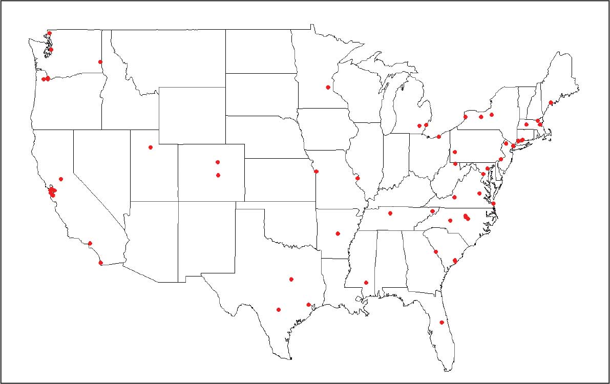
employment across all settings, July 2020 to June 2025.
The accomplishments of fellows are wide-ranging and aligned with VA’s mission. Each year, roughly 15 fellows receive new investigator awards, travel awards, and poster or presentation awards from prominent professional societies. Fellows have obtained VA Career Development Awards in diverse topics, including suicide prevention through clinician resources and training programs, firearm safety discussions, digital phenotyping and neuroimaging to enhance social integration in veterans with schizophrenia, rapid transcranial magnetic stimulation to treat nicotine use and PTSD, and evidence-based psychotherapy techniques for female veterans experiencing issues in menopause.
Several recent MIRT fellows have also received highly competitive NIH K Career Development Awards. One notable example is a fellow who studied pharmacologic approaches for treatment-resistant depression informed by novel brain circuit findings, first testing these approaches in community samples through a NIH K grant and translating findings to veterans. Fellows have gone on to become directors of important national research centers and studies, chairs of academic departments, and presidents of national medical organizations. Importantly, many MIRT fellows have become local directors and mentors to a new generation of VA fellows and researchers.
Conclusions
The AF MIRT coordinating center supports the VA’s mission of fulfilling President Lincoln’s promise to care for veterans. There are multiple benefits to evidence-based work that helps veterans and fosters a highly skilled VA workforce. Veterans are at the center of the MIRT data-driven approach, which is critical given their complex needs. Approaches to building the AF MIRT’s evidence base include randomized controlled trials open to veteran participants; program evaluation of current local, regional, or national VHA clinical services through measurement-based care and evaluation of national clinician training programs; and even smaller quality improvement projects in local VA clinics. These efforts support effective, efficient, and accessible provision of treatments that benefit veterans.
- US Department of Veterans Affairs. Our VA mission and core values. Updated April 17, 2025. Accessed March 2, 2026. https://department.va.gov/icare/
- Holliday R, Holder N. VA is a leader in mental health and social service research and operations. Fed Pract. 2025;42:S5. doi:10.12788/fp.0578
- Zeiss AM, Karlin BE. Integrating mental health and primary care services in the Department of Veterans Affairs health care system. J Clin Psychol Med Settings. 2008;15:73-78. doi:10.1007/s10880-008-9100-4
- O’Hara R, Cassidy-Eagle EL, Beaudreau SA, et al. Increasing the ranks of academic researchers in mental health: a multisite approach to postdoctoral fellowship training. Acad Med. 2010;85:41-47. doi:10.1097/ACM.0b013e3181c47c51
- US Department of Veterans Affairs. Office of Academic Affiliations. Updated March 13, 2025. Accessed March 2, 2026. https://www.va.gov/oaa/advancedfellowships /advanced-fellowships.asp
- Hantke NC, Samarina V, Hallmayer J, et al. Preparing the next generation of academic researchers during the pandemic: lessons from a national mental health research postdoctoral fellowship. Acad Psychiatry. 2022;46:466- 469. doi:10.1007/s40596-022-01613-4
The US Department of Veterans Affairs (VA) mission realizes President Abraham Lincoln’s promise to “to care for him who shall have borne the battle, and for his widow, and his orphan.”1 Evidence-based care fulfills this promise and is the backbone of Veterans Health Administration (VHA) mental health care.2,3 To ensure veterans receive state-of-the-art clinical care, a skilled workforce and investment in data-driven approaches are necessary to identify best treatments and strategies to implement them in practice.
Through scientific and clinical training tailored to VA, the 23 VA Advanced Fellowships have secured a steady flow of highly trained PhD professionals (ie, psychologists and other allied health professionals), and medical doctors (ie, psychiatrists and neurologists) into the VA workforce.4 The VA Advanced Fellows are funded by the Office of Academic Affiliations (OAA) and offer 2-year training opportunities for postresidency MDs and postdoctoral PhDs. This article describes a VA Advanced Fellowship in mental health as an example of how these programs can have a broad and positive impact on the VA health care system.
Advanced Fellows Program
The VA Advanced Fellowship in Mental Illness Research and Treatment (AF MIRT), formerly known as the VA Special Fellowship Program in Advanced Psychiatry and Psychology, educates and trains clinician and nonclinician researchers to meet VA priority mental health care needs.5 Clinical AF MIRT fellows dedicate 75% of their time to training and research activities and 25% to direct clinical services. Data science fellows complete projects that inform veteran clinical care through qualitative data collection, program evaluation, and analysis of large datasets. The full translational pathway to evidence-based clinical care is represented by fellow research spanning basic animal models, genetics, and neuroimaging to implementation science and applied clinical care for veterans.
In 2025, AF MIRT marked its 25th year of training postdoctoral-level mental health scientific practitioners and scholars. This investment in clinical research training has had profound benefits for innovation and retention of clinicians and scientists within the VA system. As of April 1, 2026, AF MIRT trained 700 fellows, including 152 MD or MD/PhD fellows, 544 PhD or PsyD fellows, 3 PharmDs fellows, and 1 doctor of nursing practice fellow.
Fellowship Structure
The AF MIRT coordinating center provides key administrative support to fellowship site directors and topical didactic training to Advanced Fellows, ensuring consistent standard of quality training across locations in 15 states and 4 times zones. The training provided by the AF MIRT coordinating center deepens the nationally-mandated focus of local translational clinical centers (eg, Mental Illness Research Education and Clinical Centers, Centers of Excellence) on posttraumatic stress disorder (PTSD), serious mental illness, dementia, and other areas.
The AF MIRT coordinating center also promotes VA workforce sustainability. Advanced Fellows in programs with a coordinating center are much more likely to be retained in VA for postfellowship employment compared with fellows in programs without such a coordinating center (60% vs 38%) according to unpublished Office of Academic Affiliations data (Joel Schmidt, oral communication, May 15, 2025). The AF MIRT coordinating center provides central standardization and uses evidence-based approaches to ensure fellows receive consistent support, resources, and training. More specifically, the coordinating center develops and delivers a standardized, core curriculum to the program’s 28 sites. The program pioneered video delivery of integrated didactics that enlist national experts, many of them VA researchers and clinicians themselves. Didactics include high priority veteran mental health topics, such as suicide prevention, new and emerging evidence-based treatments (eg, neurostimulation for treatment resistant PTSD, psychotherapeutic approaches for traumatic brain injury), and VA health system considerations for mental health treatment delivery.
This curated didactic series also covers professional and technical issues, such as statistical and methodological considerations for clinical trials, scientific writing, and grant-writing skill development. These offerings support the career pathways of advanced fellows to pursue careers as researchers, scientifically-informed clinicians, or data scientists at VA or academic medical centers. The coordinating center prepares fellows to apply for mentored career award funding or independent investigator awards through the VA, National Institutes of Health (NIH), US Department of Defense, and other organizations by offering an annual mock grant review session and monthly reviews and discussions of fellows’ grant applications.
AF MIRT continuously fine tunes the didactic series curriculum based on feedback from fellows on how the program meets their training needs. For example, learning about the strategies Advanced Fellows used to remain productive during COVID-19 pandemic lockdowns revealed a strong trend toward use of secondary data (eg, publicly available data or VA electronic health record data). This fueled curriculum adjustments to include more topics relevant to fellow interests and needs for accessing secondary data resources for high priority veteran mental health topics.6
VA Advanced Fellowships Successes
From July 2020 to June 2025, MIRT advanced fellows published 906 peer-reviewed articles in psychiatry, psychology, and other disciplines. Each year, about 20 to 25 articles are published in high-impact journals. In this 5-year period, fellows have received 153 grants (114 VA grants) as principal investigators– many examining new innovations to improve the quality of care of veterans. Of the 165 fellows who graduated since 2020, 63% continued working in veteran health care: 38% transitioned to full-time VA employment and 25% moved to VA employment with an academic-affiliated role. Nineteen percent transitioned to academic positions, 12% transitioned to the private sector, and 5% transitioned to other government, industry, or nonprofit employment where these professionals contribute to scientific and clinical innovation benefiting the US public; 1% did not provide postfellowship employment information. The Figure displays geographic locations of graduated fellows’ postfellowship employment from July 2020 to June 2025.
employment across all settings, July 2020 to June 2025.
The accomplishments of fellows are wide-ranging and aligned with VA’s mission. Each year, roughly 15 fellows receive new investigator awards, travel awards, and poster or presentation awards from prominent professional societies. Fellows have obtained VA Career Development Awards in diverse topics, including suicide prevention through clinician resources and training programs, firearm safety discussions, digital phenotyping and neuroimaging to enhance social integration in veterans with schizophrenia, rapid transcranial magnetic stimulation to treat nicotine use and PTSD, and evidence-based psychotherapy techniques for female veterans experiencing issues in menopause.
Several recent MIRT fellows have also received highly competitive NIH K Career Development Awards. One notable example is a fellow who studied pharmacologic approaches for treatment-resistant depression informed by novel brain circuit findings, first testing these approaches in community samples through a NIH K grant and translating findings to veterans. Fellows have gone on to become directors of important national research centers and studies, chairs of academic departments, and presidents of national medical organizations. Importantly, many MIRT fellows have become local directors and mentors to a new generation of VA fellows and researchers.
Conclusions
The AF MIRT coordinating center supports the VA’s mission of fulfilling President Lincoln’s promise to care for veterans. There are multiple benefits to evidence-based work that helps veterans and fosters a highly skilled VA workforce. Veterans are at the center of the MIRT data-driven approach, which is critical given their complex needs. Approaches to building the AF MIRT’s evidence base include randomized controlled trials open to veteran participants; program evaluation of current local, regional, or national VHA clinical services through measurement-based care and evaluation of national clinician training programs; and even smaller quality improvement projects in local VA clinics. These efforts support effective, efficient, and accessible provision of treatments that benefit veterans.
- US Department of Veterans Affairs. Our VA mission and core values. Updated April 17, 2025. Accessed March 2, 2026. https://department.va.gov/icare/
- Holliday R, Holder N. VA is a leader in mental health and social service research and operations. Fed Pract. 2025;42:S5. doi:10.12788/fp.0578
- Zeiss AM, Karlin BE. Integrating mental health and primary care services in the Department of Veterans Affairs health care system. J Clin Psychol Med Settings. 2008;15:73-78. doi:10.1007/s10880-008-9100-4
- O’Hara R, Cassidy-Eagle EL, Beaudreau SA, et al. Increasing the ranks of academic researchers in mental health: a multisite approach to postdoctoral fellowship training. Acad Med. 2010;85:41-47. doi:10.1097/ACM.0b013e3181c47c51
- US Department of Veterans Affairs. Office of Academic Affiliations. Updated March 13, 2025. Accessed March 2, 2026. https://www.va.gov/oaa/advancedfellowships /advanced-fellowships.asp
- Hantke NC, Samarina V, Hallmayer J, et al. Preparing the next generation of academic researchers during the pandemic: lessons from a national mental health research postdoctoral fellowship. Acad Psychiatry. 2022;46:466- 469. doi:10.1007/s40596-022-01613-4
VA Advanced Training for Clinician Researchers and Data Scientists in Mental Health
VA Advanced Training for Clinician Researchers and Data Scientists in Mental Health

The Home Improvements and Structural Alterations Program: Overview and Future Implications
The Home Improvements and Structural Alterations Program: Overview and Future Implications
The Veterans Health Administration (VHA) Home Improvements and Structural Alterations (HISA) program is a primary means through which veterans can obtain home modifications necessary to continue safe and independent living in their home, including fall risk reduction and accessibility to essential parts of the home. However, not all eligible veterans who may benefit from this program participate, for a variety of reasons.1-6 Historically, the HISA program has been administered in a decentralized and nonstandardized fashion dictated by the organizational structure of each US Department of Veterans Affairs (VA) medical center (VAMC) within a certain region or Veterans Integrated Service Network (VISN). Previous research found differential access to the HISA program by younger veterans, women, minorities, veterans with certain disability types, and veterans living in rural vs urban settings. These disparities in access and use of benefits conferred by the HISA program suggests an area of unmet need, which may improve veterans’ health care outcomes and reduce costs associated with their care.2-8
The purpose of this article is to provide information to improve equitable provision and effective eligible use of resources available through the HISA program in a more generalizable manner by providing insight to highlight common program process deficiencies and care provision gaps relevant to VAMCs nationwide. This information can be used to inform the VA Physical Medicine and Rehabilitation (PM&R) and Prosthetic and Sensory Aid Service (PSAS) national policy initiatives, as well as hiring practices, clinic organization, specific care provision, and administrative goals and metrics at each VISN and at the VA Healthcare System level.
Methods
Veterans who participated in the HISA program, VHA administrators, and VHA clinicians from select VAMCs were identified and interviewed to better understand what helps increase access to the program, barriers to access, and how existing program components and processes impact use of the service. These interviews were taken from a directed convenience sample of selected VAMCs. To obtain this directed convenience sample, 167 VAMCs that participated in the HISA program were categorized as facilities that provided either a high or low number of HISA program prescriptions based on data from 2010 to 2018. Ten facilities from the top quartiles and 10 from the bottom quartiles of prescribing locations were selected. This facility selection was driven by the proportion of rural veterans served by each facility, favoring those serving a greater proportion of rural veterans, as well geographic location, with the aim of avoiding overrepresentation of any specific region. The convenience sample included 45 individuals (20 VHA employees and 25 veterans) across 22 states from the Northeast, West, South, and Midwest US Census regions.
Interview Process
Interviews underwent a coding process. The development of topical themes followed a systematic, 2-phase approach. Initially, researchers analyzed responses to semistructured interview questions addressing specific aspects of the HISA program, such as program awareness and accessibility. These responses naturally clustered into preliminary categories based on the interview guide structure. For example, responses related to program discovery formed a marketing-related category, while recommendations about program implementation contributed to a training and development category.
Following this initial categorization, the research team conducted a more rigorous coding process. A team of 3 researchers systematically reviewed assigned interview transcripts to extract practical recommendations for the guide. The researchers first identified relevant responses individually and then convened during group meetings to discuss and finalize selections. This second phase refined the preliminary categorization while maintaining alignment with the original interview structure.
This approach allowed the team to preserve the practical utility of participant feedback while ensuring methodological rigor in the analysis process. Resulting themes reflect both the structured nature of the original inquiry and the practical recommendations identified for improving the HISA program. Information on the following areas were collected: education about the HISA program, the contracting process, use of telehealth, interaction between VHA clinical care and the PSAS, marketing of the program, program funding, and revising the application process.
Results
Interview respondents provided several recommendations for improving the HISA program (Table). Regarding training and education, respondents noted deficiencies in VHA employee communication about the HISA program to veterans. Some employees did not know details or were unaware the HISA program existed. Additionally, a lack of knowledge about HISA program alternatives, including other available programs for obtaining home modifications or other durable medical equipment alternatives (eg, provision of a portable ramp rather than construction of a permanent one), was apparent. It was strongly recommended to provide additional education to effectively disseminate knowledge about the HISA program. Specifically, VHA employees, especially those in Primary Care, Geriatrics, Home Based Primary Care, the Caregiver Support Program, and Blind Rehabilitation Services, require greater awareness of the program and its processes.
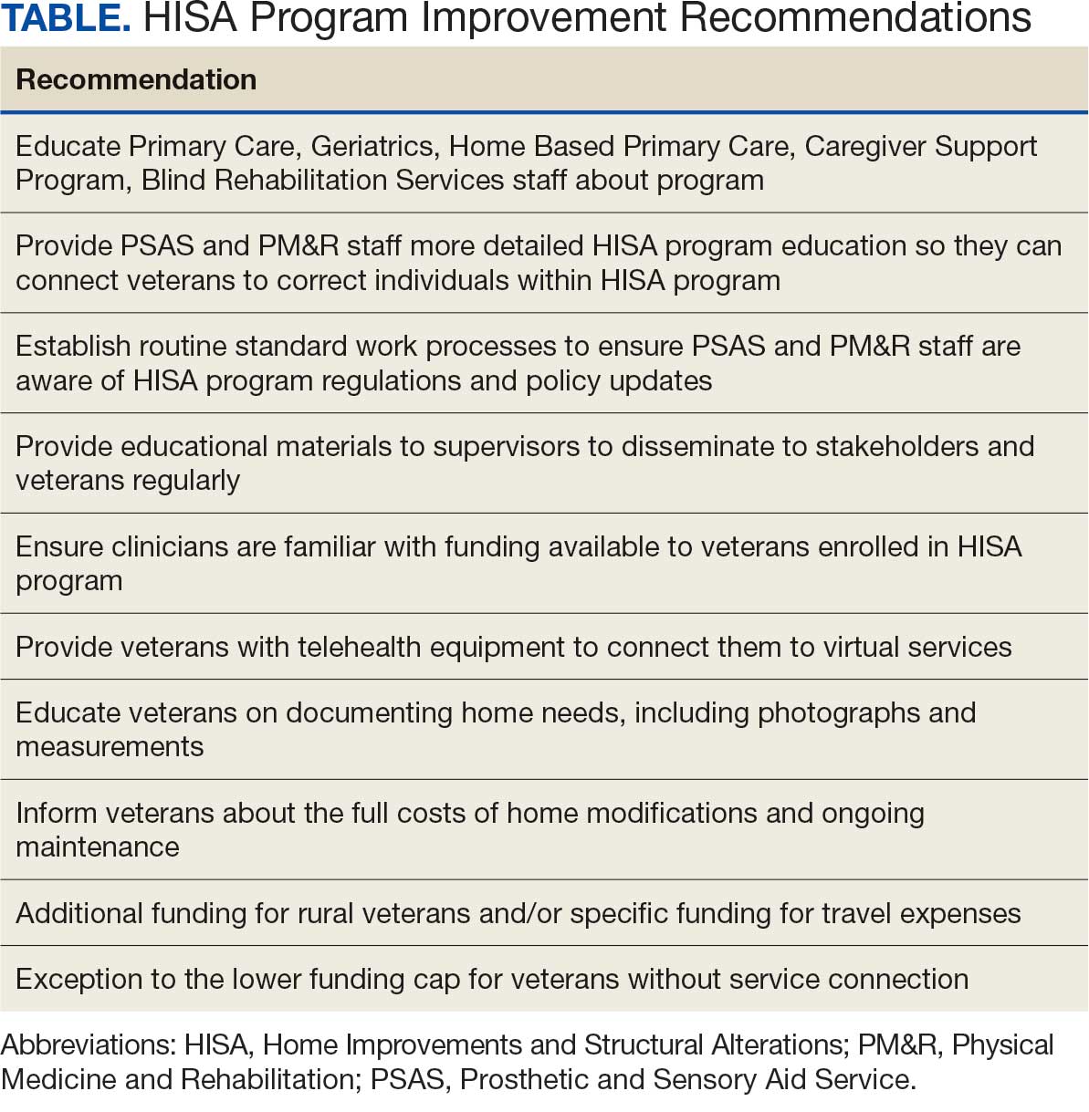
PSAS and PM&R professionals, including physicians, nurse practitioners, physician assistants, and physical and occupational therapists, would be expected to have some knowledge of the HISA program, and therefore be more likely to connect a veteran with it. However, they may lack specific details about the program such as correct contact persons in the other service (PSAS or PM&R, respectively), facility- specific processes, such as how to enter a HISA consultation within the veteran’s electronic health record, how the entered consultation would progress through the system and avoid cancellation, and what should routinely be done to avoid HISA consultation cancellation, such as referral to Occupational Therapy for a functional assessment so appropriate durable medical equipment can be trialed with the veteran prior to proceeding with more costly and time-consuming home modifications.
In addition, there is no routine standard work process to ensure that PM&R staff are aware of updates in HISA program regulations and policy. Further recommendations in this area include having supervisory employees in PSAS and PM&R work both individually and together to develop effective information dissemination methods for key stakeholders. These include targeted in-services (ie, educational trainings often scheduled and conducted during recurring meetings), whether faceto- face or virtually in real time, or recorded, that occur on an ongoing and regular basis with sister services such as Primary Care, Geriatrics, Home Based Primary Care, the Caregiver Support Program, and Blind Rehabilitation Services (eg, the facility Vision Impairment Services Team coordinator). Regularly updated educational materials should be provided to veterans and VHA adjacent stakeholders such as Veteran Service Organizations and Veteran County Service Officers, via a variety of platforms.
Successfully navigating the provision of home modifications via the HISA program involves identifying a contractor to perform the home modification and obtaining service and construction plan pricing. A key barrier in this area is that veterans and VHA clinicians perceive the funds available through HISA as insufficient, regardless of whether they have serviceconnected status or not. Service connection refers to designation of ≥ 1 medical conditions determined to be related to military service and thus eligible to receive VHA care.9 Service-connected veterans receive a lifetime maximum award of $6800 from HISA while veterans without service connection receive a lifetime maximum award of $2000.1,2
Rural veterans face a greater challenge than urban veterans, as there are fewer contractors located nearby. Thus, providing higher funding for rural veterans, or specific funding such as for travel expenses, would be especially helpful to find a willing contractor to perform home medications.1 The current requirement of working with a licensed contractor was also a barrier, especially for smaller jobs, and could result in VHA employees (including clinicians) feeling pressured to become overly involved to assist veterans to move through the process.
To that point, respondents requested resources such as a regularly updated list of licensed contractors in the area, especially those familiar with working with the HISA program, be provided to veterans and their assisting groups. In addition, respondents asked that VHA take on greater responsibility and liability with regard to contractors accessing HISA funding, such as not releasing final payment until VHA approved the completed home modification. On the other hand, respondents also expressed concerns about the length of time associated with HISA program payment and noted it should be sped up to allow contractors who participate to receive payment sooner, which many believed would increase the number of contractors willing to take on this work.
The role of telehealth was noted as a great facilitator of increased access to care, especially following the COVID-19 pandemic. Telehealth modalities adapted for the HISA program could help increase access to the program and improve processing speed. Barriers include lack of appropriate veteran telehealth equipment and poor understanding of information needed to move the process forward. Recommendations included providing veterans tablets to connect to virtual services, and developing information on home measurements needed, assistance in obtaining and sending photographs, and detailed information on successfully using telehealth for the HISA application process. Of note, some clinicians, representing home-based primary care, prosthetics services, geriatrics, rehabilitation therapy, mobile clinic, and the telehealth division, and including both clinical staff (eg, occupational therapists) and nonclinical staff (eg, prosthetics representatives and administrative personnel), have found patients expressed comparable satisfaction with the process whether faceto- face or via telehealth.
The essential relationship between PSAS and PM&R regarding the HISA program was a key finding. Both services are integral to helping veterans successfully obtain home modifications via the HISA program.1,2 Barriers include insufficient communication and a lack of clearly defined points of contact for each service, poorly defined roles, and inefficiencies because 2 services are involved in navigating the process. Recommendations therefore include addressing these issues, such as adopting a case management or liaison model between the services to better manage the process.
Respondents indicated that insufficient program funding was a concern. Veterans living in poorer quality housing, such as older homes, often require more expensive home modifications, necessitating greater out-of-pocket expenses. Veterans and VHA employees advocated for the creation of an exception to the lower funding cap for veterans without service connection in cases of financial hardship. Overall, the funding limits for both service-connected veterans and those without service connection were thought to be insufficient, especially as the COVID-19 pandemic increased the cost of construction materials.
Respondents also noted that veterans would benefit from clear messaging that receiving HISA funds does not impact eligibility for other VA benefits and services. Veterans must understand that home modifications work must be approved by VHA before being started and should be aware that if their disability rating increases so that they become eligible for the higher level service-connected benefits, they would then become eligible for the higher maximum benefit. Respondents recommended veterans should receive assistance in understanding the full costs of the home modification and ongoing maintenance, and the HISA research team recommended that the National Program develop a fact sheet that can be used to advise veterans.
Respondents consistently indicated that information about the HISA program was not disseminated effectively to key internal and external stakeholders, and opportunities to highlight the program on VHA websites, brochures throughout VHA facilities, and other outlets such as direct mailing should be used. Veterans who have used the program are overwhelmingly older (mean age 71 years), White, and male, suggesting missed opportunities and unmet need for underrepresented groups. Therefore, targeted marketing interventions would especially benefit these groups.
Respondents also noted inefficiencies throughout the HISA program application process and advocated for changes such as national standard operating procedures (SOPs) to guide navigation through the HISA process. The national SOPs could include home evaluation prior to HISA application submission, clearly identified points of contact for the HISA program in PSAS and PM&R, and standardized documentation.
Future Directions
Information from respondents provided several avenues for future studies. Recommendations were obtained from each of the 7 broad topical areas: training and educational needs, potential, contracting challenges and opportunities, telehealth as a conduit to facilitate the availability of the HISA program, PSAS, and clinical services collaboration, marketing, need for increased funding, and revision of the application process. Input from stakeholders can help direct efficient use of resources to guide future studies for the greatest impact and highlight current and future priorities. Easy areas of intervention indicated by respondents include creating a national standard work process regarding the HISA program with standardized educational materials for key stakeholders, revised at regular intervals, and readily available on national websites. A pre- and postimplementation survey could help provide quantifiable information about the benefits of such an intervention.
Conclusions
A qualitative analysis of interviews with veterans and VHA clinicians provides evidence of potential barriers for the HISA program. Addressing these barriers could allow HISA to better meet the VHA goal of providing home modifications that allow veterans to live safely and independently in their homes. There is a need for ongoing review and assessment of the program to ensure optimization and efficient use of resources across the spectrum of veteran needs.
- Semeah LM, Ahrentzen S, Jia H, et al. The Home Improvements and Structural Alterations Benefits Program: veterans with disabilities and home accessibility. J Disabil Policy Stud. 2017;28:43-51. doi:10.1177/1044207317696275
- Semeah LM, Wang X, Cowper Ripley DC, et al. Improving health through a home modification service for veterans. In: Fiedler BA, ed. Three Facets of Public Health and Paths to Improvements. 2020:381-416. doi:10.1016/B978-0-12-819008-1.00014-6
- Semeah LM, Ganesh SP, Wang X, et al. Home modification and health services utilization by rural and urban veterans with disabilities. Housing Policy Debate. 2021;31:862-874. doi:10.1080/10511482.2020.1858923
- Semeah LM, Orozco T, Wang X, et al. Home modifications for rural veterans with disabilities. Fed Pract. 2021;38:300- 310. doi:10.12788/fp.0153
- Semeah LM, Orozco T, Wang X, et al. Predictors of countylevel home modification use across the US. Fed Pract. 2022;39:274-280. doi:10.12788/fp.0279
- Semeah LM, Orozco T, Wang X, et al. Rural and urban home modification program users: a comparative study. HERD. 2023;16:223-235. doi:10.1177/19375867221142627
- US Department of of Veterans Affairs. Home Improvements and Structural Alterations (HISA) benefits program: final rule. Fed Regist. 2014;79:71658-71663
- US Department of Veterans Affairs. Home Improvement and Structural Alterations (HISA): increase in the limit for home improvement and structural alterations (HISA)-VA: final regulations. Fed Regist. 1993;58:25565.
- US Department of Veterans Affairs. Eligibility for VA disability benefits. Updated April 25, 2025. Accessed April 1, 2026. https://www.va.gov/disability/eligibility
The Veterans Health Administration (VHA) Home Improvements and Structural Alterations (HISA) program is a primary means through which veterans can obtain home modifications necessary to continue safe and independent living in their home, including fall risk reduction and accessibility to essential parts of the home. However, not all eligible veterans who may benefit from this program participate, for a variety of reasons.1-6 Historically, the HISA program has been administered in a decentralized and nonstandardized fashion dictated by the organizational structure of each US Department of Veterans Affairs (VA) medical center (VAMC) within a certain region or Veterans Integrated Service Network (VISN). Previous research found differential access to the HISA program by younger veterans, women, minorities, veterans with certain disability types, and veterans living in rural vs urban settings. These disparities in access and use of benefits conferred by the HISA program suggests an area of unmet need, which may improve veterans’ health care outcomes and reduce costs associated with their care.2-8
The purpose of this article is to provide information to improve equitable provision and effective eligible use of resources available through the HISA program in a more generalizable manner by providing insight to highlight common program process deficiencies and care provision gaps relevant to VAMCs nationwide. This information can be used to inform the VA Physical Medicine and Rehabilitation (PM&R) and Prosthetic and Sensory Aid Service (PSAS) national policy initiatives, as well as hiring practices, clinic organization, specific care provision, and administrative goals and metrics at each VISN and at the VA Healthcare System level.
Methods
Veterans who participated in the HISA program, VHA administrators, and VHA clinicians from select VAMCs were identified and interviewed to better understand what helps increase access to the program, barriers to access, and how existing program components and processes impact use of the service. These interviews were taken from a directed convenience sample of selected VAMCs. To obtain this directed convenience sample, 167 VAMCs that participated in the HISA program were categorized as facilities that provided either a high or low number of HISA program prescriptions based on data from 2010 to 2018. Ten facilities from the top quartiles and 10 from the bottom quartiles of prescribing locations were selected. This facility selection was driven by the proportion of rural veterans served by each facility, favoring those serving a greater proportion of rural veterans, as well geographic location, with the aim of avoiding overrepresentation of any specific region. The convenience sample included 45 individuals (20 VHA employees and 25 veterans) across 22 states from the Northeast, West, South, and Midwest US Census regions.
Interview Process
Interviews underwent a coding process. The development of topical themes followed a systematic, 2-phase approach. Initially, researchers analyzed responses to semistructured interview questions addressing specific aspects of the HISA program, such as program awareness and accessibility. These responses naturally clustered into preliminary categories based on the interview guide structure. For example, responses related to program discovery formed a marketing-related category, while recommendations about program implementation contributed to a training and development category.
Following this initial categorization, the research team conducted a more rigorous coding process. A team of 3 researchers systematically reviewed assigned interview transcripts to extract practical recommendations for the guide. The researchers first identified relevant responses individually and then convened during group meetings to discuss and finalize selections. This second phase refined the preliminary categorization while maintaining alignment with the original interview structure.
This approach allowed the team to preserve the practical utility of participant feedback while ensuring methodological rigor in the analysis process. Resulting themes reflect both the structured nature of the original inquiry and the practical recommendations identified for improving the HISA program. Information on the following areas were collected: education about the HISA program, the contracting process, use of telehealth, interaction between VHA clinical care and the PSAS, marketing of the program, program funding, and revising the application process.
Results
Interview respondents provided several recommendations for improving the HISA program (Table). Regarding training and education, respondents noted deficiencies in VHA employee communication about the HISA program to veterans. Some employees did not know details or were unaware the HISA program existed. Additionally, a lack of knowledge about HISA program alternatives, including other available programs for obtaining home modifications or other durable medical equipment alternatives (eg, provision of a portable ramp rather than construction of a permanent one), was apparent. It was strongly recommended to provide additional education to effectively disseminate knowledge about the HISA program. Specifically, VHA employees, especially those in Primary Care, Geriatrics, Home Based Primary Care, the Caregiver Support Program, and Blind Rehabilitation Services, require greater awareness of the program and its processes.
PSAS and PM&R professionals, including physicians, nurse practitioners, physician assistants, and physical and occupational therapists, would be expected to have some knowledge of the HISA program, and therefore be more likely to connect a veteran with it. However, they may lack specific details about the program such as correct contact persons in the other service (PSAS or PM&R, respectively), facility- specific processes, such as how to enter a HISA consultation within the veteran’s electronic health record, how the entered consultation would progress through the system and avoid cancellation, and what should routinely be done to avoid HISA consultation cancellation, such as referral to Occupational Therapy for a functional assessment so appropriate durable medical equipment can be trialed with the veteran prior to proceeding with more costly and time-consuming home modifications.
In addition, there is no routine standard work process to ensure that PM&R staff are aware of updates in HISA program regulations and policy. Further recommendations in this area include having supervisory employees in PSAS and PM&R work both individually and together to develop effective information dissemination methods for key stakeholders. These include targeted in-services (ie, educational trainings often scheduled and conducted during recurring meetings), whether faceto- face or virtually in real time, or recorded, that occur on an ongoing and regular basis with sister services such as Primary Care, Geriatrics, Home Based Primary Care, the Caregiver Support Program, and Blind Rehabilitation Services (eg, the facility Vision Impairment Services Team coordinator). Regularly updated educational materials should be provided to veterans and VHA adjacent stakeholders such as Veteran Service Organizations and Veteran County Service Officers, via a variety of platforms.
Successfully navigating the provision of home modifications via the HISA program involves identifying a contractor to perform the home modification and obtaining service and construction plan pricing. A key barrier in this area is that veterans and VHA clinicians perceive the funds available through HISA as insufficient, regardless of whether they have serviceconnected status or not. Service connection refers to designation of ≥ 1 medical conditions determined to be related to military service and thus eligible to receive VHA care.9 Service-connected veterans receive a lifetime maximum award of $6800 from HISA while veterans without service connection receive a lifetime maximum award of $2000.1,2
Rural veterans face a greater challenge than urban veterans, as there are fewer contractors located nearby. Thus, providing higher funding for rural veterans, or specific funding such as for travel expenses, would be especially helpful to find a willing contractor to perform home medications.1 The current requirement of working with a licensed contractor was also a barrier, especially for smaller jobs, and could result in VHA employees (including clinicians) feeling pressured to become overly involved to assist veterans to move through the process.
To that point, respondents requested resources such as a regularly updated list of licensed contractors in the area, especially those familiar with working with the HISA program, be provided to veterans and their assisting groups. In addition, respondents asked that VHA take on greater responsibility and liability with regard to contractors accessing HISA funding, such as not releasing final payment until VHA approved the completed home modification. On the other hand, respondents also expressed concerns about the length of time associated with HISA program payment and noted it should be sped up to allow contractors who participate to receive payment sooner, which many believed would increase the number of contractors willing to take on this work.
The role of telehealth was noted as a great facilitator of increased access to care, especially following the COVID-19 pandemic. Telehealth modalities adapted for the HISA program could help increase access to the program and improve processing speed. Barriers include lack of appropriate veteran telehealth equipment and poor understanding of information needed to move the process forward. Recommendations included providing veterans tablets to connect to virtual services, and developing information on home measurements needed, assistance in obtaining and sending photographs, and detailed information on successfully using telehealth for the HISA application process. Of note, some clinicians, representing home-based primary care, prosthetics services, geriatrics, rehabilitation therapy, mobile clinic, and the telehealth division, and including both clinical staff (eg, occupational therapists) and nonclinical staff (eg, prosthetics representatives and administrative personnel), have found patients expressed comparable satisfaction with the process whether faceto- face or via telehealth.
The essential relationship between PSAS and PM&R regarding the HISA program was a key finding. Both services are integral to helping veterans successfully obtain home modifications via the HISA program.1,2 Barriers include insufficient communication and a lack of clearly defined points of contact for each service, poorly defined roles, and inefficiencies because 2 services are involved in navigating the process. Recommendations therefore include addressing these issues, such as adopting a case management or liaison model between the services to better manage the process.
Respondents indicated that insufficient program funding was a concern. Veterans living in poorer quality housing, such as older homes, often require more expensive home modifications, necessitating greater out-of-pocket expenses. Veterans and VHA employees advocated for the creation of an exception to the lower funding cap for veterans without service connection in cases of financial hardship. Overall, the funding limits for both service-connected veterans and those without service connection were thought to be insufficient, especially as the COVID-19 pandemic increased the cost of construction materials.
Respondents also noted that veterans would benefit from clear messaging that receiving HISA funds does not impact eligibility for other VA benefits and services. Veterans must understand that home modifications work must be approved by VHA before being started and should be aware that if their disability rating increases so that they become eligible for the higher level service-connected benefits, they would then become eligible for the higher maximum benefit. Respondents recommended veterans should receive assistance in understanding the full costs of the home modification and ongoing maintenance, and the HISA research team recommended that the National Program develop a fact sheet that can be used to advise veterans.
Respondents consistently indicated that information about the HISA program was not disseminated effectively to key internal and external stakeholders, and opportunities to highlight the program on VHA websites, brochures throughout VHA facilities, and other outlets such as direct mailing should be used. Veterans who have used the program are overwhelmingly older (mean age 71 years), White, and male, suggesting missed opportunities and unmet need for underrepresented groups. Therefore, targeted marketing interventions would especially benefit these groups.
Respondents also noted inefficiencies throughout the HISA program application process and advocated for changes such as national standard operating procedures (SOPs) to guide navigation through the HISA process. The national SOPs could include home evaluation prior to HISA application submission, clearly identified points of contact for the HISA program in PSAS and PM&R, and standardized documentation.
Future Directions
Information from respondents provided several avenues for future studies. Recommendations were obtained from each of the 7 broad topical areas: training and educational needs, potential, contracting challenges and opportunities, telehealth as a conduit to facilitate the availability of the HISA program, PSAS, and clinical services collaboration, marketing, need for increased funding, and revision of the application process. Input from stakeholders can help direct efficient use of resources to guide future studies for the greatest impact and highlight current and future priorities. Easy areas of intervention indicated by respondents include creating a national standard work process regarding the HISA program with standardized educational materials for key stakeholders, revised at regular intervals, and readily available on national websites. A pre- and postimplementation survey could help provide quantifiable information about the benefits of such an intervention.
Conclusions
A qualitative analysis of interviews with veterans and VHA clinicians provides evidence of potential barriers for the HISA program. Addressing these barriers could allow HISA to better meet the VHA goal of providing home modifications that allow veterans to live safely and independently in their homes. There is a need for ongoing review and assessment of the program to ensure optimization and efficient use of resources across the spectrum of veteran needs.
- Semeah LM, Ahrentzen S, Jia H, et al. The Home Improvements and Structural Alterations Benefits Program: veterans with disabilities and home accessibility. J Disabil Policy Stud. 2017;28:43-51. doi:10.1177/1044207317696275
- Semeah LM, Wang X, Cowper Ripley DC, et al. Improving health through a home modification service for veterans. In: Fiedler BA, ed. Three Facets of Public Health and Paths to Improvements. 2020:381-416. doi:10.1016/B978-0-12-819008-1.00014-6
- Semeah LM, Ganesh SP, Wang X, et al. Home modification and health services utilization by rural and urban veterans with disabilities. Housing Policy Debate. 2021;31:862-874. doi:10.1080/10511482.2020.1858923
- Semeah LM, Orozco T, Wang X, et al. Home modifications for rural veterans with disabilities. Fed Pract. 2021;38:300- 310. doi:10.12788/fp.0153
- Semeah LM, Orozco T, Wang X, et al. Predictors of countylevel home modification use across the US. Fed Pract. 2022;39:274-280. doi:10.12788/fp.0279
- Semeah LM, Orozco T, Wang X, et al. Rural and urban home modification program users: a comparative study. HERD. 2023;16:223-235. doi:10.1177/19375867221142627
- US Department of of Veterans Affairs. Home Improvements and Structural Alterations (HISA) benefits program: final rule. Fed Regist. 2014;79:71658-71663
- US Department of Veterans Affairs. Home Improvement and Structural Alterations (HISA): increase in the limit for home improvement and structural alterations (HISA)-VA: final regulations. Fed Regist. 1993;58:25565.
- US Department of Veterans Affairs. Eligibility for VA disability benefits. Updated April 25, 2025. Accessed April 1, 2026. https://www.va.gov/disability/eligibility
The Home Improvements and Structural Alterations Program: Overview and Future Implications
The Home Improvements and Structural Alterations Program: Overview and Future Implications

The Development of a Comprehensive Wound Care Fellowship Curriculum
The Development of a Comprehensive Wound Care Fellowship Curriculum
Often disguised as comorbid conditions, nonhealing and chronic wounds have emerged as a silent epidemic that affects about 6.5 million Americans.1-3 In 2023, estimated US wound care costs were $126.86 billion.4 About 1% to 2% of individuals worldwide will experience a chronic wound in their lifetime. The Veterans Health Administration reported 277,000 inpatient and outpatient encounters for ulcers in 2011, including chronic ulcers of the lower extremity due to diabetes, venous disease, or arterial disease.5 Associated costs of chronic wounds are expected to increase as the populations of developed countries age.6 Effective treatment of chronic wounds requires a nuanced understanding of complex wound pathophysiology, best practices in interdisciplinary and multidisciplinary wound care, and advanced wound care technologies.7,8
The typical 4-year medical school curriculum, followed by residency, offers little in the way of formal didactic training in wound care.9,10 Without specialized and advanced fellowship training dedicated to wound care, health care will lack specialists prepared to manage complex wounds. As a result, wound care-related difficulties may be exacerbated by prolonged recovery time, increased costs, productivity loss, and increased mortality risk.8 Wound care is a growing field of study and practice, and there is a critical need for rigorous training, research, and quality improvement efforts to enhance outcomes for patients with nonhealing wounds.5
One of the most direct ways to address the need for more physicians with specialty training in wound medicine is to implement a comprehensive training curriculum for advanced wound care practice. Although specialized advanced wound care fellowships are available, the curricula primarily detail rotation names and areas for practice without accompanying competencies, milestones, or entrustable professional activities.11 Furthermore, wound care is not recognized as a subspecialty by the Accreditation Council for Graduate Medical Education (ACGME).
This article synthesized the literature and integrated innovative, evidence-based practices into a curriculum for a formal advanced fellowship training program. To our knowledge, no comprehensive wound care curriculum is publicly available that includes rotations, competencies, milestones, entrustable professional activities, and 360-degree evaluation forms.
Program Development
The advanced wound care fellowship program started in January 2014 at the Michael E. DeBakey Veterans Affairs Medical Center in affiliation with the Baylor College of Medicine. The fellowship program was originally designed for geriatrics fellows to extend the 1-year fellowship for an additional year to learn wound care. It has been adjusted to address formal program goals and objectives, competencies, milestones, entrustable professional activities, and evaluations, with the goal of developing an example curriculum for wound care fellowships across specialties. Although the ACGME does not recognize a wound care subspecialty, this curriculum complies with the ACGME 1-year fellowship common program requirements.12,13
Scoping Review
A scoping literature review of Google Scholar and PubMed was performed using the medical subject heading terms “wound care + curriculum” and “wound + care + curriculum” to find advanced wound care medical training, fellowship programs, boards, and related ACGME-accredited specialty curricula. The local wound care fellowship program was initially implemented based on an informal literature review by faculty and their respective contributions to curriculum (ie, process establishing wound care-specific competency domains in accordance with ACGME accreditation competency requirements of 1-year fellowships). 12,13 Standing program practice-based competencies and activities were examined and determined to align with best practices. This scoping review considered additional competencies, competency domains, and entrustable professional activities of reputable wound care fellowship training programs (eg, University of Chicago at Illinois and Wake Forest School of Medicine),8,11,14 a specialty wound care board (American Board of Wound Medicine and Surgery),15 an international wound specialist professional society (European Union of Medical Specialists), 16 and recommended curriculum guidelines for wound care residency programs.17 ACGME-accredited specialty and subspecialty milestones professional activities were examined, including vascular surgery,18 plastic surgery,19 dermatology, 20 foot and ankle,21 orthopedic surgery,22 spinal cord injury,23 and geriatric medicine.24
The competencies, milestones, and entrustable professional activities were compiled and redundancies were eliminated. Wound care specialists from geriatrics, family medicine, internal medicine, undersea and hyperbaric medicine, general surgery, podiatry, and physical therapy examined the findings and suggested eliminating redundancies, irrelevant content, and content that fell below the minimal expected level of competence for an advanced medical specialist in wound care. An expert consensus meeting further refined items presented to the panel before unanimous consensus resulted in the final set of curriculum competencies, milestones, and entrustable professional activities.
Training Program Feedback
We developed a comprehensive wound care curriculum for an advanced physician fellowship training program based on the streamlined competencies, milestones, and entrustable professional activities (Appendix). Multiple wound care experts from various interdisciplinary backgrounds reached consensus to establish this fellowship curriculum as adaptable for use across training settings. The training program is 12 core rotations and 2 elective rotations (Table 1). Additionally, we developed wound care evaluation forms for faculty-, peer-, and self-assessment of trainees which were adapted from an evidence-based 360-degree evaluation template.25 Suggestions for structured, advanced didactics are in Table 2.
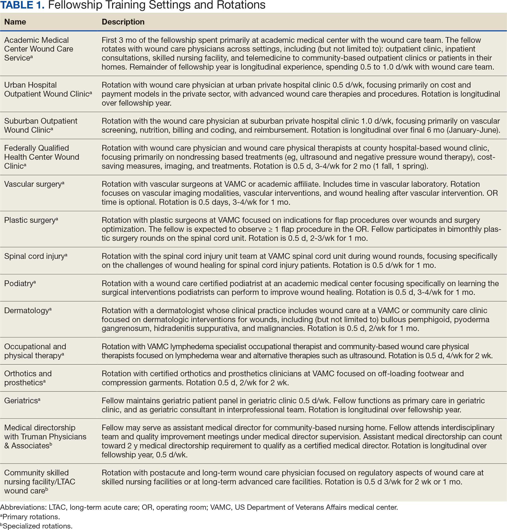
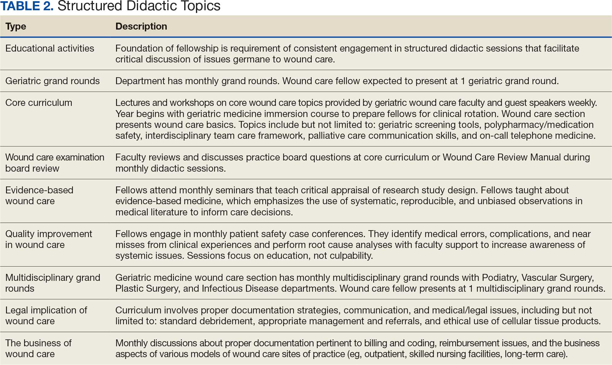
Seventeen fellows have successfully matriculated through the wound care training program. Although wound care certification is not required to work as a wound care specialist, after completion of this fellowship, graduates are able to sit for a wound care certification examination. The American Board of Wound Medicine and Surgery (ABWMS) and the American Board of Wound Management (ABWM) allow physicians to take a certification examination after 1 year of a dedicated wound fellowship program, instead of the typical wound care practice experience ≥ 3 years.
The Clinical Wound Care Fellowship Program collected data for program improvement, and 15 alumni responded (response rate, 88%) to a survey using a 5-point Likert scale. Respondents indicated high mean scores for overall satisfaction (4.7), instructional methods (4.7), program enjoyment (4.7), teaching materials (4.6), and relevance (4.6). All respondents indicated that the fellowship prepared them for a career in wound care as well as their current employment, and 13 of 15 (87%) reported they obtained immediate relevant postfellowship wound care positions and stated that the fellowship prepared them for their current roles. Nine respondents (69%) reported that they were engaged in wound care ≥ 26% of work time. Six respondents (46%) worked in private practice, 3 (23%) at academic medical centers, and 2 (15%) at government- funded hospitals. Four respondents indicated they were board certified in wound care. Program alumni are currently involved in scholarly activities, including 8 in quality improvement and 3 in research.
Discussion
An easily accessible, comprehensive wound care fellowship curriculum has not been previously developed or published. This limited the sources that informed this curriculum. However, the developmental process for this curriculum was robust, as the authors reviewed previously published materials related to wound care, including: 1) descriptive overviews of wound care fellowships; 2) details of month-long rotations for medical students and residents; and 3) practices of the specific environment in which this curriculum was created. Confidence in the practical nature of the curriculum can be assumed, as the experts involved in the development process represented diverse physician specializations, including geriatrics, family medicine, internal medicine, undersea and hyperbaric medicine, general surgery, podiatry, and physical therapy.
Most wound care clinicians have not completed a formal comprehensive fellowship program. Instead, due to the lack of a comprehensive training curriculum, clinicians have had to use various continuing medical education programs and practice in a wound care setting for ≥ 3 years to be eligible for certification in the specialty. This curriculum will help academic medical centers develop their own fellowship programs, enabling new wound care clinicians to attain certifications more efficiently. As more fellowship programs develop, the goal would be to obtain recognition as an ACGME specialty and standardize the training and competencies for graduates of wound care fellowships.
Conclusions
As new wound care fellowships develop, wound care may become formally acknowledged as its own specialty within medicine and surgery. This will provide wound care with a voice at the national level, particularly in an era of value-based care. Wound care clinicians will be able to advocate for specialty-specific quality metrics and avoid potential penalization for not meeting quality metrics that are irrelevant to wound care.
- Fife CE, Eckert KA, Carter MJ. Publicly Reported wound healing rates: the fantasy and the reality. Adv Wound Care (New Rochelle). 2018;7:77-94. doi:10.1089/wound.2017.0743
- Fife CE, Carter MJ, Walker D. Why is it so hard to do the right thing in wound care?. Wound Repair Regen. 2010;18:154-158. doi:10.1111/j.1524-475X.2010.00571.x
- Sen CK, Gordillo GM, Roy S, et al. Human skin wounds: a major and snowballing threat to public health and the economy. Wound Repair Regen. 2009;17:763-771. doi:10.1111/j.1524-475X.2009.00543.x
- Queen D, Harding K. What’s the true costs of wounds faced by different healthcare systems around the world?. Int Wound J. 2023;20:3935-3938. doi:10.1111/iwj.14491
- Greer N, Foman N, Dorrian J, et al. Advanced Wound Care Therapies for Non-Healing Diabetic, Venous, and Arterial Ulcers: A Systematic Review [Internet]. US Dept of Veterans Affairs; November 2012. https://www.ncbi.nlm.nih.gov/books/NBK132238/
- Simman R, McNevin AJ. Pursuing the path to specialized wound care: the ABWMS perspective. Todays Wound Clin. 2017;8:10,12.
- Shahin ES, Dassen T, Halfens RJ. Pressure ulcer prevalence in intensive care patients: a cross-sectional study. J Eval Clin Pract. 2008;14:563-568. doi:10.1111/j.1365-2753.2007.00918.x
- Ennis WJ, Valdes W, Meneses P. Wound care specialization: a proposal for a comprehensive fellowship program. Wound Repair Regen. 2004;12:120-128. doi:10.1111/j.1067-1927.2004.012203.x
- Patel NP, Granick MS. Wound education: American medical students are inadequately trained in wound care. Ann Plast Surg. 2007;59:53-55. doi:10.1097/SAP.0b013e31802dd43b
- Patel NP, Granick MS, Kanakaris NK, et al. Comparison of wound education in medical schools in the United States, United Kingdom, and Germany. Eplasty. 2008;8:e8.
- Ennis WJ. Wound care specialization: the current status and future plans to move wound care into the medical community. Adv Wound Care (New Rochelle). 2012;1:184- 188. doi:10.1089/wound.2011.0346
- Accreditation Council for Graduate Medical Education. ACGME common program requirements (fellowship). Updated September 3, 2025. Accessed January 15, 2026. https://www.acgme.org/globalassets/pfassets /programrequirements/2025-reformatted-requirements/cprfellowship_2025_reformatted.pdf
- Accreditation Council for Graduate Medical Education. Program directors’ guide to the common program requirements (fellowship). Updated December 2025. Accessed May 27, 2026. https://www .acgme.org/globalassets/pdfs/guide-to-the-common -program-requirements-fellowship.pdf
- Curriculum overview - wound care and hyperbaric medicine fellowship. Wake Forest University School of Medicine. 2026. Accessed January 5, 2026. https://school .wakehealth.edu/Education-and-Training/Residencies -and-Fellowships/Wound-Care-and-Hyperbaric-Medicine -Fellowship/Curriculum-Overview
- Curriculum overview - American Board of Wound Medicine and Surgery. Core Curriculum for Fellowships in Wound Care. American Board of Wound Medicine and Surgery. 2022. Accessed January 5, 2026. https://abwms.org /curriculum-overview/
- European Wound Management Association. EWMA Wound healing curriculum for physicians. February 13, 2017. Accessed January 15, 2026. https://ewma.org /wp-content/uploads/2024/02/ETR-TF-Wound-Healing -UEMS-approved.pdf
- Accreditation Council for Graduate Medical Education. Recommended Curriculum Guidelines for Family Medicine Residents. Accessed January 5, 2026. https://www.aafp .org/dam/AAFP/documents/medical_education_residency /program_directors/Wound_Care.pdf
- Accreditation Council for Graduate Medical Education. Vascular Surgery Milestones the Accreditation Council for Graduate Medical Education. Accessed January 5, 2026. https://www.acgme.org/Portals/0/PDFs/Milestones /VascularSurgeryMilestones2.0.pdf
- Accreditation Council for Graduate Medical Education. Plastic Surgery Milestones the Accreditation Council for Graduate Medical Education. Accessed January 5, 2026. https://www.acgme.org/Portals/0/PDFs /Milestones/PlasticSurgeryMilestones.pdf
- Accreditation Council for Graduate Medical Education. Dermatology Milestones the Accreditation Council for Graduate Medical Education. Accessed January 5, 2026. https://www.acgme.org/Portals/0/PDFs/Milestones /DermatologyMilestones.pdf
- Accreditation Council for Graduate Medical Education. The Foot and Ankle Milestone Project a joint initiative of the Accreditation Council for Graduate Medical Education and the American Board of Orthopaedic Surgery. July 2015. Accessed January 5, 2026. https://www.acgme.org /Portals/0/PDFs/Milestones/FootandAnkleMilestones.pdf
- Accreditation Council for Graduate Medical Education. Orthopaedic Surgery Milestones the Accreditation Council for Graduate Medical Education. Accessed January 5, 2026. https://www.acgme.org/Portals/0/PDFs/Milestones /OrthopaedicSurgeryMilestones.pdf
- Accreditation Council for Graduate Medical Education. Spinal Cord Injury Medicine Milestones the Accreditation Council for Graduate Medical Education. Accessed January 5, 2026. https://www.acgme.org/Portals/0/PDFs /Milestones/SpinalCordInjuryMedicineMilestones.pdf
- Accreditation Council for Graduate Medical Education. Geriatric Medicine Milestones the Accreditation Council for Graduate Medical Education. Accessed January 5, 2026. https://www.acgme.org/Portals/0/PDFs/Milestones /GeriatricMedicineMilestones.pdf
- Goldhamer ME, Baker K, Anne Rigg DW, et al. Development and implementation of multi-source assessment tools for ACGME residents and fellows. MedEDPORTAL. 2014. Accessed May 14, 2026. doi:10.15766/mep_2374-8265.9839
Often disguised as comorbid conditions, nonhealing and chronic wounds have emerged as a silent epidemic that affects about 6.5 million Americans.1-3 In 2023, estimated US wound care costs were $126.86 billion.4 About 1% to 2% of individuals worldwide will experience a chronic wound in their lifetime. The Veterans Health Administration reported 277,000 inpatient and outpatient encounters for ulcers in 2011, including chronic ulcers of the lower extremity due to diabetes, venous disease, or arterial disease.5 Associated costs of chronic wounds are expected to increase as the populations of developed countries age.6 Effective treatment of chronic wounds requires a nuanced understanding of complex wound pathophysiology, best practices in interdisciplinary and multidisciplinary wound care, and advanced wound care technologies.7,8
The typical 4-year medical school curriculum, followed by residency, offers little in the way of formal didactic training in wound care.9,10 Without specialized and advanced fellowship training dedicated to wound care, health care will lack specialists prepared to manage complex wounds. As a result, wound care-related difficulties may be exacerbated by prolonged recovery time, increased costs, productivity loss, and increased mortality risk.8 Wound care is a growing field of study and practice, and there is a critical need for rigorous training, research, and quality improvement efforts to enhance outcomes for patients with nonhealing wounds.5
One of the most direct ways to address the need for more physicians with specialty training in wound medicine is to implement a comprehensive training curriculum for advanced wound care practice. Although specialized advanced wound care fellowships are available, the curricula primarily detail rotation names and areas for practice without accompanying competencies, milestones, or entrustable professional activities.11 Furthermore, wound care is not recognized as a subspecialty by the Accreditation Council for Graduate Medical Education (ACGME).
This article synthesized the literature and integrated innovative, evidence-based practices into a curriculum for a formal advanced fellowship training program. To our knowledge, no comprehensive wound care curriculum is publicly available that includes rotations, competencies, milestones, entrustable professional activities, and 360-degree evaluation forms.
Program Development
The advanced wound care fellowship program started in January 2014 at the Michael E. DeBakey Veterans Affairs Medical Center in affiliation with the Baylor College of Medicine. The fellowship program was originally designed for geriatrics fellows to extend the 1-year fellowship for an additional year to learn wound care. It has been adjusted to address formal program goals and objectives, competencies, milestones, entrustable professional activities, and evaluations, with the goal of developing an example curriculum for wound care fellowships across specialties. Although the ACGME does not recognize a wound care subspecialty, this curriculum complies with the ACGME 1-year fellowship common program requirements.12,13
Scoping Review
A scoping literature review of Google Scholar and PubMed was performed using the medical subject heading terms “wound care + curriculum” and “wound + care + curriculum” to find advanced wound care medical training, fellowship programs, boards, and related ACGME-accredited specialty curricula. The local wound care fellowship program was initially implemented based on an informal literature review by faculty and their respective contributions to curriculum (ie, process establishing wound care-specific competency domains in accordance with ACGME accreditation competency requirements of 1-year fellowships). 12,13 Standing program practice-based competencies and activities were examined and determined to align with best practices. This scoping review considered additional competencies, competency domains, and entrustable professional activities of reputable wound care fellowship training programs (eg, University of Chicago at Illinois and Wake Forest School of Medicine),8,11,14 a specialty wound care board (American Board of Wound Medicine and Surgery),15 an international wound specialist professional society (European Union of Medical Specialists), 16 and recommended curriculum guidelines for wound care residency programs.17 ACGME-accredited specialty and subspecialty milestones professional activities were examined, including vascular surgery,18 plastic surgery,19 dermatology, 20 foot and ankle,21 orthopedic surgery,22 spinal cord injury,23 and geriatric medicine.24
The competencies, milestones, and entrustable professional activities were compiled and redundancies were eliminated. Wound care specialists from geriatrics, family medicine, internal medicine, undersea and hyperbaric medicine, general surgery, podiatry, and physical therapy examined the findings and suggested eliminating redundancies, irrelevant content, and content that fell below the minimal expected level of competence for an advanced medical specialist in wound care. An expert consensus meeting further refined items presented to the panel before unanimous consensus resulted in the final set of curriculum competencies, milestones, and entrustable professional activities.
Training Program Feedback
We developed a comprehensive wound care curriculum for an advanced physician fellowship training program based on the streamlined competencies, milestones, and entrustable professional activities (Appendix). Multiple wound care experts from various interdisciplinary backgrounds reached consensus to establish this fellowship curriculum as adaptable for use across training settings. The training program is 12 core rotations and 2 elective rotations (Table 1). Additionally, we developed wound care evaluation forms for faculty-, peer-, and self-assessment of trainees which were adapted from an evidence-based 360-degree evaluation template.25 Suggestions for structured, advanced didactics are in Table 2.
Seventeen fellows have successfully matriculated through the wound care training program. Although wound care certification is not required to work as a wound care specialist, after completion of this fellowship, graduates are able to sit for a wound care certification examination. The American Board of Wound Medicine and Surgery (ABWMS) and the American Board of Wound Management (ABWM) allow physicians to take a certification examination after 1 year of a dedicated wound fellowship program, instead of the typical wound care practice experience ≥ 3 years.
The Clinical Wound Care Fellowship Program collected data for program improvement, and 15 alumni responded (response rate, 88%) to a survey using a 5-point Likert scale. Respondents indicated high mean scores for overall satisfaction (4.7), instructional methods (4.7), program enjoyment (4.7), teaching materials (4.6), and relevance (4.6). All respondents indicated that the fellowship prepared them for a career in wound care as well as their current employment, and 13 of 15 (87%) reported they obtained immediate relevant postfellowship wound care positions and stated that the fellowship prepared them for their current roles. Nine respondents (69%) reported that they were engaged in wound care ≥ 26% of work time. Six respondents (46%) worked in private practice, 3 (23%) at academic medical centers, and 2 (15%) at government- funded hospitals. Four respondents indicated they were board certified in wound care. Program alumni are currently involved in scholarly activities, including 8 in quality improvement and 3 in research.
Discussion
An easily accessible, comprehensive wound care fellowship curriculum has not been previously developed or published. This limited the sources that informed this curriculum. However, the developmental process for this curriculum was robust, as the authors reviewed previously published materials related to wound care, including: 1) descriptive overviews of wound care fellowships; 2) details of month-long rotations for medical students and residents; and 3) practices of the specific environment in which this curriculum was created. Confidence in the practical nature of the curriculum can be assumed, as the experts involved in the development process represented diverse physician specializations, including geriatrics, family medicine, internal medicine, undersea and hyperbaric medicine, general surgery, podiatry, and physical therapy.
Most wound care clinicians have not completed a formal comprehensive fellowship program. Instead, due to the lack of a comprehensive training curriculum, clinicians have had to use various continuing medical education programs and practice in a wound care setting for ≥ 3 years to be eligible for certification in the specialty. This curriculum will help academic medical centers develop their own fellowship programs, enabling new wound care clinicians to attain certifications more efficiently. As more fellowship programs develop, the goal would be to obtain recognition as an ACGME specialty and standardize the training and competencies for graduates of wound care fellowships.
Conclusions
As new wound care fellowships develop, wound care may become formally acknowledged as its own specialty within medicine and surgery. This will provide wound care with a voice at the national level, particularly in an era of value-based care. Wound care clinicians will be able to advocate for specialty-specific quality metrics and avoid potential penalization for not meeting quality metrics that are irrelevant to wound care.
- Fife CE, Eckert KA, Carter MJ. Publicly Reported wound healing rates: the fantasy and the reality. Adv Wound Care (New Rochelle). 2018;7:77-94. doi:10.1089/wound.2017.0743
- Fife CE, Carter MJ, Walker D. Why is it so hard to do the right thing in wound care?. Wound Repair Regen. 2010;18:154-158. doi:10.1111/j.1524-475X.2010.00571.x
- Sen CK, Gordillo GM, Roy S, et al. Human skin wounds: a major and snowballing threat to public health and the economy. Wound Repair Regen. 2009;17:763-771. doi:10.1111/j.1524-475X.2009.00543.x
- Queen D, Harding K. What’s the true costs of wounds faced by different healthcare systems around the world?. Int Wound J. 2023;20:3935-3938. doi:10.1111/iwj.14491
- Greer N, Foman N, Dorrian J, et al. Advanced Wound Care Therapies for Non-Healing Diabetic, Venous, and Arterial Ulcers: A Systematic Review [Internet]. US Dept of Veterans Affairs; November 2012. https://www.ncbi.nlm.nih.gov/books/NBK132238/
- Simman R, McNevin AJ. Pursuing the path to specialized wound care: the ABWMS perspective. Todays Wound Clin. 2017;8:10,12.
- Shahin ES, Dassen T, Halfens RJ. Pressure ulcer prevalence in intensive care patients: a cross-sectional study. J Eval Clin Pract. 2008;14:563-568. doi:10.1111/j.1365-2753.2007.00918.x
- Ennis WJ, Valdes W, Meneses P. Wound care specialization: a proposal for a comprehensive fellowship program. Wound Repair Regen. 2004;12:120-128. doi:10.1111/j.1067-1927.2004.012203.x
- Patel NP, Granick MS. Wound education: American medical students are inadequately trained in wound care. Ann Plast Surg. 2007;59:53-55. doi:10.1097/SAP.0b013e31802dd43b
- Patel NP, Granick MS, Kanakaris NK, et al. Comparison of wound education in medical schools in the United States, United Kingdom, and Germany. Eplasty. 2008;8:e8.
- Ennis WJ. Wound care specialization: the current status and future plans to move wound care into the medical community. Adv Wound Care (New Rochelle). 2012;1:184- 188. doi:10.1089/wound.2011.0346
- Accreditation Council for Graduate Medical Education. ACGME common program requirements (fellowship). Updated September 3, 2025. Accessed January 15, 2026. https://www.acgme.org/globalassets/pfassets /programrequirements/2025-reformatted-requirements/cprfellowship_2025_reformatted.pdf
- Accreditation Council for Graduate Medical Education. Program directors’ guide to the common program requirements (fellowship). Updated December 2025. Accessed May 27, 2026. https://www .acgme.org/globalassets/pdfs/guide-to-the-common -program-requirements-fellowship.pdf
- Curriculum overview - wound care and hyperbaric medicine fellowship. Wake Forest University School of Medicine. 2026. Accessed January 5, 2026. https://school .wakehealth.edu/Education-and-Training/Residencies -and-Fellowships/Wound-Care-and-Hyperbaric-Medicine -Fellowship/Curriculum-Overview
- Curriculum overview - American Board of Wound Medicine and Surgery. Core Curriculum for Fellowships in Wound Care. American Board of Wound Medicine and Surgery. 2022. Accessed January 5, 2026. https://abwms.org /curriculum-overview/
- European Wound Management Association. EWMA Wound healing curriculum for physicians. February 13, 2017. Accessed January 15, 2026. https://ewma.org /wp-content/uploads/2024/02/ETR-TF-Wound-Healing -UEMS-approved.pdf
- Accreditation Council for Graduate Medical Education. Recommended Curriculum Guidelines for Family Medicine Residents. Accessed January 5, 2026. https://www.aafp .org/dam/AAFP/documents/medical_education_residency /program_directors/Wound_Care.pdf
- Accreditation Council for Graduate Medical Education. Vascular Surgery Milestones the Accreditation Council for Graduate Medical Education. Accessed January 5, 2026. https://www.acgme.org/Portals/0/PDFs/Milestones /VascularSurgeryMilestones2.0.pdf
- Accreditation Council for Graduate Medical Education. Plastic Surgery Milestones the Accreditation Council for Graduate Medical Education. Accessed January 5, 2026. https://www.acgme.org/Portals/0/PDFs /Milestones/PlasticSurgeryMilestones.pdf
- Accreditation Council for Graduate Medical Education. Dermatology Milestones the Accreditation Council for Graduate Medical Education. Accessed January 5, 2026. https://www.acgme.org/Portals/0/PDFs/Milestones /DermatologyMilestones.pdf
- Accreditation Council for Graduate Medical Education. The Foot and Ankle Milestone Project a joint initiative of the Accreditation Council for Graduate Medical Education and the American Board of Orthopaedic Surgery. July 2015. Accessed January 5, 2026. https://www.acgme.org /Portals/0/PDFs/Milestones/FootandAnkleMilestones.pdf
- Accreditation Council for Graduate Medical Education. Orthopaedic Surgery Milestones the Accreditation Council for Graduate Medical Education. Accessed January 5, 2026. https://www.acgme.org/Portals/0/PDFs/Milestones /OrthopaedicSurgeryMilestones.pdf
- Accreditation Council for Graduate Medical Education. Spinal Cord Injury Medicine Milestones the Accreditation Council for Graduate Medical Education. Accessed January 5, 2026. https://www.acgme.org/Portals/0/PDFs /Milestones/SpinalCordInjuryMedicineMilestones.pdf
- Accreditation Council for Graduate Medical Education. Geriatric Medicine Milestones the Accreditation Council for Graduate Medical Education. Accessed January 5, 2026. https://www.acgme.org/Portals/0/PDFs/Milestones /GeriatricMedicineMilestones.pdf
- Goldhamer ME, Baker K, Anne Rigg DW, et al. Development and implementation of multi-source assessment tools for ACGME residents and fellows. MedEDPORTAL. 2014. Accessed May 14, 2026. doi:10.15766/mep_2374-8265.9839
The Development of a Comprehensive Wound Care Fellowship Curriculum
The Development of a Comprehensive Wound Care Fellowship Curriculum


Characteristics of Applicants and Recipients of the Veterans Affairs Home Loan Program
Characteristics of Applicants and Recipients of the Veterans Affairs Home Loan Program
The US Department of Veterans Affairs (VA) Home Loan Program, administered by the Veterans Benefits Administration (VBA), is a unique benefit for veterans, active-duty service members, National Guard and Reserve members, and eligible surviving spouses. Established in 1944, the program aims to help these individuals achieve homeownership by leveraging a third-party guarantee, typically from a government agency, to enhance access to credit and improve loan terms for borrowers who may not meet conventional loan qualifications.1 Since its inception, the VA has guaranteed > 28.5 million loans, enabling millions of veterans to buy, build, repair, retain, or adapt homes for personal occupancy.2 The program is designed to support veterans and eligible individuals to become homeowners, recognizing homeownership as a pathway to financial stability and community integration. VA home loans are provided by private lenders (eg, banks, mortgage companies) with a portion guaranteed by the VA, which reduces the risk for lenders and enables them to offer competitive terms, such as no down payment and lower interest rates, making homeownership more accessible to veterans.2
Eligibility criteria for the VA Home Loan Program include military service criteria such as active-duty service members with ≥ 90 continuous days of service; veterans with an honorable discharge meeting minimum service requirements; individuals who served in the National Guard/Reserve for ≥ 90 days of active service or 6 years of service with an honorable discharge; and surviving spouses of veterans who died in service or from a service-connected disability, were designated as missing in action/ prisoner of war, and the spouse is receiving Dependency and Indemnity Compensation. Financial criteria also apply: borrowers must meet lender requirements for credit and income (although VA loans are more flexible than conventional loans) and the home must be for personal occupancy rather than an investment property.3
A June 2025 PubMed literature search did not reveal any prior research on the VA Home Loan Program, although a limited number of studies tackled a wide range of issues related to federal and private home loans.4-12 To our knowledge, there is no prior published examination of the VA Home Loan Program. Understanding VA Home Loan Program usage among Veterans Health Administration (VHA) users can inform the future direction of the program. The VHA operates the largest integrated US health care system, serving > 9 million enrolled veterans annually at 1321 facilities, including 172 medical centers and 1138 outpatient clinics, providing primary and specialized health care, and related medical and social support services for enrolled veterans, including those who are experiencing housing instability or homelessness.13 Specialized VHA programs for homeless veterans include housing, employment, health care, justice, and re-entryrelated services in collaboration with federal and community partners.14 Housing instability has been defined as the state of being at risk of losing housing due to challenges such as difficulties paying rent, overcrowding, frequent relocation, and a substantial proportion of income spent on housing.15,16 Homelessness is a severe manifestation of housing instability that has been defined as the lack of stable, safe, and functioning housing.17,18
Health care and social services, including those that address housing instability and homelessness, are major priorities for the VHA and VBA.19 The VA Home Loan Program may represent an important resource to help veterans achieve long-term housing stability through home ownership. There has been wide public concern about housing affordability and the ability of many Americans, including veterans, to achieve home ownership.20 Homeownership is considered an important part of developing financial assets and achieving financial stability. Lowincome veterans, in particular, may benefit from this program as a national study found that 8.0% of low-income veterans and 13.9% of veterans with a history of homelessness have previously experienced a home foreclosure. 21 A greater understanding of who applies for and receives assistance from the VA Home Loan Program would inform homelessness prevention services and future planning for this program.
We conducted a quality improvement (QI) project on behalf of the VHA Homeless Programs Office and in partnership with the VBA. Our goals were to: (1) describe the annual number of applicants and recipients of the VA Home Loan Program by age group, sex, race/ethnicity, presence of any diagnosed substance use and/or mental health disorder, and history of homelessness; and (2) compare demographic, clinical, and homelessness characteristics among individuals who apply and are granted a loan through this program, individuals who apply and are denied a loan through this program, and individuals who do not apply for a loan through this program.
Methods
This project involved linked VA administrative national databases and was undertaken by the VHA Homeless Programs Office in partnership with the VBA. Specifically, VHA and VBA databases were linked together using veteran identifiers and all data were managed and analyzed on secure VA servers. The project followed VA’s Program Guide 1200.21 for nonresearch activities and institutional review board approval was waived through sponsorship by the VA Homeless Programs Office. The VHA Corporate Data Warehouse (CDW) was accessed to obtain data from the Homeless Operations Management and Evaluation System (HOMES) and other clinical data systems used by VHA clinicians and administrators that capture diagnoses, workload, and other health care data.22,23 HOMES collects intake, progress, and outcome data on homeless veterans within its care system that enables the VA to assess the effectiveness of programs and strategically allocate resources to prevent homelessness.24,25
A list of veterans who filed disability compensation and pension claims was obtained from the VBA Office of Performance Analysis and Integrity, including Social Security number, name, city and state, date of claim submission, grant or increase in benefits, homeless status, VA home loan approval, and homeless aid for dependent children from fiscal year (FY) 2022 through FY 2024. VBA data were linked to VHA CDW electronic health record data from veterans who sought VA health care services and HOMES data on veteran participation in homeless programs who were also experiencing homelessness. VHA data included demographic characteristics (eg, sex, age, race, marital status, combat service) at an index date (earliest visit to the VHA between October 1, 2021, and September 30, 2024); military sexual trauma; clinical characteristics within 12 months prior to the index date (VHA disability rating, substance use disorder [SUD] diagnosis, mental health disorder diagnosis, Charlson Comorbidity Index [CCI] score), and homelessness experience ≤ 5 years prior to the index date.
History of homelessness ≤ 5 years prior to the index date was determined using an operational definition of homelessness based on multiple indicators, including International Classification of Diseases, Tenth Revision, Clinical Modification (ICD-10-CM) diagnostic code Z59.0; clinic stop codes or HOMES records indicating VA homeless programs clinical encounters; or a positive screen on an annual homelessness screener.16 US Department of Housing and Urban Development-VA Supportive Housing enrollees were excluded because they are considered to no longer be experiencing homelessness, and Veterans Justice Program enrollees were excluded because the program primarily focuses on serving criminal justice-involved veterans. The CCI predicts the risk of death ≤ 1 year by assessing the number and severity of a patient’s coexisting health conditions and is a valuable tool for understanding a patient’s overall health burden, aiding in clinical decision-making and evaluation research studies.26-29 Diagnoses based on ICD-10-CM codes were used to determine SUDs, mental health disorders, and CCI score, using methods that have been described in other publications.30
Population
The VBA cohort of veterans requesting benefits was further restricted to those who met the following eligibility criteria: (1) requested VA benefits FYs 2022 to 2024; (2) sought VHA services ≥ 1 time between FY 2022 and 2024; (3) had matching VBA/VHA records; (4) had no missing data on claim status and/ or demographic, clinical, and homelessness characteristics; and (5) had known home loan status FYs 2022 to 2024. The original VBA dataset consisted of 4,219,755 records and the original VHA dataset consisted of 7,170,199 records (Figure 1). The final linked VBA/VHA dataset after excluding 29 records with missing data on sex, 7 with missing data on age, 6 with missing data on marital status, and an additional 143,444 with unknown VBA claim status, consisted of 3,089,295 records corresponding to 2,260,851 unique veterans. Specifically, 251,796 records corresponded to veterans who had applied and received a loan, 84,751 to veterans who had applied and were nonrecipients of a loan, and 2,752,748 to veterans who did not apply for a loan.
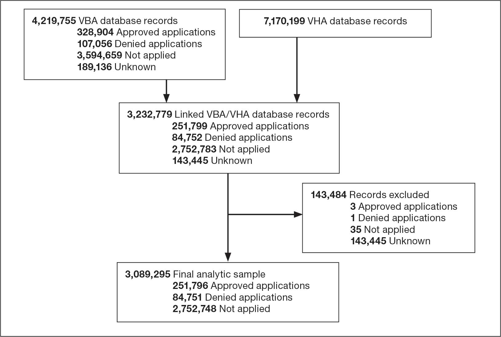
Abbreviations: FY, fiscal year; VBA, Veterans Benefits Administration; VHA, Veterans Health Administration.
Statistical Analysis
All statistical analyses were performed using SAS Enterprise Guide, an application that provides a point-and-click interface for data access, analysis, and management, accommodating both code-based and visual programming. 31 First, we relied on the final analytic sample to calculate the annual proportions of veterans who applied for and/or received a loan through the VA Home Loan Program. We also generated descriptive statistics stratified by age group, sex, race/ethnicity, SUD, mental health disorder, and homelessness, overall and within each FY. Pearson χ2 and Cochran-Armitage trend tests were applied to examine differences in application and receipt of a home loan by baseline characteristics and FY, respectively. Second, we conducted bivariate and multivariable analyses to compare demographic, clinical, and homelessness characteristics between 3 groups of veterans as they pertain to the VA Home Loan Program. Veterans who applied and were nonrecipients of a loan (group 1), veterans who applied and were recipients of a loan (group 2), and veterans who did not apply for a loan (group 3). Similar analyses compared VA Home Loan Program applicants who were recipients of a home loan vs VA Home Loan Program applicants who were nonrecipients of a home loan. Multinomial and binary logistic regression models were constructed to estimate the relative risk ratio (RR) and odds ratio (OR) with 95% CIs for comparisons between these distinct groups on demographic, clinical, and homelessness characteristics. Two-sided statistical tests were evaluated at α = 0.05.
Results
Tables 1 and 2 present the number of VBA applicants, including those who applied for and received benefits through the VA Home Loan Program, by age group, sex, race/ethnicity, as well as histories of SUDs, mental health disorders, and homelessness, overall, and by FY. As shown in Figure 2, 336,547 of 3,089,295 VBA applications (10.9%) pertained to the VA Home Loan Program, with a statistically significant decline in application rates, from 12.2% in FY 2022 to 9.9% in FY 2024 (P < .001 for trend). Among 336,547 veterans who applied for the VA Home Loan Program, 251,796 (74.8%) received a home loan during FYs 2022 to 2024, ranging between 73.8% for FY 2024 and 75.5% for FY 2023 (P < .001 for trend).
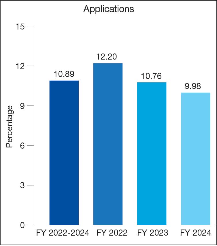
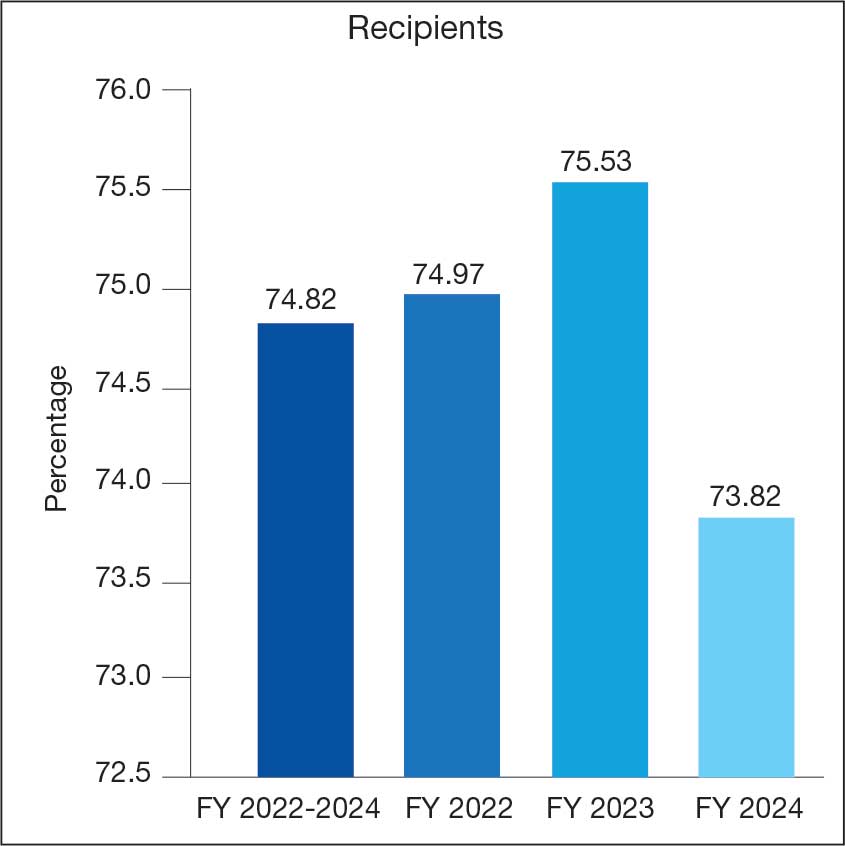
Veterans Affairs Home Loan Program, fiscal years (FY) 2022-2024.
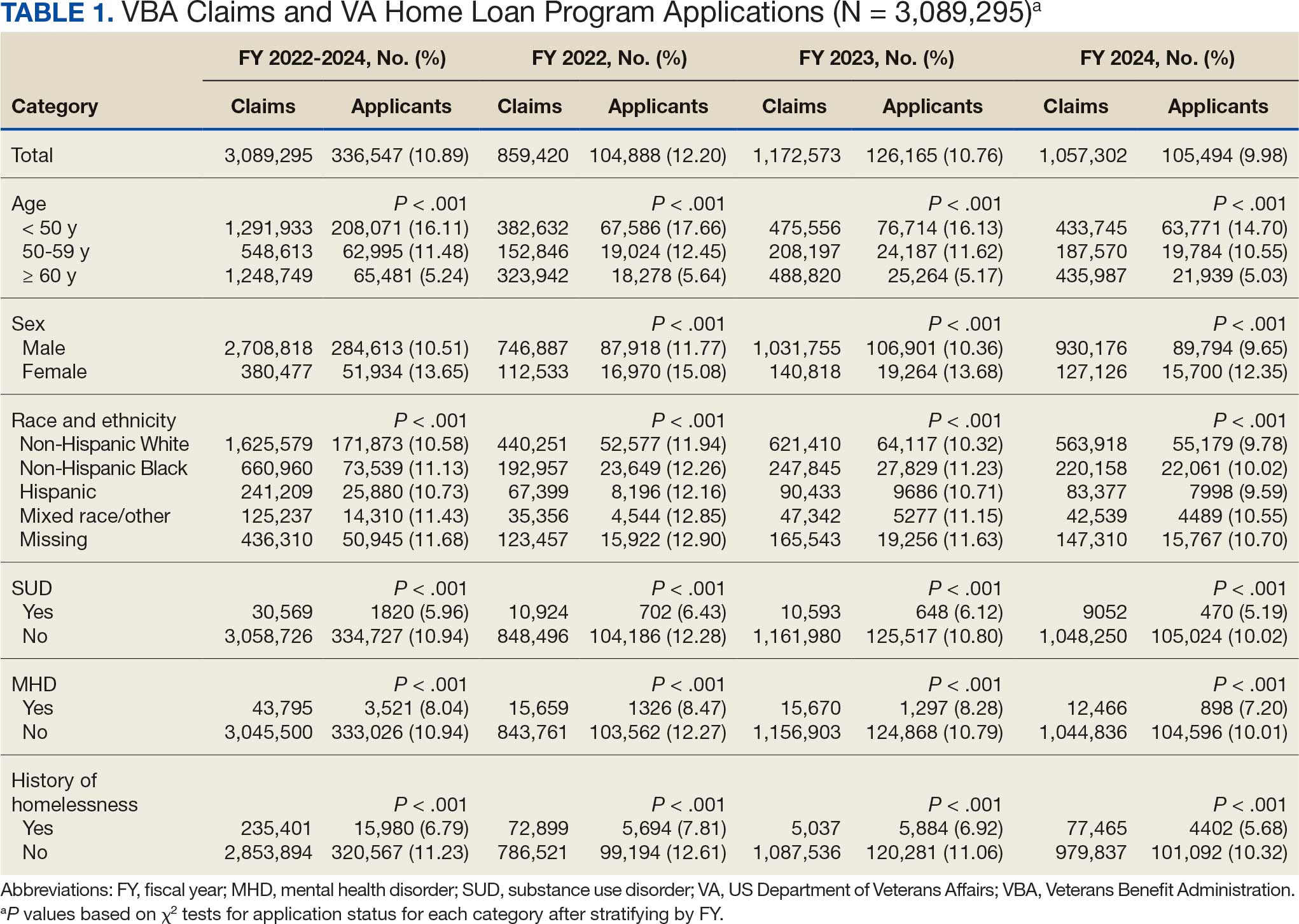
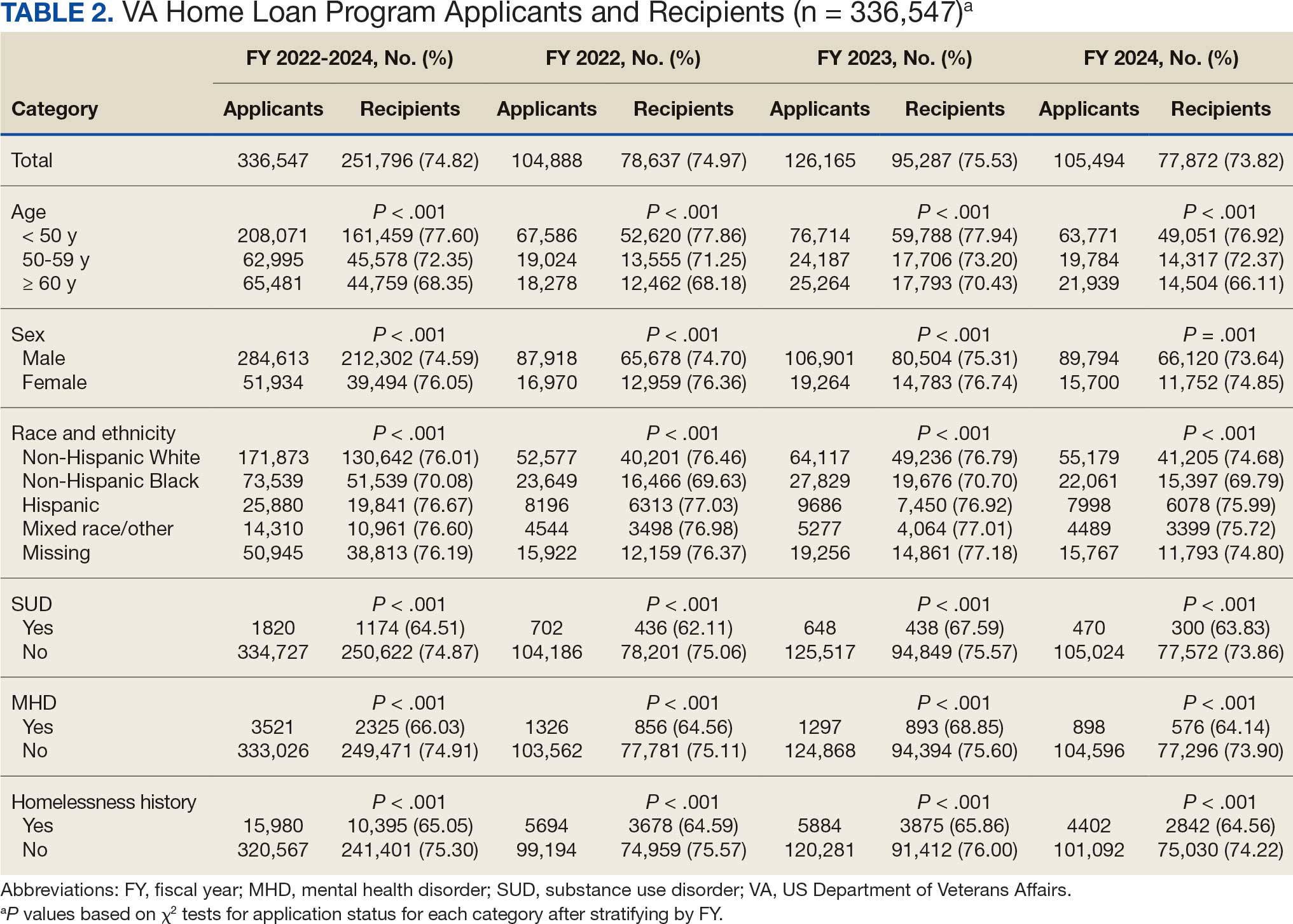
Multinomial logistic regression models for demographic, clinical, and homelessness characteristics as predictors of VA Home Loan Program status are provided in Appendix 1. Based on the fully adjusted model, compared with veterans who did not apply to the VA Home Loan Program, those who applied for a home loan were less likely to be aged ≥ 50 years, unmarried, Hispanic ethnicity, mixed race, or other race, diagnosed with a SUD, or history of homelessness. Veterans with higher VA service-connected disability ratings were more frequently recipients of VA home loans, whereas those who self-identified as non-Hispanic Black and those with higher CCI scores were less frequently recipients of VA home loans. Finally, those with mental health disorders were more likely than their counterparts to be applicants (recipients or nonrecipients) of VA home loans.
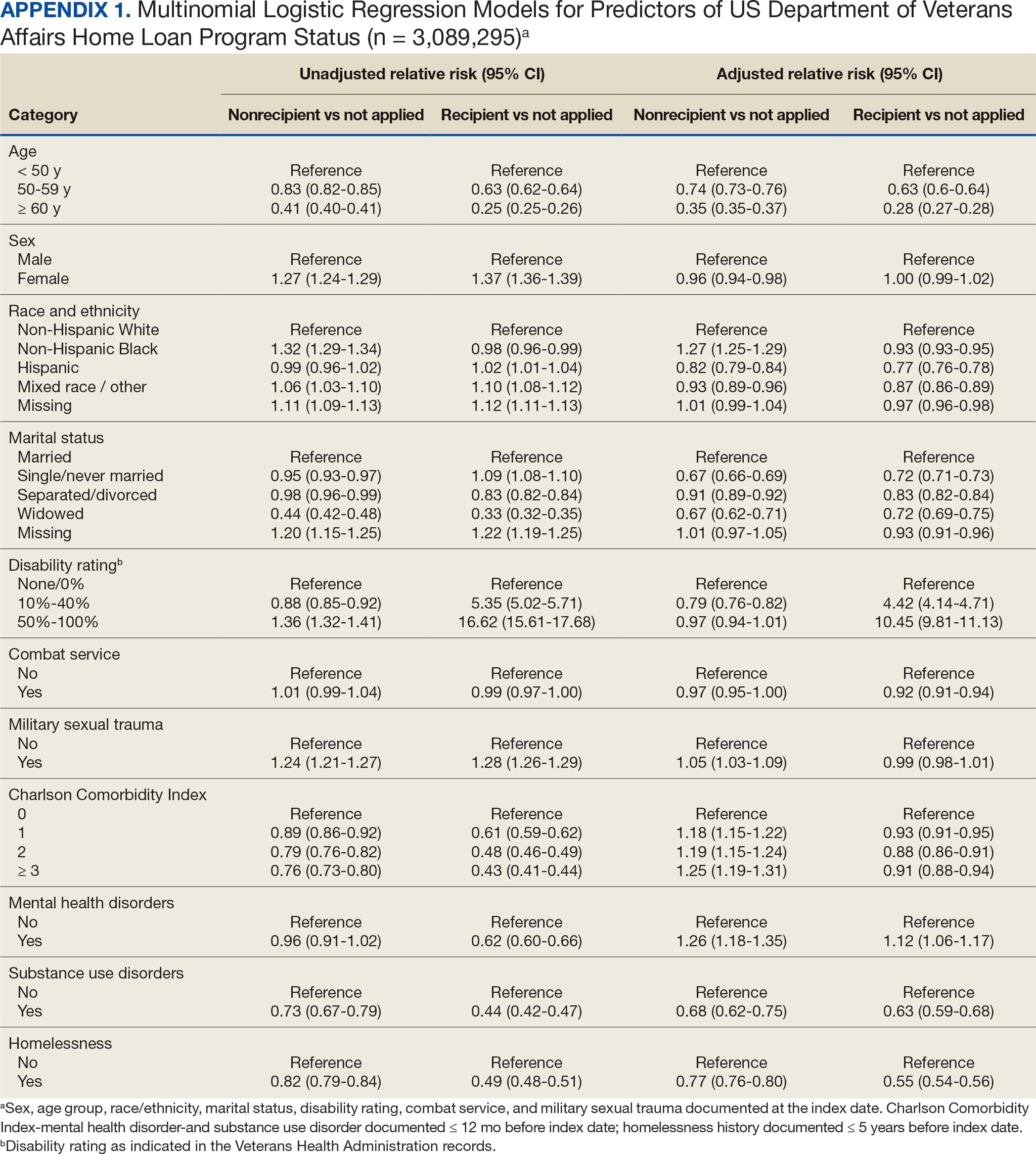
Binary logistic regression models for demographic, clinical, and homelessness characteristics as predictors of receipt status among applicants to the VA Home Loan Program are provided in Appendix 2. Among applicants, those who were granted a VA home loan were less likely to be aged ≥ 50 years; have a CCI score > 0; have experienced combat service and/or military sexual trauma; be diagnosed with a SUD and/or mental health disorder; or to have a history of homelessness compared with those denied a VA home loan. Applicants granted a VA home loan were also more likely to be female, non-Hispanic White, single or never married, and/or have a VA service-connected disability ratings > 0%.
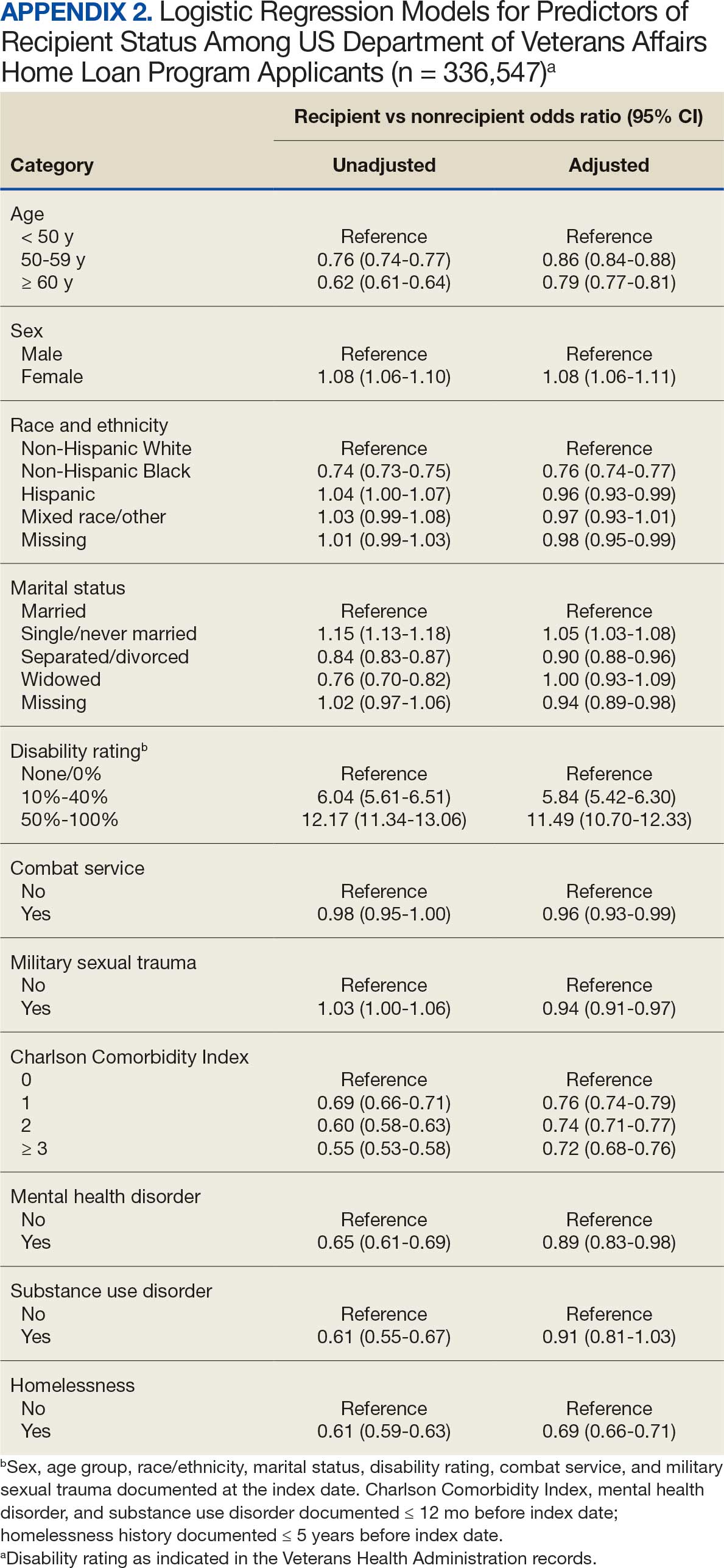
Discussion
The VA Home Loan Program is a unique benefit and resource for eligible veterans that may be increasingly important in a time of growing concern about the affordability of housing for many Americans. Research on other federally-supported home loan programs as well as private home mortgage programs has been mostly conducted in the economic realm, and studies focused on understanding these programs from a health care system perspective have been sparse.32,33 However, there is a large body of literature documenting the importance of stable, safe, and secure housing on health and well-being.34-37 This study did not focus on evaluating the effects of the VA Home Loan Program, because we wanted to first examine the characteristics of veterans who benefited from the program and how they differed from veterans who did not apply or did apply but had a denied application.
Our findings suggest that several thousands of veterans benefit from the VA Home Loan Program each year. For historical context, the time period examined was one of economic downturn with rising costs of living, including housing, and steady increases in homelessness as reported in the annual point-in-time count of sheltered and unsheltered people experiencing homelessness on a single night as mandated by the US Department of Housing and Urban Development.38-40 The Sergeant First Class Heath Robinson Honoring Our Promise to Address Comprehensive Toxics (PACT) Act of 2022 expanded health care and benefits for veterans exposed to burn pits, Agent Orange, and other toxic substances, resulting in more VA disability benefit claims, including large retroactive payments.41-43 Anecdotally, the VBA has noted that the PACT Act helped some homeless veterans with funds and stability to exit homelessness and enroll in the VA Home Loan Program.
Our analysis suggests that beneficiaries of the VA Home Loan Program were frequently aged < 50 years, female, of non-Hispanic White race, and did not have histories of psychiatric disorders or homelessness. Most of these demographic and clinical characteristics were not surprising given the composition of the veteran population, although in-depth analyses are needed to examine sex differences that may have led to more females than males benefiting from the VA Home Loan Program. In addition, it was notable that many younger and non-Hispanic Black veterans had applied. While relatively few veterans with SUDs benefited from the VA Home Loan Program, few had applied. Research is warranted into why veterans with SUDs are less likely to apply for home loans. Quite surprisingly, a sizable proportion of veterans with histories of homelessness reported they had applied to the VA Home Loan Program, although they were less likely than veterans who had not experienced homelessness to be granted a loan.
The examination of differences between veterans who did not apply, were granted, and denied a loan through the VA Home Loan Program revealed several key predictors of application outcomes in multivariable models. Specifically, veterans who applied for home loans were less likely to be aged ≥ 50 years, unmarried, of Hispanic, mixed, or other race/ethnicity, diagnosed with an SUD, or have a history of homelessness. Veterans with higher disability ratings were less frequently denied and more frequently approved, while non-Hispanic Black veterans and those with higher CCI scores were more frequently denied and less frequently approved. VBA applicants with mental health disorders were also more likely to apply for a home loan. Conversely, those granted a home loan were more likely than those denied a home loan to be female, non-Hispanic White, single/unmarried, or to have > 0% VA service-connected disability rating, but less likely to be aged ≥ 50 years, have CCI score > 0, be diagnosed with psychiatric disorders, or have a history of homelessness.
Limitations
This analysis was restricted to a subset of FY 2022 to FY 2024 linked VBA/VHA databases (ie, to veterans who had both VBA and VHA records and met prespecified eligibility criteria). Despite the large number of linked records, a small percentage of these records corresponded to veterans who were applicants or recipients of the VA Home Loan Program. Future studies should expand the time frame to examine variations in application outcomes over time and by background characteristics of veterans enrolled in VHA care who applied for VBA benefits. In addition, we relied on data and ICD-10-CM diagnostic codes from existing electronic health records and claims data to define histories of homelessness, comorbidities, SUDs, and mental health disorders. Given the time-varying nature of these conditions, the temporal sequence of events was difficult to ascertain. Third, it is worth noting that these findings can only be generalized to veterans who applied for VBA benefits and met eligibility criteria, and that these veterans may differ in terms of their demographic and clinical characteristics from those who did not apply for these benefits.
Conclusions
This study analyzed data from 251,796 individuals who applied for and received a VA home loan, 84,751 who were denied a VA home loan, and 2,752,748 veterans who did not apply for a VA home loan from FY 2022 to FY 2024. Accordingly, 11% of applications pertained to the VA Home Loan Program, and 75% of VA Home Loan Program applicants received a home loan. Distinct demographic and clinical characteristics were observed for applicants and recipients of the VA Home Loan Program, which can set the stage for future planning and evaluation of the program. Despite the broad accessibility of veterans to the VA Home Loan Program, there were differences in approval rates among applicants based on sociodemographic and clinical characteristics. Further evaluation, perhaps using qualitative methods, is needed to better understand opportunities and challenges to achieving a VA home loan, especially among underserved veteran populations. Investigation and research can guide future recommendations for any development or corrective actions that can help increase access to veterans who can benefit from the program. Future analyses are also needed to compare veterans enrolled and not enrolled in the VA Home Loan Program on health care-related outcomes.
- US Department of Veterans Affairs. Home loans. Accessed April 1, 2026. https://www.benefits.va.gov/homeloans/
- Veterans United Home Loans. VA loans: the complete guide. Accessed April 1, 2026. https://www.veteransunited.com/va-loans/
- US Department of Veterans Affairs. VA-backed veterans home loans. Accessed April 1, 2026. https://www.va.gov/housing-assistance/home-loans/
- Choplin JM, Stark DP. Whispering sweet nothings: a review of verbal behaviors that undermine the effectiveness of government-mandated home-loan disclosures. Cogn Res Princ Implic. 2019;4:6. doi:10.1186/s41235-019-0154-7
- Evans M. Borrowing boon. More explore federal home loan banks backing. Mod Healthc. 2009;39:14.
- Hogarth M. A home loan: how—and how much? Nurs Times. 1973;69:908-909.
- Jacoby SF. Home Owners’ Loan Corporation maps and place-based injury risks: a complex history. Am J Public Health. 2023;113:356-358. doi:10.2105/AJPH.2023.307242
- Merrell C. Finance. Home: a loan. Nurs Times. 1996;92:61-64.
- Namin S, Xu W, Zhou Y, et al. The legacy of the Home Owners’ Loan Corporation and the political ecology of urban trees and air pollution in the United States. Soc Sci Med. 2020;246:112758. doi:10.1016/j.socscimed.2019.112758
- Namin S, Zhou Y, Xu W, et al. Persistence of mortgage lending bias in the United States: 80 years after the Home Owners’ Loan Corporation security maps. J Race Ethn City. 2022;3:70-94. doi:10.1080/26884674.2021.2019568
- Slottow R. The home loan program. J Natl Assoc Hosp Dev. 1990:43-45.
- Wang M, Chen H, Wang L. Locus of control and home mortgage loan behaviour. Int J Psychol. 2008;43:125-129. doi:10.1080/00207590801888760
- US Dept of Veterans Affairs. Veterans Health Administration. About VHA. Updated January 20, 2025. Accessed April 1, 2026. https://www.va.gov/health/aboutvha.asp
- US Dept of Veterans Affairs. VA homeless programs. Updated May 7, 2026. Accessed May 8, 2026. https://department.va.gov/homeless/
- DiTosto JD, Holder K, Soyemi E, et al. Housing instability and adverse perinatal outcomes: a systematic review. Am J Obstet Gynecol MFM. 2021;3:100477. doi:10.1016/j.ajogmf.2021.100477
- Tsai J, Szymkowiak D, Jutkowitz E. Developing an operational definition of housing instability and homelessness in Veterans Health Administration medical records. PLoS One. 2022;17:e0279973. doi:10.1371/journal.pone.0279973
- Fowler PJ, Hovmand PS, Marcal KE, et al. Solving homelessness from a complex systems perspective: insights for prevention responses. Annu Rev Public Health. 2019;40: 465-486. doi:10.1146/annurev-publhealth-040617-013553
- US Department of Health and Human Services. Healthy People 2030: housing instability. Accessed April 1, 2026. https://health.gov/healthypeople/priority-areas/social-determinants-health/literature-summaries/housing-instability
- US Department of Veterans Affairs. VA health care priorities. Accessed April 1, 2026. https://www.va.gov/health/priorities/index.asp
- Tsai J. Federal priorities to address homelessness as a community health problem. Fam Community Health. 2025;48:57-69.
- Tsai J, Hooshyar D. Prevalence of eviction, home foreclosure, and homelessness among low-income US veterans: the National Veteran Homeless and Other Poverty Experiences study. Public Health. 2022;213:181-188. doi:10.1016/j.puhe.2022.10.017
- US Department of Veterans Affairs. Corporate Data Warehouse (CDW). Accessed April 1, 2026. https://www.hsrd.research.va.gov/for_researchers/cdw.cfm
- Price LE, Shea K, Gephart S. The Veterans Affairs Corporate Data Warehouse: uses and implications for nursing research and practice. Nurs Adm Q. 2015;39:311-318. doi:10.1097/NAQ.0000000000000118
- US Department of Veterans Affairs. Homeless Operations Management and Evaluation System (HOMES) User Manual—Phase 1. April 19, 2011. Accessed April 1, 2026. https://www.adldata.org/wp-content/uploads/2016/07/homes.pdf
- Tsai J, Kasprow WJ, Rosenheck RA. Latent homeless risk profiles of a national sample of homeless veterans and their relation to program referral and admission patterns. Am J Public Health. 2013;103:S239-S247. doi:10.2105/AJPH.2013.301322
- Sundararajan V, Henderson T, Perry C, et al. New ICD-10 version of the Charlson comorbidity index predicted inhospital mortality. J Clin Epidemiol. 2004;57:1288-1294. doi:10.1016/j.jclinepi.2004.03.012
- Beydoun HA, Szymkowiak D, Beydoun MA, et al. Comparing major comorbidity indices as predictors of all-cause mortality in the Veterans Affairs health care system. J Clin Epidemiol. 2025;182:111778. doi:10.1016/j.jclinepi.2025.111778
- Charlson ME, Carrozzino D, Guidi J, et al. Charlson Comorbidity Index: a critical review of clinimetric properties. Psychother Psychosom. 2022;91:8-35. doi:10.1159/000521288
- Glasheen WP, Cordier T, Gumpina R, et al. Charlson Comorbidity Index: ICD-9 update and ICD-10 translation. Am Health Drug Benefits. 2019;12:188-197.
- Beydoun HA, Szymkowiak D, Kinney R, et al. Is the risk of Alzheimer’s disease and related dementias among US veterans influenced by the intersectionality of housing status, HIV/AIDS, hepatitis C, and psychiatric disorders? J Gerontol A Biol Sci Med Sci. 2024;79:glae153. doi:10.1093/gerona/glae153
- SAS Institute. SAS Enterprise Guide. Accessed April 1, 2026. https://www.sas.com/en_us/software/enterprise-guide/features-list.html
- Agarwal S, Amromin G, Chomsisengphet S, et al. Mortgage refinancing, consumer spending, and competition: evidence from the Home Affordable Refinance Program. Rev Econ Stud. 2023;90:499-537.
- Ashcraft A, Bech ML, Frame WS. The Federal Home Loan Bank System: the lender of next-to-last resort? J Money Credit Bank. 2010;42:551-583.
- Gibson M, Petticrew M, Bambra C, et al. Housing and health inequalities: a synthesis of systematic reviews of interventions aimed at different pathways linking housing and health. Health Place. 2011;17:175-184. doi:10.1016/j.healthplace.2010.09.011
- Shaw M. Housing and public health. Annu Rev Public Health. 2004; 25:397-418. doi:10.1146/annurev.publhealth.25.101802.123036
- Thomson H, Petticrew M, Morrison D. Health effects of housing improvement: systematic review of intervention studies. BMJ. 2001;323:187-190. doi:10.1136/bmj.323.7306.187
- Tsai J. Theorizing pathways between eviction filings and increased mortality risk. JAMA. 2024;331:570-571. doi:10.1001/jama.2023.27978
- Bernanke B, Blanchard O. What caused the US pandemicera inflation? Am Econ J Macroecon. 2025;17:1-35.
- Hall SG, Tavlas GS, Wang Y. Drivers and spillover effects of inflation: the United States, the euro area, and the United Kingdom. J Int Money Finance. 2023;131:1-13.
- US Department of Housing and Urban Development. Point-in-Time Count and Housing Inventory Count. Accessed April 1, 2026. https://www.hudexchange.info/programs/hdx/pit-hic/
- Beckman AL, Jacobs J, Elnahal SM. The PACT Act: expanding coverage and access for veterans. JAMA. 2024;332:1423-1424. doi:10.1001/jama.2024.16013
- Zychowicz ME. The PACT Act: enhancing health care access for military personnel and veterans. N C Med J. 2023;84:379-380. doi:10.18043/001c.89208
- US Department of Veterans Affairs. The PACT Act and your VA benefits. April 2, 2026. https://www.va.gov/resources/the-pact-act-and-your-va-benefits/
The US Department of Veterans Affairs (VA) Home Loan Program, administered by the Veterans Benefits Administration (VBA), is a unique benefit for veterans, active-duty service members, National Guard and Reserve members, and eligible surviving spouses. Established in 1944, the program aims to help these individuals achieve homeownership by leveraging a third-party guarantee, typically from a government agency, to enhance access to credit and improve loan terms for borrowers who may not meet conventional loan qualifications.1 Since its inception, the VA has guaranteed > 28.5 million loans, enabling millions of veterans to buy, build, repair, retain, or adapt homes for personal occupancy.2 The program is designed to support veterans and eligible individuals to become homeowners, recognizing homeownership as a pathway to financial stability and community integration. VA home loans are provided by private lenders (eg, banks, mortgage companies) with a portion guaranteed by the VA, which reduces the risk for lenders and enables them to offer competitive terms, such as no down payment and lower interest rates, making homeownership more accessible to veterans.2
Eligibility criteria for the VA Home Loan Program include military service criteria such as active-duty service members with ≥ 90 continuous days of service; veterans with an honorable discharge meeting minimum service requirements; individuals who served in the National Guard/Reserve for ≥ 90 days of active service or 6 years of service with an honorable discharge; and surviving spouses of veterans who died in service or from a service-connected disability, were designated as missing in action/ prisoner of war, and the spouse is receiving Dependency and Indemnity Compensation. Financial criteria also apply: borrowers must meet lender requirements for credit and income (although VA loans are more flexible than conventional loans) and the home must be for personal occupancy rather than an investment property.3
A June 2025 PubMed literature search did not reveal any prior research on the VA Home Loan Program, although a limited number of studies tackled a wide range of issues related to federal and private home loans.4-12 To our knowledge, there is no prior published examination of the VA Home Loan Program. Understanding VA Home Loan Program usage among Veterans Health Administration (VHA) users can inform the future direction of the program. The VHA operates the largest integrated US health care system, serving > 9 million enrolled veterans annually at 1321 facilities, including 172 medical centers and 1138 outpatient clinics, providing primary and specialized health care, and related medical and social support services for enrolled veterans, including those who are experiencing housing instability or homelessness.13 Specialized VHA programs for homeless veterans include housing, employment, health care, justice, and re-entryrelated services in collaboration with federal and community partners.14 Housing instability has been defined as the state of being at risk of losing housing due to challenges such as difficulties paying rent, overcrowding, frequent relocation, and a substantial proportion of income spent on housing.15,16 Homelessness is a severe manifestation of housing instability that has been defined as the lack of stable, safe, and functioning housing.17,18
Health care and social services, including those that address housing instability and homelessness, are major priorities for the VHA and VBA.19 The VA Home Loan Program may represent an important resource to help veterans achieve long-term housing stability through home ownership. There has been wide public concern about housing affordability and the ability of many Americans, including veterans, to achieve home ownership.20 Homeownership is considered an important part of developing financial assets and achieving financial stability. Lowincome veterans, in particular, may benefit from this program as a national study found that 8.0% of low-income veterans and 13.9% of veterans with a history of homelessness have previously experienced a home foreclosure. 21 A greater understanding of who applies for and receives assistance from the VA Home Loan Program would inform homelessness prevention services and future planning for this program.
We conducted a quality improvement (QI) project on behalf of the VHA Homeless Programs Office and in partnership with the VBA. Our goals were to: (1) describe the annual number of applicants and recipients of the VA Home Loan Program by age group, sex, race/ethnicity, presence of any diagnosed substance use and/or mental health disorder, and history of homelessness; and (2) compare demographic, clinical, and homelessness characteristics among individuals who apply and are granted a loan through this program, individuals who apply and are denied a loan through this program, and individuals who do not apply for a loan through this program.
Methods
This project involved linked VA administrative national databases and was undertaken by the VHA Homeless Programs Office in partnership with the VBA. Specifically, VHA and VBA databases were linked together using veteran identifiers and all data were managed and analyzed on secure VA servers. The project followed VA’s Program Guide 1200.21 for nonresearch activities and institutional review board approval was waived through sponsorship by the VA Homeless Programs Office. The VHA Corporate Data Warehouse (CDW) was accessed to obtain data from the Homeless Operations Management and Evaluation System (HOMES) and other clinical data systems used by VHA clinicians and administrators that capture diagnoses, workload, and other health care data.22,23 HOMES collects intake, progress, and outcome data on homeless veterans within its care system that enables the VA to assess the effectiveness of programs and strategically allocate resources to prevent homelessness.24,25
A list of veterans who filed disability compensation and pension claims was obtained from the VBA Office of Performance Analysis and Integrity, including Social Security number, name, city and state, date of claim submission, grant or increase in benefits, homeless status, VA home loan approval, and homeless aid for dependent children from fiscal year (FY) 2022 through FY 2024. VBA data were linked to VHA CDW electronic health record data from veterans who sought VA health care services and HOMES data on veteran participation in homeless programs who were also experiencing homelessness. VHA data included demographic characteristics (eg, sex, age, race, marital status, combat service) at an index date (earliest visit to the VHA between October 1, 2021, and September 30, 2024); military sexual trauma; clinical characteristics within 12 months prior to the index date (VHA disability rating, substance use disorder [SUD] diagnosis, mental health disorder diagnosis, Charlson Comorbidity Index [CCI] score), and homelessness experience ≤ 5 years prior to the index date.
History of homelessness ≤ 5 years prior to the index date was determined using an operational definition of homelessness based on multiple indicators, including International Classification of Diseases, Tenth Revision, Clinical Modification (ICD-10-CM) diagnostic code Z59.0; clinic stop codes or HOMES records indicating VA homeless programs clinical encounters; or a positive screen on an annual homelessness screener.16 US Department of Housing and Urban Development-VA Supportive Housing enrollees were excluded because they are considered to no longer be experiencing homelessness, and Veterans Justice Program enrollees were excluded because the program primarily focuses on serving criminal justice-involved veterans. The CCI predicts the risk of death ≤ 1 year by assessing the number and severity of a patient’s coexisting health conditions and is a valuable tool for understanding a patient’s overall health burden, aiding in clinical decision-making and evaluation research studies.26-29 Diagnoses based on ICD-10-CM codes were used to determine SUDs, mental health disorders, and CCI score, using methods that have been described in other publications.30
Population
The VBA cohort of veterans requesting benefits was further restricted to those who met the following eligibility criteria: (1) requested VA benefits FYs 2022 to 2024; (2) sought VHA services ≥ 1 time between FY 2022 and 2024; (3) had matching VBA/VHA records; (4) had no missing data on claim status and/ or demographic, clinical, and homelessness characteristics; and (5) had known home loan status FYs 2022 to 2024. The original VBA dataset consisted of 4,219,755 records and the original VHA dataset consisted of 7,170,199 records (Figure 1). The final linked VBA/VHA dataset after excluding 29 records with missing data on sex, 7 with missing data on age, 6 with missing data on marital status, and an additional 143,444 with unknown VBA claim status, consisted of 3,089,295 records corresponding to 2,260,851 unique veterans. Specifically, 251,796 records corresponded to veterans who had applied and received a loan, 84,751 to veterans who had applied and were nonrecipients of a loan, and 2,752,748 to veterans who did not apply for a loan.
Abbreviations: FY, fiscal year; VBA, Veterans Benefits Administration; VHA, Veterans Health Administration.
Statistical Analysis
All statistical analyses were performed using SAS Enterprise Guide, an application that provides a point-and-click interface for data access, analysis, and management, accommodating both code-based and visual programming. 31 First, we relied on the final analytic sample to calculate the annual proportions of veterans who applied for and/or received a loan through the VA Home Loan Program. We also generated descriptive statistics stratified by age group, sex, race/ethnicity, SUD, mental health disorder, and homelessness, overall and within each FY. Pearson χ2 and Cochran-Armitage trend tests were applied to examine differences in application and receipt of a home loan by baseline characteristics and FY, respectively. Second, we conducted bivariate and multivariable analyses to compare demographic, clinical, and homelessness characteristics between 3 groups of veterans as they pertain to the VA Home Loan Program. Veterans who applied and were nonrecipients of a loan (group 1), veterans who applied and were recipients of a loan (group 2), and veterans who did not apply for a loan (group 3). Similar analyses compared VA Home Loan Program applicants who were recipients of a home loan vs VA Home Loan Program applicants who were nonrecipients of a home loan. Multinomial and binary logistic regression models were constructed to estimate the relative risk ratio (RR) and odds ratio (OR) with 95% CIs for comparisons between these distinct groups on demographic, clinical, and homelessness characteristics. Two-sided statistical tests were evaluated at α = 0.05.
Results
Tables 1 and 2 present the number of VBA applicants, including those who applied for and received benefits through the VA Home Loan Program, by age group, sex, race/ethnicity, as well as histories of SUDs, mental health disorders, and homelessness, overall, and by FY. As shown in Figure 2, 336,547 of 3,089,295 VBA applications (10.9%) pertained to the VA Home Loan Program, with a statistically significant decline in application rates, from 12.2% in FY 2022 to 9.9% in FY 2024 (P < .001 for trend). Among 336,547 veterans who applied for the VA Home Loan Program, 251,796 (74.8%) received a home loan during FYs 2022 to 2024, ranging between 73.8% for FY 2024 and 75.5% for FY 2023 (P < .001 for trend).
Veterans Affairs Home Loan Program, fiscal years (FY) 2022-2024.
Multinomial logistic regression models for demographic, clinical, and homelessness characteristics as predictors of VA Home Loan Program status are provided in Appendix 1. Based on the fully adjusted model, compared with veterans who did not apply to the VA Home Loan Program, those who applied for a home loan were less likely to be aged ≥ 50 years, unmarried, Hispanic ethnicity, mixed race, or other race, diagnosed with a SUD, or history of homelessness. Veterans with higher VA service-connected disability ratings were more frequently recipients of VA home loans, whereas those who self-identified as non-Hispanic Black and those with higher CCI scores were less frequently recipients of VA home loans. Finally, those with mental health disorders were more likely than their counterparts to be applicants (recipients or nonrecipients) of VA home loans.
Binary logistic regression models for demographic, clinical, and homelessness characteristics as predictors of receipt status among applicants to the VA Home Loan Program are provided in Appendix 2. Among applicants, those who were granted a VA home loan were less likely to be aged ≥ 50 years; have a CCI score > 0; have experienced combat service and/or military sexual trauma; be diagnosed with a SUD and/or mental health disorder; or to have a history of homelessness compared with those denied a VA home loan. Applicants granted a VA home loan were also more likely to be female, non-Hispanic White, single or never married, and/or have a VA service-connected disability ratings > 0%.
Discussion
The VA Home Loan Program is a unique benefit and resource for eligible veterans that may be increasingly important in a time of growing concern about the affordability of housing for many Americans. Research on other federally-supported home loan programs as well as private home mortgage programs has been mostly conducted in the economic realm, and studies focused on understanding these programs from a health care system perspective have been sparse.32,33 However, there is a large body of literature documenting the importance of stable, safe, and secure housing on health and well-being.34-37 This study did not focus on evaluating the effects of the VA Home Loan Program, because we wanted to first examine the characteristics of veterans who benefited from the program and how they differed from veterans who did not apply or did apply but had a denied application.
Our findings suggest that several thousands of veterans benefit from the VA Home Loan Program each year. For historical context, the time period examined was one of economic downturn with rising costs of living, including housing, and steady increases in homelessness as reported in the annual point-in-time count of sheltered and unsheltered people experiencing homelessness on a single night as mandated by the US Department of Housing and Urban Development.38-40 The Sergeant First Class Heath Robinson Honoring Our Promise to Address Comprehensive Toxics (PACT) Act of 2022 expanded health care and benefits for veterans exposed to burn pits, Agent Orange, and other toxic substances, resulting in more VA disability benefit claims, including large retroactive payments.41-43 Anecdotally, the VBA has noted that the PACT Act helped some homeless veterans with funds and stability to exit homelessness and enroll in the VA Home Loan Program.
Our analysis suggests that beneficiaries of the VA Home Loan Program were frequently aged < 50 years, female, of non-Hispanic White race, and did not have histories of psychiatric disorders or homelessness. Most of these demographic and clinical characteristics were not surprising given the composition of the veteran population, although in-depth analyses are needed to examine sex differences that may have led to more females than males benefiting from the VA Home Loan Program. In addition, it was notable that many younger and non-Hispanic Black veterans had applied. While relatively few veterans with SUDs benefited from the VA Home Loan Program, few had applied. Research is warranted into why veterans with SUDs are less likely to apply for home loans. Quite surprisingly, a sizable proportion of veterans with histories of homelessness reported they had applied to the VA Home Loan Program, although they were less likely than veterans who had not experienced homelessness to be granted a loan.
The examination of differences between veterans who did not apply, were granted, and denied a loan through the VA Home Loan Program revealed several key predictors of application outcomes in multivariable models. Specifically, veterans who applied for home loans were less likely to be aged ≥ 50 years, unmarried, of Hispanic, mixed, or other race/ethnicity, diagnosed with an SUD, or have a history of homelessness. Veterans with higher disability ratings were less frequently denied and more frequently approved, while non-Hispanic Black veterans and those with higher CCI scores were more frequently denied and less frequently approved. VBA applicants with mental health disorders were also more likely to apply for a home loan. Conversely, those granted a home loan were more likely than those denied a home loan to be female, non-Hispanic White, single/unmarried, or to have > 0% VA service-connected disability rating, but less likely to be aged ≥ 50 years, have CCI score > 0, be diagnosed with psychiatric disorders, or have a history of homelessness.
Limitations
This analysis was restricted to a subset of FY 2022 to FY 2024 linked VBA/VHA databases (ie, to veterans who had both VBA and VHA records and met prespecified eligibility criteria). Despite the large number of linked records, a small percentage of these records corresponded to veterans who were applicants or recipients of the VA Home Loan Program. Future studies should expand the time frame to examine variations in application outcomes over time and by background characteristics of veterans enrolled in VHA care who applied for VBA benefits. In addition, we relied on data and ICD-10-CM diagnostic codes from existing electronic health records and claims data to define histories of homelessness, comorbidities, SUDs, and mental health disorders. Given the time-varying nature of these conditions, the temporal sequence of events was difficult to ascertain. Third, it is worth noting that these findings can only be generalized to veterans who applied for VBA benefits and met eligibility criteria, and that these veterans may differ in terms of their demographic and clinical characteristics from those who did not apply for these benefits.
Conclusions
This study analyzed data from 251,796 individuals who applied for and received a VA home loan, 84,751 who were denied a VA home loan, and 2,752,748 veterans who did not apply for a VA home loan from FY 2022 to FY 2024. Accordingly, 11% of applications pertained to the VA Home Loan Program, and 75% of VA Home Loan Program applicants received a home loan. Distinct demographic and clinical characteristics were observed for applicants and recipients of the VA Home Loan Program, which can set the stage for future planning and evaluation of the program. Despite the broad accessibility of veterans to the VA Home Loan Program, there were differences in approval rates among applicants based on sociodemographic and clinical characteristics. Further evaluation, perhaps using qualitative methods, is needed to better understand opportunities and challenges to achieving a VA home loan, especially among underserved veteran populations. Investigation and research can guide future recommendations for any development or corrective actions that can help increase access to veterans who can benefit from the program. Future analyses are also needed to compare veterans enrolled and not enrolled in the VA Home Loan Program on health care-related outcomes.
- US Department of Veterans Affairs. Home loans. Accessed April 1, 2026. https://www.benefits.va.gov/homeloans/
- Veterans United Home Loans. VA loans: the complete guide. Accessed April 1, 2026. https://www.veteransunited.com/va-loans/
- US Department of Veterans Affairs. VA-backed veterans home loans. Accessed April 1, 2026. https://www.va.gov/housing-assistance/home-loans/
- Choplin JM, Stark DP. Whispering sweet nothings: a review of verbal behaviors that undermine the effectiveness of government-mandated home-loan disclosures. Cogn Res Princ Implic. 2019;4:6. doi:10.1186/s41235-019-0154-7
- Evans M. Borrowing boon. More explore federal home loan banks backing. Mod Healthc. 2009;39:14.
- Hogarth M. A home loan: how—and how much? Nurs Times. 1973;69:908-909.
- Jacoby SF. Home Owners’ Loan Corporation maps and place-based injury risks: a complex history. Am J Public Health. 2023;113:356-358. doi:10.2105/AJPH.2023.307242
- Merrell C. Finance. Home: a loan. Nurs Times. 1996;92:61-64.
- Namin S, Xu W, Zhou Y, et al. The legacy of the Home Owners’ Loan Corporation and the political ecology of urban trees and air pollution in the United States. Soc Sci Med. 2020;246:112758. doi:10.1016/j.socscimed.2019.112758
- Namin S, Zhou Y, Xu W, et al. Persistence of mortgage lending bias in the United States: 80 years after the Home Owners’ Loan Corporation security maps. J Race Ethn City. 2022;3:70-94. doi:10.1080/26884674.2021.2019568
- Slottow R. The home loan program. J Natl Assoc Hosp Dev. 1990:43-45.
- Wang M, Chen H, Wang L. Locus of control and home mortgage loan behaviour. Int J Psychol. 2008;43:125-129. doi:10.1080/00207590801888760
- US Dept of Veterans Affairs. Veterans Health Administration. About VHA. Updated January 20, 2025. Accessed April 1, 2026. https://www.va.gov/health/aboutvha.asp
- US Dept of Veterans Affairs. VA homeless programs. Updated May 7, 2026. Accessed May 8, 2026. https://department.va.gov/homeless/
- DiTosto JD, Holder K, Soyemi E, et al. Housing instability and adverse perinatal outcomes: a systematic review. Am J Obstet Gynecol MFM. 2021;3:100477. doi:10.1016/j.ajogmf.2021.100477
- Tsai J, Szymkowiak D, Jutkowitz E. Developing an operational definition of housing instability and homelessness in Veterans Health Administration medical records. PLoS One. 2022;17:e0279973. doi:10.1371/journal.pone.0279973
- Fowler PJ, Hovmand PS, Marcal KE, et al. Solving homelessness from a complex systems perspective: insights for prevention responses. Annu Rev Public Health. 2019;40: 465-486. doi:10.1146/annurev-publhealth-040617-013553
- US Department of Health and Human Services. Healthy People 2030: housing instability. Accessed April 1, 2026. https://health.gov/healthypeople/priority-areas/social-determinants-health/literature-summaries/housing-instability
- US Department of Veterans Affairs. VA health care priorities. Accessed April 1, 2026. https://www.va.gov/health/priorities/index.asp
- Tsai J. Federal priorities to address homelessness as a community health problem. Fam Community Health. 2025;48:57-69.
- Tsai J, Hooshyar D. Prevalence of eviction, home foreclosure, and homelessness among low-income US veterans: the National Veteran Homeless and Other Poverty Experiences study. Public Health. 2022;213:181-188. doi:10.1016/j.puhe.2022.10.017
- US Department of Veterans Affairs. Corporate Data Warehouse (CDW). Accessed April 1, 2026. https://www.hsrd.research.va.gov/for_researchers/cdw.cfm
- Price LE, Shea K, Gephart S. The Veterans Affairs Corporate Data Warehouse: uses and implications for nursing research and practice. Nurs Adm Q. 2015;39:311-318. doi:10.1097/NAQ.0000000000000118
- US Department of Veterans Affairs. Homeless Operations Management and Evaluation System (HOMES) User Manual—Phase 1. April 19, 2011. Accessed April 1, 2026. https://www.adldata.org/wp-content/uploads/2016/07/homes.pdf
- Tsai J, Kasprow WJ, Rosenheck RA. Latent homeless risk profiles of a national sample of homeless veterans and their relation to program referral and admission patterns. Am J Public Health. 2013;103:S239-S247. doi:10.2105/AJPH.2013.301322
- Sundararajan V, Henderson T, Perry C, et al. New ICD-10 version of the Charlson comorbidity index predicted inhospital mortality. J Clin Epidemiol. 2004;57:1288-1294. doi:10.1016/j.jclinepi.2004.03.012
- Beydoun HA, Szymkowiak D, Beydoun MA, et al. Comparing major comorbidity indices as predictors of all-cause mortality in the Veterans Affairs health care system. J Clin Epidemiol. 2025;182:111778. doi:10.1016/j.jclinepi.2025.111778
- Charlson ME, Carrozzino D, Guidi J, et al. Charlson Comorbidity Index: a critical review of clinimetric properties. Psychother Psychosom. 2022;91:8-35. doi:10.1159/000521288
- Glasheen WP, Cordier T, Gumpina R, et al. Charlson Comorbidity Index: ICD-9 update and ICD-10 translation. Am Health Drug Benefits. 2019;12:188-197.
- Beydoun HA, Szymkowiak D, Kinney R, et al. Is the risk of Alzheimer’s disease and related dementias among US veterans influenced by the intersectionality of housing status, HIV/AIDS, hepatitis C, and psychiatric disorders? J Gerontol A Biol Sci Med Sci. 2024;79:glae153. doi:10.1093/gerona/glae153
- SAS Institute. SAS Enterprise Guide. Accessed April 1, 2026. https://www.sas.com/en_us/software/enterprise-guide/features-list.html
- Agarwal S, Amromin G, Chomsisengphet S, et al. Mortgage refinancing, consumer spending, and competition: evidence from the Home Affordable Refinance Program. Rev Econ Stud. 2023;90:499-537.
- Ashcraft A, Bech ML, Frame WS. The Federal Home Loan Bank System: the lender of next-to-last resort? J Money Credit Bank. 2010;42:551-583.
- Gibson M, Petticrew M, Bambra C, et al. Housing and health inequalities: a synthesis of systematic reviews of interventions aimed at different pathways linking housing and health. Health Place. 2011;17:175-184. doi:10.1016/j.healthplace.2010.09.011
- Shaw M. Housing and public health. Annu Rev Public Health. 2004; 25:397-418. doi:10.1146/annurev.publhealth.25.101802.123036
- Thomson H, Petticrew M, Morrison D. Health effects of housing improvement: systematic review of intervention studies. BMJ. 2001;323:187-190. doi:10.1136/bmj.323.7306.187
- Tsai J. Theorizing pathways between eviction filings and increased mortality risk. JAMA. 2024;331:570-571. doi:10.1001/jama.2023.27978
- Bernanke B, Blanchard O. What caused the US pandemicera inflation? Am Econ J Macroecon. 2025;17:1-35.
- Hall SG, Tavlas GS, Wang Y. Drivers and spillover effects of inflation: the United States, the euro area, and the United Kingdom. J Int Money Finance. 2023;131:1-13.
- US Department of Housing and Urban Development. Point-in-Time Count and Housing Inventory Count. Accessed April 1, 2026. https://www.hudexchange.info/programs/hdx/pit-hic/
- Beckman AL, Jacobs J, Elnahal SM. The PACT Act: expanding coverage and access for veterans. JAMA. 2024;332:1423-1424. doi:10.1001/jama.2024.16013
- Zychowicz ME. The PACT Act: enhancing health care access for military personnel and veterans. N C Med J. 2023;84:379-380. doi:10.18043/001c.89208
- US Department of Veterans Affairs. The PACT Act and your VA benefits. April 2, 2026. https://www.va.gov/resources/the-pact-act-and-your-va-benefits/
Characteristics of Applicants and Recipients of the Veterans Affairs Home Loan Program
Characteristics of Applicants and Recipients of the Veterans Affairs Home Loan Program

Microcystic Adnexal Carcinoma– like Neoplasm in a Patient With POT1 Mutation
Microcystic Adnexal Carcinoma– like Neoplasm in a Patient With POT1 Mutation
A 72-year-old man with a history of multiple cancers, including melanoma, squamous cell carcinoma (SCC), and basal cell carcinoma (BCC), presented to the dermatology clinic for a regularly scheduled full-body skin examination. His family history was negative for malignancy, but due to his personal history of both primary internal cancers and skin cancers, the patient previously had been referred by dermatology to a medical geneticist for evaluation. He tested positive for a pathogenic POT1 (protection of telomeres 1) variant associated with tumor predisposition, which most often is associated with cutaneous melanoma, chronic lymphocytic leukemia (CLL), angiosarcoma, and gliomas.1
At the current presentation, physical examination revealed a small, asymmetric, pink papule on the superior thoracic spine. A biopsy of the lesion was performed (Figure 1). Pathology demonstrated cornifying cystic structures with a granulomatous response at the surface of the tumor, ductal differentiation with depth, and infiltrative strands and cords of hyperchromatic cells within a collagenous stroma at the base of the specimen (Figures 2A and 2B). One unusual finding was the presence of prominent clear-cell change within the superficial portion of the neoplasm (Figure 2C). Immunohistochemical stains revealed strong p63 and p40 positivity. Epithelial membrane antigen staining was positive in the hyperchromatic strands and cords with depth but not in the clear-cell superficial portion. Similarly, periodic acid–Schiff–positive material increased within tumor cells in proportion to depth of infiltration. Additional immunohistochemical staining showed carcinoembryonic antigen was largely negative (with rare positivity in a few ductal lumina), with negative results for S100, SOX10, CD117, BerEP4, factor XIIIa, CD34, and cytokeratin 7 (Figures 2D and 2E).

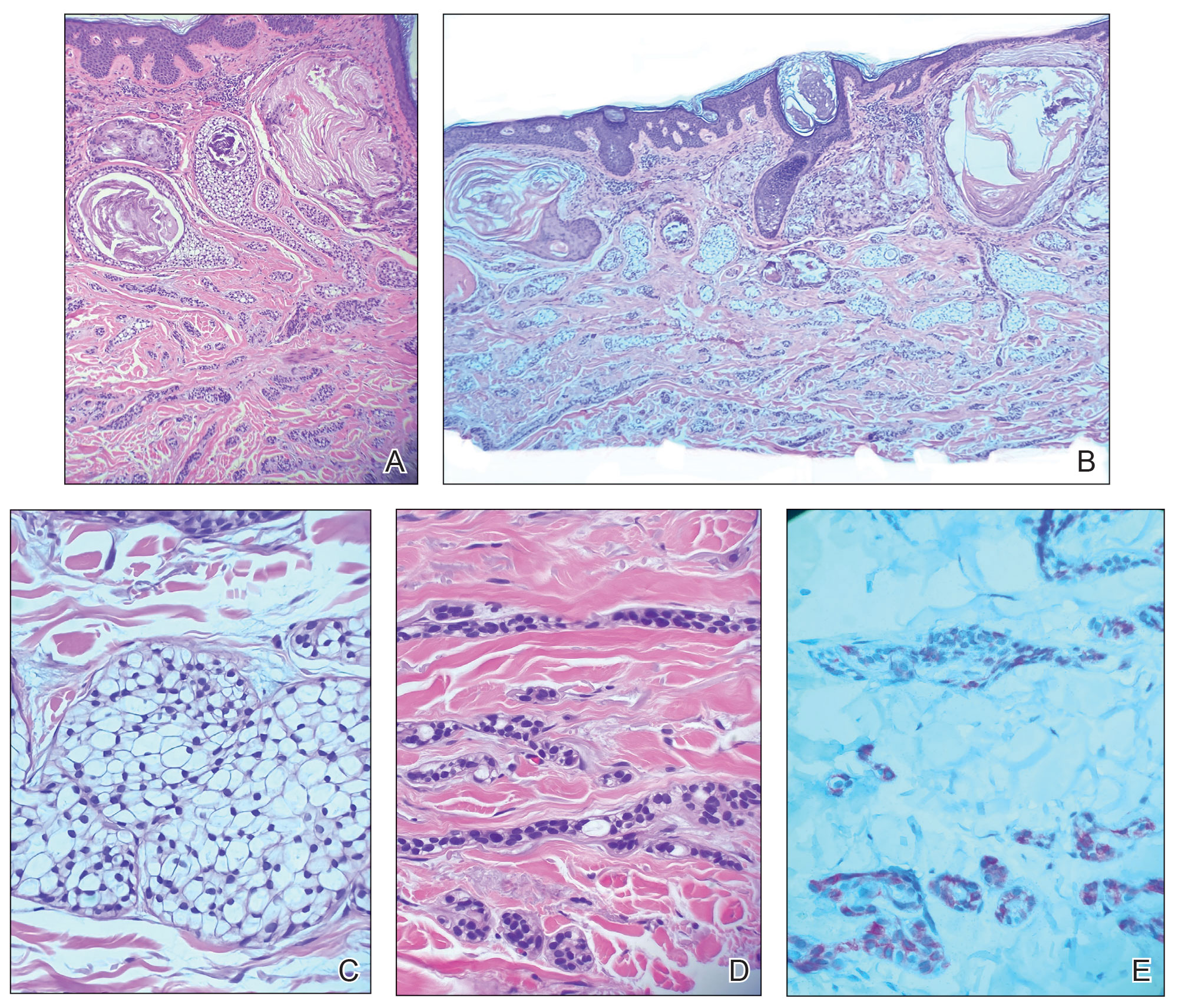
The differential diagnoses included trichilemmal carcinoma (which may manifest with CD34 expression),2 clear cell BCC, adenoid cystic carcinoma (tubular variant), sebaceous carcinoma, and eccrine carcinoma. Importantly, the patient was under continuous oncologic surveillance, with no evidence of a primary internal tumor to suggest metastasis. Despite negative carcinoembryonic antigen staining, the immunohistochemical and histopathologic findings fit best with a primary cutaneous malignant eccrine tumor, specifically microcystic adnexal carcinoma (MAC), in which p63 typically stains peripheral cells but solid variants have been described.3
Eccrine carcinoma is exceedingly rare, reported in 0.01% of diagnosed cutaneous malignancies, and demonstrates overlapping features to other malignant eccrine tumors. It possesses an inconsistent immunohistochemical staining profile, making the distinction from other malignant sweat gland tumors challenging.4 Given that the morphologic features were otherwise classic for MAC in our patient, we favored a clear-cell variant.
Sixteen years prior to the current presentation, our patient presented to urology with a history of prostatitis and increasing prostate-specific antigen levels. Biopsies were negative until prostate-specific antigen reached 13 ng/mL, confirming stage 1A prostate cancer. The patient subsequently underwent a robot-assisted radical prostatectomy. At age 63 years, dysphagia that was unresponsive to antibiotics led to a tonsillar biopsy revealing T2N2bM0 stage IVA SCC of the right tonsil with confirmed HPV type 16 with extracapsular extension. The patient underwent transoral robotic radical tonsillectomy and right neck dissection, followed by adjuvant chemoradiation consisting of intensity-modulated radiation therapy (IMRT) to a total dose of 63 Gy in 33 fractions, with concurrent weekly cisplatin. At age 67 years, dyspepsia, dysphagia, pyrosis, and gastroesophageal reflux prompted endoscopy, revealing T1aNxMx esophageal adenocarcinoma. Three months later, the patient underwent laparoscopic-assisted esophagectomy, with no recurrence. At age 68 years, an atypical intramelanocytic proliferation was found on the left cheek and was treated with Mohs micrographic surgery.
At age 71 years, acral lentiginous malignant melanoma (Breslow thickness 0.8 mm; Clark level IV; American Joint Committee on Cancer T1b) was diagnosed on the left plantar foot and treated with Mohs micrographic surgery. Sentinel lymph node biopsy was negative. Squamous cell carcinoma in situ on the frontal scalp and nodular BCC on the right upper back also were diagnosed.
While there are no guidelines for surveillance of individuals with POT1, recommendations were given in consensus from a medical genetics team,1 including comprehensive monitoring—specifically baseline imaging utilizing brain and full-body magnetic resonance imaging. Furthermore, considering the crucial role of POT1 in maintaining telomeres, it was advised to measure telomere length as part of the surveillance process. Given the patient’s susceptibility to CLL, routine complete blood count assessments were recommended. Additionally, we advised close monitoring for seizures and consideration of genetic testing in first-degree relatives.
Literature Review
Given our patient’s history of multiple skin cancers, including the most recent MAC, we sought to conduct a review of the literature to evaluate existing skin cancer associations and reports for patients with known POT1 mutations to guide recommendations for dermatologic surveillance (Table). A search of PubMed articles indexed for MEDLINE through April 2023 using the terms microcystic adnexal carcinoma, POT1, melanoma, basal cell carcinoma, squamous cell carcinoma, and skin cancer yielded no reported cases of MAC associated with POT1 mutations. POT1 is one of 6 proteins (TERF1, TERF2, RAP1, TIN2, TPP1, and POT1) belonging to the shelterin complex, which plays a crucial role in telomeric DNA remodeling and regulation of telomere length.5 Mutation in the POT1 gene disrupts the shelterin complex, causing telomeres to become elongated and unstable, resulting in chromosomal abnormalities and promoting cancer development.5
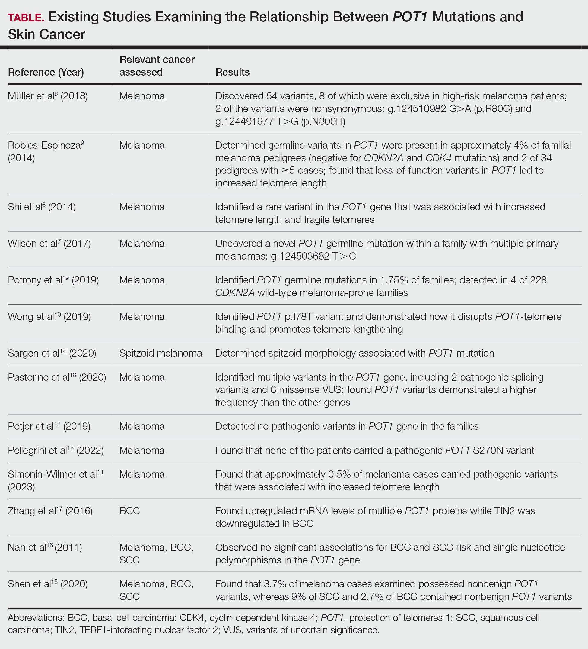
While our literature review did not reveal any associations between the shelterin complex genes and MAC, mutations in the POT1 gene have been studied in other types of skin cancer, particularly melanoma.1 One of the earliest studies was conducted in 2014 by Shi et al,6 in which whole-exome sequencing was performed on families with a history of melanoma. Multiple POT1 gene pathogenic variants associated with increased telomere length and fragility were identified in unrelated families. Subsequent studies have confirmed POT1 variants in melanoma-prone families,7 supporting an association between increased telomere length and melanoma risk8-11; however, other studies have yielded nonsignificant findings.12,13 Further investigation also has identified morphologic characteristics consistent with POT1 mutation, including spitzoid morphology.14
The association between POT1 mutations and nonmelanoma skin cancers has been relatively understudied. While a few studies have explored this link, results have shown mixed findings. Some studies have suggested a potential role for POT1 mutations in cutaneous SCC risk,15 while other studies have shown no significant associations for both BCC and SCC risk and telomere gene mutations.16 Additionally, mRNA levels of POT1 were upregulated in BCC cases compared to normal tissue in a gene expression.17
Comment
In the literature, POT1 mutations are well established as high-penetrance alterations associated with melanoma.9,18,19 However, the correlation between POT1 and other forms of skin cancer is not yet delineated. Recent insights suggest that POT1 mutations play a major role in promoting melanoma progression through telomere elongation, an established driver of melanoma progression, thereby extending the proliferative capacity of incipient cancer cells.20 This notion is supported by observations of increased telomere length in melanomaprone families with POT1 mutations. Given this association, research has focused on examining the relationship between telomere length and skin cancer.
Several studies have examined the relationship between telomere length and the risk for various types of skin cancer, including melanoma, BCC, and SCC. Prior investigations have suggested that shorter telomere length is associated with a decreased risk for melanoma and an increased risk for BCC, while no significant association has been observed for SCC.16 However, subsequent reports analyzing POT1 variants have failed to reveal any conclusive associations between BCC and SCC and telomere length.16,21
In contrast, other genetic variants associated with melanoma susceptibility have demonstrated notable associations with BCC and SCC; for instance, the CDKN2A (cyclin-dependent kinase inhibitor 2A) gene, which is the first gene linked to high-risk familial melanoma, exhibits an increased presence of mutations in individuals with BCC and SCC.22 Similarly, the MC1R (melanocortin 1 receptor) variant, a gene involved in human pigmentation and known to increase the risk for melanoma, carries a statistically significantly higher risk for BCC (summary odds ratio, 1.39; 95% CI, 1.15-1.69) and SCC (summary odds ratio, 1.61; 95% CI, 1.35-1.91) when at least one variant is present and an even greater risk with 2 or more variants.23
Considering the potential importance of POT1 mutations and their association with melanoma, as well as the inconsistencies surrounding POT1 mutations and their associations with BCC and SCC, further research may clarify the impact of POT1 mutations on the development and progression of different types of skin cancers and improve understanding of the complex interplay among telomere length, genetic variants, and skin cancer susceptibility. Given the established risk for melanoma with POT1 mutations, regular dermatology surveillance seems prudent. Dermatologists should consider referring patients with multiple skin cancers (especially melanoma) and any strong family history of internal malignancies to genetic testing for POT1. Though melanoma, CLL, angiosarcoma, and gliomas are the most commonly associated malignancies with POT1 mutations, as our case demonstrates, presentations can be heterogeneous, and the spectrum of malignancies associated with POT1 may be more expansive than previously thought.
For our patient, the current surveillance plan is fullbody skin examinations every 3 months. Given no prior family history of malignancies, presumably our patient’s case was a spontaneous mutation. Interestingly, despite his many primary cancer diagnoses and metastases, our patient has responded well to all treatments without recurrence. It is unclear if these characteristics and treatment successes are features of POT1associated cancers. Further research is needed to refine recommendations for screening and management of patients with identified POT1 mutations.
Conclusion
This case report highlights a rare occurrence of MAC in a patient with a POT1 mutation. Given the limited research conducted on investigating POT1 mutations and skin cancer, it is important to consider various forms of skin cancer, in addition to melanoma, when treating patients with a POT1 mutation.
- Accardo ML, Osborne J, Else T. POT1 tumor predisposition. GeneReviews®. October 29, 2020. Updated December 4, 2025. University of Washington.
- Chaichamnan K, Satayasoontorn K, Puttanupaab S, et al. Malignant proliferating trichilemmal tumors with CD34 expression. J Med Assoc Thai. 2010;93(suppl 6):S28-S34.
- Kavand S, Cassarino DS. “Squamoid eccrine ductal carcinoma”: an unusual low-grade case with follicular differentiation. are these tumors squamoid variants of microcystic adnexal carcinoma? Am J Dermatopathol. 2009;31:849-852.
- Kaseb H, Babiker HM. Eccrine carcinoma. StatPearls [Internet]. Updated June 26, 2023. Accessed May 11, 2026. https://www.ncbi.nlm.nih.gov/books/NBK541042
- Ye JZ, Hockemeyer D, Krutchinsky AN, et al. POT1-interacting protein PIP1: a telomere length regulator that recruits POT1 to the TIN2/TRF1 complex. Genes Dev. 2004;18:1649-1654. doi:10.1101/gad.1215404
- Shi J, Yang XR, Ballew B, et al. Rare missense variants in POT1 predispose to familial cutaneous malignant melanoma. Nat Genet. 2014;46:482-486. doi:10.1038/ng.2941
- Wilson TL, Hattangady N, Lerario AM, et al. A new POT1 germline mutation-expanding the spectrum of POT1-associated cancers. Fam Cancer. 2017;16:561-566. doi:10.1007/s10689-017-9984-y
- Müller C, Krunic M, Wendt J, et al. Germline variants in the POT1- gene in high-risk melanoma patients in Austria. G3 (Bethesda). 2018;8:1475-1480. doi:10.1534/g3.117.300394
- Robles-Espinoza CD, Harland M, Ramsay AJ, et al. POT1 loss-offunction variants predispose to familial melanoma. Nat Genet. 2014;46:478-481. doi:10.1038/ng.2947
- Wong K, Robles-Espinoza CD, Rodriguez D, et al. Association of the POT1 germline missense variant p.I78T with familial melanoma. JAMA Dermatol. 2019;155:604-609. doi:10.1001/jamadermatol.2018.3662
- Simonin-Wilmer I, Ossio R, Leddin EM, et al. Population-based analysis of POT1 variants in a cutaneous melanoma case-control cohort. J Med Genet. 2023;60:692-696. doi:10.1136/jmg-2022-108776
- Potjer TP, Bollen S, Grimbergen AJEM, et al; Dutch Working Group for Clinical Oncogenetics. Multigene panel sequencing of established and candidate melanoma susceptibility genes in a large cohort of Dutch non-CDKN2A/CDK4 melanoma families. Int J Cancer. 2019;144:2453- 2464. doi:10.1002/ijc.31984
- Pellegrini C, Raimondi S, Di Nardo L, et al; Italian Melanoma Intergroup (IMI). Melanoma in children and adolescents: analysis of susceptibility genes in 123 Italian patients. J Eur Acad Dermatol Venereol. 2022;36:213-221. doi:10.1111/jdv.17735
- Sargen MR, Calista D, Elder DE, et al. Histologic features of melanoma associated with germline mutations of CDKN2A, CDK4, and POT1 in melanoma-prone families from the United States, Italy, and Spain. J Am Acad Dermatol. 2020;83:860-869. doi:10.1016/j.jaad.2020.03.100
- Shen E, Xiu J, Lopez GY, et al. POT1 mutation spectrum in tumour types commonly diagnosed among POT1-associated hereditary cancer syndrome families. J Med Genet. 2020;57:664-670. doi:10.1136 /jmedgenet-2019-106657
- Nan H, Qureshi AA, Prescott J, et al. Genetic variants in telomere-maintaining genes and skin cancer risk. Hum Genet. 2011;129:247-253. doi:10.1007/s00439-010-0921-5
- Zhang L, Huang X, Zhu X, et al. Differential senescence capacities in meibomian gland carcinoma and basal cell carcinoma. Int J Cancer. 2016;138:1442-1452. doi:10.1002/ijc.29882
- Pastorino L, Andreotti V, Dalmasso B, et al. Insights into genetic susceptibility to melanoma by gene panel testing: potential pathogenic variants in ACD, ATM, BAP1, and POT1. Cancers (Basel). 2020;12:1007. doi:10.3390/cancers12041007
- Potrony M, Puig-Butille JA, Ribera-Sola M, et al. POT1 germline mutations but not TERT promoter mutations are implicated in melanoma susceptibility in a large cohort of Spanish melanoma families. Br J Dermatol. 2019;181:105-113. doi:10.1111/bjd.17443
- Kim WT, Hennick K, Johnson J, et al. Cancer-associated POT1 mutations lead to telomere elongation without induction of a DNA damage response. EMBO J. 2021;40:e107346.
- Ventura A, Pellegrini C, Cardelli L, et al. Telomeres and telomerase in cutaneous squamous cell carcinoma. Int J Mol Sci. 2019;20:1333. doi:10.3390/ijms20061333
- Helgadottir H, Höiom V, Jönsson G, et al. High risk of tobacco-related cancers in CDKN2A mutation-positive melanoma families. J Med Genet. 2014;51:545-552. doi:10.1136/jmedgenet-2014-102320
- Tagliabue E, Fargnoli MC, Gandini S, et al; M-SKIP Study Group. MC1R gene variants and non-melanoma skin cancer: a pooledanalysis from the M-SKIP project. Br J Cancer. 2015;113:354-363. doi:10.1038/bjc.2015.231
A 72-year-old man with a history of multiple cancers, including melanoma, squamous cell carcinoma (SCC), and basal cell carcinoma (BCC), presented to the dermatology clinic for a regularly scheduled full-body skin examination. His family history was negative for malignancy, but due to his personal history of both primary internal cancers and skin cancers, the patient previously had been referred by dermatology to a medical geneticist for evaluation. He tested positive for a pathogenic POT1 (protection of telomeres 1) variant associated with tumor predisposition, which most often is associated with cutaneous melanoma, chronic lymphocytic leukemia (CLL), angiosarcoma, and gliomas.1
At the current presentation, physical examination revealed a small, asymmetric, pink papule on the superior thoracic spine. A biopsy of the lesion was performed (Figure 1). Pathology demonstrated cornifying cystic structures with a granulomatous response at the surface of the tumor, ductal differentiation with depth, and infiltrative strands and cords of hyperchromatic cells within a collagenous stroma at the base of the specimen (Figures 2A and 2B). One unusual finding was the presence of prominent clear-cell change within the superficial portion of the neoplasm (Figure 2C). Immunohistochemical stains revealed strong p63 and p40 positivity. Epithelial membrane antigen staining was positive in the hyperchromatic strands and cords with depth but not in the clear-cell superficial portion. Similarly, periodic acid–Schiff–positive material increased within tumor cells in proportion to depth of infiltration. Additional immunohistochemical staining showed carcinoembryonic antigen was largely negative (with rare positivity in a few ductal lumina), with negative results for S100, SOX10, CD117, BerEP4, factor XIIIa, CD34, and cytokeratin 7 (Figures 2D and 2E).
The differential diagnoses included trichilemmal carcinoma (which may manifest with CD34 expression),2 clear cell BCC, adenoid cystic carcinoma (tubular variant), sebaceous carcinoma, and eccrine carcinoma. Importantly, the patient was under continuous oncologic surveillance, with no evidence of a primary internal tumor to suggest metastasis. Despite negative carcinoembryonic antigen staining, the immunohistochemical and histopathologic findings fit best with a primary cutaneous malignant eccrine tumor, specifically microcystic adnexal carcinoma (MAC), in which p63 typically stains peripheral cells but solid variants have been described.3
Eccrine carcinoma is exceedingly rare, reported in 0.01% of diagnosed cutaneous malignancies, and demonstrates overlapping features to other malignant eccrine tumors. It possesses an inconsistent immunohistochemical staining profile, making the distinction from other malignant sweat gland tumors challenging.4 Given that the morphologic features were otherwise classic for MAC in our patient, we favored a clear-cell variant.
Sixteen years prior to the current presentation, our patient presented to urology with a history of prostatitis and increasing prostate-specific antigen levels. Biopsies were negative until prostate-specific antigen reached 13 ng/mL, confirming stage 1A prostate cancer. The patient subsequently underwent a robot-assisted radical prostatectomy. At age 63 years, dysphagia that was unresponsive to antibiotics led to a tonsillar biopsy revealing T2N2bM0 stage IVA SCC of the right tonsil with confirmed HPV type 16 with extracapsular extension. The patient underwent transoral robotic radical tonsillectomy and right neck dissection, followed by adjuvant chemoradiation consisting of intensity-modulated radiation therapy (IMRT) to a total dose of 63 Gy in 33 fractions, with concurrent weekly cisplatin. At age 67 years, dyspepsia, dysphagia, pyrosis, and gastroesophageal reflux prompted endoscopy, revealing T1aNxMx esophageal adenocarcinoma. Three months later, the patient underwent laparoscopic-assisted esophagectomy, with no recurrence. At age 68 years, an atypical intramelanocytic proliferation was found on the left cheek and was treated with Mohs micrographic surgery.
At age 71 years, acral lentiginous malignant melanoma (Breslow thickness 0.8 mm; Clark level IV; American Joint Committee on Cancer T1b) was diagnosed on the left plantar foot and treated with Mohs micrographic surgery. Sentinel lymph node biopsy was negative. Squamous cell carcinoma in situ on the frontal scalp and nodular BCC on the right upper back also were diagnosed.
While there are no guidelines for surveillance of individuals with POT1, recommendations were given in consensus from a medical genetics team,1 including comprehensive monitoring—specifically baseline imaging utilizing brain and full-body magnetic resonance imaging. Furthermore, considering the crucial role of POT1 in maintaining telomeres, it was advised to measure telomere length as part of the surveillance process. Given the patient’s susceptibility to CLL, routine complete blood count assessments were recommended. Additionally, we advised close monitoring for seizures and consideration of genetic testing in first-degree relatives.
Literature Review
Given our patient’s history of multiple skin cancers, including the most recent MAC, we sought to conduct a review of the literature to evaluate existing skin cancer associations and reports for patients with known POT1 mutations to guide recommendations for dermatologic surveillance (Table). A search of PubMed articles indexed for MEDLINE through April 2023 using the terms microcystic adnexal carcinoma, POT1, melanoma, basal cell carcinoma, squamous cell carcinoma, and skin cancer yielded no reported cases of MAC associated with POT1 mutations. POT1 is one of 6 proteins (TERF1, TERF2, RAP1, TIN2, TPP1, and POT1) belonging to the shelterin complex, which plays a crucial role in telomeric DNA remodeling and regulation of telomere length.5 Mutation in the POT1 gene disrupts the shelterin complex, causing telomeres to become elongated and unstable, resulting in chromosomal abnormalities and promoting cancer development.5
While our literature review did not reveal any associations between the shelterin complex genes and MAC, mutations in the POT1 gene have been studied in other types of skin cancer, particularly melanoma.1 One of the earliest studies was conducted in 2014 by Shi et al,6 in which whole-exome sequencing was performed on families with a history of melanoma. Multiple POT1 gene pathogenic variants associated with increased telomere length and fragility were identified in unrelated families. Subsequent studies have confirmed POT1 variants in melanoma-prone families,7 supporting an association between increased telomere length and melanoma risk8-11; however, other studies have yielded nonsignificant findings.12,13 Further investigation also has identified morphologic characteristics consistent with POT1 mutation, including spitzoid morphology.14
The association between POT1 mutations and nonmelanoma skin cancers has been relatively understudied. While a few studies have explored this link, results have shown mixed findings. Some studies have suggested a potential role for POT1 mutations in cutaneous SCC risk,15 while other studies have shown no significant associations for both BCC and SCC risk and telomere gene mutations.16 Additionally, mRNA levels of POT1 were upregulated in BCC cases compared to normal tissue in a gene expression.17
Comment
In the literature, POT1 mutations are well established as high-penetrance alterations associated with melanoma.9,18,19 However, the correlation between POT1 and other forms of skin cancer is not yet delineated. Recent insights suggest that POT1 mutations play a major role in promoting melanoma progression through telomere elongation, an established driver of melanoma progression, thereby extending the proliferative capacity of incipient cancer cells.20 This notion is supported by observations of increased telomere length in melanomaprone families with POT1 mutations. Given this association, research has focused on examining the relationship between telomere length and skin cancer.
Several studies have examined the relationship between telomere length and the risk for various types of skin cancer, including melanoma, BCC, and SCC. Prior investigations have suggested that shorter telomere length is associated with a decreased risk for melanoma and an increased risk for BCC, while no significant association has been observed for SCC.16 However, subsequent reports analyzing POT1 variants have failed to reveal any conclusive associations between BCC and SCC and telomere length.16,21
In contrast, other genetic variants associated with melanoma susceptibility have demonstrated notable associations with BCC and SCC; for instance, the CDKN2A (cyclin-dependent kinase inhibitor 2A) gene, which is the first gene linked to high-risk familial melanoma, exhibits an increased presence of mutations in individuals with BCC and SCC.22 Similarly, the MC1R (melanocortin 1 receptor) variant, a gene involved in human pigmentation and known to increase the risk for melanoma, carries a statistically significantly higher risk for BCC (summary odds ratio, 1.39; 95% CI, 1.15-1.69) and SCC (summary odds ratio, 1.61; 95% CI, 1.35-1.91) when at least one variant is present and an even greater risk with 2 or more variants.23
Considering the potential importance of POT1 mutations and their association with melanoma, as well as the inconsistencies surrounding POT1 mutations and their associations with BCC and SCC, further research may clarify the impact of POT1 mutations on the development and progression of different types of skin cancers and improve understanding of the complex interplay among telomere length, genetic variants, and skin cancer susceptibility. Given the established risk for melanoma with POT1 mutations, regular dermatology surveillance seems prudent. Dermatologists should consider referring patients with multiple skin cancers (especially melanoma) and any strong family history of internal malignancies to genetic testing for POT1. Though melanoma, CLL, angiosarcoma, and gliomas are the most commonly associated malignancies with POT1 mutations, as our case demonstrates, presentations can be heterogeneous, and the spectrum of malignancies associated with POT1 may be more expansive than previously thought.
For our patient, the current surveillance plan is fullbody skin examinations every 3 months. Given no prior family history of malignancies, presumably our patient’s case was a spontaneous mutation. Interestingly, despite his many primary cancer diagnoses and metastases, our patient has responded well to all treatments without recurrence. It is unclear if these characteristics and treatment successes are features of POT1associated cancers. Further research is needed to refine recommendations for screening and management of patients with identified POT1 mutations.
Conclusion
This case report highlights a rare occurrence of MAC in a patient with a POT1 mutation. Given the limited research conducted on investigating POT1 mutations and skin cancer, it is important to consider various forms of skin cancer, in addition to melanoma, when treating patients with a POT1 mutation.
- Accardo ML, Osborne J, Else T. POT1 tumor predisposition. GeneReviews®. October 29, 2020. Updated December 4, 2025. University of Washington.
- Chaichamnan K, Satayasoontorn K, Puttanupaab S, et al. Malignant proliferating trichilemmal tumors with CD34 expression. J Med Assoc Thai. 2010;93(suppl 6):S28-S34.
- Kavand S, Cassarino DS. “Squamoid eccrine ductal carcinoma”: an unusual low-grade case with follicular differentiation. are these tumors squamoid variants of microcystic adnexal carcinoma? Am J Dermatopathol. 2009;31:849-852.
- Kaseb H, Babiker HM. Eccrine carcinoma. StatPearls [Internet]. Updated June 26, 2023. Accessed May 11, 2026. https://www.ncbi.nlm.nih.gov/books/NBK541042
- Ye JZ, Hockemeyer D, Krutchinsky AN, et al. POT1-interacting protein PIP1: a telomere length regulator that recruits POT1 to the TIN2/TRF1 complex. Genes Dev. 2004;18:1649-1654. doi:10.1101/gad.1215404
- Shi J, Yang XR, Ballew B, et al. Rare missense variants in POT1 predispose to familial cutaneous malignant melanoma. Nat Genet. 2014;46:482-486. doi:10.1038/ng.2941
- Wilson TL, Hattangady N, Lerario AM, et al. A new POT1 germline mutation-expanding the spectrum of POT1-associated cancers. Fam Cancer. 2017;16:561-566. doi:10.1007/s10689-017-9984-y
- Müller C, Krunic M, Wendt J, et al. Germline variants in the POT1- gene in high-risk melanoma patients in Austria. G3 (Bethesda). 2018;8:1475-1480. doi:10.1534/g3.117.300394
- Robles-Espinoza CD, Harland M, Ramsay AJ, et al. POT1 loss-offunction variants predispose to familial melanoma. Nat Genet. 2014;46:478-481. doi:10.1038/ng.2947
- Wong K, Robles-Espinoza CD, Rodriguez D, et al. Association of the POT1 germline missense variant p.I78T with familial melanoma. JAMA Dermatol. 2019;155:604-609. doi:10.1001/jamadermatol.2018.3662
- Simonin-Wilmer I, Ossio R, Leddin EM, et al. Population-based analysis of POT1 variants in a cutaneous melanoma case-control cohort. J Med Genet. 2023;60:692-696. doi:10.1136/jmg-2022-108776
- Potjer TP, Bollen S, Grimbergen AJEM, et al; Dutch Working Group for Clinical Oncogenetics. Multigene panel sequencing of established and candidate melanoma susceptibility genes in a large cohort of Dutch non-CDKN2A/CDK4 melanoma families. Int J Cancer. 2019;144:2453- 2464. doi:10.1002/ijc.31984
- Pellegrini C, Raimondi S, Di Nardo L, et al; Italian Melanoma Intergroup (IMI). Melanoma in children and adolescents: analysis of susceptibility genes in 123 Italian patients. J Eur Acad Dermatol Venereol. 2022;36:213-221. doi:10.1111/jdv.17735
- Sargen MR, Calista D, Elder DE, et al. Histologic features of melanoma associated with germline mutations of CDKN2A, CDK4, and POT1 in melanoma-prone families from the United States, Italy, and Spain. J Am Acad Dermatol. 2020;83:860-869. doi:10.1016/j.jaad.2020.03.100
- Shen E, Xiu J, Lopez GY, et al. POT1 mutation spectrum in tumour types commonly diagnosed among POT1-associated hereditary cancer syndrome families. J Med Genet. 2020;57:664-670. doi:10.1136 /jmedgenet-2019-106657
- Nan H, Qureshi AA, Prescott J, et al. Genetic variants in telomere-maintaining genes and skin cancer risk. Hum Genet. 2011;129:247-253. doi:10.1007/s00439-010-0921-5
- Zhang L, Huang X, Zhu X, et al. Differential senescence capacities in meibomian gland carcinoma and basal cell carcinoma. Int J Cancer. 2016;138:1442-1452. doi:10.1002/ijc.29882
- Pastorino L, Andreotti V, Dalmasso B, et al. Insights into genetic susceptibility to melanoma by gene panel testing: potential pathogenic variants in ACD, ATM, BAP1, and POT1. Cancers (Basel). 2020;12:1007. doi:10.3390/cancers12041007
- Potrony M, Puig-Butille JA, Ribera-Sola M, et al. POT1 germline mutations but not TERT promoter mutations are implicated in melanoma susceptibility in a large cohort of Spanish melanoma families. Br J Dermatol. 2019;181:105-113. doi:10.1111/bjd.17443
- Kim WT, Hennick K, Johnson J, et al. Cancer-associated POT1 mutations lead to telomere elongation without induction of a DNA damage response. EMBO J. 2021;40:e107346.
- Ventura A, Pellegrini C, Cardelli L, et al. Telomeres and telomerase in cutaneous squamous cell carcinoma. Int J Mol Sci. 2019;20:1333. doi:10.3390/ijms20061333
- Helgadottir H, Höiom V, Jönsson G, et al. High risk of tobacco-related cancers in CDKN2A mutation-positive melanoma families. J Med Genet. 2014;51:545-552. doi:10.1136/jmedgenet-2014-102320
- Tagliabue E, Fargnoli MC, Gandini S, et al; M-SKIP Study Group. MC1R gene variants and non-melanoma skin cancer: a pooledanalysis from the M-SKIP project. Br J Cancer. 2015;113:354-363. doi:10.1038/bjc.2015.231
Microcystic Adnexal Carcinoma– like Neoplasm in a Patient With POT1 Mutation
Microcystic Adnexal Carcinoma– like Neoplasm in a Patient With POT1 Mutation
PRACTICE POINTS
- Dermatologists should consider referring patients with both a history of skin cancer and a strong family history of internal malignancy for genetic testing for POT1 (protection of telomeres 1) mutations.
- Although melanoma, chronic lymphocytic leukemia, angiosarcoma, and gliomas are most commonly associated with POT1 mutations, this case suggests a broader and more heterogeneous malignancy spectrum than previously recognized.
