User login
Maggots Prove Wound-Cleaning Worth
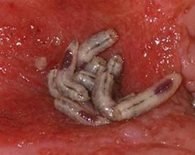
PRAGUE – Maggot debridement of wounds proved significantly faster, less painful, and less labor intensive than surgical debridement and conventional dressings in a randomized, multicenter clinical trial.
"There was quite an amazing debridement after 7 days of maggot therapy," Dr. Kristina Opletalova said of the phase III study findings presented at the annual congress of the European Academy of Dermatology and Venereology.
She reported on 119 patients hospitalized for 2 weeks for treatment of nonhealing sloughy wounds, most of which were venous ulcers on the lower limbs. Participants were randomized to maggot therapy or to thrice-weekly surgical debridement with topical anesthesia and conventional dressings.
The maggot therapy was administered via a novel delivery system: 80 maggots of Lucilia sericata were bagged in a special two-layer dressing, known as a Vitapad (BioMonde Laboratories), which allowed the critters to move and feed on the wound surface and kept them from escaping. The dressing was changed twice weekly.
Patients were blindfolded for all dressing changes so they didn’t know which study arm they were in. Wound sloughing was analyzed using computerized planimetry software, and other end points were assessed by an investigator blinded to treatment arm.
After 7 days of therapy, the wound status was significantly better in the maggot debridement group. The mean percentage of slough in wounds was 54.5%, compared with 66.5% in controls, but the significantly faster debridement didn’t boost the final healing rate. The day 15 percentage of slough in the wounds didn’t differ significantly between the two groups: 55.4% with maggot therapy and 53.8% in controls.
"So we think maggot debridement therapy should be stopped after 1 week and other types of dressings should then be used," said Dr. Opletalova, a dermatologist at the University of Caen (France).
Pain scores, which were assessed on a weekly basis, were similarly low in the two groups, but it must be noted that the control group received topical anesthetic and the maggot therapy group did not. Nursing time was four-fold greater in the control group. The study didn’t include a formal cost analysis, but the markedly reduced nursing time in the maggot therapy group is likely to spell cost savings, she said.
Maggot therapy was well tolerated. Patients expressed no reticence about it. A similar number of patients in both study arms reported a crawling sensation on their wounds.
Dr. Opletalova said that maggot therapy is likely to be particularly useful in patients with wounds that need rapid debridement, such as those with diabetes, or to prepare a wound for skin grafting or when there is an increased risk for infection.
Further details of the recently published randomized trial can be found in the Archives of Dermatology (2012;148:432-8).
The study was supported by university hospital research funding and by a grant from the French Society of Dermatology. Dr. Opletalova reported having no financial conflicts.
AT THE ANNUAL CONGRESS OF THE EUROPEAN ACADEMY OF DERMATOLOGY AND VENEREOLOGY
Major Finding: After 7 days of therapy, the mean percentage of slough in wounds was 54.5% in the maggot debridement group, compared with 66.5% in the control group.
Data Source: The phase-III findings come from a randomized, multicenter, blinded, prospective clinical trial involving 119 hospitalized patients.
Disclosures: The study was supported by university research funding and by a grant from the French Society of Dermatology. Dr. Opletalova reported having no financial conflicts.

Medicare Okays Wound Plasma Gel for Clinical Trial Patients
The Centers for Medicare and Medicaid Services has announced coverage of platelet-rich plasma gel for chronic wounds, but only for patients who are enrolled in clinical trials that meet the agency’s criteria.
"The available evidence does not permit us to conclude that use of autologous PRP improves health outcomes in beneficiaries with chronic diabetic, pressure, and/or venous wounds," the agency wrote in its decision statement Therefore, trials are needed to prove effectiveness.
The decision comes after CMS rejection of coverage since 1992, according to Cytomedix of Gaithersburg, Md., which manufactures a device called AutoloGel that produces PRP from a patient’s own blood. "This determination reverses a nearly 20-year non-coverage determination for PRP and provides for an appropriate research study with practical study designs we are confident will demonstrate that patients treated with our autologous PRP product, the AutoloGel System, experience clinically significant health outcomes," Martin P. Rosendale, CEO of Cytomedix, said in a statement.
Cytomedix submitted another request in October 2011 for CMS review of its PRP therapy, which was approved under the coverage with evidence development (CED) program.
Under the program, services, equipment, and supplies are covered through Medicare and Medicaid while a product is further investigated. CMS noted in its decision that it "is evaluating PRP as a service, and not a specific system for administrating PRP."
In the PRP process, autologous blood is centrifuged to produce a concentrate of platelets and plasma proteins. Individual growth factors are not identified or separated, but additives are used to change the consistency.
Autologous PRP has had many uses over the years, including use as an adhesive in plastic surgery and as filler for acute wounds. It has also been used to treat amateur and professional athletes – such as golfer Tiger Woods, basketball player Kobe Bryant, and baseball star Alex Rodriguez – to help heal tendons and ligaments, and is growing in popularity in the sports world, according to recent consumer news reports.
According to the CMS, to continue being covered for wound healing PRP therapy must show complete wound healing in patients and a reduction of wound size or healing trajectory. It also must help a patient’s ability to return to previous function and normal activities. The studies have to be prospective and enroll Medicare beneficiaries with chronic nonhealing diabetic, pressure, and/or venous wounds who are receiving optimal care, in addition to the therapy.
The agency must receive all pertinent data by August 2014.
Cytomedix said that it has been in talks with CMS about trials. "We look forward to continuing to work with CMS in the coming weeks in further defining the specifics of the protocols for the research studies and clinical questions to be answered through the CED program," said Mr. Rosendale.
Spray-On Cells Promote Venous Ulcer Healing
A spray formulation of allogeneic neonatal keratinocytes and fibroblasts significantly improved wound healing in a multicenter, randomized, placebo-controlled phase II study of more than 200 patients with venous leg ulcers.
The mean reduction in wound area was significantly greater among 177 patients who received 12 weeks of active treatment with the novel cell therapy, known as HP802-247, than among 50 patients who were treated with application vehicle of a modified fibrin spray alone. HP802-247 consists of cryopreserved, allogenic, growth-arrested fibroblasts and keratinocytes derived from neonatal foreskin. The largest improvement (16% on average over placebo) was seen in 44 patients assigned to treatment with 5.0x10⁶ cells per mL every 14 days, Dr. Robert S. Kirsner of the University of Miami and his colleagues reported. The study was published online in the Aug. 3 issue of The Lancet.
Reduction in wound area was between 8% and 12% over that seen with placebo in 45 patients treated with 5.0x10⁶ cells per mL every 7 days, 44 patients treated with 5.0x10⁶ cells per mL every 14 days, and 43 patients treated with 0.5x10⁶ cells per mL every 7 days, the investigators said (Lancet 2012 Aug. 3 [doi:10.1016/S0140-6736(12)60644-8]).
The study investigators included vascular surgeons, dermatologists, and podiatrists at universities and in private practice. All patients were treated with standard compression bandaging as well as the study treatment.
"Wound area began to decrease rapidly after initiation of treatment. After one application of the 0.5x10⁶/mL every 14 days dose, the mean reduction in wound area was 40%, compared with 23% for the vehicle group. This effect persisted with the maximum difference in mean reduction between the group assigned [0.5x10⁶ cells per mL] every 14 days and the vehicle group occurring at week 7 of treatment (87% vs. 65%)," they wrote.
At week 12 of treatment, the mean wound area in the group treated with 0.5x10⁶ cells per mL every 14 days was reduced by 91%, compared with 80% with vehicle, and for the other treatment groups, the mean wound area was reduced by between 84% and 87%.
"Overall, patients treated with cells had higher proportions of healed wounds than did those assigned vehicle alone, but only the group assigned [0.5x10⁶ cells per mL] every 14 days differed significantly compared with control," they noted.
Adverse events were similar across all groups, and most were nonserious, mild to moderate events that resolved. Only new skin ulcers and cellulitis occurred at a frequency of greater than 5%. Immunotoxicity testing showed that there was no treatment-induced alloantibody formation or induction of autoimmunity.
Included in this study were adult outpatients enrolled from 28 centers in the United States and Canada between 2009 and 2011. All had venous reflux confirmed by duplex Doppler ultrasonography, and up to three venous leg ulcers. Those assigned to the placebo group received vehicle alone applied every 7 days for the 12 weeks of the trial.
The findings may have important implications for the treatment of venous leg ulcers, which affect an estimated 1.65% to 1.74% of adults aged 65 years and older. Standard treatment with infection control, primary dressings, and high-strength compressions heal between 30% and 75% of such ulcers, compared with the 84%-91% healing seen with HP802-247 in this study, with the remainder becoming "chronic, with unresolved inflammatory processes within the extracellular matrix of the wound bed and dysfunction of wound fibroblasts and keratinocytes," the investigators noted.
"The results are sufficiently promising that larger randomized trials comparing HP802-247 to standard treatment for venous leg ulcers are now warranted," they concluded.
The researchers reported that their study was limited by the wound upper end boundary of 12 cm3, which represents 80% of all venous leg ulcers, but a smaller proportion of chronic venous ulcers.
The research trial is registered with ClinicalTrials.gov as NCT00852995.
Dr. Kirsner received consulting fees for his assistance with protocol design and data interpretation. Two of his coauthors also received such consulting fees, and other authors on the study disclosed that they are employed by Healthpoint, and/or hold adjunct academic appointments at the University of North Texas Health Sciences Center in Fort Worth, Tex.
The findings of this "well-done clinical trial" highlight the potential of cell-based therapies not only for venous leg ulcer treatment, but for the treatment of other chronic wounds as well, Dr. Matthias Augustin and Dr. Wolfgang Vanscheidt wrote in an accompanying editorial.
The authors have "clearly shown that a specific cell therapy for venous leg ulcers can lead to a significantly higher healing rate than for vehicle alone in hard-to-treat wounds for which compression treatment has been applied unsuccessfully," they noted, adding that "the benefits identified by Kirsner and colleagues could well be applicable in other chronic wounds such as ischemic and diabetic foot ulcers."
Dr. Augustin and Dr. Vanscheidt stressed the importance of gaining insight into the mechanism of action to further understand the best potential uses of these types of therapies, and called on the scientific community and industry to investigate the effects of tissue and cell therapy products in patients with mixed arterial-venous leg ulcers in whom revascularization is not possible (Lancet 2012 Aug. 3 [doi:10.1016/S0140-6736(12)61255-0]).
"In this indication, bioengineered tissue and cell therapy products are presumably among the very few methods that could achieve sustained healing. Because of aging of the population in most societies, this fatal combination of venous insufficiency and arterial occlusive disease will become an ever-growing problem. We believe that biotherapy of such ulcers will be a challenge but will also offer hope to wound researchers in the coming decades," they said.
Dr. Augustin and Dr. Vanscheidt are with the Institute for Health Services Research in Dermatology and Nursing and Comprehensive Wound Center, University Medical Center Hamburg (Germany), and Freiburg, Germany, respectively. They reported having no conflicts of interest.
A spray formulation of allogeneic neonatal keratinocytes and fibroblasts significantly improved wound healing in a multicenter, randomized, placebo-controlled phase II study of more than 200 patients with venous leg ulcers.
The mean reduction in wound area was significantly greater among 177 patients who received 12 weeks of active treatment with the novel cell therapy, known as HP802-247, than among 50 patients who were treated with application vehicle of a modified fibrin spray alone. HP802-247 consists of cryopreserved, allogenic, growth-arrested fibroblasts and keratinocytes derived from neonatal foreskin. The largest improvement (16% on average over placebo) was seen in 44 patients assigned to treatment with 5.0x10⁶ cells per mL every 14 days, Dr. Robert S. Kirsner of the University of Miami and his colleagues reported. The study was published online in the Aug. 3 issue of The Lancet.
Reduction in wound area was between 8% and 12% over that seen with placebo in 45 patients treated with 5.0x10⁶ cells per mL every 7 days, 44 patients treated with 5.0x10⁶ cells per mL every 14 days, and 43 patients treated with 0.5x10⁶ cells per mL every 7 days, the investigators said (Lancet 2012 Aug. 3 [doi:10.1016/S0140-6736(12)60644-8]).
The study investigators included vascular surgeons, dermatologists, and podiatrists at universities and in private practice. All patients were treated with standard compression bandaging as well as the study treatment.
"Wound area began to decrease rapidly after initiation of treatment. After one application of the 0.5x10⁶/mL every 14 days dose, the mean reduction in wound area was 40%, compared with 23% for the vehicle group. This effect persisted with the maximum difference in mean reduction between the group assigned [0.5x10⁶ cells per mL] every 14 days and the vehicle group occurring at week 7 of treatment (87% vs. 65%)," they wrote.
At week 12 of treatment, the mean wound area in the group treated with 0.5x10⁶ cells per mL every 14 days was reduced by 91%, compared with 80% with vehicle, and for the other treatment groups, the mean wound area was reduced by between 84% and 87%.
"Overall, patients treated with cells had higher proportions of healed wounds than did those assigned vehicle alone, but only the group assigned [0.5x10⁶ cells per mL] every 14 days differed significantly compared with control," they noted.
Adverse events were similar across all groups, and most were nonserious, mild to moderate events that resolved. Only new skin ulcers and cellulitis occurred at a frequency of greater than 5%. Immunotoxicity testing showed that there was no treatment-induced alloantibody formation or induction of autoimmunity.
Included in this study were adult outpatients enrolled from 28 centers in the United States and Canada between 2009 and 2011. All had venous reflux confirmed by duplex Doppler ultrasonography, and up to three venous leg ulcers. Those assigned to the placebo group received vehicle alone applied every 7 days for the 12 weeks of the trial.
The findings may have important implications for the treatment of venous leg ulcers, which affect an estimated 1.65% to 1.74% of adults aged 65 years and older. Standard treatment with infection control, primary dressings, and high-strength compressions heal between 30% and 75% of such ulcers, compared with the 84%-91% healing seen with HP802-247 in this study, with the remainder becoming "chronic, with unresolved inflammatory processes within the extracellular matrix of the wound bed and dysfunction of wound fibroblasts and keratinocytes," the investigators noted.
"The results are sufficiently promising that larger randomized trials comparing HP802-247 to standard treatment for venous leg ulcers are now warranted," they concluded.
The researchers reported that their study was limited by the wound upper end boundary of 12 cm3, which represents 80% of all venous leg ulcers, but a smaller proportion of chronic venous ulcers.
The research trial is registered with ClinicalTrials.gov as NCT00852995.
Dr. Kirsner received consulting fees for his assistance with protocol design and data interpretation. Two of his coauthors also received such consulting fees, and other authors on the study disclosed that they are employed by Healthpoint, and/or hold adjunct academic appointments at the University of North Texas Health Sciences Center in Fort Worth, Tex.
FROM THE LANCET
Major Finding: At week 12 of treatment, the mean wound area in the group treated with 0.5x10⁶ cells per mL every 14 days was reduced by 91%, compared with 80% with vehicle, and for the other treatment groups the mean wound area was reduced by between 84% and 87%.
Data Source: This was a randomized, placebo-controlled phase II trial.
Disclosures: Dr. Kirsner received consulting fees for his assistance with protocol design and data interpretation. Two of his coauthors also received such consulting fees, and other authors on the study disclosed that they are employed by Healthpoint, and/or hold adjunct academic appointments at the University of North Texas Health Sciences Center in Fort Worth, Tex. The authors of the editorial reported having no disclosures.
Laser Treatment of Scars and Keloids
Hypertrophic Scars and Keloids, Part 1: Conventional Treatments
Cyst Removal: Punch Incision Leaves Smaller Scar
RALEIGH, N.C. – Punch incision epidermal inclusion cysts located on the trunk leaves a significantly smaller scar than does elliptical excision with a similarly low recurrence rate, according to the results of a randomized trail.
Procedure time was essentially the same for the two techniques, at around 13 minutes. Although punch incision and its wound closure can be easier, it took a fair amount of time to squeeze the cyst contents through the small punch opening and remove the cyst lining using a curette, Dr. Justin T. Cheeley explained at the annual meeting of the Society for Investigative Dermatology.
He reported on 40 consecutive patients with one or more truncal epidermal inclusion cysts 1-3 cm in diameter who were randomized to elliptical excision or punch incision in a head-to-head comparative trial.
The primary study end point – cyst recurrence during 16 months of prospective follow-up – occurred in three patients in the punch incision group and two in the elliptical excision group. Predictors of cyst recurrence were sought, but none could be identified, according to Dr. Cheeley of Emory University, Atlanta.
Most secondary end points were similar for the two study arms, including early and late complication rates, as well as improvement in skin-specific quality of life and patient satisfaction as measured by change in Skindex-16 scores.

There was, however, a significant difference between the two study groups in terms of average scar length. In the punch incision group, average scar length was 1.1 cm, compared with 1.8 cm in the elliptical excision group.
The investigators employed a 4-mm punch for the most part, although they turned to a 6-mm punch in treating larger cysts. Punch incision wounds were closed with a single nylon suture. Closure of the elliptical excision sites required more extensive suturing.
Audience member Dr. Eric L. Simpson complimented Dr. Cheeley and his coinvestigators for conducting a study with important cost implications given how often epidermal inclusion cysts are encountered in practice.
“The difference between punch incision and elliptical excision with an intermediate-level repair is probably 10-fold in terms of cost,” said Dr. Simpson of Oregon Health and Science University, Portland.
Dr. Cheeley reported having no financial conflicts.
RALEIGH, N.C. – Punch incision epidermal inclusion cysts located on the trunk leaves a significantly smaller scar than does elliptical excision with a similarly low recurrence rate, according to the results of a randomized trail.
Procedure time was essentially the same for the two techniques, at around 13 minutes. Although punch incision and its wound closure can be easier, it took a fair amount of time to squeeze the cyst contents through the small punch opening and remove the cyst lining using a curette, Dr. Justin T. Cheeley explained at the annual meeting of the Society for Investigative Dermatology.
He reported on 40 consecutive patients with one or more truncal epidermal inclusion cysts 1-3 cm in diameter who were randomized to elliptical excision or punch incision in a head-to-head comparative trial.
The primary study end point – cyst recurrence during 16 months of prospective follow-up – occurred in three patients in the punch incision group and two in the elliptical excision group. Predictors of cyst recurrence were sought, but none could be identified, according to Dr. Cheeley of Emory University, Atlanta.
Most secondary end points were similar for the two study arms, including early and late complication rates, as well as improvement in skin-specific quality of life and patient satisfaction as measured by change in Skindex-16 scores.
There was, however, a significant difference between the two study groups in terms of average scar length. In the punch incision group, average scar length was 1.1 cm, compared with 1.8 cm in the elliptical excision group.
The investigators employed a 4-mm punch for the most part, although they turned to a 6-mm punch in treating larger cysts. Punch incision wounds were closed with a single nylon suture. Closure of the elliptical excision sites required more extensive suturing.
Audience member Dr. Eric L. Simpson complimented Dr. Cheeley and his coinvestigators for conducting a study with important cost implications given how often epidermal inclusion cysts are encountered in practice.
“The difference between punch incision and elliptical excision with an intermediate-level repair is probably 10-fold in terms of cost,” said Dr. Simpson of Oregon Health and Science University, Portland.
Dr. Cheeley reported having no financial conflicts.
FROM THE ANNUAL MEETING OF THE SOCIETY FOR INVESTIGATIVE DERMATOLOGY
Major Finding: In the punch incision group, average scar length was 1.1 cm, compared with 1.8 cm in the elliptical excision group.
Data Source: This was a randomized trial of 40 consecutive patients.
Disclosures: Dr. Cheeley reported having no financial conflicts.
Onion Extract Improved Scars by 36%
RALEIGH, N.C. – A new once-daily topical gel containing a proprietary onion extract resulted in a 36% improvement in the appearance of recent postsurgical dermal scars at 8 weeks, according to the results of a randomized, controlled trial.
The over-the-counter product, Merz Pharmaceuticals’ Mederma Advanced Scar Gel, was studied in 44 adults, each of whom underwent surgical shave removal of two similar-size seborrheic keratoses on the chest. At 2 weeks, after the wounds had reepithelialized, patients were randomly assigned to apply the nonprescription onion extract gel once daily to one scar and no treatment to the other.

Blinded investigator assessment was carried out after 2, 4, and 8 weeks of once-daily therapy. Each scar was graded on a 0-3 scale for improvement over baseline for overall appearance and for more specific individual domains of texture, redness, and softness. Patients independently carried out the same assessments, explained Dr. Zoe D. Draelos, a clinical dermatologist and researcher in High Point, N.C.
At week 8, investigators rated the onion extract–treated scars as demonstrating a mean 2.6-point improvement over baseline in terms of overall appearance, with comparable improvements noted in texture, redness, and scar softness. These were significantly better outcomes than was the mean 2.1-point improvement in the overall appearance of untreated control scars, she noted.
The patients rated the onion extract gel–treated scars as showing a mean 2.0-point improvement at week 8, significantly better than the 1.5-point improvement noted in the control scars.
Although optimal results were seen at week 8, the topical gel–treated scars showed a significant advantage in appearance scores, compared with control scars, as early as week 4, with a nonsignificant favorable trend noted at week 2.
The chief advantage that the transparent onion extract gel offers over other scar treatment products is the convenience of once-daily application, noted Dr. Draelos.
Merz announced the launch of Mederma Advanced Scar Gel in the spring. It is available in the first-aid section of pharmacies nationwide at a retail price of about $20 for a 20-g tube and $32 for 50 g, according to the company.
Other Merz products containing Cepalin, the proprietary onion extract, include Mederma Scar Cream plus SPF 30, Mederma for Kids, and Mederma Stretch Marks Therapy.
Dr. Draelos received research funding from Merz to conduct the clinical trial.
RALEIGH, N.C. – A new once-daily topical gel containing a proprietary onion extract resulted in a 36% improvement in the appearance of recent postsurgical dermal scars at 8 weeks, according to the results of a randomized, controlled trial.
The over-the-counter product, Merz Pharmaceuticals’ Mederma Advanced Scar Gel, was studied in 44 adults, each of whom underwent surgical shave removal of two similar-size seborrheic keratoses on the chest. At 2 weeks, after the wounds had reepithelialized, patients were randomly assigned to apply the nonprescription onion extract gel once daily to one scar and no treatment to the other.
Blinded investigator assessment was carried out after 2, 4, and 8 weeks of once-daily therapy. Each scar was graded on a 0-3 scale for improvement over baseline for overall appearance and for more specific individual domains of texture, redness, and softness. Patients independently carried out the same assessments, explained Dr. Zoe D. Draelos, a clinical dermatologist and researcher in High Point, N.C.
At week 8, investigators rated the onion extract–treated scars as demonstrating a mean 2.6-point improvement over baseline in terms of overall appearance, with comparable improvements noted in texture, redness, and scar softness. These were significantly better outcomes than was the mean 2.1-point improvement in the overall appearance of untreated control scars, she noted.
The patients rated the onion extract gel–treated scars as showing a mean 2.0-point improvement at week 8, significantly better than the 1.5-point improvement noted in the control scars.
Although optimal results were seen at week 8, the topical gel–treated scars showed a significant advantage in appearance scores, compared with control scars, as early as week 4, with a nonsignificant favorable trend noted at week 2.
The chief advantage that the transparent onion extract gel offers over other scar treatment products is the convenience of once-daily application, noted Dr. Draelos.
Merz announced the launch of Mederma Advanced Scar Gel in the spring. It is available in the first-aid section of pharmacies nationwide at a retail price of about $20 for a 20-g tube and $32 for 50 g, according to the company.
Other Merz products containing Cepalin, the proprietary onion extract, include Mederma Scar Cream plus SPF 30, Mederma for Kids, and Mederma Stretch Marks Therapy.
Dr. Draelos received research funding from Merz to conduct the clinical trial.
FROM THE ANNUAL MEETING OF THE SOCIETY FOR INVESTIGATIVE DERMATOLOGY
Major Finding: An 8-week regimen of an onion extract–based topical gel led to a 36% greater improvement in the overall appearance of new postsurgical dermal scars, compared with no treatment.
Data Source: A randomized, controlled study of 44 patients who underwent surgical removal of two similar-sized seborrheic keratoses on their chest.
Disclosures: Dr. Draelos received research funding from Merz to conduct the clinical trial.
Hyperbaric Oxygen Improves Diabetic Ulcers Regardless of Glycemia
PHILADELPHIA – Diabetic lower-extremity wound response to hyperbaric oxygen treatment was unaffected by pretreatment glycemic control in a multicenter, prospective cohort study of 22 adults with lower-extremity ulcers.
The finding suggests that hyperbaric oxygen treatment should not be delayed in patients whose glycemic control is suboptimal at the time that the therapy is prescribed, said Dr. Owaise Mansuri, an endocrinology fellow at Southern Illinois University, Springfield.
Hyperbaric oxygen is increasingly used as an adjunct to antibiotics, debridement, and revascularization for therapy of chronic nonhealing wounds associated with diabetes mellitus. The treatment enhances wound healing pathways including phagocyte function, collagen synthesis, and angiogenesis. Results have been mixed, but studies show increased rates of ulcer healing (Diabetes Care 2003;26:2378-82) and decreased amputation rates (Wound Rep. Regen. 2008;16:513-9). The impact of glycemic control at time of treatment has not been studied, Dr. Mansuri said.
There were 12 patients with hemoglobin A1c values less than 7.5% ("controlled") and 10 with values of 7.5% or above ("uncontrolled"). Other than mean HbA1c (6.5% vs. 8.8%), there were no significant demographic or disease characteristic differences between the two groups.
Patients received 20 hyperbaric oxygen sessions and routine wound care over a 1-month period. No ulcers were superficial; all were Wagner grade 2-4.
Wound volume was reduced by 65% in the controlled group and 71% in the uncontrolled group, a nonsignificant difference. Wound healing also was unaffected by presence or absence of peripheral artery disease, hypertension, tobacco use, weight, duration of diabetes, or ulcer duration, Dr. Mansuri said.
Ulcer area and depth were similarly unaffected by glycemic status, with both groups experiencing an area reduction of 46% and a depth reduction of 47% for the "controlled" group and 48% for the "uncontrolled" patients. Similar numbers of patients from both groups experienced a 50% or greater reduction in ulcer size (4 and 3, respectively).
Asked how to reconcile these findings with those from numerous previous studies suggesting that hyperglycemia delays wound healing, Dr. Mansuri said "We suspect that the effect of hyperbaric oxygen therapy was potent enough to overcome the negative effect of hyperglycemia."
This study was funded by Nevada Idea Network of Biomedical Research Excellence. Dr. Mansuri had no other disclosures.
FROM THE ANNUAL MEETING OF THE AMERICAN ASSOCIATION OF CLINICAL ENDOCRINOLOGISTS
Major Finding: Wound volume was reduced by 65% among the 12 patients with hemoglobin A1c less than 7.5%, and by 71% among the 10 with Hb A1c 7.5% or above, a nonsignificant difference.
Data Source: The findings come from a multicenter, prospective cohort study of 22 adults with lower extremity ulcers.
Disclosures: This study was funded by Nevada Idea Network of Biomedical Research Excellence. Dr. Mansuri had no other disclosures.
Ablative Fractional Resurfacing for the Treatment of Traumatic Scars and Contractures
Nathan S. Uebelhoer, DO, E. Victor Ross, MD, and Peter R. Shumaker, MD
After a decade of military conflict, thousands of wounded warriors have suffered debilitating and cosmetically disfiguring scars and scar contractures. Clearly, there is a need for effective scar treatment regimens to assist in the functional and cosmetic rehabilitation of these patients. Traditional treatments, including aggressive physical and occupational therapy and dedicated wound care, are essential. Adjunctive treatments with established laser technologies, such as vascular lasers and full-field ablative lasers, have had a somewhat limited role in scar contractures due to modest efficacy and/or an unacceptable side effect profile in compromised skin. Refractory scar contractures often require surgical
revision, which can be effective, but is associated with additional surgical morbidity and a significant risk of recurrence. Furthermore, current scar treatment paradigms often dictate scar maturation for approximately a year to allow for spontaneous improvement before surgical intervention. Since 2009, the Dermatology Clinic at the Naval Medical Center San Diego has been treating scars and scar contractures in wounded warriors and others using ablative fractionated laser technology. Although traditionally associated with the rejuvenation of aged and photo-damaged skin, our clinical experience and a handful of early reports indicate that laser ablative fractional resurfacing demonstrates promising efficacy and an excellent side effect profile when applied to the functional and cosmetic enhancement of traumatic scars and contractures. This article discusses our clinical experience with ablative fractional resurfacing and its potential prominent role in rehabilitation from traumatic injuries, including a possible shift in scar treatment paradigms toward earlier procedural intervention. Potential benefits include the optimization of scar trajectory and higher levels of full or adapted function in a more favorable time course.
*For a PDF of the full article, click on the link to the left of this introduction.