User login
AGA patient and physician advocates visit Capitol Hill to push for prior authorization reform

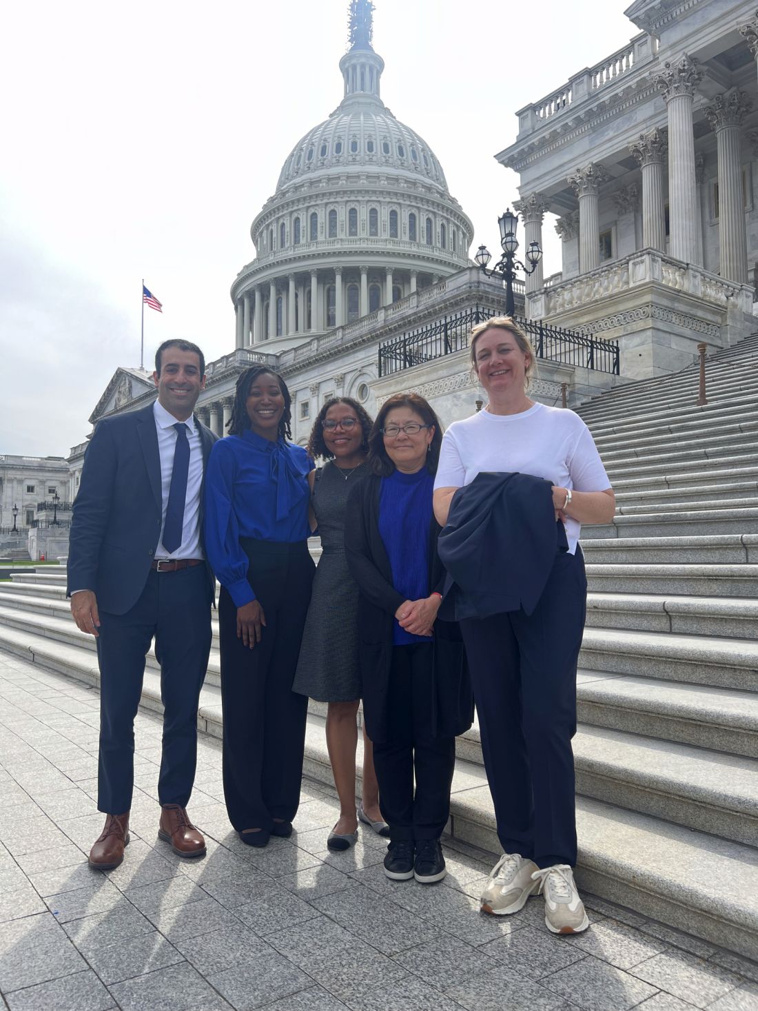
In our first in-person Advocacy Day on Capitol Hill since 2019, AGA leaders and patient advocates from 22 total states met with House and Senate offices to educate members of Congress and their staff about policies affecting GI patient care such as prior authorization and step therapy. Federal research funding and Medicare reimbursement were also on the agenda.
In the meetings, the patient shared their stories of living with various gastrointestinal diseases, including ulcerative colitis and Crohn’s disease, and the struggles they’ve gone through to get treatments approved by their insurers. AGA physicians shared the provider perspective of how policies like prior authorization negatively impact practices. According to a 2023 AGA member survey, 95% of respondents say that prior authorization restrictions have impacted patient access to clinically appropriate treatments and patient clinical outcomes and 84% described that the burden associated with prior authorization policies have increased “significantly” or “somewhat” over the last 5 years. AGA’s advocacy day came not long after UnitedHealthcare’s announcement of a new “Gold Card” prior authorization policy to be implemented in 2024, which will impact most colonoscopies and endoscopies for its 27 million commercial beneficiaries. The group expressed serious concerns about the proposed policy to lawmakers.
“It was a wonderful and empowering experience to share my personal story with my Representative/Senator and know that they were really listening to my concerns about insurer overreach,” said Aaron Blocker, a Crohn’s disease patient and advocate. “I hope Congress acts swiftly on passing prior authorization reform, so no more patients are forced to live in pain while they wait for treatments to be approved.” As gastroenterologists, too much administrative time is spent submitting onerous prior authorization requests on a near daily basis. We hope Congress takes our concerns seriously and comes together to rein in prior authorization.
AGA thanks the patient and physician advocates who participated in this year’s Advocacy Day and looks forward to continuing our work to ensure timely access to care.
Rethinking how we promote cancer screening?
Except possibly for colorectal cancer screening with sigmoidoscopy, common cancer screening tests do not extend life, according to a new study published in JAMA Internal Medicine.
The study, which was a systematic review and meta-analysis of 18 long-term randomized clinical trials involving 2.1 million individuals, found that colorectal cancer screening with sigmoidoscopy prolonged lifetime by 110 days, while fecal testing and mammography screening did not prolong life. An extension of 37 days was noted for prostate cancer screening with prostate-specific antigen testing and 107 days with lung cancer screening using CT. The study involved more than 1 decade of follow-up reporting all-cause mortality of people who had undergone mammography screening for breast cancer; colonoscopy, sigmoidoscopy, or fecal occult blood testing for colorectal cancer; CT screening for lung cancer in smokers and former smokers; or prostate-specific antigen testing for prostate cancer.

The study received a fair amount of attention in the press, but
“Cancer prevention and earlier stage diagnoses through colorectal cancer screening provides significant morbidity and cost benefits, even if all-cause mortality is not reduced,” said Lawrence Kim, MD, AGAF, AGA vice president.
The authors of the study, who were led by Michael Bretthauer, MD, PhD, of the Clinical Effectiveness Research Group, University of Oslo, are not suggesting that cancer screenings be abandoned. However, they do suggest that “organizations, institutions, and policy makers who promote cancer screening tests by their effect to save lives may find other ways of encouraging screening. It might be wise to reconsider priorities and dispassionately inform interested people about the absolute benefits, harms, and burden of screening tests that they consider undertaking.”
Except possibly for colorectal cancer screening with sigmoidoscopy, common cancer screening tests do not extend life, according to a new study published in JAMA Internal Medicine.
The study, which was a systematic review and meta-analysis of 18 long-term randomized clinical trials involving 2.1 million individuals, found that colorectal cancer screening with sigmoidoscopy prolonged lifetime by 110 days, while fecal testing and mammography screening did not prolong life. An extension of 37 days was noted for prostate cancer screening with prostate-specific antigen testing and 107 days with lung cancer screening using CT. The study involved more than 1 decade of follow-up reporting all-cause mortality of people who had undergone mammography screening for breast cancer; colonoscopy, sigmoidoscopy, or fecal occult blood testing for colorectal cancer; CT screening for lung cancer in smokers and former smokers; or prostate-specific antigen testing for prostate cancer.
The study received a fair amount of attention in the press, but
“Cancer prevention and earlier stage diagnoses through colorectal cancer screening provides significant morbidity and cost benefits, even if all-cause mortality is not reduced,” said Lawrence Kim, MD, AGAF, AGA vice president.
The authors of the study, who were led by Michael Bretthauer, MD, PhD, of the Clinical Effectiveness Research Group, University of Oslo, are not suggesting that cancer screenings be abandoned. However, they do suggest that “organizations, institutions, and policy makers who promote cancer screening tests by their effect to save lives may find other ways of encouraging screening. It might be wise to reconsider priorities and dispassionately inform interested people about the absolute benefits, harms, and burden of screening tests that they consider undertaking.”
New co–editors-in-chief named for CMGH
will be taking over their new roles beginning July 1, 2024.

“In my role as co–editor-in-chief of CMGH, I look forward to working with Jonathan Katz to advance its longstanding mission to disseminate rigorous, reproducible, and impactful digestive biology research. I am excited to launch new initiatives including the addition of special topic editors who will cover themes relating to early career investigators and diversity, equity, and inclusion,” said Dr. Battle, who is a professor in the department of cell biology, neurobiology, and anatomy at the Medical College of Wisconsin, Milwaukee.

“I am honored to work with Michele Battle and the rest of our new CMGH board of editors. Some of our goals are to expand CMGH outreach and engagement, incorporate more programs and opportunities for junior investigators, and provide rapid advancements and technical reports that drive research in the field. As such, we will strive to ensure that CMGH remains the preeminent journal focused on high-impact, basic, mechanistic research in GI and hepatology,” said Dr. Katz, an associate professor of medicine in the department of medicine gastroenterology division, director of molecular pathology and imaging care, and director of the undergraduate student scholars program at the University of Pennsylvania, Philadelphia. He is also currently an associate editor for CMGH.
Dr. Battle and Dr. Katz are former AGA Research Foundation awardees, with Dr. Battle receiving an AGA Research Scholar Award in 2009 and Dr. Katz receiving the AGA Astra Merck Advanced Research Training Award in 1998.
Over the next year, they will meet with the current editorial board and start to plan and develop new special sections and initiatives to bring to CMGH during their term.
Please join us in congratulating Dr. Battle and Dr. Katz on their new role as incoming co–editors-in chief.
will be taking over their new roles beginning July 1, 2024.
“In my role as co–editor-in-chief of CMGH, I look forward to working with Jonathan Katz to advance its longstanding mission to disseminate rigorous, reproducible, and impactful digestive biology research. I am excited to launch new initiatives including the addition of special topic editors who will cover themes relating to early career investigators and diversity, equity, and inclusion,” said Dr. Battle, who is a professor in the department of cell biology, neurobiology, and anatomy at the Medical College of Wisconsin, Milwaukee.
“I am honored to work with Michele Battle and the rest of our new CMGH board of editors. Some of our goals are to expand CMGH outreach and engagement, incorporate more programs and opportunities for junior investigators, and provide rapid advancements and technical reports that drive research in the field. As such, we will strive to ensure that CMGH remains the preeminent journal focused on high-impact, basic, mechanistic research in GI and hepatology,” said Dr. Katz, an associate professor of medicine in the department of medicine gastroenterology division, director of molecular pathology and imaging care, and director of the undergraduate student scholars program at the University of Pennsylvania, Philadelphia. He is also currently an associate editor for CMGH.
Dr. Battle and Dr. Katz are former AGA Research Foundation awardees, with Dr. Battle receiving an AGA Research Scholar Award in 2009 and Dr. Katz receiving the AGA Astra Merck Advanced Research Training Award in 1998.
Over the next year, they will meet with the current editorial board and start to plan and develop new special sections and initiatives to bring to CMGH during their term.
Please join us in congratulating Dr. Battle and Dr. Katz on their new role as incoming co–editors-in chief.
Watch the inaugural Gastro Journal Club
We are delighted to introduce the Gastro Journal Club in which an author of a study published in Gastroenterology will present their paper followed by a Q&A. The inaugural Gastro Journal Club features Loren Laine, MD, professor of medicine (digestive diseases) at Yale University, New Haven, Conn., and was hosted by the McMaster University gastroenterology division. Dr. Laine presented his article “Vonoprazan Versus Lansoprazole for Healing and Maintenance of Healing of Erosive Esophagitis: A Randomized Trial,” published in the January 2023 issue of Gastroenterology.
If you are interested in participating, please contact mpogachar@gastro.org.
Watch the inaugural Gastro Journal Club: https://rb.gy/j8mqm
Landmark obesity legislation reintroduced in Congress
(TROA) (H.R. 4818/S. 2407). This legislation is a vital first step in expanding access to obesity treatment as it would expand Medicare coverage to include screening and treatment of obesity by a diverse range of health care providers who provide obesity care. The bill also includes coverage of behavioral counseling, prescription drugs for long-term weight management, and other prevention and treatment options.
The passage of TROA could lead to improved obesity care options because many private insurance companies model their covered health benefits to reflect Medicare.
You can help lawmakers understand the urgent need for expanded access to affordable, effective obesity treatments and how greater access to these tools will equip you to better care for your patients.
Use the new obesity advocacy toolkit to find the tools and resources you need, including an email template, sample phone script, op-ed template, and more, to assist you in reaching out to your elected officials and urging them to support the passage of TROA.
CHEST SEEK releases key points feature and new print edition
Two exciting updates have come to the CHEST SEEK™ portfolio this summer.
The latest book, CHEST SEEK™ Pulmonary Medicine: 33rd Edition, was released in August. And in this newest book and certain CHEST SEEK Library collections, a feature called key points is included in the recently published 150 pulmonary medicine questions.
Key points are concise summaries of the most important takeaways of SEEK questions. Knowing the key point can help learners focus their studies.
“SEEK questions can be quite robust and intentionally detailed in their response as to why the answer options are correct or incorrect. But because of the level of detail, it can be difficult at times for the learner to correctly hone in on the author’s teaching point,” said CHEST Director, Product Strategy and Evaluation, Martha Zaborowski Pascale, CPM.
“Key points concisely summarize each question’s most important details, potentially saving the learner study time.”
CHEST SEEK™ Pulmonary Medicine: 33rd Edition was developed from the pulmonary medicine board subspecialty examination content blueprints. It tests recall, interpretation, and problem-solving skills.
Rationales provide thorough explanations and reasoning for the correct and incorrect answers. Key points are easy to find at the bottom of the pages and in a tab within SEEK Library questions.
From a printed booklet to the classic book and subscription-based library, learners have engaged with case-based questions in multiple ways. As SEEK has transformed through the years, it’s continued to be a timeless, reliable study partner.
“SEEK has evolved in many ways over its 30-year history. As technologic involvement has permitted greater advances in imaging and data presentation, SEEK has sought to make such advances from the bedside as part of the SEEK experience,” said Pascale.
“The strength of peer-reviewed, expert-written content has remained the same, but modalities such as digital flash cards and behind-the-scenes peer review discussions have enhanced this enduring product in ways that help it stand the test of time.”
Based on CHEST evaluation data, more than 90% of SEEK learners said their practice will change based on content found in the library. Plus, more than 95% of SEEK learners agreed that SEEK question authors are effective instructors.
“The success of SEEK in the past and the ability of this tool to be adapted to the changing needs of learners makes one excited about the editions to come,” said Jesse B. Hall, MD, FCCP, SEEK Editor-in-Chief and Chair of CHEST SEEK™ Pulmonary Medicine: 33rd Edition.
Looking toward the future, SEEK will continue to develop and serve the needs of chest medicine clinicians.
“One of the joys of our professional lives is the constant new discoveries and trials that change the way we practice,” said SEEK Pulmonary Medicine Vice-Chair and Deputy Editor, Jess Mandel, MD.
“However, with this comes the challenge of keeping up and staying current as the field evolves. SEEK is a terrific resource for keeping up with changes in practice and the underlying data that justify them.”
Subscribe to the SEEK Library and find CHEST SEEK™ Pulmonary Medicine: 33rd Edition at chestnet.org/Learning-and-Events/Learning/Seek-App.
Are you ready for CHEST 2023 in Hawai’i?
Just a few weeks ahead of CHEST 2023, we’re sharing the can’t-miss opportunities available on site at the meeting.
Beyond the top-tier education, CHEST 2023 has a lot to offer attendees in the way of networking, development, and unique experiences that will all make for a memorable meeting.
We’re sharing a preview of the many opportunities that will be available over the 4 days of the meeting. For more specifics on these events, including locations, visit the CHEST 2023 website. You can also download the CHEST 2023 mobile app, which will be available in mid-September.
Networking and development
• For those who want to get more involved with the CHEST community, the Networks Mixer (Monday, October 9, 4 PM HST) is open to all who’d like to learn more about the seven CHEST Networks and the 21 clinically-focused Sections within them.
• The annual Women in Chest Medicine Luncheon (Monday, October 9, 12:45 PM HST) will feature a panel of three women speaking about their experiences, their advice, how to support other women in the field, and more. This event is free, but preregistration is required. Scan the QR code to sign up.
• The first-ever Ohana Mixer (Tuesday, October 10, 6 PM HST) is an opportunity for CHEST attendees to celebrate the spirit of community that unites us across our differences. Attendees can network with each other, meet the members of our newly formed Interest Groups – including the leaders of our Women in Chest Medicine Interest Group and Respiratory Care Interest Group – and socialize with presenters from our three local CHEST Community Connections organizations.
• The Trainee Lounge will feature activities like speed mentoring, a lunch and learn with the Keynote Speaker, Dr. Cedric “Jamie” Rutland, financial wellness presentations, and more.
CHEST experiences
• The Opening Session (Sunday, October 8, 3:15 PM HST) will showcase traditional Hawaiian performances and the Keynote Address from Dr. Rutland. Immediately following, the CHEST Welcome Reception will feature live music and a traditional Hawaiian luau.
• For the second year, CHEST After Hours (Monday, October 9, 3 PM HST) will feature clinicians sharing stories of their personal triumphs, tribulations, and more experiences within medicine.
• Each year, the CHEST Challenge Championship (Tuesday, October 10, 7 PM HST) gives pulmonary and critical care medicine fellows-in-training an opportunity to compete in a live Jeopardy-style game – with bragging rights and cash prizes on the line.
• The Wellness Zone has a packed schedule of events, including beachy workouts, food demonstrations, meditation, and more.
Exhibit hall activities
• Opportunities to network with and hear presentations from local Hawaiian organizations, such as the Waianae Coast Comprehensive Health Center
• Hands-on, experiential education escape rooms
• Live educational games, including Hocus POCUS Diagnosis, PulmMemory, Peer Pressure, and more
• Simulation experiences, including Aspirated and Need for Speed – Airway Bleed
Mark your calendars now to participate in all that CHEST 2023 has to offer. We’ll see you in Hawai’i!
Environmental and occupational risk factors for lung cancer
Thoracic Oncology And Chest Imaging Network
Lung Cancer Section
Lung cancer is the third most prevalent cancer in United States, with the highest mortality (Oliver, 2022)(Siegel et al, 2023). The factors contributing to its occurrence have become more complex due to increased industrialization and worsening environmental pollution. Air pollution is a well-established environmental risk factor for lung cancer (Lu et al. 2019). On average, a full-time worker spends around 90,000 hours at work over their lifetime. It is crucial to control environmental and occupational exposures to decrease the risk of developing lung cancer. Occupations like asbestos-related work, mining, and transportation are well-known to be at risk for lung cancer (Li et al. 2021). With worsening air pollution, occupations such as firefighters, outdoor delivery workers, and forest rangers are facing an increased risk as well. Many of these carcinogens independently increase lung cancer risk (Li et al. 2021). Smoking combined with these exposures, causes a synergistic effect on lung cancer incidence. They also have a cell subtype differential risk favoring squamous and small cell lung cancer (Christiani, 2020). It is essential for workers in these high-risk occupations to use proper PPE, have regular check-ups and screenings and follow occupational safety regulations and guidelines. As air pollution continues to worsen, individuals living in these areas should reduce outdoor activities during AQI alerts, and use air purifiers and masks. Public health efforts to decrease air pollution with cleaner transportation and energy production, and better local and national air quality regulations will decrease risk in the general population (Rice et al. 2021).
Amaraja Kanitkar, MD, MBBSGuest Author
References
Christiani DC. Occupational exposures and lung cancer. Am J Respir Crit Care Med. 2020;202(3):317-19.
Li N, et al. Association of 13 occupational carcinogens in patients with cancer, individually and collectively, 1990-2017. JAMA Netw Open. 2021;4(2):e2037530.
Lu T, et al. Trends in the incidence, treatment, and survival of patients with lung cancer in the last four decades. Cancer Manag Res. 2019;11:943-53.
Oliver AL. Lung cancer: Epidemiology and screening. Surg Clin North Am. 2022;102(3):335-44.
Rice MB, et al. Respiratory impacts of wildland fire smoke: Future challenges and policy opportunities an official American thoracic society workshop report. Ann Am Thorac Soc. 2021;18(6):921-30.
Siegel RL, et al. Cancer statistics: 2023. CA Cancer J Clin. 2023;73(1):17-48.
PalliPulm: Time to expand our arsenal
Critical Care Network
Palliative and End-of-Life Section
Symptoms at the end of life in patients with COPD are just as severe as in patients with advanced cancer (Solano JP, et al. J Pain Symptom Manage. 2006;31[1]:58-69). However, despite the high symptom burden, palliative care is less common in patients with COPD (Gore J, et al. Thorax. 2000;55[12]:1000-6).

Palliative care is associated with a number of benefits, including improved symptom burden, quality of life, and patient satisfaction (Vermylen JH, et al. Int J Chron Obstruct Pulmon Dis. 2015;10:1543-51). The majority of pulmonologists report that palliative care for patients with COPD is desirable, but about half of pulmonologists indicate that they do not use the palliative care guidelines and many were not even aware they existed (Duenk RG, et al. Int J Chron Obstruct Pulmon Dis. 2017;12:299-311). Patients with COPD often have unmet needs, and the majority of patients with COPD do not have access to palliative care at their end of life (Gore JM, et al). Unfortunately, the supply of palliative care specialists is too low to meet demand, especially in outpatient settings (Kamal AH, et al. Am J Med. 2017;130:113-4).
The ATS released a multisociety policy statement in 2022 that established a framework for early palliative care in the care in patients with respiratory illnesses (Sullivan DR, et al. Am J Respir Crit Care Med. 2022;206[6]:e44-e69). However, given the paucity of specialists and the aging population, the needs of patients and their loved ones cannot be met exclusively by palliative care specialists. Pulmonologists must expand their practice to include guideline-based palliative care in order to truly serve our patients to the best of our abilities. It is incumbent on training programs to train future pulmonologists with these palliative skills, and upon medical organizations to supply time and resources to ensure the pulmonologist is able to use these skills.
Gretchen Winter, MD
Section Member-at-Large
Critical Care Network
Palliative and End-of-Life Section
Symptoms at the end of life in patients with COPD are just as severe as in patients with advanced cancer (Solano JP, et al. J Pain Symptom Manage. 2006;31[1]:58-69). However, despite the high symptom burden, palliative care is less common in patients with COPD (Gore J, et al. Thorax. 2000;55[12]:1000-6).
Palliative care is associated with a number of benefits, including improved symptom burden, quality of life, and patient satisfaction (Vermylen JH, et al. Int J Chron Obstruct Pulmon Dis. 2015;10:1543-51). The majority of pulmonologists report that palliative care for patients with COPD is desirable, but about half of pulmonologists indicate that they do not use the palliative care guidelines and many were not even aware they existed (Duenk RG, et al. Int J Chron Obstruct Pulmon Dis. 2017;12:299-311). Patients with COPD often have unmet needs, and the majority of patients with COPD do not have access to palliative care at their end of life (Gore JM, et al). Unfortunately, the supply of palliative care specialists is too low to meet demand, especially in outpatient settings (Kamal AH, et al. Am J Med. 2017;130:113-4).
The ATS released a multisociety policy statement in 2022 that established a framework for early palliative care in the care in patients with respiratory illnesses (Sullivan DR, et al. Am J Respir Crit Care Med. 2022;206[6]:e44-e69). However, given the paucity of specialists and the aging population, the needs of patients and their loved ones cannot be met exclusively by palliative care specialists. Pulmonologists must expand their practice to include guideline-based palliative care in order to truly serve our patients to the best of our abilities. It is incumbent on training programs to train future pulmonologists with these palliative skills, and upon medical organizations to supply time and resources to ensure the pulmonologist is able to use these skills.
Gretchen Winter, MD
Section Member-at-Large
Hot or cold – impact on asthma and COPD
Airways Disorders Network
Asthma & COPD Section
Earlier works investigating effects of temperature and humidity changes on the airway in patients with asthma are insightful (Strauss, et al. 1978). Heat can irritate asthmatic airways that are already hyperreactive. Cold air can remove airway moisture. Similar mechanisms with warm/hot air can affect airway inflammation in COPD. In addition, poor air quality often occurs during extreme heat events and can affect patients with COPD.
Seasonal variation in COPD exacerbations was demonstrated by the TORCH study, where a two-fold increase in COPD exacerbations and hospitalizations was noted during the winter months in both northern and southern regions of the world. This trend was not observed in tropical countries with average annual temperatures of >18 °C (64 °F). Factors accounting for this variation may include greater risk of viral infections, increased host susceptibility, and more time spent indoors, along with impact of temperature variation on lung function (Jenkins, et al. 2012). This effect was accompanied by variation in the treatment choices with antibiotics alone or in combination with steroids. A trend towards combined antibiotics and steroids was noted during winters.
Ideal conditions for patients with COPD to minimize risk for exacerbation would be home humidity between 30% and 50% with indoor temperature of 21°C at least 9 hours per day in living areas (Osman, et al. 2008).
Outdoor activities during extreme temperatures should be avoided. Air conditioning and/or humidifiers can be helpful in modifying influences.
Maria Azhar, MD
Section Fellow-in-Training
Richard George Barbers, MD, FCCP
Section Chair
References
Jenkins CR, et al. Seasonality and determinants of moderate and severe COPD exacerbations in the TORCH study. Eur Respir J. 2012;39(1):38-45.
Osman LM, et al. Home warmth and health status of COPD patients. Eur J Public Health. 2008;18(4):399-405.
Strauss RH, et al. Influence of heat and humidity on the airway obstruction induced by exercise in asthma. J Clin Invest. 1978;61(2):433-40.