User login
Does MS Begin With Radiologically Isolated Syndrome?
BOSTON—Radiologically isolated syndrome (RIS) may be the first visible manifestation of multiple sclerosis (MS), according to an overview that was presented at the 2014 Joint ACTRIMS–ECTRIMS Meeting.
Not all neurologists accept RIS as a valid and distinct entity, however. Providing a firm, scientific basis for the diagnosis may require improved characterization of the syndrome and the lesions that define it.
An important question for researchers to answer is whether MS, similar to diseases such as high blood pressure, high cholesterol, and diabetes, should be treated early (eg, at RIS onset) to improve disability over time, said Darin T. Okuda, MD, Associate Professor of Neurology and Neurotherapeutics at the University of Texas Southwestern in Dallas. “We definitely need more scientific studies to answer this very important question: Would earlier treatment result in better long-term outcomes for patients?”

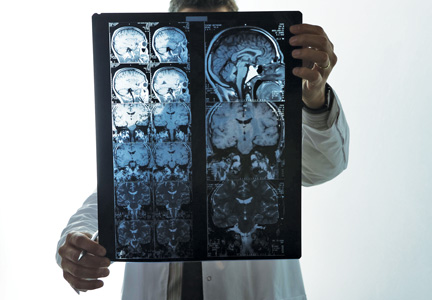
What Is RIS? A diagnosis of RIS may result from an MRI ordered to investigate a complaint unrelated to MS. “For the most part, these are individuals who were being worked up for migraine headache complaints, cerebrovascular accidents, [and] post-traumatic events,” said Dr. Okuda. A neurologist may diagnose RIS when the MRI reveals features within the brain and spinal cord that strongly suggest MS. Such features include periventricular, ovoid, circumscribed lesions within the cervical and thoracic spinal cord.
In addition, to diagnose RIS, a neurologist must confirm that the patient has never had clinical symptoms of MS. “We do our best to ensure that we are not missing another condition or disease process that could better explain why these structural anomalies are observed,” said Dr. Okuda.
The literature appears to provide evidence for asymptomatic MS. “Great scientific data support the presence of incidental anomalies [and] asymptomatic anomalies within the CNS from sporadic and familial MS cases,” said Dr. Okuda. Large collections of postmortem cases have identified and validated demyelinating changes within brain and spinal cord tissue, and researchers have described brain changes that are highly suggestive of MS. “Healthy subjects are definitely a group of interest because they appear asymptomatic and clearly possess features consistent with demyelinating disease,” said Dr. Okuda.
Skepticism About RIS
Neurologists have presented several arguments that RIS is not a distinct entity. One argument is that patients diagnosed with RIS have cryptogenic white matter changes but not MS. Many patients with RIS have changes in a brain region that MS does not typically affect, such as the subcortical or frontal region.
Another argument is that the patients, knowingly or not, have had clinical symptoms of MS. This objection often is directed against clinically isolated syndrome, too, said Dr. Okuda. Furthermore, some neurologists suggest that RIS is actually MS. The literature, however, appears to lend support to RIS as a diagnosis. Many MRI studies focusing on at-risk groups indicate the existence of presymptomatic disease, and recent scientific evidence suggests that deep gray matter volume changes may be present before the first clinical attack suggestive of MS, said Dr. Okuda.
Conventional Imaging May Lead to Overdiagnosis
Neurologists now use conventional imaging technology more often than novel techniques, but this tendency may result in the overdiagnosis of RIS, said Dr. Okuda. With conventional imaging, lesions may appear to be typical of MS when they are unrelated to the disease.
Future imaging efforts could prevent such mistakes. Newer techniques enable neurologists to see whether blood vessels traverse the lesions, and this feature is characteristic of MS-related changes, said Dr. Okuda. A preponderance of these features on brain MRI suggests that MS, rather than an unknown cause, is responsible for the findings. High-resolution imaging and qualitative imaging studies also could improve the diagnosis of RIS.
Treatment of RIS
No scientific data currently address the question of whether to treat a patient with RIS. One common reason that neurologists choose treatment is that the patient’s MRI scan has evolved and revealed more changes (eg, contrast enhancement or additional lesions) that are consistent with MS. Neurologists also may decide to treat RIS if the patient’s MRI has particularly negative findings such as tumefactive-like lesions, lesions involving the gray and white matter, or signs of old disease. Contrast enhancement within a specific brain region for an individual with features typical of MS also prompts some neurologists to initiate treatment.
Nevertheless, the effects of treating RIS with disease-modifying therapy (DMT) are unknown. “We need to treat these individuals under a controlled environment” in a prospective study, said Dr. Okuda. The RIS Consortium recently formed a strategic research alliance with Biogen Idec to assess the effect of dimethyl fumarate on extending the time to a first neurologic event associated with CNS demyelination. This study within the US will begin enrollment during the last quarter of 2014.
One-Third of Patients With RIS Had an Attack
Dr. Okuda and colleagues performed a retrospective analysis of individuals with RIS in the United States, Spain, France, Italy, and Turkey. The researchers’ goal was to collect cases and assess for risk factors for first symptom onset. They identified 451 subjects with RIS, the majority of whom were from the US. Most patients were young (mean age, 37.2), Caucasian, and female.
The most common reason the patients underwent MRI was for headache evaluation. Approximately 10% of subjects had a family history of MS, and about 65% had an abnormal CSF profile. These results indicate the specificity of the criteria that the researchers used to identify subjects, said Dr. Okuda. Approximately 17% of patients were exposed to a DMT before first symptom onset.
During the five-year study period, 34% of the cohort had a first attack suggestive of MS. This result “provided further validation that asymptomatic MS exists,” said Dr. Okuda. The investigators identified spinal cord lesions within the cervical or thoracic region, age younger than 37, and male sex as risk factors for a first attack. Having more than one risk factor was associated with a greater risk than having only one risk factor.
Among patients who had a first attack, 11% were identified with a primary progressive MS phenotype. This finding provides evidence for an asymptomatic form of progressive MS, said Dr. Okuda. In addition, for those subjects with an initial MRI study performed after 2008 that revealed anomalies suggestive of MS, 40% demonstrated worsening on MRI and had an increased risk for a seminal clinical event, but further scientific efforts would be needed to confirm these data, said Dr. Okuda.
—Erik Greb
Suggested Reading
Okuda DT, Mowry EM, Beheshtian A, et al. Incidental MRI anomalies suggestive of multiple sclerosis: the radiologically isolated syndrome. Neurology. 2009;72(9):800-805.
Okuda DT, Mowry EM, Cree BA, et al. Asymptomatic spinal cord lesions predict disease progression in radiologically isolated syndrome. Neurology. 2011;76(8):686-692.
Okuda DT, Siva A, Kantarci O, et al. Radiologically isolated syndrome: 5-year risk for an initial clinical event. PLoS One. 2014 Mar 5;9(3):e90509.
BOSTON—Radiologically isolated syndrome (RIS) may be the first visible manifestation of multiple sclerosis (MS), according to an overview that was presented at the 2014 Joint ACTRIMS–ECTRIMS Meeting.
Not all neurologists accept RIS as a valid and distinct entity, however. Providing a firm, scientific basis for the diagnosis may require improved characterization of the syndrome and the lesions that define it.
An important question for researchers to answer is whether MS, similar to diseases such as high blood pressure, high cholesterol, and diabetes, should be treated early (eg, at RIS onset) to improve disability over time, said Darin T. Okuda, MD, Associate Professor of Neurology and Neurotherapeutics at the University of Texas Southwestern in Dallas. “We definitely need more scientific studies to answer this very important question: Would earlier treatment result in better long-term outcomes for patients?”
What Is RIS? A diagnosis of RIS may result from an MRI ordered to investigate a complaint unrelated to MS. “For the most part, these are individuals who were being worked up for migraine headache complaints, cerebrovascular accidents, [and] post-traumatic events,” said Dr. Okuda. A neurologist may diagnose RIS when the MRI reveals features within the brain and spinal cord that strongly suggest MS. Such features include periventricular, ovoid, circumscribed lesions within the cervical and thoracic spinal cord.
In addition, to diagnose RIS, a neurologist must confirm that the patient has never had clinical symptoms of MS. “We do our best to ensure that we are not missing another condition or disease process that could better explain why these structural anomalies are observed,” said Dr. Okuda.
The literature appears to provide evidence for asymptomatic MS. “Great scientific data support the presence of incidental anomalies [and] asymptomatic anomalies within the CNS from sporadic and familial MS cases,” said Dr. Okuda. Large collections of postmortem cases have identified and validated demyelinating changes within brain and spinal cord tissue, and researchers have described brain changes that are highly suggestive of MS. “Healthy subjects are definitely a group of interest because they appear asymptomatic and clearly possess features consistent with demyelinating disease,” said Dr. Okuda.
Skepticism About RIS
Neurologists have presented several arguments that RIS is not a distinct entity. One argument is that patients diagnosed with RIS have cryptogenic white matter changes but not MS. Many patients with RIS have changes in a brain region that MS does not typically affect, such as the subcortical or frontal region.
Another argument is that the patients, knowingly or not, have had clinical symptoms of MS. This objection often is directed against clinically isolated syndrome, too, said Dr. Okuda. Furthermore, some neurologists suggest that RIS is actually MS. The literature, however, appears to lend support to RIS as a diagnosis. Many MRI studies focusing on at-risk groups indicate the existence of presymptomatic disease, and recent scientific evidence suggests that deep gray matter volume changes may be present before the first clinical attack suggestive of MS, said Dr. Okuda.
Conventional Imaging May Lead to Overdiagnosis
Neurologists now use conventional imaging technology more often than novel techniques, but this tendency may result in the overdiagnosis of RIS, said Dr. Okuda. With conventional imaging, lesions may appear to be typical of MS when they are unrelated to the disease.
Future imaging efforts could prevent such mistakes. Newer techniques enable neurologists to see whether blood vessels traverse the lesions, and this feature is characteristic of MS-related changes, said Dr. Okuda. A preponderance of these features on brain MRI suggests that MS, rather than an unknown cause, is responsible for the findings. High-resolution imaging and qualitative imaging studies also could improve the diagnosis of RIS.
Treatment of RIS
No scientific data currently address the question of whether to treat a patient with RIS. One common reason that neurologists choose treatment is that the patient’s MRI scan has evolved and revealed more changes (eg, contrast enhancement or additional lesions) that are consistent with MS. Neurologists also may decide to treat RIS if the patient’s MRI has particularly negative findings such as tumefactive-like lesions, lesions involving the gray and white matter, or signs of old disease. Contrast enhancement within a specific brain region for an individual with features typical of MS also prompts some neurologists to initiate treatment.
Nevertheless, the effects of treating RIS with disease-modifying therapy (DMT) are unknown. “We need to treat these individuals under a controlled environment” in a prospective study, said Dr. Okuda. The RIS Consortium recently formed a strategic research alliance with Biogen Idec to assess the effect of dimethyl fumarate on extending the time to a first neurologic event associated with CNS demyelination. This study within the US will begin enrollment during the last quarter of 2014.
One-Third of Patients With RIS Had an Attack
Dr. Okuda and colleagues performed a retrospective analysis of individuals with RIS in the United States, Spain, France, Italy, and Turkey. The researchers’ goal was to collect cases and assess for risk factors for first symptom onset. They identified 451 subjects with RIS, the majority of whom were from the US. Most patients were young (mean age, 37.2), Caucasian, and female.
The most common reason the patients underwent MRI was for headache evaluation. Approximately 10% of subjects had a family history of MS, and about 65% had an abnormal CSF profile. These results indicate the specificity of the criteria that the researchers used to identify subjects, said Dr. Okuda. Approximately 17% of patients were exposed to a DMT before first symptom onset.
During the five-year study period, 34% of the cohort had a first attack suggestive of MS. This result “provided further validation that asymptomatic MS exists,” said Dr. Okuda. The investigators identified spinal cord lesions within the cervical or thoracic region, age younger than 37, and male sex as risk factors for a first attack. Having more than one risk factor was associated with a greater risk than having only one risk factor.
Among patients who had a first attack, 11% were identified with a primary progressive MS phenotype. This finding provides evidence for an asymptomatic form of progressive MS, said Dr. Okuda. In addition, for those subjects with an initial MRI study performed after 2008 that revealed anomalies suggestive of MS, 40% demonstrated worsening on MRI and had an increased risk for a seminal clinical event, but further scientific efforts would be needed to confirm these data, said Dr. Okuda.
—Erik Greb
Suggested Reading
Okuda DT, Mowry EM, Beheshtian A, et al. Incidental MRI anomalies suggestive of multiple sclerosis: the radiologically isolated syndrome. Neurology. 2009;72(9):800-805.
Okuda DT, Mowry EM, Cree BA, et al. Asymptomatic spinal cord lesions predict disease progression in radiologically isolated syndrome. Neurology. 2011;76(8):686-692.
Okuda DT, Siva A, Kantarci O, et al. Radiologically isolated syndrome: 5-year risk for an initial clinical event. PLoS One. 2014 Mar 5;9(3):e90509.
Specialty drug coupons - a double-edged sword of improved adherence, increased costs
While the availability of coupons can help defray out-of-pocket costs and improve access and adherence to high-cost specialty drugs, it may also result in higher costs to the health system.
Patients used drug coupons to pay for $21.2 million of $35.3 million in out-of-pocket costs, according to an analysis of 265,000 prescriptions worth $911 million in 2013. The coupons generally dropped a patient’s out-of-pocket costs to less than $250 per prescription, a point below which patients were “far less likely” to abandon prescriptions in cases of specialty anti-inflammatory drugs and treatments for multiple sclerosis (Health Affairs 2014;33:1761-9 [doi:10.1377/hlthaff.2014.0497]).
But while the use of coupons demonstrated that the lower cost led to lower prescription abandonment rates, it also allowed patients to circumvent cost-containment measures that pharmacy benefit managers employ.

“We found that drug coupons were extremely effective in lowering patients’ costs to less than $50 per prescription, thus eliminating the incentive to select a preferred drug,” wrote Catherine I. Starner, Pharm.D., senior health outcomes researcher at pharmacy benefit manager Prime Therapeutics, and her colleagues.
To control costs at the health system level, tighter controls could be placed on specialty drugs at the pharmacy benefit manager level, according to Dr. Starner and her colleagues. Such controls could include prior authorization or the use of step therapies,as well as excluding coverage of nonpreferred drugs. The latter strategy, however, could make drug access a problem, especially if physicians are not aware of an insurance plan taking these steps.
Excluding specialty drugs “makes the patient responsible for the entire cost of the drug,” the authors noted. “If prescribers are unaware of such exclusions, or if patients require specific therapies, such exclusions may impose considerable costs on patients” and potentially lead to access issues.
While the availability of coupons can help defray out-of-pocket costs and improve access and adherence to high-cost specialty drugs, it may also result in higher costs to the health system.
Patients used drug coupons to pay for $21.2 million of $35.3 million in out-of-pocket costs, according to an analysis of 265,000 prescriptions worth $911 million in 2013. The coupons generally dropped a patient’s out-of-pocket costs to less than $250 per prescription, a point below which patients were “far less likely” to abandon prescriptions in cases of specialty anti-inflammatory drugs and treatments for multiple sclerosis (Health Affairs 2014;33:1761-9 [doi:10.1377/hlthaff.2014.0497]).
But while the use of coupons demonstrated that the lower cost led to lower prescription abandonment rates, it also allowed patients to circumvent cost-containment measures that pharmacy benefit managers employ.
“We found that drug coupons were extremely effective in lowering patients’ costs to less than $50 per prescription, thus eliminating the incentive to select a preferred drug,” wrote Catherine I. Starner, Pharm.D., senior health outcomes researcher at pharmacy benefit manager Prime Therapeutics, and her colleagues.
To control costs at the health system level, tighter controls could be placed on specialty drugs at the pharmacy benefit manager level, according to Dr. Starner and her colleagues. Such controls could include prior authorization or the use of step therapies,as well as excluding coverage of nonpreferred drugs. The latter strategy, however, could make drug access a problem, especially if physicians are not aware of an insurance plan taking these steps.
Excluding specialty drugs “makes the patient responsible for the entire cost of the drug,” the authors noted. “If prescribers are unaware of such exclusions, or if patients require specific therapies, such exclusions may impose considerable costs on patients” and potentially lead to access issues.
FROM HEALTH AFFAIRS

More Than 45 Novel MS Susceptibility Variants Are Identified
BOSTON—More than 45 new multiple sclerosis (MS) susceptibility variants were reported by researchers.
“We now present a comprehensive view of MS genetic susceptibility and provide a detailed map of proximal biologic effects that identify new molecular pathways involved in the transition from health to MS,” said Philip Laurence De Jager, MD, PhD, Assistant Professor, Harvard Medical School in Boston, on behalf of the International MS Genetics Consortium.
To identify MS susceptibility associations outside the validated MS susceptibility loci and uncover new biologic processes that drive the onset of MS, the researchers conducted a genomewide discovery study of approximately eight million single-nucleotide polymorphisms (SNPs) in each of 14,802 MS cases and 26,703 controls. This step was followed by a deep replication study of more than 80,000 SNPs in more than 19,217 MS cases and 17,842 controls. The 4,716 SNPs with a P value of less than 0.05 in the discovery study were included in the present study. Functional evaluations of the results were conducted using DEPICT for pathway analysis, as well as analyses of immune cell RNA expression data from ImmVar and reference epigenomic maps from the Epigenome Roadmap and ENCODE projects.
At the end of the replication study, more than 45 new susceptibility variants were identified, with 10 major histocompatibility complex (MHC) and more than 150 non-MHC SNPs meeting a threshold of genomewide significance. The depth of the replication effort identified multiple independent effects in many regions that were previously unresolvable. For example, the EVI5 region has as many as four independent susceptibility variants.
“Uncovering this multiplicity of associations in certain regions is critical to our efforts to model the biologic consequences of MS susceptibility variants and to develop predictive algorithms,” Dr. De Jager said. “With more than 150 independent susceptibility effects and a high resolution analysis of each locus in hand, we have created a reference map of MS susceptibility and now turn to the task of understanding the biology of MS susceptibility.
“With the new MS map and multiple approaches to epigenomic annotation and functional evaluations, it is clear that non-TH1/Th17/Treg processes are important in the onset of MS,” he continued. “Myeloid, NK, and CD8 cells are now implicated, and B and dendritic cell functions are suggested to be altered by MS variants.”
Leveraging RNA data from 405 subjects with purified CD4 T and monocytes, 29% of MS variants with RNA effects are unique to monocytes, which is now the same proportion as for T cells (29%). Pathway analyses highlighted an enrichment of NK and B cell activation molecular networks in addition to T cell effects.
—Glenn S. Williams
Investigational Oral Drug Reduces Disease Activity in Relapsing-Remitting MS
An oral, selective sphingosine 1-phosphate 1 receptor modulator appears to reduce MRI measures of disease activity in patients with relapsing-remitting multiple sclerosis (MS), according to data presented. The drug, RPC1063, may be safe and tolerable for these patients.
Data are from the international, combined phase II/III RADIANCE trial. Jeffrey Cohen, MD, Director of the Cleveland Clinic’s Mellen Center for MS Treatment and Research in Cleveland, and colleagues randomized 258 patients with relapsing-remitting MS. A total of 87 participants received a low dose (0.5 mg) of RPC1063, 83 participants received a high dose (1.0 mg) of RPC1063, and 88 participants received placebo for 24 weeks.
The trial’s primary end point was the cumulative number of total gadolinium-enhancing lesions on MRI at weeks 12, 16, 20, and 24. Key secondary end points included the number of gadolinium-enhancing lesions at week 24, the cumulative number of new or enlarging T2 lesions from weeks 12 to 24, and annualized relapse rate.
The researchers assessed safety using vital signs, laboratory tests, ECG, Holter monitoring, pulmonary function test, optical coherence tomography, and adverse events. Patients were titrated to their assigned doses during one week to mitigate the first doses’ effects on heart rate.
Approximately 98% of patients completed the trial. The cumulative number of gadolinium-enhancing lesions from weeks 12 to 24 was reduced by 86% in both treatment arms, compared with placebo. The numbers of gadolinium-enhancing lesions at week 24 were significantly reduced by 91% in the low-dose group and by 94% in the high-dose group, compared with placebo. The cumulative number of new or enlarging T2 lesions from weeks 12 to 24 was reduced by 84% in the low-dose group and by 91% in the high-dose group. The researchers observed a favorable trend toward a reduction in annualized relapse rate among treated patients, compared with controls.
The adverse event profiles were similar between groups. The most common adverse events in the treated groups, compared with placebo, were nasopharyngitis (9.4% vs 13.6%), headache (4.7% vs 9.1%), and urinary tract infection (4.7% vs 2.3%). During the first six hours after the first dose of RPC1063, maximum reductions in mean hourly heart rate were < 2 bpm from baseline, and no patient had a minimum hourly heart rate < 45 bpm. The investigators did not observe any notable cardiac, pulmonary, ophthalmologic, or malignancy adverse events.
“Both doses of RPC1063 demonstrated large, significant, and consistent reductions of all MRI measures of MS disease activity,” said Dr. Cohen. “The tolerability and safety results suggest a favorable risk–benefit profile of RPC1063 in the treatment of relapsing-remitting MS. These results support the ongoing phase III portion of the RADIANCE trial of RPC1063 vs interferon beta-1a in relapsing-remitting MS,” he concluded.
—Erik Greb
Improved Adherence to Disease-Modifying Therapy Reduces Health Care Resource Use and Medical Costs
Improved adherence to disease-modifying therapy (DMT) is associated with lower medical and indirect costs, reduced work-loss days, and decreased inpatient stays and emergency visits for patients with multiple sclerosis (MS), investigators reported.
According to published estimates, real-world adherence to DMTs in MS ranges from 41% to 88%, and higher adherence is associated with lower rates of relapse and lower costs. Sander Yermakov, from the Analysis Group in Boston, and researchers from Biogen Idec in Cambridge, Massachusetts, sought to estimate the effect of adherence to DMTs in health care resource use and cost outcomes in patients with MS and to model the impact of improving adherence on these outcomes.
A retrospective analysis was conducted using the OptumHealth Reporting & Insights employer claims database, which contains information on medical and pharmacy claims for more than 18 million beneficiaries in the US, as well as disability claims and salary information in a subset of approximately 4.2 million employees. Employed patients with two or more MS diagnoses (ICD-9-CM 340) initiating DMT from January 2, 2002, through March 31, 2012, were included in the analysis.
Direct medical costs (paid amounts to providers for services or drugs, excluding payments for DMTs), indirect costs (disability payments to employees and work-loss costs to employers), and resource use were analyzed in the six months prior to initiation of any DMT (baseline period) and for three years after initiation (follow-up period). Adherence to any DMT was defined as the proportion of days covered (PDC) and measured using the percentage of days in each follow-up period during which the patient had one or more MS DMT available.
Multivariate regression analyses were used to estimate the effect of PDC on follow-up period outcomes, controlling for baseline characteristics. The estimated model was used to predict the change in use and costs associated with a 10% improvement in PDC.
A total of 1,538 patients met the selection criteria (baseline age, 43.6; 63% female). PDC had a statistically significant effect on direct medical and indirect work productivity costs, work-loss days, and likelihood of an inpatient stay or emergency visit at one-, two-, and three-year follow-up. A 10% improvement in PDC was estimated to reduce direct costs by 4%, indirect costs by 3% to 4%, work-loss days by 3% to 7%, likelihood of an inpatient stay by 13% to 19%, and likelihood of an emergency visit by 8% to 19%, depending on the follow-up period.
The impact on the likelihood of an inpatient stay or emergency visit increased with the length of the follow-up period. In the first year, the decrease in direct costs associated with a 10% improvement in adherence was greater for patients with PDC of 0.8 or higher (10%), men (6%), and patients not on disability in the baseline period.
—Glenn S. Williams
Equivalence Demonstrated by a Generic Version of Glatiramer Acetate
Results of the randomized, double-blind GATE trial indicate that generic glatiramer acetate is equivalent to branded Copaxone in reducing the number of gadolinium-enhancing lesions, a clinically relevant end point in relapsing-remitting multiple sclerosis (RRMS). Other efficacy outcomes, safety, and tolerability also were comparable.
“This is the first generic glatiramer acetate with an efficacy and safety profile demonstrated to be equivalent to the currently marketed product,” said Jeffrey A. Cohen, MD, Director of the Cleveland Clinic Mellen Center for Multiple Sclerosis Treatment and Research, on behalf of the GATE Study Group.
“Generic alternatives to the currently approved therapies for RRMS are needed,” Dr. Cohen said. But because glatiramer acetate is a complex polypeptide mixture that precludes pharmacokinetic comparison, a generic version needs to demonstrate equivalence in efficacy and safety. The GATE trial aimed to show that generic glatiramer acetate was equivalent to Copaxone (Teva Pharmaceuticals; North Wales, Pennsylvania), as measured by MRI and clinical end points, safety, and tolerability in RRMS.
Patients with ambulatory RRMS (ages 18 to 55) with one or more relapse in the year prior to screening and one to 15 gadolinium-enhancing brain lesions were randomized in a 4.3:4.3:1 fashion in a multicenter, double-blind, placebo-controlled trial to receive 20 mg of generic glatiramer acetate, 20 mg of Copaxone, or placebo by daily subcutaneous injection for nine months. The primary end point was combined number of gadolinium-enhancing lesions over months seven, eight, and nine. Additional efficacy end points included other MRI parameters, annualized relapse rate, Expanded Disability Status Scale (EDSS) score, and freedom from disease activity. Safety and tolerability were assessed through monitoring of adverse events, injection site reactions, and routine blood laboratory tests.
A total of 794 patients were randomized and treated with generic glatiramer acetate (n = 353), Copaxone (n = 357), or placebo (n = 84). Of these, 735 patients (92.5%) completed the nine-month double-blind treatment period. The estimated geometric mean numbers of gadolinium-enhancing lesions were 0.42 for generic glatiramer acetate and 0.39 for Copaxone, resulting in an estimated generic/brand lesion ratio of 1.097 with a 95% confidence interval of 0.884 to 1.362, which is within the predefined equivalence margin.
The estimated geometric mean number of gadolinium-enhancing lesions for both the generic and brand drug groups was lower than for the placebo group, confirming assay sensitivity. Annualized relapse rates were 0.31 for generic glatiramer acetate, 0.41 for Copaxone, and 0.39 for placebo. Comparable proportions of patients treated with the generic and branded drug were free from disease activity. EDSS was stable in all three groups. The incidence, spectrum, and severity of reported adverse events, including injection site reactions, were similar in the two treatment groups.
The generic version of glatiramer acetate is being developed by Synthon Biopharmaceuticals BV (Nijmegen, the Netherlands).
—Glenn S. Williams
Does Brain Reserve Protect Against Physical Disability in MS?
Patients with multiple sclerosis (MS) who have larger maximal lifetime brain growth may have less physical disability, according to researchers. Larger maximal lifetime brain growth may help preserve patients’ ambulation and fine motor function.
“Clinical consideration of maximal lifetime brain growth … may help identify patients with MS at highest risk for future physical disability,” said James F. Sumowski, PhD, Senior Research Scientist of Neuropsychology and Neuroscience Research at the Kessler Foundation Research Center in West Orange, New Jersey. “At-risk patients can be enrolled in early intervention treatments or research on such treatments.”
Dr. Sumowski and colleagues studied 352 patients with MS, including 255 people with relapsing-remitting MS and 97 people with secondary progressive MS. The researchers assessed participants’ disease burden using high-resolution, 3-D, T1 fast field echo. They used software to normalize patients’ total brain, gray matter, white matter, deep gray matter, and thalamus volumes. The researchers also used dual-echo turbo spin echo to quantify T2 lesion volume. Dr. Sumowski’s group estimated maximal lifetime brain growth with SIENAX v-scaling factor (adjusted for gender), a proxy for intracranial volume.
In addition, the investigators assessed 168 participants’ ambulation with the 25-Foot Walk. Participants who used assistive devices for walking were excluded. Fine motor function was assessed with the Nine-Hole Peg Test (in 323 patients) and Finger Tapping Test (in 330 patients). Cognitive status was assessed in 333 patients with the Paced Auditory Serial Addition Test-3 (PASAT-3).
Dr. Sumowski and colleagues found that maximal lifetime brain growth significantly predicted pyramidal and cerebellar function. Maximal lifetime brain growth accounted for the variance between patients in physical disability, as measured by the Finger Tapping Test. People with larger maximal lifetime brain growth performed better on that test. Disability was worse in people with smaller maximal lifetime brain growth, and the relationship did not change when the researchers controlled for demographics and deep gray matter atrophy.
The investigators found a similar relationship for cerebellar function. Participants with larger maximal lifetime brain growth had less disability and cerebellar dysfunction, even when the researchers controlled for demographics and disease burden. Patients with larger maximal lifetime brain growth completed the 25-Foot Walk more quickly, which indicated that they had less disability.
The researchers did not find a relationship between maximal lifetime brain growth and other functional systems, such as brainstem, visual, motor sensory, and bladder function.
—Erik Greb
Computerized Cognitive Test May Be a Valid Assessment for Patients With MS
PHILADELPHIA—A computerized tool is a valid means of performing cognitive testing for patients with multiple sclerosis (MS), according to research presented at the 66th Annual Meeting of the American Academy of Neurology.
NeuroTrax provides a global cognitive score along with scores for individual cognitive domains. Results of NeuroTrax assessments correlate well with results of the Symbol Digit Modalities Test (SDMT), which provides a single, global score “and does not allow us to appreciate the richness and variability of domains of cognition that can be impaired differentially,” according to Mark Gudesblatt, MD.

“Easily utilized objective cognitive screens are needed to evaluate the cognitive impact of MS independent of Expanded Disability Status Scale score or MRI,” said Dr. Gudesblatt, a neurologist at South Shore Neurologic Associates in Patchogue, New York. “Additional studies of cognitive screening tools and correlation to previously utilized clinical trial measures are necessary to facilitate widespread acceptance and incorporation of these tools into routine MS care and patient management.”
Dr. Gudesblatt and his colleagues conducted a retrospective review of data for 113 consecutive patients with MS who were referred for cognitive testing during the course of routine clinical care. The patients were evaluated with the oral version of SDMT and NeuroTrax testing on the same day. The researchers used Centofanti (1975) age norms to standardize the SDMT raw scores. Patients’ ages ranged between approximately 20 and 83, and the population’s mean age was approximately 49. About 85% of participants were female. Patients had an average of about 15 years of education.
The patients’ standardized SDMT scores significantly correlated with NeuroTrax global cognitive scores. Standardized SDMT scores also significantly correlated with NeuroTrax scores on seven individual cognitive domain indices, including executive function, memory, attention, visual spatial processing, information processing speed, motor skill, and verbal function. The three individual cognitive domain scores on NeuroTrax that most strongly correlated with SDMT scores were executive function, memory, and attention.
In addition, SDMT score was correlated with the number of NeuroTrax index scores greater than 1 SD below average for norms of cognitive health, age, and education. The researchers classified SDMT subgroups, according to the SDMT manual, as less than 1 SD below average, less than 1.5 SD below average (low), less than 2 SD below average (moderately low), and 2 or more SD below average (very low). Mean NeuroTrax global cognitive scores for these subgroups were 99.1, 93.0, 83.9, and 76.9, respectively. The researchers observed a similar pattern for all NeuroTrax domain index scores, especially executive function and attention. Neurotrax’s ability to identify individual domains of impairment and categorize the number of domains that were impaired “improves our ability to identify and appreciate the variable impact and burden of neurologic diseases like MS,” said Dr. Gudesblatt.
This retrospective review supports the construct validity of the NeuroTrax cognitive assessment tool, said Dr. Gudesblatt. “Routine use of cognitive screening in the care of patients with MS is not as common as is likely warranted,” he added. “Computerized cognitive testing may provide an easy, independent, objective screening tool that taps into broader sets of cognitive domains.”
—Erik Greb
PHILADELPHIA—A computerized tool is a valid means of performing cognitive testing for patients with multiple sclerosis (MS), according to research presented at the 66th Annual Meeting of the American Academy of Neurology.
NeuroTrax provides a global cognitive score along with scores for individual cognitive domains. Results of NeuroTrax assessments correlate well with results of the Symbol Digit Modalities Test (SDMT), which provides a single, global score “and does not allow us to appreciate the richness and variability of domains of cognition that can be impaired differentially,” according to Mark Gudesblatt, MD.
“Easily utilized objective cognitive screens are needed to evaluate the cognitive impact of MS independent of Expanded Disability Status Scale score or MRI,” said Dr. Gudesblatt, a neurologist at South Shore Neurologic Associates in Patchogue, New York. “Additional studies of cognitive screening tools and correlation to previously utilized clinical trial measures are necessary to facilitate widespread acceptance and incorporation of these tools into routine MS care and patient management.”
Dr. Gudesblatt and his colleagues conducted a retrospective review of data for 113 consecutive patients with MS who were referred for cognitive testing during the course of routine clinical care. The patients were evaluated with the oral version of SDMT and NeuroTrax testing on the same day. The researchers used Centofanti (1975) age norms to standardize the SDMT raw scores. Patients’ ages ranged between approximately 20 and 83, and the population’s mean age was approximately 49. About 85% of participants were female. Patients had an average of about 15 years of education.
The patients’ standardized SDMT scores significantly correlated with NeuroTrax global cognitive scores. Standardized SDMT scores also significantly correlated with NeuroTrax scores on seven individual cognitive domain indices, including executive function, memory, attention, visual spatial processing, information processing speed, motor skill, and verbal function. The three individual cognitive domain scores on NeuroTrax that most strongly correlated with SDMT scores were executive function, memory, and attention.
In addition, SDMT score was correlated with the number of NeuroTrax index scores greater than 1 SD below average for norms of cognitive health, age, and education. The researchers classified SDMT subgroups, according to the SDMT manual, as less than 1 SD below average, less than 1.5 SD below average (low), less than 2 SD below average (moderately low), and 2 or more SD below average (very low). Mean NeuroTrax global cognitive scores for these subgroups were 99.1, 93.0, 83.9, and 76.9, respectively. The researchers observed a similar pattern for all NeuroTrax domain index scores, especially executive function and attention. Neurotrax’s ability to identify individual domains of impairment and categorize the number of domains that were impaired “improves our ability to identify and appreciate the variable impact and burden of neurologic diseases like MS,” said Dr. Gudesblatt.
This retrospective review supports the construct validity of the NeuroTrax cognitive assessment tool, said Dr. Gudesblatt. “Routine use of cognitive screening in the care of patients with MS is not as common as is likely warranted,” he added. “Computerized cognitive testing may provide an easy, independent, objective screening tool that taps into broader sets of cognitive domains.”
—Erik Greb

Watch for PML after switching from natalizumab to fingolimod
BOSTON– Cases of progressive multifocal leukoencephalopathy in multiple sclerosis patients who switch from natalizumab to fingolimod treatment are more likely a result of the natalizumab exposure than the fingolimod exposure, according to an analysis of cases in the Novartis safety reporting database for fingolimod.
About 15%-20% of fingolimod-treated patients have prior exposure to natalizumab, and of 11 patients from the safety reporting database who have developed progressive multifocal leukoencephalopathy (PML) to date, all had prior natalizumab exposure, according to Dr. Norman Putzki.
The 11 patients with PML had been treated with natalizumab for a median of 4 years, Dr. Putzki, an employee of Novartis Pharma AG, Basel, Switzerland, said at the joint meeting of the European and Americas Committees for Treatment and Research in Multiple Sclerosis.
The onset of PML, based on a retrospective review of MRI scans, was prior to the first fingolimod dose in 5 of the 11 patients, and between 3 weeks and 6 months after the first dose in 4 of the patients. Onset was unknown in the remaining 2 patients.
One additional case of possible PML was reported in a patient without prior natalizumab exposure. The patient had MRI features and the presence of AQP4 antibodies consistent with a diagnosis of neuromyelitis optica spectrum disorder, and clinical features that were consistent with a PML diagnosis, but the timing of onset in this patient could not be determined, Dr. Putzki said.
With more than 135,800 total patient-years of exposure, including during clinical trials and in the postmarketing period, no evidence of an association between fingolimod and PML has been found, he said.
Treatment with natalizumab, however, is an identified predisposing factor for PML development, and the risk is known to persist after treatment discontinuation. In fact, it is recommended that patients be monitored for any new signs or symptoms suggestive of PML for at least 6 months following discontinuation of natalizumab, he said, noting that this is particularly true for patients with John Cunningham virus (JCV) seropositivity, prior exposure to immunosuppressants, or long-term natalizumab exposure, as these patients may be at increased risk of developing PML.
With exposure to natalizumab for 25-48 months, the rate of PML in JCV-positive patients is 3/1,000 patients in those with no prior exposure to immunosuppressant therapy, and 13/1,000 in those with prior exposure to immunosuppressant therapy, whereas with exposure for 49-72 months, the respective rates are 7/1,000 and 9/1,000, he said.
Although definitive conclusions about PML risk and exposure to natalizumab and fingolimod cannot be made based on the current analysis, an excess risk due to an overlapping pharmacodynamic effect when switching from natalizumab to fingolimod was not established, and a risk of PML cannot be confidently attributed to fingolimod, Dr. Putzki said.
A baseline neurological examination and MRI scan prior to initiating fingolimod treatment may help – at least retrospectively – in attributing PML causality to natalizumab vs. fingolimod, he added.
“Vigilance for PML is warranted following natalizumab discontinuation irrespective of consecutive treatment approaches,” he concluded.
Key clinical point: Natalizumab-related PML risk may persist after switch to fingolimod.
Major finding: All PML cases were in patients with prior natalizumab exposure.
Data source: An analysis of 11 PML cases from the Novartis safety reporting database for fingolimod.
Disclosures: Dr. Putzki is employed by Novartis.
Sodium Intake Is Linked to Increased Disease Activity in MS
A higher sodium intake is associated with increased clinical and radiologic disease activity among patients with multiple sclerosis (MS), according to research published online ahead of print August 28 in the Journal of Neurology, Neurosurgery, and Psychiatry.
Mauricio F. Farez, MD, from the Department of Neurology at Raúl Carrea Institute for Neurological Research in Buenos Aires, and colleagues conducted an observational study of 70 patients with relapsing-remitting MS who were followed for two years. The researchers estimated sodium intake from sodium excretion in urine samples and used regression analysis to estimate the effect of sodium intake on MS disease activity. The findings were subsequently replicated in a separate group of 52 patients with MS.
Dr. Farez’s group found a positive correlation between exacerbation rates and sodium intake in a multivariate model after adjusting for age, gender, disease duration, smoking status, vitamin D level, BMI, and treatment. The exacerbation rate was 2.75-fold higher in patients with medium or high sodium intake, compared with the rate in subjects who had a low sodium intake.
In addition, participants with a high sodium intake had a 3.4-fold greater risk of having a new lesion revealed by MRI. This group also had, on average, eight more T2 lesions on MRI than other participants. The investigators observed a similar relationship in the independent replication group.
The authors noted several study limitations, including a small cohort size and the inability to exclude potential confounders such as diet, role of commensal microbiota, stress, and other behavior that may affect food preference and treatment compliance or healthy overall behavior.
“Thus, even though an association between increased sodium intake and increased disease activity was shown, we cannot claim causality and we cannot exclude the possibility of reverse causation: individuals with more relapses received more steroids, and thus their salt intake and excretion is increased because they have higher disease activity, and not the other way around,” stated the researchers. “Another possible caveat relates to changes in salt consumption over time. We tried to overcome this [variation] by retesting the same patients at different time points, finding no significant changes.”
The investigators also noted that although studies have found that dietary salt has a role in regulating blood pressure, the effects of sodium on the risk of MS may involve additional factors. For example, sodium chloride has been shown to have pleiotropic effects on kidney homeostasis and T-cell function, and other evidence suggests a reciprocal relationship between sodium chloride and the immune system.
“Whether high blood pressure interacts with the typical autoimmune mechanisms associated with MS is an interesting question that remains to be answered,” commented Dr. Farez and colleagues. “Nevertheless, our findings suggest that clinical trials with a salt intake reduction as an intervention are needed to establish whether sodium intake control benefits patients with MS.”
—Colby Stong
Suggested Reading
Farez MF, Fiol MP, Gaitán MI, et al. Sodium intake is associated with increased disease activity in multiple sclerosis. J Neurol Neurosurg Psychiatry. 2014; August 28 [Epub ahead of print].
A higher sodium intake is associated with increased clinical and radiologic disease activity among patients with multiple sclerosis (MS), according to research published online ahead of print August 28 in the Journal of Neurology, Neurosurgery, and Psychiatry.
Mauricio F. Farez, MD, from the Department of Neurology at Raúl Carrea Institute for Neurological Research in Buenos Aires, and colleagues conducted an observational study of 70 patients with relapsing-remitting MS who were followed for two years. The researchers estimated sodium intake from sodium excretion in urine samples and used regression analysis to estimate the effect of sodium intake on MS disease activity. The findings were subsequently replicated in a separate group of 52 patients with MS.
Dr. Farez’s group found a positive correlation between exacerbation rates and sodium intake in a multivariate model after adjusting for age, gender, disease duration, smoking status, vitamin D level, BMI, and treatment. The exacerbation rate was 2.75-fold higher in patients with medium or high sodium intake, compared with the rate in subjects who had a low sodium intake.
In addition, participants with a high sodium intake had a 3.4-fold greater risk of having a new lesion revealed by MRI. This group also had, on average, eight more T2 lesions on MRI than other participants. The investigators observed a similar relationship in the independent replication group.
The authors noted several study limitations, including a small cohort size and the inability to exclude potential confounders such as diet, role of commensal microbiota, stress, and other behavior that may affect food preference and treatment compliance or healthy overall behavior.
“Thus, even though an association between increased sodium intake and increased disease activity was shown, we cannot claim causality and we cannot exclude the possibility of reverse causation: individuals with more relapses received more steroids, and thus their salt intake and excretion is increased because they have higher disease activity, and not the other way around,” stated the researchers. “Another possible caveat relates to changes in salt consumption over time. We tried to overcome this [variation] by retesting the same patients at different time points, finding no significant changes.”
The investigators also noted that although studies have found that dietary salt has a role in regulating blood pressure, the effects of sodium on the risk of MS may involve additional factors. For example, sodium chloride has been shown to have pleiotropic effects on kidney homeostasis and T-cell function, and other evidence suggests a reciprocal relationship between sodium chloride and the immune system.
“Whether high blood pressure interacts with the typical autoimmune mechanisms associated with MS is an interesting question that remains to be answered,” commented Dr. Farez and colleagues. “Nevertheless, our findings suggest that clinical trials with a salt intake reduction as an intervention are needed to establish whether sodium intake control benefits patients with MS.”
—Colby Stong
Suggested Reading
Farez MF, Fiol MP, Gaitán MI, et al. Sodium intake is associated with increased disease activity in multiple sclerosis. J Neurol Neurosurg Psychiatry. 2014; August 28 [Epub ahead of print].
Suggested Reading
Farez MF, Fiol MP, Gaitán MI, et al. Sodium intake is associated with increased disease activity in multiple sclerosis. J Neurol Neurosurg Psychiatry. 2014; August 28 [Epub ahead of print].
Age and DMT Exposure May Predict Conversion From Clinically Isolated Syndrome to MS
BOSTON—Age and any exposure to disease-modifying therapy may independently affect the likelihood of conversion from clinically isolated syndrome (CIS) to clinically definite multiple sclerosis (MS), according to the results of a large prospective cohort study presented at the 2014 Joint ACTRIMS–ECTRIMS Meeting.
The findings could have important implications for treatment initiation in patients at higher risk of conversion, said Tim Spelman, of the University of Melbourne in Parkville, Australia. These results suggest the potential utility of developing a risk calculator that considers the values for the predictors identified in this study and helps identify patients who may benefit from closer monitoring and early treatment intervention.
Patients were recruited from 50 clinics in 22 countries, contributing a total of 5,379 patient years of data with more than 30,000 observation points.
Of 3,296 patients with CIS who were enrolled in the MSBase Incident Study (MSBASIS) subset of the MSBase global registry, 1,953 (59%) experienced a second attack, marking the conversion to clinically definite MS. Age was significantly associated with conversion to MS; for every additional five years of age at the time of CIS, the likelihood of relapse was reduced by 10%, said Mr. Spelman.
Relapse risk was also lower in patients with any exposure to disease-modifying therapy, compared with those with no such exposure (hazard ratio, 0.57), and in those with increasing proportion of follow-up time on disease-modifying therapy (hazard ratio, 0.35).
Having at least one T1 gadolinium-enhancing lesion on cerebral MRI was associated with an increased risk of relapse, compared with having no such lesions (hazard ratio, 1.25). In addition, having at least one infratentorial and at least one juxtacortical lesion on cerebral MRI also were associated with an increased risk of relapse, compared with having no such lesions (hazard ratios of 1.23 and 1.21, respectively).
The presence of oligoclonal bands on baseline CSF examination also was associated with an increased risk of relapse (hazard ratio, 1.52). Baseline spinal MRI lesion frequency was co-linear with oligoclonal banding in the CSF, said Mr. Spelman.
—Sharon Worcester
Suggested Reading
Arrambide G, Sastre-Garriga J. Predictive markers of disease evolution after a CIS in everyday practice. J Neurol Sci. 2014;343(1-2):8-14.
Brownlee WJ, Miller DH. Clinically isolated syndromes and the relationship to multiple sclerosis. J Clin Neurosci. 2014 Jul 11 [Epub ahead of print].
Mitjana R, Tintoré M, Rocca MA, et al. Diagnostic value of brain chronic black holes on T1-weighted MR images in clinically isolated syndromes. Mult Scler. 2014 Feb 27 [Epub ahead of print].
BOSTON—Age and any exposure to disease-modifying therapy may independently affect the likelihood of conversion from clinically isolated syndrome (CIS) to clinically definite multiple sclerosis (MS), according to the results of a large prospective cohort study presented at the 2014 Joint ACTRIMS–ECTRIMS Meeting.
The findings could have important implications for treatment initiation in patients at higher risk of conversion, said Tim Spelman, of the University of Melbourne in Parkville, Australia. These results suggest the potential utility of developing a risk calculator that considers the values for the predictors identified in this study and helps identify patients who may benefit from closer monitoring and early treatment intervention.
Patients were recruited from 50 clinics in 22 countries, contributing a total of 5,379 patient years of data with more than 30,000 observation points.
Of 3,296 patients with CIS who were enrolled in the MSBase Incident Study (MSBASIS) subset of the MSBase global registry, 1,953 (59%) experienced a second attack, marking the conversion to clinically definite MS. Age was significantly associated with conversion to MS; for every additional five years of age at the time of CIS, the likelihood of relapse was reduced by 10%, said Mr. Spelman.
Relapse risk was also lower in patients with any exposure to disease-modifying therapy, compared with those with no such exposure (hazard ratio, 0.57), and in those with increasing proportion of follow-up time on disease-modifying therapy (hazard ratio, 0.35).
Having at least one T1 gadolinium-enhancing lesion on cerebral MRI was associated with an increased risk of relapse, compared with having no such lesions (hazard ratio, 1.25). In addition, having at least one infratentorial and at least one juxtacortical lesion on cerebral MRI also were associated with an increased risk of relapse, compared with having no such lesions (hazard ratios of 1.23 and 1.21, respectively).
The presence of oligoclonal bands on baseline CSF examination also was associated with an increased risk of relapse (hazard ratio, 1.52). Baseline spinal MRI lesion frequency was co-linear with oligoclonal banding in the CSF, said Mr. Spelman.
—Sharon Worcester
Suggested Reading
Arrambide G, Sastre-Garriga J. Predictive markers of disease evolution after a CIS in everyday practice. J Neurol Sci. 2014;343(1-2):8-14.
Brownlee WJ, Miller DH. Clinically isolated syndromes and the relationship to multiple sclerosis. J Clin Neurosci. 2014 Jul 11 [Epub ahead of print].
Mitjana R, Tintoré M, Rocca MA, et al. Diagnostic value of brain chronic black holes on T1-weighted MR images in clinically isolated syndromes. Mult Scler. 2014 Feb 27 [Epub ahead of print].
Nurses’ health studies show no link between diet, multiple sclerosis
BOSTON – Diet does not appear to contribute to the development of multiple sclerosis, according to findings from more than 185,000 prospectively followed women from the Nurses’ Health Study and the Nurses’ Health Study II.
None of three dietary indices measured at baseline – the Alternative Healthy Eating Index-2010 (AHEI-2010), the Alternate Mediterranean Diet index (aMED), and the Dietary Approaches to Stop Hypertension index (DASH) – were significantly associated with the risk of developing multiple sclerosis in the two longitudinal cohort studies, Dr. Dalia Rotstein reported at the a joint meeting of the European and Americas Committees for Treatment and Research in Multiple Sclerosis.

After adjustment for known confounders of multiple sclerosis (MS) risk, including age, latitude of residence at age 15 years, body mass index at age 18 years, pack-years of smoking, total energy intake, supplemental vitamin D intake, and ethnicity, the pooled relative risk of MS for the highest vs. the lowest quintile of scores for each index was 0.89 for the AHEI-2010, 1.10 for aMED, and 1.30 for DASH, said Dr. Rotstein of Brigham and Women’s Hospital, Boston.
A principal-components analysis identified two general dietary patterns among the subjects. One was a “Western” dietary pattern with high intake of red and processed meats, refined grains, and sweets, and the other was a “prudent” dietary pattern with high intake of vegetables, fruit, legumes, fish, poultry, and whole grains. Neither was associated with MS risk (relative risk for the highest vs. lowest quintile of scores, 0.71 and 1.09, respectively).
The results were similar when looking at mean cumulative dietary scores and when looking at the two cohorts separately, she said.
An exception in the analysis using mean cumulative dietary score was for the Western dietary pattern, which was shown to have a significant inverse association with MS risk (relative risk, 0.66). “This just met statistical significance, and we believe that this result is an artifact,” Dr. Rotstein said.
Study participants completed semiquantitative food frequency questionnaires every 4 years, beginning in 1984 for the Nurses’ Health Study (NHS) and in 1991 for NHS II. The MS cases were documented as of 2004 for NHS (130 cases) and as of 2009 for NHS II (350 cases).
With the exception of studies on vitamin D intake, prior studies have yielded null or inconsistent results with respect to the role of diet in MS development, and most have been limited by a focus on individual dietary elements and by small sample size.
The current study is the first large, prospective, population-based study to investigate the relationship, and it shows no evidence of an association between overall dietary quality and MS, she said.
The study is limited by the inherent subjectivity in dietary scores and by a basis in population norms rather than ideal consumption patterns. Also, the study is conceived around recommendations for cardiovascular health, but other dietary patterns may be more relevant for immunological health, she said, noting that additional study is needed to determine if dietary quality and individual dietary elements in the early years play a role in MS development.
Dr. Rotstein reported having no disclosures.
BOSTON – Diet does not appear to contribute to the development of multiple sclerosis, according to findings from more than 185,000 prospectively followed women from the Nurses’ Health Study and the Nurses’ Health Study II.
None of three dietary indices measured at baseline – the Alternative Healthy Eating Index-2010 (AHEI-2010), the Alternate Mediterranean Diet index (aMED), and the Dietary Approaches to Stop Hypertension index (DASH) – were significantly associated with the risk of developing multiple sclerosis in the two longitudinal cohort studies, Dr. Dalia Rotstein reported at the a joint meeting of the European and Americas Committees for Treatment and Research in Multiple Sclerosis.
After adjustment for known confounders of multiple sclerosis (MS) risk, including age, latitude of residence at age 15 years, body mass index at age 18 years, pack-years of smoking, total energy intake, supplemental vitamin D intake, and ethnicity, the pooled relative risk of MS for the highest vs. the lowest quintile of scores for each index was 0.89 for the AHEI-2010, 1.10 for aMED, and 1.30 for DASH, said Dr. Rotstein of Brigham and Women’s Hospital, Boston.
A principal-components analysis identified two general dietary patterns among the subjects. One was a “Western” dietary pattern with high intake of red and processed meats, refined grains, and sweets, and the other was a “prudent” dietary pattern with high intake of vegetables, fruit, legumes, fish, poultry, and whole grains. Neither was associated with MS risk (relative risk for the highest vs. lowest quintile of scores, 0.71 and 1.09, respectively).
The results were similar when looking at mean cumulative dietary scores and when looking at the two cohorts separately, she said.
An exception in the analysis using mean cumulative dietary score was for the Western dietary pattern, which was shown to have a significant inverse association with MS risk (relative risk, 0.66). “This just met statistical significance, and we believe that this result is an artifact,” Dr. Rotstein said.
Study participants completed semiquantitative food frequency questionnaires every 4 years, beginning in 1984 for the Nurses’ Health Study (NHS) and in 1991 for NHS II. The MS cases were documented as of 2004 for NHS (130 cases) and as of 2009 for NHS II (350 cases).
With the exception of studies on vitamin D intake, prior studies have yielded null or inconsistent results with respect to the role of diet in MS development, and most have been limited by a focus on individual dietary elements and by small sample size.
The current study is the first large, prospective, population-based study to investigate the relationship, and it shows no evidence of an association between overall dietary quality and MS, she said.
The study is limited by the inherent subjectivity in dietary scores and by a basis in population norms rather than ideal consumption patterns. Also, the study is conceived around recommendations for cardiovascular health, but other dietary patterns may be more relevant for immunological health, she said, noting that additional study is needed to determine if dietary quality and individual dietary elements in the early years play a role in MS development.
Dr. Rotstein reported having no disclosures.
AT MSBOSTON 2014
Key clinical point: Evidence suggests that dietary quality does not influence risk for MS.
Major finding: The top vs. bottom quintile of scores for a “Western” diet and a “prudent” diet were not associated with relative risk values that reached statistical significance (0.71 and 1.09, respectively).
Data source: The Nurses’ Health Study and the Nurses’ Health Study II, involving more than 185,000 subjects.
Disclosures: Dr. Rotstein reported having no disclosures.

Trial probes mechanism of THC-CBD improvement of multiple sclerosis spasticity
BOSTON – A tetrahydrocannabinol and cannabidiol (THC-CBD) oromucosal spray significantly improved spasticity symptoms in a randomized, placebo-controlled study of 43 multiple sclerosis patients.
The spray, which goes by the trade name Sativex, has been shown to reduce symptoms associated with spasticity in MS, and is approved in several European countries and commercially licensed in many others, including the United States, where it received fast-track status from the Food and Drug Administration earlier this year. But little has been known about the correlates of these effects on objective measures of spasticity or corticospinal excitability, Dr. Letizia Leocani said at the joint meeting of the Americas and European Committees for Treatment and Research in Multiple Sclerosis.
The findings of the study suggest, however, that mechanisms other than corticospinal excitability and the monosynaptic component of the stretch reflex may play a role in spasticity, Dr. Leocani reported.
The treatment effect, as measured by the modified Ashworth Scale (MAS), was significantly greater in those who were randomized to receive active treatment, compared with those who received placebo (change in MAS score, –1.51 vs. 0.16, respectively). Patients in the treatment group improved by about 18%; those in the placebo group improved by less than 7%, said Dr. Leocani, clinical group leader of the experimental neurophysiology unit of San Raffaele Hospital, Milan.
A 20% or better response on the MAS occurred in 44% of patients on active treatment, compared with about 15% of those who received placebo.
Patients in the study were adults with progressive MS and clinical evidence of spasticity. They were randomized in double-blind fashion to a 2-week titration period plus 2 weeks of stable dose treatment with either Sativex or a placebo formulation. A second double-blind, crossover cycle followed after another 2-week washout.
The MAS and several other clinical neurophysiological measures – including spasticity and pain numeric rating scales (NRS) - were obtained before and after each cycle. MAS improvement and improvement in NRS spasticity were significantly correlated (r = 0.38), but other neurophysiological measures did not differ in the treatment and placebo groups, and were not correlated with clinical parameters, with the exception of a trend between percent change in the MAS and in the bilateral soleus ratio of maximum H reflex to maximum M response.
While the current findings confirm the clinical benefit of treatment for spasticity, there was a lack of corresponding changes on measures of corticospinal excitability and on the monosynaptic component of the stretch reflex; these findings point to a role for – and a need for exploration of – other spinal and supraspinal mechanisms in spasticity physiopathology, she concluded.
Dr. Leocani reported having no relevant disclosures.
AT MSBOSTON 2014
Key clinical point: Sativex reduces symptoms of MS spasticity but does not induce corresponding changes on measures of corticospinal excitability and on the monosynaptic component of the stretch reflex.
Major finding: A 20% or better response on the MAS occurred in 44% of patients on active treatment, compared with about 15% of those who received placebo.
Data source: A randomized, placebo-controlled study of 43 patients.
Disclosures: Dr. Leocani reported having no relevant disclosures.
VIDEO: Novel sphingosine 1-phosphate 1 receptor modulator has promising safety results
BOSTON – Treatment of relapsing-remitting multiple sclerosis patients with the investigational drug RPC1063, a selective, oral sphingosine 1-phosphate 1 receptor modulator, in the phase II RADIANCE trial generated few concerns about potential cardiac events or elevated liver enzymes.
These initial clinical trial results of RPC1063 hint at a potentially better safety profile than the approved sphingosine 1-phosphate receptor modulator fingolimod, which has a high affinity for the sphingosine 1-phosphate 1, 3, 4, and 5 receptor subtypes. Its affinity for the sphingosine 1-phosphate 3 and 4 subtypes are thought to contribute to its potential for cardiac adverse events. RPC1063 could prove to be a safer drug because of its specificity for sphingosine 1-phosphate receptor subtype 1, according to Dr. Jeffrey Cohen, director of the Mellon Center for Multiple Sclerosis at the Cleveland Clinic and primary investigator for the RADIANCE trial. Dr. Cohen spoke with us at the joint meeting of the European and Americas Committees for Treatment and Research in Multiple Sclerosis.
The video associated with this article is no longer available on this site. Please view all of our videos on the MDedge YouTube channel
At MSBoston 2014