User login
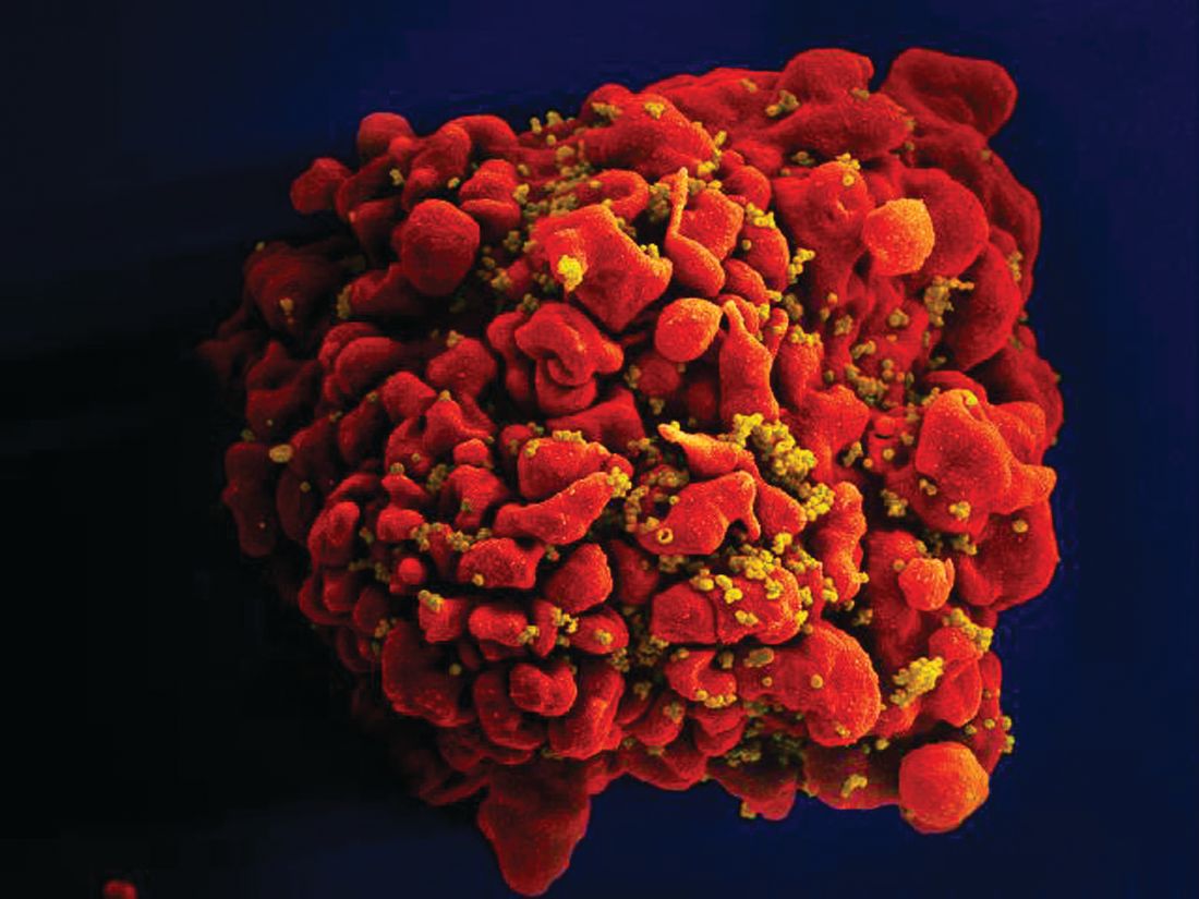
Stem cells gene edited to be HIV resistant treat ALL, but not HIV
Gene editing of donor stem cells prior to transplantation into a patient with both HIV infection and acute lymphoblastic leukemia (ALL) was safe and effectively treated the patient’s leukemia, but failed to resolve his HIV, investigators reported.

The 27-year-old man received an HLA-matched transplant of hematopoietic stem and progenitor cells (HSPCs) that had been genetically engineered to lack CCR5, a key gateway for HIV entry into cells.
Although the transplant resulted in complete remission of leukemia with full donor chimerism, only about 9% of the posttransplant lymphocytes showed disruption of CCR5, and during a brief trial of antiretroviral therapy interruption his HIV viral load rebounded, reported Hongkui Deng, PhD, and colleagues from Peking University in China.
Although the experiment did not meet its goal of a drug-free HIV remission, it serves as a proof of concept for the use of CRISPR-Cas9 (clustered regularly interspaced palindromic repeats/CRISPR-associated protein 9) gene editing to treat HIV infection, the authors contend.
“These results show the proof of principle that transplantation and long-term engraftment of CRISPR-edited allogeneic HSPCs can be achieved; however, the efficiency of the response was not adequate to achieve the target of cure of HIV-1 infection,” they wrote in a brief report published in the New England Journal of Medicine.
As previously reported, other research groups have investigated genetic editing to mimic a naturally occurring mutation that effectively disables the CCR5 HIV coreceptor, preventing the retrovirus from entering healthy cells. The mutation was first identified in a man named Timothy Brown who came to be known as “the Berlin patient” after he was apparently cured of HIV infection after a bone marrow transplant from a donor who had the mutation.
Dr. Deng and colleagues took advantage of HSPC transplantation, a standard therapy for ALL to see whether it could also have beneficial effects on concomitant HIV infection.
They treated donor HSPCs with CRISPR-Cas9 to ablate CCR5 and then delivered them to the patient along with additional CD34-depleted donor cells from mobilized peripheral blood.
The transplant was a success, with neutrophil engraftment on day 13 and platelet engraftment on day 27, and the leukemia was in morphologic complete remission at week 4 following transplantation. The patient remained in complete remission from leukemia throughout the 19-month follow-up period, with full donor chimerism .
However, when a planned interruption of antiretroviral therapy was carried out at 7 months post transplant, the serum viral load increased to 3 × 10⁷ copies/ml at week 4 following interruption, and the patient was restarted on the drug. His viral levels gradually decreased to undetectable level during the subsequent months.
The investigators noted that 2 weeks after the drug interruption trial was started there was a small increase in the percentage of CCR5 insertion/deletions.
“The low efficiency of gene editing in the patient may be due to the competitive engraftment of the coinfused HSPCs in CD34-depleted cells and the persistence of donor T cells. To further clarify the anti-HIV effect of CCR5-ablated HSPCs, it will be essential to increase the gene-editing efficiency of our CRISPR-Cas9 system and improve the transplantation protocol,” they wrote.
The study was funded by the Beijing Municipal Science and Technology Commission and others (unspecified). All authors reported having nothing to disclose.
SOURCE: Xu L et al. N Engl J Med. 2019. doi: 10.1056/NEJMoa1817426.
Gene editing of donor stem cells prior to transplantation into a patient with both HIV infection and acute lymphoblastic leukemia (ALL) was safe and effectively treated the patient’s leukemia, but failed to resolve his HIV, investigators reported.
The 27-year-old man received an HLA-matched transplant of hematopoietic stem and progenitor cells (HSPCs) that had been genetically engineered to lack CCR5, a key gateway for HIV entry into cells.
Although the transplant resulted in complete remission of leukemia with full donor chimerism, only about 9% of the posttransplant lymphocytes showed disruption of CCR5, and during a brief trial of antiretroviral therapy interruption his HIV viral load rebounded, reported Hongkui Deng, PhD, and colleagues from Peking University in China.
Although the experiment did not meet its goal of a drug-free HIV remission, it serves as a proof of concept for the use of CRISPR-Cas9 (clustered regularly interspaced palindromic repeats/CRISPR-associated protein 9) gene editing to treat HIV infection, the authors contend.
“These results show the proof of principle that transplantation and long-term engraftment of CRISPR-edited allogeneic HSPCs can be achieved; however, the efficiency of the response was not adequate to achieve the target of cure of HIV-1 infection,” they wrote in a brief report published in the New England Journal of Medicine.
As previously reported, other research groups have investigated genetic editing to mimic a naturally occurring mutation that effectively disables the CCR5 HIV coreceptor, preventing the retrovirus from entering healthy cells. The mutation was first identified in a man named Timothy Brown who came to be known as “the Berlin patient” after he was apparently cured of HIV infection after a bone marrow transplant from a donor who had the mutation.
Dr. Deng and colleagues took advantage of HSPC transplantation, a standard therapy for ALL to see whether it could also have beneficial effects on concomitant HIV infection.
They treated donor HSPCs with CRISPR-Cas9 to ablate CCR5 and then delivered them to the patient along with additional CD34-depleted donor cells from mobilized peripheral blood.
The transplant was a success, with neutrophil engraftment on day 13 and platelet engraftment on day 27, and the leukemia was in morphologic complete remission at week 4 following transplantation. The patient remained in complete remission from leukemia throughout the 19-month follow-up period, with full donor chimerism .
However, when a planned interruption of antiretroviral therapy was carried out at 7 months post transplant, the serum viral load increased to 3 × 10⁷ copies/ml at week 4 following interruption, and the patient was restarted on the drug. His viral levels gradually decreased to undetectable level during the subsequent months.
The investigators noted that 2 weeks after the drug interruption trial was started there was a small increase in the percentage of CCR5 insertion/deletions.
“The low efficiency of gene editing in the patient may be due to the competitive engraftment of the coinfused HSPCs in CD34-depleted cells and the persistence of donor T cells. To further clarify the anti-HIV effect of CCR5-ablated HSPCs, it will be essential to increase the gene-editing efficiency of our CRISPR-Cas9 system and improve the transplantation protocol,” they wrote.
The study was funded by the Beijing Municipal Science and Technology Commission and others (unspecified). All authors reported having nothing to disclose.
SOURCE: Xu L et al. N Engl J Med. 2019. doi: 10.1056/NEJMoa1817426.
FROM NEW ENGLAND JOURNAL OF MEDICINE
Key clinical point: Donor cells depleted of the HIV coreceptor CCR5 effectively treated ALL, but not HIV.
Major finding: The patient had a sustained complete remission of ALL, but HIV persisted after transplantation.
Study details: Case report of a 27-year-old man with ALL and HIV.
Disclosures: The study was funded by the Beijing Municipal Science and Technology Commission and others (unspecified). All authors reported having nothing to disclose.
Source: Xu L et al. N Engl J Med. 2019. doi: 10.1056/NEJMoa1817426.
Hematocrit (HCT) Levels and Thrombotic Events Among US Veterans With Polycythemia Vera
Background: Thrombotic events (TEs) are a leading cause of death in patients with polycythemia vera (PV), contributing to lower overall survival compared with age/sex-matched controls. This analysis evaluated the relationship between HCT levels and TEs among patients with PV from the Veteran’s Health Administration (VHA) to replicate the findings of the Cytoreductive Therapy in Polycythemia Vera (CYTO-PV) trial with a real-world patient population.
Methods: This retrospective study used VHA medical record and claims data from first PV diagnosis claim (index) until death, disenrollment, or end of study (collected October 1, 2005, through September 30, 2012). Patients were aged ≥ 18 years at index, had ≥ 2 PV claims (ICD-9-CM code, 238.4) ≥ 30 days apart during the identification period, continuous health plan enrollment from 12 months pre-index until end of study, and ≥ 3 HCT measurements/year during follow-up. This analysis focused on patients with no pre-index TE and all HCT values either < 45% or ≥ 45% during follow-up. Patient demographics and disease characteristics were assessed using descriptive statistics. Associations between HCT levels and TE occurrence were analyzed using unadjusted Cox regression models for patients with ≥ 1 HCT before TE (≥ 1 HCT subgroup). A sensitivity analysis including patients with a history of TE before the index period was also performed.
Results: Patients (n=213) were mean (SD) age 68.9 (11.5) years and predominately white (61.5%). TE during follow-up occurred in 44.1% of patients (mean follow-up, 2.3 y; rate per 100 person-years, 18.9). The 3 most common types of TE were ischemic stroke (21.6%), deep vein thrombosis (12.2%), and transient ischemic attack (9.4%). TE rates for patients with HCT values 45% vs < 45% were 54.2% and 40.3%, respectively. In the 1 HCT subgroup (n=208), the TE risk hazard ratio was 1.61 (95% CI, 1.03–2.51; P=0.036). The sensitivity analysis, which included patients with and without pre-index TEs (n=342), had similar results (76.9% vs 55.6%); hazard ratio in the 1 HCT subgroup (n=322):, 1.95 (95% CI, 1.46–2.61; P<0.0001).
Implications: These results in agreement with CYTOPV study findings and further support effective monitoring and management of HCT levels < 45% to reduce the risk of TE in veterans with PV.
Abstracts Presented at the 2019 AVAHO Annual Meeting (Digital Edition)

Standardization of the Discharge Process for Inpatient Hematology and Oncology
Background: Hematology/Oncology patients represent a complex population that requires timely follow- up to prevent clinical decompensation and delays in treatment. Previous reports have demonstrated that follow-up within 14 days is associated with decreased 30-day readmissions, and the magnitude of this impact is greater in higher risk patients. This project was designed to standardize the discharge process with the primary goal to reduce average time to hematology/oncology follow-up to 14 days.
Methods: Using Plan-Do-Study-Act (PDSA) quality improvement methodology, a multidisciplinary team of hematology/oncology staff developed and implemented a standardized discharge process. Rotating resident physicians were trained through online and inperson orientation. Additional interventions included the development of a discharge checklist handout and clinical decision support tool including a note template and embedded order set. All patients discharged during the two-month period prior to and discharged after the implementation of the standardized process were reviewed. Patients who followed with hematology/oncology at another facility, enrolled in hospice, or died during admission were excluded. Follow-up appointment scheduling data and communication between inpatient and outpatient providers were reviewed. Data was analyzed using XmR statistical process control chart and Fisher’s Exact Test using GraphPad.
Results: One hundred forty-two consecutive patients were reviewed between May - August 2018 and January - April 2019. The primary endpoint of time to hematology/ oncology follow up appointment improved from a baseline average of 17 days prior to intervention to 13 days in PDSA cycles 1 and 2 and 10 days in PDSA cycle 3. The target of 14 day average time to follow up was achieved. Furthermore, the upper control limit decreased from 58 days at baseline to 21 days in PDSA cycle 3 suggesting a decrease in variation. Outpatient hematology/oncology provider co-signature to discharge summary increased from 20% to 54% after intervention (P=0.01).
Conclusion: Our quality initiative to standardize the discharge process for the hematology & oncology service decreased time to hematology/oncology follow up appointment, improved communication between inpatient and outpatient teams, and decreased process variation. Timelier follow-up for this complex patient population will prevent clinical decompensation and delays in treatment.
Investigation of Outpatient Infusion Space Utilization to Increase Access to Same-Day Transfusion for Hematology/Oncology Patients
Background: Approximately 15% of patients with cancer require transfusion for treatment of disease- or chemotherapy-induced anemia. Previous studies have shown that anemia adversely affects patient quality of life (QoL), but QoL significant improves with transfusion. At the BVAMC, providers noted increasing delays in outpatient transfusion access averaging 2-3 days, resulting in prolongation of patient symptom burden (Plan). Additionally, outpatient transfusion is associated with significant patient time burden, patient travel burden, and health care cost, so delay in transfusion delivery also exacerbates these challenges.
Intervention: We created a process map for outpatient transfusion (Do). We learned that if a patient requires same-day transfusion, the patient must be admitted, resulting in a minimum cost of $3000 for a 24-hour hospitalization. We also learned that the outpatient infusion clinic is performing an increasing number of transfusions and non-transfusion related clinical services for other subspecialties, specifically infusion of iron, intravenous immunoglobulin, and biologic medications (i.e. infliximab, tocilizumab). There was a 2.6- times increase in blood transfusions per year since 2013 (78 to 205 units of pack red cells), possibly due to improved oncologic therapies prolonging patient survival. Furthermore, there was a 2-times increase in patient encounters for iron infusions (463 to 923) and a 1.4-times increase for biologics (876 to 1248) since 2010. This significantly increased demand has resulted in limited infusion chair access, precluding same-day transfusion availability (Study).
Outcome: The repercussions of decreased same-day transfusion access was presented to BVAMC administration. New space has been made available for seven additional chairs with transfusion capability. Iron infusions have been moved to the Hematology/Oncology chemotherapy center to increase ease of access, with a plan to move most transfusion to this space as well (Act). We project that access to same-day transfusion will avoid 2 hospitalizations per month at an annual cost of $72,000.
Implications: Access to same-day transfusion for treatment of anemia in patients with cancer decreases patient symptom and time burden and also results in cost savings. We encourage other facilities to explore their infusion space utilization, as demands will likely increase with growing use of intravenous therapies across specialties.
Use of Biosimilar Granulocyte Colony-Stimulating Factor for Mobilization in Autologous and Allogeneic Hematopoietic Stem Cell Transplantation in a Veteran Population
Purpose: The US Food and Drug Administration (FDA) approved the biologic medication filgrastim to mobilize hematopoietic progenitor cells (HPCs) into the peripheral blood for collection by leukapheresis for hematopoietic stem cell transplant (HSCT). The FDA-approved biosimilar tbo-filgrastim is currently used off-label for this indication in both autologous and allogeneic HSCT at the Tennessee Valley Healthcare System. This study compares the efficacy of filgrastim and tbo-filgrastim for these indications.
Methods: This was a retrospective, single center, observational cohort study approved by the Institutional Review Board. Patients were identified from the bone marrow transplant clinic and included in data collection if they received filgrastim or tbo-filgrastim for HPC mobilization between September 1, 2012 and September 1, 2018. The primary outcome was the proportion of patients with a CD34+ count > 15 ×103 cells/uL on day 4 of filgrastim or tbo-filgrastim mobilization for autologous transplantation. Secondary outcomes were the proportion of donors with a CD34+ count > 15 ×103 cells/uL on day 4 of filgrastim or tbo-filgrastim mobilization for allogeneic transplantation and the use of plerixafor in both patient populations.
Continuous data were described using mean and standard deviation. Associations between independent and dependent variables were assessed using t-tests for continuous variables and Fishers Exact tests for dichotomous variables.
Results: A total of 469 patients were identified for study inclusion; 367 underwent mobilization for autologous and 102 for allogeneic HSCT. Primary outcome was achieved in 47.5% of patients who received filgrastim compared with 50.24% who received tbo-filgrastim (P=0.67). There was no difference in patients eligible for collection on day 4 of filgrastim or tbo-filgrastim administration in the allogeneic HSCT population (97.6% vs 100% respectively; P=0.41). No statistically significant differences were identified in the number of patients requiring plerixafor use in the autologous or allogenic HSCT populations.
Conclusion: The use of the biosimilar tbo-filgrastim for mobilization in either autologous or allogeneic HSCT has comparable outcomes to that of the biotherapeutic reference product filgrastim at a reduced cost.
Early post-ACS bleeding may signal cancer
PARIS –, according to work presented at the annual congress of the European Society of Cardiology.
Of 3,644 patients discharged with dual-antiplatelet therapy after acute coronary syndrome (ACS), 1,215 (33%) had postdischarge bleeding. Taken together, patients who bled had a hazard ratio (HR) of 3.43 for a new cancer diagnosis (P less than .001).
Of the patients in the post-ACS cohort, 227 were newly diagnosed with cancer after discharge, making up 1% of the patients who did not bleed after discharge, and 3.9% of the patients who experienced postdischarge bleeding. Put another way, “[t]he positive predictive value for cancer diagnosis of post-discharge bleeding was 7.7%,” wrote Isabel Muñoz Pousa, MD, and her colleagues in the poster accompanying the presentation.
This elevated risk for cancer diagnosis was driven primarily by the 827 incidents of spontaneous bleeding; here, the HR was 4.38 (P less than .001). The 389 bleeds occuring after trauma, such as bladder catheterization or a fall, did not carry an increased risk for a new cancer diagnosis.
“Spontaneous post-discharge bleeding in ACS patients is strongly associated with subsequent cancer diagnosis within the first 6 months,” wrote Dr. Muñoz Pousa and her colleagues of the Hospital Universitario Alvaro Cunqueiro, Vigo, Spain. The investigators found a median time of 4.6 months from the bleeding episode to cancer diagnosis.
Of all anatomic locations, genitourinary bleeds were the most strongly associated with new cancer: 228 patients saw a HR of 8.63 for a new cancer diagnosis (P less than .001). Bronchopulmonary bleeds, sustained by 56 patients, carried a HR of 4.26 for new cancer diagnosis, and gastrointestinal bleeds a HR of 3.78 (P = .001 and P less than .001, respectively). Dr. Muñoz Pousa and her coinvestigators aggregated data from patients who had bleeding at other sites and saw no significant association with new cancers in this group of patients.
Though patients were initially discharged on dual-antiplatelet therapy, many patients stopped taking the medication over the mean 56.2 months of follow-up. The risk of bleeding did not differ significantly between those who were taking DAPT and those off DAPT, wrote Dr. Muñoz Pousa and her colleagues, adding: “We found a higher incidence of cancer in the first six months after discharge regardless of whether patients were taking dual-antiplatelet therapy or not.”
In their statistical analysis, Dr. Muñoz Pousa and colleagues adjusted for potential confounders, and looked at the effect of bleeding as a time-varying covariate on subsequent cancer diagnosis, using Cox regression models.
“Most of the bleeding episodes in the study were mild,” noted Dr. Munoz Pousa in a press statement. However, she said, “The bleeding events more strongly related with a new cancer diagnosis were severe hemorrhages of unknown cause requiring surgery – for example digestive bleeding needing endoscopic treatment.”
Breaking bleeding severity down by Bleeding Academic Research Consortium (BARC) criteria, the investigators found that most patients had relatively mild bleeding episodes categorized as BARC 1 or 2, with about half of all bleeding falling into the BARC 1 category.
Still, the 436 patients who had BARC 2 bleeding had a hazard ratio of 4.88 for cancer diagnosis, and the 71 BARC 3A patients saw the HR climb to 7.30. The risk for cancer subsequent to bleeding peaked at BARC 3B, with a HR of 12.29 for these 46 individuals (P less than .001 for all). Just 37 patients experienced BARC 3C bleeds, which were associated with a nonsignificant HR of 3.17 for new cancer diagnosis.
Although it’s not known why the post ACS–cancer bleeding association exists, Dr. Munoz Pousa put forward a plausible reason for the link. “A possible explanation is that there is a preexisting subclinical lesion in an organ that is triggered to become cancer by antiplatelet drugs or a stressful situation such as heart attack,” she said in the press release.
Antiplatelet therapy should be taken as prescribed post-ACS, and the physician threshold for further evaluation should be low when a significant spontaneous bleed is seen soon after ACS. “A prompt evaluation of bleeding could be useful for enabling an early detection of cancer in these patients,” said Dr. Munoz Pousa and her colleagues. “Our results suggest that patients should seek medical advice if they experience bleeding after discharge for a heart attack.”
The authors reported no conflicts of interest.
SOURCE: Munoz Pousa, I. et al. ESC Congress 2019, Abstract P677.
FROM ESC CONGRESS 2019
Ibrutinib-rituximab induction yields ‘unprecedented’ responses in MCL
LUGANO, Switzerland – In younger patients with previously untreated mantle cell lymphoma, the chemotherapy-free combination of ibrutinib and rituximab followed by a short course of chemotherapy was associated with an “unprecedented” 3-year progression-free survival rate, investigators in the phase 2 WINDOW-1 trial reported.

Among 50 patients aged 65 years and younger who received ibrutinib and rituximab until they achieved a complete or partial response, followed by four cycles of chemotherapy with rituximab plus hyper-CVAD (cyclophosphamide, vincristine, doxorubicin and dexamethasone) and rituximab plus methotrexate, the 3-year progression-free survival (PFS) rate was 88%, said Michael Wang, MD, from the University of Texas MD Anderson Cancer Center in Houston.
Additionally, for patients with the low-risk features, the 3-year PFS rate was 90%.
“Chemo-free ibrutinib-rituximab induced unprecedented – unprecedented – efficacy before chemo consolidation,” he said at the International Conference on Malignant Lymphoma.
Dr. Wang presented data from an interim analysis of the investigator-initiated single-center trial. Fifty patients aged 65 years or younger with untreated mantle cell lymphoma (MCL), good performance status, and good organ function were enrolled.
The patients were treated with ibrutinib and rituximab for two cycles and then evaluated for response with PET-CT scan, bone marrow biopsy, and for some patients, esophagogastroduodenoscopy (EGD) and colonoscopy with random biopsies.
In the induction phase, patients received ibrutinib daily on days 1-28 and rituximab intravenously over 6-8 hours on days 1, 8, 15, and 22 of cycle 1, and then over 4 hours on day 1 of cycles 3-12. The treatment was repeated every 28 days for up to 12 cycles in the absence of disease progression or unacceptable toxicity, or until patients achieved a complete response.
In the consolidation phase, patients received rituximab IV over 6 hours on day 1; oral or IV dexamethasone on days 1-4; cyclophosphamide IV over 3 hours twice daily on days 2-4; doxorubicin IV over 15-30 minutes on day 5; and vincristine IV over 15-30 minutes on day 5 of cycles one, three, five, and seven. Patients also received rituximab IV over 6 hours on day 1; methotrexate IV over 24 hours on day 2; and cytarabine IV over 2 hours twice daily on days 3 and 4 of cycles two, four, six, and eight. Treatments were repeated every 28 days for up to eight cycles in the absence of disease progression or unacceptable toxicity.
Patients who had a complete response (CR) after two cycles of induction and those who had disease progression on induction went on to consolidation. Patients with partial responses (PR) to induction continued on ibrutinib/rituximab until either the loss of a PR or best response for up to 12 cycles, with those who achieved a CR then moving on to consolidation.
Patients who had a CR after induction received four cycles of R-hyperCVAD, no subsequent stem cell transplant, and no maintenance therapy. Patients who had a PR after induction received two cycles of R-hyperCVAD, were reassessed, and then continued on R-hyperCVAD until CR or for up to eight total cycles.
Patients with either stable disease or progression during R-hyperCVAD were taken off the study.
Of the 50 patients enrolled, all 50 were evaluable for part A (induction), and 48 were evaluable after induction and consolidation (two patients withdrew for personal reasons).
After a median follow-up of 36 months, the overall response rate (ORR) following induction was 100%, consisting of 46 CRs (92%) and four PRs (8%).
In an intention-to-treat analysis (including the two patients who withdrew), the ORR was 96%, consisting of CRs in 47 patients (94%) and a PR in 1 patient (2%).
Neither the median PFS nor median overall survival had been reached at the time of data cutoff, and no patients have died.
Of the 50 enrolled patients, four experienced disease progression after 17, 24, 34, and 35 months of treatment. The patients with disease progression included one with Ki-67 of less than 30%, and three with KI-67 of 30% or greater.
Grade 3-4 toxicities during induction including myelosuppression in 4%; fatigue, myalgia, and rashes in 8% each; and oral mucositis in 4%.
Dr. Wang said that future studies on minimal residual disease and clonal evolution are ongoing, and that data on more patients will be presented at the next annual meeting of the American Society of Hematology, scheduled for December 2019.
He also noted that the WINDOW-2 trial, in which ibrutinib and rituximab are followed by veneotclax and hyper-CVAD chemotherapy in patients with newly diagnosed MCL, is open and rapidly enrolling patients.
The study is supported by the National Cancer Institute. Dr. Wang reported financial relationships with Janssen, Pharmacyclics, and other companies.
SOURCE: Wang M et al. ICML-15, Abstract 12.
LUGANO, Switzerland – In younger patients with previously untreated mantle cell lymphoma, the chemotherapy-free combination of ibrutinib and rituximab followed by a short course of chemotherapy was associated with an “unprecedented” 3-year progression-free survival rate, investigators in the phase 2 WINDOW-1 trial reported.
Among 50 patients aged 65 years and younger who received ibrutinib and rituximab until they achieved a complete or partial response, followed by four cycles of chemotherapy with rituximab plus hyper-CVAD (cyclophosphamide, vincristine, doxorubicin and dexamethasone) and rituximab plus methotrexate, the 3-year progression-free survival (PFS) rate was 88%, said Michael Wang, MD, from the University of Texas MD Anderson Cancer Center in Houston.
Additionally, for patients with the low-risk features, the 3-year PFS rate was 90%.
“Chemo-free ibrutinib-rituximab induced unprecedented – unprecedented – efficacy before chemo consolidation,” he said at the International Conference on Malignant Lymphoma.
Dr. Wang presented data from an interim analysis of the investigator-initiated single-center trial. Fifty patients aged 65 years or younger with untreated mantle cell lymphoma (MCL), good performance status, and good organ function were enrolled.
The patients were treated with ibrutinib and rituximab for two cycles and then evaluated for response with PET-CT scan, bone marrow biopsy, and for some patients, esophagogastroduodenoscopy (EGD) and colonoscopy with random biopsies.
In the induction phase, patients received ibrutinib daily on days 1-28 and rituximab intravenously over 6-8 hours on days 1, 8, 15, and 22 of cycle 1, and then over 4 hours on day 1 of cycles 3-12. The treatment was repeated every 28 days for up to 12 cycles in the absence of disease progression or unacceptable toxicity, or until patients achieved a complete response.
In the consolidation phase, patients received rituximab IV over 6 hours on day 1; oral or IV dexamethasone on days 1-4; cyclophosphamide IV over 3 hours twice daily on days 2-4; doxorubicin IV over 15-30 minutes on day 5; and vincristine IV over 15-30 minutes on day 5 of cycles one, three, five, and seven. Patients also received rituximab IV over 6 hours on day 1; methotrexate IV over 24 hours on day 2; and cytarabine IV over 2 hours twice daily on days 3 and 4 of cycles two, four, six, and eight. Treatments were repeated every 28 days for up to eight cycles in the absence of disease progression or unacceptable toxicity.
Patients who had a complete response (CR) after two cycles of induction and those who had disease progression on induction went on to consolidation. Patients with partial responses (PR) to induction continued on ibrutinib/rituximab until either the loss of a PR or best response for up to 12 cycles, with those who achieved a CR then moving on to consolidation.
Patients who had a CR after induction received four cycles of R-hyperCVAD, no subsequent stem cell transplant, and no maintenance therapy. Patients who had a PR after induction received two cycles of R-hyperCVAD, were reassessed, and then continued on R-hyperCVAD until CR or for up to eight total cycles.
Patients with either stable disease or progression during R-hyperCVAD were taken off the study.
Of the 50 patients enrolled, all 50 were evaluable for part A (induction), and 48 were evaluable after induction and consolidation (two patients withdrew for personal reasons).
After a median follow-up of 36 months, the overall response rate (ORR) following induction was 100%, consisting of 46 CRs (92%) and four PRs (8%).
In an intention-to-treat analysis (including the two patients who withdrew), the ORR was 96%, consisting of CRs in 47 patients (94%) and a PR in 1 patient (2%).
Neither the median PFS nor median overall survival had been reached at the time of data cutoff, and no patients have died.
Of the 50 enrolled patients, four experienced disease progression after 17, 24, 34, and 35 months of treatment. The patients with disease progression included one with Ki-67 of less than 30%, and three with KI-67 of 30% or greater.
Grade 3-4 toxicities during induction including myelosuppression in 4%; fatigue, myalgia, and rashes in 8% each; and oral mucositis in 4%.
Dr. Wang said that future studies on minimal residual disease and clonal evolution are ongoing, and that data on more patients will be presented at the next annual meeting of the American Society of Hematology, scheduled for December 2019.
He also noted that the WINDOW-2 trial, in which ibrutinib and rituximab are followed by veneotclax and hyper-CVAD chemotherapy in patients with newly diagnosed MCL, is open and rapidly enrolling patients.
The study is supported by the National Cancer Institute. Dr. Wang reported financial relationships with Janssen, Pharmacyclics, and other companies.
SOURCE: Wang M et al. ICML-15, Abstract 12.
REPORTING FROM 15-ICML
Hemophilia carriers face elevated risk of joint comorbidities
Individuals who are carriers of hemophilia genes and have reduced clotting factor activity have at least a twofold higher risk of joint-related comorbidities, compared with the general population, according to new research.

In a population-based cohort study using patient registry data, Swedish researchers identified 539 potential carriers of impaired factor VIII or IX gene in the X chromosome – 213 of whom had documented factor activity – and paired them with sex‐ and birthdate‐matched controls from the general population.
They found that carriers with reduced factor activity had a 2.3-fold higher risk of a joint diagnosis, compared with the general population (95% confidence interval, 1.1-4.5). Carriers with normal factor activity did not show a statistically significant increase in joint diagnosis hazard, however carriers with unknown factor activity had a 2.4-fold higher risk of joint diagnosis, compared with controls (95% CI, 1.8-3.2). The findings were published in Haemophilia.
By the age of 60 years, around 37% of carriers with reduced or unknown factor activity had received a joint diagnosis, compared with 23% of carriers with normal factor activity.
The most common joint diagnoses across carriers and controls were knee related, including gonarthrosis and internal derangement, but these were more common among carriers. Five carriers also recorded a diagnosis of hemophilic arthropathy or systemic disorders of connective tissue in diseases classified elsewhere.
Researchers also saw a 10-fold higher risk of joint surgery (95% CI, 1.0-3.7) among carriers with reduced factor activity – although the numbers were small – and even among carriers with normal factor activity, there was a nearly twofold higher rate (95% CI, 0.9-4.6), compared with the control population.
Carriers with reduced or unknown factor activity also had a higher risk of outpatient hospitalization, compared with the general population, although no effect was seen in carriers with normal factor activity.
“Although the frequency of joint comorbidities overall was relatively low, our results clearly indicate and confirm a higher burden of joint afflictions, including an earlier age at joint diagnosis, for carriers with reduced or unknown factor activity compared with the general population, as well as more joint surgeries and related hospitalizations,” wrote Mehdi Osooli, PhD, from the Skåne University Hospital in Malmö, Sweden, and his coauthors.
The authors noted that the findings correlated with their earlier research on the incidence of arthropathy among males with mild hemophilia, who have previously been found to have a ninefold higher incidence of arthropathy‐related hospital admissions and a 16‐fold higher incidence of joint disease.
“The relatively higher incidence in the male population compared with the carriers in the current study may be explained by the lower median factor activity level, for example, levels between 5% and 40% in males with mild haemophilia compared with a median overall of 50% in carriers,” they wrote.
All authors declared that they had no conflict of interest related to the study findings. Four of the authors reported financial ties to companies including Novo Nordisk, Shire, and Bayer.
SOURCE: Osooli M et al. Haemophilia. 2019 Aug 14. doi: 10.1111/hae.13831.
Individuals who are carriers of hemophilia genes and have reduced clotting factor activity have at least a twofold higher risk of joint-related comorbidities, compared with the general population, according to new research.
In a population-based cohort study using patient registry data, Swedish researchers identified 539 potential carriers of impaired factor VIII or IX gene in the X chromosome – 213 of whom had documented factor activity – and paired them with sex‐ and birthdate‐matched controls from the general population.
They found that carriers with reduced factor activity had a 2.3-fold higher risk of a joint diagnosis, compared with the general population (95% confidence interval, 1.1-4.5). Carriers with normal factor activity did not show a statistically significant increase in joint diagnosis hazard, however carriers with unknown factor activity had a 2.4-fold higher risk of joint diagnosis, compared with controls (95% CI, 1.8-3.2). The findings were published in Haemophilia.
By the age of 60 years, around 37% of carriers with reduced or unknown factor activity had received a joint diagnosis, compared with 23% of carriers with normal factor activity.
The most common joint diagnoses across carriers and controls were knee related, including gonarthrosis and internal derangement, but these were more common among carriers. Five carriers also recorded a diagnosis of hemophilic arthropathy or systemic disorders of connective tissue in diseases classified elsewhere.
Researchers also saw a 10-fold higher risk of joint surgery (95% CI, 1.0-3.7) among carriers with reduced factor activity – although the numbers were small – and even among carriers with normal factor activity, there was a nearly twofold higher rate (95% CI, 0.9-4.6), compared with the control population.
Carriers with reduced or unknown factor activity also had a higher risk of outpatient hospitalization, compared with the general population, although no effect was seen in carriers with normal factor activity.
“Although the frequency of joint comorbidities overall was relatively low, our results clearly indicate and confirm a higher burden of joint afflictions, including an earlier age at joint diagnosis, for carriers with reduced or unknown factor activity compared with the general population, as well as more joint surgeries and related hospitalizations,” wrote Mehdi Osooli, PhD, from the Skåne University Hospital in Malmö, Sweden, and his coauthors.
The authors noted that the findings correlated with their earlier research on the incidence of arthropathy among males with mild hemophilia, who have previously been found to have a ninefold higher incidence of arthropathy‐related hospital admissions and a 16‐fold higher incidence of joint disease.
“The relatively higher incidence in the male population compared with the carriers in the current study may be explained by the lower median factor activity level, for example, levels between 5% and 40% in males with mild haemophilia compared with a median overall of 50% in carriers,” they wrote.
All authors declared that they had no conflict of interest related to the study findings. Four of the authors reported financial ties to companies including Novo Nordisk, Shire, and Bayer.
SOURCE: Osooli M et al. Haemophilia. 2019 Aug 14. doi: 10.1111/hae.13831.
FROM HAEMOPHILIA
ACOG advises bleeding disorder screening for teens with heavy menstruation
Adolescent girls with heavy menstrual bleeding should be assessed for bleeding disorders, according to a Committee Opinion issued by the American College of Obstetricians and Gynecologists.
A bleeding disorder is secondary only to anovulation as a cause of heavy menstrual bleeding in adolescents.
Bleeding disorders affect 1%-2% of the general population, but are “found in approximately 20% of adolescent girls who present for evaluation of heavy menstrual bleeding and in 33% of adolescent girls hospitalized for heavy menstrual bleeding,” wrote Oluyemisi Adeyemi-Fowode, MD, and Judith Simms-Cendan, MD, and members of the ACOG Committee on Adolescent Health Care in the opinion, published in Obstetrics & Gynecology.
The committee advised that physical examination of teens with acute heavy menstrual bleeding should include assessment of hemodynamic stability with orthostatic blood pressure and pulse measurements. A speculum exam is not usually needed in teen girls with heavy menstrual bleeding. Evaluation should include screening for anemia attributable to blood loss with serum ferritin, endocrine disorders, and bleeding disorders. In suspected cases of bleeding disorders, laboratory evaluation and medical management should be done in consultation with a hematologist.
Those who are actively bleeding or hemodynamically unstable should be hospitalized for medical management, they said.
Ultrasonography is not necessary for an initial work-up of teens with heavy menstrual bleeding, but could be useful in patients who fail to respond to medical management.
Adolescent girls without contraindications to estrogen can be treated with hormone therapy in various forms including intravenous conjugated estrogen every 4-6 hours or oral 30-50 mg ethinyl estradiol every 6-8 hours until cessation of bleeding. Antifibrinolytics also can be used to stop bleeding.
Maintenance therapy after correction of acute heavy bleeding can include a combination of treatments such as hormonal contraceptives, oral and injectable progestins, and levonorgestrel-releasing intrauterine devices, the committee wrote. They also recommended oral iron replacement therapy for all women of reproductive age with anemia caused by menstrual bleeding.
If a patient fails to respond to medical therapy, nonmedical options or surgery may be considered, according to the committee. In addition, all teen girls with bleeding disorders should be advised about safe medication use, including the use of aspirin or NSAIDs only on the recommendation of a hematologist.
Patients and their families need education on menstrual issues including possible options for surgery in the future if heavy menstruation does not resolve. If a patient has a known bleeding disorder and is considering surgery, preoperative evaluation should include a consultation with a hematologist and an anesthesiologist, the committee noted.
Melissa Kottke, MD, MPH, said in an interview, “Every ob.gyn. will see a young patient with ‘heavy menstrual bleeding.’ And it becomes part of the art and challenge to work with the patient and family to collectively explore if this is, indeed, ‘heavy’ and of concern … or is it is a ‘normal’ menstrual period and simply reflects a newer life experience that would benefit from some education? And the stakes are high. Young people who have heavy menstrual cycles are much more likely to have an underlying bleeding disorder than the general population (20% vs. 1%-2%), and 75%-80% of adolescents with bleeding disorders report heavy menses as the most common clinical manifestation of their disorder.
“Fortunately, Committee Opinion 785, ‘Screening and Management of Bleeding Disorders in Adolescents with Heavy Menstrual Bleeding’ from the ACOG Committee on Adolescent Health Care is detailed and pragmatic. It outlines how to translate everyday conversations with young people about their menses into a quantifiable estimate of bleeding, including a very teen-friendly Pictorial Blood Loss Assessment Chart. It also gives ob.gyns. ever-important guidance about what to do next for evaluation and diagnosis. This committee opinion nicely outlines how to help manage heavy bleeding in an adolescent with a detailed algorithm. And very importantly, it gives clear management guidance and encourages ob.gyns. to avoid frequently unnecessary (speculum exams and ultrasounds) and excessive (early transfusion or surgical interventions) approaches to management for the young patient. I think it will be a great resource for any provider who is taking care of heavy menstrual bleeding for a young person,” said Dr. Kottke, who is director of the Jane Fonda Center for Adolescent Reproductive Health and associate professor of gynecology and obstetrics, both at Emory University, Atlanta. Dr. Kottke is not a member of the ACOG Committee on Adolescent Health and was asked to comment on the opinion.*
The complete opinion, ACOG Committee Opinion number 785, includes recommended laboratory tests, an eight-question screening tool, and a management algorithm.
The committee members had no financial conflicts to disclose. Dr. Kottke said she had no relevant financial disclosures.
SOURCE: Adeyemi-Fowode O and Simms-Cendan J. Obstet Gynecol. 2019 Sep. 134:e71-83.
*This article was updated on 9/9/2019.
FROM OBSTETRICS AND GYNECOLOGY