User login
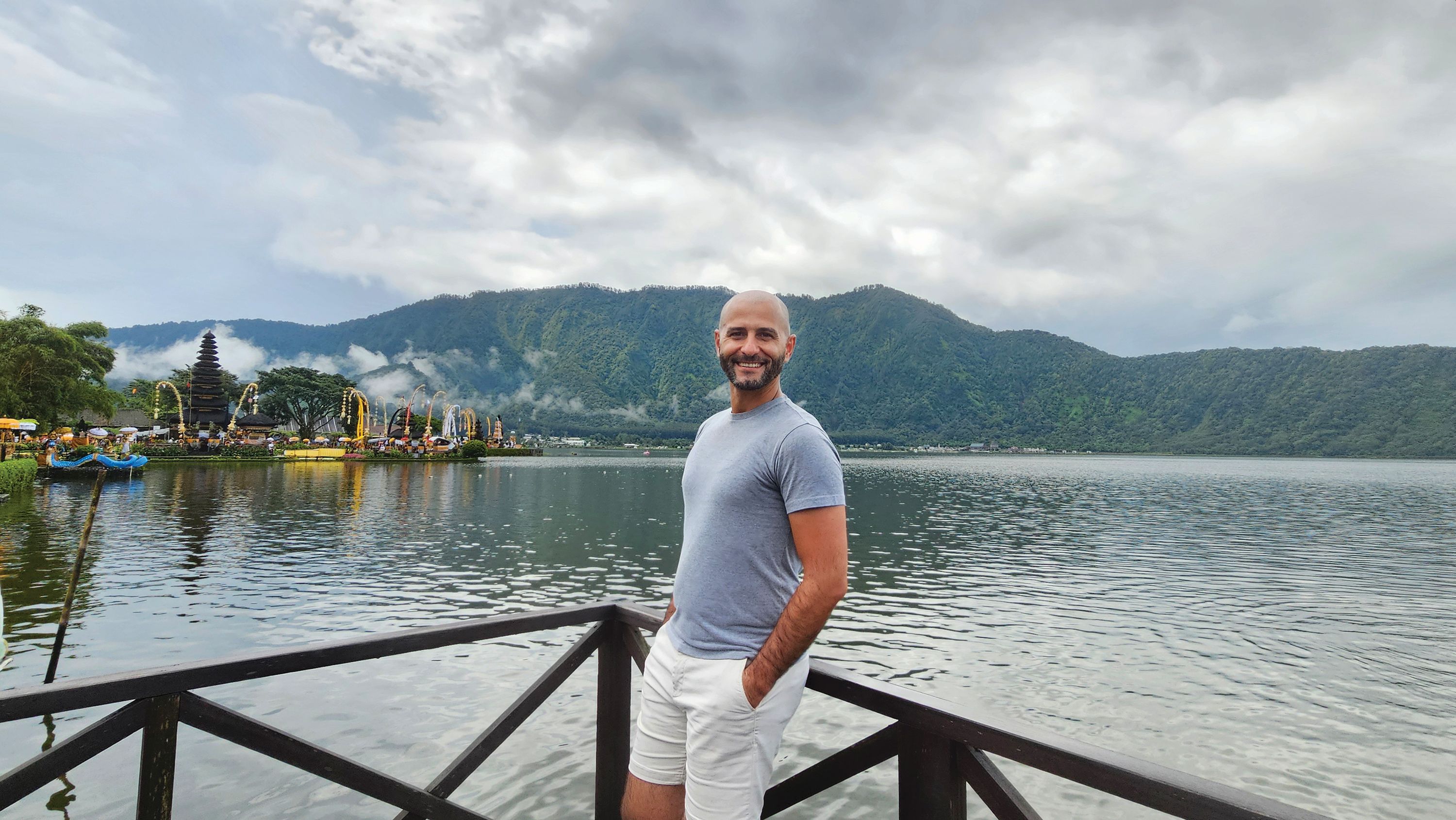
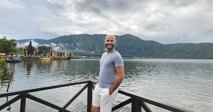
Balancing the Challenge of Research with the Joys of Clinical Care
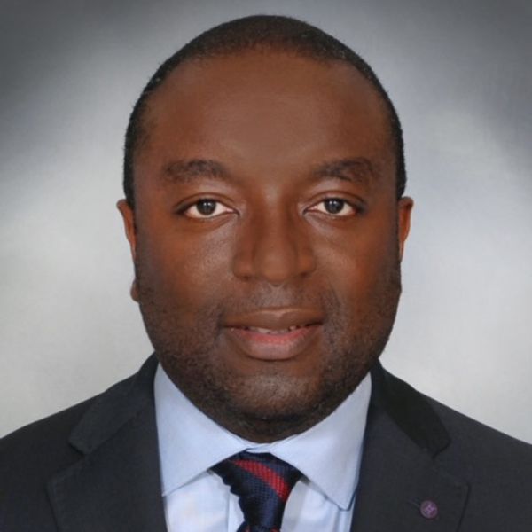
Andrew Ofosu, MD, MPH, loves the variety that GI medicine offers on a day-to-day basis.
Some days are spent in the endoscopy suite, performing endoscopic retrograde cholangiopancreatography in patients with cholangitis, “which is usually a high-stakes situation,” he said. Other days he might be in clinic, helping to manage a patient with chronic pancreatitis.
“The contrast of the immediate impact of a procedure combined with the continuity of long-term relationships, is special to me,” said Dr. Ofosu, an associate professor of medicine at Cincinnati College of Medicine, in Cincinnati, Ohio. He’s also a member of AGA’s Future Leaders program, which provides early career GI physicians with opportunities to network and develop leadership skills.
In an interview, he discussed his research pursuits in the areas of pancreatic cancer and artificial intelligence (AI), and his unique methods for connecting with patients. The art of listening to patient concerns is crucial, he says, especially following a difficult diagnosis.
What’s it like to be part of the AGA Future Leaders Class of 2025-2026? How has the experience enriched your career?
Dr. Ofosu: My time being part of this group has been very transformative. It’s provided mentorship from national leaders. It’s enabled me to collaborate with peers across different institutions and given me opportunities to refine my leadership skills. It’s changed my perspective and created a network that has equipped me to contribute meaningfully to the gastroenterology community and to my institution.
What is the most challenging clinical case you’ve encountered?
Dr. Ofosu: One case that stands out was a young patient with recurrent idiopathic pancreatitis. We went through all the potential differential etiologies that includes genetics, autoimmune disease and structural etiologies. It became a long, diagnostic journey. The challenge wasn’t just the medical aspect of it, but the emotional aspect of it…when you don’t have all the answers available. We were eventually able to figure out what the cause of the pancreatitis was. It was genetic, and the patient is doing great now.
One of your research interests has been developing innovative ways to use AI in endoscopic ultrasound to identify and characterize lesions. Can you discuss some of those innovations?
Dr. Ofosu: It’s definitely an area that I’m looking to explore at this time; to leverage AI to improve diagnostic capability of endoscopic ultrasound. The whole idea is to be able to use AI to analyze images in real time that can help highlight features, which can ultimately help in distinguishing both benign and malignant tumors, and allowing AI to provide real time diagnostic support, improving accuracy of diagnosis and reducing unnecessary treatment.
In 2021, you conducted a study to investigate the demographics, clinical outcomes and survival outcomes of patients diagnosed with early and late onset pancreatic adenocarcinoma. What did your study reveal and what are the next steps?
Dr. Ofosu: Our study looked at over 136,000 patients with pancreatic adenocarcinoma and compared those diagnosed under age 40 to older patients. We found that although pancreatic cancer is rare in the young, both groups are presenting more often with advanced disease, and incidence is rising. Younger patients tend to have tumors in the head of the pancreas, while older patients more often show growth in the body and tail. Survival overall remains very poor—about 6 to 7 months—but slightly better in younger patients.
I think the next step is to better understand the biological drivers of early onset PAC to look at integrating molecular profiling to see if there are distinct genetic patterns that can guide therapy. Ultimately the goal is to improve early detection and tailor management strategy for this subset of patients.
What is your approach to patient communication and education?
Dr. Ofosu: I aim for clarity and empathy. Some GI diagnoses can be intimidating, with all the terminologies, and so I use a lot of analogies and visuals to simplify complex conditions. I also ensure that patients understand what we are discussing because I found that what a patient hears isn’t always what they think I explained.
I believe being honest and compassionate should go hand in hand. I don’t shy away from delivering difficult news, but I always take time to pause, listen, and acknowledge emotions. I found that patients and families appreciate transparency even when the prognosis is tough, as long as they know I’m fully present with them.
Can you share a memorable patient interaction that impacted you?
Dr. Ofosu: There was one patient with chronic pancreatitis due to alcohol who had limited economic and social support. Beyond the medical management, what made a difference was sitting and listening to the patient, helping them connect to resources and social support – a social network. I think this reinforces that medicine isn’t just about lab values. It’s all about restoring dignity and focus with the patient.
What do you think is the biggest misconception about your specialty?
Dr. Ofosu: That gastroenterology is all about procedures, that all we do is scope. In reality, it’s a combination of technical expertise as well as the cognitive aspect of providing long-term management of complex diseases that affect patients, which takes a diverse skillset beyond endoscopy.
Lightning Round
What’s your favorite season of the year?
Fall. I like the colors of changing leaves
What’s your favorite way to spend a weekend?
Watching soccer with family and friends
If you could have dinner with any historical figure, who would it be?
Nelson Mandela
What’s your go-to karaoke song?Don’t Stop Believin’ by Journey
What’s one thing on your bucket list?
Travel to Europe, experience different cultures
What’s your favorite childhood memory?
When I learned how to fly a kite
If you could instantly learn any skill, what would it be?
Playing piano
Are you a planner or more spontaneous?
Planner
What’s your favorite holiday tradition?
Sharing Christmas dinner with family.
“Don’t Take Shortcuts,” Endoscopy Researcher Advises
But the work he’s most proud of took place when he was a graduate student at Harvard, working on a master’s degree in epidemiology and biostatistics.
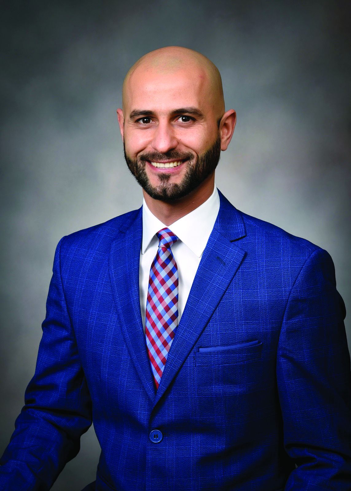
Jovani compared two different types of needles for tissue acquisition with endoscopic ultrasound. His finding that fine needle biopsy is better than fine needle aspiration for lesions isn’t groundbreaking, yet “the reason why I feel proud of that one is because it’s the first paper I did completely by myself,” said Jovani, medical director for advanced therapeutic endoscopy with Gastro Health Florida, in Miami, Florida.
Dr. Jovani has since contributed to countless peer-reviewed articles and book chapters and has presented research findings at meetings across the globe. He will be program director of the upcoming gastroenterology fellowship program at Florida International University School of Medicine, Miami, and participates in several endoscopy panels in the U.S. and in Europe to set guidelines and improve the quality of endoscopic procedures.
Therapeutic endoscopy is a clinical interest of his, specifically in the areas of third space, biliopancreatic and bariatric endoscopy. In an interview, he discussed how he used third space endoscopy to save a patient and improve her quality of life
Indeed, helping patients feel better is the most satisfying part of his career.
“A lot of people may have acute pain or an early cancer or many other problems that they need solving. As a physician, you can be the one who solves it,” said Jovani.
But training in medicine involves hard work, he advised. In the interview, he explained why young doctors should never rely on shortcuts to solve problems.
Therapeutic endoscopy is a specific interest of yours. How has this field advanced since you’ve been practicing gastroenterology?
Dr. Jovani: In the last 10 to 15 years, significant improvements have come along. As an example, lumen-apposing metal stents have revolutionized the way we do therapeutic endoscopy. A lot of procedures were not possible beforehand and we would have to send patients to surgery. Now, these can be done with endoscopy.
Examples include drainage of pancreatic collections, gallbladder drainage, or gastrojejunostomy (a connection between the stomach and the intestine) or reversal of Roux-en-Y gastric bypass to reach and drain the bile duct. Many of these procedures can be done with these metal stents that were not possible beforehand. Bariatric endoscopy is a relatively new field, and that has significantly changed the management of obesity.
There’s also third space endoscopy for the treatment of gastroparesis, achalasia, and early cancer.
What is third space endoscopy and how are you applying it in your practice?
Dr. Jovani: Third space endoscopy refers to a new space that’s created between the mucosa and the muscularis propria into the submucosa. We go in the submucosa, we inject some fluid there, and we cut the submucosa and we separate the mucosa from the muscle.
This allows us to do a lot of procedures. For patients with achalasia, we can tunnel through the submucosa, get into the muscle and perform myotomy, meaning that we can cut the muscle. By doing so, we can treat achalasia with a minimally invasive method. Patients can either go home the next day or even on the same day. The same thing applies for gastroparesis. With early cancer, we can go through in the submucosa, and if the cancer is in the mucosa only, or if it is in the very superficial submucosa, we can treat it without a need for surgery. Sometimes the procedure is simple, but other times it can be very challenging.
Can you discuss a challenging case where you applied third space endoscopy?
Dr. Jovani: It was a gastric cancer case. I did an endoscopic ultrasound for staging purposes. When I saw the lesion, it looked very superficial, like an early cancer of the stomach. I called the surgeon and said I could take it out with endoscopy. And it was in a very difficult location, so it was a very challenging procedure. It took about 12 hours to do it, but I was able to completely take it out. More than a year later, the patient was cancer free and more importantly, we preserved the stomach. Before I did this, she was prepared to undergo total gastrectomy, which meant I would have taken out her entire stomach.
Instead, with this minimally invasive procedure, I was able to take the cancer away and keep the stomach, which preserved her quality of life as well.
When you don’t have the stomach, obviously you adapt, but the quality of life is never the same. The type of food you eat, the frequency of eating, the quality of food you eat is not the same. The fact that we could avoid that in this patient feels very good.
What advice would you give to aspiring medical students?
Dr. Jovani: Do the hard work that’s required to be a doctor. Being a physician is a hard job, but it’s very rewarding. It’s like going to the gym—there really are no shortcuts. You have to do the work, you have to get tired, you have to study hard. You may study things you might not think will be useful to you necessarily in the future field that you choose. If it is GI, you still need to study all the other fields because sometimes patients may have GI diseases that are connecting with other diseases and you won’t know that if you haven’t studied the other diseases.
Patients are not only one disease, but they are also complex patients. Sometimes if you try to correct one disease, you create a complication with the other disease and you might not be aware of that.
Don’t create shortcuts like ChatGPT, things that are becoming fashionable with younger people today. Do the hard work the old way in which you have to memorize things. Knowledge is the only thing that really can help the patient.
Go to GI meetings. Offer to meet people, collaborate, network. Don’t be shy about it. Even if it is not natural to you, just do it. It’ll become more natural as you do it. GI, like any other field, any other endeavor in human society, is something that also depends on interactions. Therefore, it’s good to learn how to interact, how to network, how to do research projects. Even with people from far away, communication is very easy. You don’t really need to do research projects only with people in your local environment. You can do research projects with people who are on the other side of the state or even on the other side of the world.
You place an emphasis on individualized patient care. Can you discuss what that means to you?
Dr. Jovani: It basically means that there isn’t one size fits all in the management of diseases. Obviously there are some general principles that are applicable to everybody, but sometimes for the single specific patient, what works for one patient might not necessarily work for the next patient.
With Endoscopic Retrograde Cholangiopancreatography (ERCP) for example, there are so many things that go into that. Most papilla are in a certain position and it’s relatively easy to cannulate. But there are others that are in very different positions or in different angulations and they might require specific techniques that are not applicable in the majority of cases. You have to adapt to the single patient.How you speak to the patient is also important. Some may prefer a certain type of communication and other patients may prefer another type of communication involving patients or family. You have to adapt to the single patient. You have to understand the different types of personalities and adapt how you explain things or how you communicate disease, or management of disease or even complications to the specific patient. Different approaches are more appropriate for different patients with different needs. At the end of the day, patients are single individuals after all.
Where do you see the field of GI medicine advancing internationally over the next 5 years?
Dr. Jovani: Artificial intelligence or AI is a big player. It will help with diagnostics primarily, at least over the short term. Potentially it can help with therapeutics as well. There’s a lot of investment and excitement and interest in artificial intelligence.
Therapeutic endoscopy robotics, especially in interventional endoscopy, third space endoscopy, is also gaining attention.
With regards to bariatric endoscopy, we should have a CPT code for it in January 2027. This will increase volume because it’ll be covered more by insurance. These are things that will help advance GI in the next five or 10 years.
Lightning Round
What’s one hobby you’d like to pick up?
Kite surfing
What’s your favorite season of the year?
Summer
What’s your favorite way to spend a weekend?
Traveling or going to the beach
If you could have dinner with any historical figure, who would it be?
Jesus Christ
What’s your favorite holiday tradition?
New Year’s Eve
Are you a planner or more spontaneous?
Planner
What’s the best piece of advice you’ve ever received?
You can do it!
What’s your comfort food?
Lasagna
But the work he’s most proud of took place when he was a graduate student at Harvard, working on a master’s degree in epidemiology and biostatistics.
Jovani compared two different types of needles for tissue acquisition with endoscopic ultrasound. His finding that fine needle biopsy is better than fine needle aspiration for lesions isn’t groundbreaking, yet “the reason why I feel proud of that one is because it’s the first paper I did completely by myself,” said Jovani, medical director for advanced therapeutic endoscopy with Gastro Health Florida, in Miami, Florida.
Dr. Jovani has since contributed to countless peer-reviewed articles and book chapters and has presented research findings at meetings across the globe. He will be program director of the upcoming gastroenterology fellowship program at Florida International University School of Medicine, Miami, and participates in several endoscopy panels in the U.S. and in Europe to set guidelines and improve the quality of endoscopic procedures.
Therapeutic endoscopy is a clinical interest of his, specifically in the areas of third space, biliopancreatic and bariatric endoscopy. In an interview, he discussed how he used third space endoscopy to save a patient and improve her quality of life
Indeed, helping patients feel better is the most satisfying part of his career.
“A lot of people may have acute pain or an early cancer or many other problems that they need solving. As a physician, you can be the one who solves it,” said Jovani.
But training in medicine involves hard work, he advised. In the interview, he explained why young doctors should never rely on shortcuts to solve problems.
Therapeutic endoscopy is a specific interest of yours. How has this field advanced since you’ve been practicing gastroenterology?
Dr. Jovani: In the last 10 to 15 years, significant improvements have come along. As an example, lumen-apposing metal stents have revolutionized the way we do therapeutic endoscopy. A lot of procedures were not possible beforehand and we would have to send patients to surgery. Now, these can be done with endoscopy.
Examples include drainage of pancreatic collections, gallbladder drainage, or gastrojejunostomy (a connection between the stomach and the intestine) or reversal of Roux-en-Y gastric bypass to reach and drain the bile duct. Many of these procedures can be done with these metal stents that were not possible beforehand. Bariatric endoscopy is a relatively new field, and that has significantly changed the management of obesity.
There’s also third space endoscopy for the treatment of gastroparesis, achalasia, and early cancer.
What is third space endoscopy and how are you applying it in your practice?
Dr. Jovani: Third space endoscopy refers to a new space that’s created between the mucosa and the muscularis propria into the submucosa. We go in the submucosa, we inject some fluid there, and we cut the submucosa and we separate the mucosa from the muscle.
This allows us to do a lot of procedures. For patients with achalasia, we can tunnel through the submucosa, get into the muscle and perform myotomy, meaning that we can cut the muscle. By doing so, we can treat achalasia with a minimally invasive method. Patients can either go home the next day or even on the same day. The same thing applies for gastroparesis. With early cancer, we can go through in the submucosa, and if the cancer is in the mucosa only, or if it is in the very superficial submucosa, we can treat it without a need for surgery. Sometimes the procedure is simple, but other times it can be very challenging.
Can you discuss a challenging case where you applied third space endoscopy?
Dr. Jovani: It was a gastric cancer case. I did an endoscopic ultrasound for staging purposes. When I saw the lesion, it looked very superficial, like an early cancer of the stomach. I called the surgeon and said I could take it out with endoscopy. And it was in a very difficult location, so it was a very challenging procedure. It took about 12 hours to do it, but I was able to completely take it out. More than a year later, the patient was cancer free and more importantly, we preserved the stomach. Before I did this, she was prepared to undergo total gastrectomy, which meant I would have taken out her entire stomach.
Instead, with this minimally invasive procedure, I was able to take the cancer away and keep the stomach, which preserved her quality of life as well.
When you don’t have the stomach, obviously you adapt, but the quality of life is never the same. The type of food you eat, the frequency of eating, the quality of food you eat is not the same. The fact that we could avoid that in this patient feels very good.
What advice would you give to aspiring medical students?
Dr. Jovani: Do the hard work that’s required to be a doctor. Being a physician is a hard job, but it’s very rewarding. It’s like going to the gym—there really are no shortcuts. You have to do the work, you have to get tired, you have to study hard. You may study things you might not think will be useful to you necessarily in the future field that you choose. If it is GI, you still need to study all the other fields because sometimes patients may have GI diseases that are connecting with other diseases and you won’t know that if you haven’t studied the other diseases.
Patients are not only one disease, but they are also complex patients. Sometimes if you try to correct one disease, you create a complication with the other disease and you might not be aware of that.
Don’t create shortcuts like ChatGPT, things that are becoming fashionable with younger people today. Do the hard work the old way in which you have to memorize things. Knowledge is the only thing that really can help the patient.
Go to GI meetings. Offer to meet people, collaborate, network. Don’t be shy about it. Even if it is not natural to you, just do it. It’ll become more natural as you do it. GI, like any other field, any other endeavor in human society, is something that also depends on interactions. Therefore, it’s good to learn how to interact, how to network, how to do research projects. Even with people from far away, communication is very easy. You don’t really need to do research projects only with people in your local environment. You can do research projects with people who are on the other side of the state or even on the other side of the world.
You place an emphasis on individualized patient care. Can you discuss what that means to you?
Dr. Jovani: It basically means that there isn’t one size fits all in the management of diseases. Obviously there are some general principles that are applicable to everybody, but sometimes for the single specific patient, what works for one patient might not necessarily work for the next patient.
With Endoscopic Retrograde Cholangiopancreatography (ERCP) for example, there are so many things that go into that. Most papilla are in a certain position and it’s relatively easy to cannulate. But there are others that are in very different positions or in different angulations and they might require specific techniques that are not applicable in the majority of cases. You have to adapt to the single patient.How you speak to the patient is also important. Some may prefer a certain type of communication and other patients may prefer another type of communication involving patients or family. You have to adapt to the single patient. You have to understand the different types of personalities and adapt how you explain things or how you communicate disease, or management of disease or even complications to the specific patient. Different approaches are more appropriate for different patients with different needs. At the end of the day, patients are single individuals after all.
Where do you see the field of GI medicine advancing internationally over the next 5 years?
Dr. Jovani: Artificial intelligence or AI is a big player. It will help with diagnostics primarily, at least over the short term. Potentially it can help with therapeutics as well. There’s a lot of investment and excitement and interest in artificial intelligence.
Therapeutic endoscopy robotics, especially in interventional endoscopy, third space endoscopy, is also gaining attention.
With regards to bariatric endoscopy, we should have a CPT code for it in January 2027. This will increase volume because it’ll be covered more by insurance. These are things that will help advance GI in the next five or 10 years.
Lightning Round
What’s one hobby you’d like to pick up?
Kite surfing
What’s your favorite season of the year?
Summer
What’s your favorite way to spend a weekend?
Traveling or going to the beach
If you could have dinner with any historical figure, who would it be?
Jesus Christ
What’s your favorite holiday tradition?
New Year’s Eve
Are you a planner or more spontaneous?
Planner
What’s the best piece of advice you’ve ever received?
You can do it!
What’s your comfort food?
Lasagna
Ergonomic ‘Timeouts’ Make Endoscopy Easier For GIs
Amandeep Shergill, MD, MS, AGAF, always thought she had good hand-eye coordination until she entered her gastroenterology fellowship.
“You’re learning how to scope and the endoscope just feels so awkward in the hands. It can be such a difficult instrument to both learn and to use,” said Dr. Shergill, professor of clinical medicine at University of California, San Francisco.
Her attendings and mentors couldn’t give her the feedback she needed.
“I was told that I wasn’t holding it right. But every time I tried to do something that someone was trying to tell me, it seemed like my hands were too small. I couldn’t hold it the way that they were teaching me to hold it.” She began to wonder: Was this about her or the tool itself?
A deep dive into hand tool interactions and medical device designs led her to human factors and ergonomics. Her fellowship mentor, Ken McQuaid, MD, AGAF, had gone to medical school with David Rempel, MD, MPH who was one of the top-funded ergonomists in the country. “He emailed David and wrote: I have a fellow who’s interested in learning more about ergonomics and applying it to endoscopy,” said Dr. Shergill.
Through her work with Dr. Rempel, she was able to uncover the mechanisms that lead to musculoskeletal disorders in endoscopists.
Over time, she has become a trailblazer in this field, helming the UC Berkeley Center for Ergonomic Endoscopy with Carisa Harris-Adamson PhD, CPE, her ergonomics collaborator. In an interview, she described the unique “timeout” algorithm she created to ease the process of endoscopy for GI physicians.
What is your favorite aspect of being a GI physician?
I really love the diversity of patients and cases. You’re always learning something new. It’s an internal medicine subspecialty and a cognitive field, so we must think about differential diagnoses, risks and benefits of procedures for patients. But as a procedural field, we get to diagnose and immediately treat certain disorders. What’s exciting about GI right now is there’s still so much to learn. I think that we’re still discovering more about how the brain-gut interaction works every day. There’s been additional research about the microbiome and the immense influence it has on both health and disease. The field is continuing to evolve rapidly. There’s always something new to learn, and I think it keeps us fresh.
Tell me about your work in ergonomics and endoscopy.
Ken McQuaid connected me with David Rempel. I worked with David to approach this problem of endoscopy ergonomics from a very rigorous ergonomics perspective. Early in my fellowship, endoscopy ergonomics wasn’t well known. There were few survey-based studies, including one from the American Society for Gastrointestinal Endoscopy (ASGE) that documented a high prevalence of endoscopist injury. But not a lot was known about what was causing injury in endoscopists.
What were the risk factors for endoscopist injury? Instead of just doing another survey, I wanted to show that there was this potential for causation given the design of the endoscopes. I worked with David to do a pilot study where we collected some pinch forces and forearm muscle loads. I was able to collect some pilot data that I used to apply for the ASGE Endoscopic Research Award. And luckily, ASGE supported that work.
Another award I received, the ASGE Career Development Award, was instrumental in allowing me to become more proficient in the science of ergonomics. I was able to leverage that career development award to go back to school. I went to UC Berkeley and got a master’s in environmental health sciences with a focus on ergonomics. It really helped me to lay the foundation and understanding for ergonomics and then apply that to endoscopy to generate a more rigorous scientific background for endoscopy ergonomics and start that conversation within the field of GI.
What leads to musculoskeletal disorders in endoscopists and how can it be prevented?
Musculoskeletal disorders are associated with the repetitive procedures that we’re performing, often utilizing high forces and in non-neutral postures. This is because of how we’re interacting with our tools and how we’re interacting with our environments. The studies I have done with Carisa Harris-Adamson have been able to demonstrate and document the high forces that are required to interact with the endoscope. To turn the control section dials and to torque and manipulate the insertion tube, there are really high distal upper extremity muscle loads that are being applied.
We were able to compare the loads and the forces we were seeing to established risk thresholds from the ergonomics literature and demonstrate that performing endoscopy was associated with moderate to high risk of development of distal upper extremity disorders.
What research are you doing now?
We’re trying to focus more on interventions. We’ve done some studies on engineering controls we can utilize to decrease the loads of holding the scope. First, it was an anti-gravity support arm. More recently we’re hoping to publish data on whether a scope stand can alleviate some of those left distal upper extremity loads because the stand is holding the scope instead of the hand holding the scope. Can we decrease injury risk by decreasing static loading?
Neck and back injuries, which have a high prevalence in endoscopists, are usually associated with how the room is set up. One of the things that I’ve tried to help promote is a pre-procedure ergonomic “timeout.” Before an endoscopist does a procedure, we’re supposed to perform a timeout focused on the patient’s safety. We should also try to advocate for physician safety and an ergonomic timeout. I developed a mnemonic device utilizing the word “MYSELF” to help endoscopists remember the ergonomic timeout checklist: M = monitor, Y = upside-down Y stance, S = scope, E = elbow/ bed position, L = lower extremities, F = free movement of endoscope/ processor placement.
First, thinking about the monitor, “M”, and fixing the monitor height so that the neck is in neutral position. Then, thinking of an upside down “Y” standing straight with the feet either hip width or shoulder width apart, so that the physician has a stable, neutral standing posture. Then “S” is for checking the scope to ensure you have a scope with optimal angulation that’s working properly.
“E” is for elbows — adjusting the bed to an optimal position so that elbows and shoulders are in neutral position. “L” is for lower extremities — are the foot pedals within an easy reach? Do you have comfortable shoes on, an anti-fatigue floor mat if you need it? And then the “F” in “MYSELF” is for the processor placement, to ensure “free movement” of the scope. By placing the processor directly behind you and lining up the processor with the orifice to be scoped, you can ensure free movement of the scope so that you can leverage large movements of the control section to result in tip deflection.
We studied the MYSELF mnemonic device for a pre-procedure ergonomic timeout in a simulated setting and presented our results at Digestive Disease Week (DDW) 2024, where we showed a reduction in ergonomic risk scores based on the Rapid Entire Body Assessment tool.
We presented the results of the scope stand study at DDW 2025 in San Diego this May.
What has been the feedback from physicians who use these supportive tools?
While physicians are very grateful for bringing attention to this issue, and many have found utility in some of the tools that I proposed, I think we still have so much work to do. We’re just all hoping to continue to move this field forward for better tools that are designed more with the breadth of endoscopists in mind.
How do you handle stress and maintain work-life balance?
A few years ago, during DDW I gave a talk entitled “Achieving Work-Life Harmony.” I disclosed at the beginning of the talk that I had not achieved work-life harmony. It’s definitely a difficult thing to do, especially in our field as GI proceduralists, where we’re frequently on call and there are potentially on-call emergencies.
One of the key things that I’ve tried to do is create boundaries to prioritize both things in my personal life and my professional life and really try to stay true to the things that are important to me. For instance, things like family time and mealtimes, I think that’s so critical. Trying to be home on evenings for dinnertime is so important.
One of my GI colleagues, Raj Keswani, MD, MS gave a talk about burnout and described imagining life as juggling balls; trying to figure out which balls are glass balls and need to be handled with care, and which balls are rubber balls.
More often, work is the rubber ball. If you drop it, it’ll bounce back and the work that you have will still be there the next day. Family, friends, our health, those are the glass balls that if they fall, they can get scuffed or shatter sometimes. That image helps me think in the moment. If I need to decide between two competing priorities, which one will still be here tomorrow? Which is the one that’s going to be more resilient, and which is the one that I need to focus on? That’s been a helpful image for me.
I also want to give a shout out to my amazing colleagues. We all pitch in with the ‘juggling’ and help to keep everyone’s ‘balls’ in the air, and cover for each other. Whether it’s a sick patient or whatever’s going on in our personal lives, we always take care of each other.
What advice would you give to aspiring GI fellows or graduating fellows?
GI is such an amazing field and many people end up focusing on the procedural aspect of it. What I think defines an exceptional gastroenterologist and physician in general is adopting both a “growth mindset” and a “mastery mindset.” And really, it starts out with when you’re exploring an area of focus, listening to what consistently draws your attention, what you’re excited about learning more about.
Finding mentors, getting involved in projects, doing deep learning, and really trying to develop an expertise in that area through additional training, coursework, and education. I think that idea of a mastery mindset will really help set you up for becoming deeply knowledgeable about a field.
Lightning Round
Coffee or tea?
Coffee
What’s your favorite book?
Project Hail Mary (audiobook)
Beach vacation or mountain retreat?
Mountain retreat
Early bird or night owl?
Night owl
What’s your go-to comfort food?
Chaat (Indian street food)
Do you prefer dogs or cats?
Dogs
What’s one hobby you’d like to pick up?
Sewing
If you could have dinner with any historical figure, who would it be?
Ruth Bader Ginsburg
What’s your go-to karaoke song?
I Wanna Dance with Somebody
What’s one thing on your bucket list?
To see the Northern Lights

Endoscopist Brings Cutting-Edge Tech to Asia-Pacific Region
As the COVID-19 crisis unfolded in early 2020, Tossapol Kerdsirichairat, MD, faced another challenge: his mother’s ovarian cancer diagnosis.
“She chose to remain in Thailand, so I decided to relocate to care for her,” said Dr. Kerdsirichairat, an interventional endoscopist who completed fellowships at the University of Michigan, Ann Arbor, and Johns Hopkins University in Baltimore. The move to Bangkok turned out to be one of the best decisions of his life, he said, as he could support his mother while introducing advanced endoscopic techniques and devices to the region.
“Bangkok is a hub for medical innovation in Asia, offering opportunities to work with a diverse patient population and access to cutting-edge technology,” said Dr. Kerdsirichairat, who works at Bumrungrad International Hospital as a clinical associate professor.
The program is the first of its kind in Thailand and one of the few in the Asia-Pacific region.
“I guide patients and families through understanding their risks and implementing preventive strategies, collaborating with multidisciplinary teams to ensure comprehensive care. It’s incredibly rewarding to see the impact of early tumor detection,” said Dr. Kerdsirichairat, an international member of AGA who was a participant in the AGA Young Delegates Program.
He has set several records in Thailand for the smallest tumor detected, including a 0.3-millimeter (mm) esophageal tumor, a 0.8-mm tumor for stomach cancer, a 5-mm pancreatic tumor, and a 1-mm tumor for colon cancer.
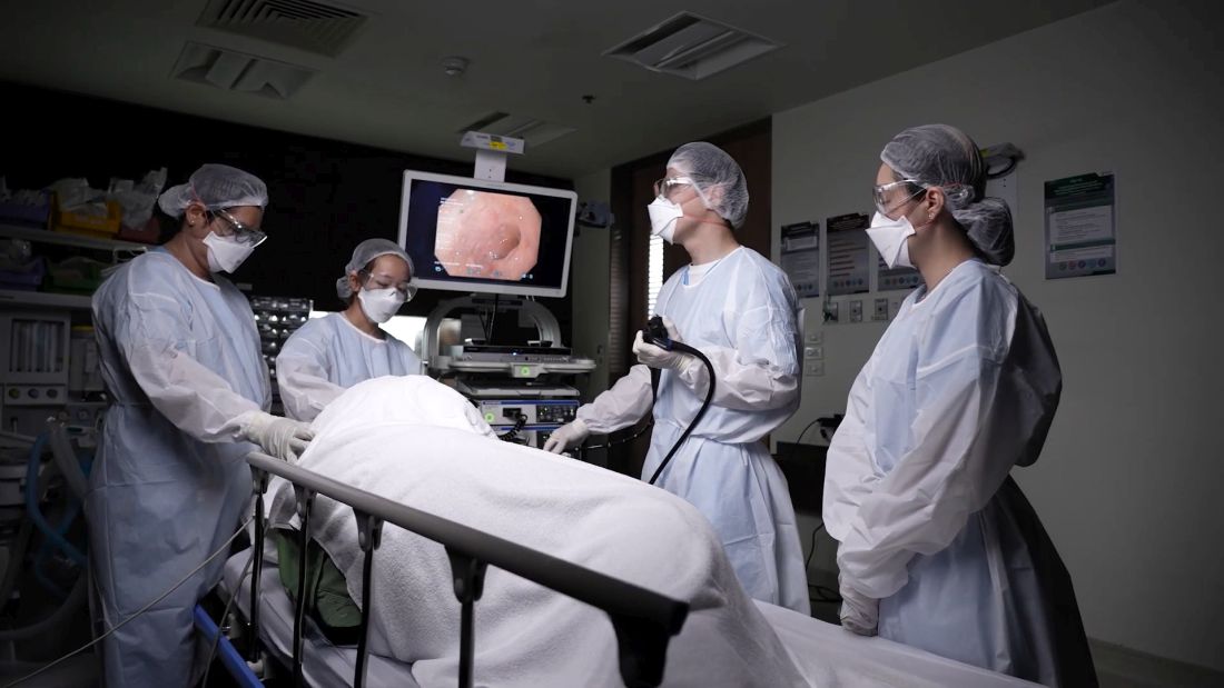
“These were detected through high-standard screening programs, as patients often do not develop symptoms from these subtle lesions,” said Dr. Kerdsirichairat, who discussed in an interview the unique challenges of practicing overseas.
Why did you choose GI?
Gastroenterology is a specialty that uniquely integrates procedural skill, clinical decision making, and a deep understanding of complex biological systems. I was drawn especially to the ability to make a direct and meaningful impact in patients’ lives through advanced endoscopic procedures, while also addressing both acute and chronic diseases, and focusing on cancer prevention. It is incredibly rewarding to perform an endoscopic retrograde cholangiopancreatography (ERCP) for cholangitis and see a patient return to normal the very next day, or to perform an endoscopic ultrasound (EUS) for pancreatic cancer screening in high-risk individuals and detect a sub-centimeter pancreatic tumor.
Realizing that early detection can improve survival by threefold after surgery is a powerful reminder of the difference we can make in patients’ lives. This specialty requires a delicate balance of precision and empathy, which perfectly aligns with my strengths and values as a physician.
You have a wide variety of clinical interests, from endoscopic procedures to cancer research to GERD. What’s your key subspecialty and why?
My primary specialty is advanced endoscopy, which includes techniques such as EUS, ERCP, and endoscopic resection of precancerous and early cancerous lesions. I also focus on cutting-edge, evidence-based techniques recently included in clinical guidelines, such as Transoral Incisionless Fundoplication (TIF). These minimally invasive options allow me to diagnose and treat conditions that once required surgery. The precision and innovation involved in advanced endoscopy enable me to effectively manage complex cases—from diagnosing early cancers to managing bile duct obstructions and resecting precancerous lesions.
Can you describe your work in cancer genetics and screening?
I am deeply committed to the early detection of gastrointestinal cancers, particularly through screening for precancerous conditions and hereditary syndromes. During my general GI training at the University of Michigan, I had the privilege of working with Grace Elta, MD, AGAF, and Michelle Anderson, MD, MSc, renowned experts in pancreatic cancer management. I was later trained by Anne Marie Lennon, PhD, AGAF, who pioneered the liquid biopsy technique for cancer screening through the CancerSEEK project, and Marcia (Mimi) Canto, MD, MHS, who initiated the Cancer of the Pancreas Screening project for high-risk individuals of pancreatic cancer.
I also had the distinction of being the first at Bumrungrad International Hospital to perform endoscopic drainage for pancreatic fluid collections in the setting of multi-organ failure. This endoscopic approach has been extensively validated in the medical literature as significantly improving survival rates compared to surgical drainage. My training in this specialized procedure was conducted under the guidance of the premier group for necrotizing pancreatitis, led by Martin Freeman, MD, at the University of Minnesota.
Later, I contributed to overseeing the Inherited Gastrointestinal Malignancy Clinic of MyCode, a large-scale population-based cohort program focused on cancer screening in Pennsylvania. By December 2024, MyCode had collected blood samples from over 258,000 individuals, analyzed DNA sequences from over 184,000, and provided clinical data that benefits over 142,000 patients. It’s not uncommon for healthy 25-year-old patients to come to our clinic for colon cancer screening after learning from the program that they carry a cancer syndrome, and early screening can potentially save their lives.
What are the key differences between training and practicing medicine in the United States and in an Asian country?
The U.S. healthcare system is deeply rooted in evidence-based protocols and multidisciplinary care, driven by an insurance-based model. In contrast, many Asian countries face challenges such as the dependency on government approval for certain treatments and insurance limitations. Practicing in Asia requires navigating unique cultural, economic, and systemic differences, including varying resource availability and disease prevalence.
What specific challenges have you faced as a GI in Thailand?
As an advanced endoscopist, one of the biggest challenges I faced initially was the difficulty in obtaining the same devices I used in the U.S. for use in Thailand. With support from device companies and mentors in the U.S., I was able to perform groundbreaking procedures, such as the TIF in Southeast Asia and the first use of a full-thickness resection device in Thailand. I am also proud to be part of one of the first few centers worldwide performing the combination of injectable semaglutide and endoscopic sleeve gastroplasty, resulting in a remarkable weight reduction of 44%, comparable to surgical gastric bypass.
In addition, Bumrungrad International Hospital, where I practice, sees over 1.1 million visits annually from patients from more than 190 countries. This offers a unique opportunity to engage with a global patient base and learn from diverse cultures. Over time, although the hospital has professional interpreters for all languages, I have become able to communicate basic sentences with international patients in their preferred languages, including Chinese, Japanese, and Arabic, which has enriched my practice.
What’s your favorite thing to do when you’re not practicing GI?
I enjoy traveling, exploring new cuisines, and spending quality time with family and friends. These activities help me recharge and offer fresh perspectives on life.
Lightning Round
Texting or talking?
Talking. It’s more personal and meaningful.
Favorite city in the U.S.?
Ann Arbor, Michigan
Cat or dog person?
Dog person
Favorite junk food?
Pizza
How many cups of coffee do you drink per day?
Two – just enough to stay sharp, but not jittery.
If you weren’t a GI, what would you be?
Architect
Best place you went on vacation?
Kyoto, Japan
Favorite sport?
Skiing
Favorite ice cream?
Matcha green tea
What song do you have to sing along with when you hear it?
“Everybody” by Backstreet Boys
Favorite movie or TV show?
Forrest Gump and Friends
Optimist or pessimist?
Optimist. I believe in focusing on solutions and possibilities.
As the COVID-19 crisis unfolded in early 2020, Tossapol Kerdsirichairat, MD, faced another challenge: his mother’s ovarian cancer diagnosis.
“She chose to remain in Thailand, so I decided to relocate to care for her,” said Dr. Kerdsirichairat, an interventional endoscopist who completed fellowships at the University of Michigan, Ann Arbor, and Johns Hopkins University in Baltimore. The move to Bangkok turned out to be one of the best decisions of his life, he said, as he could support his mother while introducing advanced endoscopic techniques and devices to the region.
“Bangkok is a hub for medical innovation in Asia, offering opportunities to work with a diverse patient population and access to cutting-edge technology,” said Dr. Kerdsirichairat, who works at Bumrungrad International Hospital as a clinical associate professor.
The program is the first of its kind in Thailand and one of the few in the Asia-Pacific region.
“I guide patients and families through understanding their risks and implementing preventive strategies, collaborating with multidisciplinary teams to ensure comprehensive care. It’s incredibly rewarding to see the impact of early tumor detection,” said Dr. Kerdsirichairat, an international member of AGA who was a participant in the AGA Young Delegates Program.
He has set several records in Thailand for the smallest tumor detected, including a 0.3-millimeter (mm) esophageal tumor, a 0.8-mm tumor for stomach cancer, a 5-mm pancreatic tumor, and a 1-mm tumor for colon cancer.
“These were detected through high-standard screening programs, as patients often do not develop symptoms from these subtle lesions,” said Dr. Kerdsirichairat, who discussed in an interview the unique challenges of practicing overseas.
Why did you choose GI?
Gastroenterology is a specialty that uniquely integrates procedural skill, clinical decision making, and a deep understanding of complex biological systems. I was drawn especially to the ability to make a direct and meaningful impact in patients’ lives through advanced endoscopic procedures, while also addressing both acute and chronic diseases, and focusing on cancer prevention. It is incredibly rewarding to perform an endoscopic retrograde cholangiopancreatography (ERCP) for cholangitis and see a patient return to normal the very next day, or to perform an endoscopic ultrasound (EUS) for pancreatic cancer screening in high-risk individuals and detect a sub-centimeter pancreatic tumor.
Realizing that early detection can improve survival by threefold after surgery is a powerful reminder of the difference we can make in patients’ lives. This specialty requires a delicate balance of precision and empathy, which perfectly aligns with my strengths and values as a physician.
You have a wide variety of clinical interests, from endoscopic procedures to cancer research to GERD. What’s your key subspecialty and why?
My primary specialty is advanced endoscopy, which includes techniques such as EUS, ERCP, and endoscopic resection of precancerous and early cancerous lesions. I also focus on cutting-edge, evidence-based techniques recently included in clinical guidelines, such as Transoral Incisionless Fundoplication (TIF). These minimally invasive options allow me to diagnose and treat conditions that once required surgery. The precision and innovation involved in advanced endoscopy enable me to effectively manage complex cases—from diagnosing early cancers to managing bile duct obstructions and resecting precancerous lesions.
Can you describe your work in cancer genetics and screening?
I am deeply committed to the early detection of gastrointestinal cancers, particularly through screening for precancerous conditions and hereditary syndromes. During my general GI training at the University of Michigan, I had the privilege of working with Grace Elta, MD, AGAF, and Michelle Anderson, MD, MSc, renowned experts in pancreatic cancer management. I was later trained by Anne Marie Lennon, PhD, AGAF, who pioneered the liquid biopsy technique for cancer screening through the CancerSEEK project, and Marcia (Mimi) Canto, MD, MHS, who initiated the Cancer of the Pancreas Screening project for high-risk individuals of pancreatic cancer.
I also had the distinction of being the first at Bumrungrad International Hospital to perform endoscopic drainage for pancreatic fluid collections in the setting of multi-organ failure. This endoscopic approach has been extensively validated in the medical literature as significantly improving survival rates compared to surgical drainage. My training in this specialized procedure was conducted under the guidance of the premier group for necrotizing pancreatitis, led by Martin Freeman, MD, at the University of Minnesota.
Later, I contributed to overseeing the Inherited Gastrointestinal Malignancy Clinic of MyCode, a large-scale population-based cohort program focused on cancer screening in Pennsylvania. By December 2024, MyCode had collected blood samples from over 258,000 individuals, analyzed DNA sequences from over 184,000, and provided clinical data that benefits over 142,000 patients. It’s not uncommon for healthy 25-year-old patients to come to our clinic for colon cancer screening after learning from the program that they carry a cancer syndrome, and early screening can potentially save their lives.
What are the key differences between training and practicing medicine in the United States and in an Asian country?
The U.S. healthcare system is deeply rooted in evidence-based protocols and multidisciplinary care, driven by an insurance-based model. In contrast, many Asian countries face challenges such as the dependency on government approval for certain treatments and insurance limitations. Practicing in Asia requires navigating unique cultural, economic, and systemic differences, including varying resource availability and disease prevalence.
What specific challenges have you faced as a GI in Thailand?
As an advanced endoscopist, one of the biggest challenges I faced initially was the difficulty in obtaining the same devices I used in the U.S. for use in Thailand. With support from device companies and mentors in the U.S., I was able to perform groundbreaking procedures, such as the TIF in Southeast Asia and the first use of a full-thickness resection device in Thailand. I am also proud to be part of one of the first few centers worldwide performing the combination of injectable semaglutide and endoscopic sleeve gastroplasty, resulting in a remarkable weight reduction of 44%, comparable to surgical gastric bypass.
In addition, Bumrungrad International Hospital, where I practice, sees over 1.1 million visits annually from patients from more than 190 countries. This offers a unique opportunity to engage with a global patient base and learn from diverse cultures. Over time, although the hospital has professional interpreters for all languages, I have become able to communicate basic sentences with international patients in their preferred languages, including Chinese, Japanese, and Arabic, which has enriched my practice.
What’s your favorite thing to do when you’re not practicing GI?
I enjoy traveling, exploring new cuisines, and spending quality time with family and friends. These activities help me recharge and offer fresh perspectives on life.
Lightning Round
Texting or talking?
Talking. It’s more personal and meaningful.
Favorite city in the U.S.?
Ann Arbor, Michigan
Cat or dog person?
Dog person
Favorite junk food?
Pizza
How many cups of coffee do you drink per day?
Two – just enough to stay sharp, but not jittery.
If you weren’t a GI, what would you be?
Architect
Best place you went on vacation?
Kyoto, Japan
Favorite sport?
Skiing
Favorite ice cream?
Matcha green tea
What song do you have to sing along with when you hear it?
“Everybody” by Backstreet Boys
Favorite movie or TV show?
Forrest Gump and Friends
Optimist or pessimist?
Optimist. I believe in focusing on solutions and possibilities.
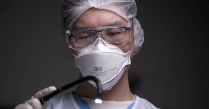
Expert Advice for Difficult GERD Cases
PHOENIX – , said Kristle L. Lynch, MD, in a presentation at the American College of Gastroenterology (ACG) 2025 Annual Scientific Meeting.
Patients with persistent GERD who have cough and asthma, as well as those with hoarseness, globus, or other ear, nose, and throat concerns, may first undergo workup by other clinicians, said Lynch, who is a professor of clinical medicine and director of the Physiology and Motility Laboratory at the Perelman School of Medicine at the University of Pennsylvania in Philadelphia.
With these patients, “given the low pretest probability of an acid reflux source, it makes sense to bypass a trial of acid-suppressing medication if symptoms persist and instead move on to consideration of upper endoscopy,” she said.
“If negative for damage or sequelae of reflux, this test is often followed by advanced reflux testing and high-resolution manometry, to help diagnose the cause of reflux and swallowing problems,” she noted.
For patients with heartburn, regurgitation, or noncardiac chest pain (and no other concerning symptoms), moving to an empiric trial of antisecretory therapy before endoscopy can be reasonable, Lynch said. But if symptoms persist, an endoscopy should be done before any advanced pH testing.
Advanced pH Testing Options
Advanced reflux testing options for persistent GERD after an endoscopy include catheter-based pH-impedance testing and wireless pH testing, said Lynch.
“Wireless pH monitoring offers improved patient comfort by eliminating the need for a nasal catheter, allowing individuals to maintain normal daily activities and diet during testing,” Lynch told GI & Hepatology News.
“It also enables extended monitoring periods of up to 96 hours, enhancing the detection of intermittent acid reflux episodes,” she said. “However, unlike pH-impedance catheter-based testing, wireless pH measures only acid exposure and cannot identify nonacid reflux events.”
In addition, the wireless capsule may detach prematurely or cause mild chest discomfort, and its endoscopic placement adds procedural complexity with the need for anesthesia, as well as additional cost, she noted.
Catheter-based pH-impedance testing involves sedation-free catheter placement while the patient is awake, followed by monitoring for 24 hours, and involves assessment of bolus movement in tandem with pH measurements, Lynch said. The catheter monitors levels of acid refluxing into the esophagus and transmits this information to the smartphone-sized computer worn by the patient over the 24-hour period.
Unlike wireless testing, impedance testing allows for adjunctive measures of mean nocturnal baseline impedance (MNBI) and post-reflux swallow-induced peristaltic waves (PSPW), the latter of which is shown to predict response to proton pump inhibitor (PPI) therapy in patients with reflux.
Logistics include a concurrent manometry for the lower esophageal sphincter to optimize location, followed by a transnasal placement. Challenges associated with the catheter-based test include potential catheter migration and patient discomfort, she added.
Assessing pH Testing Results
After pH testing of either type, abnormal acid exposure time (AET) when the pH in the lower esophagus is greater than 6% supports a diagnosis of GERD; AET between 4% and 6% is considered borderline GERD; and AET less than 4% is considered normal and outside the bounds of pathologic reflux, as per the Lyon Consensus, Lynch explained.
Treatment for patients meeting the GERD criteria based on AET includes not only diet and lifestyle changes but also potential pharmacologics, including acid suppressants, alginates, gamma-aminobutyric acid agonists, and prokinetics. In some cases, anti-reflux surgery may be considered, such as magnetic sphincter augmentation, transoral incisionless fundoplication, or Roux-en-Y gastric bypass, Lynch said.
But not all heartburn in patients with persistent GERD is acid-related, she said. Functional heartburn and reflux hypersensitivity can be seen in these cases.
Patients not meeting the criteria for GERD on advanced pH testing and diagnosed with reflux hypersensitivity or functional heartburn may be treated with neuromodulators or nonpharmacologic therapies such as hypnosis, diaphragmatic breathing, cognitive-behavioral therapy, or acupuncture, said Lynch in her presentation.
Overall, “advanced pH testing should be assessed within the framework of other investigations and the patient as a whole,” she emphasized.
Challenges in the Clinic
One of the biggest mistakes while diagnosing a patient with persistent GERD symptoms despite PPI therapy is always assuming their symptoms are driven by ongoing pathologic acid exposure, said Michael Kingsley, MD, a gastroenterologist specializing in gastrointestinal motility disorders and a clinical assistant professor of medicine at the University of Pittsburgh, Pittsburgh, in an interview.
He agreed with Lynch that patients with reflux hypersensitivity or functional heartburn are more likely to benefit from interventions other than a PPI, such as a neuromodulator.
But managing GERD in the clinic also requires making time to assess whether patients are taking PPI correctly and to counsel them on lifestyle modifications, he noted.
One of the current biggest challenges in GERD is how best to manage a patient who falls into the inconclusive range of not definitely normal or abnormal on pH testing, Kingsley told GI & Hepatology News.
“As alluded to in this talk, MNBI and PSPW are emerging metrics, both of which require a pH-impedance study (as opposed to a wireless pH study),” he said. “Further elucidating the ideal application and cutoffs of these metrics may provide additional tools for best treating patients who fall into the “inconclusive” range with standard metrics.”
Lynch and Kingsley disclosed no financial conflicts of interest.
A version of this article appeared on Medscape.com .
FROM ACG 2025
Journal Highlights: July-November 2025
Endoscopy
Barkun AN, et al. Canadian Association of Gastroenterology Clinical Practice Guideline for the Endoscopic Management of Nonvariceal Nonpeptic Ulcer Upper Gastrointestinal Bleeding. Gastroenterology. 2025 Oct. doi: 10.1053/j.gastro.2025.04.041.
Kindel TL, et al. Multisociety Clinical Practice Guidance for the Safe Use of Glucagon-like Peptide-1 Receptor Agonists in the Perioperative Period. Clin Gastroenterol Hepatol. 2025 Nov. doi: 10.1016/j.cgh.2024.10.003.
Roy A, et al. Endohepatology: Evolving Indications, Challenges, Unmet Needs and Opportunities. Gastro Hep Advances. 2025 Oct. doi: 10.1016/j.gastha.2025.100838.
Esophagus
Wani S, et al. AGA Clinical Practice Guideline on Surveillance of Barrett’s Esophagus. Gastroenterology. 2025 Nov. doi: 10.1053/j.gastro.2025.09.012.
Reed CC, et al. Worsening Disease Severity as Measured by I-SEE Associates With Decreased Treatment Response to Topical Steroids in Eosinophilic Esophagitis Patients. Clin Gastroenterol Hepatol. 2025 Sep. doi: 10.1016/j.cgh.2025.01.015.
Kagzi Y, et al. Safety and Efficacy of Transoral Incisionless Fundoplication for Post–Esophageal Peroral Endoscopic Myotomy Gastroesophageal Reflux Disease With Esophagitis: A Meta-Analysis. Tech Innov Gastrointest Endosc. 2025 Oct. doi:10.1016/j.tige.2025.250953.
Stomach
Staller K, et al. AGA Clinical Practice Guideline on Management of Gastroparesis. Gastroenterology. 2025 Oct. doi: 10.1053/j.gastro.2025.08.004.
Colon
Bergman D, et al. Cholecystectomy Is a Risk Factor for Microscopic Colitis: A Nationwide Population-based Matched Case Control Study. Clin Gastroenterol Hepatol. 2025 Nov. doi: 10.1016/j.cgh.2024.12.032.
Liver
Younossi ZM, et al. Global Consensus Recommendations for Metabolic Dysfunction-Associated Steatotic Liver Disease and Steatohepatitis. Gastroenterology. 2025 Oct. doi: 10.1053/j.gastro.2025.02.044.
Kabelitz MA, et al. Early Occurrence of Hepatic Encephalopathy Following Transjugular Intrahepatic Portosystemic Shunt Insertion is Linked to Impaired Survival: A Multicenter Cohort Study. Clin Gastroenterol Hepatol. 2025 Nov. doi: 10.1016/j.cgh.2025.01.024.
Brar G, et al. Association of Cirrhosis Etiology with Outcomes After TIPS: A National Cohort Study. Gastro Hep Advances. 2025 Nov. doi: 10.1016/j.gastha.2025.100850.
IBD
Kucharzik T, et al. Role of Noninvasive Imaging in the Diagnosis and Management of Patients With Suspected and Established Inflammatory Bowel Disease. Gastroenterology. 2025 Nov. doi: 10.1053/j.gastro.2025.06.002.
Griffiths BJ, et al. Hypercoagulation After Hospital Discharge in Acute Severe Ulcerative Colitis: A Prospective Study. Clin Gastroenterol Hepatol. 2025 Sep. doi: 10.1016/j.cgh.2024.10.031.
Disorders of Gut-Brain Interaction
Trindade IA, et al. Implications of Shame for Patient-Reported Outcomes in Bowel Disorders of Gut-Brain Interaction. Gastroenterology. 2025 Aug. doi: 10.1053/j.gastro.2025.06.030.
Salwen-Deremer JK, et al. A Practical Guide to Incorporating a Psychologist Into a Gastroenterology Practice. Gastroenterology. 2025 Nov. doi: 10.1053/j.gastro.2025.05.014.
Misc
Monahan K, et al. In Our Scope of Practice: Genetic Risk Assessment and Testing for Gastrointestinal Cancers and Polyposis in Gastroenterology. Gastroenterology. 2025 Nov. doi: 10.1053/j.gastro.2025.06.001.
Dr. Trieu is assistant professor of medicine, interventional endoscopy, in the Division of Gastroenterology at Washington University in St. Louis School of Medicine, Missouri.
Office-Based Endoscopy Model Offers Way Forward for Outpatient GI
After decades of successful growth, the ambulatory surgery center (ASC) model may be turning a corner, opening up opportunity for office-based endoscopy models, according to a recent practice management editorial published in Clinical Gastroenterology and Hepatology.
Although office endoscopy has been an option, it hasn’t always felt practical or financially viable in the past. However, the paradigm appears to be shifting as ASC-based revenue streams show signs of stress and fail to keep pace with inflation. As healthcare regulatory and economic environments continue to change, gastroenterologists need a new model to support equity, efficiency, and growth in gastrointestinal (GI) care delivery, the authors wrote.
“Through the course of my 40-year career, I’ve been hit with a lot of changes related to regulations, insurance, and the market. You can’t stay entrenched in your old ways. You have to remain pivotable and come up with new strategic positions,” said Lawrence Kosinski, MD, AGAF, lead author and founder of SonarMD and VOCnomics.
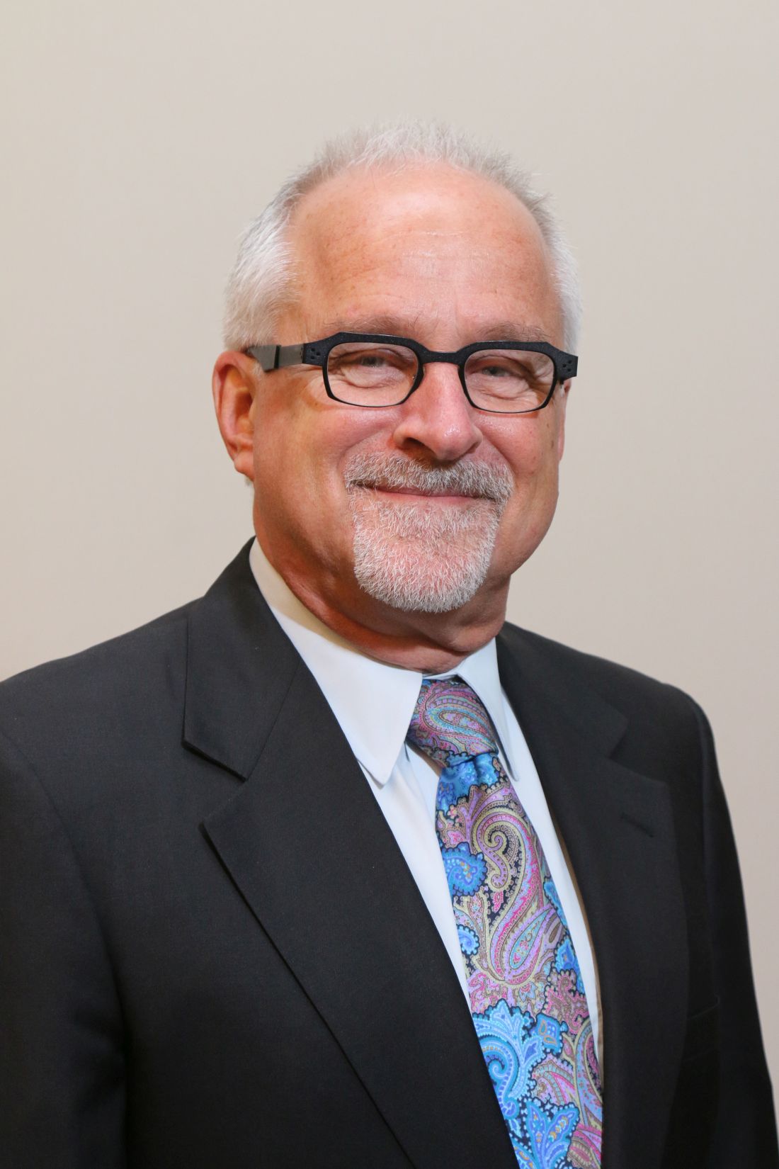
During his private practice career, Kosinski built one of the largest GI practices in Illinois, which had seven ASCs and is now part of one of the largest GI groups in the country. Across 30 years of experience with ASCs, Kosinski has watched the reimbursement for professional services decline, as well as for added revenue streams such as pathology and anesthesia.
Looking for a better solution, Kosinski served on the governing board for the American Gastroenterological Association as the councilor for development and growth. During the past 3 years, he has spoken with GI practices and worked with a national anesthesia company — Ambulatory Anesthesia Care — to better understand the office endoscopy setting.
“In the ’90s, all I wanted was to have an ASC because that was in vogue,” he said. “But if you look critically at what has happened to the business of outpatient endoscopy in the past 25 years, you’ll see that professional fees haven’t kept up, and trying to replace that lost revenue is a losing battle.”
Considering Financial Shifts
Since 2001, professional reimbursement for colonoscopies has fallen by more than 40% while ASC revenue has risen, decreasing the percentage of revenue from professional fees (from 34% to 23%) and increasing the facility component (from 44% to 60%), Kosinski and colleagues wrote.
When looking at profit, compression of professional service fees appears even greater, especially with surging costs of anesthesia care due to high demand and provider shortages. Beyond that, about a third of ASCs are owned at least partially by national entities, as of 2024, leading to even lower realization of profit.
“The profit margins have really been crushed, so what is a GI doc to do? Go where there is opportunity,” Kosinski said. “The difference between hospitals and ASCs has been compressed, so what about the office?”
The proposed 2026 Medicare Physician Fee Schedule includes a 14% increase in reimbursement for office-based procedures, including endoscopy, as well as a 7% decrease for facility-based procedures.
In several states — such as Illinois, Oregon, Virginia, Washington, and Wisconsin — health plans are introducing programs to promote the transition of outpatient endoscopy to office settings rather than hospital-based or ASC-based settings due to costs, the authors wrote.
“The decision to start offering office-based endoscopy services was an easy one for our practice, as it provides a way for us to provide patients convenient, easy-to-access endoscopy that is high quality yet much more affordable than hospital-based settings,” said Neil Gupta, MD, managing partner at Midwest Digestive Health & Nutrition in Des Plaines, Illinois.

The practice has used office-based endoscopy for nearly 2 years, Gupta said, performing about 5000 GI endoscopy procedures per year.
“As we all try to find better ways to provide high-quality but affordable care for patients, office-based endoscopy is a great way to help achieve those goals,” he said. “Healthcare professionals and patients should all be asking, ‘What type of site am I getting my GI endoscopy scheduled at — hospital, surgery center, or physician’s office?’”
Regaining Autonomy and Time
Beyond the financial dynamics, , Kosinski and colleagues wrote.
Looking ahead, office-based models can also provide the agility and infrastructure to compete in value-based care models, they wrote. In turn, value-based models can create relevance and resilience in a continually changing healthcare environment.
Without the involvement of ASC managers, investors, or health system partners, physicians retain control of scheduling, clinical protocols, financial decisions, and operational workflows, the authors wrote. This could create better alignment with personal preferences, clinical judgment, and patient needs, they noted.
“GI physicians should no longer feel trapped in a hospital setting where they lack independence and influence over decision-making,” said Rock Rockett, PhD, owner and principal consultant of Rockett Healthcare Strategies, which partners with GI groups nationwide to help with development, accreditation, and payer contracting for office endoscopy.
“GI physicians should also no longer feel trapped in a ‘bad marriage’ with partners in an ASC or partners in a practice who create a difficult work environment,” he said. “The viability of office endoscopy allows them to strike out on their own or set up a new partnership on more equitable terms that are attractive for them.”
Patient safety and quality also appear to be similar or better in office-based settings, based on benchmarking data analyzed so far. Hospital transfers were lower, falls were similar, and patient experience was positive, the authors wrote.
At the same time, Kosinski and colleagues noted the difficulty in shifting to office-based models. Most practices have committed to ASCs, for instance, and adding an office-based room can be challenging. Otherwise, practices already use their available office space and don’t have extra rooms available. In that case, an office endoscopy suite may be best suited for expansion sites, allowing practices to grow into new service areas, they wrote.
“You can’t fight the market. You have to focus on what the market wants and needs,” Kosinski said. “To do that, you have to be able to pivot and change direction, looking for new ways to change your mission. This could be an option to do that.”
Kosinski, Gupta, and Rockett declared having no conflicts of interest other than their current employments.
A version of this article appeared on Medscape.com.
After decades of successful growth, the ambulatory surgery center (ASC) model may be turning a corner, opening up opportunity for office-based endoscopy models, according to a recent practice management editorial published in Clinical Gastroenterology and Hepatology.
Although office endoscopy has been an option, it hasn’t always felt practical or financially viable in the past. However, the paradigm appears to be shifting as ASC-based revenue streams show signs of stress and fail to keep pace with inflation. As healthcare regulatory and economic environments continue to change, gastroenterologists need a new model to support equity, efficiency, and growth in gastrointestinal (GI) care delivery, the authors wrote.
“Through the course of my 40-year career, I’ve been hit with a lot of changes related to regulations, insurance, and the market. You can’t stay entrenched in your old ways. You have to remain pivotable and come up with new strategic positions,” said Lawrence Kosinski, MD, AGAF, lead author and founder of SonarMD and VOCnomics.
During his private practice career, Kosinski built one of the largest GI practices in Illinois, which had seven ASCs and is now part of one of the largest GI groups in the country. Across 30 years of experience with ASCs, Kosinski has watched the reimbursement for professional services decline, as well as for added revenue streams such as pathology and anesthesia.
Looking for a better solution, Kosinski served on the governing board for the American Gastroenterological Association as the councilor for development and growth. During the past 3 years, he has spoken with GI practices and worked with a national anesthesia company — Ambulatory Anesthesia Care — to better understand the office endoscopy setting.
“In the ’90s, all I wanted was to have an ASC because that was in vogue,” he said. “But if you look critically at what has happened to the business of outpatient endoscopy in the past 25 years, you’ll see that professional fees haven’t kept up, and trying to replace that lost revenue is a losing battle.”
Considering Financial Shifts
Since 2001, professional reimbursement for colonoscopies has fallen by more than 40% while ASC revenue has risen, decreasing the percentage of revenue from professional fees (from 34% to 23%) and increasing the facility component (from 44% to 60%), Kosinski and colleagues wrote.
When looking at profit, compression of professional service fees appears even greater, especially with surging costs of anesthesia care due to high demand and provider shortages. Beyond that, about a third of ASCs are owned at least partially by national entities, as of 2024, leading to even lower realization of profit.
“The profit margins have really been crushed, so what is a GI doc to do? Go where there is opportunity,” Kosinski said. “The difference between hospitals and ASCs has been compressed, so what about the office?”
The proposed 2026 Medicare Physician Fee Schedule includes a 14% increase in reimbursement for office-based procedures, including endoscopy, as well as a 7% decrease for facility-based procedures.
In several states — such as Illinois, Oregon, Virginia, Washington, and Wisconsin — health plans are introducing programs to promote the transition of outpatient endoscopy to office settings rather than hospital-based or ASC-based settings due to costs, the authors wrote.
“The decision to start offering office-based endoscopy services was an easy one for our practice, as it provides a way for us to provide patients convenient, easy-to-access endoscopy that is high quality yet much more affordable than hospital-based settings,” said Neil Gupta, MD, managing partner at Midwest Digestive Health & Nutrition in Des Plaines, Illinois.
The practice has used office-based endoscopy for nearly 2 years, Gupta said, performing about 5000 GI endoscopy procedures per year.
“As we all try to find better ways to provide high-quality but affordable care for patients, office-based endoscopy is a great way to help achieve those goals,” he said. “Healthcare professionals and patients should all be asking, ‘What type of site am I getting my GI endoscopy scheduled at — hospital, surgery center, or physician’s office?’”
Regaining Autonomy and Time
Beyond the financial dynamics, , Kosinski and colleagues wrote.
Looking ahead, office-based models can also provide the agility and infrastructure to compete in value-based care models, they wrote. In turn, value-based models can create relevance and resilience in a continually changing healthcare environment.
Without the involvement of ASC managers, investors, or health system partners, physicians retain control of scheduling, clinical protocols, financial decisions, and operational workflows, the authors wrote. This could create better alignment with personal preferences, clinical judgment, and patient needs, they noted.
“GI physicians should no longer feel trapped in a hospital setting where they lack independence and influence over decision-making,” said Rock Rockett, PhD, owner and principal consultant of Rockett Healthcare Strategies, which partners with GI groups nationwide to help with development, accreditation, and payer contracting for office endoscopy.
“GI physicians should also no longer feel trapped in a ‘bad marriage’ with partners in an ASC or partners in a practice who create a difficult work environment,” he said. “The viability of office endoscopy allows them to strike out on their own or set up a new partnership on more equitable terms that are attractive for them.”
Patient safety and quality also appear to be similar or better in office-based settings, based on benchmarking data analyzed so far. Hospital transfers were lower, falls were similar, and patient experience was positive, the authors wrote.
At the same time, Kosinski and colleagues noted the difficulty in shifting to office-based models. Most practices have committed to ASCs, for instance, and adding an office-based room can be challenging. Otherwise, practices already use their available office space and don’t have extra rooms available. In that case, an office endoscopy suite may be best suited for expansion sites, allowing practices to grow into new service areas, they wrote.
“You can’t fight the market. You have to focus on what the market wants and needs,” Kosinski said. “To do that, you have to be able to pivot and change direction, looking for new ways to change your mission. This could be an option to do that.”
Kosinski, Gupta, and Rockett declared having no conflicts of interest other than their current employments.
A version of this article appeared on Medscape.com.
FROM CLINICAL GASTROENTEROLOGY AND HEPATOLOGY
Nailing Neoplastic Lesions in Barrett’s Esophagus
, said Prateek Sharma, MD, in a presentation on the management of BE at the American College of Gastroenterology (ACG) 2025 Annual Scientific Meeting.
However, clinicians often make mistakes such as failing to remove debris such as saliva and bile from the esophagus prior to assessing a patient, said Sharma, professor of medicine and the Elaine Blaylock Endowed Professor at the University of Kansas School of Medicine and the Cancer Center, Kansas City, Kansas.
More than 90% of neoplasias in patients with BE are found on an index endoscopy or within 6 months, as shown by Sharma and his colleagues in a systematic review, which highlights the importance of a high-quality index endoscopy, he told meeting attendees.
To improve the index endoscopy, Sharma developed a new algorithm called “CLEAN.”
The algorithm is composed of five steps, he said, the first of which is Clear: clear the esophagus of debris, including saliva and bile. Adequate prep is essential to detecting clinically significant lesions in patients with BE, he explained. In a study published in 2024, Sharma and colleagues found adequate cleanliness of the upper gastrointestinal tract was associated with a significantly higher detection rate of clinically significant lesions.
The second step of the algorithm is Learn: pay attention to BE inspection time and learn slow withdrawal strategies.
It’s important not to shortchange inspection time, Sharma emphasized. He cited a previous study in which the percentage of patients with BE who had high-grade dysplasia or esophageal adenocarcinoma during a surveillance endoscopy was 15% with inspection times of 2 minutes or less but jumped to 69% with inspection times of 7 minutes or more.
The third step of CLEAN is Endoscope: conduct a high-definition white-light endoscopy, which should be coupled with the fourth step, Acquire: acquire education on BE-related neoplasia, to learn how to recognize neoplastic lesions, he stressed.
The final step of the algorithm is Neoplasia detection rate (NDR): follow a quality metric to measure NDR.
The algorithm emphasizes a comprehensive approach in conjunction with resection of visible lesions followed by ablation for complete eradication, Sharma told GI & Hepatology News.
After Identification: What’s Next?
If lesions are identified, the next step is resection and/or ablation, Sharma said.
“Resection is typically used for visible lesions, nodules, or masses, while ablation is used to treat the remaining underlying Barrett tissue,” he told GI & Hepatology News. “A combination of both is often necessary to fully treat advanced cases, such as when a nodule is resected and the surrounding area is subsequently ablated.”
“It’s important to understand why we need to resect,” he said.
“Resection removes the lesion” and “provides more accurate histopathology reading and staging of how deep the lesion is,” he explained. Options for resection of cancerous or precancerous lesions in patients with BE include endoscopic mucosal resection (EMR) and endoscopic submucosal dissection (ESD).
The treatment algorithm for BE continues to evolve, Sharma said in his presentation. Currently, evidence supports EMR for most cases, but ESD is based on factors including lesion size ≥ 25-30 mm and potential submucosal invasion, he said.
He cited a study of 1000 adults with early BE who were managed with EMR that showed a 96% curative response after 5 years. Similarly, a review of ESD for early BE neoplasia including 501 patients showed a 75% curative response rate overall and a 93% en bloc resection rate, he noted.
Ablation
In terms of ablation, radiofrequency ablation, hybrid argon plasma coagulation, and the multifocal cryoballoon procedure have shown significant effectiveness, Sharma said.
In a 2020 multicenter, prospective study of 120 adult patients with BE, 76% achieved complete eradication of dysplasia, and 72% achieved complete eradication of intestinal metaplasia. As for safety, data from nine European centers including 154 patients who underwent ablation after resection had an adverse event rate of 6%, said Sharma.
In the Clinic
“It is sometimes difficult to detect subtle nodularity and irregularity that would benefit more from resection therapy/EMR rather than ablation,” said Gyanprakash A. Ketwaroo, MD, associate professor of medicine (digestive diseases) at Yale University, New Haven, Connecticut.
“Lesions can be obscured by esophagitis, peristalsis, or the shape of the [gastroesophageal] GE junction,” he noted. Therefore, careful scope cleaning and inspection with high-definition white light and narrow band imaging are important, he said. “Using a cap on the scope to better distend or manipulate the gastroesophageal junction also helps identify obscured lesions,” he added.
“Any acronym or approach that reminds us to slow down, and examine carefully, is welcome,” Ketwaroo told GI & Hepatology News. The CLEAN algorithm provides a useful summary of some of the key steps all clinicians should incorporate into approaching BE and could be useful for teaching trainees, he added.
Sharma disclosed serving as a consultant for the Olympus Corporation and Exact Sciences and receiving grant support from Fujifilm, Erbe Medical, and Braintree Pharmaceuticals.
A version of this article appeared on Medscape.com.
FROM ACG 2025
Duodenal Mucosal Resurfacing Curbs Weight Gain Post-GLP-1
, initial results of the open-label, multistage REMAIN-1 trial showed.
In addition, “the procedure was well tolerated, with only minor, transient TEAEs [treatment-emergent adverse events] consistent with routine upper endoscopy,” said Shailendra Singh, MD, of West Virginia University in Morgantown, West Virginia, who presented the findings at The Obesity Society’s Obesity Week 2025 meeting in Atlanta.
DMR uses hydrothermal ablation to treat the duodenal mucosa, which may be dysfunctional in both obesity and impaired glucose tolerance. A previous pooled clinical trial analysis of more than 100 patients with type 2 diabetes demonstrated that DMR helped patients maintain body weight loss up to 48 weeks post-procedure.
Metabolic therapeutics company Fractyl Health, Burlington, Massachusetts, developed the procedure, called Revita, and is sponsoring the current study. The trial’s aim is to determine the effect of DMR on weight-loss maintenance in patients with ≥ 15% total body weight loss using a GLP-1 RA in both an open-label arm and a prospective, randomized, double-blind, sham-controlled multicenter arm.
‘Encouraging Preliminary Findings’
The open-label arm included 15 DMR-treated participants (mean age, 49 years, 87% female ), all of whom had taken tirzepatide for a minimum of 5 months and a maximum of 3 years prior to DMR and had lost at least 15% of their total body weight.
Participants had a mean pre-GLP-1 RA weight of 104.8 kg and a mean weight prior to DMR of 79.4 kg, for a mean total body weight loss from the start of GLP-1 RA of 23.8%. Weight loss was heterogeneous and reflective of the real-world patient population taking GLP-1 medications, according to the poster presentation.
Participants discontinued their GLP-1 medication, underwent the DMR procedure, and were followed for 3 months. A total of 12 of 13 patients maintained or lost weight at that point, with 6 of 13 losing additional weight.
Specifically, participants experienced a median of 0.46% weight change (approximately 1 lb) compared with the 5%-6% weight regain (10-15 lb) observed after GLP-1 discontinuation in the literature.
The procedure was well tolerated, with most patients experiencing no TEAEs and none experiencing an event greater than grade 1. Grade 1 events occurred in three patients; 23% were transient in nature, lasting 2-5 days, and were similar to those typically seen with a routine upper endoscopy.
“These encouraging preliminary findings suggest that DMR may safely achieve durable weight maintenance for patients who wish to discontinue GLP-1 RA therapy,” the study authors stated.
Randomization is anticipated in early 2026, with 6-month topline data and a potential premarket approval filing expected in the second half of 2026.
A version of this article appeared on Medscape.com.
GI Endoscopists Want More Training in Moderate Sedation
PHOENIX — , and a majority would be interested in providing physician-directed propofol sedation, especially after in-person or online training, according to results from an ongoing survey presented at the American College of Gastroenterology (ACG) 2025 Annual Scientific Meeting.
The dwindling supply of anesthesiology professionals in the US puts pressure on endoscopists, Dayna S. Early, MD, professor of medicine in the Gastroenterology Division at the Washington University, director of endoscopy at Barnes-Jewish Hospital, both in St. Louis, and chair of an ACG task force on anesthesia issues, told meeting attendees. However, preliminary results from the survey found that only about 4% of respondents said they used solely endoscopist-directed moderate sedation.
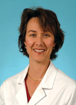
This could be because — as the survey also showed — GI fellows are not receiving adequate training in moderate sedation, which requires no interventions to maintain a patient airway, she reported. About 80% of program directors and 75% of senior fellows responding to the survey said they received training in moderate/conscious sedation during their fellowship.
These numbers are not impressive, said Early.
The Accreditation Council for Graduate Medical Education (ACGME) requires gastroenterology fellows to demonstrate competence in conscious sedation, along with other core skills, she explained. “What if I substituted training in mucosal biopsy or training in colonoscopy with polypectomy, which are other core requirements? I think you’d be shocked.”
The survey was small, with only 92 of 250 program directors and 33 of 655 fellows responding, but Early said the task force continues to collect responses.
Is Existing Training Enough?
Ten percent of fellows who replied to the survey did not participate in any moderate sedation procedures during training. And about a third of program directors said fellows participated in less than 100 such procedures.
“We really don’t know if that’s enough, in this era of competency-based assessment, which really values competency measures over numbers,” said Early.
Of the fellows who did receive training, 37% received hands-on training, a quarter received didactic lecture training, 11% used online modules, and 17% received a combination of the above training methods.
Just two thirds of program directors said they or their fellows were competent in moderate sedation, while close to 70% of fellows judged themselves competent.
While the majority of program directors (80%) knew that training in conscious sedation was a core ACGME requirement, only around a quarter of fellows were aware of the requirement.
Most gastroenterologists rely on anesthesiologists or certified registered nurse anesthetists (CRNAs) to deliver moderate or deep sedation, said Early, citing results from a separate survey sent to practicing clinicians.
Ongoing Shortages of CRNAs and Anesthesiologists
Shortages of anesthesiologists and CRNAs will continue to limit endoscopy procedure volume, especially in rural areas of the US, said Early.
The nation is expected to be short by 450,000 CRNAs this year and by 6300 anesthesiologists within a decade, she reported. Anesthesia providers are burned out or nearing retirement age, and there are not enough residency programs to produce new anesthesiologists at the rate needed to meet the demand, she said.
Gastroenterologists have become reliant on anesthesia providers, but adding a clinician is more expensive and “doesn’t appear to resolve and improve safety as compared with endoscopist-directed sedation for routine procedures,” said Early.
When practicing clinicians were asked if they’d be interested in providing physician-directed propofol sedation, 20% said yes, while 35% said no. But 16% said they would want to provide moderate sedation after completing in-person training, and 19% said they would after completing online training.
It may take time for gastroenterologists to get appropriate training and reduce reliance on anesthesia providers, Early said. But she said it may be increasingly possible in states allowing endoscopist-directed, nurse-administered propofol, and with medications such as remimazolam, a rapid-acting benzodiazepine that has shown similar efficacy and lower adverse event rates than propofol.
There will have to be a really deliberate step in order to take back control of endoscopic sedation from anesthesia and start performing more modest sedation, she said.
Early reported having no conflicts.
A version of this article first appeared on Medscape.com.
PHOENIX — , and a majority would be interested in providing physician-directed propofol sedation, especially after in-person or online training, according to results from an ongoing survey presented at the American College of Gastroenterology (ACG) 2025 Annual Scientific Meeting.
The dwindling supply of anesthesiology professionals in the US puts pressure on endoscopists, Dayna S. Early, MD, professor of medicine in the Gastroenterology Division at the Washington University, director of endoscopy at Barnes-Jewish Hospital, both in St. Louis, and chair of an ACG task force on anesthesia issues, told meeting attendees. However, preliminary results from the survey found that only about 4% of respondents said they used solely endoscopist-directed moderate sedation.
This could be because — as the survey also showed — GI fellows are not receiving adequate training in moderate sedation, which requires no interventions to maintain a patient airway, she reported. About 80% of program directors and 75% of senior fellows responding to the survey said they received training in moderate/conscious sedation during their fellowship.
These numbers are not impressive, said Early.
The Accreditation Council for Graduate Medical Education (ACGME) requires gastroenterology fellows to demonstrate competence in conscious sedation, along with other core skills, she explained. “What if I substituted training in mucosal biopsy or training in colonoscopy with polypectomy, which are other core requirements? I think you’d be shocked.”
The survey was small, with only 92 of 250 program directors and 33 of 655 fellows responding, but Early said the task force continues to collect responses.
Is Existing Training Enough?
Ten percent of fellows who replied to the survey did not participate in any moderate sedation procedures during training. And about a third of program directors said fellows participated in less than 100 such procedures.
“We really don’t know if that’s enough, in this era of competency-based assessment, which really values competency measures over numbers,” said Early.
Of the fellows who did receive training, 37% received hands-on training, a quarter received didactic lecture training, 11% used online modules, and 17% received a combination of the above training methods.
Just two thirds of program directors said they or their fellows were competent in moderate sedation, while close to 70% of fellows judged themselves competent.
While the majority of program directors (80%) knew that training in conscious sedation was a core ACGME requirement, only around a quarter of fellows were aware of the requirement.
Most gastroenterologists rely on anesthesiologists or certified registered nurse anesthetists (CRNAs) to deliver moderate or deep sedation, said Early, citing results from a separate survey sent to practicing clinicians.
Ongoing Shortages of CRNAs and Anesthesiologists
Shortages of anesthesiologists and CRNAs will continue to limit endoscopy procedure volume, especially in rural areas of the US, said Early.
The nation is expected to be short by 450,000 CRNAs this year and by 6300 anesthesiologists within a decade, she reported. Anesthesia providers are burned out or nearing retirement age, and there are not enough residency programs to produce new anesthesiologists at the rate needed to meet the demand, she said.
Gastroenterologists have become reliant on anesthesia providers, but adding a clinician is more expensive and “doesn’t appear to resolve and improve safety as compared with endoscopist-directed sedation for routine procedures,” said Early.
When practicing clinicians were asked if they’d be interested in providing physician-directed propofol sedation, 20% said yes, while 35% said no. But 16% said they would want to provide moderate sedation after completing in-person training, and 19% said they would after completing online training.
It may take time for gastroenterologists to get appropriate training and reduce reliance on anesthesia providers, Early said. But she said it may be increasingly possible in states allowing endoscopist-directed, nurse-administered propofol, and with medications such as remimazolam, a rapid-acting benzodiazepine that has shown similar efficacy and lower adverse event rates than propofol.
There will have to be a really deliberate step in order to take back control of endoscopic sedation from anesthesia and start performing more modest sedation, she said.
Early reported having no conflicts.
A version of this article first appeared on Medscape.com.
FROM ACG 2025